Embed Size (px)
Citation preview

Congenital, developmental & cystic diseases of the kidney
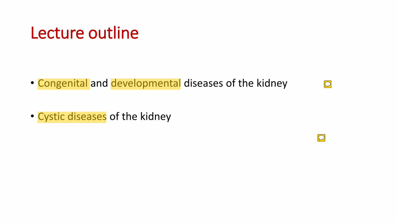
Lecture outline
• Congenital and developmental diseases of the kidney
• Cystic diseases of the kidney
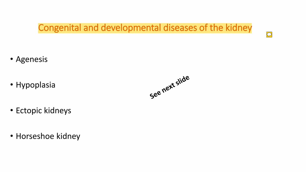
Congenital and developmental diseases of the kidney
• Agenesis
• Hypoplasia
• Ectopic kidneys
• Horseshoe kidney
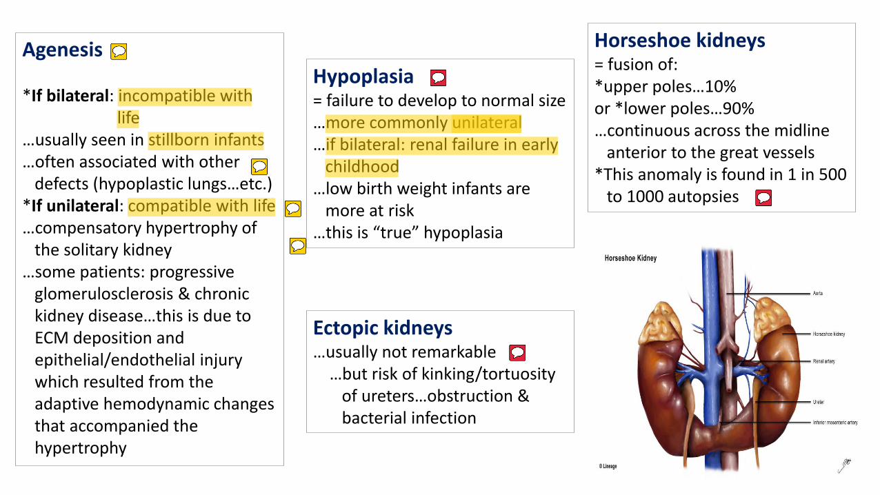
Agenesis *If bilateral: incompatible with life …usually seen in stillborn infants …often associated with other defects (hypoplastic lungs…etc.) *If unilateral: compatible with life …compensatory hypertrophy of the solitary kidney …some patients: progressive glomerulosclerosis & chronic kidney disease…this is due to ECM deposition and epithelial/endothelial injury which resulted from the adaptive hemodynamic changes that accompanied the hypertrophy
Hypoplasia = failure to develop to normal size …more commonly unilateral …if bilateral: renal failure in early childhood …low birth weight infants are more at risk …this is “true” hypoplasia
Ectopic kidneys …usually not remarkable …but risk of kinking/tortuosity of ureters…obstruction & bacterial infection
Horseshoe kidneys = fusion of: *upper poles…10% or *lower poles…90% …continuous across the midline anterior to the great vessels *This anomaly is found in 1 in 500 to 1000 autopsies
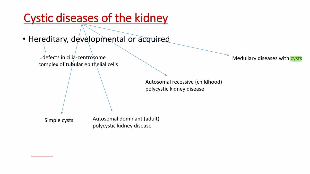
Cystic diseases of the kidney • Hereditary, developmental or acquired
…defects in cilia-centrosome complex of tubular epithelial cells
Simple cysts Autosomal dominant (adult) polycystic kidney disease
Autosomal recessive (childhood) polycystic kidney disease
Medullary diseases with cysts
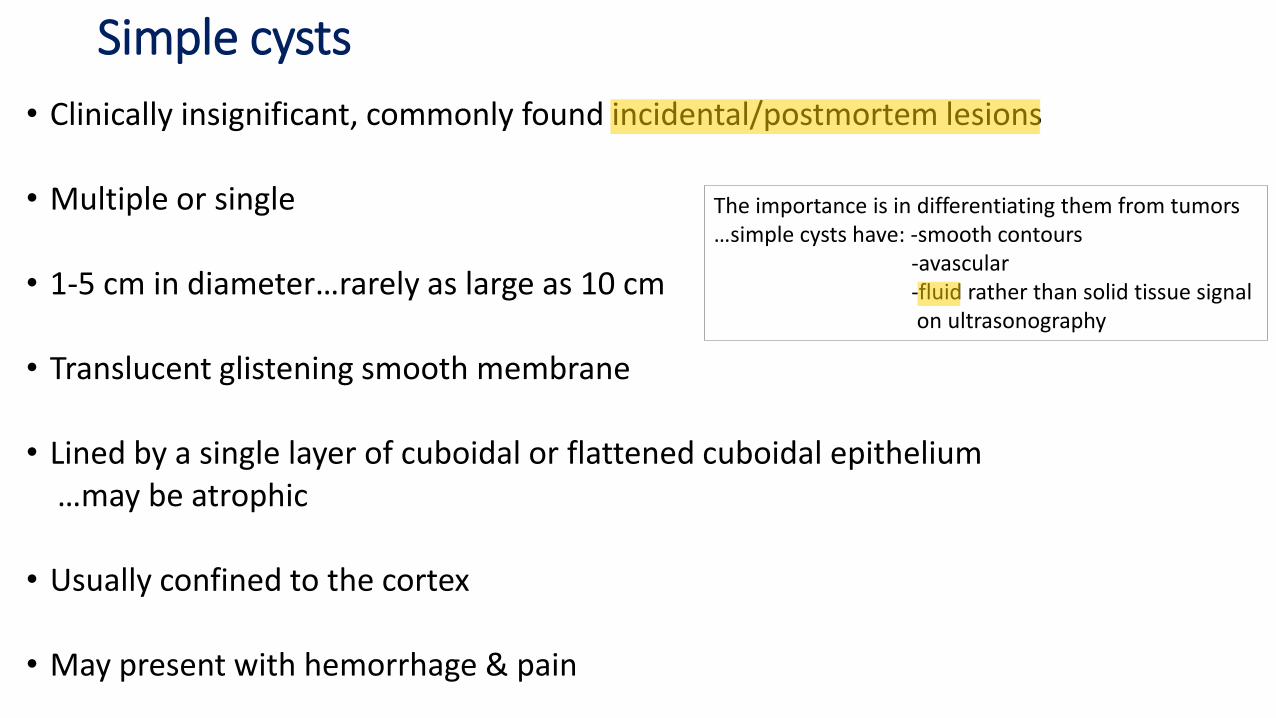
Simple cysts
• Clinically insignificant, commonly found incidental/postmortem lesions
• Multiple or single
• 1-5 cm in diameter…rarely as large as 10 cm
• Translucent glistening smooth membrane
• Lined by a single layer of cuboidal or flattened cuboidal epithelium …may be atrophic • Usually confined to the cortex
• May present with hemorrhage & pain
The importance is in differentiating them from tumors …simple cysts have: -smooth contours -avascular -fluid rather than solid tissue signal on ultrasonography
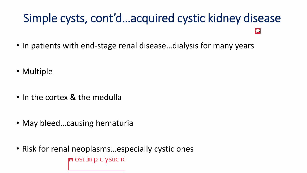
Simple cysts, cont’d…acquired cystic kidney disease
• In patients with end-stage renal disease…dialysis for many years
• Multiple
• In the cortex & the medulla
• May bleed…causing hematuria
• Risk for renal neoplasms…especially cystic ones
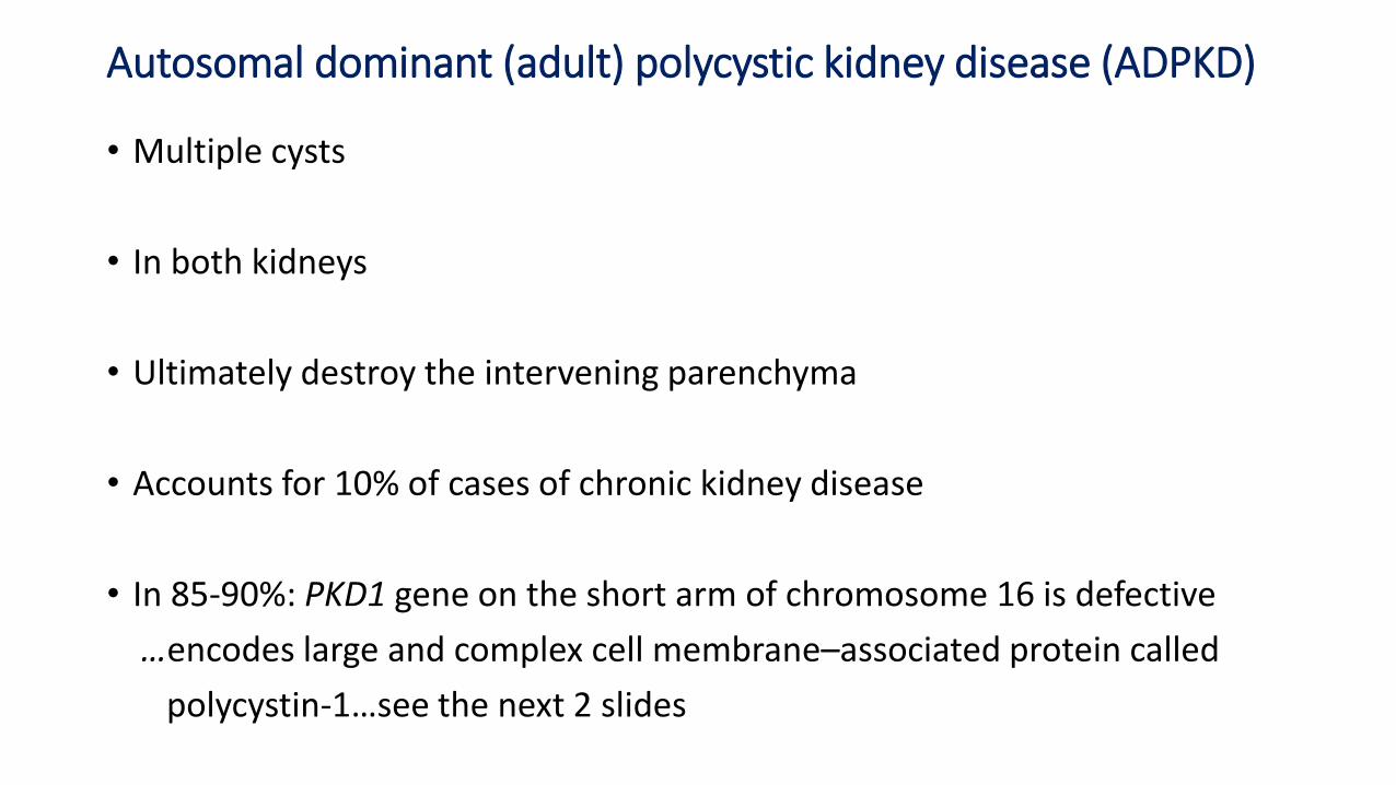
Autosomal dominant (adult) polycystic kidney disease (ADPKD) • Multiple cysts
• In both kidneys
• Ultimately destroy the intervening parenchyma
• Accounts for 10% of cases of chronic kidney disease
• In 85-90%: PKD1 gene on the short arm of chromosome 16 is defective
…encodes large and complex cell membrane–associated protein called
polycystin-1…see the next 2 slides
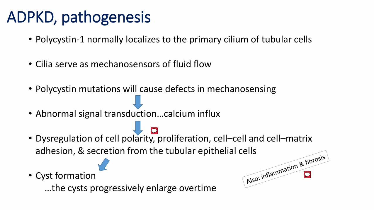
ADPKD, pathogenesis • Polycystin-1 normally localizes to the primary cilium of tubular cells
• Cilia serve as mechanosensors of fluid flow
• Polycystin mutations will cause defects in mechanosensing
• Abnormal signal transduction…calcium influx
• Dysregulation of cell polarity, proliferation, cell–cell and cell–matrix adhesion, & secretion from the tubular epithelial cells • Cyst formation …the cysts progressively enlarge overtime
ADPKD, pathogenesis…cont’d
• Germline mutations of the PKD1 gene are present in all renal tubular
cells of affected individuals…but: cysts develop in only some tubules
…which means: a second “somatic hit” is required for cyst
development
ADPKD, pathogenesis…cont’d
• PKD2 gene
…10-15% of the cases
…on chromosome 4, encodes polycystin-2
…a calcium-permeable membrane channel …also localized to cilia …acts together with polycstin-1 to form heterodimer …so: a defect in any of the two will cause the same result …However, PKD2 mutations: slower disease progression
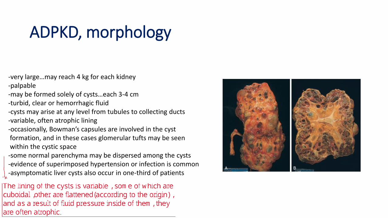
-very large…may reach 4 kg for each kidney -palpable -may be formed solely of cysts…each 3-4 cm -turbid, clear or hemorrhagic fluid -cysts may arise at any level from tubules to collecting ducts -variable, often atrophic lining -occasionally, Bowman’s capsules are involved in the cyst formation, and in these cases glomerular tufts may be seen within the cystic space -some normal parenchyma may be dispersed among the cysts -evidence of superimposed hypertension or infection is common -asymptomatic liver cysts also occur in one-third of patients
ADPKD, morphology
ADPKD, clinical features
• Usually does not produce symptoms until the fourth decade of life
• The most common presenting complaint is flank pain or a heavy, dragging sensation • Acute distention of a cyst, either by intracystic hemorrhage or by obstruction, may cause excruciating pain • Sometimes attention is first drawn to the lesion on palpation of an abdominal mass • Intermittent gross hematuria commonly occurs
• The most important: hypertension (75% of patients) & infection
ADPKD, clinical features…cont’d
• Saccular aneurysms of the circle of Willis…10-30% of patients
• More favorable than with most chronic kidney diseases…although it is ultimately fatal
• Slow progression
…end-stage renal disease occurs at about 50 years of age
…even nearly normal life spans are reported
• Treatment: renal transplantation
• Death: usually due to uremia or HTN
Autosomal recessive (childhood) polycystic kidney disease (ARPKD)
• Rare
• Subcategories:
-Perinatal
-Neonatal
-Infantile
-Juvenile
• The defect is in PKHD1 gene, coding for a membrane receptor protein called
fibrocystin
• Fibrocystin is found in cilia in tubular epithelial cells…unknown function
most common
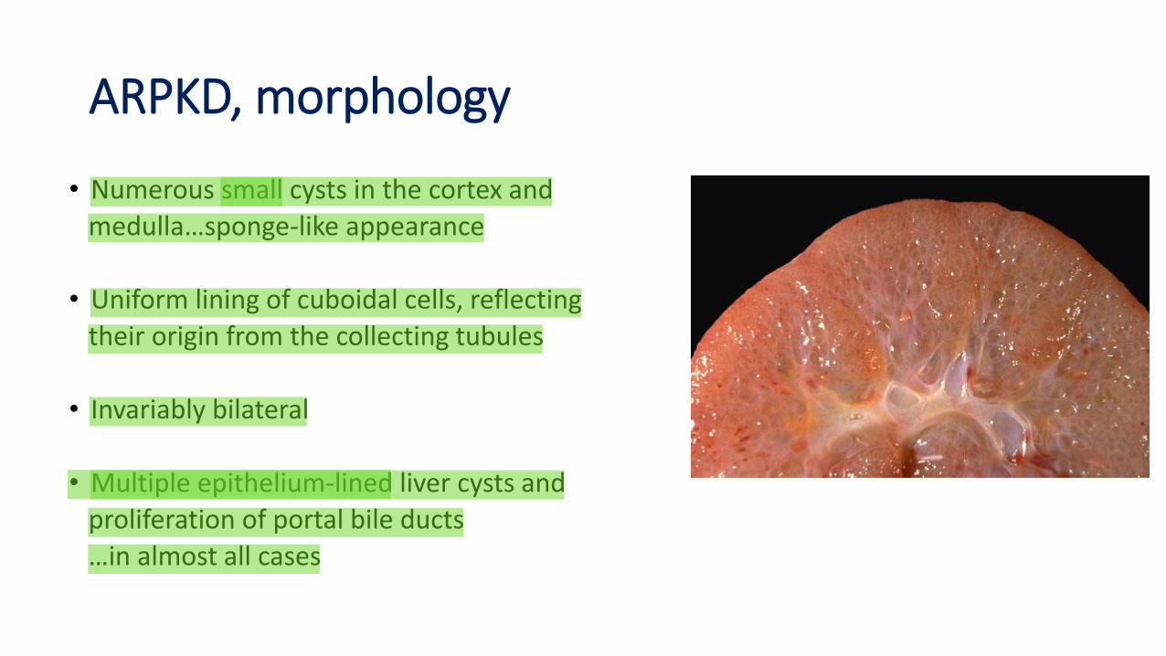
ARPKD, morphology
• Numerous small cysts in the cortex and
medulla…sponge-like appearance
• Uniform lining of cuboidal cells, reflecting
their origin from the collecting tubules
• Invariably bilateral
• Multiple epithelium-lined liver cysts and
proliferation of portal bile ducts
…in almost all cases
ARPKD, clinical features
• Serious manifestations usually are present at birth
• Young infants may die quickly from hepatic or renal failure
• Patients who survive infancy develop liver cirrhosis
…congenital hepatic fibrosis
Medullary diseases with cysts
• 2 major types of cystic disease affecting the medulla:
-medullary sponge kidney
…relatively common
…usually innocuous
…occasionally associated with nephrolithiasis
-nephronophthisis-medullary cystic disease complex
…almost always associated with renal dysfunction
Nephronophthisis-medullary cystic disease complex
• Usually begins in childhood
• 4 variants:
-Infantile
-Juvenile…the most common
-Adolescent nephronophthisis
-Medullary cystic disease developing later in adult life
Nephronophthisis-medullary cystic disease complex …juvenile form
• 15-20% have extrarenal manifestations:
…retinal abnormalities: retinitis pigmentosa and even early onset
blindness
…oculomotor apraxia
…mental retardation
…cerebellar malformations
…liver fibrosis
Nephronophthisis-medullary cystic disease complex, pathogenesis
• At least nine gene loci (NHP1 to NHP9) have been identified for the
autosomal recessive forms of the nephronophthisis complex
• The majority of these genes encode proteins that are components of
epithelial cilia
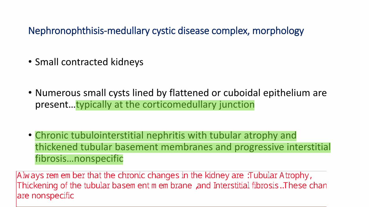
Nephronophthisis-medullary cystic disease complex, morphology
• Small contracted kidneys
• Numerous small cysts lined by flattened or cuboidal epithelium are present…typically at the corticomedullary junction
• Chronic tubulointerstitial nephritis with tubular atrophy and thickened tubular basement membranes and progressive interstitial fibrosis…nonspecific
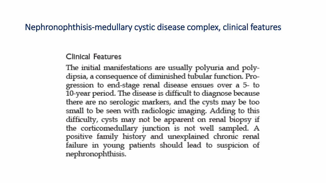
Nephronophthisis-medullary cystic disease complex, clinical features