Embed Size (px)
Citation preview
10
Congenital Diaphragmatic Hernia Survivors: Outcomes in Infancy, Childhood and
Adolescence
Jennifer R. Benjamin1 and C. Michael Cotten2
1Yale University School of Medicine 2Duke University Medical Centre
USA
1. Introduction
Congenital diaphragmatic hernia (CDH) remains highly challenging. CDH is complex and often results in multi-system morbidity among survivors. In the past decade, significant technologic and medical advancements have enhanced the care of critically ill infants, including those born with CDH. Such advancements have led to improved survival rates among CDH infants at certain high-volume centres, but this has occurred at the expense of increased short- and long-term morbidities for patients who previously would have died.
As collective knowledge has improved regarding the range of outcomes experienced by CDH survivors, the focus of much clinical research has shifted toward reducing survivor morbidity. Accordingly, many centres now offer multidisciplinary follow-up care for CDH survivors throughout infancy and early childhood in order to effectively diagnose and treat problems with the goal of maximizing the potential of the individual patient.
The aim of this chapter is to describe the short- and long-term medical and neurodevelopmental outcomes of CDH survivors in infancy, childhood, and adolescence. We will also outline an approach to follow-up care for these highly complex and intriguing patients.
2. Cardiopulmonary morbidity
Because the diaphragmatic defect allows abdominal organs to herniate into the chest cavity during the time of bronchial and pulmonary artery branching, lung growth and development are subsequently impaired. Long-term pulmonary morbidity is variable and depends on the degree of underlying pulmonary hypoplasia as well as the severity of iatrogenic injury the lungs incurred in the neonatal period. Treatment with extracorporeal membrane oxygenation (ECMO) and patch repair of the diaphragm are also associated with persistent cardiorespiratory complications (Jaillard et al., 2003; Muratore et al., 2001). Up to 50% of CDH survivors have long-lasting cardiopulmonary compromise, manifested as chronic lung disease, reactive airway disease, recurrent respiratory infections, and persistent pulmonary hypertension (Bagolan & Morini, 2007; Lally & Engle, 2008; Trachsel et al., 2005; Wischermann et al., 1995).
Congenital Diaphragmatic Hernia – Prenatal to Childhood Management and Outcomes
140
2.1 Chronic lung disease
Lung structure is fundamentally altered in CDH due to the decreased number of bronchi and alveoli in the ipsilateral and, to a lesser degree, contralateral lung. Although the number of alveoli may increase over time (Beals et al., 1992), the number of larger airways does not, because bronchial development is complete at around 16 weeks of gestation (Reid, 1984). Postnatally, exposure to supplemental oxygen and mechanical ventilation results in pulmonary edema and protein leak, causing surfactant denaturation and lung injury. The suboptimal nutrition often experienced by critically ill infants may further worsen the problem. Taken together, these factors lead to the development of chronic lung disease - a highly significant problem for between 40% and 60% of CDH survivors (D’Agostino et al., 1995; Kinsella et al., 1997; van den Hout et al., 2010).
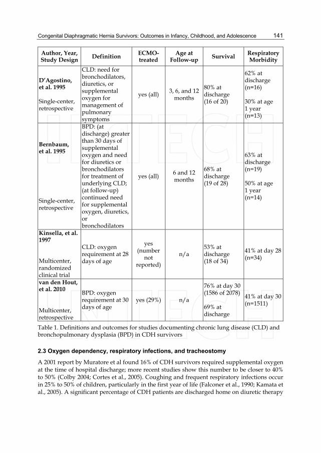
D’Agostino et al documented a 62% incidence of chronic lung disease among ECMO-treated CDH survivors at hospital discharge (D’Agostino et al., 1995). Bernbaum et al found a similar incidence of 63% with bronchopulmonary dysplasia (BPD, a form of chronic lung disease) at discharge, with only a slight improvement to 50% at age one. Strikingly, four of seven CDH-ECMO patients in this report continued to need artificial ventilatory support at one year of age (Bernbaum et al., 1995). In a study of infants with pulmonary hypertension, Kinsella et al found CDH infants to have a 41% incidence of chronic lung disease; this incidence was significantly higher than the incidence of chronic lung disease due to meconium aspiration syndrome or respiratory distress syndrome, and suggests the possibility of decreased total lung volume combined with more severe underlying parenchymal lung disease in the CDH group (Kinsella et al., 1997). Van den Hout et al documented a 41% prevalence of BPD in a study of 2078 CDH patients based on data from the CDH Study Group. The authors identified early gestational age, presence of cardiac abnormality, prenatal diagnosis, right-sided defect, low 5-minute Apgar score, and intial treatment with high-frequency ventilation as factors predictive of BPD in CDH survivors. They suggest that in addition to injury from volutrauma, barotrauma, and atelectotrauma, the hypoplastic lungs in CDH may be more susceptible to oxygen toxicity than lungs of newborns with other respiratory disorders (van den Hout et al., 2010). Differences in definitions of respiratory morbidity among survivors makes comparison of heterogenous studies somewhat difficult, yet overall morbidity ranges between 30-50% in general (Table 1).
2.2 Reactive airway disease
Approximately 25% of CDH infants demonstrate evidence of obstructive airway disease (Jaillard et al., 2003), with up to 45% of survivors showing asthma-like symptoms during childhood and adolesence (Davis et al., 2004; Trachsel et al., 2005). A study by Crankson and colleagues found 45% of 31 CDH survivors had recurrent wheezing attacks and required bronchodilators and/or inhaled steroids (Crankson et al., 2005). In another report, bronchodilators were prescribed in 40% and inhaled steroids in 35% of CDH survivors during the first year of life (Muratore et al., 2001). Lung function abnormalities appear to improve over time as compensatory lung growth occurs, although these data are limited to a relatively small number of patients (Peetsold et al., 2007; Trachsel et al., 2005). According to some experts, although CDH survivors may exhibit mild to moderate abnormalities on pulmonary function testing in late childhood through adulthood, they appear not to have an important reduction of total lung capacity, and may be expected to achieve normal exercise function (Ijsselstijn et al., 1997; Marven et al., 1998; Peetsold et al., 2007).
Congenital Diaphragmatic Hernia Survivors: Outcomes in Infancy, Childhood, and Adolescence
141
Author, Year, Study Design Definition ECMO-
treated Age at
Follow-up Survival Respiratory Morbidity
D’Agostino, et al. 1995 Single-center, retrospective
CLD: need for bronchodilators, diuretics, or supplemental oxygen for management of pulmonary symptoms
yes (all) 3, 6, and 12 months
80% at discharge (16 of 20)
62% at discharge (n=16) 30% at age 1 year (n=13)
Bernbaum, et al. 1995 Single-center, retrospective
BPD: (at discharge) greater than 30 days of supplemental oxygen and need for diuretics or bronchodilators for treatment of underlying CLD; (at follow-up) continued need for supplemental oxygen, diuretics, or bronchodilators
yes (all) 6 and 12 months
68% at discharge (19 of 28)
63% at discharge (n=19) 50% at age 1 year (n=14)
Kinsella, et al. 1997 Multicenter, randomized clinical trial
CLD: oxygen requirement at 28 days of age
yes (number
not reported)
n/a 53% at discharge (18 of 34)
41% at day 28 (n=34)
van den Hout, et al. 2010 Multicenter, retrospective
BPD: oxygen requirement at 30 days of age
yes (29%) n/a
76% at day 30 (1586 of 2078) 69% at discharge
41% at day 30 (n=1511)
Table 1. Definitions and outcomes for studies documenting chronic lung disease (CLD) and bronchopulmonary dysplasia (BPD) in CDH survivors
2.3 Oxygen dependency, respiratory infections, and tracheostomy
A 2001 report by Muratore et al found 16% of CDH survivors required supplemental oxygen at the time of hospital discharge; more recent studies show this number to be closer to 40% to 50% (Colby 2004; Cortes et al., 2005). Coughing and frequent respiratory infections occur in 25% to 50% of children, particularly in the first year of life (Falconer et al., 1990; Kamata et al., 2005). A significant percentage of CDH patients are discharged home on diuretic therapy
Congenital Diaphragmatic Hernia – Prenatal to Childhood Management and Outcomes
142
for management of pulmonary edema (Muratore et al., 2001). Additionally, up to 4% of CDH infants ultimately require tracheostomy for respiratory failure in the setting of severe lung disease (Bagolan & Morini, 2007; Jaillard et al., 2003).
2.4 Pulmonary hypertension
Chronic pulmonary hypertension is one of the major complicating factors in CDH (Kinsella et al., 2005), and, according to one report, occurs in up to 21% of CDH infants (Kinsella et al., 2003). Recurrence of pulmonary hypertension beyond the neonatal period may lead to prolonged mechanical ventilation, a second ECMO run, or death (Dela Cruz et al., 1996; Lally & Breaux, 1995). In a small retrospective study of 8 infants with CDH, 100% were found to have evidence of pulmonary hypertension on echocardiography (Benjamin et al., 2010). Overall mortality attributable to pulmonary hypertension approaches 50% in the CDH population (Kinsella et al., 1997; The Neonatal Inhaled Nitric Oxide Study Group (NINOS), 1997).
In an attempt to correlate outcome with the severity of pulmonary hypertension among a cohort of CDH infants, Dillon et al investigated estimates of pulmonary artery pressures using serial echocardiography. Based on their findings, the authors propose that almost half of CDH patients will resolve their pulmonary hypertension within the first 21 days, and survival may be nearly 100% in this subpopulation. Conversely, a certain number of CDH infants will show persistent systemic or suprasystemic pressures despite all interventions (including ECMO), and survival among this group can be presumed zero. Finally, there exists a group with protracted pulmonary hypertension that is responsive to maximum medical management; survival rates for infants with this degree of pulmonary hypertension lie somewhere in between the two extremes (Dillon et al., 2004).
Schwartz et al investigated the outcome of 21 CDH-ECMO survivors at a mean age of 3.2 years and found 38% with pulmonary hypertension based on echocardiographic findings. Of those, 88% had some evidence of reactive airway disease and were on bronchodilators at the time of the study. There is, however, no mention of whether the patients were being treated with medication for pulmonary hypertension. The authors surmise that CDH infants may undergo a gradual increase in vascular smooth muscle proliferation leading to an increase in pulmonary arterial pressures over time. It remains unclear whether pulmonary vascular pressures eventually normalize as repair and growth progress (Schwartz et al., 1999).
Management strategies for CDH infants who do not require mechanical ventilation but have pulmonary hypertension include inhaled nitric oxide (iNO) via nasal cannula and oral sildenafil, a highly selective phosphodiesterase inhibitor type 5 (Keller et al., 2004; Kinsella et al., 2003). Sildenafil has been used to treat older children and adults with pulmonary hypertension (Wilkens et al., 2001) by limiting degradation of cyclic GMP and thereby enhancing endogenous nitric oxide activity (Tulloh, 2009). Results from a 2007 study indicate sildenafil may augment cardiac output in infants with CDH during the first two weeks of administration (Noori et al., 2007).
Though some reports document the presence of pulmonary hypertension in CDH beyond the neonatal period, little is known about the long-term structural pulmonary vascular abnormalities that remain, and how these structural abnormalities translate into clinical outcomes for older children and adults with chronic pulmonary hypertension following
Congenital Diaphragmatic Hernia Survivors: Outcomes in Infancy, Childhood, and Adolescence
143
CDH repair. More research is needed to assess the prevalence of persistent pulmonary hypertension, investigate the natural course of changing pulmonary pressures, determine the best methods for diagnosing pulmonary hypertension in CDH survivors, and determine the optimal dosing, timing, and indications for pharmacologic intervention.
3. Growth and nutritional morbidity
Numerous studies have documented the high incidence of gastrointestinal morbidity in CDH survivors (Fasching et al., 2000; Muratore et al., 2001; Van Meurs et al., 1993). Symptomatic gastroesophageal reflux (GER), feeding difficulties, and chronic growth failure necessitating supplemental enteral tube feedings are among the issues these infants and children face.
3.1 Gastroesophageal reflux
GER occurs in the vast majority of infants and children born with CDH, and is perhaps the most commonly encountered long-term problem among survivors. Several mechanisms have been proposed to explain the high incidence of GER in CDH, including fetal esophageal obstruction resulting in impaired esophageal motility, shortened esophageal length, disruption of the angle of His due to the abnormal location of the stomach in utero, and the complete or partial absence of the parahiatal diaphragm. The clinical presentation varies: some infants and children will experience recurrent vomiting, while in others GER may manifest as persistent bradycardic spells or aspiration pneumonia (Kieffer et al., 1995). Although many CDH survivors can be successfully managed with medical treatment, a significant number – up to 23% in one series – have severe GER requiring surgical intervention with fundoplication (Su et al., 2007). Factors associated with need for fundoplication are patch repair, ECMO treatment, and intrathoracic liver position (Diamond et al., 2007; Su et al., 2007).
Recently, Maier et al attempted to determine whether performing preventive fundoplication at the time of CDH repair would impact GER symptoms in infancy. The authors found a clinically (but not statistically) significant difference between the degree of GER in the first postnatal year among those infants who did (n = 36) and did not (n = 43) have preventive fundoplication performed at the time of hernia repair, but there was no difference by the second year of follow-up. These authors concluded that preventive anti-reflux surgery cannot be recommended as standard of care (Maier et al., 2011).
In contrast to the plethora of studies focusing on the gastrointestinal sequelae of CDH in infancy and childhood, there are relatively few studies describing outcomes among adult CDH survivors. Reports suggest a certain percentage of adult CDH survivors may have continued GER resulting in esophagitis and, in severe cases, Barrett’s esophagus. Vanamo et al found 63% of adult survivors to have symptoms suggestive of GERD and 54% with evidence of esophagitis on endoscopy. Further, 2 of 11 patients with early postoperative GER developed esophageal stricture and underwent fundoplication (Vanamo et al., 1996). Steven et al described a case of esophageal adenocarcinoma in a 22 year-old CDH survivor; the malignancy was presumed to be related to long-standing GER associated with CDH (Steven et al., 2007). More studies are needed in adolescents and adults to determine whether the significant degree of GER experienced by CDH survivors in infancy and childhood remains a problem over time, and to what extent it impacts the health and wellbeing of adult survivors.
Congenital Diaphragmatic Hernia – Prenatal to Childhood Management and Outcomes
144
3.2 Feeding problems
Protracted hospitalization combined with symptoms of GER and delayed initiation of oral feedings contributes to the development of feeding difficulties in CDH infants. In one series, oral aversion was observed in 25% of infants with CDH and contributed largely to growth failure (Jaillard et al., 2003). Van Meurs et al found 22% of CDH-EMO survivors to have ”extreme food refusal” in the first two years of life, 75% of whom required long-term supplemental nutrition. In general, one-third to one-half of CDH infants require supplementation with enteral tube feedings to support growth in the first year of life, either via nasogastric or gastrostomy tube feedings (Muratore et al., 2001; Van Meurs et al., 1993).
Symptoms of GER and resultant feeding problems may be more clinically apparent among CDH-ECMO survivors. In a study of 16 CDH-ECMO survivors, 81% were diagnosed with GER, and 69% were discharged requiring supplemental tube feedings (D’Agostino et al., 1995). Similarly, in Van Meurs’ population of 18 CDH-ECMO survivors, at the time of hospital discharge, reflux precautions were in place for 22%, feedings were thickened with rice cereal for 22%, and 33% were being treated with metoclopramide (Van Meurs et al., 1993).
3.3 Failure to thrive
In conjunction with an increased metabolic demand due to pulmonary morbidity and underlying GER, feeding-related problems often result in failure to thrive. Muratore et al documented weight and height below the 25th percentile in more than 50% of CDH infants during the first year of life. In this study, ECMO treatment and need for oxygen at discharge were independent predictors of growth failure in the first postnatal year (Muratore et al., 2001). Others have similarly reported a relationship between ECMO and subsequent poor growth among CDH survivors; this finding is specific to the CDH-ECMO population, as non-CDH ECMO children were shown to have essentially normal growth at 12 and 24 months in Van Meurs’ study (Nobuhara et al., 1996; Van Meurs et al., 1993). Further, Van Meurs et al found that parental discontinuation of supplemental tube feedings contributed to findings of severe malnourishment at one year follow-up (Van Meurs et al., 1993). A significant proportion of CDH survivors remain below the 5th percentile for weight in the first three years (Kamata et al., 2005; Lund et al., 1994). Catch-up growth can occur with aggressive management (Cortes et al., 2005), but poor growth remains a problem for a subset of patients.
4. Musculoskeletal morbidity
Chest wall and spinal deformities occur relatively commonly as a sequelae of CDH. One study of adult survivors found 48% with chest asymmetry, 27% with significant scoliosis, and 18% with pectus excavatum (Vanamo et al., 1996). The incidence increases with a larger diaphragmatic defect, presumably because repair of a large defect puts tension on the spine and interferes with normal development of the thorax. Other contributing factors include a smaller thoracic cavity on the affected side and increased work of breathing leading to development of a pectus abnormality via retraction of the cartilaginous anterior chest wall. For the most part, these deformities are mild and rarely require surgical intervention. However, the exact relationship between chest wall deformities and long-term pulmonary function remains unclear. Trachsel et al found a correlation between moderate to severe chest wall abnormalities and impaired pulmonary functioning in
Congenital Diaphragmatic Hernia Survivors: Outcomes in Infancy, Childhood, and Adolescence
145
adulthood (Trachsel et al., 2005). Additionally, there is some evidence of abnormal lung function among patients with pectus excavatum (Koumbourlis & Stolar, 2004).
5. Surgical morbidity
The incidence of reherniation at the site of the diaphragmatic defect is directly related to the use of synthetic patch repair and ranges from 5% to 80% (Atkinson & Poon, 1992; Van Meurs et al., 1993). With the increasing popularity of minimally invasive surgical (MIS) techniques, studies have begun to investigate the short- and long-term outcomes following this approach. Using data from the CDH Study Group, Tsao et al looked at the in-hospital hernia recurrence rate following MIS and found a significantly higher proportion of recurrence in the MIS group compared with open repair; the authors estimated an almost 4-fold increased odds of recurrence with MIS repairs. In this study, infants who required a patch repair had a higher recurrence rate, and MIS repairs that used a patch had the highest recurrence rate of approximately 9% (Tsao et al., 2011). The overall benefit for MIS in CDH patients remains to be determined.
Timing of reherniation varies from months to years after the initial repair. Patients may present with gastrointestinal symptoms (feeding difficulties, vomiting), respiratory symptoms (coughing, wheezing), or they may be asymptomatic. Thus, regularly scheduled surveillance chest radiographs are advised (Lally & Engle, 2008; Moss et al., 2001).
Almost invariably, infants with CDH have intestinal non-rotation or malrotation. Consequently, some are at risk for developing intestinal obstruction due to midgut volvulus if a Ladd’s procedure was not performed at the time of diaphragmatic repair. Postoperative intra-abdominal adhesions may also lead to intestinal obstruction – a life-threatening complication if not recognized and treated in a timely manner.
6. Neurodevelopmental morbidity
The underlying mechanisms of brain injury in CDH remain incompletely defined. Chronic lung disease, postnatal growth failure, and prolonged hospitalization are factors known to adversely affect neurodevelopment. In addition, most infants with CDH are exposed to perinatal and postnatal hypoxia, hypotension, hypercapnia, inflammation, and acidosis – all of which interfere with central nervous system development. Poor neurodevelopmental outcome in infants with CDH is likely the result of a combination of multiple interrelated factors. All CDH survivors should be considered at risk for cognitive delay and followed regularly in a multidisciplinary clinic.
6.1 Neurodevelopmental outcomes among non-ECMO-treated CDH survivors
In spite of heightened awareness about the potential for long-lasting adverse neurodevelopmental outcome in CDH survivors, most studies have focused on the 18- to 36-month follow up period, with a considerable paucity of information detailing longer-term outcomes. Similarly, few studies describe the neurodevelopmental outcome of CDH survivors not treated with ECMO. Only recently have investigators begun to look at the developmental and psychological outcomes of survivors into late childhood and adolescence.
One such series looking at CDH survivors between 8 and 12 years of age found only 54% to be functioning cognitively at expected school level. In addition, there was a higher
Congenital Diaphragmatic Hernia – Prenatal to Childhood Management and Outcomes
146
prevalence of emotional and behavioural problems among survivors in comparison to the general population (Bouman et al., 2000). In a study of adolescent CDH survivors, Jakobson and colleagues studied the visual and fine motor outcomes of CDH survivors between ages 10 through 16 and found 13% had severe cognitive impairment, with the majority of the remaining showing poorer oral motor control and visual-motor integration skills as compared to peers (Jakobson et al., 2009). Notably, none of the survivors in these two studies were treated with ECMO as infants.
6.2 Neurodevelopmental outcomes among CDH-ECMO survivors
The relationship between CDH infants treated with ECMO and adverse neurocognitive outcome remains controversial. Some experts conclude that although ECMO can support the most critically ill infants, its use in the CDH population is associated with significant mortality and long-term morbidity (Bernbaum et al., 2005; Davis et al., 2004). On the contrary, others question whether adverse outcomes in this population may be due to the underlying disease process or an intrinsic neurologic abnormality rather than ECMO per se (D’Agostino et al., 1995; Jaillard et al., 2003; Lally & Engle, 2008).
The physiologic basis for ECMO treatment leading to cognitive delay rests on several factors. First, infants who require ECMO to survive represent the sickest subset of patients in the newborn intensive care unit (NICU). They are at significant risk for hypoxia and hypoperfusion before and during ECMO treatment (Bagolan & Morini, 2007) and, therefore, are very likely subject to hypoxic-ischemic brain injury. Furthermore, ECMO support requires systemic anticoagulation (in all) and ligation of the carotid artery (in venoarterial). The increase in neurologic complications in infants treated with venoarterial ECMO (seizures, cerebral infarction) may be attributable to decreased cerebral blood flow resulting from ligation of the right internal carotid artery (Dimmitt et al., 2001). One report suggests that venovenous ECMO may be sufficient to support CDH patients, and when compared with venoarterial ECMO, is associated with decreased risk of neurologic impairment (Guner et al., 2009).
A study by Van Meurs et al focusing on the outcome of 18 CDH-ECMO survivors between ages one and four years, 15 of whom underwent developmental testing, found 46% of the group to be normal, 20% with suspected developmental delays, and 13% with definitely abnormal developmental outcome (Van Meurs et al., 1993). In a report by Nobuhara and colleagues, 37% of 78 CDH survivors had developmental delay at follow-up; notably, 72% of the survivors with mild to moderate delays were treated with ECMO as neonates (Nobuhara et al., 1996). Similarly, McGahren et al described a statistically significant difference in neurologic abnormalities between CDH survivors who were and were not treated with ECMO: 67% of 12 CDH-ECMO infants had evidence of significant neurologic deficit at follow-up compared with 24% of 21 non-ECMO-treated CDH survivors (McGahren et al., 1997).
A 2004 study of CDH-ECMO survivors found 19% to have severe neurodevelopmental problems, defined as speech, language, or motor delay, at a median age of 5 years. In this report, only 25% of 27 CDH children were free of significant neurodevelopmental deficit at follow-up (Davis et al., 2004). Similarly, among 149 5 year-old ECMO survivors, Nijhuis-van der Sanden et al found the 36 with CDH to have the worst outcomes: nearly 42% died, 72% had growth problems, and almost 16% had severe disabilities with only 37% functioning
Congenital Diaphragmatic Hernia Survivors: Outcomes in Infancy, Childhood, and Adolescence
147
normally (Nijhuis-van der Sanden et al., 2009). A recent study of 41 infants prospectively enrolled in an interdisciplinary follow-up program found 46% of CDH-ECMO survivors had deficits in developmental outcome at 24 months as well as an increased risk for psychomotor dysfunction and motor delays. Autism was diagnosed in 7% of this cohort (Danzer et al., 2010).
Several studies document the relationship between ECMO and hypotonia in infancy and early childhood. A report by D’Agostino and colleagues followed 13 CDH-ECMO survivors into early childhood and found a significant number to have problems with hypotonia, which subsequently had a large impact on motor skill acquisition. Fifty-four percent had normal cognitive and motor outcomes at 12 months while 23% had delays in both areas (D’Agostino et al., 1995). In Nobuhara’s study, decreased lower extremity tone was the most common finding among CDH survivors with developmental delay (Nobuhara et al., 1996). Bernbaum et al studied a cohort of ECMO survivors and found the CDH-ECMO population to have significantly lower motor and slightly lower cognitive scores at one year of age compared with groups treated with ECMO for other reasons. In this study, 75% percent of CDH-ECMO infants with hypotonia at discharge continued to be hypotonic at age one, which affected the attainment of gross motor skills (Bernbaum et al., 1995).
In a study by Tracy et al looking at the relationship between neuroimaging abnormalities and neurodevelopmental outcomes in a cohort of 45 CDH survivors, the authors found hypotonia to be the most frequently noted neurologic finding on physical examination. Hypotonia was present in 37% of survivors at age one and 71% of survivors at age three years. Motor problems seemed to be associated with hypotonia, as motor problems were diagnosed in 46% and 71% of the cohort at ages one and three, respectively. Additionally, motor problems at one year of age were correlated with abnormal findings on postnatal neuroimaging, continued respiratory compromise at age one, and a history of prolonged mechanical ventilation (Tracy et al., 2010).
6.3 School-age follow-up of CDH survivors
In a study looking at predictors of neurocognitive delay among a cohort of 16 school-age CDH survivors, 44% of the study population had evidence of persistent neurocognitive delay between ages 4 and 7 years. In this study, eight subjects underwent developmental testing at both 2 year and school age follow-up, and 50% had evidence of both early impairment and continued delay at school age. The remaining four subjects who scored within the average range on neurodevelopmental testing at 2 years remained with average scores at early school age. We found patch repair, ECMO treatment, days on ECMO, days of mechanical ventilation, and post-operative use of iNO to be associated with neurocognitive delay at early school age (Benjamin et al., 2010).
6.4 Neuroimaging in CDH
As brain imaging techniques have become more widely available, researchers have begun to study the effects of CDH on the developing central nervous system. A study looking at eight CDH survivors – none treated with ECMO – found all to have abnormalities on brain MRI, including ventricular dilatation, abnormal myelination of the posterior limb of the internal capsule, and abnormal signal in the white matter and basal ganglia (Hunt et al., 2004). Tracy et al showed that by age three years, 88% of CDH survivors who underwent brain MRI
Congenital Diaphragmatic Hernia – Prenatal to Childhood Management and Outcomes
148
studies had abnormal imaging findings, including extra axial fluid and periventricular leukomalacia. Additionally, abnormalities were present in 79% of those who underwent head computed tomography (CT) as a mode of neuroimaging (Tracy et al., 2010). Similarly, among a cohort of CDH-ECMO infants, 35% had central nervous system abnormalities on CT scan or at autopsy, including ventricular dilatation, intracranial hemorrhage, and cerebral atrophy. At two-year follow-up, the survivors were noted to have low-average scores on cognitive and motor testing (Ahmed et al., 1999).
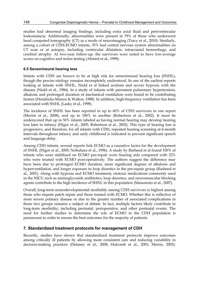
6.5 Sensorineural hearing loss
Infants with CDH are known to be at high risk for sensorineural hearing loss (SNHL), though the precise etiology remains incompletely understood. In one of the earliest reports looking at infants with SNHL, Nield et al linked acidosis and severe hypoxia with the disease (Nield et al., 1986). In a study of infants with persistent pulmonary hypertension, alkalosis and prolonged duration of mechanical ventilation were found to be contributing factors (Hendricks-Munoz & Walton, 1988). In addition, high-frequency ventilation has been associated with SNHL (Lasky et al., 1998).
The incidence of SNHL has been reported in up to 60% of CDH survivors in one report (Morini et al., 2008), and up to 100% in another (Robertson et al., 2002). It must be underscored that up to 50% infants labeled as having normal hearing may develop hearing loss later in infancy (Fligor et al., 2005; Robertson et al., 2002). This type of hearing loss is progressive, and therefore, for all infants with CDH, repeated hearing screening at 6-month intervals throughout infancy and early childhood is indicated to prevent significant speech and language delay.
Among CDH infants, several reports link ECMO as a causative factor for the development of SNHL (Fligor et al., 2005; Nobuhara et al., 1996). A study by Rasheed et al found 100% of infants who were stabilized on ECMO pre-repair wore hearing aids compared with 22% who were treated with ECMO post-operatively. The authors suggest the difference may have been due to prolonged ECMO duration, more significant degrees of alkalosis and hyperventilation, and longer exposure to loop diuretics in the pre-repair group (Rasheed et al., 2001). Along with hypoxia and ECMO treatment, ototoxic medications commonly used in the NICU such as aminoglycoside antibiotics, loop diuretics, and neuromuscular blocking agents contribute to the high incidence of SHNL in this population (Masumoto et al., 2007).
Overall, long-term neurodevelopmental morbidity among CDH survivors is highest among those who require patch repair and those treated with ECMO. Whether this is reflective of more severe primary disease or due to the greater number of associated complications in those two groups remains a subject of debate. In fact, multiple factors likely contribute to long-term morbidity, including perinatal, perioperative, and other postnatal events. The need for further studies to determine the role of ECMO in the CDH population is paramount in order to ensure the best outcomes for the majority of patients.
7. Standardized treatment protocols for management of CDH
Recently, studies have shown that standardized treatment protocols improve outcomes among critically ill patients by allowing more consistent care and reducing variability in decision-making practices (Delaney et al., 2008; Holcomb et al., 2001; Morris, 2003).
Congenital Diaphragmatic Hernia Survivors: Outcomes in Infancy, Childhood, and Adolescence
149
Although a universal approach to management of the CDH infant does not yet exist, several authors have documented their successes after adopting centre-specific CDH management protocols. In one such report, the authors demonstrate that the institution of a treatment protocol is independently associated with decreased mortality risk among infants with CDH; in their study survival rates increased significantly from 67% to 88% following implementation of the protocol (van den Hout et al., 2011). Tracy et al noted similar results after the development and execution of an evidence-based care protocol for CDH infants, with survival to discharge improving from 55% to 85% despite comparable expected survival rates between the pre-protocol and protocol groups (Tracy et al., 2010).
With the aim of providing European neonatologists and pediatric intensive care physicians with a protocolized approach to the infant with CDH, the European task force for CDH (CDH EURO Consortium) recently drafted and published a consensus statement describing a standardized management protocol for CDH (Reiss et al., 2010). One may hope that a similar consensus statement will soon be realized in the United States and worldwide to allow more streamlined and evidence-based care for these highly complex patients.
Given the small numbers of CDH cases occurring at individual centres, cooperation and collaboration between centres and internationally is essential to develop such protocols, and to enhance further research in this field via randomized controlled trials and investigation into long-term outcomes among survivors (van den Hout et al., 2011; Wright et al., 2011).
8. Comprehensive follow-up for CDH survivors
Prior to leaving the hospital, standard discharge practices should be applied to infants with CDH, including measurement of weight, length, and head circumference, physical and neurologic examinations, and newborn hearing screening. Pre-discharge chest radiography may be helpful for comparision over time and to monitor for asymptomatic reherniation at the repair site. An echocardiogram should be considered to evaluate for residual pulmonary hypertension and/or persistent elevations in right-sided heart pressures. Neuroimaging (brain MRI or head CT) may be considered for infants with abnormalities on ultrasound or neurologic examination, and for all ECMO survivors. For the CDH infant with symptomatic GER, imaging studies may be useful in delineating the severity of the problem to aid in deciding whether surgical intervention is warranted. All routine newborn immunizations, including pneumococal and influenza vaccines, should be administered according to recommeded guidelines. In addition, CDH survivors with chronic lung disease are suggested to receive palivizumab (Synagis) for prophylaxis against respiratory syncytial virus (American Academy of Pediatrics, 2009).
Infants with CDH require long-term periodic follow-up evaluation to identify and effectively treat potential challenges; the goal is early and appropriate intervention to prevent additional disability and maximize developmental and cognitive outcomes. Ideally, this would be provided by a multidisciplinary team in a structured neonatal follow-up program in order to enhance communication and collaboration among providers while allowing the most comprehensive care possible. If such a program is not available, follow-up appointments should be arranged with the infant’s primary care provider and with pediatric surgery at minimum. Follow-up with other specialists such as pediatric cardiology, pediatric neurology, and developmental-behavioral pediatrics should be arranged on an as-needed basis depending on the infant’s degree of illness.
Congenital Diaphragmatic Hernia – Prenatal to Childhood Management and Outcomes
150
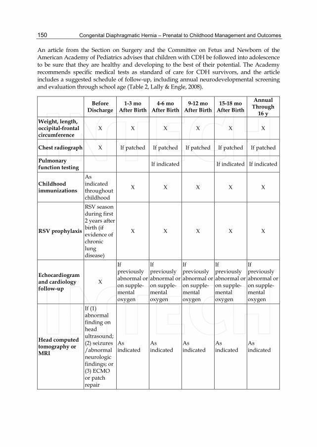
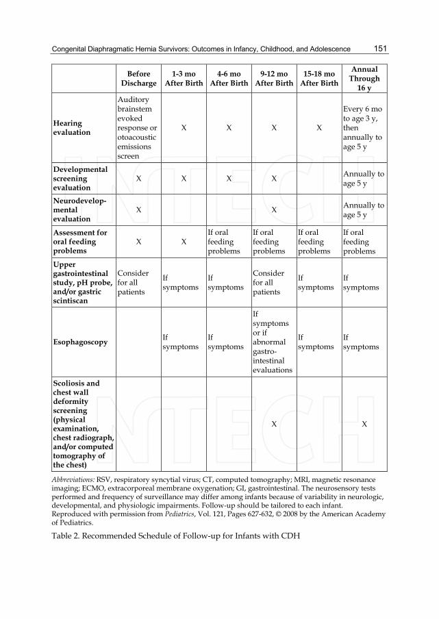
An article from the Section on Surgery and the Committee on Fetus and Newborn of the American Academy of Pediatrics advises that children with CDH be followed into adolescence to be sure that they are healthy and developing to the best of their potential. The Academy recommends specific medical tests as standard of care for CDH survivors, and the article includes a suggested schedule of follow-up, including annual neurodevelopmental screening and evaluation through school age (Table 2, Lally & Engle, 2008).
Before Discharge
1-3 mo After Birth
4-6 mo After Birth
9-12 mo After Birth
15-18 mo After Birth
Annual Through
16 y Weight, length, occipital-frontal circumference
X X X X X X
Chest radiograph X If patched If patched If patched If patched If patched
Pulmonary function testing If indicated If indicated If indicated
Childhood immunizations
As indicated throughout childhood
X X X X X
RSV prophylaxis
RSV season during first 2 years after birth (if evidence of chronic lung disease)
X X X X X
Echocardiogram and cardiology follow-up
X
If previously abnormal or on supple-mental oxygen
If previously abnormal or on supple-mental oxygen
If previously abnormal or on supple-mental oxygen
If previously abnormal or on supple-mental oxygen
If previously abnormal or on supple-mental oxygen
Head computed tomography or MRI
If (1) abnormal finding on head ultrasound; (2) seizures /abnormal neurologic findings; or (3) ECMO or patch repair
As indicated
As indicated
As indicated
As indicated
As indicated
Congenital Diaphragmatic Hernia Survivors: Outcomes in Infancy, Childhood, and Adolescence
151
Before
Discharge 1-3 mo
After Birth 4-6 mo
After Birth 9-12 mo
After Birth 15-18 mo
After Birth
Annual Through
16 y
Hearing evaluation
Auditory brainstem evoked response or otoacoustic emissions screen
X X X X
Every 6 mo to age 3 y, then annually to age 5 y
Developmental screening evaluation
X X X X Annually to age 5 y
Neurodevelop- mental evaluation
X X Annually to age 5 y
Assessment for oral feeding problems
X X If oral feeding problems
If oral feeding problems
If oral feeding problems
If oral feeding problems
Upper gastrointestinal study, pH probe, and/or gastric scintiscan
Consider for all patients
If symptoms
If symptoms
Consider for all patients
If symptoms
If symptoms
Esophagoscopy If symptoms
If symptoms
If symptoms or if abnormal gastro-intestinal evaluations
If symptoms
If symptoms
Scoliosis and chest wall deformity screening (physical examination, chest radiograph, and/or computed tomography of the chest)
X X
Abbreviations: RSV, respiratory syncytial virus; CT, computed tomography; MRI, magnetic resonance imaging; ECMO, extracorporeal membrane oxygenation; GI, gastrointestinal. The neurosensory tests performed and frequency of surveillance may differ among infants because of variability in neurologic, developmental, and physiologic impairments. Follow-up should be tailored to each infant. Reproduced with permission from Pediatrics, Vol. 121, Pages 627-632, © 2008 by the American Academy of Pediatrics.
Table 2. Recommended Schedule of Follow-up for Infants with CDH
Congenital Diaphragmatic Hernia – Prenatal to Childhood Management and Outcomes
152
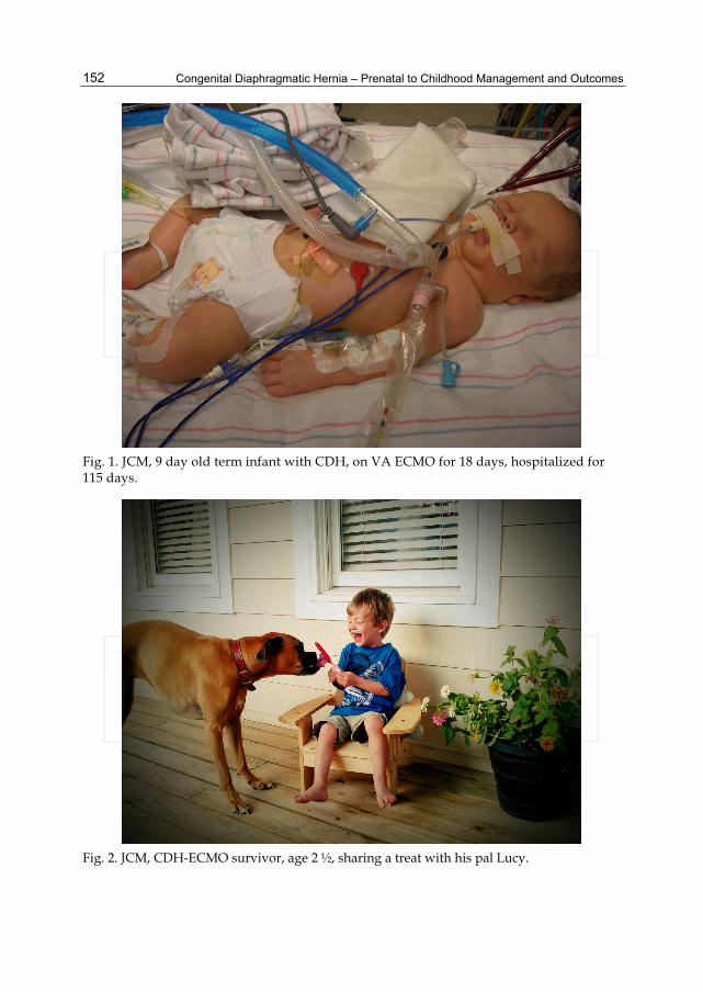
Fig. 1. JCM, 9 day old term infant with CDH, on VA ECMO for 18 days, hospitalized for 115 days.
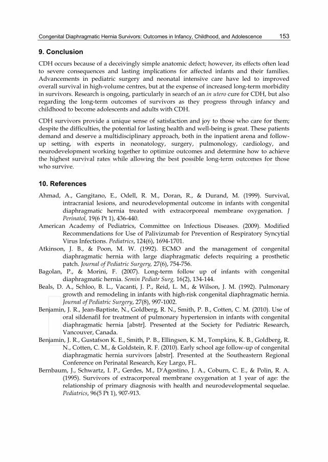
Fig. 2. JCM, CDH-ECMO survivor, age 2 ½, sharing a treat with his pal Lucy.
Congenital Diaphragmatic Hernia Survivors: Outcomes in Infancy, Childhood, and Adolescence
153
9. Conclusion
CDH occurs because of a deceivingly simple anatomic defect; however, its effects often lead to severe consequences and lasting implications for affected infants and their families. Advancements in pediatric surgery and neonatal intensive care have led to improved overall survival in high-volume centres, but at the expense of increased long-term morbidity in survivors. Research is ongoing, particularly in search of an in utero cure for CDH, but also regarding the long-term outcomes of survivors as they progress through infancy and childhood to become adolescents and adults with CDH.
CDH survivors provide a unique sense of satisfaction and joy to those who care for them; despite the difficulties, the potential for lasting health and well-being is great. These patients demand and deserve a multidisciplinary approach, both in the inpatient arena and follow-up setting, with experts in neonatology, surgery, pulmonology, cardiology, and neurodevelopment working together to optimize outcomes and determine how to achieve the highest survival rates while allowing the best possible long-term outcomes for those who survive.
10. References
Ahmad, A., Gangitano, E., Odell, R. M., Doran, R., & Durand, M. (1999). Survival, intracranial lesions, and neurodevelopmental outcome in infants with congenital diaphragmatic hernia treated with extracorporeal membrane oxygenation. J Perinatol, 19(6 Pt 1), 436-440.
American Academy of Pediatrics, Committee on Infectious Diseases. (2009). Modified Recommendations for Use of Palivizumab for Prevention of Respiratory Syncytial Virus Infections. Pediatrics, 124(6), 1694-1701.
Atkinson, J. B., & Poon, M. W. (1992). ECMO and the management of congenital diaphragmatic hernia with large diaphragmatic defects requiring a prosthetic patch. Journal of Pediatric Surgery, 27(6), 754-756.
Bagolan, P., & Morini, F. (2007). Long-term follow up of infants with congenital diaphragmatic hernia. Semin Pediatr Surg, 16(2), 134-144.
Beals, D. A., Schloo, B. L., Vacanti, J. P., Reid, L. M., & Wilson, J. M. (1992). Pulmonary growth and remodeling in infants with high-risk congenital diaphragmatic hernia. Journal of Pediatric Surgery, 27(8), 997-1002.
Benjamin, J. R., Jean-Baptiste, N., Goldberg, R. N., Smith, P. B., Cotten, C. M. (2010). Use of oral sildenafil for treatment of pulmonary hypertension in infants with congenital diaphragmatic hernia [abstr]. Presented at the Society for Pediatric Research, Vancouver, Canada.
Benjamin, J. R., Gustafson K. E., Smith, P. B., Ellingsen, K. M., Tompkins, K. B., Goldberg, R. N., Cotten, C. M., & Goldstein, R. F. (2010). Early school age follow-up of congenital diaphragmatic hernia survivors [abstr]. Presented at the Southeastern Regional Conference on Perinatal Research, Key Largo, FL.
Bernbaum, J., Schwartz, I. P., Gerdes, M., D'Agostino, J. A., Coburn, C. E., & Polin, R. A. (1995). Survivors of extracorporeal membrane oxygenation at 1 year of age: the relationship of primary diagnosis with health and neurodevelopmental sequelae. Pediatrics, 96(5 Pt 1), 907-913.
Congenital Diaphragmatic Hernia – Prenatal to Childhood Management and Outcomes
154
Bouman, N. H., Koot, H. M., Tibboel, D., & Hazebroek, F. W. (2000). Children with congenital diaphragmatic hernia are at risk for lower levels of cognitive functioning and increased emotional and behavioral problems. Eur J Pediatr Surg, 10(1), 3-7.
Colby, C. E. (2004). Surfactant replacement therapy on ECMO does not improve outcome in neonates with congenital diaphragmatic hernia. Journal of Pediatric Surgery, 39(11), 1632-1637.
Cortes, R. A., Keller, R. L., Townsend, T., Harrison, M. R., Farmer, D. L., Lee, H., et al. (2005). Survival of severe congenital diaphragmatic hernia has morbid consequences. J Pediatr Surg, 40(1), 36-45.
Crankson, S. J., Al Jadaan, S. A., Namshan, M. A., Al-Rabeeah, A. A., & Oda, O. (2006). The immediate and long-term outcomes of newborns with congenital diaphragmatic hernia. Pediatr Surg Int, 22(4), 335-340.
D'Agostino, J. A., Bernbaum, J. C., Gerdes, M., Schwartz, I. P., Coburn, C. E., Hirschl, R. B., et al. (1995). Outcome for infants with congenital diaphragmatic hernia requiring extracorporeal membrane oxygenation: The first year. Journal of Pediatric Surgery, 30(1), 10-15.
Danzer, E., Gerdes, M., Bernbaum, J., D'Agostino, J., Bebbington, M. W., Siegle, J., et al. (2010). Neurodevelopmental outcome of infants with congenital diaphragmatic hernia prospectively enrolled in an interdisciplinary follow-up program. Journal of Pediatric Surgery, 45(9), 1759-1766.
Davis, P. J., Firmin, R. K., Manktelow, B., Goldman, A. P., Davis, C. F., Smith, J. H., et al. (2004). Long-term outcome following extracorporeal membrane oxygenation for congenital diaphragmatic hernia: the UK experience. The Journal of Pediatrics, 144(3), 309-315.
Dela Cruz, T. V., Stewart, D. L., Robinson, T. W., & Bond, S. J. (1996). The use of a second course of extracorporeal membrane oxygenation in neonatal patients. ASAIO Journal, 42(3), 230-232.
Delaney, A., Angus, D. C., Bellomo, R., Cameron, P., Cooper, D. J., Finfer, S., et al. (2008). Bench-to-bedside review: the evaluation of complex interventions in critical care. Crit Care, 12(2), 210.
Diamond, I. R., Mah, K., Kim, P. C. W., Bohn, D., Gerstle, J. T., & Wales, P. W. (2007). Predicting the need for fundoplication at the time of congenital diaphragmatic hernia repair. Journal of Pediatric Surgery, 42(6), 1066-1070.
Dillon, P. W., Cilley, R. E., Mauger, D., Zachary, C., & Meier, A. (2004). The relationship of pulmonary artery pressure and survival in congenital diaphragmatic hernia. Journal of Pediatric Surgery, 39(3), 307-312.
Dimmitt, R. A., Moss, R. L., Rhine, W. D., Benitz, W. E., Henry, M. C. W., & VanMeurs, K. P. (2001). Venoarterial versus venovenous extracorporeal membrane oxygenation in congenital diaphragmatic hernia: The extracorporeal life support organization registry, 1990-1999. Journal of Pediatric Surgery, 36(8), 1199-1204.
Falconer, A. R., Brown, R. A., Helms, P., Gordon, I., & Baron, J. A. (1990). Pulmonary sequelae in survivors of congenital diaphragmatic hernia. Thorax, 45(2), 126-129.
Fasching, G., Huber, A., Uray, E., Sorantin, E., Lindbichler, F., & Mayr, J. (2000). Gastroesophageal reflux and diaphragmatic motility after repair of congenital diaphragmatic hernia. European Journal of Pediatric Surgery, 10(6), 360-364.
Congenital Diaphragmatic Hernia Survivors: Outcomes in Infancy, Childhood, and Adolescence
155
Guner, Y. S., Khemani, R. G., Qureshi, F. G., Wee, C. P., Austin, M. T., Dorey, F., et al. (2009). Outcome analysis of neonates with congenital diaphragmatic hernia treated with venovenous vs venoarterial extracorporeal membrane oxygenation. J Pediatr Surg, 44(9), 1691-1701.
Hendricks-Munoz, K. D., & Walton, J. P. (1988). Hearing loss in infants with persistent fetal circulation. Pediatrics, 81(5), 650-656.
Holcomb, B. W., Wheeler, A. P., & Ely, E. W. (2001). New ways to reduce unnecessary variation and improve outcomes in the intensive care unit. Curr Opin Crit Care, 7(4), 304-311.
Hunt, R. W., Kean, M. J., Stewart, M. J., & Inder, T. E. (2004). Patterns of cerebral injury in a series of infants with congenital diaphragmatic hernia utilizing magnetic resonance imaging. Journal of Pediatric Surgery, 39(1), 31-36.
Ijsselstijn, H., Tibboel, D., Hop, W. J., Molenaar, J. C., & de Jongste, J. C. (1997). Long-term pulmonary sequelae in children with congenital diaphragmatic hernia. American Journal of Respiratory and Critical Care Medicine, 155(1), 174-180.
Jakobson, L. S., Frisk, V., Trachsel, D., & O'Brien, K. (2009). Visual and fine-motor outcomes in adolescent survivors of high-risk congenital diaphragmatic hernia who did not receive extracorporeal membrane oxygenation. Journal of Perinatology, 29(9), 630-636.
Jaillard, S. M., Pierrat, V., Dubois, A., Truffert, P., Lequien, P., Wurtz, A. J., et al. (2003). Outcome at 2 years of infants with congenital diaphragmatic hernia: a population-based study. Ann Thorac Surg, 75(1), 250-256.
Kamata, S., Usui, N., Kamiyama, M., Tazuke, Y., Nose, K., Sawai, T., et al. (2005). Long-term follow-up of patients with high-risk congenital diaphragmatic hernia. J Pediatr Surg, 40(12), 1833-1838.
Keller, R. L., Hamrick, S. E. G., Kitterman, J. A., Fineman, J. R., & Hawgood, S. (2004). Treatment of rebound and chronic pulmonary hypertension with oral sildenafil in an infant with congenital diaphragmatic hernia. Pediatric Critical Care Medicine, 5(2), 184-187.
Kinsella, J. P., Parker, T. A., Ivy, D. D., & Abman, S. H. (2003). Noninvasive delivery of inhaled nitric oxide therapy for late pulmonary hypertension in newborn infants with congential diaphragmatic hernia. The Journal of Pediatrics, 142(4), 397-401.
Kinsella, J. P., Truog, W. E., Walsh, W. F., Goldberg, R. N., Bancalari, E., Mayock, D. E., et al. (1997). Randomized, multicenter trial of inhaled nitric oxide and high-frequency oscillatory ventilation in severe, persistent pulmonary hypertension of the newborn. Journal of Pediatrics, 131(1 Pt 1), 55-62.
Kinsella, J. P., Ivy, D. D., & Abman, S. H. (2005). Pulmonary Vasodilator Therapy in Congenital Diaphragmatic Hernia: Acute, Late, and Chronic Pulmonary Hypertension. Seminars in Perinatology, 29(2), 123-128.
Koumbourlis, A. C., & Stolar, C. J. (2004). Lung growth and function in children and adolescents with idiopathic pectus excavatum. Pediatric Pulmonology, 38(4), 339-343.
Lally, K. P., & Breaux, C. W., Jr. (1995). A second course of extracorporeal membrane oxygenation in the neonate--is there a benefit? Surgery, 117(2), 175-178.
Lally, K. P., & Engle, W. (2008). Postdischarge follow-up of infants with congenital diaphragmatic hernia. Pediatrics, 121(3), 627-632.
Congenital Diaphragmatic Hernia – Prenatal to Childhood Management and Outcomes
156
Lasky, R. E., Wiorek, L., & Becker, T. R. (1998). Hearing loss in survivors of neonatal extracorporeal membrane oxygenation (ECMO) therapy and high-frequency oscillatory (HFO) therapy. Journal of the American Academy of Audiology, 9(1), 47-58.
Lund, D. P., Mitchell, J., Kharasch, V., Quigley, S., Kuehn, M., & Wilson, J. M. (1994). Congenital diaphragmatic hernia: the hidden morbidity. J Pediatr Surg, 29(2), 258-262; discussion 262-254.
Maier, S., Zahn, K., Wessel, L. M., Schaible, T., Brade, J., & Reinshagen, K. (2011). Preventive antireflux surgery in neonates with congenital diaphragmatic hernia: a single-blinded prospective study. Journal of Pediatric Surgery, 46(8), 1510-1515.
Marven, S. S., Smith, C. M., Claxton, D., Chapman, J., Davies, H. A., Primhak, R. A., et al. (1998). Pulmonary function, exercise performance, and growth in survivors of congenital diaphragmatic hernia. Archives of Disease in Childhood, 78(2), 137-142.
Masumoto, K., Nagata, K., Uesugi, T., Yamada, T., & Taguchi, T. (2007). Risk factors for sensorineural hearing loss in survivors with severe congenital diaphragmatic hernia. European Journal of Pediatrics, 166(6), 607-612.
McGahren, E. D., Mallik, K., & Rodgers, B. M. (1997). Neurological outcome is diminished in survivors of congenital diaphragmatic hernia requiring extracorporeal membrane oxygenation. J Pediatr Surg, 32(8), 1216-1220.
Morini, F., Capolupo, I., Masi, R., Ronchetti, M. P., Locatelli, M., Corchia, C., et al. (2008). Hearing impairment in congenital diaphragmatic hernia: the inaudible and noiseless foot of time. J Pediatr Surg, 43(2), 380-384.
Morris, A. H. (2003). Treatment algorithms and protocolized care. Curr Opin Crit Care, 9(3), 236-240.
Moss, L. R., Chen, C. M., & Harrison, M. R. (2001). Prosthetic patch durability in congenital diaphragmatic hernia: A long-term follow-up study. Journal of Pediatric Surgery, 36(1), 152-154.
Muratore, C. S., Utter, S., Jaksic, T., Lund, D. P., & Wilson, J. M. (2001). Nutritional morbidity in survivors of congenital diaphragmatic hernia. J Pediatr Surg, 36(8), 1171-1176.
Muratore, C. S., Kharasch, V., Lund, D. P., Sheils, C., Friedman, S., Brown, C., et al. (2001). Pulmonary morbidity in 100 survivors of congenital diaphragmatic hernia monitored in a multidisciplinary clinic. J Pediatr Surg, 36(1), 133-140.
The Neonatal Inhaled Nitric Oxide Study Group (NINOS). (1997). Inhaled Nitric Oxide and Hypoxic Respiratory Failure in Infants With Congenital Diaphragmatic Hernia. Pediatrics, 99(6), 838-845.
Nield, T. A., Schrier, S., Ramos, A. D., Platzker, A. C., & Warburton, D. (1986). Unexpected hearing loss in high-risk infants. Pediatrics, 78(3), 417-422.
Nield, T. (2000). Neurodevelopmental outcome at 3.5 years of age in children treated with extracorporeal life support: Relationship to primary diagnosis. The Journal of Pediatrics, 136(3), 338-344.
Nijhuis-van der Sanden, M. W., van der Cammen-van Zijp, M. H., Janssen, A. J., Reuser, J. J., Mazer, P., van Heijst, A. F., et al. (2009). Motor performance in five-year-old extracorporeal membrane oxygenation survivors: a population-based study. Crit Care, 13(2), R47.
Congenital Diaphragmatic Hernia Survivors: Outcomes in Infancy, Childhood, and Adolescence
157
Nobuhara, K. K., Lund, D. P., Mitchell, J., Kharasch, V., & Wilson, J. M. (1996). Long-term outlook for survivors of congenital diaphragmatic hernia. Clin Perinatol, 23(4), 873-887.
Noori, S., Friedlich, P., Wong, P., Garingo, A., & Seri, I. (2007). Cardiovascular effects of sildenafil in neonates and infants with congenital diaphragmatic hernia and pulmonary hypertension. Neonatology, 91(2), 92-100.
Peetsold, M. G., Vonk-Noordegraaf, A., Heij, H. H., & Gemke, R. J. B. J. (2007). Pulmonary function and exercise testing in adult survivors of congenital diaphragmatic hernia. Pediatric Pulmonology, 42(4), 325-331.
Rasheed, A., Tindall, S., Cueny, D. L., Klein, M. D., & Delaney-Black, V. (2001). Neurodevelopmental outcome after congenital diaphragmatic hernia: Extracorporeal membrane oxygenation before and after surgery. J Pediatr Surg, 36(4), 539-544.
Reid, L. M. (1984). Lung growth in health and disease. British Journal of Diseases of the Chest, 78(2), 113-134.
Reiss, I., Schaible, T., van den Hout, L., Capolupo, I., Allegaert, K., van Heijst, A., et al. (2010). Standardized postnatal management of infants with congenital diaphragmatic hernia in Europe: the CDH EURO Consortium consensus. Neonatology, 98(4), 354-364.
Schwartz, I. P., Bernbaum, J. C., Rychik, J., Grunstein, M., D'Agostino, J., & Polin, R. A. (1999). Pulmonary hypertension in children following extracorporeal membrane oxygenation therapy and repair of congenital diaphragmatic hernia. Journal of Perinatology, 19(3), 220-226.
Steven, M. J., Fyfe, A. H. B., Raine, P. A. M., & Watt, I. (2007). Esophageal adenocarcinoma: a long-term complication of congenital diaphragmatic hernia? Journal of Pediatric Surgery, 42(7), e1-e3.
Su, W., Berry, M., Puligandla, P. S., Aspirot, A., Flageole, H., & Laberge, J. M. (2007). Predictors of gastroesophageal reflux in neonates with congenital diaphragmatic hernia. J Pediatr Surg, 42(10), 1639-1643.
Trachsel, D., Selvadurai, H., Bohn, D., Langer, J. C., & Coates, A. L. (2005). Long-term pulmonary morbidity in survivors of congenital diaphragmatic hernia. Pediatr Pulmonol, 39(5), 433-439.
Tracy, S., Estroff, J., Valim, C., Friedman, S., & Chen, C. (2010). Abnormal neuroimaging and neurodevelopmental findings in a cohort of antenatally diagnosed congenital diaphragmatic hernia survivors. Journal of Pediatric Surgery, 45(5), 958-965.
Tsao, K., Lally, P. A., & Lally, K. P. (2011). Minimally invasive repair of congenital diaphragmatic hernia. Journal of Pediatric Surgery, 46(6), 1158-1164.
Tulloh, R. (2009). Etiology, Diagnosis, and Pharmacologic Treatment of Pediatric Pulmonary Hypertension. Pediatric Drugs, 11(2), 115-128.
Vanamo, K., Rintala, R. J., Lindahl, H., & Louhimo, I. (1996). Long-term gastrointestinal morbidity in patients with congenital diaphragmatic defects. Journal of Pediatric Surgery, 31(4), 551-554.
Vanamo, K., Peltonen, J., Rintala, R., Lindahl, H., Jääskeläinen, J., & Louhimo, I. (1996). Chest wall and spinal deformities in adults with congenital diaphragmatic defects. Journal of Pediatric Surgery, 31(6), 851-854.
Congenital Diaphragmatic Hernia – Prenatal to Childhood Management and Outcomes
158
van den Hout, L., Reiss, I., Felix, J. F., Hop, W. C., Lally, P. A., Lally, K. P., et al. (2010). Risk factors for chronic lung disease and mortality in newborns with congenital diaphragmatic hernia. Neonatology, 98(4), 370-380.
van den Hout, L., Schaible, T., Cohen-Overbeek, T. E., Hop, W., Siemer, J., van de Ven, K., et al. (2011). Actual outcome in infants with congenital diaphragmatic hernia: the role of a standardized postnatal treatment protocol. Fetal Diagnosis and Therapy, 29(1), 55-63.
Van Meurs, K. P., Robbins, S. T., Reed, V. L., Karr, S. S., Wagner, A. E., Glass, P., et al. (1993). Congenital diaphragmatic hernia: long-term outcome in neonates treated with extracorporeal membrane oxygenation. J Pediatr, 122(6), 893-899.
Wilkens, H., Guth, A., König, J., Forestier, N., Cremers, B., Hennen, B., et al. (2001). Effect of Inhaled Iloprost Plus Oral Sildenafil in Patients With Primary Pulmonary Hypertension. Circulation, 104(11), 1218-1222.
Wischermann, A., Holschneider, A. M., & Hübner, U. (1995). Long-Term Follow-Up of Children with Diaphragmatic Hernia. European Journal of Pediatric Surgery, 5(01), 13,18.
Wright, J. C. E., Budd, J. L. S., Field, D. J., & Draper, E. S. (2011). Epidemiology and outcome of congenital diaphragmatic hernia: a 9-year experience. Paediatric and Perinatal Epidemiology, 25(2), 144-149.