Embed Size (px)
Citation preview
J. clin. Path., 1972, 25, 1063-1070
Congenital stenosis and atresia of the jejunumand ileumD. J. deSA
From the Department ofPathology, Radcliffe Infirmary, Oxford
sxtNoPsis The macroscopic and microscopic findings in 12 cases of stenosis and/or atresia of thejejunum and ileum are presented. There was considerable uniformity within the series with over-
lapping of cases of atresia, stenosis, and gut infarction. An analysis of associated lesions in cases
coming to necropsy suggests that the infants were suffering from shock. In nine out of the 12 cases
there was evidence of intrapartum asphyxia and in eight cases evidence of retardation of intrauterinegrowth. It is argued that, since many of the associated complications of pregnancy are known to beof importance in the aetiology of gut infarction in the neonatal period, they are likely to be ofaetiological significance in the development of atresia and stenosis of the gut. A review of perinataldeaths shows that gut ischaemia of varying degrees of severity is a common finding at necropsy,
being noted in 19 out of 56 cases studied. It is suggested that stenosis and atresia are sequelae ofprevious gut ischaemia.
There is controversy regarding the pathogenesis ofcongenital stenosis and atresia of the small bowel.The traditional explanation favoured by Stowens(1966) and Morison (1970) is that atresia andstenosis are caused by a failure of recanalization ofthe bowel lumen following a developmental stage ofobliterative epithelial proliferation. Alternatively,Louw and Barnard (1955) have led the opposingschool who suggest that the development of stenosisand/or atresia is socondary to a vascular insult.What may be interpreted as a refinement of the latterproposition is the view that intestinal stenosis/atresia may develop as a consequence of intus-susception (Parkkulainen, 1958).
This paper presents the findings in cases ofcongenital stenosis/atresia of the jejunum and ileumoccurring in the United Oxford Hospitals duringthe 10 years 1962-71. From the changes seen thepossible pathogenesis will be suggested.
Material and Methods
The records of the Department of Pathology at theRadcliffe Infirmary, Oxford, were searched forcases of intestinal obstruction in stillbirths andneonatal infants. Those cases where intestinalobstruction was due to volvulus, herniae, intus-susception, paralytic ileus, or meconium ileus wereexcluded.Received for publication 3 October 1972.
Sixteen cases of stenosis or atresia were traced butattention was concentrated on the 12 cases wherehistological material was available. The older slideswere reassessed, fresh sections cut where necessaryand stained as thought to be most appropriate.
Details of age, sex, birth weight, gestation,obstetrical history, postnatal progress, age at whichsymptoms presented, and associated malformationswere extracted from the clinical notes and, whereapplicable, the necropsy reports.
In view of the apparent clinical associationsfurther neonatal necropsy material was studied. Thisincluded sections of small bowel from 15 cases asso-ciated with preeclamptic toxaemia, 12 associatedwith maternal antepartum haemorrhage, 21 caseswith intrapartum asphyxia, and eight cases wherecongenital cardiac malformations were associatedwith neonatal death.
Results
A summary of the main findings in the 12 cases isshown in Table I.
MACROSCOPIC FEATURESSingle lesions of the bowel were found in eight ofthe 12 cases, and multiple lesions were seen in thefour remaining cases.
AtresiaIn two cases there was at least one site of complete
1063
copyright. on 1 M
ay 2018 by guest. Protected by
http://jcp.bmj.com
/J C
lin Pathol: first published as 10.1136/jcp.25.12.1063 on 1 D
ecember 1972. D
ownloaded from
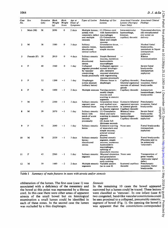
Case Sex Gestation Birth Birth Age at Types ofLesion Pathology of Gut Associated Vascular Associated ClinicalNo. (wk) Weight Weight Onset of Lesions Lesions (Necropsy Findings
(g) Score Symptoms Cases Only)
I Male (M) 38 2890 0 2 days Multiple lesions; (I) Fibrous cord Intatresia with with haemosiderin hamesenteric defect macrophages bilaand multiple (2) Granulation catstenoses tissue and simple
mucosa2 M 38 3380 0 3 days Solitary Granulation tissue, -
stenotic lesion, haemosiderindiscoloured simple mucosaserosal surface
3 Female (F) 39 2910 0 4 days Solitary stenotic Simple flattenedlesion, mucosa, numerousdiscoloured haemosiderinserosa macrophages
4 F 40 3360 0 1 day Collapsed Collapsed segment -
segment preceded normal histologic-by vascular ally; extensiveconstricting mucosal ulcerationbands proximally with regenerating
mucosa proximallyS F 32 1200 - 1 1 day Diaphragm Fibrous tissue in Ca
across jejunum diaphragm bil(solitary lesion) nec
gla6 F 36 1800 -1 2 days Multiple stenoses Varying pattern; Capillary t}
mostly simplemucosa andscarring in stenoticsegments
7 M 37 2300 -1 3 days Solitary stenotic Granulation tissue Extensive bsegment near and marked scar- segmental r
terminal ileum ring of muscularis in adrenalin stenotic segment Capillary ti
8 M 39 2670 _I I day Solitary stenotic (1) Granulation Bilateral inlesion with tissue and early ventricularpatch of acute scarring in stenotic haemorrhalnecrosis segment Capillary tIproximally (2) Acute full-
thickness necrosis9 F 41 2640 -I I day Solitary stenotic Extensive scarring None seen
lesion of muscularis andsimple mucosa;arterial intimalplaques
10 M 38 2520 -1 3 days Solitary stenotic Scarred stenotic -
lesion; segment, simplediscoloured mucosa, numerousserosa haemosiderin
macrophages,splitting ofmuscularis mucosa
It F 43 2560 I I day Solitary stenotic Mucosal ulceration None seensegment and granulation
tissue replacement;early scarring
12 M 39 1480 -2 2 days Multiple stenotic Variable pattern Scattered c;segments (as in case 6) thrombi
traventricular Threatened abortion,emorrhage,ateral,pillary thrombi
old retroplacentalclot noted ondelivery
Marked foetalbradycardia,meconium in liquor(intrapartumasphyxia)Rhesusisoimmunization
Severe foetalbradycardia(intrapartumasphyxia)
apillary thrombi; Preeclampticlateral segmental toxaemia; foetalcrosis of adrenal bradycardiaands
:hrombi Antepartumhaemorrhage; foetalbradycardia
bilateral Preeclampticnecrosis toxaemia; foetalglands bradycardia:hrombiitra- Severe foetal
bradycardiatges (intrapartum:hrombi asphyxia)
Foetal bradycardia(intrapartumasphyxia)
Foetal bradycardia(Placenta said tobe extensively'infarcted')
Transposition ofgreat vessels;ventricular septaldefect
apillary Preeclamptictoxaemia; foetalbradycardia
Table I Summary ofmain features in cases with atresia and/or stenosis
obliteration of the lumen. The first case (case 1) wasassociated with a deficiency of the mesentery andthe bowel at this point was represented by a fibrouscord. In this case there were other areas of apparentatresia of the small bowel but on histologicalexamination a small lumen could be identified ineach of these zones. In the second case the lumenwas occluded by a thin diaphragm.
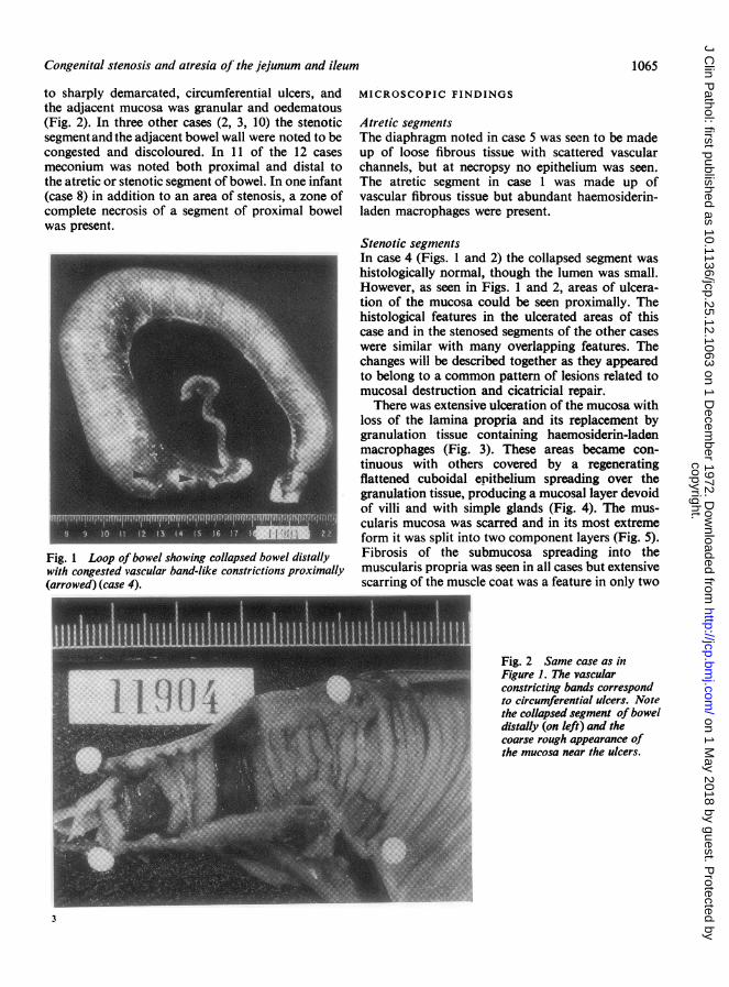
StenosisIn the remaining 10 cases the bowel appearednarrowed but a lumen could be traced. These lesionswere classified as 'stenoses'. In one infant (case 4)two congested, band-like vascularconstrictions couldbe seen proximal to a collapsed, presumably stenotic,segment of bowel (Fig. 1). On opening the bowel itwas apparent that the constrictions corresponded
1064 D. J. dc-Sa
copyright. on 1 M
ay 2018 by guest. Protected by
http://jcp.bmj.com
/J C
lin Pathol: first published as 10.1136/jcp.25.12.1063 on 1 D
ecember 1972. D
ownloaded from
Congenital stenosis and atresia of the jejunum and ileum
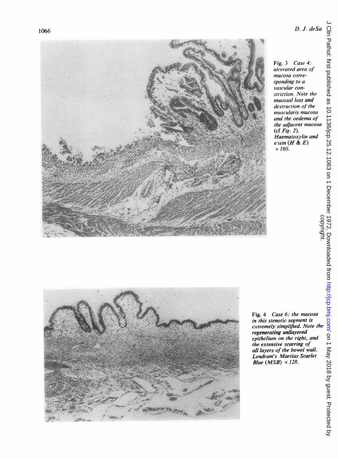
to sharply demarcated, circumferential ulcers, andthe adjacent mucosa was granular and oedematous(Fig. 2). In three other cases (2, 3, 10) the stenoticsegment and the adjacent bowel wall were noted to becongested and discoloured. In 11 of the 12 casesmeconium was noted both proximal and distal tothe atretic or stenotic segment of bowel. In one infant(case 8) in addition to an area of stenosis, a zone ofcomplete necrosis of a segment of proximal bowelwas present.
Fig. 1 Loop ofbowel showing collapsed bowel distallywith congested vascular band-like constrictions proximally(arrowed) (case 4).
MICROSCOPIC FINDINGS
Atretic segmentsThe diaphragm noted in case 5 was seen to be madeup of loose fibrous tissue with scattered vascularchannels, but at necropsy no epithelium was seen.The atretic segment in case 1 was made up ofvascular fibrous tissue but abundant haemosiderin-laden macrophages were present.
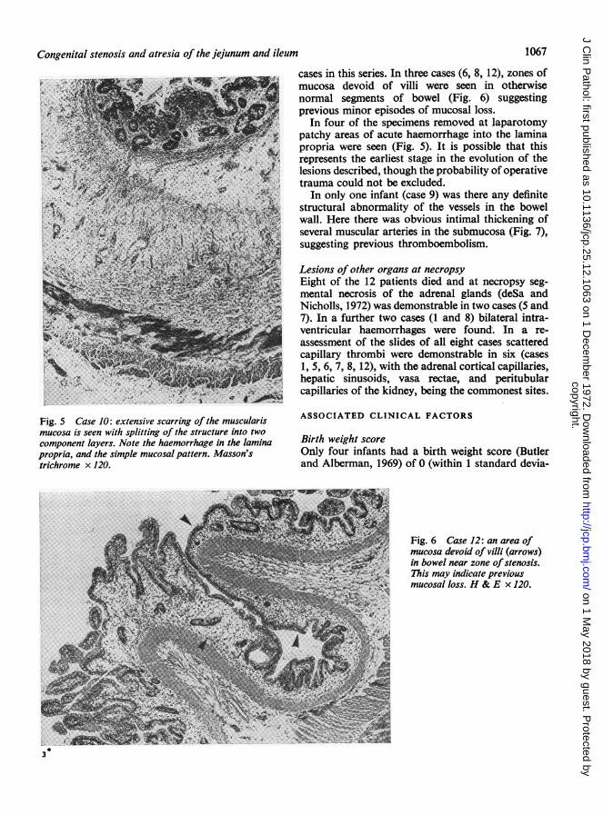
Stenotic segmentsIn case 4 (Figs. 1 and 2) the collapsed segment washistologically normal, though the lumen was small.However, as seen in Figs. 1 and 2, areas of ulcera-tion of the mucosa could be seen proximally. Thehistological features in the ulcerated areas of thiscase and in the stenosed segments of the other caseswere similar with many overlapping features. Thechanges will be described together as they appearedto belong to a common pattern of lesions related tomucosal destruction and cicatricial repair.
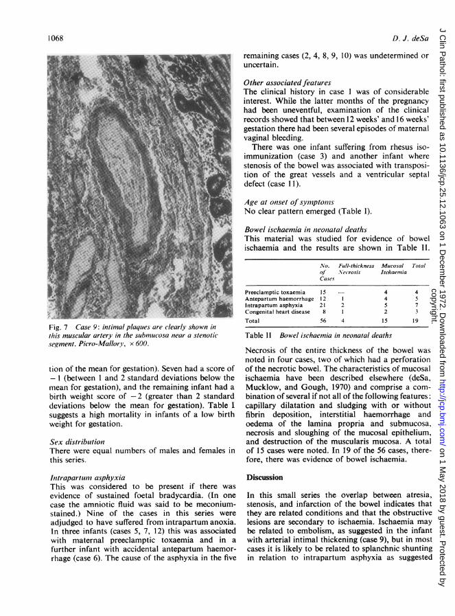
There was extensive ulceration of the mucosa withloss of the lamina propria and its replacement bygranulation tissue containing haemosiderin-ladenmacrophages (Fig. 3). These areas became con-tinuous with others covered by a regeneratingflattened cuboidal enitbelium spreading over thegranulation tissue, producing a mucosal layer devoidof villi and with simple glands (Fig. 4). The mus-cularis mucosa was scarred and in its most extremeform it was split into two component layers (Fig. 5).Fibrosis of the submucosa spreading into themuscularis propria was seen in all cases but extensivescarring of the muscle coat was a feature in only two
Fig. 2 Same case as inFigure 1. The vascularconstricting bands correspondto circumferential ulcers. Notethe collapsed segment ofboweldistally (on left) and thecoarse rough appearance of
I the mucosa near the ulcers.
1065
copyright. on 1 M
ay 2018 by guest. Protected by
http://jcp.bmj.com
/J C
lin Pathol: first published as 10.1136/jcp.25.12.1063 on 1 D
ecember 1972. D
ownloaded from
D. J. deSa
F Fig. 3 Case 4:ulcerated area ofmucosa corre-
^ sponding to a-. vascular con-
4 striction. Note the*i~§ mucosal loss and
destruction of theri -' muscularis mucosa
and the oedema ofthe adjacent mucosa
4~:t (cf Fig. 2).Haematoxylin ande9sin (H & E)x 160.
Ii'
Fig. 4 Case 6: the mucosain this stenotic segment isextremely simplified. Note theregenerating unilayeredepithelium on the right, andthe extensive scarring ofall layers of the bowel wall.Lendrum's Martius ScarletBlue (MSB) x 128.
4 . &K Apf4....
¼ *iZ2..-. .44 . .-
* . *.- b..-S%.C -'-. a
54...."...... %#
.VI,
'S --
V
& ...
1066
copyright. on 1 M
ay 2018 by guest. Protected by
http://jcp.bmj.com
/J C
lin Pathol: first published as 10.1136/jcp.25.12.1063 on 1 D
ecember 1972. D
ownloaded from
Congenital stenosis and atresia of the jejunum and ileum
.-
I E
Fig. 5 Case 10: extensive scarring of the muscularismucosa is seen with splitting of the structure into twocomponent layers. Note the haemorrhage in the laminapropria, and the simple mucosal pattern. Masson'strichrome x 120.
cases in this series. In three cases (6, 8, 12), zones ofmucosa devoid of villi were seen in otherwisenormal segments of bowel (Fig. 6) suggestingprevious minor episodes of mucosal loss.
In four of the specimens removed at laparotomypatchy areas of acute haemorrhage into the laminapropria were seen (Fig. 5). It is possible that thisrepresents the earliest stage in the evolution of thelesions described, though the probability of operativetrauma could not be excluded.
In only one infant (case 9) was there any definitestructural abnormality of the vessels in the bowelwall. Here there was obvious intimal thickening ofseveral muscular arteries in the submucosa (Fig. 7),suggesting previous thromboembolism.
Lesions of other organs at necropsyEight of the 12 patients died and at necropsy seg-mental necrosis of the adrenal glands (deSa andNicholls, 1972) was demonstrable in two cases (5 and7). In a further two cases (1 and 8) bilateral intra-ventricular haemorrhages were found. In a re-assessment of the slides of all eight cases scatteredcapillary thrombi were demonstrable in six (cases1, 5, 6, 7, 8, 12), with the adrenal cortical capillaries,hepatic sinusoids, vasa rectae, and peritubularcapillaries of the kidney, being the commonest sites.
ASSOCIATED CLINICAL FACTORS
Birth weight scoreOnly four infants had a birth weight score (Butlerand Alberman, 1969) of 0 (within 1 standard devia-
Fig. 6 Case 12: an area ofmucosa devoid of villi (arrows)
b->F:f* in bowel near zone of stenosis.^e--Y-g This may indicate previous
mucosal loss. H & E x 120.
3
1067
copyright. on 1 M
ay 2018 by guest. Protected by
http://jcp.bmj.com
/J C
lin Pathol: first published as 10.1136/jcp.25.12.1063 on 1 D
ecember 1972. D
ownloaded from
D. J. deSa
remaining cases (2, 4, 8, 9, 10) was undetermined or
uncertain.
Other associated featur-esThe clinical history in case 1 was of considerableinterest. While the latter months of the pregnancy
had been uneventful, examination of the clinicalrecords showed that between 12 weeks' and 16 weeks'gestation there had been several episodes of maternalvaginal bleeding.
There was one infant suffering from rhesus iso-immunization (case 3) and another infant wherestenosis of the bowel was associated with transposi-tion of the great vessels and a ventricular septaldefect (case 11).
Age at onset of syIn?ptolnlsNo clear pattern emerged (Table 1).
Fig. 7 Case 9: intimal plaques are clearly shown inthis muscular artery in the submucosa near a stenoticsegment. Picro-Mallory, x 600.
tion of the mean for gestation). Seven had a score of- I (between 1 and 2 standard deviations below themean for gestation), and the remaining infant had a
birth weight score of -2 (greater than 2 standarddeviations below the mean for gestation). Table Isuggests a high mortality in infants of a low birthweight for gestation.
Sex distributionThere were equal numbers of males and females inthis series.
Iltrapartumii asphyxiaThis was considered to be present if there was
evidence of sustained foetal bradycardia. (In one
case the amniotic fluid was said to be meconium-stained.) Nine of the cases in this series were
adjudged to have suffered from intrapartum anoxia.in three infants (cases 5, 7, 12) this was associatedwith maternal preeclamptic toxaemia and in a
further infant with accidental antepartum haemor-rhage (case 6). The cause of the asphyxia in the five
Bowel ischaemia in neonatal deathsThis material was studied for evidence of bowelischaemia and the results are shown in Table II.
No. Full-thickness Mucosal Totalof Necrosis IschlaeniaCases
Preeclamptic toxaemia 15 - 4 4Antepartum haemorrhage 12 1 4 5Intrapartum asphyxia 21 2 5 7Congenital heart disease 8 1 2 3
Total 56 4 15 19
Table 11 Bowel isehaemia in neonatal deaths
Necrosis of the entire thickness of the bowel wasnoted in four cases, two of which had a perforationof the necrotic bowel. The characteristics of mucosalischaemia have been described elsewhere (deSa,Mucklow, and Gough, 1970) and comprise a com-bination of several if not all of the following features:capillary dilatation and sludging with or withoutfibrin deposition, interstitial haemorrhage andoedema of the lamina propria and submucosa,necrosis and sloughing of the mucosal epithelium,and destruction of the muscularis mucosa. A totalof 15 cases were noted. In 19 of the 56 cases, there-fore, there was evidence of bowel ischaemia.
Discussion
In this small series the overlap between atresia,stenosis, and infarction of the bowel indicates thatthey are related conditions and that the obstructivelesions are secondary to ischaemia. Ischaemia maybe related to embolism, as suggested in the infantwith arterial intimal thickening (case 9), but in mostcases it is likely to be related to splanchnic shuntingin relation to intrapartum asphyxia as suggested
1 068
copyright. on 1 M
ay 2018 by guest. Protected by
http://jcp.bmj.com
/J C
lin Pathol: first published as 10.1136/jcp.25.12.1063 on 1 D
ecember 1972. D
ownloaded from
Congenital steinosis and atresia of the jejunum and ileum
earlier (deSa et al, 1970). This would place mostcases of neonatal and congenital stenosis of thebowel as an end-stage in the spectrum ofgut ischaemiaaffecting this age group. It is of interest that case 5of deSa et al (1970) developed a stricture of the largebowel following a previous perforation, and thatcase 7 in the same series showed evidence of regenera-tion. Further, the mucosal changes seen in thestenotic segments are extremely similar to the persist-ent mucosal changes in the colon following theischaemic phenomena associated with Hirsch-sprung's disease (Berry, 1969).The aetiological importance of the complications
of pregnancy noted in this series is suggested by thestudy of the morphology of the small bowel in theneonatal deaths not associated with stenosis (TableII), where it can be seen that ischaemic bowel lesionsare a frequent finding in babies suffering from intra-partum asphyxia. The case for the importance ofintrapartum asphyxia, and in particular the com-plications noted in this series, in the aetiology ofischaemia of the gastrointestinal tract has beenclearly stated by Lloyd (1969).
It would be unrealistic to suppose that the com-plications of pregnancy listed in this small series ofcases were of importance as acute phenomena only.In an examination of placentae from a large numberof pregnancies complicated by the same abnormali-ties, a high proportion were found to have organiz-ing thrombi of varying age in their foetal chorionicveins (deSa, 1971). These venous thrombi were thestarting point in the development of intimal cushionsin the affected veins and are commonest in infantsof low birth weight score, a point noted earlier byother observers (Gruenwald, 1963; Blanc, 1968).The importance of the placental changes is that theyindicate longstanding damage occurring within animportant part of the foetal circulation. It is regret-table that the placenta was not available for histo-logical examination in any of the cases of the presentseries. The relatively large number of infants of lowbirth weight score in the present series, however, isnotable, and the apparently high mortality amongthese infants has been remarked upon.Table II demonstrates further that ischaemic lesions
of the small bowel are not uncommon in congenitalheart disease, a situation that finds a parallel inadults with cardiovascular disease and bowelischaemia (Marston, Pheils, Thomas, and Morson,1966; McKinnell and Kearney, 1967).The presence of other ischaemic lesions and scat-
tered fibrin thrombi in infants with stenosis oratresia suggests that the affected infants were suffer-ing from shock (McGovern, 1971), and providesfurther support for the aetiological role of the com-plications of pregnancy noted in this series. The
presence of scattered ischaemic lesions and fibrinthrombi in other organs would tend to suggest thatan incidental intussusception was not of majorimportance in the cases studied.
If an episode (or episodes) of ischaemia is the basicaetiological factor in the development of stenosis ofthe bowel it is easy to understand why lanugo,squames, and meconium may be present distal to anatretic segment (Santulli and Blanc, 1961) or whysegments of atretic bowel may be separated by rela-tively normal bowel lined by normal intestinalmucosa and containing meconium (Schultz andLawrence, 1960). Such findings are incapable ofbeing explained by a failure of recanalization duringthe organogenesis of the bowel, and it seems appro-priate to suggest that this traditional theory shouldbe discarded. In any case, a phase of obliterativeepithelial proliferation has never been seen in thesmall bowel below the duodenum (Johnson, 1910),and it seems inappropriate that it was ever advancedas an explanation of stenosis/atresia of the small orlarge bowel.The overlap between frank necrosis and oblitera-
tive scarring, the high incidence of complications ofpregnancy, and the evidence of necrotic and throm-botic phenomena in other organs all point in thesame direction and the uniformity in this small seriesis striking. There is nothing new in suggesting thatischaemia can produce scarring, and the example ofmyocardial infarction is familiar to every pathologist.Strictures are an accepted complication of ischaemiccolitis in adults (Marston et al, 1966) and it wouldappear that ischaemic episodes are of primaryimportance in the development of the homologouscomplications in the small bowel of the newborn.
I am grateful to the Director of the Gibson Labora-tories, Dr A. H. T. Robb-Smith, for permission tostudy the records.
References
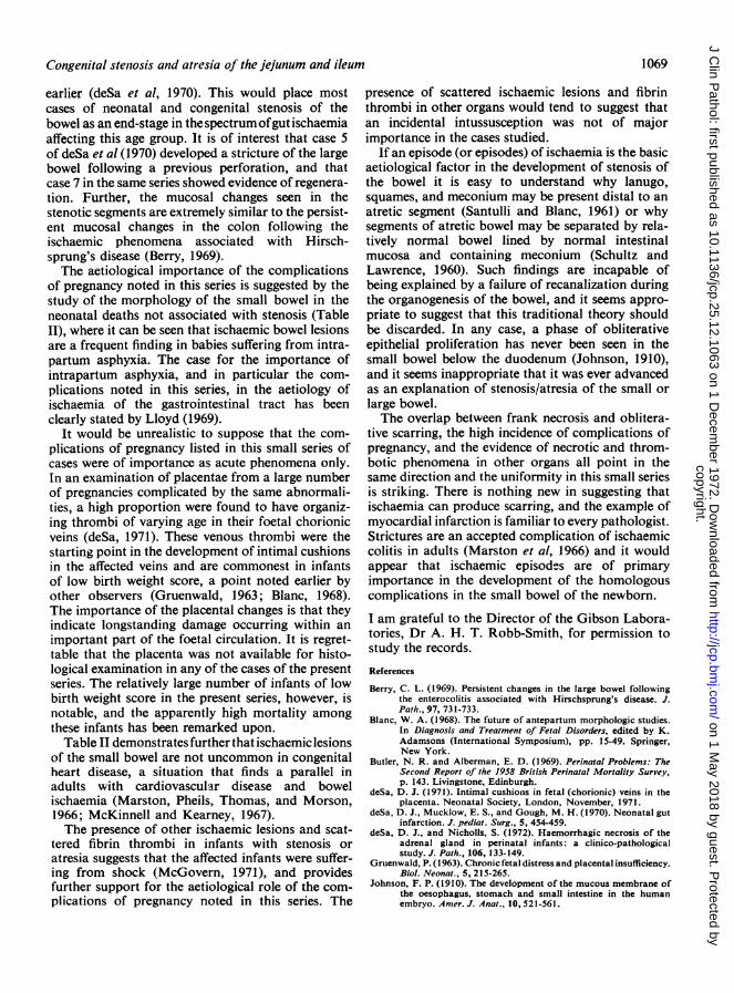
Berry, C. L. (1969). Persistent changes in the large bowel followingthe enterocolitis associated with Hirschsprung's disease. J.Path., 97, 731-733.
Blanc, W. A. (1968). The future of antepartum morphologic studies.In Diagnosis and Treatment of Fetal Disorders, edited by K.Adamsons (International Symposium), pp. 15-49. Springer,New York.
Butler, N. R. and Alberman, E. D. (1969). Perinatal Problems: TheSecond Report of the 1958 British Perinatal Mortality Survey,p. 143. Livingstone, Edinburgh.
deSa, D. J. (1971). Intimal cushions in fetal (chorionic) veins in theplacenta. Neonatal Society, London, November, 1971.
deSa, D. J., Mucklow, E. S., and Gough, M. H. (1970). Neonatal gutinfarction. J. pediat. Surg., 5, 454-459.
deSa, D. J., and Nicholls, S. (1972). Haemorrhagic necrosis of theadrenal gland in perinatal infants: a clinico-pathologicalstudy. J. Path., 106, 133-149.
Gruenwald, P. (1963). Chronic fetal distress and placental insufficiency.Biol. Neonat., 5, 215-265.
Johnson, F. P. (1910). The development of the mucous membrane ofthe oesophagus, stomach and small intestine in the humanembryo. Amer. J. Anat., 10, 521-561.
1069
copyright. on 1 M
ay 2018 by guest. Protected by
http://jcp.bmj.com
/J C
lin Pathol: first published as 10.1136/jcp.25.12.1063 on 1 D
ecember 1972. D
ownloaded from
D. J. deSa
Lloyd, J. R. (1969). The etiology of gastrointestinal perforations inthe newborn. J. pediat. Surg., 4, 77-84.
Louw, J. H., and Barnard, C. N. (1955). Congenital intestinal atresia:observations on its origin. Lancet, 2, 1065-1067.
McGovern, V. J. (1971). Shock. Path. Ann., 6,279-298.McKinneil, J. S., and Kearney, M. S. (1967). Haemorrhagic necrosis
of the intestine. Brit. med. J., 2, 460-463.Marston, A., Pheils, M. T., Thomas, M. L., and Morson, B. C. (1966).
Ischaemic colitis. Gut, 7, 1-15.Morison, J. E. (1970). Foetal and Neonatal Pathology, 3rd ed., pp. 341-
343. Butterworths, London.
Parkkulainen, K. V. (1958). Intrauterine intussuseeption as a cause ofintestinal atresia: a contribution to the etiology of intestinalatresias. Surgery, 44, 1106-1111.
Santulli, T. V., and Blanc, W. A. (1961). Congenital atresia of theintestine: pathogenesis and treatment. Ann. Surg., 154, 939-948.
Schultz, L. R., and Lawrence, G. H. (1960). Associated rectal andjejunal atresia in the newborn: report of a case. Pediatrics, 26,122-125.
Stowens, D. (1966). Pediatric Patholog>. 2nd ed., pp. 569-570. Williamsand Wilkins, Baltimore.
Reports and Bulletins prepared by the Association of Clinical Biochemists
The following reports and bulletins are published by the Association of Clinical Biochemists. They may be obtainedfrom The Administrative Office, Association ofClinical Biochemists, 7Warwick Court, Holborn, London, WC1R5DP.The prices include postage, but airmail will be charged extra. Overseas readers should remit by British Postal or MoneyOrder. If this is not possible the equivalent of 50p is the minimum amount that can be accepted.
SCIENTIFIC REPORTS3 Automatic Dispensing Pipettes. An assessment of 35commercial instruments 1967 P. M. G. BROUGHTON,A. H. GOWENLOCK, G. M. WIDDOWSON, and K. A. AHLQUIST80p ($2)
4 An Evaluation of five Commercial Flame Photometerssuitable for the Simultaneous Determination of Sodiumand Potassium March 1970 P. M. G. BROUGHTON andJ. B. DAWSON 80p ($2)
SCIENTIFIC REVIEWS
1 The Assessment of Thyroid Function March 1971F. V. FLYNN and J. R. HOBBS 60p ($1.50)
2 Renal Function Tests Suitable for Clinical PracticeJanuary 1972 F. L. MITCHELL, N. VEALL, and R. W. E.WATTS 60p ($1.50)
TECHNICAL BULLETINS
9 Determination of Urea by AutoAnalyzer November1966 RUTH M. HASLAM 40p ($1)
11 Determination of Serum Albumin by AutoAnalyzerusing Bromocresol Green October 1967 B. E. NORTHAMand G. M. WIDDOWSON 40p ($1)
13 An Assessment of the Technicon Type II SamplerUnit March 1968 B. C. GRAY and G. K. MCGOWAN40p ($1)
14 Atomic Absorption Spectroscopy. An outline of itsprinciples and a guide to the selection of instrumentsMay 1968 J. B. DAWSON and P. M. G. BROUGHTON40p ($1)
15 A Guide to Automatic Pipettes (2nd edition) June1968 P. M. G. BROUGHTON 40p ($1)
16 A Guide to Automation in Clinical Chemistry May1969 P. M. G. BROUGHTON 60p ($1.50)
17 Flame Photometers (2nd edition) 1969 P. WILDING60p ($1.50)18 Control Solutions for Clinical Biochemistry (4thedition) March 1970 P. M. G. BROUGHTON 60p($1.50)19 Spectrophotometers. A comparative list of low-pricedinstruments readily available in Britain May 1970C. E. WILDE and P. SEWELL 60p ($1.50)20 Quantities and Units in Clinical Biochemistry June1970 P. M. 0. BROUGHTON 60p ($1.50) More than30 copies in units of 10 at 20p21 Filter Fluorimeters: A comparative list of 18 instru-ments September 1970 H. BRAUNSBERG and s. s.BROWN 60p ($1.50)22 Bilirubin standards and the Determination of Bilirubinby Manual and Technicon AutoAnalyzer MethodsJanuary 1971 BARBARA BILLING, RUTH HASLAM, andN. WALD 60p ($1.50)23 Interchangeable Cells for Spectrophotometers andFluorimeters September 1971 E. S. BROWN and A. H.GOWENLOCK 60p ($1.50)24 Simple Tests to Detect Poisons March 1972 B. W.MEADE et al. 60p ($1.50)25 Blood Gas Analysers May 1972 K. DIXON 60p($1.50)26 Kits for Enzyme Activity Determination September1972 s. B. ROSALKI and D. TARLOW 80p ($2.00)27 Assessment of Pumps Suitable for Incorporation intoExisting Continuous Flow Analytical Systems November1972 A. FLECK et al 60p ($1.50)
1070
copyright. on 1 M
ay 2018 by guest. Protected by
http://jcp.bmj.com
/J C
lin Pathol: first published as 10.1136/jcp.25.12.1063 on 1 D
ecember 1972. D
ownloaded from