Embed Size (px)
Citation preview
Congenital Vascular
Malformations Chad Simon, M.D.
Faculty Advisor: Sekin Ulualp, M.D.
The University of Texas Medical Branch
Department of Otolaryngology
Grand Rounds Presentation
December 21, 2006
Introduction
History and Classification
Diagnosis
Lymphatic Malformations
Venous Malformations
Capillary Malformations
Arteriovenous Malformations
Introduction
Among the most common congenital and neonatal abnormalities.
In the past sometimes confusing classifications were developed
Understanding of the various biological characteristics of vascular tumors has been impeded.
Misconception that most of these lesions spontaneously disappear within the first years of life.
As a consequence congenital vascular malformations were often misdiagnosed and left untreated.
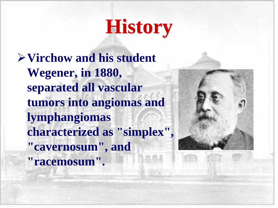
History
Virchow and his student
Wegener, in 1880,
separated all vascular
tumors into angiomas and
lymphangiomas
characterized as "simplex",
"cavernosum", and
"racemosum".
Classification
Mulliken and Glowacki , in 1982, developed
a biological classification encompassing
physical findings, clinical behavior and
cellular kinetics.
They distinguished hemangiomas from
vascular malformations with two main
characteristics distinguishing each.
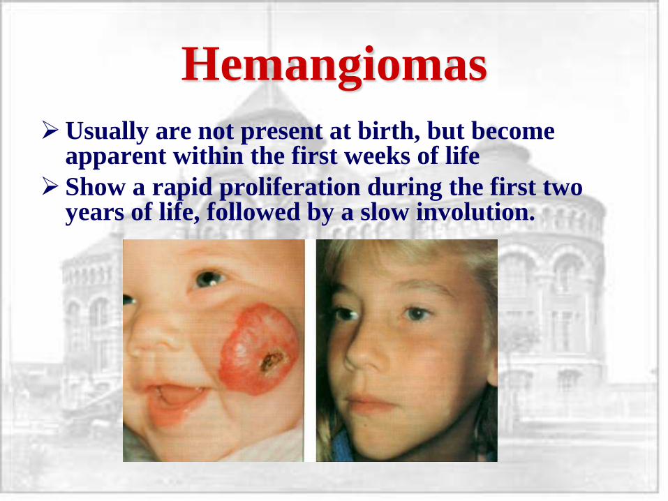
Hemangiomas
Usually are not present at birth, but become apparent within the first weeks of life
Show a rapid proliferation during the first two years of life, followed by a slow involution.
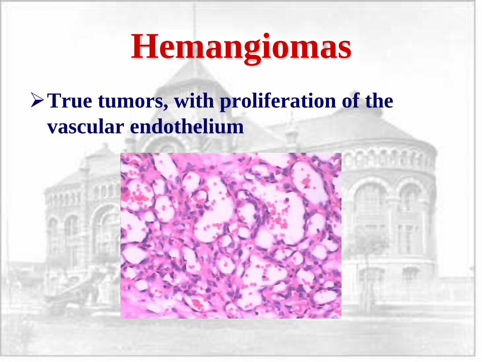
Hemangiomas
True tumors, with proliferation of the
vascular endothelium
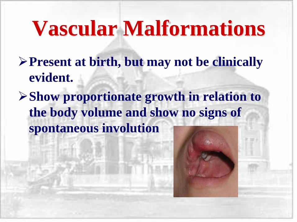
Vascular Malformations
Present at birth, but may not be clinically
evident.
Show proportionate growth in relation to
the body volume and show no signs of
spontaneous involution
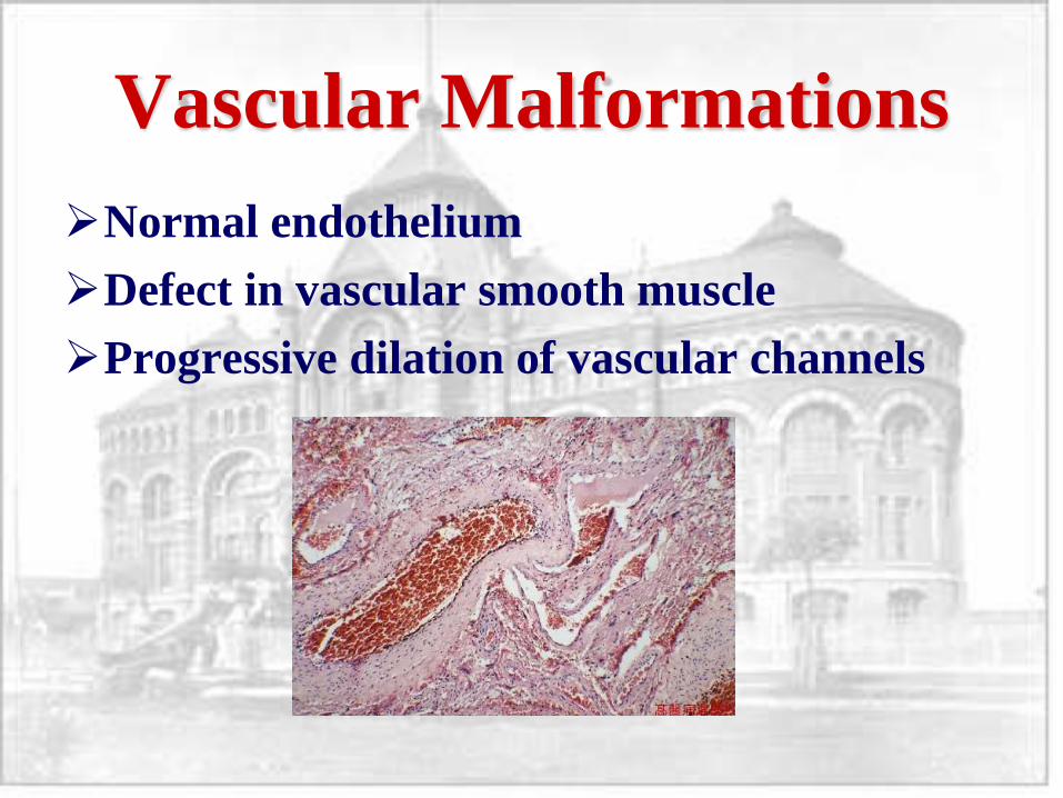
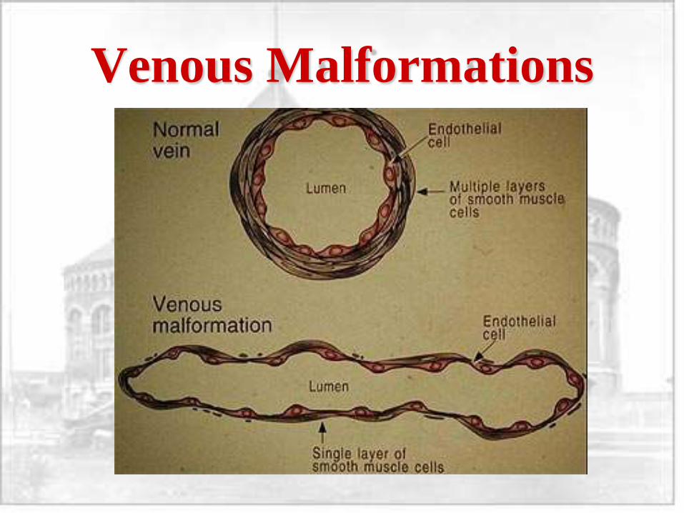
Vascular Malformations
Normal endothelium
Defect in vascular smooth muscle
Progressive dilation of vascular channels
Classification
Burrows, in conjunction with Mulliken, in
1983, further described malformations as
either:
High flow - having a connection to the
arterial or capillary system
Low flow - having a connection to the
venous or lymphatic system.
Classification
High-flow vascular anomalies, such as
arteriovenous fistulas and arteriovenous
malformations, are traditionally addressed
by means of transarterial embolization.
Low-flow malformations found to be
solitary or combined in capillary, venous, or
lymphatic vessels are successfully treated
with sclerotherapy.

Classification
1988, 7th meeting of the
International
workshop on Vascular
Malformations,
Hamburg
Classification
differentiates between
truncular and
extratruncular
malformations
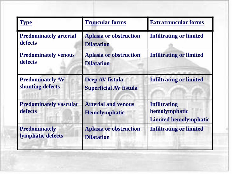
Type Truncular forms Extratruncular forms
Predominately arterial
defects
Aplasia or obstruction
Dilatation
Infiltrating or limited
Predominately venous
defects
Aplasia or obstruction
Dilatation
Infiltrating or limited
Predominately AV
shunting defects
Deep AV fistula
Superficial AV fistula
Infiltrating or limited
Predominately vascular
defects
Arterial and venous
Hemolymphatic
Infiltrating
hemolymphatic
Limited hemolymphatic
Predominately
lymphatic defects
Aplasia or obstruction
Dilatation
Infiltrating or limited
Diagnosis
Noninvasive studies
– MRI
– Duplex ultrasound
– Whole body blood pool scintigraphy
– Transarterial lung perfusion scintigraphy
– Lymphoscintigraphy
– CT
Diagnosis
Invasive studies
– Selective and Superselective angiography
– Direct puncture (percutaneous) arteriography
– Standard phlebography
– Direct puncture (percutaneous) phlebography
– Direct puncture (percutaneous)
lymphaniography
Diagnosis
3 non-invasive tests are sufficiently accurate
and obviate the need for invasive studies
Invasive procedures are usually reserved
for treatment planning
MRI
Imaging modality most commonly used
Should include T1- and T2-weighted spin-echo imaging in multiple planes, fat-saturated T1-weighted imaging with the intravenous administration of a gadolinium-based contrast agent, and gradient-recalled echo (GRE) imaging.
T2-weighted images are used mainly to evaluate the extent of the abnormality.
GRE images are used to identify the hemodynamic nature of the condition (high- vs low-flow lesions); and contrast-enhanced images are used to determine the extent of the malformation and to distinguish low-flow vascular anomalies (venous malformation versus lymphatic malformation).
MRI
For any vascular anomaly, the basic approach is
first, to evaluate fat-suppressed T2-weighted
images to determine the extent of the anomaly,
and second, to evaluate the GRE images to decide
whether the anomaly is a high-flow lesion.
If the anomaly is a low-flow lesion, arteriovenous
malformation, arteriovenous fistula, and
hemangioma can be excluded from the differential
diagnosis.
MRI
Low-flow vascular anomalies (venous
malformation, lymphatic malformation,
capillary-lymphatic-venous malformation)
can be further differentiated on the basis of
their morphologic appearances and
contrast-enhancement patterns.
MRI
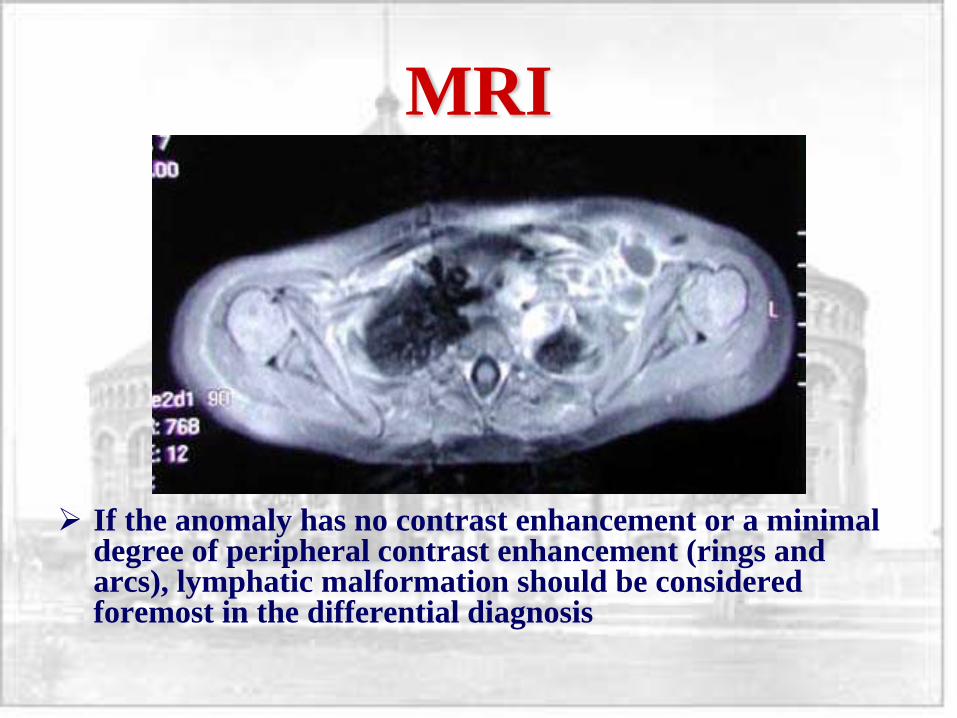
If the anomaly has no contrast enhancement or a minimal degree of peripheral contrast enhancement (rings and arcs), lymphatic malformation should be considered foremost in the differential diagnosis
MRI
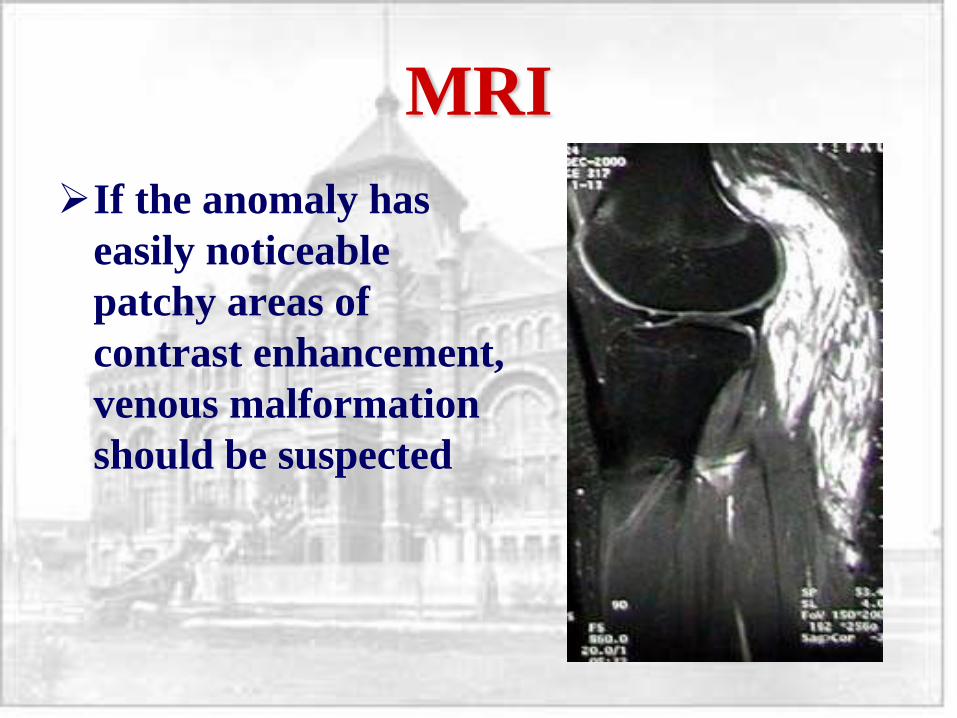
If the anomaly has
easily noticeable
patchy areas of
contrast enhancement,
venous malformation
should be suspected
MRI
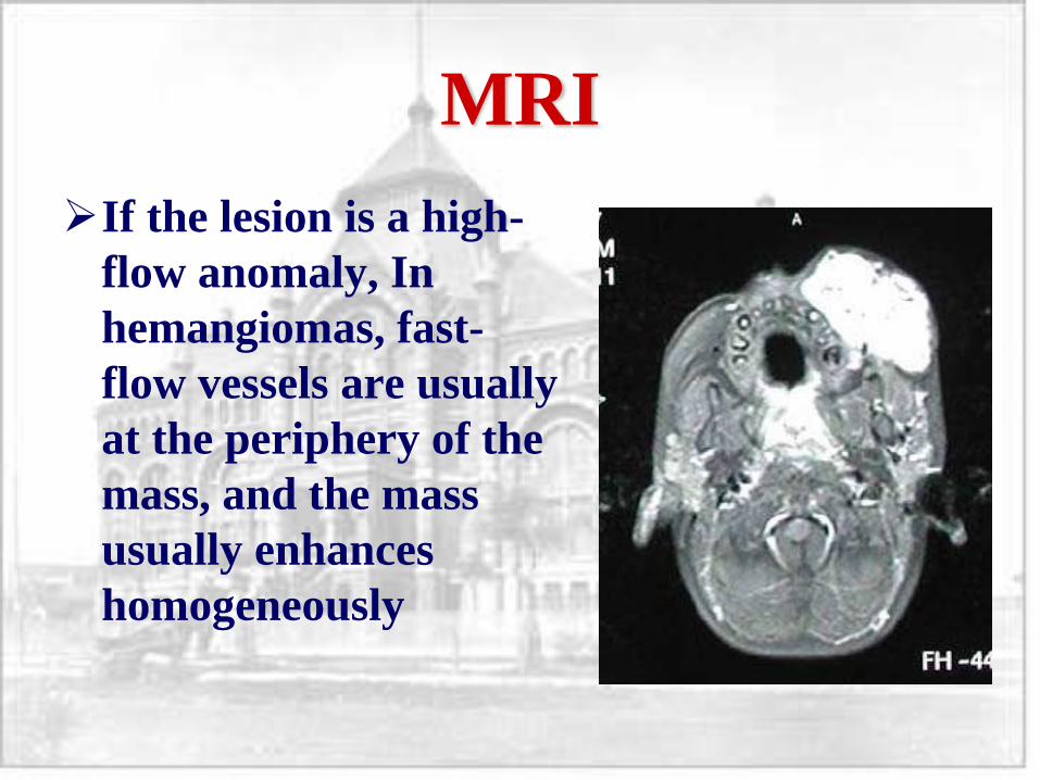
If the lesion is a high-
flow anomaly, In
hemangiomas, fast-
flow vessels are usually
at the periphery of the
mass, and the mass
usually enhances
homogeneously
MRI
A mass lesion is not expected in an
arteriovenous malformation
If there are any remaining questions, the
high-flow nature of an arteriovenous
malformation can be easily confirmed with
Doppler examination, which reveals high-
flow, low-resistance arteries and an
arterialized waveform in the draining veins.
Treatment Indications
Hemorrhage
Risk of high-output heart failure
Chronic venous hypertension
Airway impingement
Lesion threatens vital functions

Treatment Indications
Disabling pain
Functional disability
Cosmetic deformity
Recurrent infection
Persistent lymph leakage
Low flow malformations
Lymphatic Malformations
-Microcystic
-Macrocystic
Venous Malformations
Capillary Malformations
Combined types
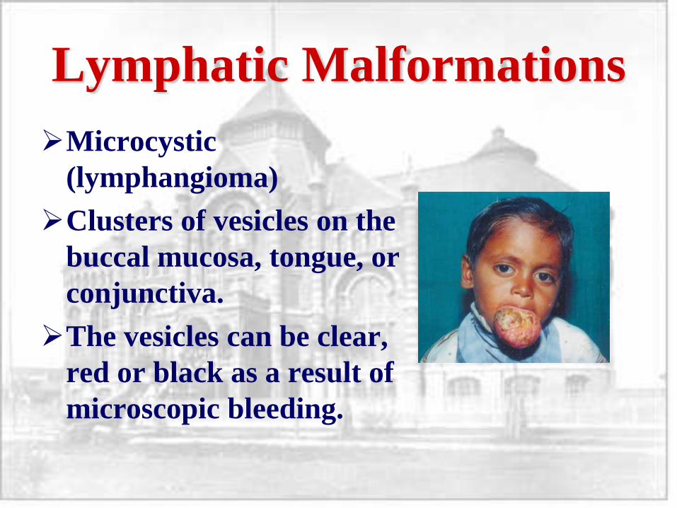
Lymphatic Malformations
Microcystic
(lymphangioma)
Clusters of vesicles on the
buccal mucosa, tongue, or
conjunctiva.
The vesicles can be clear,
red or black as a result of
microscopic bleeding.
Lymphatic Malformations
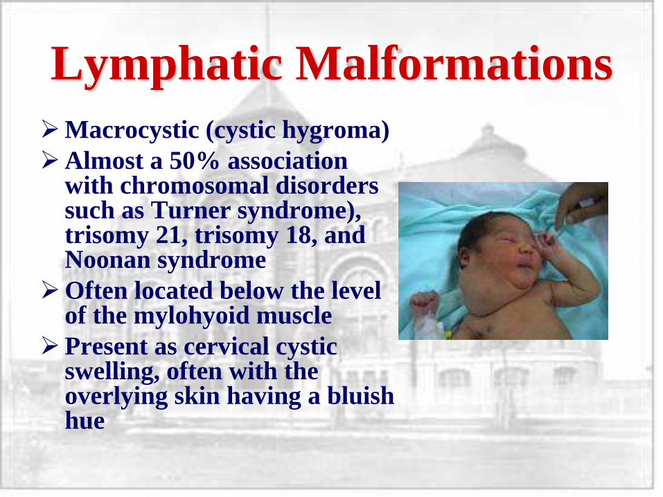
Macrocystic (cystic hygroma)
Almost a 50% association with chromosomal disorders such as Turner syndrome), trisomy 21, trisomy 18, and Noonan syndrome
Often located below the level of the mylohyoid muscle
Present as cervical cystic swelling, often with the overlying skin having a bluish hue
Lymphatic Malformations
Since most lymphatic malformations are mixed-form malformations (macro- and microcystic), the most common therapeutic approach is sclerotherapy for the macrocystic portion of the malformation, then surgical excision of the remaining microcystic portion if needed
Aspiration or drainage results only in temporary shrinkage
Macrocystic lesions, if excised are ideally removed in one procedure, because repeated excisions are complicated by fibrosis and anatomic distortion
Microcystic lesions are often difficult to resect, because there are no distinct tissue planes between the malformed and normal structures
Lymphatic Malformations
Lee, 2005, reviewed 315 patients treated for
LM.
All head and neck LM were of the
extratruncular form.
Sclerotherapy with OK-432 showed 90%
success rate with macrocystic LM, but 50%
with microcystic type
Lymphatic Malformations
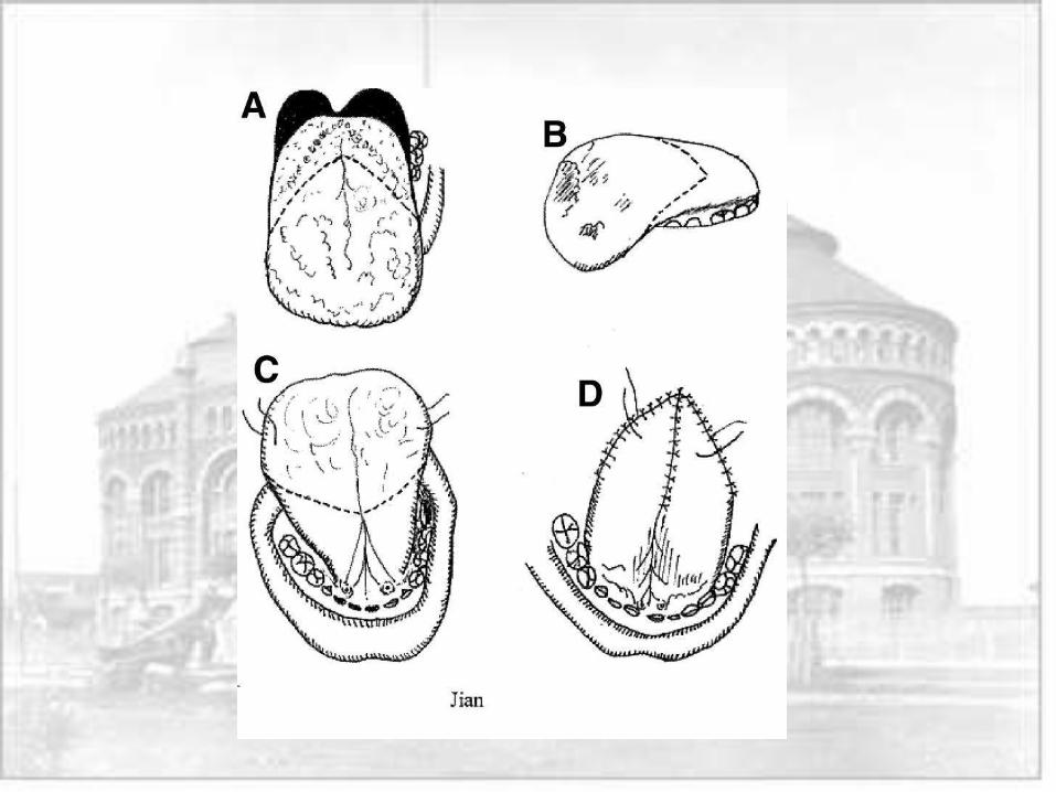
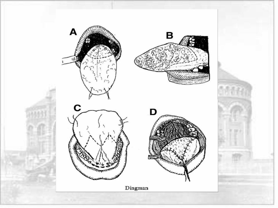
Jian, 2005, published a retrospective study
to evaluate the results of Jian or Dingman
glossectomy for lymphangiomatous
macroglossia
Lymphatic Malformations
Cosmesis and function improved after surgery in
7/7 patients. The tongue healed well, and the
patients had no long-term complications.
The authors conclude that although partial
surgical excision, injection of sclerosing solutions,
electrocoagulation, and radiation have been the
chief modalities of treatment of diffuse
lymphangioma of the tongue, surgical
management is the most effective treatment.
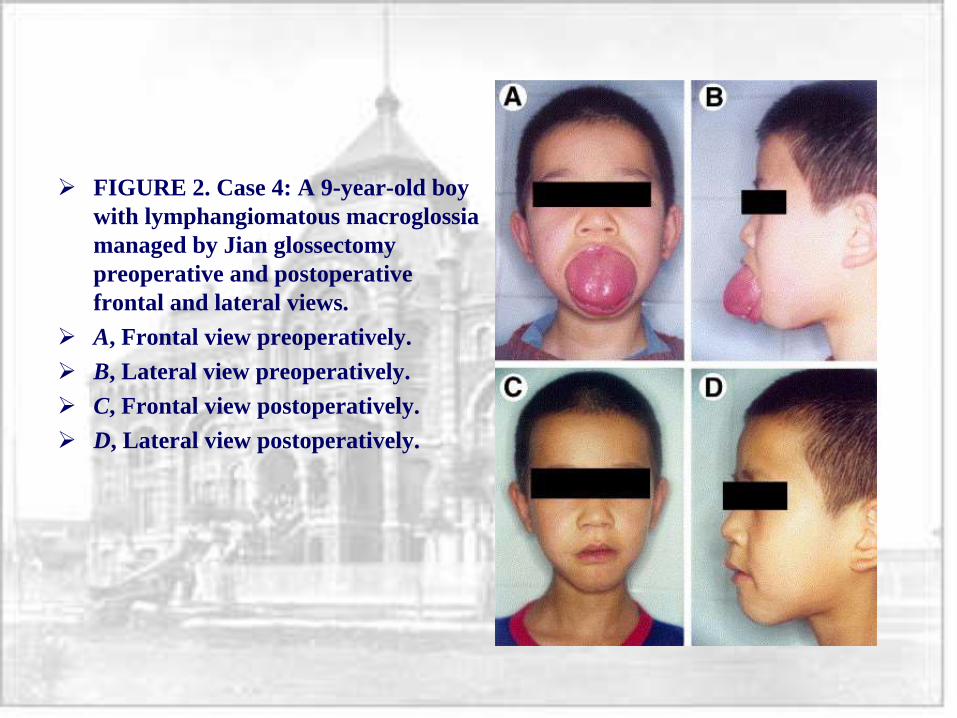
FIGURE 2. Case 4: A 9-year-old boy
with lymphangiomatous macroglossia
managed by Jian glossectomy
preoperative and postoperative
frontal and lateral views.
A, Frontal view preoperatively.
B, Lateral view preoperatively.
C, Frontal view postoperatively.
D, Lateral view postoperatively.
Venous Malformations
2/3 of all vascular malformations.
Are low-flow lesions.
Present in a spectrum, ranging from an
isolated skin varicosity or localized spongy
mass to complex lesions infiltrating various
tissue planes
May occur in the craniofacial skeleton, most
commonly in the mandible
Venous Malformations
Venous Malformations
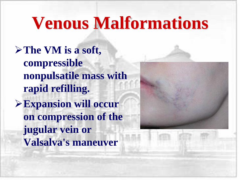
The VM is a soft,
compressible
nonpulsatile mass with
rapid refilling.
Expansion will occur
on compression of the
jugular vein or
Valsalva's maneuver
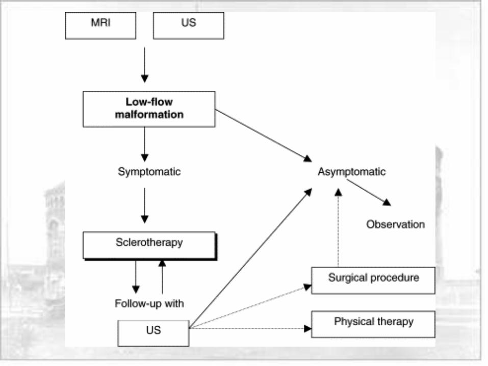
Venous Malformations
Treatment algorithm proposed by Yao et al,
2001
Venous Malformations
Treatment of choice is transcutaneous
sclerotherapy
80% ethanol is the most commonly used
agent
According to literature complications occur
in 10-15% of ethanol injections
Venous Malformations
Complications of ethanol sclerotherapy
include:
Necrosis or ulceration of the skin
Neuropathy
Complications of systemic absorption, such
as pulmonary vasoconstriction, or direct
depressant activity on the myocardium
Venous Malformations
Systemic absorption can be decreased by
Selecting only lesions with minimal
connection to the systemic circulation
Using a double needle technique
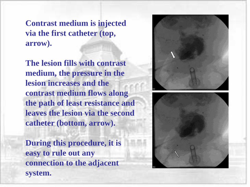
Contrast medium is injected
via the first catheter (top,
arrow).
The lesion fills with contrast
medium, the pressure in the
lesion increases and the
contrast medium flows along
the path of least resistance and
leaves the lesion via the second
catheter (bottom, arrow).
During this procedure, it is
easy to rule out any
connection to the adjacent
system.
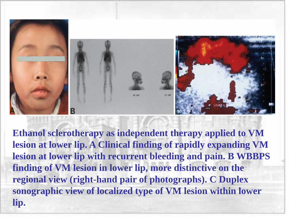
Ethanol sclerotherapy as independent therapy applied to VM
lesion at lower lip. A Clinical finding of rapidly expanding VM
lesion at lower lip with recurrent bleeding and pain. B WBBPS
finding of VM lesion in lower lip, more distinctive on the
regional view (right-hand pair of photographs). C Duplex
sonographic view of localized type of VM lesion within lower
lip.
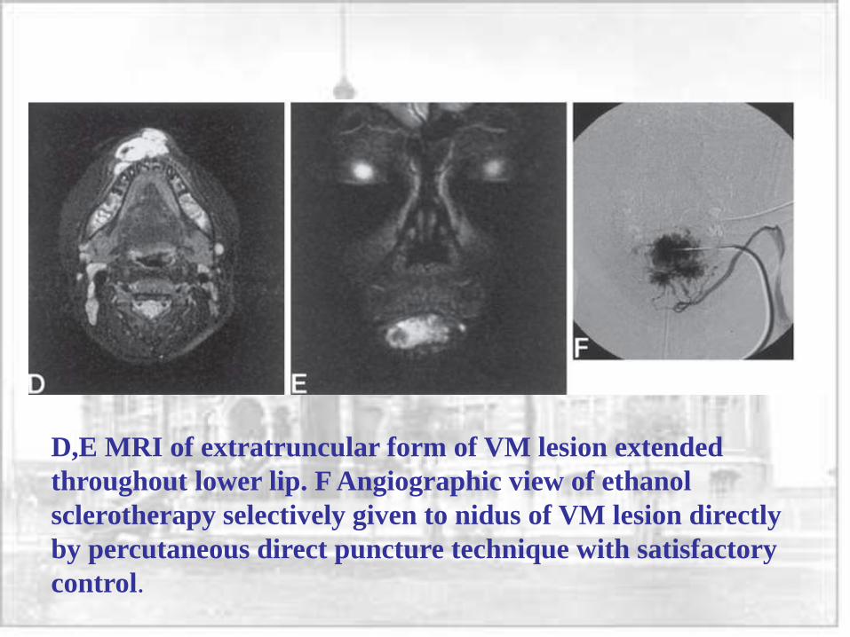
D,E MRI of extratruncular form of VM lesion extended
throughout lower lip. F Angiographic view of ethanol
sclerotherapy selectively given to nidus of VM lesion directly
by percutaneous direct puncture technique with satisfactory
control.
Venous Malformations
Alternative sclerosing agents have been tested
OK-432, derived from Streptococcus pyogenes
Luzatto, in 2000, reported 100% success rate
treating lymphangiomas
Giguere, in 2002, however, reported only 60%
success
No toxicity has been reported in the literature
Capillary Malformations
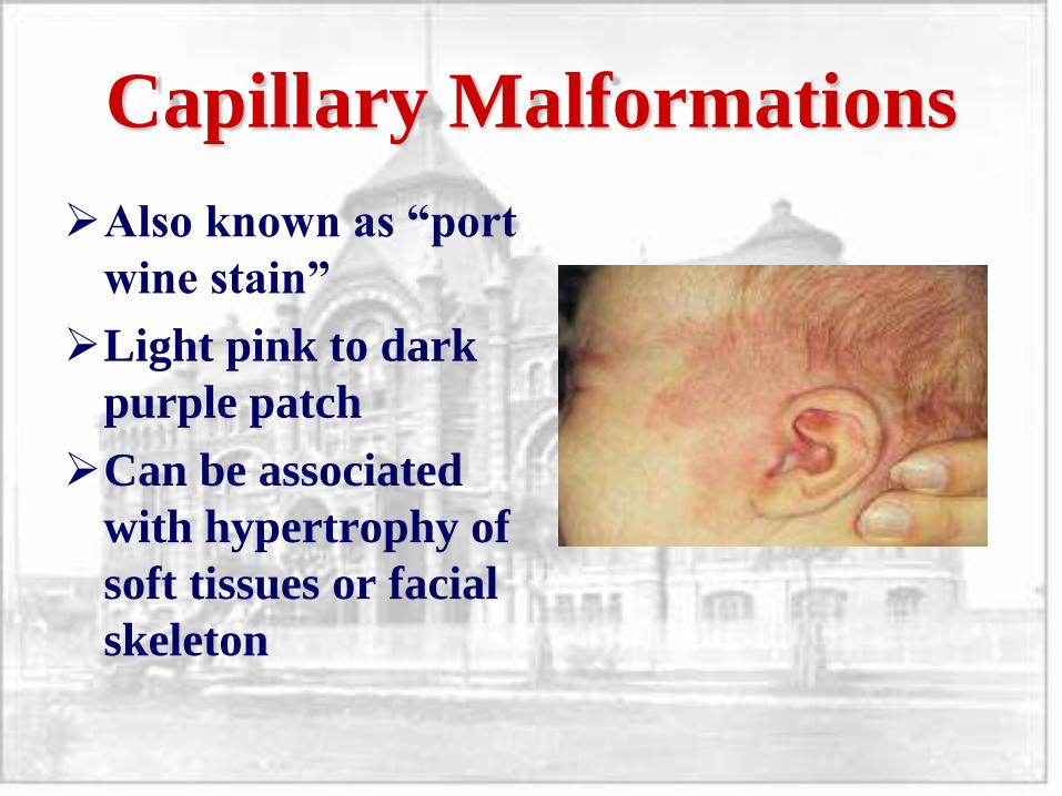
Also known as “port
wine stain”
Light pink to dark
purple patch
Can be associated
with hypertrophy of
soft tissues or facial
skeleton
Capillary Malformations
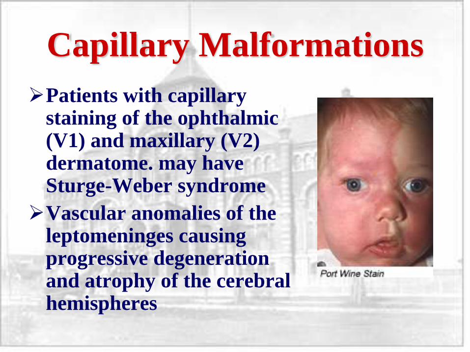
Patients with capillary staining of the ophthalmic (V1) and maxillary (V2) dermatome. may have Sturge-Weber syndrome
Vascular anomalies of the leptomeninges causing progressive degeneration and atrophy of the cerebral hemispheres
Capillary Malformations
Tunable flashlamp pulsed-dye laser (585-
nm wavelength) is widely regarded as the
optimum treatment
Complications include hypo- or
hyperpigmentation and scarring
Capillary Malformations
Goldman, 1993
43 children with 49 separate port-wine stain vascular malformations were treated with pulsed dye laser at 585 nm. Overall, 16% of patients had more than 95% resolution of their port-wine stains after an average of 4.8 (1 to 11) treatments.
Average improvement of 69% in those lesions not clearing completely
Lesions in patients less than 4 years of age were almost twice as likely to clear than were those in older children (20% vs 12%), and in fewer treatments (3.8 vs 6.5).
No episodes of scarring or persistent pigmentary changes in any of the patients.
Capillary Malformations
Theories exist on reasons for persistence of lesions after laser thermolysis.
In 2005, Sivarajan et al. investigated changes in capillary depth and diameter that occur with serial laser treatments.
Their findings show that persistent vessels in capillary malformations after treatment are deeper and narrower than those in untreated lesions.
The authors suggest that since depth and diameter are crucial to the most effective wavelength and pulse duration, respectively, of a therapeutic laser, adjusting these laser parameters for treating resistant lesions may be effective.
Arteriovenous Malformations
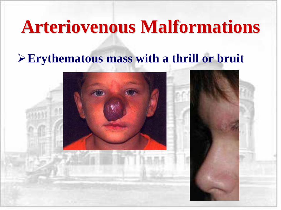
Erythematous mass with a thrill or bruit
Arteriovenous Malformations
Arteriovenous Malformations
There are four recognized stages of AVMs:
A stage I lesion has a pinkish-bluish stain and warmth.
In stage II, the lesion has pulsations, thrill, and bruit.
In stage III, the patient has dystrophic skin changes, ulceration, bleeding, and pain.
Finally, in stage IV, the patient has high-output cardiac failure.
Arteriovenous Malformations
Extracranial arteriovenous malformations of the head and neck differ from AVM of the extremities.
Decompensation failure resulting from AVM is very rare in the head and neck region
HNAVM communicate with the deep venous system less frequently because they usually occur in the more superficial tissue layers.
The main symptoms of HNAVM are cosmetic disfigurements, deformities caused by the expansion of the lesions, and bleeding with pain or ulceration
Arteriovenous Malformations
Small, superficial arteriovenous malformations can be removed surgically.
However, embolization has been the only feasible treatment option for most arteriovenous malformations.
Embolization, which closes off the arterial feeders of the malformation, is generally effective in arteriovenous malformations to stabilize the malformation.
In some patients, AVMs can be cured with repetitive embolizations. Most AVMs require several sessions, generally 2 months apart, with regular follow-ups.
Also, with successful embolization, a surgical excision can become feasible in some AVMs.
Arteriovenous Malformations
During the embolization procedure, the nidus
needs to be embolized, but the large arterial
feeders should not be embolized.
Similarly, surgical ligation of the arterial feeders
should not be performed. If the arterial feeders
are embolized percutaneously or ligated
surgically, the arteriovenous malformation nidus
recruits new smaller arterial feeders, which then
can not be accessed for nidus embolization and
makes AVM management very difficult.
Arteriovenous Malformations
Lee, 2004, retrospectively reviewed 66 patients treated for AVM with embolosclerotherapy and/or surgery
The authors report 100% success rate using preoperative embolism with NBCA glue plus surgery
They also report >90% success rate using embolosclerotherpy alone for surgically inaccessable (infiltrating) lesions
Arteriovenous Malformations
Han et al 2006 reviewed 20 patients over 7 years treated for AVM of the head and neck
Ethanol sclerotherapy, surgical excision and embolization were used as treatments, either alone or in various combinations.
Ethanol sclerotherapy had a success rate of 50.0% and a permanent complication rate of 8%.
Surgical excision combined with embolization yielded 100% successful resolution of their HNAVM
15% suffered from permanent complications including CN VII weakness
In total, 16/20 patients (80.0%) eventually achieved a ≥75% reduction in the size of their lesions.

Arteriovenous Malformations
Pearls
If you suspect CVM, consult your friendly
neighborhood interventional radiologist
U/S and MRI are good initial tests
Don’t rush into treatment if the patient is
asymptomatic