Embed Size (px)
Citation preview
1
Converse: Chapter 25
Orbital and Naso-Orbital Fractures
John Marquis Converse, Byron Smith, Donald Wood-Smith
Orbital fractures may occur independently or may be associated with other facialfractures. One should obtain specific details as to where and how the accident occurred, andif there was a period of unconsciousness, loss of vision, or diplopia. A history of priormedical or surgical treatment is also important.
Prior to the primary treatment of the fracture, an ophthalmologic examination shouldbe done routinely to verify visual acuity and check the possibility of intraocular or cornealinjury. Obviously, if the patient is unconscious, the examination is limited. In the consciouspatient, a delay of the operative procedure for a few days will allow the edema to subside andpermit a more thorough examination. Facial bone fractures are not surgical emergencies unlessthe airway is obstructed and a tracheotomy is indicated. Treatment of an orbital fractureshould not be delayed more than necessary, however, as fibrosis of the entrapped extraocularmusculature, loss of orbital fat, and organization of the fracture occur rapidly and complicatethe treatment.
When feasible, extraocular muscle movements should be checked at far and neardistance and in all cardinal fields, as ocular muscle imbalance is often associated withfractures. A fundus examination often uncovers congenital anomalies or macula or optic nerveinjuries that may limit the postoperative result because of diminution or loss of vision andmay reduce the probability of satisfactory extraocular muscle fusion.
In trauma involving the medial canthus, when there is an associated naso-orbitalfracture, an evaluation of the lacrimal system should be obtained, if feasible, depending onthe magnitude of the injury. Irrigation of the secretory system showing drainage of the fluidinto the nose demonstrates that the continuity of the lacrimal apparatus has not beeninterrupted.
Orbital fractures occur in association with zygomaticomaxillary, naso-orbital, and highmaxillary (Le Fort III) fractures, as well as in pyramidal (Le Fort II) fractures, where the lineof fracture traverses the orbital floor (see Chapter 24). The backward displacement of thefractured thick inferior orbital rim comminutes the thinner portion of the orbital floor. Thedownward displacement of the zygoma results in a separation at the frontozygomatic junctionand a lowering of the orbital floor.
Orbital fractures complicated by diplopia are frequently associated with midfacialfractures. McCoy, Chandler, Magnan, Moore, and Siemsen (1962) found an incidence of 15per cent of ocular complications is a series of 855 patients with facial fractures. Morgan,Madan, and Bergerot (1972), in a review of 300 cases of midfacial fractures, found persistentdiplopia in 11 per cent of the patients.
2
The Orbit: Anatomical Considerations
The orbits are paired bony structures separated in the midline by the interorbital space.The interorbital space, the portion of the nasal cavity situated between the orbits, is delimitedabove by the floor of the anterior cranial fossa, formed in this portion by the roof of eachethmoid sinus laterally and by the cribriform plate medially. The orbits are situatedimmediately below the floor of the anterior cranial fossa, a portion of the fossa being formedby the roofs of the orbits (see Chapter 56).
The orbital contents are protected by strong bony abutments: the nasal bones, the nasalspine of the frontal bone, and the frontal processes of the maxilla medially; the supraorbitalarch of the frontal bone above; the frontal process of the zygoma and zygomatic process ofthe frontal bone laterally; and the thick infraorbital rim formed by the zygoma and maxillainferiorly.
The Skeletal Anatomy of the Orbit. The skeletal components of the orbital cavityare the frontal bone, the lesser and greater wings of the sphenoid, the zygoma, the maxilla,the lacrimal bone, and the ethmoid.
The bony orbit has been described as cone-shaped or pyramidal in shape. Both ofthese analogies are somewhat inaccurate. The widest orbital diameter is not located at theorbital rim but approximately 1.5 cm within the orbital cavity. The medial wall has aquadrangular rather than a triangular configuration. The optic foramen lies on a medial andslightly superior plane in the apex of the orbit. In children, the orbital floor is situated at alower level in relation to the orbital rim because the maxillary sinus has not reached fulldevelopment.
The orbits are protected anteriorly by the orbital rims formed of thick bone, whereasthe orbital walls consist of relatively thin bone. Although it represents an artificial division,it is helpful in studying the bony orbit to divide it into four component parts: the roof, lateralwall, medial wall, and floor.
The floor of the orbit, a frequent site of fracture, has no sharp line of demarcation withthe medial wall because the orbital floor tilts upward in its medial aspect, while the lowerportion of the medial wall has a progressively lateral inclination. The floor is separated fromthe lateral wall by the inferior orbital (sphenomaxillary) fissure. The floor of the orbit (theroof of the maxillary sinus) is composed mainly of the orbital plate of the maxilla, a paper-thin structure medial to the infraorbital groove, and partly by the zygomatic bone anterior tothe inferior orbital fissure. The infraorbital groove (or canal) traverses the floor of the orbitbeginning at about the middle of the inferior orbital fissure. Anteriorly it penetrates the thickinferior orbital rim as the infraorbital canal, which opens on the anterior surface of the maxillaas the infraorbital foramen.
The orbital floor has a general upward inclination; the anterior portion is concave andthe posterior portion is convex. In the blowout fracture mechanism, the force transmitted tothe orbital contents tends to fracture the floor at its weakest point and may be nature's wayof protecting the ocular globe from rupture by decompressing the orbit. In the posteriorportion of this inclined plane there is an area of thin bone. This "weak area" represents the
3
thinnest bone of the orbit; its medial extension is the lamina papyracea of the ethmoid, aportion of the medial orbital wall which, as its name implies, is a plate of bone of paperlikethinness. The medial half of the orbital floor is also weakened by the canal (or groove) forthe passage of the infraorbital nerve.
The inferior oblique muscle arises from the medial aspect of the orbital floor, lateralto the lacrimal groove, near the anterior margin of the orbit.
The medial wall, reinforced anteriorly by the frontal process of the maxilla, isrelatively fragile and is formed from the frontal bone, the lacrimal bone, the lamina papyraceaof the ethmoid, and part of the lesser wing of the sphenoid around the optic foramen. Thelamina papyracea is the largest component and accounts for the structural weakness of themedial wall. The lesser wing of the sphenoid and the optic foramen are posterior to thelamina papyracea. Thus the optic foramen is located close to the posterior portion of theethmoid sinus, not at the apex of the orbit. Consequently, in severe fractures involving themedial wall in its posterior portion, the optic nerve can be injured.
The groove for the lacrimal sac is a broad vertical fossa lying partly on the anterioraspect of the lacrimal bone and partly in the frontal process of the maxilla; the anterior andposterior margins of the lacrimal groove form the respective lacrimal crests. The groove iscontinuous with the nasolacrimal duct at the junction of the floor and medial wall of the orbit,passing down into the inferior meatus of the nose.
Between the roof and medial wall of the orbit are the anterior and posterior ethmoidalforamina which lead into canals communicating with the medial part of the anterior cranialfossa.
The lateral wall is relatively stout in its anterior portion. It is formed by the greaterwing of the sphenoid, the frontal process of the zygomatic bone, and the lesser wing of thezygomatic bone, and the lesser wing of the sphenoid lateral to the optic foramen. The superiororbital fissure is a cleft which runs forward and upward from the apex between the roof andlateral wall. The fissure, which separates the greater and lesser wings of the sphenoid, givespassage to the three motor nerves to the extraocular muscles of the orbit and leads back intothe middle cranial fossa. The lateral wall of the orbit is situated in an anterolateral andposterior medial plane. It is related to the temporal fossa; posteriorly a small part of the walllies between the orbit and the middle cranial fossa and temporal lobe of the brain. Betweenthe floor and lateral wall of the orbit is the inferior orbital fissure which communicates withthe infratemporal fossa.
The roof of the orbit is composed mainly of the orbital plate of the frontal bone, butposteriorly it receives a minor contribution from the lesser wing of the sphenoid. The fossalodging the lacrimal gland is a depression situated along the anterior and lateral aspect undershelter of the zygomatic process of the frontal bone. The anterior portion of the roof can beinvaded by the supraorbital extension of the frontal sinus or by an extension of the ethmoidsinus, a frontoethmoidal cell (see also Chapter 56). The roof separates the orbit from theanterior cranial fossa and from the middle cranial fossa on the posterolateral aspect.
4
Often consisting of brittle bone, the orbital roof varies in thickness and may be quitethin in its medial portion.
The supratrochlear and supraorbital nerves and, more medially, the trochlea of thesuperior oblique muscle are located along the superior rim of the orbit. The tendon of thesuperior oblique muscle functions in a cartilaginous pulley or trochlea, which is fixed byligamentous fibers immediately behind the superomedial angle of the orbital margin. Fracturesinvolving the superior rim of the orbit may result in compression of the supraorbital nerve,with consequent anesthesia of its area of distribution. Diplopia may also result from injuryto the pulley of the superior oblique muscle, thus affecting the balance of the extraocularmusculature.
The Orbital Fat and the Ocular Globe. The ocular globe is surrounded by a cushionof orbital fat within the orbital cavity. The ocular globe occupies only the anterior half of theorbital cavity; the posterior half of the orbital cavity is filled with orbital fat, muscles, vessels,and nerves supplying the ocular globe. The two halves of the orbital cavity, anterior andposterior, are separated by Tenon's capsule, which subdivides the orbital cavity into ananterior or precapsular segment and a posterior or retrocapsular segment.
The Septum Orbitale. The orbital contents are maintained in position by the septumorbitale (orbital septum), a fascia inserted on the inner aspect of the rim of the orbit. Theseptum orbitale attaches to and blends with the levator aponeurosis in the upper eyelid for adistance of a few millimeters above the upper border of the tarsus; in the lower eyelid, theseptum orbital is attached to the lower border of the tarsus.
The Periorbita. The periosteum lining the periphery of the orbit is known as theperiorbita. The periorbita is continuous with the dura at those sites where the orbitcommunicates with the cranial cavity, eg, the optic foramen, the superior orbital fissure, andthe anterior and posterior ethmoidal canals.
The Optic Foramen and the Optic Canal. The optic foramen is situated at thejunction of the lateral and medial walls of the orbit. The foramen is not located on ahorizontal plane with the orbital floor but above it.
The optic canal, 4 to 10 mm in length, is the passage through which the optic nerveand ophthalmic artery pass from an intracranial to an intraorbital position. The canal is framedmedially by the body of the sphenoid and laterally by the lesser wing and is thus in closeapproximation to the sphenoid sinus and the posterior ethmoidal cells.
Blowout Fractures of the Floor of the Orbit
A blowout fracture is caused by a sudden increase in the intraorbital pressure, resultingfrom the application of a traumatic force to the soft tissues of the orbit (Converse and Smith,1957, 1960). The fracture is often complicated by diplopia, which is caused by a verticalmuscle imbalance secondary to entrapment of the orbital contents which may include theinferior rectus and inferior oblique muscles and the surrounding fascial expansions into thedehiscence in the orbital floor. The escape of orbital fat through the blowout dehiscences isa major cause of enophthalmos.
5
One of the first descriptions of a blowout fracture was given by King and Samuel in1944: "We would like to add one other type of fracture of great importance, which is notinfrequent. In this there is a downward displacement of part of the orbital floor, unassociatedwith any damage to the margin of the orbit surrounding the facial bones. The cause of sucha fracture is difficult to visualize. The most ready explanation is trauma transmitted throughthe eye to the orbital floor".
After the application of a traumatizing force over the orbital contents by anonpenetrating object, such as a tennis ball or the human fist, the orbital contents are forcedbackward into the narrower portion of the orbit (Table 25-1). The increased intraorbitalpressure thus exerted causes a blowout at the weakest area of the orbital floor withoutfracturing the orbital rim. This type of fracture may be referred to as "pure" blowout fracture(Table 25-2) (Converse and Smith, 1957). The strong rim of the orbit protects against objectswith a radius of curvature greater than 5 cm; an object having a curvature of less than 5 cmmay penetrate this protective barrier and damage the globe. Such objects are golf balls,hockey pucks, and the tip of a football. Damage to the globe leading to blindness may occur.
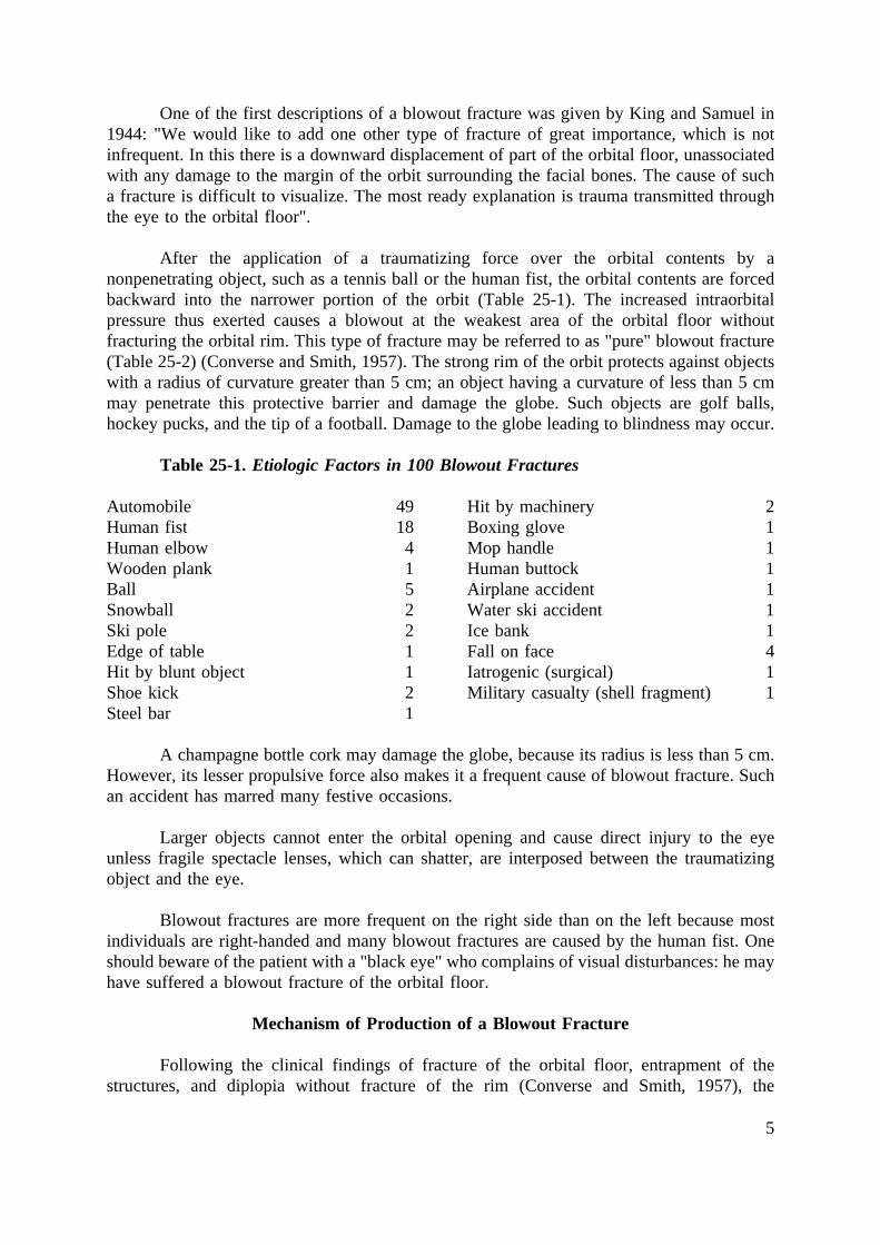
Table 25-1. Etiologic Factors in 100 Blowout Fractures
Automobile 49Human fist 18Human elbow 4Wooden plank 1Ball 5Snowball 2Ski pole 2Edge of table 1Hit by blunt object 1Shoe kick 2Steel bar 1
Hit by machinery 2Boxing glove 1Mop handle 1Human buttock 1Airplane accident 1Water ski accident 1Ice bank 1Fall on face 4Iatrogenic (surgical) 1Military casualty (shell fragment) 1
A champagne bottle cork may damage the globe, because its radius is less than 5 cm.However, its lesser propulsive force also makes it a frequent cause of blowout fracture. Suchan accident has marred many festive occasions.
Larger objects cannot enter the orbital opening and cause direct injury to the eyeunless fragile spectacle lenses, which can shatter, are interposed between the traumatizingobject and the eye.
Blowout fractures are more frequent on the right side than on the left because mostindividuals are right-handed and many blowout fractures are caused by the human fist. Oneshould beware of the patient with a "black eye" who complains of visual disturbances: he mayhave suffered a blowout fracture of the orbital floor.
Mechanism of Production of a Blowout Fracture
Following the clinical findings of fracture of the orbital floor, entrapment of thestructures, and diplopia without fracture of the rim (Converse and Smith, 1957), the
6
mechanism of production of the orbital blowout fracture was demonstrated experimentally.It was verified in a cadaver by duplicating a force similar to that which had produced ablowout fracture in one of our patients, who had been hit by a ball used in the Irish game ofhurling (Smith and Regan, 1957). The dried out condition of the cadaver globe was correctedby the intraocular injection of normal saline solution. A hurling ball was placed over theclosed lid of the cadaver orbit, and the ball was struck sharply with a mallet. A crackingsound was heard and was interpreted as having been caused by fracturing bone. Anexploratory incision through the skin of the infraorbital margin and elevation of the orbitalcontents from the floor exposed a depressed comminuted fracture of the floor of the orbit.Exenteration of the orbital contents exposed the fracture in its entirety. There was also acomminuted fracture without displacement involving the lamina papyracea of the ethmoidbone. No fracture of the orbital rim or zygomatic arch was observed. This experimentduplicated almost exactly the injury sustained by several of our patients.
Table 25-2. Classification of Orbital Fractures
1. Orbital blowout fracturesA. Pure blowout fractures: Fractures through the thin areas of the orbital floor,
medial and lateral wall. The orbital rim is intact.B. Impure blowout fractures: Fractures associated with fracture of the adjacent
facial bones. The thick orbital rim is fractured, and its backward displacementcauses a comminution of the orbital floor; the posterior displacement of theorbital rim permits the traumatizing force to be applied against the orbitalcontents, which produces a superimposed blowout fracture.
2. Orbital fractures without blowout fractureA. Linear fractures, in upper maxillary and zygomatic fractures. These fractures
are often uncomplicated from the standpoint of the orbit.B. Comminuted fractures of the orbital floor with prolapse of the orbital contents
into the maxillary sinus is often associated with fracture of the midfacialbones.
C. Fracture of the zygoma with frontozygomatic separation and downwarddisplacement of the zygomatic portion of the orbital floor and of the lateralattachment of the suspensory ligament of Lockwood.
In a second experiment, the opposite orbit of the cadaver was exenterated. The softtissue covering the orbital rim was excised to allow direct contact of the bony orbital rim withthe surface of the hurling ball. Repeated blows of similar force with the hammer failed tofracture either the floor or the rim of the orbit. However, when the striking force wassufficiently increased, the orbital rim and orbital floor were comminuted simultaneously.
The mechanism of blowout fracture (an increased hydraulic pressure) has beenquestioned by a number of authors (Rény and Stricker, 1969; Fujino, 1974a, b). Rény andStricker suggested the following hypothesis: the traumatic force striking the inferior orbitalrim, which is sufficiently resilient to transmit the force to the orbital floor, fractures the latterwhile the rim rebounds without fracturing. Fujino (1974a, b), in a series of experiments incollaboration with engineers, demonstrated on a dried human skull, without orbital contents,that a brass striker weighing 120 g with a flat silicone plate, when dropped on the infraorbital
7
margin from a height of 15 cm, produced a linear fracture of the orbital floor. When theweight was dropped from a height of 20 cm, a punched-out fracture in the convex portion ofthe orbital floor was produced. Both of these fractures occurred without fracture of the orbitalrim.
While Rény and Stricker's statement is obviously conjectural, Fujino's experiments,performed with mathematical precision, fail to demonstrate the most important clinicalconsequence of the blowout fracture: the entrapment. How does he explain the entrapment ofthe orbital contents without the increased intraorbital pressure forcing the tissues into the siteof fracture? And how is it possible for the increased infraorbital hydraulic pressure to occurwithout contact of the orbital contents with the traumatizing object>
Whatever the theory of the mechanism of the blowout fracture, the fact remains that,in the presence of diplopia due to entrapment and inability to rotate the globe by means ofthe forced duction test, release of the entrapment is the only means of relieving the patientof the extraocular muscle imbalance and diplopia.
"Impure" Blowout Fracture
According to Garrett (1963), ocular-orbital damage occurs in approximately 10 percent of all head injuries sustained in automobile accidents in the United States. In the typicalautomotive injury in which the passenger's face is projected against the dashboard, the thickorbital rim is fractured and backwardly displaced, resulting in an eggshell comminution of theorbital floor. The continuing momentum and the pressure against the orbital contents producea superimposed blowout fracture. It is to this type of orbital fracture that the term "impure"blowout fracture, as suggested by Cramer, Tooze, and Lerman (1965), may be applied (Table25-2). The human fist was the principal factor in the causation of pure blowout fractures inthe series studied by Emery and his associated (1971). In the series studied by Converse andhis associates (1967), automobile accidents caused the largest proportion of fractures; mostof these were impure and complicated fractures (see Table 25-1). Impure blowout fracturesoften occur in association with midfacial fractures.
Pyramidal maxillary fractures (Le Fort II) and craniofacial disjunction (Le Fort III) arecharacterized by fracture lines extending through the orbital floor, findings which furthercontribute to the comminution of the bone produced by the fractured orbital rim. Massiveprolapse of the floor into the maxillary sinus may occur.
Not All Orbital Floor Fractures Are Blowout Fractures! The term "blowout"fracture (Converse and Smith, 1957) has been used to refer to all orbital floor fractures. Notevery fracture of the floor of the orbit is a blowout fracture (see Table 25-2). The term"blowout" fracture defines a particular type of fracture mechanism. Orbital floor fractures, asstated earlier in the text, occur in fractures of the zygoma and in upper maxillary fractures,and comminuted fractures of the floor may result in a downward sagging of the orbitalcontents into the maxillary sinus.
Blowout Fracture in Children. The maxillary sinus is undeveloped in children. Asa result, the floor of the orbit is situated in a low position, dipping downward from the orbital
8
rim. Despite the resiliency and elasticity of young bones, blowout fractures are notinfrequently seen in children. The mechanism of entrapment is similar to that seen in adults.
Surgical Pathology
Diplopia. Extraocular muscle imbalance and subjective diplopia are the result ofdeviation of the visual axes. The deviation has several causes: the major one is entrapmentof the soft tissue structures in the blowout fracture area, a finding which explains theconstancy of vertical muscle imbalance (Table 25-3). These soft tissue structures may includethe inferior rectus muscle, inferior oblique muscle, suspensory ligament of Lockwood,periorbita, and fascial expansions.
A downward displacement of one ocular globe does not always result in diplopia.Massive comminution of the orbital floor will cause a downward displacement of the ocularglobe without entrapment: there is no diplopia.
The most common site of the blowout fracture is the portion of the floor that isweakened by the infraorbital canal or groove. The inferior oblique muscle arises from themaxillary portion of the orbital floor lateral to the lacrimal groove, and the inferior rectusmuscle is situated immediately above the infraorbital canal on the undersurface of the orbitalcontents. It is not surprising, therefore, that these two muscles are frequently involved in theblowout fracture. Absence of elasticity in the impounded inferior rectus muscle restrictsrotation in the field of action of its antagonist, the superior rectus. Because the inferior rectusand the inferior oblique muscles are intimately connected at the point where the inferioroblique crosses beneath the inferior rectus, disturbance of function of the inferior obliquemuscle is usually observed in blowout fractures. When the fracture is located lateral to theinfraorbital groove or canal, the inferior rectus and inferior oblique muscles may not beinvolved. These variations in the site of the blowout fracture explain variations in thesymptoms and clinical signs in these fractures.
Nerve Injury. Injury to the motor nerves of the inferior oblique and inferior rectusmuscles must also be considered. The inferior oblique and the inferior rectus muscles areinnervated by the inferior division of the third cranial nerve. The branch to the inferior rectusmuscle passes along its upper surface to pierce it at the junction of the posterior and middlethirds of the muscle. The branch to the inferior oblique muscle runs along the lateral edge ofthe inferior rectus muscle, enters the ocular surface of the inferior oblique muscle, and isexposed to injury in blowout fractures. The relatively short course of the nerve to the inferiorrectus muscle renders it less vulnerable to injury. Electromyographic examination will assistin determining whether nerve conduction has been interrupted by the injury.
Other Causes of Diplopia. Other causes of diplopia are injury to the third, fourth, orsixth cranial nerves, direct injury to the extraocular muscles, laceration of the muscle by bonefragments, disruption of the muscle attachments, hemorrhage into the muscle, or muscleimbalance caused by a change in orbital shape. Secondary muscle imbalance occurs whenptosis of the globe is associated with enophthalmos. Secondary muscle imbalance occurs whenptosis of the globe is associated with enophthalmos. Secondary deviations are commonly dueto overaction of the yoke or conjugate muscles of the opposite eye. A factor to beremembered is that no extraocular muscle acts singularly to produce ocular movements.

9
Ocular rotation is the sum of the action, counteraction, and relaxation of 12 extraocularmuscles (6 per ocular globe). It is not within the scope of this chapter to discuss the complexsubject of the physiology of oculorotary muscles; only the aspects which pertain to theproblem under discussion are explained.
Not only do paralytic deviations occur, but also trauma may often uncover tropias(constant imbalance) or phorias (latent imbalance occurring only with disruption of fusion)after temporary immobilization of the injured eye. These are usually horizontal in nature.
The typical blowout fracture is usually not seen in fracture-dislocation of the zygomaif the bone is displaced as a single fragment. In such a fracture, the site of impact is lateralto the orbital cavity and the orbital contents are usually not directly involved. In zygomaticfractures which are severely comminuted following an exceptionally strong impact, the lateralorbital wall and the rim and floor of the orbit may also be severely comminuted; the ocularglobe is injured to the extent that enucleation may be required. A blowout with entrapmentmay have occurred, and release is necessary, even if the ocular globe is enucleated, to permitmovement of the ocular prosthesis for purely esthetic reasons.
Enophthalmos. Enophthalmos, the second major complication of the blowout fracture,is the result of a number of causative factors. The first, the escape of fat from the orbitalcavity, occurs when the periorbita is ruptured and the orbital fat escapes into the maxillarysinus. A second cause of enophthalmos is the retention of the ocular globe in a backwardposition when the structures are entrapped in the fracture site. A third causative factor is theenlargement of the orbital cavity resulting from the fracture and the downward displacementof the orbital floor; the orbital fat is distributed in a large cavity and is no longer sufficientin quantity to prevent a sinking in of the globe. A fourth factor is orbital fat necrosis resultingfrom pressure caused by orbital hematoma and a low grade inflammatory process.
The mechanism opposite to a blowout fracture is the "blown-in" fracture, in whichpenetration of bone fragments into the orbit diminishes the size of the orbital cavity, resultingin exophthalmos.
Enophthalmos, when it is conspicuous and particularly when orbital contents aredownwardly displaced, results in a pseudoptosis of the upper eyelid, a deepening of thesupratarsal fold, and a shortening of the horizontal dimension of the palpebral fissure. Thedeformity becomes more complex in orbital fractures associated with fractures of adjacentbones, especially a concomitant naso-orbital fracture.
Lower animals possess an orbital muscle which spans the floor of the orbit, coveringthe inferior orbital fissure; this muscle protrudes the eyeball for purposes of focusing vision.In man a vestige of this muscle has been designated as the orbital muscle. Some anatomistsdiscount its importance; others claim that paralysis of the muscle is a factor in the productionof enophthalmos in conditions such as Horner's syndrome, caused by paralysis of the thirdcranial nerve. Others feel that atrophic changes in the orbital fat due to injury of thesympathetic innervation may be responsible for the production of enophthalmos. Other factorswhich have been held responsible for the development of traumatic enophthalmos aredislocation of the trochlea of the superior oblique muscle, cicatricial contraction of retrobulbartissue, and rupture of the orbital ligaments or fascial bands.
10
Enlargement of the orbital cavity as a factor in the causation of enophthalmos wassuggested by Lang (1889): "I suggest that the injury may have produced a fracture and adepression of a portion of the orbital wall; the orbital fat would then be no longer sufficientin quantity to fill this enlarged postocular area without a sinking-in of the globe fromatmospheric pressure and a resulting limitation in ocular movements."
The enlargement of the orbital cavity from the depression of the floor is a frequentfactor in the production of enophthalmos. However, there may be no entrapment of the orbitalstructures and no diplopia (see Table 25-3).
Enophthalmos in Orbital Fractures Without Blowout Fracture. Fractures of thebones of the midfacial area often involve the orbital floor: Le Fort III fractures lines traversethe floor of the orbit; Le Fort II fractures also involve the orbital floor in its medial portion(see Table 25-2).
Variations in Diplopia and Enophthalmos in Orbital Fractures. Table 25-3classifies the variations which occur in orbital fractures according to the anatomical damagesuffered by the orbit and its contents.
Examination and Diagnosis
Clinical Examination. In the typical blowout fracture, the patient complains ofdiplopia in the primary position which increases in the upward gaze. The patient may notrecognize diplopia early if the eye is temporarily closed by edema of the lids or dressings orif there is an intraocular injury. When examined during the first hours after the fracture, theocular globe appears displaced backward and downward, and the supratarsal sulcus isdeepened. Edema and hematoma may obscure such clinical findings when the patient is notexamined during the first hours after injury. Ocular globe injury, eyelid damage, andlacerations and hematoma in the levator muscle or aponeurosis are not infrequently observed.
Diplopia is the most frequent complaint of the patient but is not necessarily anindication for operation; diplopia may be caused by hematoma and edema and may resolvespontaneously. Subjective diplopia is not an indication for surgical exploration.
When an object is held approximately two feet from the patient's eye and the patientis asked to look at the object, the affected eye is not able to rotate upward in the normalrange as does the unaffected eye; restriction in rotation in other directions is also observed.The function of the inferior rectus and inferior oblique muscles is restricted by theirentrapment in the floor of the orbit, and the superior rectus cannot rotate the globe becauseof the resistance offered by the short rein of the entrapped structures; when released, the globeis again able to rotate upward. In a child, the authors have observed nearly complete fixityof the globe.
When the infraorbital rim is fractured (impure blowout fracture), one may observe thatthe lower lid is shortened vertically and everted. The infraorbital rim is displaced backwardand in its forced retreat carries with it the insertion of the septum orbitale. This findingaccounts for the vertical shortening of the lower eyelid.
11
Indications for Surgical Intervention. Although diplopia is the most frequentcomplaint of the patient, it is not an indication for operation, as diplopia may be caused byhematoma, edema, and neurogenic factors.
Indications for operation are: (1) limitation of forced rotation of the eyeball, (2)radiologic evidence of fracture, and (3) enophthalmos.
1. Limitation of forced rotation of the eyeball. This test, known as the "traction test"or the "forced duction test", provides a means of differentiating entrapment of the inferiorrectus muscle from weakness or paralysis of the superior rectus, and it is the pathognomonicsign of a blowout fracture of the floor of the orbit. A few drops of local anesthetic solutioninstilled into the conjunctival sac provide sufficient anesthesia to permit grasping the eyeballwith forceps at the insertion of the inferior rectus muscle at a point of approximately 7 mmfrom the limbus.
2. Radiologic evidence of fracture. Radiologic diagnosis is essential, and tomographyis of additional assistance in locating the area of the blowout fracture. Careful roentgenexamination will show a variety of findings and define the location, size, and type of thefracture site.
3. Enophthalmos. Clinically obvious enophthalmos is another indication for surgicalexploration, as it suggests a gross derangement of the orbit - enlargement of the volume ofthe orbit resulting from the fracture of the floor or escape of orbital fat.
Sensory Nerve Conduction Loss. In a suspected orbital floor blowout fracture,anesthesia or hypoesthesia in the area of distribution of the infraorbital nerve is suggestiveevidence of a blowout fracture involving the infraorbital groove or canal. This finding assistsin locating the site of the blowout fracture. Absence of infraorbital anesthesia implies that thefractured area is either lateral, medial, or posterior to the infraorbital groove or canal and isnot a sign that the orbital floor is not fractured. In the patient shown a linear fracture occurredposterior to the infraorbital canal, entrapping the soft structures.
Radiologic Examination. Because of the superimposition of both thick and thin bones,the roentgen picture is apt to be difficult to interpret. The diagnosis of orbital fracture byroentgenography is made by means of a variety of positions: the Caldwell position, the Watersposition, the fronto-occipital position, the anteroposterior projection, the reverse Watersposition, and the oblique orbital-optical foramen view.
Diagnosis of blowout fracture of the orbit is frequently missed if the radiologicexamination is not comprehensive. Fracture lines may be mistaken for superimposed bonysepta or suture lines, or they may be hidden by disease processes in the underlying maxillarysinus. The thin orbital floor, partially transparent on radiographs, may be obscured against thebackground of other bones of the skull. Tomography will often disclose the presence of ablowout fracture and its location (Zizmor and coworkers, 1962).
Polytomography, as well as hypocycloidal movement, is advocated in all skullradiography. It brings into focus a 1-mm thin layer of tissue with reasonable clarity andsharpness. It is far superior to the curvilinear tomography formerly used.
12
With adequate technique, blowout fractures of the orbit can be diagnosed in over 90per cent of the cases with conventional radiography. Polytomography has a similar degree ofdiagnostic accuracy and, in addition, can delineate the location, depth, and extent of thefracture with a degree of clarity and accuracy not possible with conventional radiography.
The type of blowout fractures varies: a lowering of the orbital floor; the "hangingdrop", seen in the blowout fracture through which the orbital fat has extruded into themaxillary sinus; the trapdoor fracture, one or two bone fragments hanging into the sinus ona periosteal hinge; the massive extrusion of orbital contents into the maxillary sinus;associated fracture of the medial wall. Such positive radiologic signs, combined with positiveclinical signs, are indications for surgical intervention. Crikelair, Rein, Potter, and Cosman(1972) have drawn attention to the danger of "overoperating" when only presumptiveradiologic signs (such as opacity of the maxillary sinus) are present. Certainly in the absenceof positive clinical signs, surgical intervention should not be undertaken on the basis ofpresumptive radiologic signs alone.
Treatment
The three main purposes of surgical treatment are to (1) disengage entrapped structuresand restore oculorotary function; (2) replace orbital fat into the orbital cavity if it hasprolapsed into the maxillary sinus; and (3) restore orbital cavity size and form to minimizeextraocular muscle imbalance and enophthalmos.
Once these primary objectives of treatment have been achieved and the bonyarchitecture of the orbit is restored, additional surgery may be required to restore oculorotaryfunction and to correct residual deformities or malfunction of the ocular adnexa.
Timing of Surgery. It is not necessary to operate immediately, particularly ifposttraumatic edema is present. It is usually advisable to wait a few days, as subsidence ofedema can be expected in this period of time. Delay beyond seven days is dangerous,particularly in children, as bone regeneration is rapid and the freeing of incarcerated orbitalcontents becomes more difficult. If treatment is postponed for two or three weeks,complications consisting of late motility problems as well as enophthalmos may beencountered. Undue delay, therefore, is not advocated.
In a large series of facial fractures studied by Hakelius and Pontén (1973), 21.8 percent of the patients with midfacial fractures had double vision. By comparing a series of casestreated within two weeks after the accident and another series in which treatment was delayed,Hakelius and Pontén, in a follow-up study, found that 16 per cent of the patients in the firstgroup reported the presence of diplopia only when they were tired (93 per cent werecompletely free of diplopia); in the second group, 24 per cent still had unchanged diplopia.As a result of the study an early, active surgical approach was recommended. It is significantthat in a series of 50 patients with blowout fracture and other complications referred followingunsuccessful, delayed treatment (mean time between trauma and surgery 3.5 weeks), 43patients showed extraocular muscle imbalance (Converse and coworkers, 1967). Emery, vonNoorden and Schlernitzauer (1971) also reported the clinical findings in 159 patients withorbital floor fractures. They reported late diplopia in 60 per cent of patients with untreatedblow out fractures when the diplopia was still present 15 days after injury.
13
Surgical Treatment: Yes or No? The authors have had a few patients in whom theentrapment was relieved by the forced duction test; these patients often do not require anyfurther treatment. It is difficult to agree with Putterman, Stevens, and Urist 91974), whoadvocate the nonsurgical management of blowout fractures of the orbital floor. Putterman andhis associates reported 25 per cent residual diplopia in a retrospective study and 27 per centresidual diplopia in a prospective study. Enophthalmos occurred in 65 per cent of the patientsin the retrospective study and in 36 per cent of the patients in the prospective study.
Each case should be considered individually, and the decision for or againstexploratory surgery is made on the basis of the criteria set forth above. One must bear inmind that diplopia is not the only major consequence of a blowout fracture; enophthalmos canbe a major complication if the surgeon is content to watch the ocular globe sink progressivelyinto the orbital cavity.
Operative Technique. A number of questions are usually asked concerning themethod of treating blowout fractures. First and foremost is whether the method of approachto the orbital floor is through the eyelid or through the canine fossa and the maxillary sinus.
Although the eyelid or conjunctival approach to the orbital floor is preferred becauseit facilitates disengagement of the entrapped orbital tissues, the authors recognize that theapproach to the orbital floor through the canine fossa and the maxillary sinus is indicatedoccasionally for the removal of bone fragments in the maxillary sinus and has merit incomminuted fractures of the maxilla and other bones of the midfacial area. Indeed, the placingof gauze packing or an inflatable balloon may be the only method of maintaining the contourof the orbital floor when these bones are fragmented into small pieces. A trapdoor type offracture can be supported by gauze packing once the entrapment has been relieved.
In the absence of a blowout fracture without entrapment, intramaxillary sinus packingmay effective support the comminuted orbital floor at a suitable level. However, maxillarysinus packing may be dangerous when it is excessive. In the patient shown, the globe waspushed upward under considerable pressure. Simultaneous observation of the floor of the orbitthrough the eyelid approach at the time of maxillary sinus packing would have prevented thiscomplication. Suppuration has also been observed after gauze packing of the maxillary sinus,and blindness has been reported following this procedure. McCoy and associates (1962)reported a case in which packing of the maxillary sinus caused fragments of bone to damagethe optic nerve with ensuing blindness. They condemn the method as dangerous, archaic, andineffective in giving support to the fragments.
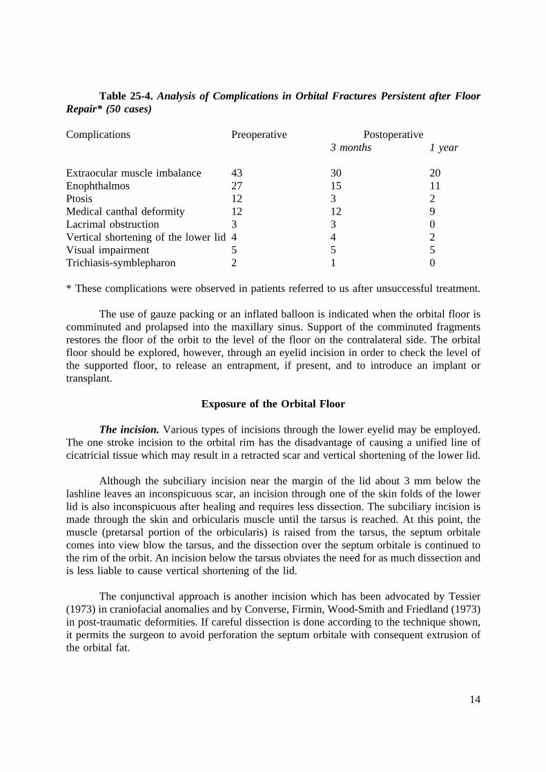
The maxillary sinus approach alone is not satisfactory for the release of the entrappedorbital soft tissues or for placing the orbital floor graft or implant. In a follow-up study of aseries of 50 complicated cases, eight patients whose fractures had been repaired through themaxillary sinus alone required the trans-eyelid approach to release the incarcerated orbitalcontents from the surrounding impacted healed bony fragments (Table 25-4).
14
Table 25-4. Analysis of Complications in Orbital Fractures Persistent after FloorRepair* (50 cases)
Complications Preoperative Postoperative3 months 1 year
Extraocular muscle imbalance 43 30 20Enophthalmos 27 15 11Ptosis 12 3 2Medical canthal deformity 12 12 9Lacrimal obstruction 3 3 0Vertical shortening of the lower lid 4 4 2Visual impairment 5 5 5Trichiasis-symblepharon 2 1 0
* These complications were observed in patients referred to us after unsuccessful treatment.
The use of gauze packing or an inflated balloon is indicated when the orbital floor iscomminuted and prolapsed into the maxillary sinus. Support of the comminuted fragmentsrestores the floor of the orbit to the level of the floor on the contralateral side. The orbitalfloor should be explored, however, through an eyelid incision in order to check the level ofthe supported floor, to release an entrapment, if present, and to introduce an implant ortransplant.
Exposure of the Orbital Floor
The incision. Various types of incisions through the lower eyelid may be employed.The one stroke incision to the orbital rim has the disadvantage of causing a unified line ofcicatricial tissue which may result in a retracted scar and vertical shortening of the lower lid.
Although the subciliary incision near the margin of the lid about 3 mm below thelashline leaves an inconspicuous scar, an incision through one of the skin folds of the lowerlid is also inconspicuous after healing and requires less dissection. The subciliary incision ismade through the skin and orbicularis muscle until the tarsus is reached. At this point, themuscle (pretarsal portion of the orbicularis) is raised from the tarsus, the septum orbitalecomes into view blow the tarsus, and the dissection over the septum orbitale is continued tothe rim of the orbit. An incision below the tarsus obviates the need for as much dissection andis less liable to cause vertical shortening of the lid.
The conjunctival approach is another incision which has been advocated by Tessier(1973) in craniofacial anomalies and by Converse, Firmin, Wood-Smith and Friedland (1973)in post-traumatic deformities. If careful dissection is done according to the technique shown,it permits the surgeon to avoid perforation the septum orbitale with consequent extrusion ofthe orbital fat.
15
A simplified technique employed by Tenzel and Miller (1971) consists of a directincision in the fornix which reaches the orbital rim and a retroseptal approach which exposesthe orbital fat. A Desmarres retractor is used to retract the lower eyelid away from the globe.A malleable retractor placed posterior to the orbital rim gives adequate exposure. The incisionis made through the conjunctiva to the orbital rim and includes the periosteum. This incisionof necessity penetrates through the septum orbitale and exposes orbital fat. Tenzel and Millerhave employed this type of incision in patients with small blowout fractures withoutrestriction of ocular rotary movements of the globe. They did not employ the incision inpatients with massive fractures with herniation of the orbital contents into the maxillary sinus.
The conjunctival incision avoids an external scar, albeit inconspicuous, and claimshave been made that it prevents postoperative lower lid lagophthalmos in the upward gaze.
Because of the need to preserve the orbital fat and recuperate fat which has extrudedinto the maxillary sinus, it is preferable to avoid extrusion of fat through the septum orbitale,whenever possible.
When the orbital rim has been reached by following the septum orbital downward, anincision through the periosteum is made immediately below the orbital rim. Subperiostealelevation is extended backward until the area of the blowout fracture is identified. Theinfraorbital nerve should be respected; with the aid of visual magnification with binocularloupes, the nerve may be carefully dissected from the herniated and entrapped soft tissues.
The orbital structures should be raised using a caterpillar technique with two retractors.Dural pliable retractors with rounded edges minimize pressure on the ocular globe. Retractionof the intraorbital contents should be relaxed periodically. When necessary, a wide exposurecan be obtained through the eyelid incision.
If the infraorbital rim is also fractured (impure blowout fracture), the fragments arerealigned and fixation is maintained by interosseous wiring. If the orbital rim has beendisplaced backward, it is essential that the fragments be realigned in their former position.This measure will prevent postoperative vertical shortening of the lower eyelid.
The inferior rectus muscle and orbital structures are liberated from the areas of theblowout. The floor must be explored sufficiently far back into the orbit until the posterioredge of the defect can be identified. Verification that the ocular globe is freed from thefracture site is obtained by the forced duction test; it is essential to demonstrate the full rangeof all oculorotary movements. The most common cause of failure to release the entrappedstructures is inadequate exposure of the floor in depth. The fracture may be far back.
Restoration of the Continuity of the Orbital Floor. Restoration of the continuity ofthe orbital floor is required in all orbital floor fractures, except in small fractures in which theentrapped structures can be freed readily and the forced duction test shows that free rotationof the eyeball has been reestablished.
Bone grafts. An iliac bone graft (a split rib graft in children) taken from the smoothinner aspect of the ilium is preferable in fractures in which there is a wide area ofcommunication between the orbit and the maxillary sinus. The bone graft, as it becomes
16
vascularized, is better able to resist bacterial invasion than an inorganic implant. The authorshave given preference to the bone graft in all major fractures with disruption of the orbitalfloor. We have also used the anterior wall of the maxillary sinus in the area of the caninefossa, the perpendicular plate of the ethmoid, and the septal cartilage for the restoration ofsmall defects of the floor. Costal cartilage has also been employed and constitutes anexcellent, seldom used transplant. Irradiated cartilage allografts have been employed byDingman and Grabb (1961).
The mucous membrane lining of the subjacent maxillary sinus, often ruptured, willrepair itself and line the undersurface of the transplant.
Inorganic implants. Inorganic materials employed in the orbital region have includedsolids, sponges, gels, and liquids. Tantalum, stainless steel, Vitallium, Paladon,methylmethacrylate, polyvinyl sponge, polyurethane, polyethylene, Teflon, Silastic, andSupramid have been commonly used.
Ballen (1964) employed Cranioplast, a rapidly polymerizing methylmethacrylate, whichis prepared by mixing powdered acrylic with a liquid catalyst. The material is molded in situand hardens by a process of polymerization, which gives off considerable heat. Ballen hasused this procedure in 31 patients but does not mention complications. Miller and Tenzel(1969) have employed prefabricated Cranioplast implants, which are prepared in various sizesand thicknesses, in over 300 patients (Tenzel, 1974). The prefabrication has the advantage ofeliminating the time interval required for the polymerization of the methylmethacrylate.
Freeman (1962) implanted sheets of Teflon in 36 patients with orbital floor fracture,despite communication with the maxillary sinus in several; Browning and Walker (1965) havereported the successful use of Teflon in 45 patients with orbital blowout fractures. Our ownexperience confirms these findings. Teflon is available in sheets 1-mm thick, and Silastic mayalso be carved to fit the specific defect. Supramid sheets 0.3-mm thick are also available.
The inorganic implant offers the advantage of obviating the need for an additionalconcomitant operation for the removal of a bone graft, and it has been satisfactory in mostsimple fractures. The authors have also had successful results in large defects with a widearea of communication with the maxillary sinus. In the course of secondary operations,regeneration of the maxillary sinus lining under the implant and, in moderate-sized defects,bone regeneration have been observed.
The purpose of the orbital floor insert, whether bone graft or inorganic implant, is toreestablish the continuity of the floor, seal off the orbit from the maxillary sinus, and restorethe volume of the orbital cavity. The orbital floor insert should bridge the defect and rest onthe stable adjacent portions of the floor. Smooth materials such as Teflon tend to slideforward and protrude under the skin of the eyelid. A tongue is prepared by making two cutsin the implant; the tongue is introduced under the anterior edge of the bony defect in theorbital floor, thus maintaining it in position and avoiding forward displacement and extrusion.Care should also be taken to avoid dead space between the inorganic implant and the boneof the orbital floor, as the accumulated fluid in the dead space constitutes a favorable mediumfor the growth of bacteria.
17
Blowout Fractures: Variations
A concomitant naso-orbital fracture suggests the possibility of an associated blowoutfracture through the lamina papyracea of the medial orbital wall.
A major portion of the floor may be collapsed into the maxillary sinus. More limitedblowout fractures are located medially, centrally, or laterally. The central blowout is typicalof the pure blowout fracture. The medial blowout often occurs in the impure type associatedwith a naso-orbital fracture, a fracture of the medial orbital wall, and a blowout through thelamina papyracea. The lateral blowout occurs in the impure type associated with fracture ofthe zygoma.
There is no standard pattern in blowout fractures; fixity of the eyeball may beobserved in massive blowout fractures as well as in small blowout fractures. In one case, theinferior orbital contents were pierced and pinned to the floor by a sharp bone fragment. Theentire orbital floor may be fragmented and hanging hammocklike into the maxillary sinus, andthe patient suffers no diplopia because oculorotary action is only slightly impaired. Theprolapse of the orbital contents into the maxillary sinus may be extreme; in some of thesecases, the ocular globe is difficult to find. The authors recall a patient in whom a long nasalspeculum was necessary to retract the edematous eyelids in order to locate the ocular globe.The most dramatic case of this sort involved a fireman who inadvertently turned on the fullpower of his fire hose as he was inspecting the inside of the nozzle. The resultant blowoutfracture was of such extent that the ocular globe disappeared from the orbital cavity and waspresumed to have been enucleated by the force of the projected blast of water. The eyeball,which underwent several choroidal tears, was subsequently located in the maxillary sinus andreplaced in position. After repair of the orbital floor and a normal postoperative course, thepatient's vision was 20/20 for reading and 20/60 for distant vision.
Fractures of the Medial Orbital Wall. Medial orbital wall fractures usually occurin conjunction with an orbital floor fracture or a naso-orbital fracture. A special etiologicfactor is the ski pole, the tip of which has struck the medial canthal area.
Clinical Findings. Rougier (1965) reported tethering of the medial rectus musclefollowing a blowout fracture, strongly suggesting an associated fracture of the medial orbitalwall into the ethmoid sinus with entrapment of the medial rectus by a blowout mechanismsimilar to that which occurs in the orbital floor. Fractures of the medial wall were also notedby Miller and Glaser (1966), Edwards and Ridley (1968), Trochel and Potter (1969(, Dodick,Galin, Littleton and Sod (1971), and Rumelt and Ernest (1972). The clinical signs wereprogressively increasing enophthalmos, narrowing of the palpebral fissure, horizontal diplopiawith restriction of abduction, and increasing enophthalmos on abduction.
It has been suggested that medial orbital wall fractures are associated with orbital floorblowout fracture in an incidence varying from 5 per cent to 50 per cent (Gould and Titus,1966; Jones and Evans, 1967; Dodick and coworkers, 1971). The high percentage can beexplained by the structural relationships between the orbital floor and the medial walldescribed in an earlier section of the chapter. Our own experience, as well as that of Prasad(1973), is that entrapment of the medial rectus muscle is rare and that many of these fracturesare found on radiographic examination to be in association with a naso-orbital fracture. The
18
cellular structure of the ethmoid bone offers resistance which the hollow maxillary sinusbeneath the orbital floor does not. The possibility of a concomitant blowout fracture of themedial orbital wall should be suspected, however, if enophthalmos develops followingadequate treatment of an orbital floor fracture.
Radiologic Findings. The diagnosis is often made by radiography; the presence of airwithin the orbit, clouding of the ethmoid sinus, and medial displacement of the medial orbitalwall or displaced fragments of bone often seen on tomography demonstrate a medial wallfracture. The radiologic examination is invaluable in verifying the integrity of the medial wallpreoperatively so that adequate measures can be taken at the time of surgery.
Treatment. Depending on the severity of the fracture, exposing the medial orbitalwall, freeing the medial rectus muscle if it is entrapped, and placing an inorganic implant overthe area of fracture will usually suffice to restore the architecture of the orbit. Primary bonegrafting may be required in massive comminuted fractures. The treatment varies, therefore,according to the clinical and radiological findings; abstention from surgical treatment isindicated when only a linear fracture is present.
Progressively developing enophthalmos may be the price to pay for the failure todiagnose a medial wall fracture.
Fractures of the Lateral Orbital Wall. The lateral orbital wall consists of a strong,resistant anterior frontozygomatic rim which is exposed to facial trauma and a thinnerposterior portion formed by the orbital process of the greater wing of the sphenoid.
The most severe fractures of the lateral wall of the orbit occur in conjunction withmassive trauma to the zygomatic area with frontozygomatic disjunction and downwarddisplacement of the lateral portion of the orbital floor. The lateral canthus is dislocateddownward, with ectropion of the lower eyelid. This type of fracture requires a direct approachsimilar to that employed in multiple fractures of the midfacial skeleton (see Chapter 24).Direct interosseous wiring of the fragments and primary bone grafting to restore the orbitalfloor, lateral wall, and zygomatic osseous framework are indicated. In such severe fractures,the ocular globe suffers injury of varying degree, and loss of vision is not infrequent.
Lateral wall fractures are probably more frequent that is generally assumed. Theauthors have noted a number of such fractures during craniofacial operations performed forthe correction of grossly malunited fractures. In some cases, orbital fat was found in thetemporal fossa, suggesting a blowout fracture of the posterior portion of the lateral orbitalwall. Behind the thick, lateral orbital rim is an area of thin bone; fracture of the rim maycomminute this thin portion of the lateral orbital wall, facilitating a blowout of the area. Sucha fracture may also be an unsuspected cause of persistent postoperative enophthalmos.
Fractures of the Orbital Roof. LaGrange (1918), in his classic monograph, showedthat the thin medial portion of the orbital roof is fractured and displaced in its posterior partin the region of the superior orbital fissure and optic foramen. A fracture of this type can leadto serious complications, such as optic nerve atrophy and injury to the nerves to theextraocular muscles which enter the superior orbital fissure. Dodick, Galin, Littleton and Sod(1971), in a series of 22 cases of suspected blowout fractures of the orbit, obtained radiologic
19
evidence of fracture of the orbital floor in 15 cases; in two cases there was a concomitantfracture of the orbital roof.
Fracture of the orbital roof may also occur in conjunction with naso-orbital fractures,as the medial portion of the orbital roof is thinner and more susceptible to fracture.
If the superior rim of the orbit is fractured and the trochlea of the superior obliquemuscle is displaced, consequent impairment of the function of the superior oblique musclemay result in diplopia, which is usually temporary.
Fractures of the orbital roof usually occur in conjunction with fractures of thesupraorbital rim and frontal bone. A combined craniofacial approach is required in thesefractures. The dura, which may be torn or penetrated by comminuted fragments, is raised andretracted. In such cases, after exposure of the anterior cranial fossa and neurosurgical repair,the orbital roof is repaired by a suitable thin bone graft.
Orbital Floor Fractures Without Blowout Fracture
Examination and Diagnosis
Clinical Examination. The symptoms and signs of fractures of the floor of the orbitwithout blowout fracture are similar to those of a blowout fracture, with the fundamentaldifference that the patient is able to effect oculorotary movements in an essentially normalfashion. The forced duction test is negative. There may be transitory diplopia.
Roentgenographic Examination. Tomograms will fail to reveal the characteristicexcrescence of the orbital contents into the maxillary sinus, although in crushing fractures ofthe orbital rim and the floor of the orbit may have collapsed into the sinus. Maxillary andzygomatic fracture lines involving the orbital floor and irregularity of the contour of theinfraorbital rim are noted on roentgenograms in these types of fractures.
Treatment
Fractures of the orbital floor occurring in zygomatic and maxillary fractures often donot require orbital intervention other than realignment and wiring of the fragments of thefractured orbital rim. The treatment is that required for the maxillary or zygomatic fracture.Verification should always be made, however, that the patient does not have a blowoutfracture. Careful checking of the oculorotary movements and radiologic examination willeliminate this possibility.
In fractures with bony displacement, the risk of enlargement of the orbital cavity andof consequent enophthalmos is an important consideration. Treatment of an orbital fractureis done in conjunction with the reduction and fixation of fractures of other bones of themidfacial area. Measures are taken to restore the bony continuity of the displaced orfragmented bones. This is best done by direct exposure of the fractured area, reduction, andinterosseous wire fixation.
20
When there is doubt as to the integrity of the orbital floor, exploration is indicated inorder to eliminate an occult comminuted and depressed fracture.
Comminuted Fractures of the Orbital Floor. In cases of exceptionally severe"crush" and "crash" injuries seen following accidents in automobiles, helicopters, or airplanesand usually associated with other fractures of the midfacial skeleton, the orbital rim and floormay be completely demolished. The fragments of bone, most of them suspended hammocklikefrom the periosteum, and the orbital contents sink into the maxillary sinus. The bone ispulverized or reduced to small particles. If bone fragments can be salvaged, they are used toreconstruct the orbital rim. The lateral wall must be stabilized by interosseous fixation priorto reconstruction of the orbital floor. Ocular globe injury often requires enucleation. Openingthe maxillary sinus through the canine fossa provides an approach to the fragmented orbitalfloor, which is elevated by packing the maxillary sinus with gauze impregnated with antibioticointment. Small portions of the orbital floor may remain laterally and medially and serve tosupport the bone graft used to restore the orbital floor and rim. Wire fixation is often requiredto stabilize the graft. In one of our patients whose eye was enucleated, a shelf of bone wasfound only in the posterior reaches of the orbit.
Naso-Orbital Fractures
Severe injuries of the midfacial area associated with fractures of the maxilla, nasalbones, zygomas, or orbits may also be complicated by fracture of the bones of thefrontoethmoidal area of the facial skeleton. The bones of the middle third of the face are alsoin close anatomical relationship to the floor of the anterior cranial fossa and the frontal lobesof the brain through the frontal and ethmoid sinuses and the cribriform plate. The possibilityof a concomitant blowout fracture of the orbital floor has also been discussed.
Because of the possibility of brain damage, patients suffering these fractures shouldbe observed for neurologic complications, such as progressive loss of consciousness, and signsof epidural hematoma, aerocele, and chronic subdural hematoma. Fracture of the odontoidprocess, which requires early reduction, has been reported. Pulmonary edema is anothercomplication of head injuries.
Structural Aspects
The thin areas of the medial orbital wall transilluminate readily and thus contrast withthe heavier abutments formed by the nasal process of the frontal bone, the frontal processesof the maxilla, and the thick upper portion of the nasal bones. Posterior to the frontal processof the maxilla, the thinner lacrimal bone and the delicate lamina papyracea are vulnerable totrauma. The anterior and posterior ethmoidal foramina are situated along the upper border ofthe lamina papyracea in the frontoethmoidal suture, where the orbital plate of the frontal boneand the lamina papyracea of the ethmoid are joined. The anterior ethmoidal foramen transmitsthe nasociliary nerve and the anterior ethmoidal vessels; the posterior ethmoidal foramen givespassage to the posterior ethmoidal nerve and vessels. The rupture of these vessels in naso-orbital fractures with backward penetrating fragments is one of the causes of orbitalhematoma, a complication which may require immediate incision and drainage.
21
The most posterior portion of the medial orbital wall is formed by the body of thesphenoid immediately in front of the optic foramen. In severe skeletal disruption of this area,the fracture lines involving the optic foramen and the optic nerve may result in blindness.
The Interorbital Space. The term "interorbital space" designates an area between theorbits, beneath the floor of the anterior cranial fossa. The interorbital space contains the twoethmoidal labyrinths, one on each side.
The interorbital space is roughly cuboidal, being wider anteriorly than posteriorly. Itis limited above by the cribriform plate in the midline and by the roof of each ethmoidal masson the sides and is divided into two approximately equal halves by the nasal septum. Theinterorbital space is limited below at the level of a horizontal line through the lower borderof the ethmoidal labyrinth. The lateral walls of the interorbital space are the medial walls ofthe orbit. Anteriorly the interorbital space is limited by the frontal processes of the maxillaand by the nasal process and spine of the frontal bone.
The interorbital space contains cellular bony structures, the ethmoidal cells; spongybony structures, the superior and middle turbinates; and a median thin plate of bone, theperpendicular plate of the ethmoid bone which forms the posterosuperior portion of the nasalseptal framework.
The Frontal Sinus. The size of the frontal sinus varies greatly; it may be that of anethmoidal cell, or it may be a very large sinus, pneumatizing the frontal bone. Occasionallyit is absent.
The sinus has the shape of a pyramid with inferior, anterior, and posterior walls. Theinferior wall or floor of the frontal sinus corresponds to the roof of the orbit and is thethinnest portion of the frontal sinus. The anterior wall is thickest and is composed ofcancellous bone. The posterior wall is thinner than the anterior wall and is entirely composedof compact bone which separates the sinus from the frontal lobe.
The Ethmoidal Sinus. The ethmoid bone occupies the lateral portion of theinterorbital space. Below the interorbital space, the lower half of the nasal cavity is flankedby the maxillary sinuses. Each lateral mass of the ethmoid is connected medially to thecribriform plate; the roof of each ethmoidal mass is inclined upward from the cribriform plateand projects, in its lateral portion, about 0.25 cm above the cribriform plate.
The ethmoid is pyramidal or cuboidal, measuring 3.5 to 5 cm long and 1.5 to 2.5 cmwide. It is cellular in structure and contains eight to ten cells with thin lamellar walls; thesecells drain into the middle meatus of the nose. The frontal sinus drains through the ethmoid,either through a distinct duct or by emptying into an anterior ethmoidal cell and into themiddle meatus. Thus there is an intimate anatomical relationship with the frontal sinusthrough the frontonasal duct. It will be recalled that, in embryological development, thefrontal sinus is formed by an outcropping ethmoidal cell. A large ethmoidal cell, thefrontoethmoidal cell, may be seen in the frontal bone between the frontal sinus and the roofof the orbit.
22
Surgical Pathology
Situated in the upper and central part of the middle third of the face, anterior to theanatomical crossroads between the cranial, orbital, and nasal cavities, the bones forming theskeletal framework of the nose may be projected backward between the orbits when they aresubjected to a strong traumatic force. The term "naso-orbital" is employed to designate thistype of fracture (Converse and Smith, 1963, 1964, 1966). A typical cause of naso-orbitalfracture is an impact force applied over the upper portion of the bridge of the nose caused bythe projection of the face against the dashboard or steering column of an automobile whenit comes to a crash stop. A crushing injury with comminuted fractures is thus produced.Bursting of the soft tissues due to the severity of the impact and penetrating lacerations of thesoft tissue resulting from projection through the windshield may transform the closed fractureinto a compound fracture.
If the impact force suffered by the strong anterior abutments is sufficient to causebackward displacement of these structures, no further resistance is offered by the matchbox-like structures of the interorbital space; indeed, these structures collapse and splinter as woulda pile of matchboxes struck by a hammer. The roof of the interorbital space is frequentlyinvolved in these fractures, and the anterior cranial fossa is penetrated, the fracture occurringeither medially through the cribriform plate or laterally through the roof of the ethmoid sinus.
Some of the neurologic complications resulting from naso-orbital fractures arelaceration of the dura covering the frontal lobes, laceration of the tubular sheaths envelopingthe olfactory nerves as they perforate the cribriform plate, penetration of the brain by a sharp-edged ethmoidal cell wall, and necrosis of brain tissue.
An additional point of interest in the skeletal structure of this area is the continuity ofthe thin lamina papyracea of the medial orbital wall with the thin portion of the floor of theorbit. The splintering of the lamina papyracea facilitates a blowout fracture in this area andmay occur in patients who suffer a blowout fracture of the floor of the orbit concurrently witha naso-orbital fracture.
Lacerations of the soft tissues may sever the levator palpebrae superioris or penetratethrough the medial canthal area, severing the medial canthal tendon and the lacrimal canaliculior sac.
Fractures of the other facial bones, particularly of the midfacial skeleton, arefrequently seen. In some of our patients, the frontal bone was also involved.
The Nasal Area: The Weakest Portion of the Facial Skeleton. Studies confirm thatthe nasal area is the weakest portion of the facial skeleton; fractures occur in this area withan impact load of 35 to 80 g. In Swearington's study (1965), 45 impacts were made oncadaver heads to determine the fracture points of the various portions of the facial skeleton.The comparative forces that can be tolerated over the various facial areas without fracture areillustrated. With the exception of the neck of the condyle, the zygomatic area is the nextweakest area, being unable to sustain impact forces greater than 50 g. The upper portion ofthe middle third of the face, which includes both the nasal and orbital areas, is structurallysusceptible to fracture. In contrast, the lower portion of the maxilla sustains impact forces of

23
up to 100 g, and the major portion of the body of the frontal bone, with the exception of thecentral portion which is weakened by the frontal sinus cavities, sustains impact forces of upto 200 g.
Although padding of the rigid dashboard decreases the severity of the injuriessustained by the right front passenger, the padded dashboard lip in many automobiles has acontour suitable for the production of the "pushback" of the nasal structures between theorbits. Such fractures occur even though the passenger is wearing a lap seat belt; they occurless frequently when he is protected by the shoulder harness type of belt. The passengerwithout a seat belt is often projected through the windshield and suffers various types of softtissue lacerations, including penetrating lacerations of the naso-orbital tissues.
Traumatic Telecanthus. In many naso-orbital fractures, the patient has a characteristicappearance; the bony bridge of the nose is depressed and widened, and the eyes appear farapart as in orbital hypertelorism. The appearance of the patient is the result of traumatictelecanthus, an increase in the distance between the medial canthi (intercanthal distance) asa result of displacement by fracture of the bones forming the skeletal framework of the noseand medial orbital walls. Naso-orbital fractures may be unilateral but are most often bilateralfollowing severe trauma. Traumatic orbital hypertelorism is a deformity characterized by anincrease in the distance between the orbits and ocular globes and occurs in massive disruptionof the midfacial skeleton and frontal bone (Converse, Smith and Wood-Smith, 1975).
Traumatic telecanthus is produced by two varieties of backward displacement of thebony structures. In the first, the frontal processes of the maxilla and the nasal bones penetratethe interorbital space, comminuting the ethmoid cells and out-fracturing the medial wall ofthe orbit. The medial canthal tendon attachments are displaced with the bones, and the medialcanthus is displaced laterally and deformed, assuming a rounded shape.
In the second type of fracture, the nasal bones and frontal processes of the maxilla aresplayed outward and projected backward into the medial portion of the orbital cavity alongthe lateral surface of the medial orbital wall, severing the medial tendon, transecting thelacrimal sac, or severing the canaliculi. Thus traumatic telecanthus is also caused by increasein the thickness of the medial orbital wall from the overlapping of bone fragments.
Loss of bone in the area may result from injudicious debridement and removal of bonefragments or the expulsion of bone fragments into the nasal cavity at the time of fracture.
Occasionally the medial portion of the inferior rim of the orbit is also fractured anddisplaced backward.
Examination and Diagnosis
Clinical Examination. The appearance of the patient who has suffered a naso-orbitalfracture is typical: the nose is flattened, appearing to have been pushed between the eyes; themedial canthal areas are swollen and distorted, the caruncles and plicae semilunares beingcovered by the edematous and displaced structures; ecchymosis and subjunctival hemorrhageare usual findings.
24
Intranasal examination shows the findings observed in fractures of the nasal bones andseptum. Fracture of the septum is suggested by septal hematoma. Naso-orbital fractures areoften accompanied by signs of orbital blowout fracture or fracture of the maxilla or zygoma,which are frequently associated fractures. Edema and hematoma often mask the extent of theskeletal distortion of the area, particularly if the patient is not seen during the first hours afterthe accident.
The patient may be unconscious or have had loss of consciousness of long or shortduration. Loss of consciousness is suggestive of brain injury. The patient may be irritable,restless, or even thrashing about after a severe injury. As in other fractures around the orbit,extensive edema of the lids and orbital structures may cause mechanical limitation of eyeballmovements.
There may be little evidence of skeletal deformity because of hematoma or swelling.In some cases the deformity is evident when the frontal bone has been crushed inward or thenasal structures have been projected into the interorbital space. The bones may be loose, andcrepitation may be felt when they are mobilized. The entire upper jaw may be movable, andmotion may be felt in the bones of the interorbital space. A portion of the forehead skin maybe avulsed in compound fractures, exposing the bone and revealing the site of fracture.
Clear fluid escaping from the nose is strongly suggestive of cerebrospinal fluidrhinorrhea show an initial escape of blood from the fracture site; the fluid then becomesbrownish in color and finally clear. The fluid may be seen to pulsate within the nose.
Roentgenographic Examination. Roentgenograms and tomograms are required toestimate the amount of damage. As stated earlier in the text, a fracture of the medial orbitalwall may be associated with a blowout fracture of the orbital floor. Careful tomographic studywill show suggestive signs of medial wall fracture. Fractures of the cribriform plate may beimpossible to detect by ordinary radiographic examination. The presence of air in thesubdural, subarachnoid space or in the ventricle is a sign of communication with the nasalcavity or sinuses, establishing a direct pathway to infection, and is an indication forneurosurgical intervention. Air may not be detected during the first 24 hours. Roentgenogramsshould therefore be repeated if the patient shows increasing signs of frontal lobe dysfunctionin the nature of mental changes.
Fragmentation and a "buckled" appearance of the cribriform plate are suggestive ofpenetration of fragments of ethmoidal cells into the brain; this is an additional indication forneurosurgical operation. Tomograms may be of help in evaluating the location of the damageand the degree of displacement of the fragments.
Treatment
Brain injury should be suspected when the patient has been unconscious. Neurosurgicalintervention is required in patients who have suffered destructive lesions of the brain orpenetration of bone fragments into the brain. Careful neurologic and radiologic examinationis required. Cerebrospinal fluid rhinorrhoea should not be a contraindication for treatment ofthe fractures. A delay of a number of days may be required to allow for subsidence of
25
swelling and hematoma and clarification of the neurosurgical status of the patient; such adelay does not jeopardize an ultimate satisfactory result.
The technique of the treatment of naso-orbital fracture consists of the elevation of thecomminuted fragments by means of an instrument placed inside the nasal cavity. Externaldigital pressure and, if necessary, realignment of the fragments restore the position of themedial orbital walls. In naso-orbital fractures, the wired plate technique and the figure-of-eightwire suspension have their selective applications. In severe cases of naso-orbital fractures, theopen-sky method is the treatment of choice.
The Open-Sky Technique (Converse and Hogan, 1970). In compound naso-orbitalfractures, the external wound permits direct inspection of the area, and the comminutedfragments can be realigned under direct vision. In severely comminuted naso-orbital fracturesthat are not compound, an open reduction is indicated. Bilateral vertical incisions through theskin over the lateral wall of the nose provide exposure; if additional exposure is required, thevertical incisions are joined by a transverse incision placed over the root of the nose. Thebilateral vertical incisions, if they are of adequate length, give adequate exposure in mostcases, and the resulting scars are not conspicuous, usually being hardly perceptible. After thefragments are dissected one by one, each lacrimal sac and the nasolacrimal duct exposed andpreserved, and the medial canthal tendon identified, subperiosteal exposure of the medialwalls of the orbit is extended posteriorly behind the area of fracture. It is then possible, underdirect vision, to replace the fragments in acceptable alignment, aided by an instrument placedintranasally. Repair of the lacrimal apparatus in any of its sections can also be done underdirect vision. Fragments of the nasal bones and the frontal processes of the maxilla may bejoined to each other by interosseous wiring as well as to the frontal bone. Direct exposure ofthe fracture area is indispensable in fractures involving the cribriform plate with cerebrospinalrhinorrhea, as it permits inspection of the cribriform plate through the area of fracture.
The principle of the open treatment of naso-orbital fractures is the preservation of allfragments of bone, even where they are detached from soft tissues.
A frequent occurrence is the lateral displacement of the portion of the medial orbitalwall containing the anterior lacrimal crest and the attachment of the medial canthal tendon.The orbicularis oculi muscle exerts a lateral traction, deforming the medial canthal area; thisis another mechanism in the production of traumatic telecanthus.
Primary Bone Grafting. Primary bone grafting is indicated to restore bone continuitywhen the medial orbital wall is reduced to a pulp. The bone graft establishes an area ofpurchase for the attachment of the medial canthal tendon and also restores the medial orbitalwall and the size and shape of the orbit (see Chapter 28).
In the preceding chapter (see Chapter 24), certain indications for primary bone graftingwere given - namely, when bone is destroyed, and when assistance is required in maintainingthe projection of the maxilla by bone grafting in the pterygomaxillary area. Primary bonegrafting has also been advocated for the repair of defects of the floor of the orbit, of themedian and lateral walls, and of the orbital roof. Moreover, primary bone grafting in severelycomminuted naso-orbital fractures is indicated when the clinical conditions are favorable.
26
Supraorbital And Glabellar Fractures AndFractures Involving The Frontal Sinus
Either independently or in conjunction with naso-orbital fractures, fractures of thelower portion of the frontal bone in the supraorbital and glabellar regions are relativelyinfrequent. Schultz (1970) estimated the incidence of such fractures at approximately 5 percent of those patients suffering fractures of the facial bones. Furthermore, he noted thatpatients suffering supraorbital and glabellar fractures require a longer average hospital staythan other facially injured patients irrespective of the cause.
Fractures of the supraorbital ridge are clinically evident by the observation of an areaof local depression, especially in the early stages. In later stages, edema and periorbitalecchymosis may mask the deformity. The degree of depression of the supraorbital arch maydepend on the size of the frontal sinus, which is extremely variable from one individual toanother. Individuals with large frontal sinuses are more susceptible to fracture. When thedepressed area involves the trochlea and the pulley of the superior oblique muscle, the patientmay complain of diplopia, which is usually transitory. Some of our patients with supraorbitalfractures had penetrating injuries which severed the levator palpebrae superioris. The resultingptosis of the upper eyelid was masked in the early stages by edema and ecchymosis.Restriction of global movements secondary to the ecchymosis might lead one to the suspicionof a concomitant blowout fracture. If the lid, however, is carefully raised with a smallelevator, unrestricted upward rotation of the globe can be observed and can be confirmed bythe forced duction test. These findings eliminate the presumptive diagnosis of blowout fractureof the orbital floor.
The authors have observed concomitant fracture of the supraorbital arch, depressedfracture of the glabellar region, and naso-orbital fracture in the same patient. More frequently,however, the glabellar fracture is associated with a severe naso-orbital fracture, and thesupraorbital arch fracture occurs independently.
When the causative impact is extremely violent and when an associated brain injuryis present, the fragmented bones may be so badly comminuted that their realignment is notpossible; some of them may even be ejected by the explosive force. In such cases, the typeof deformity illustrated may be seen, with loss of nasal, glabellar, and supraorbital archskeletal framework.
Roentgenographic diagnosis may be difficult, particularly in attempting to demonstratea fracture of the posterior wall of the frontal sinus by lateral, Waters, or posteroanterior views.Tomograms, however, may be helpful in evaluating the extent of the damage and fracture.
Treatment nearly always involves open reduction through the wound or through asurgical incision. Exposure for supraorbital arch fractures can be achieved through theeyebrow if the incision is correctly placed. Objections have been made to such incisionsbecause of subsequent hairless scars separating the upper and lower portions of the eyebrow.The hair follicles of the eyebrow are implanted in an oblique fashion; the incision should beslanted downward in order to parallel the direction of the hair follicles. In this manner, aninconspicuous scar will result in a well-furnished eyebrow, particularly in male patients. In
27
female patients with plucked eyebrows, it may be preferable to make the incision immediatelybelow the orbital margin.
The approach to glabellar fractures, often associated with the telescoping type of naso-orbital fracture, can be achieved through the incisions made for the open-sky approach tonaso-orbital fractures. If wider exposure is required, a transverse incision at the root of thenose is extended laterally on each side immediately below the orbital margin.
The best technique of exposure in major fractures involving the frontal bone is thecoronal flap advocated by Tessier. In severe fractures involving the anterior cranial fossa, anintracranial approach and neurosurgical collaboration are indicated.
Reduction and realignment of fragments depend essentially upon the type of fracture.When large fragments are present, they may be levered upward and will often remain inposition without interosseous wire fixation. When smaller fragments are present, directinterosseous wiring of the multiple fragments, as in the treatment of naso-orbital fractures,may be necessary.
Treatment of Fractures Involving the Frontal Sinus. Excluding the possibility ofa concomitant head injury which may require craniotomy, fracture with backward crushingof the anterior wall of the frontal sinus may not cause any functional disturbance, but it leavesthe patient with a depression deformity if no treatment is applied.
Open reduction of the depressed anterior wall of the frontal sinus, in a manner similarto that employed in glabellar fractures, is the method of choice. The depressed bone iselevated and maintained by wire fixation. When the anterior wall of the frontal sinus iscomminuted, the following approach can be practiced.
In fractures of the anterior wall of the frontal sinus, an incision is made along theupper portion of the lateral wall of the nose from the point where the root of the nose joinswith the supraorbital arch to a few centimeters below. The incision is through the skin andperiosteum, equidistant between the dorsum of the nose and the medial canthus of the eye.Subperiosteal elevation is extended upward to below the supraorbital arch. The lacrimal sacis temporarily elevated and retracted from the lacrimal groove. Posterior to the area of thelacrimal groove, the lamina papyracea of the ethmoid is cut through, and the ethmoidal sinusis penetrated. By working upward from the ethmoidal sinus with a small Kerrison punch, itis possible to follow the ethmoidal cells until the medial aspect of the frontal sinus is reached.Part of the floor of the frontal sinus is removed, and the frontal sinus is entered. The fracturedanterior wall of the frontal sinus is pried forward with a blunt probe or a hard rubber catheter.Often, if the depressed fracture consists of one large fragment, the fragment will maintain itsposition after it has been repositioned.
If the anterior wall of the frontal sinus is severely comminuted and if the generalcondition of the patient is satisfactory, a thin iliac bone graft is resected and placed over thefrontal sinus to restore contour, if a satisfactory soft tissue covering can be assured.
If clinical and radiologic examination shows intracranial injury and a posterior wallfracture of the frontal sinus, neurosurgical exploration and treatment should be undertaken.
28
Complications of Orbital and Naso-Orbital Fractures
The type of injury, the force of the impact, associated tissue damage, and inadequateor delayed treatment are the major causes of complications in orbital and naso-orbitalfractures.
Early diagnosis and adequate repair of the orbital floor result in few latecomplications. Depending on the quality of the initial treatment and despite adequatetreatment, some cases are subjected to an inexorable evolution toward late complications (seeTable 25-4).
The sequelae and their treatment have been discussed in some detail (Converse andcoworkers, 1967; Kazanjian and Converse, 1974), and enumeration of these sequelae follows:structural deformities; complications with the implant or transplant; muscular imbalance andenophthalmos; ocular complications; lacrimal system disturbances; hematoma and blindness;blepharoptosis; medial and lateral canthal deformities; vertical shortening of the lower eyelid;and infraorbital nerve anesthesia. These complications and their treatment will be discussedin Chapter 28. A discussion of a few of the early complications follows.
Structural Deformities. Deformities and functional impairment are late complicationswhich can be reduced by early diagnosis and treatment, but often the diagnosis is obscuredby more severe cranial and facial injuries which demand primary treatment. The unconsciouspatient cannot experience diplopia, and orbital edema, hemorrhage, and ptosis can mask theenophthalmos. After several weeks, fibrous cicatrization is established, and reconstruction ofthe orbital cavity and restoration of symmetrical ocular function in a malunited fracture mustbe undertaken in the presence of scarred, atrophic orbital fat and muscles.
Complications with the Implant or Transplant. Dead space between the inorganicimplant and the bone of the orbital floor should be avoided, as the accumulated fluid in thedead space constitutes a favorable medium for the growth of bacteria. Orbital infection andsuppuration are indications for incisions, drainage, and removal of the implant, which isreplaced after the infection has subsided. Antibiotic therapy should be routinely employed inall cases to avoid this complication.
In the average blowout fracture caused by a fist punch, a 1-mm Teflon implant isadequate to restore the continuity of the floor. Excessive thickness of the implant may causethe eyeball to be elevated. If the latter complications occurs, the implant should be removedand replaced by a thinner one. Furthermore, excessive overcorrection may result in elevationof the ocular globe and excessive compression on the orbital contents and the optic nerve.
Occasionally in severe trauma, hematoma may elevate the ocular globe. Gradualresorption of the hematoma will reduce the ocular globe to a level commensurate with thecontralateral ocular globe.
An implant of excessive anteroposterior dimensions may compress the optic nerve andcause blindness. Extrusion of the implant by progressive forward migration can occur and canbe avoided by the technique shown.
29
There is no perfect substitute for the orbital floor at the present time. The materialused, however, should fit certain criteria. It should be well tolerated by the patient,sufficiently strong to support the orbital contents, relatively nonreactive to prevent adhesionsto the orbital capsule, easily obtainable at the time of surgery, and workable into the desiredshape. In early fracture repairs, the thin inorganic prosthesis is best, as exophthalmos canresult from overcorrection. While autogenous bone is well tolerated by the patient and is themost physiologic substitute, it is not immune to absorption. Every effort should be made toensure close contact of the bone graft with the bone of the orbital floor by carefully shapingthe bone graft and eliminating dead space with small slivers of cancellous bone. Teflon,Silastic, Supramid, and Cranioplast implants are the preferred inorganic implants presentlyavailable. They can be shaped or carved and fitted to the floor of the orbit withoutpreliminary preparation or delay.
Muscular Imbalance and Enophthalmos. Despite adequate early treatment of theorbital fracture, progressive ocular muscle imbalance and enophthalmos may ensue. Manyadequately treated orbital floor fracture patients do not recover complete extraocular musclefunction. Slight limitation in the upward gaze results in diplopia in this position, a relativelyslight handicap. Extraocular muscle surgery on the affected eye or on the contralateralunaffected eye is a frequent requirement to restore eye muscle balance.
The complicated blowout fracture is often accompanied by multiple fractures of thefacial bones and injuries of the soft tissues. Many of the fractures with complications treatedby us have fallen into this category. These patients show impairment of oculorotary action anddiplopia, enophthalmos, depression of the zygomatic prominence, ptosis of the upper eyelid,downward displacement of the orbital contents, medial canthal deformities, shortening of thehorizontal dimension of the palpebral fissure, shortening of the vertical dimension of thelower eyelid, saddle deformity and/or widening of the nasal bony bridge, and occasionallydeformities of the supraorbital arch (see Table 25-4). Most complicated orbital fracturesrequire extraocular muscle surgery during the months following the injury.
Ocular Complications. Ocular injury following orbital fractures has been reported asvarying between 14 per cent (Milauskas and Fueger, 1966), 17 per cent (Miller and Tenzel,1967), and 29 per cent (Jabaley and coworkers, 1975) in different series. Ocular globe injuryalso varies in severity from a corneal abrasion to loss of vision from a ruptured globe or afracture involving the optic canal. Blindness or loss of the eye is remarkably infrequent, inview of the severity of some of the injuries sustained.
The importance of the ophthalmologic examination in all fractures of the orbit hasalready been discussed. Vitreous hemorrhage, dislocated lens, rupture of the sclera, traumaticcataract, choroidal rupture and hemorrhage, ruptured iris sphincter, glaucoma, retinaldetachment, and diminution or loss of vision are some of the complications which may beavoidable if treatment is instituted early.
Verification of vision is essential in the course of an ophthalmologic examination. Anexcellent prognostic sign is the Marcus Gunn pupillary sign. A light is moved rapidly fromone eye to the other alternately. If conduction of the optic nerve is lessened, the pupil on theinvolved side will appear to dilate as the light is brought from the sound eye to the involvedeye. Monocular vision should also be considered.
30
The need for preliminary ophthalmologic examination is dramatically illustrated in acase reported by Miller (1968). Miller reported a patient with a midfacial fracture in whomvision in the left eye was 20/70 a few hours after the injury but dropped to no lightperception by the fifth day. No surgery had been done. If the surgery had been performedbefore the fifth day, the resulting blindness could have been attributed to the operation.
Blindness has never resulted from repair of the orbital floor in any of the patientstreated by the authors. We have seen patients whose vision was lost following repair byothers. Nicholson and Guzak (1971) reported six cases in which vision was lost in a seriesof 72 patients who underwent orbital floor repair by means of silicone implants inserted byvarious surgeons in the same hospital. This high rate of visual loss, occurring in a reputablehospital, may be explained by the fact that the patients were operated upon by a number ofdifferent surgeons. It is preferable to place such patients under the care of specializedexperienced surgeons.
Lacrimal System Complications. Interruption of the continuity of the lacrimalapparatus, chronic inflammatory condition of the lacrimal sac, or cystic dilation (known asa mucocele) with ensuing epiphora requires dacryocystorhinostomy or other procedures (seeChapter 28).
Hematoma and Blindness. Hematoma is unusual. It occurs if continuous bleedingfrom the anterior and posterior ethmoidal arteries is not spontaneously arrested in fracturesinvolving the medial orbital wall. Blindness may be the consequence of a hematoma occurringunder a firm pressure dressing. The use of continuous suction drainage is recommended ifbleeding is excessive at the end of the operation. The treatment of hematoma of the orbit isusually conservative (see Chapter 37).
Blepharoptosis. True ptosis of the upper lid is to be differentiated from pseudoptosisresulting from the downward displacement of the eyeball and enophthalmos. True ptosisresults from loss of function of the levator palpebrae superioris. This may occur either as aresult of transection of the levator aponeurosis or of intramuscular hematoma and subsequentfibrosis, or from an injury to the third cranial nerve. In most cases the levator aponeurosis istransected, usually not through its entire width. The levator aponeurosis can usually besuccessfully repaired (see Chapter 28).
Vertical Shortening of the Lower Eyelid. Vertical shortening of the lower eyelidwith baring of the sclera below the limbus of the globe in the primary position (scleral show)may result from downward and backward displacement of the fractured inferior orbital rim.Release of the septum orbitale attachment from the orbital rim and restoration of the positionof the orbital rim after osteotomy may be required. If such operative procedures fail, atarsoconjunctival graft from the same or opposite upper eyelid will elevate the lower lidmargin up to 4 mm. Often pseudoptosis and depression of the supratarsal fold accompanyvertical shortening of the lower lid; these three problems can also be at least partly resolvedby means of a tarsoconjunctival graft.
The authors have received a number of personal communications concerning verticalshortening of the lower lid following the approach to the floor through a subciliary incision.
31
We are not using a slightly lower incision, below the inferior tarsus, to eliminate such acomplication.
Infraorbital Nerve Anesthesia. Infraorbital nerve anesthesia may be verydisconcerting to some patients. The area of sensory loss usually extends from the lower lidover the cheek and lateral ala to the upper lip. Release of the infraorbital nerve from thepressure of bone fragments within the canal may be indicated (see Chapter 28). Sensation mayreturn spontaneously as late as one year after fracture.
Cerebrospinal Fluid Rhinorrhea. This complication occurs in naso-orbital fractures.The present trend is toward a conservative approach, influenced by a diminished fear ofmeningitis which results from the protection provided by antibiotic therapy. However, therehave been reports of cases of traumatic cerebrospinal fluid rhinorrhea with recurring bouts ofmeningitis 15 years after injury.
When the radiologic examination fails to show evidence of damage other than afracture line, the patient is treated conservatively while being observed for signs of impendingcomplications, such as meningitis or extradural or intracerebral abscess. No packing is placedin the nasal fossae, smoking is forbidden, and the head of the bed is elevated to an angle of60 degrees. The patient should be warned against blowing his nose, because the leakage mightrecur, or he might force tissue or air into the cranial cavity. If the cerebrospinal fluidrhinorrhea is prolonged, an operation to close the fistula should be considered; this is adecision to be made by the neurosurgeon. Collins (1973) stated that spinal fluid drainage isconfirmed by the presence of glucose in amounts of more than 30 mg of glucose per 100 mLof fluid. The use of glucose oxidative paper is not reliable as a test for glucose. As high as75 per cent positive reactions have been obtained when using the oxidative paper test inpatients with normal secretions. The fistula may be located by isotope dyes or by dyes placedin the lumbar or ventricular cerebrospinal spaces.
As stated in Chapter 24, Collins (1973) advocated early reduction of facial fracturesin the presence of cerebrospinal fluid rhinorrhea (Dingman, 1974). He stated that the objectiveis to obtain reduction and fixation of the fractured bones, thus providing support of the areaof injury. In 19 patients treated by him in whom the facial bone fractures have been reducedin less than 48 hours, only two patients required operative repair of the dural fistulae, and inboth the fistulae were not in proximity to the facial bone fractures.