Embed Size (px)
Citation preview
Coordinate activation of c-Src by SH3 and SH2-binding sites on a novel, plSO^^ -related protein, Sin Konstantina Alexandropoulos and David Baltimore^ Massachusetts Institute of Technology, Cambridge, Massachusetts 02139 USA
To understand how protein-protein interactions mediated by the Src-SH3 domain affect c-Src signaling, we screened for proteins that interact with the Src-SH3. We found a novel protein. Sin (Src interacting or signal integrating protein), that binds to Src-SH3 with high affinity, contains numerous tyrosine residues in configurations suggestive of SH2-binding sites, and is related to the v-Src substrate pi30* ^ . In cotransfection assays, a small fragment of Sin retaining the Src-SH3-binding site and one tyrosine-containing motif induced c-Src activation as measured by a transcriptional reporter. Phosphorylation of the peptide on tyrosine by c-Src, as a consequence of Src-SH3 binding, was necessary for its stable interaction with c-Src in vivo and for transcriptional activation. Phosphorylation of multiple tyrosine-containing motifs found on Sin correlated with c-Crk and cellular phosphoprotein binding to Sin as well as increased c-Src activity. These data suggest that (1) SH2 and SH3 ligand sites on Sin cooperatively activate the signaling potential of c-Src, (2) Sin acts as both an activator and a substrate for c-Src, and (3) phosphorylated Sin may serve as a signaling effector molecule for Src by binding to multiple cellular proteins.
{Key Words: Sin; plSO' '' ; Src activation; Src substrate; Crk] Received February 28, 1996; revised version accepted April 12, 1996.
Src was discovered as an oncoprotein that had constitutive tyrosine kinase activity (Brugge and Erickson 1977; Hunter and Sefton 1980; Sefton et al. 1980). The normal cellular c-Src, however, has a regulated kinase activity that can be activated by removing an autoinhibitory interaction in the protein (Cooper and King 1986; Cart-wright et al. 1987; Kmiecik and Shalloway 1987; Pi-wnica-Worms et al. 1987). The inhibited state is thought to be achieved through an intramolecular interaction in which the carboxy-terminal tail of c-Src interacts with the amino-terminal SH3 and SH2 domains (Roussel et al. 1991; Liu et al. 1993; Murphy et al. 1993; Okada et al. 1993; Superti et al. 1993; Bagrodia et al. 1994). That interaction depends on phosphorylation of Src tyrosine 527 by the Csk kinase (Nada et al. 1991; Okada et al. 1991). The phosphorylated tyrosine must interact with SH2, but the binding site for SH3 remains uncertain. A dimeric complex of the tyrosine-phosphorylated carboxy-terminal tail of Lck—a kinase related to Src—with its SH2 and SH3 domains has been crystalized and its structure determined, but its direct relevance to Src remains uncertain (Eck et al. 1994). Ligands for the Src-SH2 domain, such as tyrosine-phosphorylated motifs found on activated platelet-derived growth factor (PDGF) receptors, can activate Src enzymatic activity in vitro by
^Corresponding author.
outcompeting intramolecular inhibitory interactions (Liu et al. 1993). Changes in the phosphorylation state of tyrosine 527 by mutation (Kmiecik and Shalloway 1987; Piwnica-Worms et al. 1987), association with polyoma mT antigen (Cartwtight et al. 1987), or antibody binding (Cooper and King 1986), can also activate the c-Src kinase.
SH3 and SH2 domains are modular polypeptide units that mediate protein-protein interactions and are parts of many cytoplasmic signaling proteins. Structures of both domains have been solved from a variety of proteins and their ligand specificities have been determined (Cohen et al. 1995). SH2 domains bind to phosphotyrosine-containing sequences (Anderson et al. 1990; Mayer et al. 1991), whereas SH3 domains recognize proline-rich peptides with a PXXP core motif (Ren et al. 1993; Yu et al. 1994). Ligands can bind to SH3 domains in either an amino-carboxy or a carboxy-amino orientation and are therefore classified as class I or class II, respectively (Feng et al. 1994). SH2 and SH3 domains mediate a variety of physiological responses and are found together on many proteins (Cohen et al. 1995), suggesting that their activities may be coordinated. Identification of physiological regulators of the enzymatic activity of Src and its substrates has proved difficult. In recent years, a number of proteins have been identified that associate with v-Src through its modular SH2 and SH3 domains, although the biological role of these interactions is not
GENES & DEVELOPMENT 10:1341-1355 © 1996 by Cold Spring Harbor Laboratory Press ISSN 0890-9369/96 $5.00 1341
Cold Spring Harbor Laboratory Press on April 28, 2018 - Published by genesdev.cshlp.orgDownloaded from
Alexandiopoulos and Baltimore
clear. Among the proteins identified are ones that contain multiple ligands for the Src-SH2 and Src-SH3 domains—pl30^^" (Sakai et al. 1994), paxillin (Weng et al. 1993), and p62/Sam68 (Taylor and Shalloway 1994; Richard et al. 1995; Taylor et al. 1995; Lock et al. 1996)— suggesting a cooperative role for these domains in Src regulation. Therefore, it has been proposed that these proteins, when phosphorylated on tyrosines, can act as signal integration sites by forming multiprotein complexes with cytoplasmic SH2-containing proteins. Such a role would be equivalent to that had by the insert and tail components of tyrosine kinase-containing surface receptor proteins like the PDGF receptor (Kazlauskas and Cooper 1989; Courtneidge et al. 1991; Fantl et al. 1992; Ronnstrand et al. 1992; Valius et al. 1993), and this mechanism could be used to propagate signals through the cytoplasm.
In this paper we describe a novel protein, Sin, that contains two ligand sites that exhibit high-affinity binding for Src-SH3, as well as multiple tyrosine-containing motifs that could bind to SH2 domains. We show that on binding to Src-SH3, Sin activates c-Src kinase activity and becomes highly tyrosine-phosphorylated. Both events are required for stable association of Src with Sin and subsequent signaling through Src. In addition, we show that an increased number of Sin tyrosine-containing motifs correlates with increased phosphorylation and increased gene expression, as well as with c-Crk association, suggesting that Sin may serve as a signal-integrating molecule in Src signal transduction.
Results Isolation of Sin
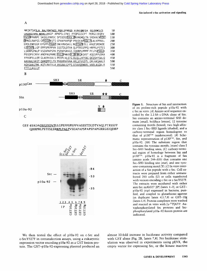
We have described previously the isolation of novel pro-line-rich sequences from a mouse embryonic library that bind to SH3 domains with different specificities (Alex-andropoulos et al. 1995). One such clone (10a) bound specifically to Src-SH3 with high affinity using an RPLPALP motif. This binding specificity for Src-SH3 was consistent with results from phage display (Rickles et al. 1994) or selection from chemically synthesized libraries (Yu et al. 1994). A 2.2-kb c-DNA clone was isolated from a mouse embryonic cDNA library using a 5' fragment of clone 10a as a probe. This clone encodes a predicted 560-amino-acid protein we have called Sin (for Src interacting or signal integrating protein) (Fig. lA). This clone shares regions of homology with the recently isolated pl30*^^^ (Fig. IB), a bigger protein that is highly phosphorylated in v-Src and v-Crk transformed cells (Sakai et al. 1994). Sin and pl30^^^ contain 84% homologous (66% identical) amino-terminal SH3 domains, and their carboxyl termini are 57% homologous (42% identical) over a stretch of 134 amino acids. In addition, both proteins have a central substrate region (Sakai et al. 1994) that contains repeated tyrosine motifs, although the number and primary sequences of these motifs differ between the two proteins (Table 1). The overall architecture of mouse Sin and rat pl30'^''* are similar, but the
DNA sequences encoding these proteins are not homologous outside of the regions coding for the amino-terminal SH3s and the carboxyl termini; we therefore believe that these proteins are possibly related members of a novel family of proteins, rather than homologs from different species. The molecular cloning and definition of the expression patterns of Sin were recently discovered independently (Ishino et al. 1995).
Regulation of c-Src by a Sin peptide
Because Sin contains two high-affinity class I Src-SFi3-binding sites (Alexandropoulos et al. 1995; Fig. lA), we believed that it might be involved in the regulation of c-Src. For instance, its binding could interfere with the SH3-mediated intramolecular control of c-Src and activate signaling by the c-Src kinase domain. To test such possibilities, we have examined the interaction of portions of Sin with Src-SH3. Initially, we used a 92-amino-acid fragment of Sin (plOa-92) (sequence in Fig. IC). Extracts of 293 cells overexpressing c-Src were incubated with purified glutathionine S-transferase (GST)-pl0a-92 fusion protein coupled to glutathione beads, or with anti-Src antibody, and the captured proteins were revealed by in vitro kinase assays. The antibody bound no detectable protein from control cells but recovered Src as a phosphorylated band from cell lysates overexpressing c-Src (Fig. IC, lanes 1,2). The pIOa-92 peptide also bound to c-Src and served as a very efficient substrate for the c-Src kinase (Fig. IC, lanes 4,5). In contrast, a peptide that specifically binds to Abl-SH3 (clone lOg; Alexandropoulos et al. 1995) did not precipitate c-Src (Fig. IC, lane 6). As seen for c-Src, plOa-92 was bound to and phosphorylated by the constitutively activated c-SrcY527Y mutant (Fig. IC, lanes 3,7-9). These data suggested that the Src-SH3-binding sequence present on plOa-92 could bind to Src-SH3 in solution and serve as a substrate for c-Src.
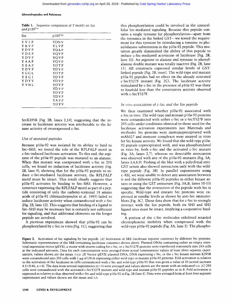
To test the ability of the plOa-92 peptide to modulate c-Src signaling in cells, we used a transient expression protocol with retroviral vectors expressing c-Src or c-SrcY527F (Kmiecik and Shalloway 1987). Signaling by the overexpressed proteins was assessed by activation of a cotransfected reporter luciferase construct containing four serum response elements that regulate the expression of the primary response transcription factor Egr-1 (Fig. 2A) (Tsai-Morris et al. 1988). These elements were shown previously to mediate activation of Egr-1 transcription in response to v-Src (Qureshi et al. 1991). Over-expression of c-Src had a barely detectable effect on luciferase activation as expected, attributable to the intramolecular negative regulation of the kinase by its phosphorylated carboxy-terminal tail (Superti et al. 1993) (Fig. 2A). In contrast, the transforming derivative c-SrcY527F stimulated luciferase activity in proportion to the amount of DNA transfected into 293 cells (Fig. 2A). Neither the empty vector (pEVX; Fig. 2A) nor a luciferase construct lacking the SRE-containing promoter displayed stimulated luciferase activity (data not shown). The various Src proteins were expressed at similar levels (data not shown).
1342 GENES & DEVELOPMENT
Cold Spring Harbor Laboratory Press on April 28, 2018 - Published by genesdev.cshlp.orgDownloaded from
Sin-induced c-Src activation and signaling
MAIATSAQLA RALYDNTAES PQELSFRRGD VLRVLQREGA GGLDGWCLCS 50 LHGOQGIVPA NRVKLLPAGP APKPSLCPAS PTQPGSSCPT PERGCEEQEV 100 fYVm^PARPr SASGLPARSC SPSSDSlg^LEt^VNGMQLTA SKDVAEVEDS] 150 E^NILRAPSS CFIYDS^SFS CPVAPVVPQP PREDEAPED5LB.ALKPPAEL 200 ERDLEWEGGR EPGPPLJYAAPI SNLKRASALL NLjYEAPfeELL ANGESRDADE 250 GJYDVPLLGP EPPSPEPPVA SSSTDLDTVA QLPTRSSPPQ HRPRLPSTES 300 LSRRPLPALP VSEAPAPSPA PSPAPGRKGS IQDRPLPPPP PCLPGEnniK 350 PEGDPECREV ANDPAGPHNE lYEGIJ^MAEdY DIYVHLKGVDT AQGSRPLDKA 400 FPVDPELLER GLAERKEALS PEEPLVLSTG DLOLLHFYAG OCOSHYSALO 450 AAVAALVAST OANQPPCLFV PHGKRVVVAA HRLVFVGDTL GRLAASAALR 500 AQVGAAGTML AOTLRATVLA VKGAALGYPS DTAVOEMARC VAELAGOALR 550 FTTLLAGLLP 560 B
SH3 SR pl30Cas
SH3 SR Sin
itif
plOa-92
C
Y *
GST-ESRDADEGIYDVPLLGPEPPSPEPPVASSSTDLDTVAQLPTRSSPP OHRPRLPSTESLSRRPLPALPVSEAPAPSPAPSPAPGRKGSIQDRP
^ ^
Src
plOa-92
1 2 3 4 5 6 7 8 9
IP C4 bO Cti DO
o o o o \r* h* h^ b^ C/3 C/3 00 on O O O O
84
53
-35
Figure 1. Structure of Sin and interaction of its proline-rich peptide plOa-92 with c-Src in vitro. [A] Amino acid sequence encoded by the 2.2-kb c-DNA clone of Sin. Sin contains an amino-terminal SH3 domain (small, boldface letters), 12 tyrosine containing motifs (boxed), two high-affinity class I Src-SH3 ligands (shaded), and a carboxy-terminal region homologous to that of pl30^^" (underscored). [B] Schematic representation of pl30^^^ Sin, and plOa-92. (SR) The substrate region that contains the tyrosine motifs; (stars) class I Src-SH3 binding sites; (C) carboxy-terminal region of homology between Sin and pl30'^^'. plOa-92 is a fragment of Sin (amino acids 244-335) that contains one Src-SH3 binding site (star), and one tyro-sine-containing motif (Y). (C) In vitro interaction of a Sin peptide with c-Src. Cell extracts were prepared from either untrans-fected 293 cells (U) or cells transfected with vectors encoding c-Src or c-SrcY527F. The extracts were incubated with either anti-Src mAb327 (IP) (lanes 1-3), or GST-plOa-92 [top] expressed in bacteria, purified, and coupled to glutathione-agarose (in duplicate lanes 4,5,7,8] or GST-lOg (lanes 6,9). Protein complexes were washed and reacted in vitro with l7-- ^P]ATP. Au-tophosphorylated Src proteins and Src-phosphorylated plOa-92 fusion protein are indicated.
We then tested the effect of plOa-92 on c-Src and c-SrcY527F in cotransfection assays, using a eukaryotic expression vector encoding plOa-92 as a GST fusion protein. The GST-plOa-92-expressing plasmid produced an
almost 10-fold increase in luciferase activity compared with GST alone (Fig. 2B, lanes 7,8). No luciferase stimulation v^as observed in experiments using pEVX, the empty vector for expressing Src, or the kinase inactive
GENES & DEVELOPMENT 1343
Cold Spring Harbor Laboratory Press on April 28, 2018 - Published by genesdev.cshlp.orgDownloaded from
Alexandtopoulos and Baltimore
Table 1. Sequence comparison of Y motifs on Sin andpiaO^"'
Sin pl30^
Y V I P Y KVP YDV P YDS P YDVP Y AAP Y E AP YDVP YGGL Y EG I YDYV Y VHL
YDNV YLVP YQAP YQVP YQVP YQVP YDTP YDVP YDTP YDVP YDVP YDVP YDVP YDVP YDVP YAVP YDYV
SrcK295R (Fig. 2B, lanes 2,14), suggesting that the increase in luciferase activity was attributable to the kinase activity of overexpressed c-Src.
Use of mutated peptides
Because plOa-92 was isolated by its ability to bind to Src-SH3, we tested the role of the RPLPALP motif in c-Src-induced luciferase activation. To this end, the argi-nine of the plOa-92 peptide was mutated to an alanine. When this mutant was coexpressed with c-Src in 293 cells, we found no induction of luciferase activity (Fig. 2B, lane 9), showing that for the plOa-92 peptide to induce c-Src-mediated luciferase activity, the RPLPALP motif must be intact. This result clearly suggests that plOa-92 activates by binding to Src-SH3. However, a construct expressing the RPLPALP motif as part of a peptide consisting of only the carboxy-terminal 18 amino acids of plOa-92 (Alexandropoulos et al. 1995) failed to induce luciferase activity when cotransfected with c-Src (Fig. 2B, lane 12). This suggests that binding of a ligand to Src-SH3 may be necessary but is certainly not sufficient for signaling, and that additional elements on the longer peptide are involved.
A previous experiment showed that pIOa-92 can be phosphorylated by c-Src in vitro (Fig. IC), suggesting that
this phosphorylation could be involved in the intracellular Src-mediated signaling. Because this peptide contains a single tyrosine for phosphorylation—apart from the tyrosines in the linked GST—we tested the requirement for this tyrosine by introducing a tyrosine to phenylalanine substitution in the plOa-92 peptide. This mutation greatly diminished the ability of this peptide to induce c-Src-mediated activation of luciferase (Fig. 2B, lane 10). An arginine to alanine and tyrosine to phenylalanine double mutant was totally inactive (Fig. 2B, lane 11). All constructs expressed similar levels of GST-linked peptide (Fig. 2B, inset). The wild-type and mutant plOa-92 peptides had no effect on the already activated c-SrcY527F mutant (Fig. 2C). The luciferase activity stimulated by c-Src in the presence of plOa-92 was three to fourfold less than the constitutive activity observed with c-SrcY527F.
In vivo association of c-Sic and the Sin peptide
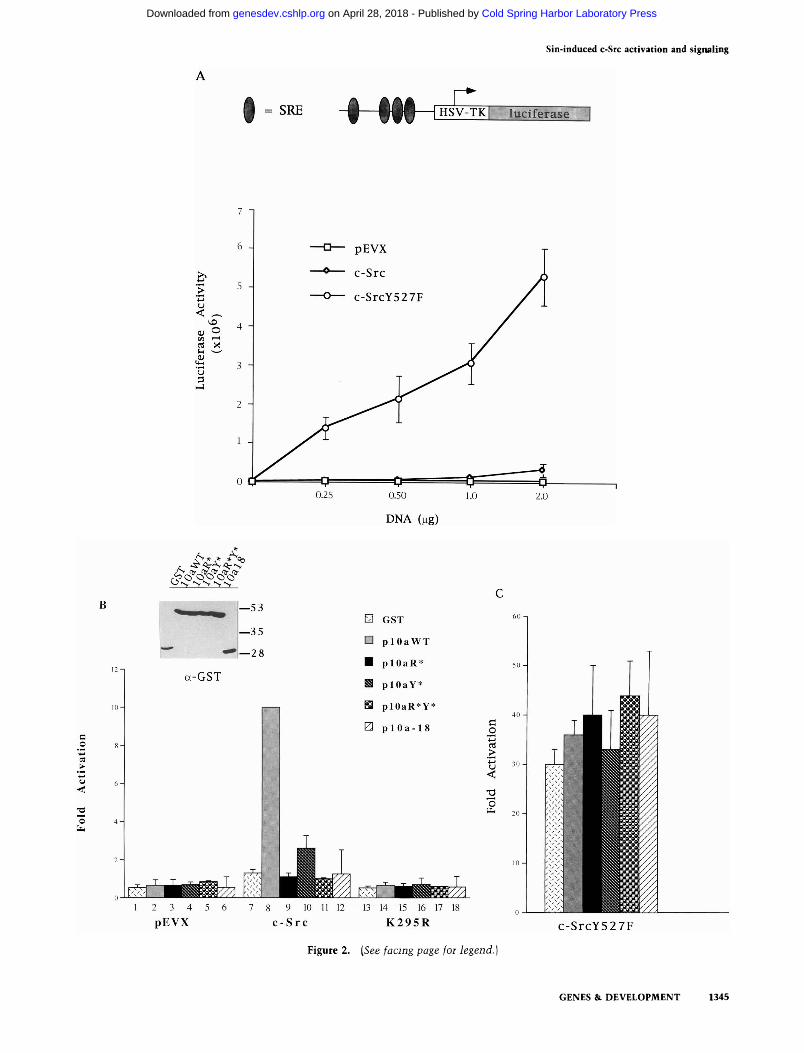
We then examined whether plOa-92 associated with c-Src in vivo. The wild-type and mutant plOa-92 proteins were cotransfected with either c-Src or c-SrcY527F into 293 cells under conditions identical to those used for the luciferase activation experiments (see Materials and methods). Src proteins were immunoprecipitated with mAb327 and immune complexes were assayed in vitro for Src kinase activity. We found that the wild-type plOa-92 peptide coprecipitated with, and was phosphorylated in vitro by, both c-Src and the activated c-Src mutant (Fig. 3A, lanes 2,7), whereas no detectable association was observed with any of the plOa-92 mutants (Fig. 3A, lanes 3,4,8,9). Probing of the blot with a polyclonal anti-GST serum also showed interaction only with the wild-type peptide (Fig. 3B). In parallel experiments using c-Abl, we were unable to detect any association between it and the different plOa-92 peptides in either kinase assays or using the GST antiserum (Fig. 3A,B; lanes 10-15) suggesting that the interaction of the peptide with Src is specific. Wild-type and mutant Src proteins were expressed at similar levels as shown by anti-Src immuno-blots (Fig. 3C). These data show that for c-Src to strongly interact with the Sin peptide, both its SH3 and SH2 ligand sites must be intact, implying a cooperative binding.
A portion of the c-Src molecules exhibited retarded electrophoretic mobility when coexpressed with the wild-type plOa-92 peptide (Fig. 3A, lane 2). The phospho-
Figure 2. Activation of Src signaling by Sin peptide. [A] Activation of SRE-luciferase reporter construct by different Src proteins. Schematic representation of the SRE-containing luciferase construct shown above. Plasmid DNAs containing either an empty retroviral expression vector (pEVX), a vector with inserts coding for c-Src, or c-SrcY527F proteins were transfected transiently into 293 cells at the indicated amounts. Data for each concentration were averaged from actual luminometer values of least three separate experiments; values shown are the mean ±S.D. [B] Vector (pEVX) plasmid DNA, DNA expressing c-Src, or the c-Src kinase mutant K295R were cotransfected into 293 cells with 1 |xg of DNA expressing either wild-type or mutant plOa-92 proteins. Fold activation is relative to the activation of the luciferase in cells cotransfected with c-Src and wild-type plOa-92 that was given a value of 10 (actual increase 8.5±0.86, n = 6). The data from several experiments [n>5) were averaged and values shown are the mean with an indicated s.D. (C) 293 cells were cotransfected with the activated c-SrcY527F mutant and wild-type and mutant plOa-92 peptides as in B. Fold activation is expressed as relative to that observed with c-Src and wild-type piOa-92 in Fig. 2B (lane 8). Data were averaged from at least four separate experiments and values shown are the mean and s.D.
1344 GENES &. DEVELOPMENT
Cold Spring Harbor Laboratory Press on April 28, 2018 - Published by genesdev.cshlp.orgDownloaded from
Sin-induced c-Src activation and signaling
• = SRE
r: HSV-TKI Ittciferase
u w (4-1
'u
O-S NN' N'N
—53
—35
'—28
a-GST
Q GST
□ p l O a W T
■ p lOaR ' '
B p lOaY*
H plOaR*Y*
0 p l O a - 1 8
9 10 c -Sr c
13 14 15 16 17 18 K295R
4-1
01
<
o
c-SrcY527F
Figure 2. [See facing page for legend.]
GENES & DEVELOPMENT 1345
Cold Spring Harbor Laboratory Press on April 28, 2018 - Published by genesdev.cshlp.orgDownloaded from
Alexandropoulos and Baltimore
c-Src SrcY527F c-Abl
A, * * A * * /N * * / s (> (fr '6- / v (V '0' ttr c ^ fir ^ ^ ■h Of O 0 \N , <h 0 \ 0 O ^ - r<' O O O O'
Figure 3. Interaction of plOa-92-derived peptides with c-Src in vivo. (A) Cell extracts were prepared from cells overexpressing c-Src, activated c-SrcY527F, or c-Abl (-) and wild-type or mutant plOa-92 peptides, as shown. The transfection conditions used were identical to those used for Fig. 2B. Extracts were immunoprecipitated with Src mAb327 or Abl mAb-3 and immune complexes were incubated for Src kinase activity in vitro. Phosphorylated proteins were separated on SDS-PAGE and transferred to nitrocellulose. Autora-diographs were obtained by exposing the blots for 1 hr at room temperature. Results are representative of several independent experiments. [B] The blots from 3A were immunoblotted with a rabbit polyclonal antiserum to GST, and visualized by enhanced chemiluminescent detection jECL, Am-ersham). Exposures were performed for 15 sec and no background exposure from the radioactive proteins was observed under these conditions. (C) Al-iquots of total cell extracts from A, containing overexpressed c-Src of c-SrcY527F, were normalized for protein content and immunoblotted with anti-Src mAb327.
p l O a - 9 2 - —
— 1 9 9 — 1 2 0
— 87
— 4 8
B 1 2 3 4 5 6 7 8 9 1 0 1 1 1 2 1 3 1415
p l O a - 9 2 -
a-GST
S r c -
1 2 3 4 5 6 7 8 9 a-Src
rylation pattern of the constitutively active c-SrcY527F mutant showed both the retarded and unretarded forms, irrespective of the presence or absence of the different peptides (Fig. 3A, lanes 6-9). c-Src hyperphosphorylation and a concommitant mobility shift of a small percentage of Src molecules has been observed in other situations of c-Src activation, such as during mitosis (Kypta et al. 1990; Bagrodia et al. 1991; Taylor and Shalloway 1994).
Lack of stable association of the mutant peptides v^ith c-Src correlates with the inability of these proteins to induce signaling through c-Src (Fig. 2B). It appears that both the Src-SH3 ligand present on the plOa-92 peptide and its DEGIYDVP tyrosine are required for activation and stable in vivo association of this peptide with c-Src.
In vitro activation of c-Src
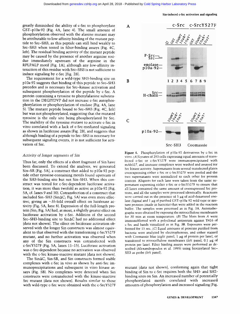
To show further that plOa-92 can directly activate c-Src, we assayed the effect of purified^ bacterially expressed, GST-plOa-92 fusion proteins on c-Src autophosphoryla-tion in vitro. Extracts from transfected 293 cells overexpressing c-Src and c-SrcY527F, were immunoprecipitated with Src mAb327. When immune complexes containing c-Src were incubated with purified GST-plOa-92, a plOa-92-dependent increase of c-Src autophosphoryla-tion was observed, which also caused a partial decrease in Src mobility (Fig. 4A, lane 3). In contrast, no change of
mobility occurred with the c-SrcY527F mutant (Fig. 4A, lanes 6-9). Autophosphorylation of c-SrcY527F was lower than that observed with c-Src. This difference may have been attributable to a lower substrate concentration because tyrosine 416 was already phosphorylated in the mutant protein. To examine this question, the blots were reprobed with a polyclonal antiserum that specifically recognizes the phosphorylated form of SrcY416 but not the nonphosphorylated Y4I6 or other phosphorylated tyrosines (Fig. 4B) (Mukhopadhyay et al. 1995; S. Iyer, R. Russel, K. Flynn, D. Shalloway, and A. Laudano, in prep.). Although the in vitro phosphorylation levels of c-SrcY527F were lower than those of c-Src (Fig. 4A, lanes 2,6), tyrosine 416 phosphorylation was higher in c-SrcY527F (Fig. 4B, lanes 2,6). Furthermore, the increased autophosphorylation activity of c-Src induced by plOa-92 correlated with an increased Y416 level (Fig. 4B, lane 3), showing that the peptide markedly increases the autophosphorylation activity of c-Src.
Src did phosphorylate the plOa-92 peptide when it was presented in vitro (Fig. 4A, lane 3). Enolase, a classic Src substrate, did not show any increased phosphorylation when the peptide was present perhaps because the peptide blocked access to the c-Src catalytic site. In fact, c-SrcY527F phosphorylated enolase less efficiently when the peptide was present (Fig. 4A, cf. lanes 6 and 7). An arginine to alanine substitution in the RPLPALP motif
1346 GENES & DEVELOPMENT
Cold Spring Harbor Laboratory Press on April 28, 2018 - Published by genesdev.cshlp.orgDownloaded from
Sin-induced C-STC activation and signaling
greatly diminished the abiUty of c-Src to phosphorylate GST-plOa-92 (Fig. 4A, lane 4). The small amount of phosphorylation observed with the alanine mutant may be attributable to low-affinity binding of the mutant peptide to Src-SH3, as this peptide can still bind weakly to Src-SH3 when tested in filter-binding assays (Fig. 4C, left). The residual binding activity of the mutant peptide may be caused by the presence of another arginine residue immediately upstream of the arginine in the RPLPALP motif (Fig. lA), although any low-affinity interaction of this residue with Src-SH3 is not sufficient to induce signaling by c-Src (Fig. 2B).
The requirement for a wild-type SH3-binding site on plOa-92 suggests that binding of this peptide to Src-SH3 precedes and is necessary for Src-kinase activation and subsequent phosphorylation of the peptide by c-Src. A protein containing a tyrosine to phenylalanine substitution in the DEGIYDVP did not increase c-Src autophos-phorylation or phosphorylation of enolase (Fig. 4A, lane 5). The mutant peptide bound to Src-SF13 (Fig. 4C, left) but was not phosphorylated, suggesting that the mutated tyrosine is the only site being phosphorylated by Src. The inability of the tyrosine mutant to activate c-Src in vitro correlated with a lack of c-Src-mediated signaling as shown in luciferase assays (Fig. 2B), and suggests that although binding of a peptide to Src-SH3 is necessary for subsequent signaling events, it is not sufficient for activation of Src.
Activity of longer segments of Sin
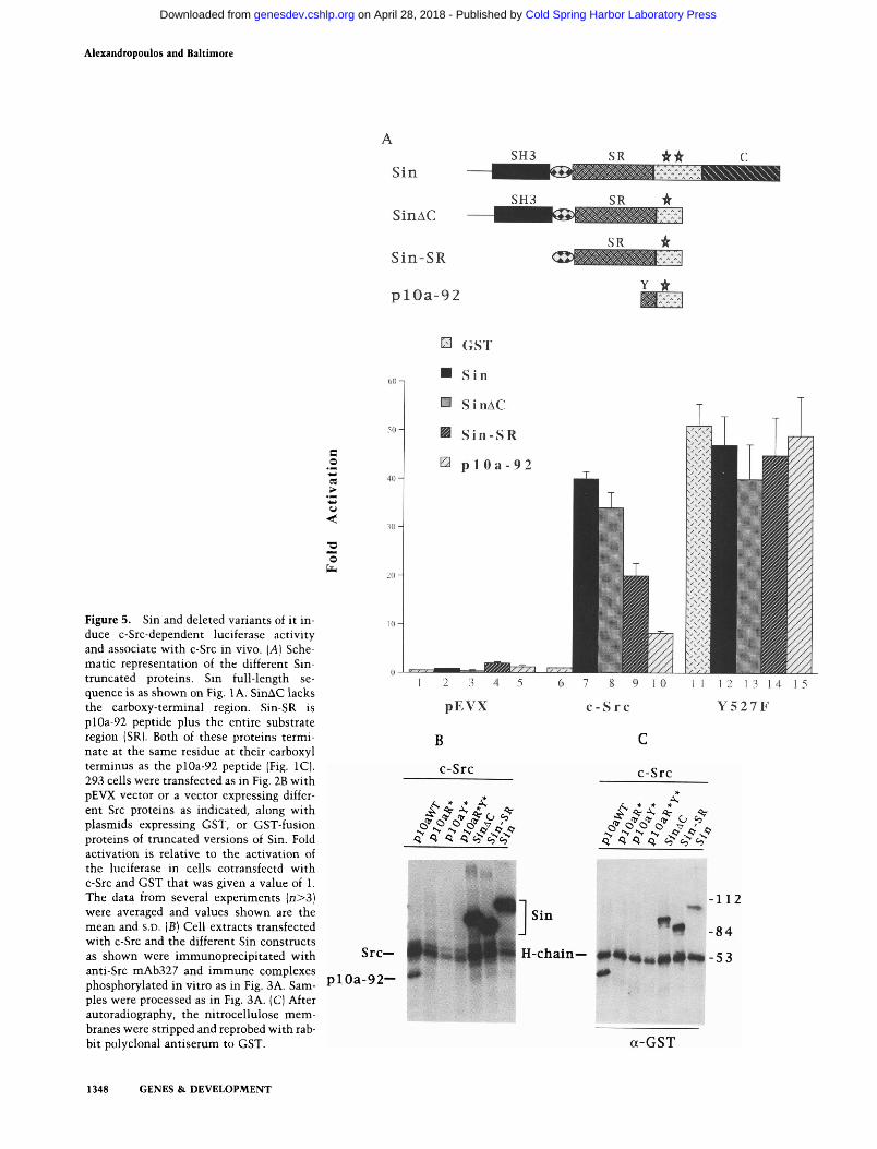
Thus far, only the effects of a short fragment of Sin have been discussed. To extend the analysis, we generated Sin-SR (Fig. 5A), a construct that added to plOa-92 peptide other tyrosine-containing motifs found upstream of the SH3-binding site but not Sin-SH3. When this construct was tested for c-Src-dependent luciferase activation, it was more than twofold as active as plOa-92 (Fig. 5A, cf. lanes 9 and 10). Cotransfection of a construct that included Sin-SH3 (SinAC; Fig. 5A) was even more effective, giving an —35-fold overall effect on luciferase activity (Fig. 5A, lane 8). Expression of the full-length protein (Sin; Fig. 5A) had, at most, a slightly greater effect on luciferase activation by c-Src. Addition of the second Src-SFi3-binding site to SinAC had no additional effect (data not shown). The effect on luciferase activation observed with the longer Sin constructs was almost equivalent to that observed with the transforming c-SrcY527F mutant, and no further activation was observed when any of the Sin constructs was cotransfected with c-SrcY527F (Fig. 5A, lanes 11-15). Luciferase activation was c-Src-dependent because no activation was observed with the c-Src kinase-inactive mutant (data not shown).
The SinAC, Sin-SR, and Sin constructs formed stable complexes with c-Src in vivo as shown by anti-Src im-munoprecipitations and subsequent in vitro kinase assays (Fig. 5B). No complexes were detected when Sin constructs were cotransfected with the kinase-inactive Src mutant (data not shown). Results similar to those with wild-type c-Src were obtained with the c-SrcY527F
C-Src c -SrcY527F
<? O N N N <S "vN 's
enolase-plOa-92-
B
• » j
1 2 3 4 5 6 7 8 9
, Src— H-chain—
/«v * *
N N -v
/v * *
V N' «v
plOa-92-
Src-SH3 Coomass ie
Figure 4. Phosphorylation of plOa-92 derivatives by c-Src in vitro. [A] Lysates of 293 cells expressing equal amounts of trans-fected c-Src or c-SrcY527F were immunoprecipitated with mAb327, and immune complexes were washed and assayed for Src kmase activity. Supernatants from several transfected plates overexpressing either c-Src or c-SrcY527F were pooled and the two supernatants were normalized to each other for protein content. Aliquots for each lane were taken from the same supernatant expressing either c-Src or c-SrcY527F to ensure that all lanes contained the same amount of overexpressed Src proteins, and all the samples were processed identically. Reactions were carried out in the presence of 5 |jLg of acid-denatured enolase (Sigma) and 1 |xg of purified GST-plOa-92 wild-type or mutant proteins (made in bacteria) that were added in the reaction buffer. The samples were processed as in Fig. 3A. Autoradio-graphs were obtained by exposing the nitrocellulose membranes for 30 min at room temperature. [B] The blots from A were immunoblotted with a polyclonal antiserum against Y416 of c-Src and bands visualized as in Fig. 3B. Exposures were performed for 15 sec. [C] Equal amounts of proteins purified from bacteria were analyzed by electrophoresis, and either stained with Coomassie blue [right panel, 1 jig of protein per lane), or transferred to nitrocellulose membranes [left panel, 0.1 M-g of protein per lane). Filter binding assays were performed as described (Alexandropoulos et al. 1995) using biotinylated Src-SH3 as probe {left panel).
mutant (data not shown), confirming again that tight binding of Sin to c-Src requires both the SH3- and SH2-binding sites on Sin. An increased number of potentially phosphorylated motifs correlated with increased amounts of phosphorylation and increased signaling (Fig.
GENES & DEVELOPMENT 1347
Cold Spring Harbor Laboratory Press on April 28, 2018 - Published by genesdev.cshlp.orgDownloaded from
Alexandropoulos and Baltimore
Sin
SinAC
Sin-SR
p l O a - 9 2
S H 3 SR ifir
S H 3 SR it
SR
Y •
Figure 5. Sin and deleted variants of it induce c-Src-dependent luciferase activity and associate with c-Src in vivo. [A] Schematic representation of the different Sin-truncated proteins. Sin full-length sequence is as shown on Fig. 1 A. SinAC lacks the carboxy-terminal region. Sin-SR is plOa-92 peptide plus the entire substrate region (SR). Both of these proteins terminate at the same residue at their carboxyl terminus as the plOa-92 peptide (Fig. IC). 293 cells were transfected as in Fig. 2B with pEVX vector or a vector expressing different Src proteins as indicated, along with plasmids expressing GST, or GST-fusion proteins of truncated versions of Sin. Fold activation is relative to the activation of the luciferase in cells cotransfectd with c-Src and GST that was given a value of 1. The data from several experiments (n>3) were averaged and values shown are the mean and s.D. [B] Cell extracts transfected with c-Src and the different Sin constructs as shown were immunoprecipitated with anti-Src mAb327 and immune complexes phosphorylated in vitro as in Fig. 3A. Samples were processed as in Fig. 3A. (C) After autoradiography, the nitrocellulose membranes were stripped and reprobed with rabbit polyclonal antiserum to GST.
C
■*-i (J
a ii4
2 3 4 5 6 7
pEVX c-
B
c-Src
.* ; ^ ^ ^^<J ^
8 9
S r c
10 11 12 1 3 1 4 15
Y 5 2 7 F
C
c-Src *
A, i * » *
t^ ilr tr tr 0 ^ 3 < >- N' N ^ >5'-4^
< <i <i <i c^ 'c^e^
S r c -
p l O a - 9 2 -
- 1 1 2
-84
53
a-GST
1348 GENES & DEVELOPMENT
Cold Spring Harbor Laboratory Press on April 28, 2018 - Published by genesdev.cshlp.orgDownloaded from
Sin-induced c-Src activation and signaling
5A;B) under conditions where all Sin proteins were bound at similar levels (Fig. 5C). The results obtained with these proteins on c-Src-induced luciferase activation could be attributable to GST-induced dimerization of the Sin proteins that might have affected the results. To examine the possibility, we expressed one of the proteins, SinAC, from the same expession vector as GST-SinAC but without the GST. This protein activated c-Src to the same extent as the GST form when tested in luciferase assays and coprecipitated with, and was phos-phorylated by, c-Src in kinase assays (data not shown). Therefore, the results shown in Figure 5 do not appear to result from GST dimerization.
Binding of Crk and other phosphoproteins to Sin
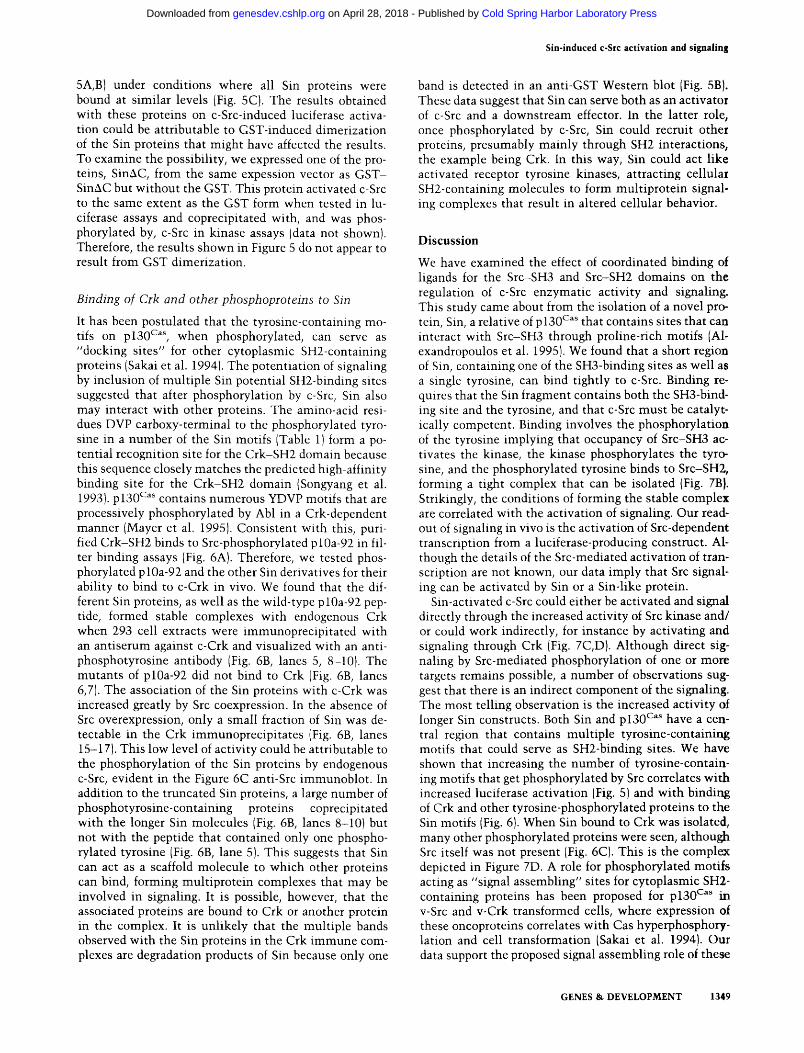
It has been postulated that the tyrosine-containing motifs on plSO* '' when phosphorylated, can serve as "docking sites" for other cytoplasmic SH2-containing proteins (Sakai et al. 1994). The potentiation of signaling by inclusion of multiple Sin potential SH2-binding sites suggested that after phosphorylation by c-Src, Sin also may interact with other proteins. The amino-acid residues DVP carboxy-terminal to the phosphorylated tyrosine in a number of the Sin motifs (Table 1) form a potential recognition site for the Crk-SH2 domain because this sequence closely matches the predicted high-affinity binding site for the Crk-SH2 domain (Songyang et al. 1993). pl30*^^^ contains numerous YDVP motifs that are processively phosphorylated by Abl in a Crk-dependent manner (Mayer et al. 1995). Consistent with this, purified Crk-SH2 binds to Src-phosphorylated plOa-92 in filter binding assays (Fig. 6A). Therefore, we tested phosphorylated plOa-92 and the other Sin derivatives for their ability to bind to c-Crk in vivo. We found that the different Sin proteins, as well as the wild-type plOa-92 peptide, formed stable complexes with endogenous Crk when 293 cell extracts were immunoprecipitated with an antiserum against c-Crk and visualized with an anti-phosphotyrosine antibody (Fig. 6B, lanes 5, 8-10). The mutants of plOa-92 did not bind to Crk (Fig. 6B, lanes 6,7). The association of the Sin proteins with c-Crk was increased greatly by Src coexpression. In the absence of Src overexpression, only a small fraction of Sin was detectable in the Crk immunoprecipitates (Fig. 6B, lanes 15-17). This low level of activity could be attributable to the phosphorylation of the Sin proteins by endogenous c-Src, evident in the Figure 6C anti-Src immunoblot. In addition to the truncated Sin proteins, a large number of phosphotyrosine-containing proteins coprecipitated with the longer Sin molecules (Fig. 6B, lanes 8-10) but not with the peptide that contained only one phosphorylated tyrosine (Fig. 6B, lane 5). This suggests that Sin can act as a scaffold molecule to which other proteins can bind, forming multiprotein complexes that may be involved in signaling. It is possible, however, that the associated proteins are bound to Crk or another protein in the complex. It is unlikely that the multiple bands observed with the Sin proteins in the Crk immune complexes are degradation products of Sin because only one
band is detected in an anti-GST Western blot (Fig. 5B). These data suggest that Sin can serve both as an activator of c-Src and a downstream effector. In the latter role, once phosphorylated by c-Src, Sin could recruit other proteins, presumably mainly through SH2 interactions, the example being Crk. In this way. Sin could act like activated receptor tyrosine kinases, attracting cellular SH2-containing molecules to form multiprotein signaling complexes that result in altered cellular behavior.
Discussion We have examined the effect of coordinated binding of ligands for the Src-SH3 and Src-SH2 domains on the regulation of c-Src enzymatic activity and signaling. This study came about from the isolation of a novel protein. Sin, a relative of p 130' ''* that contains sites that can interact with Src-SH3 through proline-rich motifs (Al-exandropoulos et al. 1995). We found that a short region of Sin, containing one of the SH3-binding sites as well as a single tyrosine, can bind tightly to c-Src. Binding requires that the Sin fragment contains both the SH3-bind-ing site and the tyrosine, and that c-Src must be catalyt-ically competent. Binding involves the phosphorylation of the tyrosine implying that occupancy of Src-SH3 activates the kinase, the kinase phosphorylates the tyrosine, and the phosphorylated tyrosine binds to Src-SH2, forming a tight complex that can be isolated (Fig. 7B). Strikingly, the conditions of forming the stable complex are correlated with the activation of signaling. Our readout of signaling in vivo is the activation of Src-dependent transcription from a luciferase-producing construct. Although the details of the Src-mediated activation of transcription are not known, our data imply that Src signaling can be activated by Sin or a Sin-like protein.
Sin-activated c-Src could either be activated and signal directly through the increased activity of Src kinase and/ or could work indirectly, for instance by activating and signaling through Crk (Fig. 7C,D). Although direct signaling by Src-mediated phosphorylation of one or more targets remains possible, a number of observations suggest that there is an indirect component of the signaling. The most telling observation is the increased activity of longer Sin constructs. Both Sin and pi30* ''* have a central region that contains multiple tyrosine-containing motifs that could serve as SH2-binding sites. We have shown that increasing the number of tyrosine-containing motifs that get phosphorylated by Src correlates with increased luciferase activation (Fig. 5) and with binding of Crk and other tyrosine-phosphorylated proteins to the Sin motifs (Fig. 6). When Sin bound to Crk was isolated, many other phosphorylated proteins were seen, although Src itself was not present (Fig. 6C). This is the complex depicted in Figure 7D. A role for phosphorylated motifs acting as "signal assembling" sites for cytoplasmic SH2-containing proteins has been proposed for pl30^^^ in v-Src and v-Crk transformed cells, where expression of these oncoproteins correlates with Cas hyperphosphory-lation and cell transformation (Sakai et al. 1994). Our data support the proposed signal assembling role of these
GENES & DEVELOPMENT 1349
Cold Spring Harbor Laboratory Press on April 28, 2018 - Published by genesdev.cshlp.orgDownloaded from
Alexandropoulos and Baltimore
Figure 6. Association of phosphorylated Sin truncated proteins with c-Crk in vivo. [A] Total cell lysates of 293 cells overexpressing c-Src in the presence or absence of plOa-92 were frac-tioned by SDS-PAGE and transferred to nitrocellulose. Membranes were probed with anti-pY monoclonal antiserum 4G10 or biotinylated GST-Crk-SH2 and GST as indicated. [B] Cell extracts overexpressing plOa-92 wild-type and mutant peptides and the Sin truncated proteins described in Fig. 5 in the presence or absence of overexpressed c-Src as indicated, were immuno-precipitated with anti-Crk monoclonal antibody. Proteins were separated by SDS-PAGE, transferred to nitrocellulose, and immunobloted with anti-pY monoclonal antibody. (Lanes 1,2] West-em blots of untreated (U) 293 cell extracts and lysates overexpressing c-Src probed with Src mAb 327; (lane 3) Western blot probed with anti-Crk antiserum, of lysates of untreated cells immuno-precipitated with anti-Crk antibody. (C) Western blots of total cell extracts from B normalized for protein content were probed with anti-Src mAb 327.
f^ ^ ^ p l O a - 9 2 - + + - +
p l O a - 9 2
6
S r c -H-chain —
p l O a - 9 2 -Crk-
+ c - S r c c-Src
^ f^ * *
o ^ dP^ <; <; cy c^ c^ < > ^ ^ O <t <t < <o <yc,^»^
- 1 1 2
- 8 4
- 5 3
1 2 3 4 5 6 7 8 9 10 1112 13 1 4 1 5 1 6 1 7 1 8
S r c -
motifs because a large number of tyrosine-phosphory-lated cellular proteins coprecipitated with the phosphorylated Sin proteins (Fig. 6), and suggest that normal and neoplastic pathv^ays may share common intermediates.
Signaling specificities of Sin and Cas
The sequence surrounding the proposed signaling tyrosines differ betw^een Cas and Sin (Table 1). The majority of the motifs found on Cas contain amino acids car-boxy-terminal to the tyrosine that are consistent with an Abl- (YXXP) or C rk - (YDXP) SH2 domain preference (Songyang et al. 1993, Mayer et al. 1995), whereas the motifs found on Sin are more diverse. We found that the YDVP motif of plOa-92 interacts with endogenous Crk in vivo, and binds to Crk-SH2 in vitro (Fig. 6). In contrast, incubating cell extracts overexpressing c-Abl and plOa-92 with anti-Abl mAb showed no association of Abl with the plOa-92 peptide (Fig. 3A), data that support
a YDVP/Crk-SH2 interaction. Additional motifs that are unique to Cas and Sin may reflect signaling specifity in a stimulus- or tissue-specific manner by binding to distinct subsets of signaling intermediates. Consistent with this, the expression patterns of Cas and Sin differ; whereas Cas is expressed in many adult tissues (Sakai et al. 1994), Sin expression is low and is restricted to the brain, thymus, and skeletal muscle (data not shown; Ish-ino et al. 1995). In addition, Sin contains a tyrosine motif, YAAP—not present on pi30*^^^—that serves as a high affinity ligand for the Abl kinase (Songyang et al. 1995). It is possible that this motif may mediate Sin-Abl interactions in the thymus where both proteins are expressed more highly. Low expression of Sin in specific tissues and different cell lines may explain why Sin has not been recognized previously as a Src-bound protein. For instance, we have not been able to detect endogenous Sin in 293 cells in our immunoprecipitation assays.
The amino-terminal SH3 domains of Cas and Sin were
1350 GENES & DEVELOPMENT
Cold Spring Harbor Laboratory Press on April 28, 2018 - Published by genesdev.cshlp.orgDownloaded from
Sin-induced c-Src activation and signaling
Other Signaling
Intermediates Signal Output?
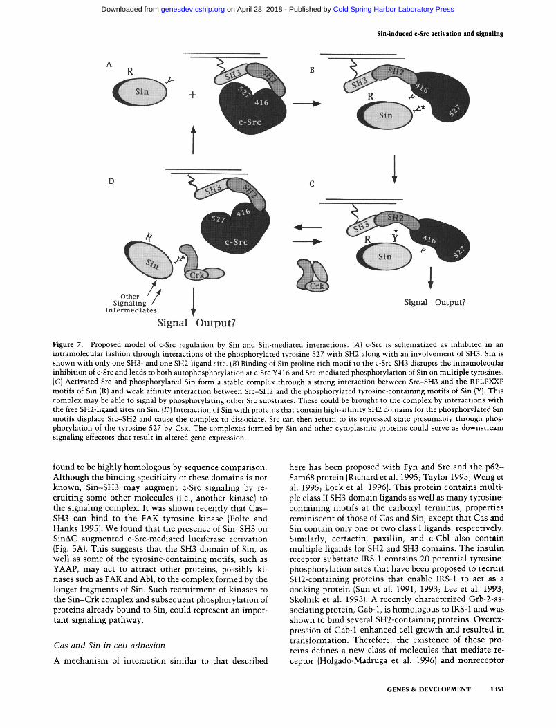
Signal Output? Figure 7. Proposed model of c-Src regulation by Sin and Sin-mediated interactions. [A] c-Src is schematized as inhibited in an intramolecular fashion through interactions of the phosphorylated tyrosine 527 with SH2 along with an involvement of SH3. Sin is shown with only one SH3- and one SH2-ligand site. [B] Binding of Sin proline-rich motif to the c-Src SH3 disrupts the intramolecular inhibition of c-Src and leads to both autophosphorylation at c-Src Y416 and Src-mediated phosphorylation of Sin on multiple tyrosines. (C) Activated Src and phosphorylated Sin form a stable complex through a strong interaction between Src-SH3 and the RPLPXXP motifs of Sin (R) and weak affinity interaction between Src-SH2 and the phosphorylated tyrosine-containmg motifs of Sin (Y). This complex may be able to signal by phosphorylating other Src substrates. These could be brought to the complex by interactions v^th the free SH2-ligand sites on Sin. (D) Interaction of Sin with proteins that contain high-affinity SH2 domains for the phosphorylated Sin motifs displace Src-SH2 and cause the complex to dissociate. Src can then return to its repressed state presumably through phosphorylation of the tyrosine 527 by Csk. The complexes formed by Sin and other cytoplasmic proteins could serve as downstream signaling effectors that result in altered gene expression.
found to be highly homologous by sequence comparison. Although the binding specificity of these domains is not known, Sin-SH3 may augment c-Src signaling by recruiting some other molecules (i.e., another kinase) to the signaling complex. It was shown recently that Cas -SH3 can bind to the FAK tyrosine kinase (Poke and Hanks 1995). We found that the presence of Sin-SH3 on SinAC augmented c-Src-mediated luciferase activation (Fig. 5A). This suggests that the SH3 domain of Sin, as well as some of the tyrosine-containing motifs, such as YAAP, may act to attract other proteins, possibly kinases such as FAK and Abl, to the complex formed by the longer fragments of Sin. Such recruitment of kinases to the Sin-Crk complex and subsequent phosphorylation of proteins already bound to Sin, could represent an important signaling pathway.
Cas and Sin in cell adhesion
A mechanism of interaction similar to that described
here has been proposed with Fyn and Src and the p62-Sam68 protein (Richard et al. 1995; Taylor 1995; Weng et al. 1995; Lock et al. 1996). This protein contains multiple class II SH3-domain ligands as well as many tyrosine-containing motifs at the carboxyl terminus, properties reminiscent of those of Cas and Sin, except that Cas and Sin contain only one or two class I ligands, respectively. Similarly, cortactin, paxillin, and c-Cbl also contain multiple ligands for SH2 and SH3 domains. The insulin receptor substrate IRS-1 contains 20 potential tyrosine-phosphorylation sites that have been proposed to recruit SH2-containing proteins that enable IRS-1 to act as a docking protein (Sun et al. 1991, 1993; Lee et al. 1993; Skolnik et al. 1993). A recently characterized Grb-2-as-sociating protein, Gab-1, is homologous to IRS-1 and was shown to bind several SH2-containing proteins. Overex-pression of Gab-1 enhanced cell growth and resulted in transformation. Therefore, the existence of these proteins defines a new class of molecules that mediate receptor (Holgado-Madruga et al. 1996) and nonreceptor
GENES & DEVELOPMENT 1351
Cold Spring Harbor Laboratory Press on April 28, 2018 - Published by genesdev.cshlp.orgDownloaded from
Alexandiopoulos and Baltimore
tyrosine kinase signaling (this paper) by acting as signal assemblers (this paper; Sakai et al. 1994) or signal integrating sites that form multiprotein complexes by binding to distinct adapter molecules and other cytoplasmic proteins that in turn serve as downstream effectors. The differences in recruiting different combinations of adapter molecules through SH2-mediated interactions, as well as the presence of the different types of SH3 ligands, for example, class I verus class II, may reflect signaling specificity.
Recent reports suggest that plSO* '"'* may be involved in integrin-receptor signaling, because integrin-mediated cell adhesion promotes tyrosine phosphorylation of Cas (Nojima et al. 1995; Vuori and Ruoslahti 1995). It has also been shown that integrin-mediated cell adhesion induces autophosphorylation of FAK and subsequent binding of c-Src to FAK, through interaction of the FAK autophosphorylation site Y-397 and the Src-SH2 domain (Schlaepfer et al. 1994). Therefore, it is likely that Cas and Sin may modulate c-Src activity in response to extracellular stimuli, such as interactions of the extracellular matrix with integrin receptors, where Sin may act as tissue-specific modulator of cell adhesion. Because the Src-SH3 ligand of Sin can interact with the SH3 domains of Fyn, Lyn, and Hck as well as SH3 of neuronal Src (Alexandropoulos et al. 1995), and its expression is restricted to the brain and thymus (K. Alexandropoulos and D. Baltimore, unpubl., Ishino et al. 1995), it is possible that Sin interacts with Src-related kinases in lymphocytes to modulate cell-adhesion events in response to lymphocyte-activating signals, or it may interact with neuronal Src to modulate LI-mediated neuronal outgrowth, because Src has been shown to mediate such processes (Ignelzi et al. 1994).
Cas and FAK have been shown to be phosphorylated in v-Src transformed cells and this phosphorylation is independent of cell adhesion (Schlaepfer et al. 1994; Vuori and Ruoslahti 1995). Because cell transformation by v-Src correlates with increased cellular tyrosine phosphorylation and anchorage-independent growth, Cas and FAK appear to be involved in the process of anchorage dependence. In this report, we show that Sin can induce its own phosphorylation by wild-type c-Src in a Src-SH3-dependent manner. It therefore appears that wild-type and transforming Src molecules can utilize the same signaling intermediates to modulate cellular processes, in which case c-Src activation would be subject to regulation by external stimuli, whereas v-Src, having escaped regulation, would be constitutively active and therefore transforming. Correlation of neoplastic pathways to physiological processes may provide insight into the physiological role of nonreceptor tyrosine kinases that will help develop strategies to block the transforming activities of deregulated protein tyrosine kinases.
Materials and methods
Library screening
A 16-day mouse embryo expression library (Novagen) was screened for SH3-binding proteins according to the manufacter-
er's protocols and as described previously (Cicchetti et al. 1992; Alexandropoulos et al. 1995). Biotinylated GST-SH3 fusion pro-tems of c-Abl, c-Src, c-Crk, and neuronal Src were used as probes to screen the library. Positive phage plaques were purified and recombinant phage were converted into plasmids through Cre-mediated excision from XEXlox by plating phage with the appropriate host. Recombinant plasmids (pEXlox) containing sequences that bound to the Src-SH3 were then transformed into competent bacteria (Novagen) that permitted expression of the cloned cDNAs as fusion proteins of gene 10 of phage T7. A 5' 400-bp fragment of clone 10a (1.4 kb) that was initially isolated (Alexandropoulos et al. 1995), was used to screen the same library for a full-length cDNA clone according to the manufacterer's protocols. The longest cDNA isolated contained a 2.2-kb cDNA piece that encoded for full-length Sin.
Cells and antibodies Human embryonic kidney carcinoma 293 cells (Pear et al. 1993) were grown in Dulbecco's modified Eagle medium (DMEM) containing 10% fetal bovine serum, 100 U/ml of penicillin, and 100 mg/ml of streptomycin. Rabbit polyclonal antibodies to tyrosine 416 of c-Src (Andy Laudano, University of New Hampshire, Durham! and GST, and mouse monoclonal antibodies 327 to Src (Joan Brugge, Ariad Pharmaceuticals, Cambridge, MA), Crk (Pharmigen), Ab-3 to Abl, and mouse monoclonal anti-pY 4G10 (UBII were used in this study.
DNA constructs DNA manipulations were performed by standard protocols. GST- fusion proteins were produced by plasmids based on the pEBG eukaryotic expression vector (Mayer et al. 1995). plOa-92 wild-type and mutant proteins were expressed as GST-fusion proteins by cloning a PCR-amplified fragment of clone 10a (Fig. lAl encoding 92 amino acids into the BamHl-Notl sites of pEBG. Mutated GST-fusion proteins were derived by PCR, using mutated synthetic oligonucleotides, and subsequently cloned in-frame into the BamHl-Notl sites of pEBG. In addition to the NotI restriction site, a translation termination codon was included in the 3' oligonucleotide. The construct expressing full-length Sin as a GST-fusion protein was constructed by PCR amplification of a 1.68-kb fragment (encoding amino acids 1-560) that was subsequently cloned into the Spel-Notl sites of pEBG. Similarly, PCR-amplified DNA fragments expressing GST-SinAC (amino acids 1-335) and GST-Sin-SR (amino acids 68-335) were cloned into the Spel-Notl sites of pEBG. Constructs for prokaryotic expression of wild-type and mutant plOa-92 GST-fusion proteins were prepared by cloning PCR-amplified fragments into the BflmHI-£coRI of GEX2T (Pharmacia). plOa-18 was cloned as described previously (Alexandropoulos et al. 1995). The luciferase reporter construct was constructed by cloning a Hindlll-Bgill 297-bp fragment from pE425-250TKCAT (Qureshi et al. 1991) containing -425/ - 250 upstream regulatory sequences of the Egr-1 promoter and the herpes simplex virus thymidine kinase (HSV-tk) minimal promoter into Hindlll-Bgill-digested pXP-2 luciferase construct (Nordeen, 1988). Plasmids that expressed wild-type Src protein pM5HHB5, psrc527, and psrcR295 have been described (Kmiecik and Shalloway 1987). The construct expressing c-Abl has been described (Ben-Neriah et al. 1986).
Transfections 293 cells (2x10^ per 60mm dish) were plated 16-20 hr before transfection. DNA was transiently introduced into cells by cal-
1352 GENES & DEVELOPMENT
Cold Spring Harbor Laboratory Press on April 28, 2018 - Published by genesdev.cshlp.orgDownloaded from
Sin-induced c-Src activation and signaling
cium phosphate-mediated transfection as described (Pear et al. 1993). Two micrograms of each pM5HHB5(c-Src), pcsrcF527, pcsrcR295, expressing vectors (Kmiecik and Shalloway 1987), or pEVX empty vector were used in all transfections. Transfection mixtures also included 1 (xg of wild-type and mutant plOa-92 constructs and plOa-18 as indicated in the figures. Serum response element (SRE)-luciferase reporter DNA and the MFG-lacZ plasmid expressing p-galactosidase (1 \i.g each) were also included in the transfection mixtures. When necessary, the total amount of DNA in each transfection mixture was kept constant by the addition of empty vector DNA (pEVX) as carrier. Each plate was treated with 0.5 ml of DNA-calcium phosphate coprecipitate. After 18 hr of incubation at 37°C, the medium was replaced and incubation was continued for 24 hr. Cells were lysed and extracts were either assayed for luciferase activity using a Promega kit according to the manufacturer's protocol, or assayed for ^-galactosidase activity to normalize for transfection efficiency between the different samples according to standard protocols (Sambrook et al. 1989). Transfections for cell extracts used in immunoprecipitation and in vitro kinase assays were performed the same way except that cells were lysed in NP-40 lysis buffer.
Immunopiecipitations
Cells were lysed in 1 ml of ice-cold lysis buffer [1% NP-40, 20 mM Tris-HCl (pH 8.0), 150 mM NaCl, 10% glycerol, 10 mM NaF, 1 mM sodium orthovanadate, 1 mM phenylmethylsulfonyl fluoride, 10 jJLg/ml of aprotinin, 10 |xg/ml of leupeptin] and incubated on ice for 10 min. The cell debris and nuclei were removed by centrifugation in an Eppendorf centrifuge for 10 min at 4°C. The cell lysates were then incubated with the specified antibody at concentrations suggested by the manufacturers for 2 hr at 4°C. The immune complexes were collected after the addition of 20 |xl of protein G-plus/protein A (Oncogene Science) agarose and incubation at 4°C for 30 min. The pellets of agarose beads were washed three times with 1 ml of lysis buffer and then subjected to kinase reactions in vitro or immunoblotting.
Kinase reactions
Protein complexes obtained by immunoprecipitation were washed three times in kinase buffer and reactions were carried out in 20 \il of buffer containing 20 mM HEPES at pH 7.4, 5 mM MnClj, 10 JLM ATP, and 1 \x\ of [^-^^PIATP (7000 Ci/mmole) (ICN), at room temperature for 10 min. The pellets were resus-pended in 1 x Laemmli buffer, boiled for 2 min, and phospho-rylated proteins were analyzed by SDS-PAGE and autoradiography.
Western blot analysis
Total cell extracts normalized for protein content or immuno-precipitates were boiled in Laemmli sample buffer, electropho-retically separated on 10% SDS-PAGE, and transferred to nitrocellulose membranes. Filters were blocked in TEST buffer (10 mM Tris-HCl at pH 8.0, 150 mM NaCl, 0.05% Tween 20) plus 2% nonfat dry milk. Filters were blotted with the appropriate monoclonal antiserum at concentrations suggested by the manufacturers in TBST/milk at room temperature for 2 hr. Rabbit polyclonal antibodies to GST and anti-pY416 were used at 1:500 and 1:1000 dilutions, respectively. The filters were washed in TEST, and consequently incubated with anti-mouse IgG-conju-gated horseradish peroxidase at a 1:4000 dilution in TEST at room temperature for 1 hr. Filters were then washed and developed with ECL (Amersham), as described by the manufacturer.
Filter binding assay
Src-SH3 and Crk-SH2 binding to plOa-92 wild type and mutant GST fusion proteins purified from bacteria was performed as described (Alexandropoulos et al. 1995; Mayer et al. 1991). The GST-Src-SH3 and Crk-SH2 biotynilated probes were prepared as described (Cicchetti et al. 1991). GST-fusion plOa-92 and mutant proteins expressed in bacteria were purified using glutathione-agarose beads as described (Mayer et al. 1991). Eacteria cell lysates were prepared as described (Ren et al. 1993). Proteins were fractionated by electrophoresis through polyacrylamide gels and transferred to nitrocellulose filters that were processed as described (Ren et al. 1993).
Solution-binding assays
Cell extracts overexpressing c-Src or c-SrcY527F were prepared as described above and were either immunoprecipitated with Src mAb327, or mixed with 10 fig of GST-plOa-92 or GST-lOg purified, bacterially expressed proteins that had been coupled to glutathione-agarose beads previously by incubating in binding buffer [100 mM EDTA and 1% Triton X in PBS) for 1 hr at 4°C with constant rotation. Incubations of coupled beads with cell extracts were carried out for 2 hr at 4°C, protein complexes were harvested, washed extensively, and subjected to phosphorylation in vitro as described above. Phosphorylated proteins were analyzed by 10% SDS-PAGE and autoradiography of the dried gel for 15 min at room temperature.
Acknowledgments
We are grateful to D. Shalloway for the Src-expressing vectors, M. Curzo and D. Foster for the luciferase reporter construct, }. Brugge for the mAb 327, B. Mayer for the GST-SH2-expressing constructs, A. Laudano for the anti-pY416 antiserum. We thank B. Mayer, I. Stancovski, C. Roman, and Y. Yamanashi for critically reading the manuscript, and C. Roman and S. Cherry for many helpful discussions. K.A. was supported by National Institutes of Health grant 7F32 CA60458-02. This work was supported by National Institutes of Health grant 2R01 CA51462-08 to D.B.
The pubhcation costs of this article were defrayed in part by payment of page charges. This article must therefore be hereby marked "advertisement" in accordance with 18 USC section 1734 solely to indicate this fact.
References
Alexandropoulos, K., G. Cheng, and D. Baltimore. 1995. Pro-line-rich sequences that bind to Src homology 3 domains with individual specificities. Proc. Natl. Acad. Sci. 92:3110-3114.
Anderson, D., C.A. Koch, L. Gray, C. Ellis, M.F. Moran, and T. Pawson. 1990. Binding of SH2 domains of phospholipase C-7I, GAP, and Src to activated growth factor receptors. Science 250: 979-982.
Bagrodia, S., I. Chackalaparampil, T.E. Kmiecik, and D. Shalloway. 1991. Altered tyrosine 527 phosphorylation and mitotic activation of p60c-src. Nature 349: 172-175.
Bagrodia, S., A.P. Laudano, and D. Shalloway. 1994. Accessibility of the c-Src SH2-domain for binding is increased during mitosis. /. Biol. Chem. 269: 10247-10251.
Ben-Neriah, Y.A., M. Bernards, G.Q. Daley, and D. Baltimore. 1986. Alternative 5' exons in c-abl mRNA. Cell 44: 577-586.
Brugge, J.S. and R.L. Erickson. 1977. Identification of a transformation-specific antigen induced by an avian sarcoma virus. Nature 269: 346-348.
GENES & DEVELOPMENT 1353
Cold Spring Harbor Laboratory Press on April 28, 2018 - Published by genesdev.cshlp.orgDownloaded from
Alexandropoulos and Baltimore
Cartwright, C.A., W. Eckhart, S. Simon, and P.L. Kaplan. 1987. Cell transformation by pp60c-Src mutated in the carboxy-terminal regulatory domain. Cell 49: 83-91.
Cicchetti, P., B.J. Mayer, G. Thiel, and D. Baltimore. 1992. Identification of a protein that binds to the SH3 region of Abl and is similar to Bcr and GAP-rho. Science 257: 803-806.
Cohen, G.B., R. Ren, and D. Baltimore. 1995. Modular binding domains in signal transduction proteins. Cell 80: 237-248.
Cooper, J.A. and C.S. King. 1986. Dephosphorylation or antibody to the carboxy terminus stimulates pp60c-Src. Mol. Cell. Biol. 6: 4467-4477.
Courtneidge, S.A., R.M. Kypta, J.A. Cooper, and A. Kazlauskas. 1991. Platelet-derived growth factor receptor sequences important for binding of src family tyrosine kinases. Cell Growth Differ. 2: 483-448.
Eck, M.J., S.K. Atwell, S.E. Shoelson, and S.C. Harrison. 1994. Structure of the regulatory domains of the Src-family tyrosine kinase Lck. Nature 368: 764-769.
Fantl, W.J., J.A. Escobedo, G.A. Martin, C.W. Turck, M. del Rosario, F. McCormick, and L.T. Williams. 1992. Distinct phosphotyrosines on a growth factor receptor bind to specific molecules that mediate different signaling pathways. Cell 69: 413-423.
Feng, S., J.K Chen, H. Yu, J.A. Simon, and S.L. Schreiber. 1994. Two binding orientations for peptides to the Src SH3 domain: Development of a general model for SH3-ligand interactions. Science 266: 1241-1247.
Holgado-Madruga, M., D.R. Emlet, D.K. Moscatello, A.K. Godwin, and A.J. Wong. 1996. A Grb2-associated docking protein in EGF- and insulin-receptor signalling. Nature 379: 560-564.
Hunter, T. and B.M. Sefton. 1980. Transforming gene product of Rous sarcoma virus phosphotylates tyrosine. Proc. Natl. Acad. Sci. 77: 1311-1315.
Ignelzi, M.A.J., D.R. Miller, P. Soriano, and P.F. Maness. 1994. Impaired neurite outgrowth of src-minus cerebellar neurons on the cell adhesion molecule LI. Neuron 12: 873-884.
Ishino, M., T. Ohba, H. Sasaki, and T. Sasaki. 1995. Molecular cloning of a cDNA encoding a phosphoprotein, Efs, which contains a Src homology 3 domain and associates with Fyn. Oncogene 11: 2331-2338.
Kazlauskas, A. and J.A. Cooper. 1989. Autophosphorylation of the PDGF receptor in the kinase insert region regulates interactions with cell proteins. Cell 58: 1121-1133.
Kmiecik, T.E. and D. Shalloway. 1987. Activation and suppression of pp60c-src transforming ability by mutation of its primary sites of tyrosine phosphorylation. Cell 49: 65-73.
Kypta, R.M., Y. Goldberg, E.T. Ulug, and S.A. Courtneidge. 1990. Association between the PDGF receptor and members of the src family of tyrosine kinases. Cell 62: 4 8 1 ^ 9 2 .
Lee, C.H., W. Li, R. Nishimura, M. Zhou, A.G. Batzer, M.G. Myers, M.F. White, J. Schlessinger, and E.Y. Skolnik. 1993. Nek associates with the SH2 domain-docking protien IRS-1 in insulin-stimulated cells. Proc. Natl. Acad. Sci. 90: 11713-11717.
Liu, X., S.R. Brodeur, G. Gish, Z. Songyang, L.C. Cantley, A.P. Laudano, and T. Pawson. 1993. Regulation of c-Src tyrosine kinase activity by the Src SH2 domain. Oncogene 8: 1119-1126.
Lock, P., S. Fumagali, P. Polakis, F. McCormick, and S.A. Courtneidge. 1996. The human p62 cDNA encodes Sam68 and not the RasGAP-associated p62 protein. Cell 84: 23-24.
Mayer, B.J., P.K. Jackson, and D. Baltimore. 1991. The noncat-alytic src homology region 2 segment of abl tyrosine kinase binds to tyrosine-phosphorylated cellular proteins with high affinity. Proc. Natl. Acad. Sci. 88: 627-631.
Mayer, B.J., H. Hirai, and R. Sakai. 1995. Evidence that SH2 domains promote processive phosphorylation by protein-ty-rosine kinases. Cun. Biol. 5: 296-305.
Mukhopadhyay, D., L. Tsiokas, X. Zhou, D. Foster, J.S. Brugge, and V.P. Sukhatme. 1995. Hypoxic induction of human vascular endothelial growth factor expression through c-Src activation. Nature 375: 577-581.
Murphy, S.M., M. Bergman, and D.O. Morgan. 1993. Suppression of c-Src activity by C-terminal Src kinase involves the c-Src SH2 and SH3 domains: Analysis with Saccharomyces cerevisiae. Mol. Cell. Biol. 13: 5290-5300.
Nada, S., M. Okada, A. MacAuley, J.A. Cooper, and H. Naka-gawa. 1991. Cloning of a complementary DNA for a protein-tyrosine kinase that specifically phosphorylates a negative regulatory site of p60c-Src. Nature 351: 69-72.
Nojima, Y., N. Morino, T. Mimura, K. Hamasaki, H. Furuya, R. Sakai, T. Sato, K. Tachibana, C. Morimoto, Y. Yazaki, and H. Hirai. 1995. Integrin-mediated cell adhesion promotes tyrosine phosphorylation of pl30Cas, a Src homology 3-contain-ing molecule having multiple Src homology 2-binding motifs. /. Biol. Chem. 270: 15398-15402.
Nordeen, S.K. 1988. Luciferase reporter gene vectors for analysis of promoters and enhancers. Biotechniques 6: 454—457.
Okada, M., S. Nada, Y. Yamanashi, T. Yamamoto, and H. Na-kagawa. 1991. CSK: A protein-tyrosine kinase involved in regulation of src family kinases. /. Biol. Chem. 266: 24249-24252.
Okada, M., B.W. Howell, M.A. Broome, and J.A. Cooper. 1993. Deletion of the SH3 domain of Src interferes with regulation by the phosphorylated carboxyl-terminal tyrosine. /. Biol. Chem. 268: 18070-18075.
Pear, W.S., G.P. Nolan, M.L. Scott, and D. Baltimore. 1993. Production of high-titer helper-free retroviruses by transient transfection. Proc. Natl. Acad. Sci. 90: 8392-8396.
Piwnica-Worms, H., K.B. Saunders, T.M. Roberts, A.E. Smith, and S.H. Cheng. 1987. Tyrosine phosphorylation regulates the biochemical and biological properties of pp60c-Src. Cell 49: 75-82.
Poke, T.R. and S.K. Hanks. 1995. Interaction between focal adhesion kinase and Crk-associated tyrosine kinase substrate pl30^"^ Proc. Natl. Acad. Sci. 92: 10678-10682.
Qureshi, S.A., X.M. Cao, V.P. Sukhatme, and D.A. Foster. 1991. v-Src activates mitogen-responsive transcription factor Egr-1 via serum response elements. /. Biol. Chem. 266: 10802-10806.
Ren, R., Z. Ye, and D. Baltimore. 1994. Abl protein-tyrosine kinase selects the Crk adapter as a substrate using SH3-bind-ing sites. Genes a Dev. 8: 783-795.
Richard, S., D. Yu, K.J. Blumer, D. Hausladen, M.W. Olszowy, P.A. Connely, and A. Shaw. 1995. Association of p62, a multifunctional SH2- and SH3-domain-binding protein, with src family tyrosine kinases, Grb-2, and Phospholipase C7-I. Mol. Cell. Biol. 15: 186-197.
Rickles, R.J., M.C. Botfield, Z. Weng, J.A. Taylor, O.M. Green, J.S. Brugge, and M.J. Zoller. 1994. Identification of Src, Fyn, Lyn, PI3K, and Abl SH3 domain ligands using phage display libraries. Proc. Natl. Acad. Sci. 13: 5598-5604.
Ronnstrand, L., S. Mori, A.K. Arridsson, A. Eriksson, C. Wem-stedt, U. Hellman, L. Claesson-Welsh, and C.H. Heldin. 1992. Identification of two C-terminal autophosphorylation sites in the PDGF beta-receptor: Involvement in the interaction with phospholipase C-gamma. EMBO f. 11:3911-3919.
Roussel, R.R., S.R. Brodeur, D. Shalloway, and A.P. Laudano. 1991. Selective binding of activated pp60c-src by an immobilized synthetic phosphopeptide modeled on the carboxyl
1354 GENES & DEVELOPMENT
Cold Spring Harbor Laboratory Press on April 28, 2018 - Published by genesdev.cshlp.orgDownloaded from
Sin-induced c-Src activation and signaling
terminus of pp60c-src. Proc. Natl. Acad. Sci. 88: 10696-10700.
Sakai, R., A. Iwamatsu, N. Hirano, S. Ogawa, T. Tanaka, H. Mano, Y. Yazaki, and H. Hirai. 1994. A novel signaling molecule, pl30, forms stable complexes in vivo with v-Crk and v-Src in a tyrosine phosphorylation-dependent manner. EMBO f. 13: 3748-3756.
Sambrook, J., E.F. Fritsch, and T. Maniatis. 1989. Molecular cloning: A laboratory manual. Cold Spring Harbor Laboratory Press, Cold Spring Harbor, NY.
Schlaepfer, D.D., S.K. Hanks, T. Hunter, and P. van der Geer. 1994. Integrin-mediated signal transduction linked to Ras pathway by GRB2 binding to focal adhesion kinase. Nature 3 7 2 : 7 8 ^ 7 9 1 .
Sefton, B.M., T. Hunter, K. Beemon, and W. Eckhart. 1980. Evidence that the phosphorylation of tyrosine is essential for cellular transformation by Rous sarcoma virus. Cell 20:807-816.
Skolnik, E.Y., A. Batzer, N. Li, C.H. Lee, E. Lowenstein, M. Mohammadi, B. Margolis, and J. Schlessinger. 1993. The function of GRB-2 in linking the insulin receptor to Ras signaling pathways. Science 260: 1953-1955.
Songyang, Z., S.E. Shoelson, M. Chaudhuri, G. Gish, T. Pawson, W.G. Haser, F. King, T. Roberts, S. Ratnofsky, R.f. Lechlei-der, et al. 1993. SH2 domains recognize specific phosphopep-tide sequences. Cell 72: 767-778.
Songyang, Z., K.L. Carraway 111, M.J. Eck, S.C. Harrison, R.A. Feldman, M. Mohammadi, J. Schlessinger, S.R. Hubbard, D.P. Smith, C. Eng, et al. 1995. Catalytic specificity of pro-tein-tyrosine kinases is ctitical for selective signaling. Nature 373: 536-539.
Sun, X.J., P. Rothenberg, C.R. Kahn, J.M. Baker, E. Araki, P.A. Wilden, D.A. Cahil, B.J. Goldstein, and M.F. White. 1991. Structure of the insulin receptor substrate IRS-1 defines a unique signal transduction protein. Nature 352: 73-77.
Sun, X.J., D.L. Grimmins, M.G. Myers, M. Miralpeix, and M.F. White. 1993. Pleitropic insulin signals are engaged by mul-tisite phophorylation of IRS-1. Mol. Cell. Biol. 13: 7418-7428.
Superti, F.G., S. Fumagalli, M. Koegl, S.A. Courtneidge, and G. Draetta. 1993. Csk inhibition of c-Src activity requires both the SH2 and SH3 domains of Src. EMBO f. 12: 2625-2634.
Taylor, S.J. and D. Shalloway. 1994. An RNA-binding protein associated with Src through its SH2 and SH3 domains in mitosis. Nature 368: 867-871.
Taylor, S.J., M. Anafi, T. Pawson, and D. Shalloway. 1995. Functional interaction between c-Src and its mitotic target, Sam 68. /. Bioi. Chem. 270: 10120-10124.
Tsai-Morris, C.H., X.M. Cao, and V.P. Sukhatme. 1988. 5' flanking sequence and genomic structure of Egr-1, a murine mitogen inducible zinc finger encoding gene. Nucleic Acids Res. 16: 8835-8846.
Valius, M., C. Bazenet, and A. Kazlauskas. 1993. Tyrosines 1021 and 1009 are phosphorylation sites in the carboxy terminus of the platelet-derived growth factor receptor beta subunit and are required for binding of phospholypase C gamma and a 64-kilodalton protein, respectively. Mol. Cell. Biol. 13:133-143.
Vuori, K. and E. Ruoslahti. 1995. Tyrosine phosphorylation of pl30Cas and cortactin accompanies integrin-mediated cell adhesion to extracellular matrix. /. Biol. Chem. 270: 22259-22262.
Weng, Z., J.A. Taylor, C.E. Turner, J.S. Brugge, and C. Seidel-Dugan. 1993. Detection of Src homology 3-binding proteins, including paxillin, in normal and v-Src-transformed Balb/c 3T3 cells. /. Biol. Chem. 268: 1495^14963.
Weng, Z., R.J. Rickles, S. Feng, S. Richard, A.S. Shaw, SX. Schreiber, and J.S. Brugge. 1995. Structure-function analysis of SH3 domains: SH3 binding specificity altered by single amino acid substitutions. Mol. Cell. Biol. 15: 5627-5634.
Yu, H., J.K. Chen, S. Feng, D.C. Dalgamo, A.W. Brauer, and SX. Schreiber. 1994. Structural basis for the binding of proline-rich peptides to SH3 domains. Cell 76: 933-945.
GENES & DEVELOPMENT 1355
Cold Spring Harbor Laboratory Press on April 28, 2018 - Published by genesdev.cshlp.orgDownloaded from
10.1101/gad.10.11.1341Access the most recent version at doi: 10:1996, Genes Dev.
K Alexandropoulos and D Baltimore novel p130Cas-related protein, Sin.Coordinate activation of c-Src by SH3- and SH2-binding sites on a
References
http://genesdev.cshlp.org/content/10/11/1341.full.html#ref-list-1
This article cites 59 articles, 27 of which can be accessed free at:
License
ServiceEmail Alerting
click here.right corner of the article or
Receive free email alerts when new articles cite this article - sign up in the box at the top
Copyright © Cold Spring Harbor Laboratory Press
Cold Spring Harbor Laboratory Press on April 28, 2018 - Published by genesdev.cshlp.orgDownloaded from


![Uniform convexity and smoothness, and their applications ...sohta/papers/Funi.pdf · the geometry of Banach spaces. We refer to [BCS], [Eg], [Sh1], [Sh2], [Sh3] and [WX] for known](https://img.pdfslide.net/doc/110x75/5edb03b009ac2c67fa68ab72/uniform-convexity-and-smoothness-and-their-applications-sohtapapersfunipdf.jpg)
![SH2, SH3 and SH4 Debugger - Lauterbach · SH2, SH3 and SH4 Debugger 10 ©1989-2020 Lauterbach GmbH Enable 8-bit AUD Trace Interface of SH4-202 The CPUs AUD trace lines AUD[7..4] are](https://img.pdfslide.net/doc/110x75/5fb904018f400302fc36e759/sh2-sh3-and-sh4-debugger-lauterbach-sh2-sh3-and-sh4-debugger-10-1989-2020.jpg)
![COMPUTATIONAL METHODOLOGIES FOR PREDICTING PROTEIN … · FOR PREDICTING PROTEIN-PROTEIN INTERACTIONS ... recognition modules SH3, SH2, WW, PDZ and LRR [see Figure 1]. This objective](https://img.pdfslide.net/doc/110x75/5f4fad2a762d1e1f941b4f64/computational-methodologies-for-predicting-protein-for-predicting-protein-protein.jpg)


![Profiling human Src homology 2 (SH2) domain … · Profiling human Src homology 2 ... coupling with Fmoc-Tyr[PO(OBzl)OH]-OH ... The slides were allowed to stand for 5 on the overnight](https://img.pdfslide.net/doc/110x75/5b7c6fbc7f8b9adb4c8e848e/profiling-human-src-homology-2-sh2-domain-profiling-human-src-homology-2-.jpg)