Embed Size (px)
Citation preview
Copyright © 2009 Wolters Kluwer Health | Lippincott Williams & Wilkins
Injury Assessment
Chapter 5
Copyright © 2009 Wolters Kluwer Health | Lippincott Williams & Wilkins
Injury Evaluation ProcessInjury Evaluation Process
• symptom
– information provided by the injured person regarding their perception of the problem
• sign
– objective, measurable physical finding
Copyright © 2009 Wolters Kluwer Health | Lippincott Williams & Wilkins
Injury Evaluation Process (Cont’d)Injury Evaluation Process (Cont’d)
• establish a reference point by assessing the opposite, non-injured body part
• methods
– HOPS
• subjective – history
• objective – observation, palpation, special tests
– SOAP
• subjective & objective – same as HOPS
• additional – assessment and planning
Copyright © 2009 Wolters Kluwer Health | Lippincott Williams & Wilkins
History of InjuryHistory of Injury• can be most important step in assessment
• involves not only asking questions, but establishing a professional & comfortable atmosphere
• information provided is subjective, but should be gathered & recorded as quantitatively as possible
• document HX in writing
• includes:
– primary complaint
– mechanism of injury
– characteristics of symptoms
– related medical Hx

Copyright © 2009 Wolters Kluwer Health | Lippincott Williams & Wilkins
History of Injury (Cont’d)History of Injury (Cont’d)
• primary complaint
– what the individual believes is the current injury
– questions
• mechanism of injury
– attempt to visualize injury to identify possible injured structures
– questions
Copyright © 2009 Wolters Kluwer Health | Lippincott Williams & Wilkins
History of Injury (Cont’d)History of Injury (Cont’d)• characteristics of symptoms
– location; onset; severity; frequency; duration; limitations due to pain
– questions
– pain
• somatic
• deep
• diffuse or nagging; w/ possible stabbing pain; longer lasting
• injury to bone, internal joint structures, or muscles
• superficial
• sharp, prickly; brief duration
• injury to skin
• visceral
• deep, nagging, and pressing; often accompanied by nausea & vomiting
• injury to internal organ
• referred pain
Copyright © 2009 Wolters Kluwer Health | Lippincott Williams & Wilkins
History of Injury (Cont’d)History of Injury (Cont’d)
• F5.1
Visceral organs can refer pain to specific cutaneous areas
Copyright © 2009 Wolters Kluwer Health | Lippincott Williams & Wilkins
History of Injury (Cont’d)History of Injury (Cont’d)
• disability resulting from injury
– determine limitations due to pain, weakness, or disability
– questions
• related medical history
– information regarding other problems/ conditions potentially affecting this injury
– use of preseason physical exam
Copyright © 2009 Wolters Kluwer Health | Lippincott Williams & Wilkins
Observation & InspectionObservation & Inspection• observation
– assess state of consciousness & body language that may indicate pain, disability, or other conditions
– note posture, willingness/ ability to move, overall attitude
– symmetry & appearance
• congenital & functional problems
• gait
– motor function
• assess general motor function
• rule out injury to other joints
Copyright © 2009 Wolters Kluwer Health | Lippincott Williams & Wilkins
Observation & Inspection (Cont’d)Observation & Inspection (Cont’d)• inspection
– factors seen at the actual injury sitee.g., deformity, discoloration, swelling, signs of infection, scars
Copyright © 2009 Wolters Kluwer Health | Lippincott Williams & Wilkins
PalpationPalpation• prior to contact, permission much be granted to the AT to touch the
patient
• bilateral palpation
– temperature
– swelling
– point tenderness
– crepitus
– deformity
– muscle spasm
– cutaneous sensation
– pulse
• gentle, circular pressure followed by gradual, deeper pressure
• begin away from inj. site and move toward inj.
Copyright © 2009 Wolters Kluwer Health | Lippincott Williams & Wilkins
Palpation (Cont’d)Palpation (Cont’d)
• determining a possible fracture
• F5.2
Copyright © 2009 Wolters Kluwer Health | Lippincott Williams & Wilkins
Physical Examination TestsPhysical Examination Tests• functional testing
– objectively measure using goniometer
– age & gender may influence ROM
– AROM
• joint motion performed voluntarily by the individual through muscular contraction
• perform before PROM
• indicates willingness & ability to move body part
• determines possible damage to contractile tissue;measures muscle strength and movement coordination
• measurement of all motions, except rotation, starts with the body in anatomical position
Copyright © 2009 Wolters Kluwer Health | Lippincott Williams & Wilkins
Physical Examination Tests (Cont’d)Physical Examination Tests (Cont’d)
– PROM
• the injured body part is moved through the ROM with no assistance from the injured individual
• distinguishes injury to contractile tissues from noncontractile or inert tissues
• end of the range, gentle overpressure to determine end feel
• differences in ROM between AROM & PROM
• accessory movements
• loose packed position
• close packed position
Copyright © 2009 Wolters Kluwer Health | Lippincott Williams & Wilkins
Physical Examination Tests (Cont’d)Physical Examination Tests (Cont’d)
– RROM
• can assess muscle strength and detect injury to the nervous system
• break test or entire ROM
• F 5.6
Copyright © 2009 Wolters Kluwer Health | Lippincott Williams & Wilkins
Physical Examination Tests (Cont’d)Physical Examination Tests (Cont’d)
• ligamentous & capsular testing
– assess joint function & integrity of joint structures
– laxity vs. instability
– test at proper angle
• F5.7
Copyright © 2009 Wolters Kluwer Health | Lippincott Williams & Wilkins
Physical Examination Tests (Cont’d)Physical Examination Tests (Cont’d)
• neurologic testing
– nerve root
• somatic
• visceral
– CNS - assess using dermatomes, myotomes, & reflexes
• dermatome – area of skin supplied by a single nerve root
• assess sensation
• abnormal: hypoesthesia; hyperesthesia; paresthesia
Copyright © 2009 Wolters Kluwer Health | Lippincott Williams & Wilkins
Physical Examination Tests (Cont’d)Physical Examination Tests (Cont’d)
The cutaneous sensation patterns of the spinal nerves dermatomes differ from the patterns innervated by the peripheral nerves.
• F5.8
Copyright © 2009 Wolters Kluwer Health | Lippincott Williams & Wilkins
Physical Examination Tests (Cont’d)Physical Examination Tests (Cont’d)
• neurologic testing (cont’d)
• myotome – group of muscles primarily innervated by a single nerve root
• assess muscle contraction (hold at least 5 seconds)
• abnormal: paresis; paralysis
Copyright © 2009 Wolters Kluwer Health | Lippincott Williams & Wilkins
Physical Examination Tests (Cont’d)Physical Examination Tests (Cont’d)
• neurologic testing (cont’d)
• reflexes
• DTRs
• abnormal: diminished; exaggerated or distorted; absent
• superficial reflexes
• pathological
• F5.9
Copyright © 2009 Wolters Kluwer Health | Lippincott Williams & Wilkins
Physical Examination Tests (Cont’d)Physical Examination Tests (Cont’d)• peripheral nerve testing
• manual muscle testing
• cutaneous sensation testing
• special compression tests
• activity-specific functional testing
– typical, active movements performed during activity participation
– movements should assess: strength, agility, flexibility, joint stability, endurance, coordination, balance, and sport-specific skill performance
Copyright © 2009 Wolters Kluwer Health | Lippincott Williams & Wilkins
Emergency Medical Services SystemEmergency Medical Services System
• process that activates the emergency health care services of the athletic training facility & community to provide immediate health care to an injured individual
• the team physician, athletic trainer, and coach have a legal duty to develop and implement an emergency plan to provide health care for participants
Copyright © 2009 Wolters Kluwer Health | Lippincott Williams & Wilkins
Emergency Medical Services System (Cont’d)Emergency Medical Services System (Cont’d)• preseason preparation
– meet with representatives from local EMS agencies to discuss, develop, and evaluate plan
– written plan for each activity site
– practice the emergency plan
• responsibilities of medical personnel
– team physician
• prior to season, delineate responsibilities of all personnel
• on-the-field
– athletic trainer
• event set-up
• home vs. away
• presence or absence of physician
Copyright © 2009 Wolters Kluwer Health | Lippincott Williams & Wilkins
Emergency Injury AssessmentEmergency Injury Assessment
• primary survey
– determines level of responsiveness
– identifies immediate life-threatening situations (ABCs)
– dictates necessary actions
• triage
– rapid assessment of all injured individuals followed by return to the most seriously injured for treatment
– charge person versus call person
• “red flags”
• on-site assessment; ascertain presence of serious or moderate injury
Copyright © 2009 Wolters Kluwer Health | Lippincott Williams & Wilkins
Emergency Injury Assessment (Cont’d)Emergency Injury Assessment (Cont’d)• on-site history
– obtained from the individual or bystanders who witnessed the injury
– relatively brief as compared to a comprehensive clinical evaluation
– critical areas (refer to Field Strategy 5.6)
• location of pain
• presence of abnormal neurologic signs
• mechanism of injury
• associated sounds
• history of the injury
Copyright © 2009 Wolters Kluwer Health | Lippincott Williams & Wilkins
Emergency Injury Assessment (Cont’d)Emergency Injury Assessment (Cont’d)• on-site observation & inspection
– begin en route to individual
– critical areas
• surrounding area
• body position
• movement of the athlete
• level of responsiveness
• primary survey
• inspection for head trauma
• inspection of injured body part
Copyright © 2009 Wolters Kluwer Health | Lippincott Williams & Wilkins
Emergency Injury Assessment (Cont’d)Emergency Injury Assessment (Cont’d)
• body posturing
• F5.10
Copyright © 2009 Wolters Kluwer Health | Lippincott Williams & Wilkins
Emergency Injury Assessment (Cont’d)Emergency Injury Assessment (Cont’d)
• on-site palpation
– general head-to-toe assessment
– determine
• abnormal joint angulation
• bony palpation
• soft tissue palpation
• skin temperature
Copyright © 2009 Wolters Kluwer Health | Lippincott Williams & Wilkins
Emergency Injury Assessment (Cont’d)Emergency Injury Assessment (Cont’d)
• on-site functional testing
– when not contraindicated, the individual’s willingness to move the injured body part
– AROM;PROM;RROM
– weight bearing
• on-site stress testing
– performed prior to any muscle guarding or swelling to prevent obscuring the extent of injury
Copyright © 2009 Wolters Kluwer Health | Lippincott Williams & Wilkins
Emergency Injury Assessment (Cont’d)Emergency Injury Assessment (Cont’d)• on-site neurologic testing
– critical to prevent a catastrophic injury
– areas
• cutaneous sensation
• motor function
• vital signs
– pulse
• variety of factors influence pulse
• count carotid for 30 seconds (and double it)
• normal ranges
• adults 60-100
• children 120-140
Copyright © 2009 Wolters Kluwer Health | Lippincott Williams & Wilkins
Emergency Injury Assessment (Cont’d)Emergency Injury Assessment (Cont’d)
– respiratory rate
• varies with gender and age
• count for 30 seconds (and double it)
• normal ranges
• adults 10 - 25
• children 20 -25
– blood pressure
• pressure or tension of the blood within the systemic arteries
• changes in BP are very significant
– temperature
• normal 98.6, but can fluctuate considerably
• methods
Copyright © 2009 Wolters Kluwer Health | Lippincott Williams & Wilkins
Emergency Injury Assessment (Cont’d)Emergency Injury Assessment (Cont’d)– skin color
• can indicate abnormal blood flow & low blood oxygen concentration in a particular body part
• lightly pigmented individuals
• red, white, and blue
• dark-skinned individuals
• skin pigments mask cyanosis
– pupils
• sensitive to situations affecting the CNS
• pupillary light reflex
• eye movement
• tracking ability
• depth perception
– disposition
• can the situation be handled on-site or should the individual be referred to a physician?
Copyright © 2009 Wolters Kluwer Health | Lippincott Williams & Wilkins
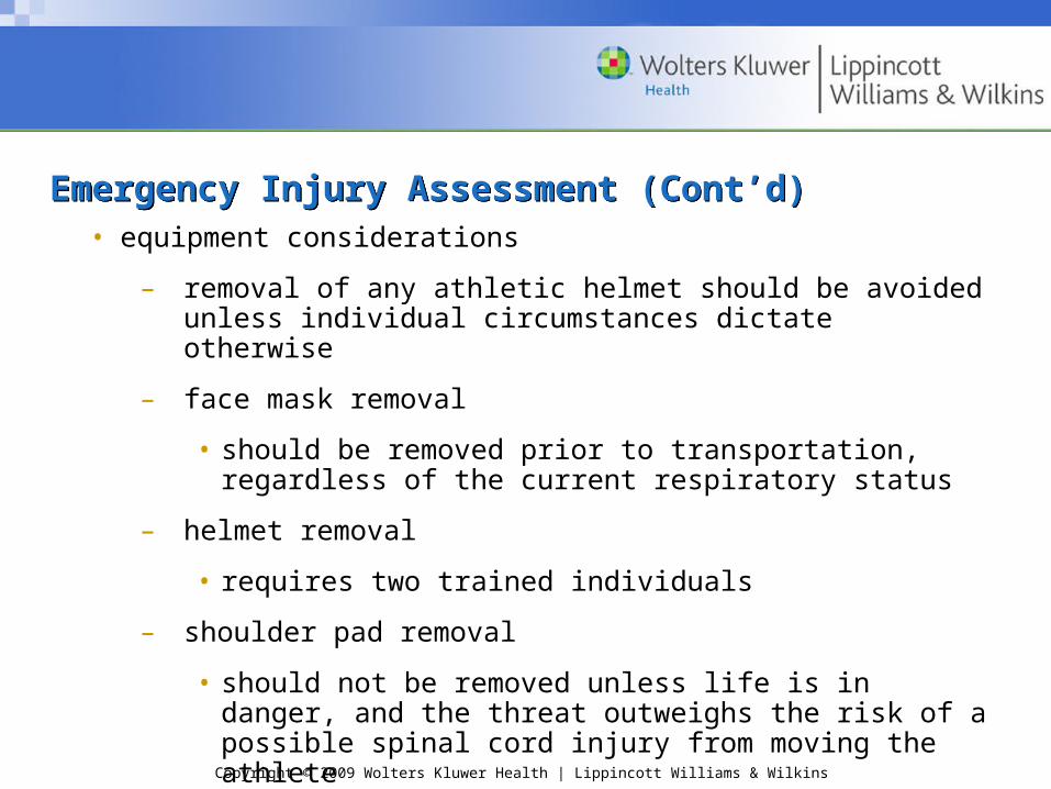
Emergency Injury Assessment (Cont’d)Emergency Injury Assessment (Cont’d)• equipment considerations
– removal of any athletic helmet should be avoided unless individual circumstances dictate otherwise
– face mask removal
• should be removed prior to transportation, regardless of the current respiratory status
– helmet removal
• requires two trained individuals
– shoulder pad removal
• should not be removed unless life is in danger, and the threat outweighs the risk of a possible spinal cord injury from moving the athlete
Copyright © 2009 Wolters Kluwer Health | Lippincott Williams & Wilkins
Moving the Injured ParticipantMoving the Injured Participant
• ambulatory assistance
– aid an injured ind. able to walk
• manual conveyance
– ind. unable to walk or distance is too great to walk
• transport by spine board
– safest method
Copyright © 2009 Wolters Kluwer Health | Lippincott Williams & Wilkins
Diagnostic TestingDiagnostic Testing
• the team physician or medical specialist orders tests and interprets the results…. the athletic trainer should have a basic understanding of the purpose of the tests
Copyright © 2009 Wolters Kluwer Health | Lippincott Williams & Wilkins
Diagnostic Testing (Cont’d)Diagnostic Testing (Cont’d)
• laboratory tests
– blood test; urinalysis
• radiographs (X-rays)
– can rule out fractures, infections, & neoplasms
– use of radio-opaque dyes
• myelogram
• arthrogram
• F5.11
Copyright © 2009 Wolters Kluwer Health | Lippincott Williams & Wilkins
Diagnostic Testing (Cont’d)Diagnostic Testing (Cont’d)
• computed tomography (CT scan)
– can reveal abnormalities in bone, fat, and soft tissue
– can detect tendon & ligament inj. in varying jt. positions
• F5.12
Copyright © 2009 Wolters Kluwer Health | Lippincott Williams & Wilkins
Diagnostic Testing (Cont’d)Diagnostic Testing (Cont’d)
• magnetic resonance imaging (MRI)
– can reveal soft tissue differentiation
– can demonstrate space-occupying lesions in the brain
– can demonstrate joint damage
– can view blood vessels & blood flow w/out use of a contrast medium
• F5.13
Copyright © 2009 Wolters Kluwer Health | Lippincott Williams & Wilkins
Diagnostic Testing (Cont’d)Diagnostic Testing (Cont’d)
• radionuclide scintigraph (bone scan)
– can detect stress fractures of the long bones and vertebrae, degenerative diseases, infections, or tumors of the bone
• F15.4
Copyright © 2009 Wolters Kluwer Health | Lippincott Williams & Wilkins
Diagnostic Testing (Cont’d)Diagnostic Testing (Cont’d)
• ultrasonic imaging
– used to view tendon and other soft-tissue imaging
• electromyography
– used to detect denervated muscles, nerve root compression injuries, and other muscle diseases