Embed Size (px)
Citation preview
Review ArticleCorneal Collagen Cross-Linking for Keratoconus:Systematic Review
Hidenaga Kobashi1,2 and Shi Song Rong3
1Department of Ophthalmology, University of Kitasato School of Medicine, Kanagawa, Japan2Schepens Eye Research Institute, Massachusetts Eye and Ear Infirmary, Department of Ophthalmology,Harvard Medical School, Boston, MA, USA3Massachusetts Eye and Ear Infirmary, Harvard Medical School, Boston, MA, USA
Correspondence should be addressed to Hidenaga Kobashi; hidenaga [email protected]
Received 3 December 2016; Accepted 9 May 2017; Published 11 June 2017
Academic Editor: Janusz Blasiak
Copyright © 2017 Hidenaga Kobashi and Shi Song Rong. This is an open access article distributed under the Creative CommonsAttribution License, which permits unrestricted use, distribution, and reproduction in any medium, provided the original work isproperly cited.
Purpose.To evaluate the efficacy of collagen cross-linking (CXL) one year after treatment for keratoconus compared to no treatmentby summarizing randomized controlled trials (RCTs) using a systematic review. Methods. Trials meeting the selection criteriawere quality appraised, and the data were extracted by two independent authors. The outcome parameters included maximumkeratometry (𝐾max), corneal thickness at the thinnest point, best spectacle-corrected visual acuity (BSCVA), uncorrected visualacuity (UCVA), spherical equivalent (SE) refraction, and cylindrical refraction one year after CXL. We compared the changes inthe above parameters with the control group. Results.We identified five RCTs involving 289 eyes that met the eligibility criteria forthis systematic review. The changes in BSCVA from baseline to one year exhibited a significant difference between the two groups.There was no statistically significant difference between the two groups for changes in corneal thickness and cylindrical refraction.We did not conduct a meta-analysis in𝐾max, UCVA, and SE refraction because their 𝐼2 values were greater than 50%. Conclusions.According to the systematic review, CXL may be effective in halting the progression of keratoconus for one year under certainconditions, although evidence is limited due to the significant heterogeneity and paucity of RCTs.
1. Introduction
Keratoconus is characterized as a bilateral, noninflammatory,progressive corneal ectasia [1]. It results in corneal thinningand protrusion, progressive myopia, and irregular astigma-tism. Although only 26.8% of patients with keratoconusprogress to requiring corneal transplantation for visual recov-ery [2], keratoconus remains themost common indication forcorneal transplantation surgery [3].
Corneal collagen cross-linking (CXL) was first intro-duced by Wollensak et al. as a promising technique to slowor stop the progression of keratoconus [4]. In CXL, riboflavin(vitamin B2) is administered in conjunction with ultravioletA (UVA, 365 nm). The interaction of riboflavin and UVAleads to the formation of reactive oxygen species, which leadsto the formation of additional covalent bonds between colla-gen molecules, with consequent biomechanical stiffening of
the cornea [5]. Since the first clinical study was published byWollensak et al. [4], there have been an increasing numberof published studies reporting the safety and efficacy of thetreatment in slowing down or halting the progression ofkeratoconus. CXL received Food and Drug Administrationapproval in 2016 in the United States [6]. Previous studies,however, are limited by their lack of a control group andrelatively short-term follow-up, particularly considering theinherent variability in the course of keratoconus [7] andthe limited reproducibility of the measurement of outcomeparameters [8]. Several studies focusing on the successfultreatment of keratoconus with CXL have been performed andpublished as randomized controlled trials (RCTs). Previousmeta-analyses and a Cochrane review of all published RCTsof CXL for the treatment of keratoconus tried to verifythe efficacy of CXL as treatment in stabilizing keratoconus;however, the meta-analysis by Li et al. [9] included one
HindawiBioMed Research InternationalVolume 2017, Article ID 8145651, 7 pageshttps://doi.org/10.1155/2017/8145651
2 BioMed Research International
short-term RCT with a follow-up three months postopera-tively [10], and the Cochrane review arrived at inconclusivefindings due to low quality of evidence and small samplesizes of RCTs conducted until August 2014 [11]. Li et al. [9]calculated the outcomes without adjusting the postoperativetime period, and they used two studies from same authors,namely, Wittig-Silva. Sykakis et al. [11] reviewed three RCTsto determine whether there is evidence that CXL is aneffective treatment compared to no treatment for haltingthe progression of keratoconus. However, their Cochranereview was unable to conduct a quantitative synthesis of theevidence because of the small number of RCTs. The twoother meta-analyses included comparative and retrospectivestudies, which may lack evidence [12, 13]. Our hypothesisis that CXL may be effective in halting the progression ofkeratoconus for long-term follow-ups. The purpose of thisstudy is to evaluate efficacy of CXL one year after treatmentof keratoconus compared to no treatment by conducting asystematic review of the literature.
2. Materials and Methods
2.1. Study Selection. Two reviewers searched the MEDLINE,EMBASE, and the Cochrane Central Register of ControlledTrials (CENTRAL) databases for publications from January1, 2003, to December 31, 2015. Our search was performedon January 1, 2016. The first published trial report evaluatingthe effect of CXL in patients with keratoconus was pub-lished in 2003 [4]; therefore, we used 2003 as the startingpoint for the literature search. The keywords in our searchstrategy include “corneal cross-linking”, “corneal collagencross-linking”, “collagen cross-linkage”, and “keratoconus”.Two reviewers reviewed the titles and abstracts of the searchresults and retrieved full-text articles if the title or abstractappeared to meet the eligibility criteria for this review.
2.2. Study Inclusion and Exclusion Criteria. Studies wereincluded if they discussed the diagnosis of progressive kera-toconus (Amsler-Krumeich grades I and III) [14].We definedthe progression of keratoconus as an increase of at least0.75 diopter (D) in the steepest keratometry, a degradationof visual acuity, and an increase of 0.75D or more in themanifest cylinder over the preceding 12 months.We includedstudies that had a one-year minimum follow-up time andfollowed the Dresden protocol for CXL. When the same trialwas drawn by a screening, we used the most recent trialreport. Only studies including human research participantsand published in the English language were included. Weexcluded studies that included patients with a history ofcorneal surgery and corneal pachymetry less than 300mm.Articles on corneal collagen cross-linking combined withother treatments, such as topography-guided photorefractivekeratectomy or intrastromal corneal ring segments, wereexcluded. We also excluded cohort studies, case-controlstudies, and studies that did not use a random method toprospectively assign participants to two groups.
We included trials that compared CXL to contralateraleyeswithout any treatment or eyes fromdifferent keratoconuspatients. Eyes that received riboflavin ophthalmic solution
alone as the sham control were excluded. All articles that wefound were carefully reviewed to select those that reportedoriginal clinical data pre- and postoperatively. Data frompreviously reported cases included in different articles wereomitted to avoid duplication of data.
2.3. Assessment of Risk of Bias in Included Studies. Tworeview authors independently assessed the risk of bias of theincluded studies in accordance with the Cochrane Hand-book for Systematic Reviews of Interventions [15] usingthe following parameters: adequacy of sequence generation;allocation concealment; blinding of participants, personnel,and outcome assessors; incomplete outcome data; and selec-tive outcome reporting. Disagreements were resolved bydiscussion.
2.4. Types of OutcomeMeasures. Our primary outcomeswerethe changes in the following parameters between baseline andone-year follow-up:
(i) Maximum keratometry value (𝐾max): the steepestkeratometry value obtained using topographies of arotating Scheimpflug camera or computerized vide-okeratography
(ii) Thinnest corneal thickness: the thickness of thethinnest point using ultrasound pachymetry
(iii) Best spectacle-corrected visual acuity (BSCVA): thevisual acuity corrected by only glasses
(iv) Uncorrected visual acuity (UCVA): the visual acuitywithout correction
Our secondary outcomes were the following:
(i) Spherical equivalent (SE) refraction: the manifestsubjective refraction of the SE
(ii) Cylindrical refraction: the manifest subjective refrac-tion of the cylinder
Best-corrected visual acuity with a contact lens was in-cluded in this analysis because the evaluation of visual acuitywas limited to BSCVA or UCVA in most previous trials.
2.5. Data Extraction. Two reviewers independently extracteddata from the included trials using a standardized form. Wecollected the above outcome measures and details of theinterventions, such as setting, sample size, age, mean baseline𝐾max, control design, and follow-up period. We requestedthe unpublished data from the corresponding authors of theindividual trials via email and waited for their replies for sixmonths.
2.6. Assessment of Heterogeneity. We planned to assess het-erogeneity by looking at the clinical and methodologicaldiversity of the included studies and by examining the forestplots and I2 statistics as described in the CochraneHandbookfor Systematic Reviews of Interventions [15].
2.7. Data Synthesis and Statistical Analysis. We examinedthe study characteristics and I2 statistic as described above.
BioMed Research International 3
Table 1: Characteristics of included trials evaluating corneal collagen cross-linking.
Study∗ (year) Country Number oftreated eyes
Mean age(years)
Mean baseline𝐾max (diopters)
Control design Follow-up(months)
Greenstein [16] (2011) United States 49 Not available 60.4 Contralateraleye 12
O’Brart [17] (2011) United Kingdom 22 29.6 53.9 Contralateraleye 18
Wittig-Silva [18] (2014) Australia 46 25.7 52.87 Differentpatients 36
Lang [19] (2015) Germany 15 29.5 47.3 Differentpatients 36
Seyedian [20] (2015) Iran 26 25.6 49.43 Contralateraleye 12
∗First author.
Reports identi�ed from search:1073
Studies obtainedfor full paper review: 80
Included in systematic review:5
Studies excludedas irrelevant to our subject: 993
Studies excluded: 75Non-RCT: 36No data indicated: 32Same trial: 7
Figure 1: Flow of trial selection. RCT: randomized controlled trial.
We did not conduct a meta-analysis if there was significantheterogeneity. An I2 value greater than 50% was consideredevidence of significant heterogeneity.
For comparisons where it was appropriate to conduct ameta-analysis, we calculated weighted mean differences and95% confidence intervals (CIs). We used a fixed-effect modelif there were three or fewer studies and a random-effectsmodel if more studies were available. The statistical optionused for this analysis was the weighted mean difference forcomparing mean changes ± standard deviation values foreach parameter from baseline to one-year follow-up betweenthe study and control groups. All statistical analyses wereperformed with RevMan software (version 5.2, InformationManagement Systems Group, Cochrane Collaboration).
3. Results
3.1. Results of the Search. There were 1073 articles relevantto the search terms. After screening titles and abstracts, weexcluded 993 studies. Of the 80 publications that were initiallyconsidered as potentially relevant, we excluded 75 studiesbecause they did not meet the predefined inclusion criteria(Figure 1). Five prospective RCTs involving 289 eyes wereincluded in this systematic review [16–20]. As the same trial,we excluded seven publications which were composed of sixstudies by Greenstein et al. and one by Wittig-Silva et al. We
obtained the primary and secondary outcomes data at one-year follow-up as unpublished information fromO’Brart et al.[17] and Lang et al. [19]. Data on our primary and secondaryoutcomes were available in the papers by Greenstein et al.[16], Wittig-Silva et al. [18], and Seyedian et al. [20]
3.2. Characteristics of the Included Studies. Table 1 shows themain characteristics of the included trials. Three [16, 17, 20]of the five trials were studies that used the contralateral eye asthe control; the contralateral eye wasmatched for progressionof keratoconus, and age and sex matching were not required.Two studies [18, 19] used two different populations thatmatched groups for age and progression of keratoconus. Nodescription of the mean age was available in the paper byGreenstein et al. [16]
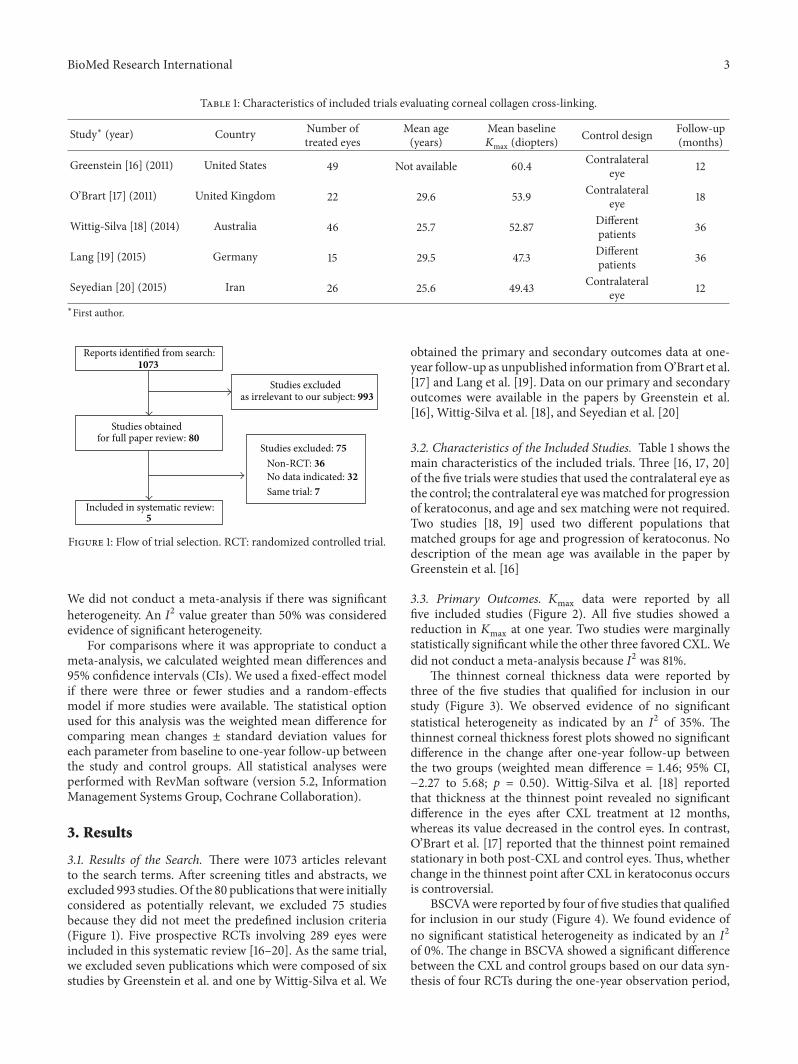
3.3. Primary Outcomes. 𝐾max data were reported by allfive included studies (Figure 2). All five studies showed areduction in 𝐾max at one year. Two studies were marginallystatistically significant while the other three favored CXL.Wedid not conduct a meta-analysis because I2 was 81%.
The thinnest corneal thickness data were reported bythree of the five studies that qualified for inclusion in ourstudy (Figure 3). We observed evidence of no significantstatistical heterogeneity as indicated by an I2 of 35%. Thethinnest corneal thickness forest plots showed no significantdifference in the change after one-year follow-up betweenthe two groups (weighted mean difference = 1.46; 95% CI,−2.27 to 5.68; p = 0.50). Wittig-Silva et al. [18] reportedthat thickness at the thinnest point revealed no significantdifference in the eyes after CXL treatment at 12 months,whereas its value decreased in the control eyes. In contrast,O’Brart et al. [17] reported that the thinnest point remainedstationary in both post-CXL and control eyes. Thus, whetherchange in the thinnest point after CXL in keratoconus occursis controversial.
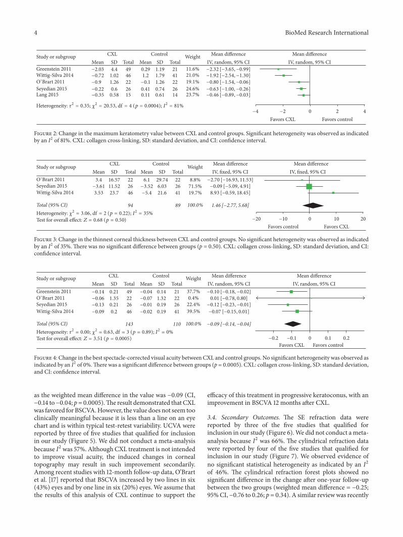
BSCVAwere reported by four of five studies that qualifiedfor inclusion in our study (Figure 4). We found evidence ofno significant statistical heterogeneity as indicated by an I2of 0%. The change in BSCVA showed a significant differencebetween the CXL and control groups based on our data syn-thesis of four RCTs during the one-year observation period,
4 BioMed Research International
Study or subgroupMean MeanSD SDTotal Total
CXL Control Weight Mean di�erenceIV, random, 95% CI
Mean di�erenceIV, random, 95% CI
−4 −2 0 2 4
Favors CXL Favors control
Greenstein 2011Wittig-Silva 2014/’Brart 2011Seyedian 2015Lang 2015
−2.03−0.72−0.9
−0.22−0.35
4.41.021.26
0.60.58
494622
2615
0.291.2−0.1
0.410.11
1.191.791.26
0.740.61
214122
2614
11.6%21.0%19.1%24.6%23.7%
−2.32 [−3.65, −0.99]−1.92 [−2.54, −1.30]−0.80 [−1.54, −0.06]
−0.63 [−1.00, −0.26]−0.46 [−0.89, −0.03]
Heterogeneity: 2 = 0.35; 2 = 20.53, d@ = 4 (p = 0.0004); I2 = 81%
Figure 2: Change in the maximum keratometry value between CXL and control groups. Significant heterogeneity was observed as indicatedby an I2 of 81%. CXL: collagen cross-linking, SD: standard deviation, and CI: confidence interval.
Study or subgroupMean MeanSD SDTotal Total
CXL Control Weight Mean di�erenceIV, �xed, 95% CI
Mean di�erenceIV, �xed, 95% CI
−20 −10 0 10 20
Favors control Favors CXL
Wittig-Silva 2014
/’Brart 2011Seyedian 2015
3.4−3.613.53
16.5711.5223.7
222646
6.1−3.52−5.4
29.746.0321.6
222641
8.8%71.5%19.7%
−2.70 [−16.93, 11.53]−0.09 [−5.09, 4.91]8.93 [−0.59, 18.45]
Total (95% CI) 94 89 100.0% 1.46 [−2.77, 5.68]Heterogeneity: 2 = 3.06, d@ = 2 (p = 0.22); I2 = 35%
Test for overall e�ect: Z = 0.68 (p = 0.50)
Figure 3: Change in the thinnest corneal thickness between CXL and control groups. No significant heterogeneity was observed as indicatedby an I2 of 35%. There was no significant difference between groups (p = 0.50). CXL: collagen cross-linking, SD: standard deviation, and CI:confidence interval.
Study or subgroupMean MeanSD SDTotal Total
CXL Control Weight Mean di�erenceIV, random, 95% CI
Mean di�erenceIV, random, 95% CI
Greenstein 2011
Wittig-Silva 2014
/’Brart 2011Seyedian 2015
−0.14−0.06−0.13
−0.09
0.211.350.21
0.2
492226
46
−0.04−0.07−0.01
−0.02
0.141.320.19
0.19
212226
41
37.7%0.4%22.4%39.5%
−0.10 [−0.18, −0.02]0.01 [−0.78, 0.80]
−0.12 [−0.23, −0.01]
−0.07 [−0.15, 0.01]
Total (95% CI) 143 110 100.0% −0.09 [−0.14, −0.04]
Test for overall e�ect: Z = 3.51 (p = 0.0005)Heterogeneity: 2 = 0.00; 2 = 0.63, d@ = 3 (p = 0.89); I2 = 0%
−0.2 −0.1 0 0.1 0.2
Favors CXL Favors control
Figure 4: Change in the best spectacle-corrected visual acuity between CXL and control groups. No significant heterogeneity was observed asindicated by an I2 of 0%.There was a significant difference between groups (p = 0.0005). CXL: collagen cross-linking, SD: standard deviation,and CI: confidence interval.
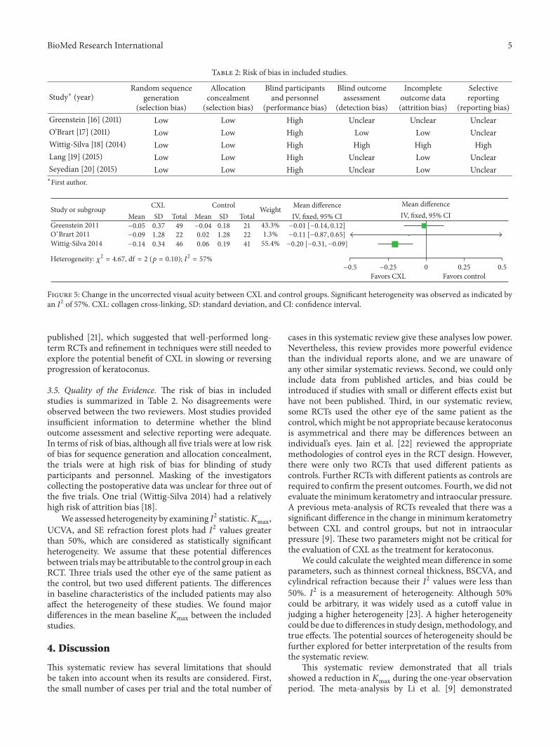
as the weighted mean difference in the value was −0.09 (CI,−0.14 to−0.04; p= 0.0005).The result demonstrated that CXLwas favored for BSCVA.However, the value does not seem tooclinically meaningful because it is less than a line on an eyechart and is within typical test-retest variability. UCVA werereported by three of five studies that qualified for inclusionin our study (Figure 5). We did not conduct a meta-analysisbecause I2 was 57%. Although CXL treatment is not intendedto improve visual acuity, the induced changes in cornealtopography may result in such improvement secondarily.Among recent studies with 12-month follow-up data, O’Brartet al. [17] reported that BSCVA increased by two lines in six(43%) eyes and by one line in six (20%) eyes. We assume thatthe results of this analysis of CXL continue to support the
efficacy of this treatment in progressive keratoconus, with animprovement in BSCVA 12 months after CXL.
3.4. Secondary Outcomes. The SE refraction data werereported by three of the five studies that qualified forinclusion in our study (Figure 6).We did not conduct a meta-analysis because I2 was 66%. The cylindrical refraction datawere reported by four of the five studies that qualified forinclusion in our study (Figure 7). We observed evidence ofno significant statistical heterogeneity as indicated by an I2of 46%. The cylindrical refraction forest plots showed nosignificant difference in the change after one-year follow-upbetween the two groups (weighted mean difference = −0.25;95%CI,−0.76 to 0.26; p = 0.34). A similar reviewwas recently
BioMed Research International 5
Table 2: Risk of bias in included studies.
Study∗ (year)Random sequence
generation(selection bias)
Allocationconcealment(selection bias)
Blind participantsand personnel
(performance bias)
Blind outcomeassessment
(detection bias)
Incompleteoutcome data(attrition bias)
Selectivereporting
(reporting bias)Greenstein [16] (2011) Low Low High Unclear Unclear UnclearO’Brart [17] (2011) Low Low High Low Low UnclearWittig-Silva [18] (2014) Low Low High High High HighLang [19] (2015) Low Low High Unclear Low UnclearSeyedian [20] (2015) Low Low High Unclear Low Unclear∗First author.
Study or subgroupMean MeanSD SDTotal Total
CXL Control Weight Mean di�erenceIV, �xed, 95% CI
Mean di�erenceIV, �xed, 95% CI
Greenstein 2011
Wittig-Silva 2014/’Brart 2011
−0.5 −0.25 0 0.25 0.5Favors CXL Favors control
−0.05−0.09
−0.14
0.371.28
0.34
4922
46
−0.040.02
0.06
0.181.28
0.19
2122
41 55.4%
−0.01 [−0.14, 0.12]−0.11 [−0.87, 0.65]−0.20 [−0.31, −0.09]
43.3%1.3%
Heterogeneity: 2 = 4.67, d@ = 2 (p = 0.10); I2 = 57%
Figure 5: Change in the uncorrected visual acuity between CXL and control groups. Significant heterogeneity was observed as indicated byan I2 of 57%. CXL: collagen cross-linking, SD: standard deviation, and CI: confidence interval.
published [21], which suggested that well-performed long-term RCTs and refinement in techniques were still needed toexplore the potential benefit of CXL in slowing or reversingprogression of keratoconus.
3.5. Quality of the Evidence. The risk of bias in includedstudies is summarized in Table 2. No disagreements wereobserved between the two reviewers. Most studies providedinsufficient information to determine whether the blindoutcome assessment and selective reporting were adequate.In terms of risk of bias, although all five trials were at low riskof bias for sequence generation and allocation concealment,the trials were at high risk of bias for blinding of studyparticipants and personnel. Masking of the investigatorscollecting the postoperative data was unclear for three out ofthe five trials. One trial (Wittig-Silva 2014) had a relativelyhigh risk of attrition bias [18].
We assessed heterogeneity by examining I2 statistic.𝐾max,UCVA, and SE refraction forest plots had I2 values greaterthan 50%, which are considered as statistically significantheterogeneity. We assume that these potential differencesbetween trialsmay be attributable to the control group in eachRCT. Three trials used the other eye of the same patient asthe control, but two used different patients. The differencesin baseline characteristics of the included patients may alsoaffect the heterogeneity of these studies. We found majordifferences in the mean baseline 𝐾max between the includedstudies.
4. Discussion
This systematic review has several limitations that shouldbe taken into account when its results are considered. First,the small number of cases per trial and the total number of
cases in this systematic review give these analyses low power.Nevertheless, this review provides more powerful evidencethan the individual reports alone, and we are unaware ofany other similar systematic reviews. Second, we could onlyinclude data from published articles, and bias could beintroduced if studies with small or different effects exist buthave not been published. Third, in our systematic review,some RCTs used the other eye of the same patient as thecontrol, whichmight be not appropriate because keratoconusis asymmetrical and there may be differences between anindividual’s eyes. Jain et al. [22] reviewed the appropriatemethodologies of control eyes in the RCT design. However,there were only two RCTs that used different patients ascontrols. Further RCTs with different patients as controls arerequired to confirm the present outcomes. Fourth, we did notevaluate theminimum keratometry and intraocular pressure.A previous meta-analysis of RCTs revealed that there was asignificant difference in the change in minimum keratometrybetween CXL and control groups, but not in intraocularpressure [9]. These two parameters might not be critical forthe evaluation of CXL as the treatment for keratoconus.
We could calculate the weighted mean difference in someparameters, such as thinnest corneal thickness, BSCVA, andcylindrical refraction because their I2 values were less than50%. I2 is a measurement of heterogeneity. Although 50%could be arbitrary, it was widely used as a cutoff value injudging a higher heterogeneity [23]. A higher heterogeneitycould be due to differences in study design,methodology, andtrue effects. The potential sources of heterogeneity should befurther explored for better interpretation of the results fromthe systematic review.
This systematic review demonstrated that all trialsshowed a reduction in𝐾max during the one-year observationperiod. The meta-analysis by Li et al. [9] demonstrated
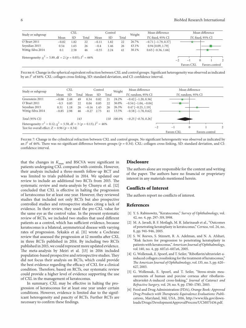
6 BioMed Research International
Study or subgroup CXL Control Mean di�erenceIV, �xed, 95% CI
Wittig-Silva 2014
/’Brart 2011Seyedian 2015
Mean−0.820.54
0.1
SD1.821.65
2.58
Total2226
46
Mean−0.11−0.4
−0.55
SD1.821.46
2.24
Total2226
41
Weight
26.7%43.1%30.1%
Mean di�erenceIV, �xed, 95% CI
−0.71 [−1.79, 0.37]0.94 [0.09, 1.79]
0.65 [−0.36, 1.66]
−2 −1 0 1 2
Favors CXL Favors control
Heterogeneity: 2 = 5.89, d@ = 2 (p = 0.05); I2 = 66%
Figure 6: Change in the spherical equivalent refraction between CXL and control groups. Significant heterogeneity was observed as indicatedby an I2 of 66%. CXL: collagen cross-linking, SD: standard deviation, and CI: confidence interval.
Study or subgroup CXL Control
Greenstein 2011
Wittig-Silva 2014
/’Brart 2011Seyedian 2015
Heterogeneity: 2 = 0.12; 2 = 5.58, d@ = 3 (p = 0.13); I2 = 46%
Mean−0.08−0.50.31−0.85
SD2.480.851.182.98
Mean0.340.04−0.16−0.27
SD0.820.851.452.75
Total (95% CI)
Total49222646
143
Total21222641
110
Weight
24.2%36.0%26.3%13.5%
100.0%
Mean di�erenceIV, random, 95% CI−0.42 [−1.20, 0.36]
0.47 [−0.25, 1.19]−0.58 [−1.78, 0.62]
−0.54 [−1.04, −0.04]
−0.25 [−0.76, 0.26]
Mean di�erenceIV, random, 95% CI
−2 −1 0 1 2
Favors CXL Favors controlTest for overall e�ect: Z = 0.96 (p = 0.34)
Figure 7: Change in the cylindrical refraction between CXL and control groups. No significant heterogeneity was observed as indicated byan I2 of 46%. There was no significant difference between groups (p = 0.34). CXL: collagen cross-linking, SD: standard deviation, and CI:confidence interval.
that the changes in 𝐾max and BSCVA were significant inpatients undergoing CXL compared with controls. However,their analysis included a three-month follow-up RCT andwas limited to trials published in 2014. We updated ourreview to include an additional two RCTs from 2015. Thesystematic review and meta-analysis by Chunyu et al. [12]concluded that CXL is effective in halting the progressionof keratoconus for at least one year. However, they reviewedstudies that included not only RCTs but also prospectivecontrolled studies and retrospective studies citing a lack ofevidence. In their review, they used the pre-CXL value forthe same eye as the control value. In the present systematicreview of RCTs, we included two studies that used differentpatients as a control, which has sufficient evidence, becausekeratoconus is a bilateral, asymmetrical disease with varyingrates of progression. Sykakis et al. [11] wrote a Cochranereview that assessed the progression at 12 months after CXLin three RCTs published in 2014. By including two RCTspublished in 2015, we could representmore updated evidence.The meta-analysis by Meiri et al. [13] in 2016 includedpopulation-based prospective and retrospective studies.Theydid not focus their analysis on RCTs, which could providethe best evidence regarding the efficacy of CXL for a clinicalcondition. Therefore, based on RCTs, our systematic reviewcould provide a higher level of evidence supporting the useof CXL in the management of keratoconus.
In summary, CXL may be effective in halting the pro-gression of keratoconus for at least one year under certainconditions. However, evidence is limited due to the signif-icant heterogeneity and paucity of RCTs. Further RCTs arenecessary to confirm these findings.
Disclosure
The authors alone are responsible for the content and writingof the paper. The authors have no financial or proprietaryinterest in any materials mentioned herein.
Conflicts of Interest
The authors report no conflicts of interest.
References
[1] Y. S. Rabinowitz, “Keratoconus,” Survey of Ophthalmology, vol.42, no. 4, pp. 297–319, 1998.
[2] M. A. Javadi, B. F. Motlagh, M. R. Jafarinasab et al., “Outcomesof penetrating keratoplasty in keratoconus,” Cornea, vol. 24, no.8, pp. 941–946, 2005.
[3] S. W. Reeves, S. Stinnett, R. A. Adelman, and N. A. Afshari,“Risk factors for progression to penetrating keratoplasty inpatients with keratoconus,”American Journal of Ophthalmology,vol. 140, no. 4, pp. 607.e1–607.e6, 2005.
[4] G.Wollensak, E. Spoerl, and T. Seiler, “Riboflavin/ultraviolet-a-induced collagen crosslinking for the treatment of keratoconus,”TheAmerican Journal of Ophthalmology, vol. 135, no. 5, pp. 620–627, 2003.
[5] G. Wollensak, E. Spoerl, and T. Seiler, “Stress-strain mea-surements of human and porcine corneas after riboflavin-ultraviolet-A-induced cross-linking,” Journal of Cataract andRefractive Surgery, vol. 29, no. 9, pp. 1780–1785, 2003.
[6] Food and Drug Administration (FDA), Orange Book: ApprovedDrug Products with Therapeutic Equivalence Evaluations: Publi-cations, Maryland, Md, USA, 2016, http://www.fda.gov/down-loads/Drugs/DevelopmentApprovalProcess/UCM071436.pdf.
BioMed Research International 7
[7] S. E. Brown, R. Simmasalam, N. Antonova, N. Gadaria, and P.A. Asbell, “Progression in keratoconus and the effect of cornealcross-linking on progression,” Eye and Contact Lens, vol. 40, no.6, pp. 331–338, 2014.
[8] E. Szalai, A. Berta, Z. Hassan, and L. Modis Jr., “Reliability andrepeatability of swept-source Fourier-domain optical coherencetomography and Scheimpflug imaging in keratoconus,” Journalof Cataract and Refractive Surgery, vol. 38, no. 3, pp. 485–494,2012.
[9] J. Li, P. Ji, and X. L. Lin, “Efficacy of corneal collagen cross-linking for treatment of keratoconus: a meta-analysis of ran-domized controlled trials,” PLoS ONE, vol. 10, no. 5, Article IDe0127079, 2015.
[10] A. Da Candelaria Renesto, L. A. S. Melo Jr., M. De FilippiSartori, and M. Campos, “Sequential topical riboflavin withor without ultraviolet a radiation with delayed intracornealring segment insertion for keratoconus,” American Journal ofOphthalmology, vol. 153, no. 5, pp. 982–e3, 2012.
[11] E. Sykakis, R. Karim, J. R. Evans et al., “Corneal collagen cross-linking for treating keratoconus,” The Cochrane Database ofSystematic Reviews, vol. 3, Article ID CD010621, 2015.
[12] T. Chunyu, P. Xiujun, F. Zhengjun, Z. Xia, and Z. Feihu,“Corneal collagen cross-linking in keratoconus: a systematicreview andmeta-analysis,” Scientific Reports, vol. 4, article 5652,2014.
[13] Z. Meiri, S. Keren, A. Rosenblatt, T. Sarig, L. Shenhav, andD. Varssano, “Efficacy of corneal collagen cross-linking forthe treatment of keratoconus: A systematic review and meta-analysis,” Cornea, vol. 35, no. 3, pp. 417–428, 2016.
[14] J. L. Alio, M. H. Shabayek, and A. Artola, “Intracorneal ringsegments for keratoconus correction: long-term follow-up,”Journal of Cataract and Refractive Surgery, vol. 32, no. 6, pp.978–985, 2006.
[15] J. P. Higgins and S. Green, “Cochrane Handbook for SystematicReviews of Interventions: Cochrane Book Series,” CochraneHandbook for Systematic Reviews of Interventions: CochraneBook Series, pp. 1–649, 2008.
[16] S. A. Greenstein, K. L. Fry, and P. S. Hersh, “Corneal topographyindices after corneal collagen crosslinking for keratoconusand corneal ectasia: one-year results,” Journal of Cataract andRefractive Surgery, vol. 37, no. 7, pp. 1282–1290, 2011.
[17] D. P. S. O’Brart, E. Chan, K. Samaras, P. Patel, and S. P. Shah,“A randomised, prospective study to investigate the efficacyof riboflavin/ultraviolet A (370 nm) corneal collagen cross-linkage to halt the progression of keratoconus,” British Journalof Ophthalmology, vol. 95, no. 11, pp. 1519–1524, 2011.
[18] C. Wittig-Silva, E. Chan, and F. M. Islam, “A randomized,controlled trial of corneal collagen cross-linking in progressiveKeratoconus: three-year results,” Ophthalmology, vol. 121, pp.812–821, 2014.
[19] S. J. Lang, E. M. Messmer, G. Geerling et al., “Prospective, ran-domized, double-blind trial to investigate the efficacy and safetyof corneal cross-linking to halt the progression of keratoconus,”BMC Ophthalmology, vol. 15, no. 1, article no. 78, 2015.
[20] M. A. Seyedian, S. Aliakbari, M. Miraftab, H. Hashemi, S.Asgari, and M. Khabazkhoob, “Corneal collagen cross-linkingin the treatment of progressive keratoconus: a randomizedcontrolled contralateral eye study,”Middle East African Journalof Ophthalmology, vol. 22, no. 3, pp. 340–345, 2015.
[21] D. P. O’Brart, “Corneal collagen cross-linking: a review,” Journalof Optometry, vol. 7, pp. 113–124, 2014.
[22] R. Jain, S. Basu, and P. Garg, “Corneal collagen cross-linkage,inkeratoconus,” British Journal of Ophthalmology, vol.97, no. 1, pp. 108-109, 2013.
[23] J. P. Higgins, S. G. Thompson, J. J. Deeks, and D. G. Altman,“Measuring inconsistency in meta-analyses,” British MedicalJournal, vol. 327, pp. 557–560, 2003.

Submit your manuscripts athttps://www.hindawi.com
Stem CellsInternational
Hindawi Publishing Corporationhttp://www.hindawi.com Volume 2014
Hindawi Publishing Corporationhttp://www.hindawi.com Volume 2014
MEDIATORSINFLAMMATION
of
Hindawi Publishing Corporationhttp://www.hindawi.com Volume 2014
Behavioural Neurology
EndocrinologyInternational Journal of
Hindawi Publishing Corporationhttp://www.hindawi.com Volume 2014
Hindawi Publishing Corporationhttp://www.hindawi.com Volume 2014
Disease Markers
Hindawi Publishing Corporationhttp://www.hindawi.com Volume 2014
BioMed Research International
OncologyJournal of
Hindawi Publishing Corporationhttp://www.hindawi.com Volume 2014
Hindawi Publishing Corporationhttp://www.hindawi.com Volume 2014
Oxidative Medicine and Cellular Longevity
Hindawi Publishing Corporationhttp://www.hindawi.com Volume 2014
PPAR Research
The Scientific World JournalHindawi Publishing Corporation http://www.hindawi.com Volume 2014
Immunology ResearchHindawi Publishing Corporationhttp://www.hindawi.com Volume 2014
Journal of
ObesityJournal of
Hindawi Publishing Corporationhttp://www.hindawi.com Volume 2014
Hindawi Publishing Corporationhttp://www.hindawi.com Volume 2014
Computational and Mathematical Methods in Medicine
OphthalmologyJournal of
Hindawi Publishing Corporationhttp://www.hindawi.com Volume 2014
Diabetes ResearchJournal of
Hindawi Publishing Corporationhttp://www.hindawi.com Volume 2014
Hindawi Publishing Corporationhttp://www.hindawi.com Volume 2014
Research and TreatmentAIDS
Hindawi Publishing Corporationhttp://www.hindawi.com Volume 2014
Gastroenterology Research and Practice
Hindawi Publishing Corporationhttp://www.hindawi.com Volume 2014
Parkinson’s Disease
Evidence-Based Complementary and Alternative Medicine
Volume 2014Hindawi Publishing Corporationhttp://www.hindawi.com