Embed Size (px)
Citation preview
This article is protected by copyright. All rights reserved
Acc
epte
d A
rtic
le
Acc
epte
d A
rtic
le
Correlation between placental underperfusion, histologic signs and
perinatal morbidity in late-onset small for gestational age fetuses
Miguel Parra-Saavedra1,2,MD; Serena Simeone, MD1,3; Stefania Triunfo1,4,PhD; Francesca Crovetto1,5,MD; Botet F6, MD; Alfons Nadal7, PhD; Eduard Gratacos1, PhD; Francesc Figueras1, PhD
1Department of Maternal-Fetal Medicine, Institute Clínic of Gynecology, Obstetrics and Neonatology (ICGON), Hospital Clinic-IDIBAPS, University of Barcelona and Centre for Biomedical Research on Rare Diseases (CIBER-ER), Barcelona, Spain 2Maternal-Fetal Unit, CEDIFETAL, Centro de Diagnóstico de Ultrasonido e Imágenes, CEDIUL, Barranquilla, Colombia 3High Risk Pregnancy Unit, Department of Child and Women's Health, Careggi University Hospital-Florence, Italy 4Department of Obstetric and Gynecology, Catholic University of the Sacred Heart, Rome, Italy
5Fondazione Ca’ Granda, Ospedale Maggiore Policlinico, Dipartimento Ostetricia e Ginecologia, Università degli Studi di Milano, Milan, Italy 6Department of Neonatology, Institute Clínic of Gynecology, Obstetrics and Neonatology (ICGON), Hospital Clinic-IDIBAPS, University of Barcelona and Centre for Biomedical Research on Rare Diseases (CIBER-ER), Barcelona, Spain 7Department of pathology, Hospital Clinic -IDIBAPS
Correspondence and reprint requests to:
Francesc Figueras Maternal-Fetal Medicine Department Hospital Clinic, University of Barcelona Sabino de Arana 1, 08028 Barcelona Spain Telephone: +34 93 227 5600 Fax: +34 (0) 93 227 5605 E-mail: [email protected] Keywords: Fetal Growth Restriction; Neonatal complications; Ultrasonography, Doppler; Placenta; Fetal Development
This article has been accepted for publication and undergone full peer review but has not
been through the copyediting, typesetting, pagination and proofreading process, which
may lead to differences between this version and the Version of Record. Please cite this
article as doi: 10.1002/uog.13415

This article is protected by copyright. All rights reserved
Acc
epte
d A
rtic
le
ABSTRACT
Objective: To investigate whether signs of placental underperfusion (PUP), defined as any
maternal and/or fetal vascular pathology, confer a higher risk of neonatal morbidity in late-
onset small for gestational age (SGA) fetuses with normal umbilical artery Doppler.
Methods: A cohort of 126 SGA singleton fetuses with normal umbilical artery Doppler
delivered after 34 weeks was created. For each case, the placenta was histologically
evaluated for signs of PUP using a hierarchical and standardized classification system.
Neonatal morbidity was assessed by calculation of the morbidity assessment index for
newborns (MAIN) score, a validated outcome scale. The independent association between
PUP and neonatal morbidity was evaluated using multivariable median regression.
Results: In a total of 84 placentas (66.7%), there were 97 placental histological findings that
qualified as signs of PUP. These cases had a significantly higher incidence of emergent
delivery for non-reassuring fetal status (44.1% vs. 21.4%; p=0.013) and neonatal metabolic
acidosis at birth (33.3% vs. 14.3%; p=0.023). The median MAIN score significantly differed
between groups (89 vs. 0; p=0.025). This difference remained significant after adjustment
for potential confounders. The proportion of cases with mild to severe morbidity scores was
also significantly higher in the PUP group (31% vs. 14.3%; p=0.043).
Conclusion: In late-onset SGA with normal umbilical artery Doppler, signs of PUP confer
higher neonatal morbidity. These findings allow phenotypic profiling of fetal growth
restriction among the general population of late-onset SGA.
INTRODUCTION
Near-term babies born small for gestational age (SGA) with no signs of placental disease, as
reflected in the umbilical artery (UA) Doppler, are typically viewed as constitutionally small
This article is protected by copyright. All rights reserved
Acc
epte
d A
rtic
le
neonates displaying satisfactory perinatal outcomes1, 2; however, recent studies have
reported poor perinatal outcomes, suboptimal neurodevelopment, and higher postnatal
cardiovascular risk in these newborns 3 45. , supporting the hypothesis that a subset of SGA
fetuses undergoes late-onset fetal growth restriction (FGR), in which placental insufficiency
is not detected by UA Doppler. Thus, latent placental insufficiency is a key aspect in
differentiating true FGR from constitutional smallness 6-10.
In pregnancies with late-onset SGA, hypoxic/ischemic injury due to placental underperfusion
(PUP) defined as any maternal and/or fetal vascular pathology is documented in roughly
two-thirds of placentas11 12 , and the presence of PUP has been correlated with abnormal
uterine and umbilical vein Doppler before delivery phenotype.13
This finding suggests that this pattern constitutes the pathological basis of placental
insufficiency late in pregnancy; however, the association between PUP and neonatal
outcomes in late-onset SGA has not yet been explored. This information is critical for
consideration of PUP as a criterion to define the late-onset FGR clinical phenotype.
Most tools used to assess neonatal morbidity are not sufficiently sensitive or precise to
provide valid measurements in near-term babies14, 1516. The morbidity assessment index for
newborns17 (MAIN) score overcomes such limitations by including a comprehensive
inventory of standard assessment items that reflect pathophysiology in the early newborn
period. This inventory has previously been demonstrated to be sensitive in the population of
term SGA babies18 .
This article is protected by copyright. All rights reserved
Acc
epte
d A
rtic
le
The main purpose of this study was to determine whether signs of PUP confer a higher risk
of neonatal morbidity in late-onset-SGA fetuses that exhibit normal umbilical artery
Doppler.
METHODS
Study population
Between January 2012 and January 2014, a cohort of consecutive pregnancies that were
attended at a single university hospital was created from those that fulfilled the following
inclusion criteria: i) singleton pregnancy; ii) estimated fetal weight (EFW) below the 10th
centile at the routine third trimester ultrasound (30-34 weeks’ gestation), after adjustment
for gestational age (GA) at delivery and gender according to local standards19, and iii)
normal UA Doppler [defined as UA pulsatility index (PI) <95th centile20] at the time of
diagnosis of SGA. The following exclusion criteria were considered: i) congenital or
chromosomal abnormalities, ii) GA at delivery less than 34 weeks, and iii) development of
abnormal UA Doppler during follow-up, and iv) a birth weight > the 10th centile19 .
Pregnancies were dated by first-trimester crown-rump length measurement21. The EFW was
calculated using the Hadlock formula22. The hospital ethics committee approved the study
protocol, and written consent was obtained from all recruited patients (IRB 2008/4422).
Doppler measurements
Prenatal Doppler ultrasound examinations were performed by one of three experienced
operators (M.P., F.C., or S.S.) using either a Siemens Sonoline Antares (Siemens Medical
Systems, Malvern, PA, USA) or a General Electric Voluson E8 (GE Medical Systems, Zipf,
Austria) ultrasound machine equipped with a 6-2-MHz linear curved-array transducer.
Doppler recordings were performed in the absence of fetal movements and voluntarily
This article is protected by copyright. All rights reserved
Acc
epte
d A
rtic
le
suspended maternal breathing. Spectral Doppler parameters were performed automatically
from three or more consecutive waveforms with the angle of insonation as close to 0° as
possible. The UA pulsatility index (PI) was calculated from a free-floating cord loop. The
middle cerebral artery (MCA) PI was measured in a transverse view of the fetal head at the
level of its origin from the circle of Willis. The cerebroplacental ratio (CPR) was calculated as
the ratio of the MCA PI to the UA PI. All cases had a Doppler examination within 7 days of
delivery, and only the last Doppler examination was considered for this study.
Clinical protocol
Labor induction was indicated in the presence of a persistent (12-h apart) CPR <5th centile
beyond 37 weeks’ gestation23 and at 40 weeks’ gestation if CPR remained within normal
ranges. Labor induction was performed by cervical ripening with a slow-release
prostaglandin E2 vaginal pessary (10 mg). If the onset of labor did not occur within 12 hours,
oxytocin induction was initiated. Indication for cesarean delivery for non-reassuring fetal
status was based on abnormal fetal heart rate monitoring and abnormal fetal scalp blood pH
during intrapartum monitoring. Continuous fetal heart monitoring was carried out, and
tracings were classified according to the following three-tiered system24: (i) normal, baseline
110–160 beats per minute (bpm), variability >5 bpm and absence of decelerations; (ii)
suspicious (one non-reassuring criterion present), baseline 100–109 or 161–180 bpm,
variability <5 bpm for less than 90 min, recurrent (>50% contractions) typical variable
decelerations for more than 90 min, and a single prolonged deceleration for up to 3 min; or
(iii) pathological, more than one non-reassuring criterion or the presence of any abnormal
feature, including baseline <100 or >180 bpm or sinusoidal patterns for more than 10 min,
variability <5 bpm for more than 90 min, recurrent atypical variable decelerations for more
This article is protected by copyright. All rights reserved
Acc
epte
d A
rtic
le
than 30 min, late decelerations for more than 30 min, and a single prolonged deceleration
for more than 3 min.
In cases with pathological fetal heart rate or a suspicious pattern not presenting with a
fetal heart rate acceleration after digital fetal scalp stimulation25, fetal scalp blood sampling
was performed. Levels were considered abnormal with values less than 7.15 or 7.20 on two
occasions within 30 min. If cervical conditions did not allow fetal scalp sampling, cesarean
delivery was considered for non-reassuring fetal status based on the persistence of
abnormal tracings after pessary withdrawal, oxytocin suspension, and 10 min of intravenous
infusion of 200 µg/min ritrodine. All cases with adverse outcome were formally assessed to
ensure that the management protocol had been followed correctly.
Data collection and outcome measures
Data on maternal characteristics, including age, ethnicity, body mass index, parity, smoking
status, known chronic disease (hypertension, diabetes mellitus, renal disease, and
autoimmune disease), and previous obstetrical history, were recorded in the hospital
database at inclusion. In addition, data regarding pregnancy follow-up, complications
developed during pregnancy, ultrasound evaluation, and perinatal data were prospectively
collected. Preeclampsia (PE) was defined according to the guidelines of the International
Society for the Study of Hypertension in Pregnancy26. Neonatal metabolic acidosis at birth
was defined as the presence of a UA pH less than the 10th centile (7.18) for term babies and
base excess greater than the 90th centile (-12 mEq/L) at birth27.
Placental evaluation
Placental examinations were performed according to standard laboratory protocol. Fresh
and trimmed (after removal of the membranes, cord, and any blood clots) placental weights
were recorded. Trimmed placental weight centiles were assigned based on GA-specific
This article is protected by copyright. All rights reserved
Acc
epte
d A
rtic
le
placental weight charts28. The fetoplacental weight ratio (birth weight:fresh placental
weight) was also expressed as a centile, based on GA-specific ranges29.
Placentas were fixed in 10% buffered formalin. After gross examination, samples of each
specimen were taken for routine processing: one transverse section of cord, one rolled strip
of membranes, and three blocks spanning the entire thickness of the villous parenchyma. All
macroscopic lesions were sampled as well. Slides were stained with hematoxylin and eosin.
A single senior pathologist (A.N.) blinded to the neonatal outcome supervised all
examinations. All pathological examinations were made by pathologists blinded to the
Doppler result and the perinatal outcome.
For purposes of this study PUP-related histologic manifestations were further designated as
maternal or fetal in origin30, 31. Among maternal vascular supply disruptions, specific
vascular alterations that qualified as maternal vascular maldevelopment included superficial
implantation/decidual arteriopathy (acute atherosis and mural hypertrophy [mean wall
diameter >30% of overall vessel diameter of arterioles in the decidua parietalis]),
undergrowth/distal villous hypoplasia (decrease in the number and modal diameter of distal
villi at the center of the lobule after adjustment for plane of section and gestational age in
the lower 75% of a full-thickness section), excessive intervillous fibrin (basal layer of
fibrinoid material involving >30% of the placental maternal surface), and migration disorders
( including accessory lobes, peripheral cord insertions, and placenta previa). Specific
vascular alterations that qualified for maternal vascular obstruction were syncytial knots
(aggregates of syncytial nuclei at the surface of terminal villi)
involving terminal villi (affecting >50% of the terminal villi), villous agglutination (>50%),
intervillous fibrin deposition (eccentric aggregates on intervillous fibrin on proximal and
distal villi affecting >50% of the villi), and villous infarcts (>30% of villous loss). Specific
This article is protected by copyright. All rights reserved
Acc
epte
d A
rtic
le
vascular alterations that qualified for maternal vascular loss of integrity were arterial
rupture (abruption placenta) and venous rupture (acute or chronic marginal abruption).
Among fetal vascular supply disruptions, lesions that qualified for maldevelopment were
chorioangioma( in the form of proliferative nodules), chorioangiosis(more pervasive with a
patchy or generalized increase in capillaries), and distal villous immaturity. Lesions that
qualified for obstruction were those that were considered secondary to vascular thrombo-
occlusive disease (thrombosis of chorionic plate and stem villous channels and villous
avascularity [avascular villi are defined as more than 15 villi in a section showing a total lack
of villous vessels and a hyalinised fibrotic stroma]affecting large groups). PUP was indicated
for placentas with the presence of any of the above mentioned lesions.
Neonatal morbidity assessment
Morbidity was calculated using the MAIN score17, 32. This score was designed to provide a
numeric index of early neonatal outcomes reflecting prenatal care and adverse prenatal
exposures in babies delivered after 28 weeks.This score is a sensitive and discriminative
outcome measure for studies with outcomes other than preterm delivery. The MAIN score
consists of 47 binary items that describe 24 attributes of early neonatal morbidity (including
physiological variables, such as blood pressure, pCO2, temperature, oxygen saturation,
Apgar score, and the presence of apnea). These data items were obtained from the hospital
discharge records by a single evaluator (S.S.), who was blinded to the placental histology
findings. According to normative ranges, the scores were divided into two morbidity
categories: 150 (no/minimal) and 150 (mild to severe morbidity)17.
Statistical analysis
This article is protected by copyright. All rights reserved
Acc
epte
d A
rtic
le
Normal distributions were examined using the Shapiro-Wilk test. The Student’s t-test and
Pearson’s Chi-squared test were used to compare quantitative and categorical data,
respectively. Non-normally distributed quantitative variable data were compared using
Mood´s median test33.
The association between PUP-related histopathologic lesions and the MAIN score (log-
transformed) was analyzed using non-parametric quantile regression with a tau of 0.5
(median regression), where adjustment was performed for parity, smoking,
cerebroplacental ratio, and birth weight centile. All statistical analyses were performed
using SPSS Statistics 20 (SPSS Inc., Chicago IL) and R version 2.15.1 (The R Foundation for
Statistical Computing; quantreg package 5.05). A p-value ≤ 0.05 was considered statistically
significant.
RESULTS
A total of 134 pregnancies fulfilled the inclusion criteria. Subsequently, three cases were
excluded due to preterm birth before 34 weeks (one case with clinical and histological
evidence of choriomanionitis), and three additional cases were excluded because of the
development of abnormal UA Doppler before delivery, and additionally two cases were
excluded for a birth weight > the 10th centile The baseline characteristics of the remaining
126 cases are displayed in Table 1.
Elective cesarean section was performed in seven cases (three breech presentations, three
cases with more than one previous cesarean section, and one case with a previous vaginal
delivery with an anal sphincter lesion). One hundred and two women (81%) underwent
induction of labor; the remaining cases (n=17, 13.5%) had spontaneous onset of delivery.
This article is protected by copyright. All rights reserved
Acc
epte
d A
rtic
le
In a total of 84 cases (66.7%), 97 findings that qualified for PUP were found. Table 2 details
the histological findings in the study population. Placentas with signs of PUP were not
statistically different from those without signs of PUP with regard to weight (383.5 vs. 396.2;
p=0.45) or feto-placental weight ratio (5.8 vs. 6.2; p=0.053). Similarly, when expressed as GA
centiles, these parameters were not statistically different between the two groups (weight,
3.20 vs. 3.26, p=0.95 and feto-placental weight ratio, 34.3 vs. 37.1, p=0.62).
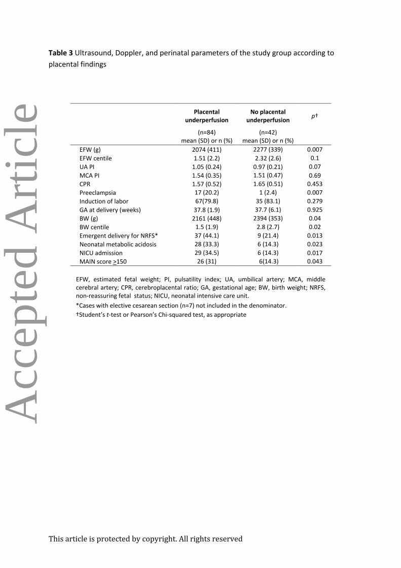
Table 3 presents the ultrasound and Doppler findings before delivery and the perinatal
outcomes according to the presence of PUP. Of note, the proportion of cases requiring
emergent delivery for non-reassuring fetal status significantly differed between cases with
and without PUP (44.1% vs. 21.4%; p=0.013). Similarly, the occurrence of neonatal
metabolic acidosis was more frequent in the PUP group compared to the group without
signs of PUP (33.3 vs. 14.3%; p=0.023). The proportion of newborns with moderate to
severe neonatal morbidity scores (>150) was also significantly higher in the PUP group
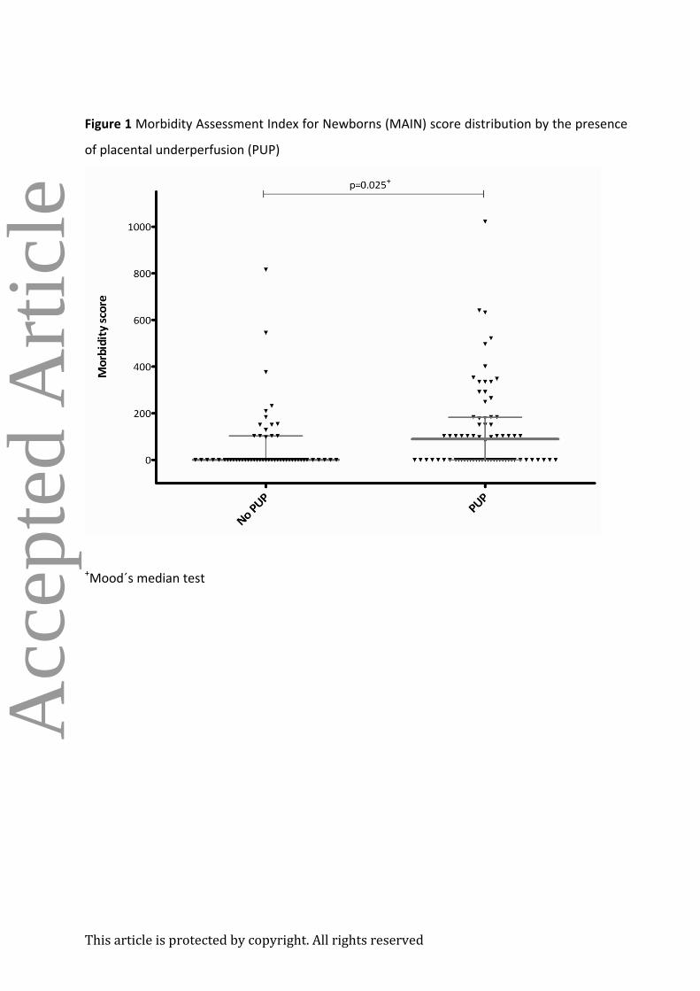
compared to the group without signs of PUP (31% vs. 14.3%; p=0.043). Finally, Figure 1
shows the distribution of MAIN scores by the presence of PUP. Median MAIN scores
significantly differed between groups (89 vs. 0; p=0.025). This difference remained
significant after adjustment for potential confounders (Table 4).
DISCUSSION
In this study, we report that in late-onset SGA, where we have previously found that the
degree of placental damage is not reflected in the UA Doppler 11 , the presence of
histological signs of PUP confers a poorer neonatal outcome. This finding supports the
notion that PUP is a key feature for phenotypic discernment of which cases among the
This article is protected by copyright. All rights reserved
Acc
epte
d A
rtic
le
overall population of late SGA babies correspond with true late-onset FGR secondary to
placental insufficiency.
Previous studies30, 31, 34-38 have shown that PUP and adverse perinatal outcomes are linked in
a variety of clinical conditions. This study extends this association to late-onset FGR even in
the presence of normal UA Doppler. One might speculate that PUP confers diminished
placental reserve, which, in turn, lowers tolerance to labor and heightens the likelihood of
neonatal morbidity. Moreover, we have also previously reported that in near-term SGA
babies, PUP undermines the neurodevelopment of early infancy39 . Taken together, this
evidence points to PUP as a surrogate of placental insufficiency and indicates that PUP is
worthy of targeting in clinical practice. These findings are particularly important because
late-onset SGA occurs in 5-10% of all pregnancies, and this pattern has been documented in
roughly two-thirds of placentas12 .
We found that, albeit not significantly, UA pulsatility was higher in the PUP group. The
precise reason why maternal underperfusion almost uniformly corresponds with abnormal
UA Doppler results in early-onset IUGR, although rarely in late-onset IUGR, is open to
debate; but because the nature of pathology is similar, one might speculate that the extent
of pathology is key. Indeed, animal models40 and mathematical projections41 of placental
vascular obliteration have suggested that abnormalities of UA Doppler studies surface only
in advanced stages of placental dysfunction. Nevertheless, term SGA babies suffering lesser
degrees of underperfusion that escape Doppler detection may be exposed to subtle but
chronic hypoxia and undernutrition with delayed neurologic consequences39 .
SGA is a descriptive term that is applied to all infants with birth weights below a given
threshold (generally 10th centile). Consequently, these infants comprise a heterogeneous
This article is protected by copyright. All rights reserved
Acc
epte
d A
rtic
le
group containing infants with true FGR and newborns who are constitutionally small but
otherwise healthy. Thus, SGA should not be considered an outcome in and of itself. In fact,
ongoing research with respect to screening and monitoring of late-onset SGA fetuses has
been hampered by consolidation of pregnancies with and without placental insufficiency,
which in essence, collapses several phenotypes into one single condition. Similar to other
obstetrical syndromes syndromes42 , we believe that the key to more effective clinical
management of these pregnancies with late-onset SGA is identifying biomarkers that reflect
the placental insufficiency phenotype as supported by histologic evidence of PUP.
In the Netherlands, a large trial compared systematic induction at term with expectant
management for late-onset SGA showed no differences in the perinatal and neonatal
outcomes (also measured by the MAIN score) between both strategies.18 43 This evidence
has been translated into some guidelines that generally recommend labor induction at 37-
38 weeks44-46 in order to avoid the rare but devastating instances of stillbirth; however, with
such a strategy, the births of a large fraction of constitutionally small yet healthy SGA babies
are unnecessarily induced, which has the potential to result in lower satisfaction and poorer
fulfillment in the birth experience.47 In a previous study, we found that in late-onset SGA
pregnancies, uterine Doppler and umbilical vein flow are surrogates of PUP13 . We speculate
that prenatal selecting for labor induction on the basis of these Doppler parameters may
result in improved neonatal outcomes.
We concede that our study has limitations. First, complete Doppler information (i.e., uterine
Doppler or umbilical vein) was not obtained for a substantial fraction of our cases. This
information may have shed additional light on to the complex relationship between fetal-
maternal hemodynamic parameters, placental insufficiency, and neonatal morbidity.
This article is protected by copyright. All rights reserved
Acc
epte
d A
rtic
le
Acc
epte
d A
rtic
le
Additionally, examination of maternal levels of angiogenic factors may also have provided
relevant information. In a previous study perinatal 48 on late-onset SGA, we demonstrated
that both cerebral Doppler and maternal levels of placental growth factor were associated
with adverse perinatal outcome with respect to neonatal acidosis and non-reassuring fetal
status requiring emergent cesarean delivery. Finally, our sample size may have rendered our
study underpowered to evaluate specific histological findings and their clinical correlations.
In summary, in late-onset SGA pregnancies with normal umbilical artery Doppler, signs of
PUP confer higher neonatal morbidity. These findings will allow better phenotypic profiling
of FGR cases among the general population of late-onset SGA fetuses.
References
1. Soothill PW, Bobrow CS, Holmes R. Small for gestational age is not a diagnosis. Ultrasound Obstet Gynecol 1999;13:225-8. 2. Barker DJ, Gluckman PD, Godfrey KM, Harding JE, Owens JA, Robinson JS. Fetal nutrition and cardiovascular disease in adult life. Lancet 1993;341:938-41. 3. Figueras F, Gardosi J. Intrauterine growth restriction: new concepts in antenatal surveillance, diagnosis, and management. Am J Obstet Gynecol 2010;204:288-300. 4. Crispi F, Bijnens B, Figueras F, Cruz-Lemini M, Bartrons J, Bijnens B, Gratacos E. Fetal growth restriction results in remodeled and less efficient hearts in children. Circulation 2012;121:2427-36 5. Savchev S, Sanz-Cortes M, Cruz-Martinez R, Arranz A, Botet F, Gratacos E, Figueras F . Neurodevelopmental outcome of full-term small-for-gestational-age infants with normal placental function. Ultrasound Obstet Gynecol 2013;42:201-6. 6. Garcia AG. Placental morphology of low-birth-weight infants born at term. Contrib Gynecol Obstet 1982;9:100-12. 7. Rayburn W, Sander C, Compton A. Histologic examination of the placenta in the growth-retarded fetus. Am J Perinatol 1989;6:58-61. 8. Salafia CM, Vintzileos AM, Silberman L, Bantham KF, Vogel CA. Placental pathology of idiopathic intrauterine growth retardation at term. Am J Perinatol 1992;9:179-84. 9. Bjoro K, Jr. Gross pathology of the placenta in intrauterine growth retardation. Ann Chir Gynaecol 1981;70:316-22. 10. Sandstedt B. The placenta and low birth weight. Curr Top Pathol 1979;66:1-55. 11. Parra-Saavedra M, Crovetto F, Triunfo S, Savchev S, Peguero A, Nadal A, Parra G, Gratacos E, Figueras F. Placental findings in late-onset SGA births without Doppler signs of placental insufficiency. Placenta 2013;34: 1136-41.
This article is protected by copyright. All rights reserved
Acc
epte
d A
rtic
le
12. Benton SJ, Hu Y, Xie F, Kupfer K, Lee SW, Magee LA, von Dadelszen P. Can placental growth factor in maternal circulation identify fetuses with placental intrauterine growth restriction? Am J Obstet Gynecol 2012;206:163 e1-7. 13. Parra-Saavedra M, Crovetto F, Triunfo S, Savchev S, Peguero A, Nadal A, Gratacos E, Figueras F. Association of Doppler parameters with placental signs of underperfusion in late-onset SGA pregnancies. Ultrasound in Obstetrics & Gynecology (accepted) 2014. 14. de Courcy-Wheeler RH, Wolfe CD, Fitzgerald A, Spencer M, Goodman JD, Gamsu HR. Use of the CRIB (clinical risk index for babies) score in prediction of neonatal mortality and morbidity. Arch Dis Child Fetal Neonatal Ed 1995;73:F32-6. 15. Richardson DK, Gray JE, McCormick MC, Workman K, Goldmann DA. Score for Neonatal Acute Physiology: a physiologic severity index for neonatal intensive care. Pediatrics 1993;91:617-23. 16. Palta M, Gabbert D, Fryback D, Widjaja I, Peters ME, Farrell P, Johnson J. Development and validation of an index for scoring baseline respiratory disease in the very low birth weight neonate. Severity Index Development and Validation Panels and Newborn Lung Project. Pediatrics 1990;86:714-21. 17. Verma A, Weir A, Drummond J, Mitchell BF. Performance profile of an outcome measure: morbidity assessment index for newborns. J Epidemiol Community Health 2005;59:420-6. 18. Boers KE, van Wyk L, van der Post JA, Kwee A, van Pampus MG, Spaanderdam ME, Duvekot JJ, Bremer HA, Delemarre FM, Bloemenkamp KW, de Groot CJ,Willekes C, Rijken M, Roumen FJ, Thornton JG, van Lith JM, Mol BW, le Cessie S, Scherjon SA. Neonatal morbidity after induction vs expectant monitoring in intrauterine growth restriction at term: a subanalysis of the DIGITAT RCT. Am J Obstet Gynecol 2012;206:344 e1-7. 19. Figueras F, Meler E, Iraola A, Eixarch E, Coll O, Figueras J, Francis A, Gratacos E, Gardosi J. Customized birthweight standards for a Spanish population. Eur J Obstet Gynecol Reprod Biol 2008;136:20-4 20. Baschat AA, Gembruch U, Harman CR. The sequence of changes in Doppler and biophysical parameters as severe fetal growth restriction worsens. Ultrasound Obstet Gynecol 2001;18:571-7. 21. Robinson HP, Fleming JE. A critical evaluation of sonar "crown-rump length" measurements. Br J Obstet Gynaecol 1975;82:702-10. 22. Hadlock FP, Harrist RB, Sharman RS, Deter RL, Park SK. Estimation of fetal weight with the use of head, body, and femur measurements--a prospective study. Am J Obstet Gynecol 1985;151:333-7. 23. Arduini D, Rizzo G. Normal values of Pulsatility Index from fetal vessels: a cross-sectional study on 1556 healthy fetuses. J Perinat Med 1990;18:165-72. 24. National Institute for Health and Clinical Excellence (NICE). Intrapartum care: care of healthy women and their babies during childbirth. NICE Clinical Guideline 55. 2012. 25. Elimian A, Figueroa R, Tejani N. Intrapartum assessment of fetal well-being: a comparison of scalp stimulation with scalp blood pH sampling. Obstet Gynecol 1997;89:373-6. 26. Brown MA, Lindheimer MD, de Swiet M, Van Assche A, Moutquin JM. The classification and diagnosis of the hypertensive disorders of pregnancy: statement from the International Society for the Study of Hypertension in Pregnancy (ISSHP). Hypertens Pregnancy 2001;20:IX-XIV.
This article is protected by copyright. All rights reserved
Acc
epte
d A
rtic
le
27. Gregg AR, Weiner CP. "Normal" umbilical arterial and venous acid-base and blood gas values. Clin Obstet Gynecol 1993;36:24-32. 28. Almog B, Shehata F, Aljabri S, Levin I, Shalom-Paz E, Shrim A. Placenta weight percentile curves for singleton and twins deliveries. Placenta;32:58-62. 29. Burkhardt T, Schaffer L, Schneider C, Zimmermann R, Kurmanavicius J. Reference values for the weight of freshly delivered term placentas and for placental weight-birth weight ratios. Eur J Obstet Gynecol Reprod Biol 2006;128:248-52. 30. Redline RW, Heller D, Keating S, Kingdom J. Placental diagnostic criteria and clinical correlation--a workshop report. Placenta 2005;26 Suppl A:S114-7. 31. Redline RW. Placental pathology: a systematic approach with clinical correlations. Placenta 2008;29 Suppl A:S86-91. 32. Verma A, Okun NB, Maguire TO, Mitchell BF. Morbidity assessment index for newborns: a composite tool for measuring newborn health. Am J Obstet Gynecol 1999;181:701-8. 33. Corder G, Foreman D. Nonparametric Statistics for Non-Statisticians: A Step-by-Step Approach: Wiley; 2009. 34. Aviram R, T BS, Kidron D. Placental aetiologies of foetal growth restriction: clinical and pathological differences. Early Hum Dev;86:59-63. 35. Burton GJ, Yung HW, Cindrova-Davies T, Charnock-Jones DS. Placental endoplasmic reticulum stress and oxidative stress in the pathophysiology of unexplained intrauterine growth restriction and early onset preeclampsia. Placenta 2009;30 Suppl A:S43-8. 36. van Vliet EO, de Kieviet JF, van der Voorn JP, Been JV, Oosterlaan J, van Elburg RM. Placental pathology and long-term neurodevelopment of very preterm infants. Am J Obstet Gynecol 2012;206:489 e1-7. 37. Redline RW, Minich N, Taylor HG, Hack M. Placental lesions as predictors of cerebral palsy and abnormal neurocognitive function at school age in extremely low birth weight infants (<1 kg). Pediatric and developmental pathology : the official journal of the Society for Pediatric Pathology and the Paediatric Pathology Society 2007;10:282-92. 38. Redline RW, Patterson P. Patterns of placental injury. Correlations with gestational age, placental weight, and clinical diagnoses. Archives of pathology & laboratory medicine 1994;118:698-701. 39. Parra-Saavedra M, Crovetto F, Triunfo S, Savchev S, Peguero A, Nadal A, Parra G, Gratacos E, Figueras F. Neurodevelopmental outcomes of near-term small-for-gestational-age infants with and without signs of placental underperfusion. Placenta 2014;35:269-74 40. Morrow RJ, Adamson SL, Bull SB, Ritchie JW. Effect of placental embolization on the umbilical arterial velocity waveform in fetal sheep. Am J Obstet Gynecol 1989;161:1055-60. 41. Thompson RS, Stevens RJ. Mathematical model for interpretation of Doppler velocity waveform indices. Med Biol Eng Comput 1989;27:269-76. 42. Villar J, Papageorghiou AT, Knight HE,Gravett, M. G.Iams, J.Waller, S. A.Kramer, M.Culhane, J. F.Barros, F. C.Conde-Agudelo, A.Bhutta, Z. A.Goldenberg, R. L . The preterm birth syndrome: a prototype phenotypic classification. Am J Obstet Gynecol 2012;206:119-23. 43. Boers KE, Vijgen SM, Bijlenga D, van der Post JA, Bekedam DJ, Kwee A, van der Salm PC, van Pampus MG, Spaanderman ME, de Boer K, Duvekot JJ,Bremer HA, Hasaart TH, Delemarre FM, Bloemenkamp KW, van Meir CA, Willekes C, Wijnen EJ, Rijken M, le

This article is protected by copyright. All rights reserved
Cessie S, Roumen FJ, Thornton JG, van Lith JM,Mol BW, Scherjon SA;. Induction versus expectant monitoring for intrauterine growth restriction at term: randomised equivalence trial (DIGITAT). BMJ 2010;341:c7087. 44. Royal College of Obstetrics and Gynaecology Green-Top Guidelines. The Investigation and Management of the Small-for-Gestational-Age Fetus. 2013. 45. ACOG Practice bulletin no. 134: fetal growth restriction. Obstet Gynecol 2013;121:1122-33. 46. Spong CY, Mercer BM, D'Alton M, Kilpatrick S, Blackwell S, Saade G. Timing of indicated late-preterm and early-term birth. Obstet Gynecol;118:323-33. 47. Shetty A, Burt R, Rice P, Templeton A. Women's perceptions, expectations and satisfaction with induced labour--a questionnaire-based study. Eur J Obstet Gynecol Reprod Biol 2005;123:56-61. 48. Lobmaier SM, Figueras F, Mercade I,Perello M, Peguero A, Crovetto F, Ortiz JU, Crispi F, Gratacós E. Angiogenic factors versus Doppler follow up in the prediction of adverse outcome among late pregnancy small-for-gestational-age fetuses. Ultrasound Obstet Gyneco 2013.

This article is protected by copyright. All rights reserved
Acc
epte
d A
rtic
le
Table 1 Baseline characteristics of the study population (n=126)
Variable Mean (SD) or n (%)
Maternal age (years) 32.6 (5.8)
Non-Caucasian ethnicity 24 (19)
Low socioeconomic class* 44 (34.9)
Maternal BMI (kg/m2) 22.9 (4.1)
Nullipary 78 (61.9)
Smoking status No 91 (72)
<10 cigarettes/day 16 (12.7)
≥10 cigarettes/day 19 (15.1)
Chronic hypertension 7 (5.5)
Diabetes mellitus 2 (1.6)
Renal disease 0 (0)
Autoimmune disease 1 (0.8)
Gestational hypertension 2 (1.6)
Preeclampsia 18(14.3)
GA at delivery (weeks) 37.8 (3.8)
Cesarean delivery 46 (36.5)
Operative vaginal delivery 7 (5.6)
Birth weight (g) 2239 (431)
Birth weight centile 1.9 (2.3)
NICU admission 37 (29.4)
NICU (days)† 4.7 (12.5)
BMI, body mass index; GA, gestational age; NICU, neonatal intensive care unit.
*Routine occupations, long-term unemployment, or never worked. †Refers only to neonates admitted to NICU (n=37)

This article is protected by copyright. All rights reserved
Acc
epte
d A
rtic
le
Table 2 Categories and subcategories of placental findings (n=97) consistent with placental
underperfusion in 84 SGA pregnancies
Categories of placental injury n (%)
Subcategories of placental injury n (%)
Maternal vascular supply 77 (79.4)
Maldevelopment 45 (58.4)
Obstruction 26(33.8)
Loss of integrity 6 (7.8)
Fetal vascular supply 20 (20.6)
Maldevelopment 5 (25)
Obstruction 12 (60)
Loss of integrity 3(15)
This article is protected by copyright. All rights reserved
Acc
epte
d A
rtic
le
Table 3 Ultrasound, Doppler, and perinatal parameters of the study group according to
placental findings
Placental
underperfusion No placental
underperfusion p†
(n=84)
mean (SD) or n (%) (n=42)
mean (SD) or n (%)
EFW (g) 2074 (411) 2277 (339) 0.007
EFW centile 1.51 (2.2) 2.32 (2.6) 0.1
UA PI 1.05 (0.24) 0.97 (0.21) 0.07
MCA PI 1.54 (0.35) 1.51 (0.47) 0.69
CPR 1.57 (0.52) 1.65 (0.51) 0.453
Preeclampsia 17 (20.2) 1 (2.4) 0.007
Induction of labor 67(79.8) 35 (83.1) 0.279
GA at delivery (weeks) 37.8 (1.9) 37.7 (6.1) 0.925
BW (g) 2161 (448) 2394 (353) 0.04
BW centile 1.5 (1.9) 2.8 (2.7) 0.02
Emergent delivery for NRFS* 37 (44.1) 9 (21.4) 0.013
Neonatal metabolic acidosis 28 (33.3) 6 (14.3) 0.023
NICU admission 29 (34.5) 6 (14.3) 0.017
MAIN score >150 26 (31) 6(14.3) 0.043
EFW, estimated fetal weight; PI, pulsatility index; UA, umbilical artery; MCA, middle cerebral artery; CPR, cerebroplacental ratio; GA, gestational age; BW, birth weight; NRFS, non-reassuring fetal status; NICU, neonatal intensive care unit.
*Cases with elective cesarean section (n=7) not included in the denominator.
†Student’s t-test or Pearson’s Chi-squared test, as appropriate

This article is protected by copyright. All rights reserved
Acc
epte
d A
rtic
le
Table 4 Multivariable analyses (median regression) of the association between PUP and
MAIN scores (log-transformed)
Estimate SE p
PUP 1.2 0.53 0.026 Nullipary 0.64 0.52 0.217 Smoking -0.94 0.55 0.093 CPR 0.31 0.61 0.611 BW centile -0.11 0.1 0.289
SE, standard error; PUP, placental underperfusion; CPR, cerebroplacental ratio; BW, birth weight
This article is protected by copyright. All rights reserved
Acc
epte
d A
rtic
le
Figure 1 Morbidity Assessment Index for Newborns (MAIN) score distribution by the presence
of placental underperfusion (PUP)
+Mood´s median test