Embed Size (px)
Citation preview
MAY 2009 I CATARACT & REFRACTIVE SURGERY TODAY EUROPE I 1
COVER STORY
INTRODUCTIONNo two cataract surgeries are alike. Although some cases
are uncomplicated and progress smoothly from start to fin-ish, others are plagued with complications that require thesurgeon to make difficult decisions.
The lesson all cataract surgeons have learned at somepoint is that there is no standard technique that can beused for all procedures. It is best to adjust your tech-nique depending on the case and the cataract type. Inthis article, key opinion leaders in cataract surgery offertheir pearls for cataract removal in soft, medium, andhard cataract cases. As most imply, you must be able tochange your strategy at any time to achieve the optimaloutcome in any given case.
DAVID ALLEN, FRCS, FRCOPHTH
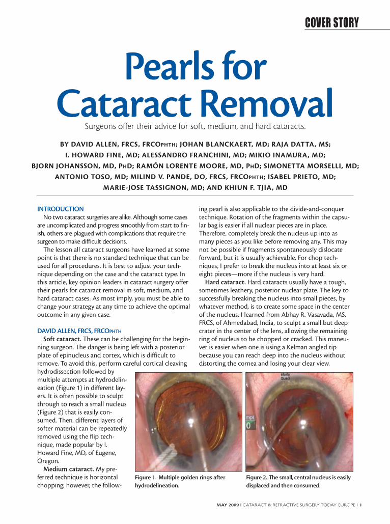
Soft cataract. These can be challenging for the begin-ning surgeon. The danger is being left with a posteriorplate of epinucleus and cortex, which is difficult toremove. To avoid this, perform careful cortical cleavinghydrodissection followed bymultiple attempts at hydrodelin-eation (Figure 1) in different lay-ers. It is often possible to sculptthrough to reach a small nucleus(Figure 2) that is easily con-sumed. Then, different layers ofsofter material can be repeatedlyremoved using the flip tech-nique, made popular by I.Howard Fine, MD, of Eugene,Oregon.
Medium cataract. My pre-ferred technique is horizontalchopping; however, the follow-
ing pearl is also applicable to the divide-and-conquertechnique. Rotation of the fragments within the capsu-lar bag is easier if all nuclear pieces are in place.Therefore, completely break the nucleus up into asmany pieces as you like before removing any. This maynot be possible if fragments spontaneously dislocateforward, but it is usually achievable. For chop tech-niques, I prefer to break the nucleus into at least six oreight pieces—more if the nucleus is very hard.
Hard cataract. Hard cataracts usually have a tough,sometimes leathery, posterior nuclear plate. The key tosuccessfully breaking the nucleus into small pieces, bywhatever method, is to create some space in the centerof the nucleus. I learned from Abhay R. Vasavada, MS,FRCS, of Ahmedabad, India, to sculpt a small but deepcrater in the center of the lens, allowing the remainingring of nucleus to be chopped or cracked. This maneu-ver is easier when one is using a Kelman angled tipbecause you can reach deep into the nucleus withoutdistorting the cornea and losing your clear view.
Pearls for Cataract Removal
Surgeons offer their advice for soft, medium, and hard cataracts.
BY DAVID ALLEN, FRCS, FRCOPHTH; JOHAN BLANCKAERT, MD; RAJA DATTA, MS;
I. HOWARD FINE, MD; ALESSANDRO FRANCHINI, MD; MIKIO INAMURA, MD;
BJORN JOHANSSON, MD, PHD; RAMÓN LORENTE MOORE, MD, PHD; SIMONETTA MORSELLI, MD;
ANTONIO TOSO, MD; MILIND V. PANDE, DO, FRCS, FRCOPHTH; ISABEL PRIETO, MD;
MARIE-JOSE TASSIGNON, MD; AND KHIUN F. TJIA, MD
Figure 1. Multiple golden rings after
hydrodelineation.
Figure 2. The small, central nucleus is easily
displaced and then consumed.
JOHAN BLANCKAERT, MDSoft cataract. In most soft cataract cases, the patient is
young and would like to continue leading an active lifestyle. Apremium IOL solution or a multifocal IOL is the best optionfor these active patients. Its obvious that in any cataract caseyou always need to do whatever you can to avoid posteriorcapsule rupture and zonulolysis. These two complications cancompromise IOL centration, an essential component whenimplanting premium IOLs. For this reason, in those softercataracts I frequently opt to do either a phaco roll or nucleusrotation technique, which has been so well described by JoseL. Guell, MD, of Spain. This technique reduces zonular stressand is safe for the posterior capsule. The most importantpearl is achieving a good hydrodissection.
Medium cataract. Patients with medium cataracts aretypically standard cataract patients. My first pearl would be:Do not fall into a routine. Secondly, a good anterior capsu-lorrhexis must be centered on the first Purkinje reflex of thecornea. A 5.5-mm capsulorrhexis is the best choice in thesepatients because it is large enough to avoid anterior capsulecontraction and small enough to provide an overlap of theIOL optic. If posterior capsule rupture forces you to use asulcus-fixated IOL, then optic capture can still be done withexcellent IOL centration.
Hard cataract. Avoiding corneal burn is the most impor-tant point in patients with hard cataracts. I would chooseeither torsional phaco with the Infiniti platform (AlconLaboratories, Inc., Fort Worth, Texas) or the WhitestarSignature Ice platform (Abbott Medical Optics, Inc., SantaAna, California). Both systems are notorious for the coolphaco needle temperature needed in those hard, longcataract emulsifications.
RAJA DATTA, MSSoft cataract. Cracking the nucleus and completely
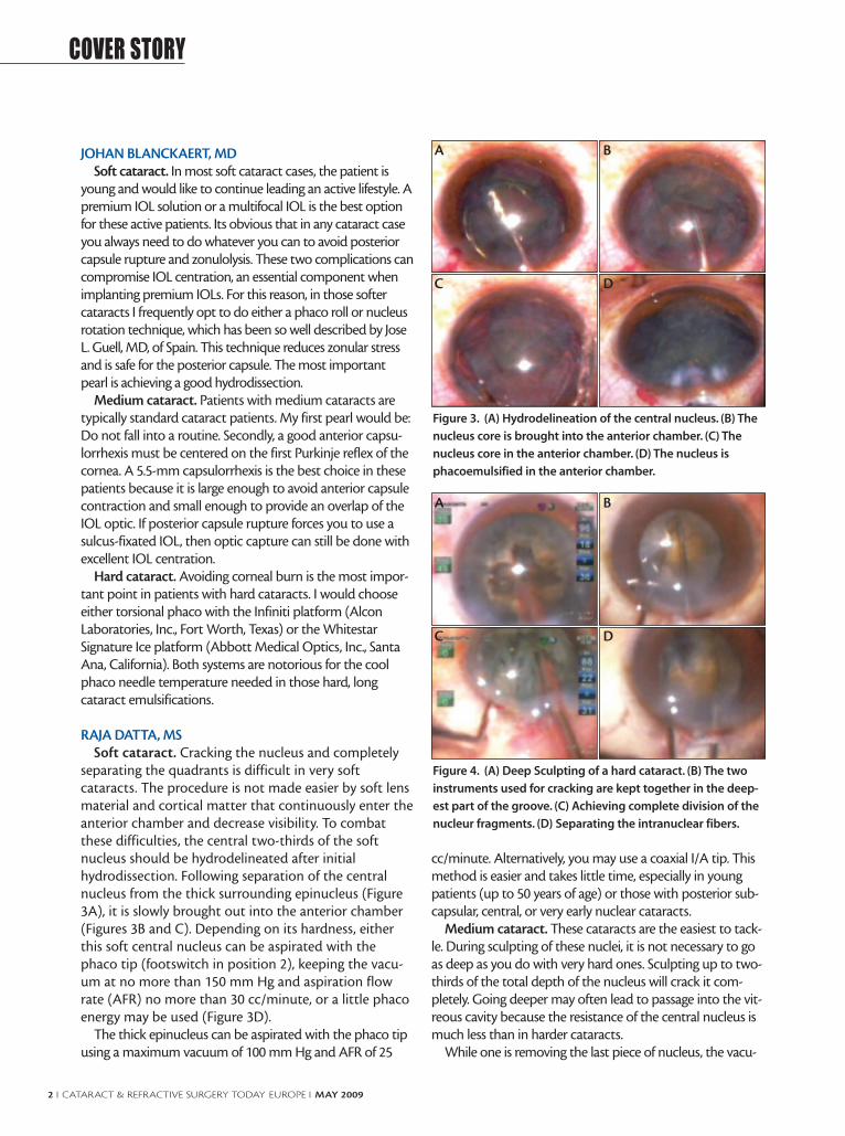
separating the quadrants is difficult in very softcataracts. The procedure is not made easier by soft lensmaterial and cortical matter that continuously enter theanterior chamber and decrease visibility. To combatthese difficulties, the central two-thirds of the softnucleus should be hydrodelineated after initialhydrodissection. Following separation of the centralnucleus from the thick surrounding epinucleus (Figure3A), it is slowly brought out into the anterior chamber(Figures 3B and C). Depending on its hardness, eitherthis soft central nucleus can be aspirated with thephaco tip (footswitch in position 2), keeping the vacu-um at no more than 150 mm Hg and aspiration flowrate (AFR) no more than 30 cc/minute, or a little phacoenergy may be used (Figure 3D).
The thick epinucleus can be aspirated with the phaco tipusing a maximum vacuum of 100 mm Hg and AFR of 25
cc/minute. Alternatively, you may use a coaxial I/A tip. Thismethod is easier and takes little time, especially in youngpatients (up to 50 years of age) or those with posterior sub-capsular, central, or very early nuclear cataracts.
Medium cataract. These cataracts are the easiest to tack-le. During sculpting of these nuclei, it is not necessary to goas deep as you do with very hard ones. Sculpting up to two-thirds of the total depth of the nucleus will crack it com-pletely. Going deeper may often lead to passage into the vit-reous cavity because the resistance of the central nucleus ismuch less than in harder cataracts.
While one is removing the last piece of nucleus, the vacu-
2 I CATARACT & REFRACTIVE SURGERY TODAY EUROPE I MAY 2009
COVER STORY
Figure 3. (A) Hydrodelineation of the central nucleus. (B) The
nucleus core is brought into the anterior chamber. (C) The
nucleus core in the anterior chamber. (D) The nucleus is
phacoemulsified in the anterior chamber.
A B
C D
Figure 4. (A) Deep Sculpting of a hard cataract. (B) The two
instruments used for cracking are kept together in the deep-
est part of the groove. (C) Achieving complete division of the
nucleur fragments. (D) Separating the intranuclear fibers.
A B
C D
um should be between 100 and 150 mm Hg and AFRbetween 25 and 30 cc/minute. The reason for these settingsis not because of the jumping fragments that may hit andrupture the posterior capsule, but in case of accidental occlu-sion of the phaco tip with posterior cortical matter and theposterior capsule, which can cause a rent. With these param-eters set low in the event of accidental occlusion, you canimmediately switch to footswitch position 1, which shouldrelease the posterior capsule without causing damage.
Hard cataract. Cracking the nucleus in the two extremesof nuclear hardness (ie, very soft and very hard) is a difficulttask. Hard to very hard cataracts, especially those that areamber or black in color, are the most difficult to crack. Inthese cases, it is advisable to adopt the four-quadrant orstop-and-chop method.
Sculpting should be done deeply, until the posteriornuclear plate is reached (Figure 4A). The two instrumentsused to crack the nucleus are placed so that the tips toucheach other as well as the floor of the groove (Figure 4B). Thetips are then slowly separated to create a crack in the nucle-us that should start from the periphery, under full visibility,and gradually move toward the center. Care must be takento ensure complete separation of the nuclear fragments,including the posterior nuclear plate (Figure 4C).
Difficulty arises in these cases due to the resilient intranu-clear fibers, which are tenacious and difficult to separate. It isalways advisable to break these fibers by separating the frag-ments at their site of origin (Figure 4D).
Utmost care must be taken while separating thenuclear fragments to achieve a complete crack, whichshould extend throughout the depth of the nucleus.Since these nuclei are not only hard and leathery butalso extra large in size, they occupy almost the whole ofthe capsular bag, which is distended, with little or nocortical matter separating them.
Care must also be taken while creating the crack. Pullingthe pieces away from each other to crack the nucleus trans-mits pressure directly to the capsular bag. The more dis-tance between pieces, especially at the periphery, the morethe stretch force on the already distended capsular bag,which may lead to a capsular tear. Therefore, it is imperativeto start cracking at the periphery and go slowly down to thecenter of the nucleus.
I. HOWARD FINE, MDSoft cataract. For soft nuclei, I hydroexpress the lens
into the capsulorrhexis plane and carousel it with thebevel of the phaco tip pointed toward the lens equator.I place the second instrument above the nucleus, pro-tecting the cornea.
Medium cataract. For these cataracts, I prefer hori-zontal chopping. Prior to embedding the tip, I go over
the equator with the horizontal chopper, pulling up andtoward the incision to stabilize the nucleus as I bury thephaco tip. This ensures that I am not transmitting anyforce to the capsule or zonules.
Hard cataract. For hard cataracts, I use vertical choppingand perform all chopping in the traditional, longitudinalphaco mode prior to mobilizing the segments. Therefore,the tip is not widened as it is buried. Then I change parame-ters for segment mobilization to include torsional or ellipti-cal phaco.
ALESSANDRO FRANCHINI, MDSoft cataract. Removing very soft nuclei and cracking the
lens into small fragments can be harder than it would seemduring either divide-and-conquer or chop techniques.Especially for surgeons in training, it can be difficult to sepa-rate the two epinuclei or the four nuclear quadrants aftersculpting. The nuclear fragment substance can be gummy,and manipulators or choppers can easily penetrate the sub-stance without applying the necessary strength to separateit. In these cases, it may be useful to deepen the groove asmuch as possible; however, this maneuver risks a posteriorcapsule break, especially in the hands of a young surgeon. Ifthe division does not appear in the middle of the nucleus,and two different-sized fragments result, one should refrac-ture the larger piece.
I prefer to use cracking forceps in patients with soft nuclei.These forceps have two flat paddles that apply lateral sepa-ration onto a larger area of tissue, allowing the perfect divi-sion of two equal fragments—even if the groove has notbeen well performed or if the nucleus is particularly soft.The most recent cracking forceps models can be easilyinserted into the anterior chamber through a 2.2-mm inci-sion. Furthermore, the lower part of the two paddles has afine edge so that if the groove is not sufficiently deep, theycan be used to perform a type of karate chop.
Medium cataract. Separating the lens material from thecapsule (ie, hydrodissection) and the nucleus from the epin-ucleus (ie, hydrodelineation) represent the most crucial stepsin patients with a medium-hard cataract. These cataracts areconsidered the easiest to remove; however, the surgeonshould avoid beginning phacoemulsification before thenucleus is mobilized with these two maneuvers and thenrotated. When a good separation between the lens layers isobtained, difficulties aspirating the cataract are avoided.
Avoid initiating hydrodissection if the anterior chamber isover-inflated with an ophthalmic viscosurgical device(OVD). When performing microincision cataract surgery(MICS), be aware of the risk of increased pressure in theanterior chamber.
To perform hydrodissection, a cannula is inserted into theanterior chamber, under the capsulorrhexis border and
MAY 2009 I CATARACT & REFRACTIVE SURGERY TODAY EUROPE I 3
COVER STORY
between the capsule and lens cortex. This is done in anattempt to slightly raise the capsule and direct the fluidstream along the inner surface of the capsular bag. Balancedsaline solution is injected to separate the lens from the cap-sule. It is necessary to continue injecting fluid until the wavecompletely crosses behind the nucleus. The center of thenucleus is pushed gently backward to allow the fluid to passthrough the capsular equator and obtain complete separa-tion from the capsule. This maneuver is repeated as manytimes as necessary to facilitate good lens rotation.
Using a straight cannula, the surgeon then injects bal-anced saline solution into only the contraincisional portionof the bag. It is preferable to use a 90º angled cannula toinject fluid in the right and left portions of the bag, guaran-teeing complete hydrodissection.
To facilitate aspiration of a medium-hard cataract, thenucleus is separated from the epinucleus with hydrodelin-eation. The cannula is inserted deeply inside the peripheryof the nucleus, and balanced saline solution is injected untila golden ring appears between the nucleus and epinucleus.
After hydrodissection and hydrodelineation have beenwell performed, the surgeon can easily aspirate the nucleus,epinucleus, and cortex without moving the phaco tip fromthe pupillary field. This technique decreases surgical timeand increases efficiency and, above all, safety.
Hard cataract. Often with the brunescent cataract, weare dealing with a nucleus of enlarged dimension andincreased density. There is no epinucleus and often no corti-cal layer. Hard cataracts are somewhat like a pillow in thatthey absorb the transmission of force applied to the posteri-or capsule and zonula. Therefore, mechanical forces appliedduring sculpting, cracking, and rotation are transmittedstraight to the posterior capsule and the zonula, which mayalready be weak in these patients. The nuclear fragments areoften hard and sharp, increasing the risk for a break in theposterior capsule.
The enlarged depth and width of the cataract can create anumber of problems, whether we perform a divide-and-con-quer or horizontal chop technique. Breaking the most poste-rior layers of the nuclear plate requires digging deep and closeto the posterior capsule. If one performs horizontal chop,inserting the chopper as deeply as possible can be dangerousbecause of the absence of an epinucleus. However, it is oftendifficult to obtain a clean fracture with horizontal chopbecause tissue bridges connecting the two fragments canremain intact. A vertical chop technique is generally moresuccessful in breaking these bridges, so this may be the besttechnique for patients with brunescent cataract.
With both the divide-and-conquer and horizontal chop-ping techniques, we cut only part of the nucleus to avoidbreaking the posterior capsule. To complete the fracture, weseparate the two heminuclei; however, the force of either
technique only partially spreads the nuclei posteriorly. In thecase of very hard nuclei, the propagating fracture may con-tinue only horizontally without advancing further posterior-ly. This is the reason the tissues bridges remain together.
Alternatively, the backward-and-forward vertical move-ment applied during vertical chop favors propagation of thefracture to the most posterior layers. In patients with awhite cataract with a very hard nucleus, vertical chop is thebest choice in terms of efficiency and safety, making it possi-ble to work only in the pupillary field without concern forwhere the posterior capsule is located.
MIKIO INAMURA, MDSoft cataract. The Infiniti OZil Torsional phaco on the
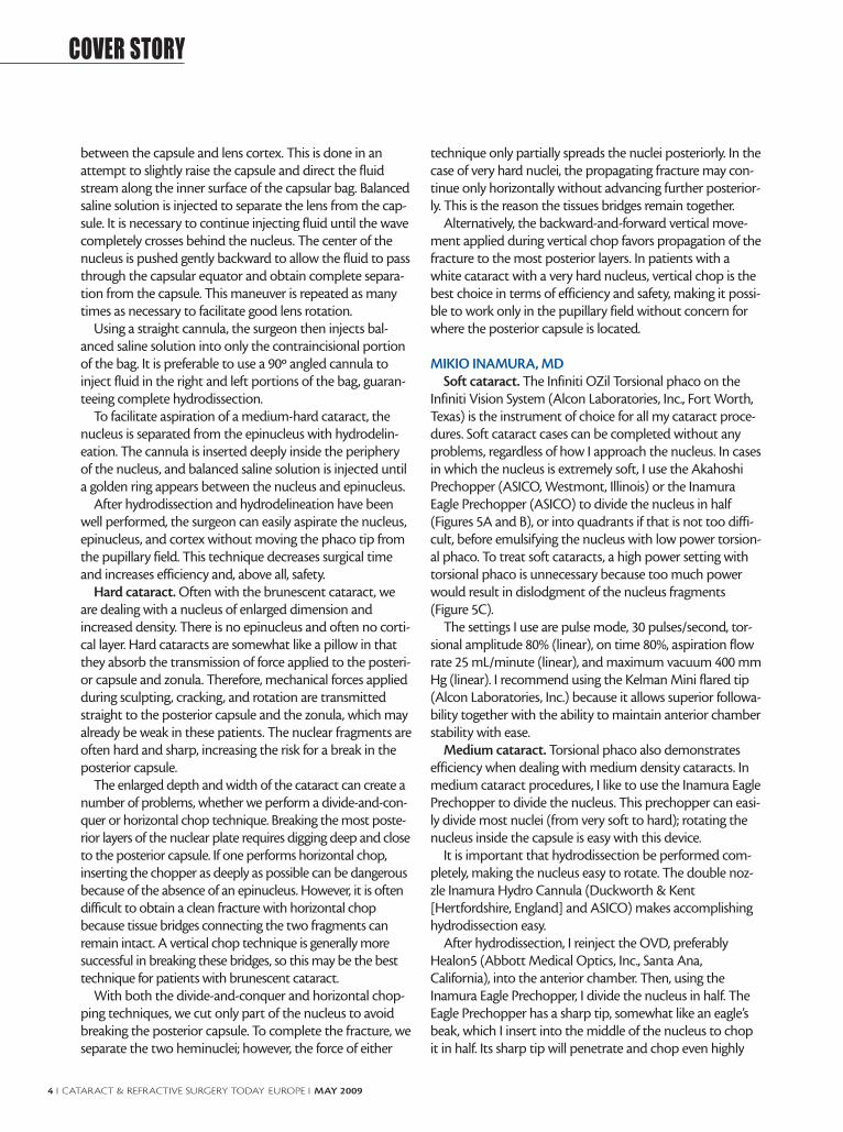
Infiniti Vision System (Alcon Laboratories, Inc., Fort Worth,Texas) is the instrument of choice for all my cataract proce-dures. Soft cataract cases can be completed without anyproblems, regardless of how I approach the nucleus. In casesin which the nucleus is extremely soft, I use the AkahoshiPrechopper (ASICO, Westmont, Illinois) or the InamuraEagle Prechopper (ASICO) to divide the nucleus in half(Figures 5A and B), or into quadrants if that is not too diffi-cult, before emulsifying the nucleus with low power torsion-al phaco. To treat soft cataracts, a high power setting withtorsional phaco is unnecessary because too much powerwould result in dislodgment of the nucleus fragments(Figure 5C).
The settings I use are pulse mode, 30 pulses/second, tor-sional amplitude 80% (linear), on time 80%, aspiration flowrate 25 mL/minute (linear), and maximum vacuum 400 mmHg (linear). I recommend using the Kelman Mini flared tip(Alcon Laboratories, Inc.) because it allows superior followa-bility together with the ability to maintain anterior chamberstability with ease.
Medium cataract. Torsional phaco also demonstratesefficiency when dealing with medium density cataracts. Inmedium cataract procedures, I like to use the Inamura EaglePrechopper to divide the nucleus. This prechopper can easi-ly divide most nuclei (from very soft to hard); rotating thenucleus inside the capsule is easy with this device.
It is important that hydrodissection be performed com-pletely, making the nucleus easy to rotate. The double noz-zle Inamura Hydro Cannula (Duckworth & Kent[Hertfordshire, England] and ASICO) makes accomplishinghydrodissection easy.
After hydrodissection, I reinject the OVD, preferablyHealon5 (Abbott Medical Optics, Inc., Santa Ana,California), into the anterior chamber. Then, using theInamura Eagle Prechopper, I divide the nucleus in half. TheEagle Prechopper has a sharp tip, somewhat like an eagle’sbeak, which I insert into the middle of the nucleus to chopit in half. Its sharp tip will penetrate and chop even highly
4 I CATARACT & REFRACTIVE SURGERY TODAY EUROPE I MAY 2009
COVER STORY
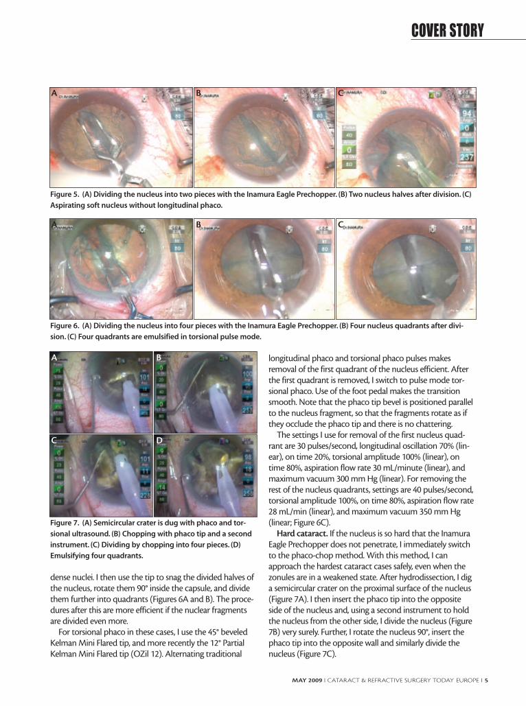
dense nuclei. I then use the tip to snag the divided halves ofthe nucleus, rotate them 90° inside the capsule, and dividethem further into quadrants (Figures 6A and B). The proce-dures after this are more efficient if the nuclear fragmentsare divided even more.
For torsional phaco in these cases, I use the 45° beveledKelman Mini Flared tip, and more recently the 12° PartialKelman Mini Flared tip (OZil 12). Alternating traditional
longitudinal phaco and torsional phaco pulses makesremoval of the first quadrant of the nucleus efficient. Afterthe first quadrant is removed, I switch to pulse mode tor-sional phaco. Use of the foot pedal makes the transitionsmooth. Note that the phaco tip bevel is positioned parallelto the nucleus fragment, so that the fragments rotate as ifthey occlude the phaco tip and there is no chattering.
The settings I use for removal of the first nucleus quad-rant are 30 pulses/second, longitudinal oscillation 70% (lin-ear), on time 20%, torsional amplitude 100% (linear), ontime 80%, aspiration flow rate 30 mL/minute (linear), andmaximum vacuum 300 mm Hg (linear). For removing therest of the nucleus quadrants, settings are 40 pulses/second,torsional amplitude 100%, on time 80%, aspiration flow rate28 mL/min (linear), and maximum vacuum 350 mm Hg(linear; Figure 6C).
Hard cataract. If the nucleus is so hard that the InamuraEagle Prechopper does not penetrate, I immediately switchto the phaco-chop method. With this method, I canapproach the hardest cataract cases safely, even when thezonules are in a weakened state. After hydrodissection, I diga semicircular crater on the proximal surface of the nucleus(Figure 7A). I then insert the phaco tip into the oppositeside of the nucleus and, using a second instrument to holdthe nucleus from the other side, I divide the nucleus (Figure7B) very surely. Further, I rotate the nucleus 90°, insert thephaco tip into the opposite wall and similarly divide thenucleus (Figure 7C).
MAY 2009 I CATARACT & REFRACTIVE SURGERY TODAY EUROPE I 5
COVER STORY
Figure 5. (A) Dividing the nucleus into two pieces with the Inamura Eagle Prechopper. (B) Two nucleus halves after division. (C)
Aspirating soft nucleus without longitudinal phaco.
A B C
Figure 6. (A) Dividing the nucleus into four pieces with the Inamura Eagle Prechopper. (B) Four nucleus quadrants after divi-
sion. (C) Four quadrants are emulsified in torsional pulse mode.
A B C
Figure 7. (A) Semicircular crater is dug with phaco and tor-
sional ultrasound. (B) Chopping with phaco tip and a second
instrument. (C) Dividing by chopping into four pieces. (D)
Emulsifying four quadrants.
A B
C D
After dividing the nucleus in this way, I set about toremove the fragments with torsional phaco (Figure 7D).Aspirating dense nuclear material with torsional phacoalone can cause clogging, but when torsional is combinedwith longitudinal oscillations, clogging can be minimized.Another way to prevent clogging is by slowly emulsifyingthe nuclear fragments piece by piece. It is most importantto avoid completely occluding the phaco tip with nucleusfragments. This is strikingly different from traditional longi-tudinal phaco and must be observed.
The settings for removal of the first nucleus quadrant are30 pulses/second, longitudinal oscillation 70% (linear), ontime 20%, torsional amplitude 100%, on time 80%, aspirationflow rate 30 mL/minute (linear), and maximum vacuum 300mm Hg (linear). For removal of the rest of the nuclear quad-rants, settings are 40 pulses/second, torsional amplitude100%, on time 80%, aspiration flow rate 28 mL/minute (lin-ear), and maximum vacuum 350 mm Hg (linear).
If clogging occurs, I get rid of it by switching the setting tolongitudinal oscillation, declogging mode at 20 pulses/sec-ond, longitudinal oscillation 100% (linear), on time 20%,aspiration flow rate 25 mL/min fixed, and maximum vacu-um 300 mm Hg (linear).
Recently I have been using the Akahoshi Bent wobblesquare tip (ASICO), and I found it to be efficient for remov-ing the nuclear fragments. It causes less injury to the corneaand iris compared to the Kelman Mini Flared tip, and it iseasier to use because of its straight tip. It can be used oncataracts of all densities. It is also not prone to clogging. Thistip is not yet on the market, but I feel there is great potentialfor its use.
BJORN JOHANSSON, MD, PHDSoft cataract. If the soft cataract goes unnoticed preoper-
atively, you will recognize it as you initiate hydrodelineationafter cortical cleaving hydrodissection—two crucial steps inevery safe cataract procedure. Your blunt cannula will notmeet resistance, or you will find a very small nucleus whenbalanced saline solution is injected into the deeper layers ofthe lens.
Attempts to crack or chop soft nuclei are, of course, sel-dom fruitful. In these cases, aspiration without ultrasound isusually enough to evacuate the lens, either by stopping atposition 2 on a monolinear pedal or activating only vacu-um/aspiration on a dual-linear foot control. I consume thecentral nucleus first and then catch the anterior rim of theremaining bowl opposite the phaco tunnel, flipping itupside-down with help from the second instrument. Thismakes it easy to aspirate efficiently at a safe distance fromthe posterior capsule in the center of the pupil.
Medium cataract. Several years ago, I transitioned from astop-and-chop technique to a direct horizontal chop with
the Nagahara chopper (Katena Products, Inc., Denville, NewJersey). Earlier, I was a traditional four-groove divide-and-conquer surgeon. Creating four grooves is advantageous forthe beginning surgeon because it allows you to get a goodgrasp of the anatomy of the lens and capsule in various sce-narios, including exfoliation and shallow or deep chambers. Ifound it a helpful precursor to chop techniques.
When I first tried direct horizontal chop, it was a bit diffi-cult. If you wish to progress to chopping, first master stop-and-chop: Create one initial groove; crack the nucleus intohalves, which can usually be mobilized; capture one half ofthe nucleus with the phaco tip, with vacuum increasedfrom the groove setting; and chop it with the side instru-ment. This technique allows you to try various choppers onlens pieces that are freed from the capsule, which I thinkmakes learning easier.
Hard cataract. With hard nuclei, I often have a problemgetting a chop completely down through to the back side ofthe nucleus, which can be fibrotic and leathery. In thesecases, my tactic is one of the following: Either I use the sideinstrument and viscoelastic cannula, inserted through thephaco tunnel, to separate the nuclear pieces (after divide-and-conquer, stop-and-chop, or direct chop); or, if theinstrument is available, I use an Akahoshi prechopper. Bothmethods allow one to easily apply forces at the deepest pos-sible point in the nucleus and to direct the splitting force inthe most efficacious directions.
RAMÓN LORENTE MOORE, MD, PHDSoft cataract. Although often considered an easy surgery,
the soft cataract may pose refractive and technical chal-lenges. As a refractive cataract procedure, there is thedemand to achieve accurate biometry and uneventful sur-gery, leading to extra pressure for the surgeon. Technicalproblems when handling the soft nuclei during phacoemul-sification are twofold: First, there is difficulty rotating thenucleus-cortex complex because cortical fibers tend to stickto the capsule. Second, there is an inability to divide thenucleus due to its softness. The following points summarizemy strategy to overcome these problems.
An adequate hydroprocedure is mandatory to achievecomplete nuclear rotation. First, cortical cleaving hydrodis-section must be performed to detach the cortex from thecapsule so that the cortex remains attached to the epinucle-us. It may be helpful to repeat the same maneuver in theopposite distal quadrant. Given that an excessive amount ofOVD can increase the resistance to fluid egress from thecapsular bag via the capsulorrhexis, an important stepbefore starting hydrodissection is to evacuate some of OVDfrom the anterior chamber. After hydrodissection, thenucleous-cortex complex is rotated bimanually (using thetwo-Sinskey hook method) to confirm that there is no
6 I CATARACT & REFRACTIVE SURGERY TODAY EUROPE I MAY 2009
COVER STORY
adherence between the cortex and the capsule. The nextstep is hydrodelineation, which by cleaving the centralnucleus from the epinucleus, facilitates phacoemulsification.A golden ring of the delineated nucleus is its hallmark sign.
My preferred method for nuclear emulsification of softnuclei is the chip-and-flip technique using AquaLase tech-nology (Alcon Laboratories, Inc.). It is not only the safesttechnology but it could also be associated with a lower rateof posterior capsule opacification.
The rest of the procedure is the same that will bedescribed for medium cataracts.
Medium cataract. My surgical technique in mediumcataracts corresponds to the procedure I perform in stan-dard cases. Thus, I will describe my personal technique inthis setting. I use topical anesthesia with intracameral pre-servative-free lidocaine 2%. The anterior chamber is filledwith a viscoadaptative OVD (DisCoVisc; Alcon Laboratories,Inc.) through the paracentesis, and a temporal clear cornealincision (2.2 x 1.75 mm) is made. Should a multifocal IOL beimplanted, the incision is placed at the steepest meridian. Acircular, well-centered, 5.25-mm capsulorrhexis is perfomedwith Utrata forceps. The hydroprocedures are performed aspreviously described for soft cataracts.
During phacoemulsification, I usually work with torsionalultrasound technology for two main reasons: it is easier andsafer. These advantages are related to its shearing action,which minimizes repulsion and allows use of lower vacuumparameters to decrease surge. To optimize the torsionaleffect, achieve better holdability, and reduce clogging, aKelman Mini flared 45º bevel tip is strongly recommended.My preferred technique is vertical chop using a Rosen chop-per to divide the nucleous into four or five fragments withthe following parameters: 90% torsional amplitude withburst mode (50 milliseconds [mSec] on, 150 mSec off); vac-uum, 420 mm Hg; flow, 35 cc/minute; and bottle height, 95cm.
Once the nucleus is divided, parameters are adapted toemulsify the fragments: continuous torsional ultrasound(maximum amplitude, 90% and starting amplitude, 20%);vacuum, 320 mm Hg; flow, 25 cc/minute; bottle height, 95cm. The footpedal set-up is adjusted to 18% to shorten therange of footpedal position 3. There is no need for longitu-dinal ultrasound.
During irrigation and aspiration, the cortex is aspiratedwith a curved silicone I/A tip (Alcon Laboratories, Inc.) dueto its greater levels of safety—the incidence of posteriorcapsule rupture is lower than using metal tip. Wheneversubincisional cortex removal becomes difficult, it may be agood option to use two separate cannulas, one for irrigatingthe anterior chamber and another for aspirating corticalmaterial. This bimanual I/A technique requires two paracen-tesis placed approximately 50º apart from the main incision.
I routinely polish the posterior capsule with the silicone tipor with an irrigating polisher.
I typically implant the AcrySof IQ (Alcon Laboratories,Inc.) using the Monarch III injector and Monarch D car-tridge (Alcon Laboratories, Inc.). I do not need to enlargethe incision. Residual OVD trapped under the IOL is thenremoved. Cefuroxime is used both for intracameral adminis-tration and stromal hydration at both edges of the clearcorneal incision.
Hard cataract. Handling the hard nucleus is a major chal-lenge, even for the experienced surgeon. It requires higherpower ultrasound and prolonged phaco time. Preoperativeevaluation is of paramount importance. We must considerthe following points: endothelial cell count, anterior cham-ber depth, zonular instability, pupil dilation, and a B-scan ifthere is no view of posterior fundus.
Phacoemulsification in hard cataracts requires the use ofspecific strategies that differ from routine phacoemulsifica-tion. The main modifications of each surgical step to suc-cessfully operate such cataracts are as follows. Local anes-thesia is recommended in cases of weak zonulas, poor col-laboration, or risk of intraoperative complications. Whenthe red reflex is poor, we must stain the capsule with trypanblue 0.06% to enhance capsular visualization enough to per-form capsulorrhexis and also visualization of the edge of therhexis during phacoemulsification.
OVD plays an important role in protecting the endo-thelium in hard cataracts. For this reason, I use StevenArshinoff´s soft shell technique, using Viscoat to coat theendothelium and a highly cohesive OVD (Provisc; AlconLaboratories, Inc.) to maintain the anterior chamber.
Phacoemulsification for hard lenses poses two main risks:higher endothelial cell loss and increased risk of posteriorcapsule rupture. Furthermore, dividing the hard nucleusbecomes difficult. Posterior layer fibers can be cohesive andtenacious and resist to all conventional methods of division.Extra precautions must be taken to protect the endotheli-um and the posterior capsule.
Torsional ultrasound is the best technology to protectthe endothelium due to minimal repulsion and less turbu-lence in the anterior chamber. My preferred technique isvertical chop because it causes less endothelial cell loss andless stress on the zonules versus divide and conquer. I do notchange many parameters with respect to medium cataracts,apart from elevating the starting torsional amplitude settingto 30%. However, there are a few variations to my techniquein hard cataracts: (1) I use a karate chopper, which is longerand sharper, to facilitate embedding the dense nucleus with-out displacing it. (2) The irrigation sleeve must be retractedmore than usual. This will expose a longer segment of themetal needle and maximize penetration of the tip, which iscrucial to divide the nucleus. It is easier to begin by sculpting
MAY 2009 I CATARACT & REFRACTIVE SURGERY TODAY EUROPE I 7
COVER STORY
a small, deep, pit centrally. This pit allows the nucleus to beimpaled more deeply. (3) It is more efficient to alter theangle of the vertical chop slightly and approach the embed-ded phaco tip more diagonally. This provides more of a hor-izontal vector that pushes the nucleus against the tip whilethe vertical vector initiates the downward fracture, combin-ing the mechanical advantages of both strategies. (4) If thereare leathery fibers at the posterior layer, it is best to transectthem with the chopper while the nucleus is engaged andstabilized by the vacuum of the phaco tip. (5) The nucleusshould be divided in smaller fragments to emulsify themsecurely. (6) To maximize endothelial protection, we shouldrefill the anterior chamber with Viscoat during fragmentemulsification. (7) A dispersive OVD injected behind the lastremaining fragments creates and artificial epinucleus thatwill restrain the lax and fragile posterior capsule from tram-polining toward the phaco tip, minimizing the risk of rup-ture.
SIMONETTA MORSELLI, MD; AND ANTONIO TOSO, MD
Soft cataract. We use very high vacuum and very lowultrasound power to remove soft cataracts. We suggestusing a manipulator rather than a chopper, avoiding dam-age to the posterior capsule during nuclear fragmentremoval with high vacuum.
Medium cataract. We use the Stellaris (Bausch & Lomb,Rochester, New York), a phacoemulsification platform creat-ed specifically for MICS. This system is exceptionally fast andsafe. Its capability for high vacuum allows quick removal ofnuclear fragments while the chamber remains perfectly sta-ble. This machine gives one the ability to set the ultrasoundin two different modes, burst and micropulse. By changingthe duration and the duty cycle of the ultrasound, we canadapt the power to any type of cataract.
We set sub mode 1 for cataracts of 2+ to 3+ hardness;sub mode 2 for 3+ to 4+ cataracts; and sub mode 3 for 5+to 6+ cataracts.
Our parameters for sub mode 1 for normal or medium2+ to 3+ cataracts are: dual foot pedal system; 10% linearultrasound, pulse mode 80 pps; 35% duty cycle.
Hard cataract. Even with very hard cataracts, we are ableto remove pieces with not more than 10% ultrasoundpower.
Our parameters for sub mode 2, used for 3+ to 4+cataracts, are: dual foot pedal system; 10% linear ultrasound;fixed burst 160 millisecond duration; 320 millisecond inter-val of pulse duration.
Our parameters for sub mode 3, used for 5+ to 6+cataracts, are: dual foot pedal system; 10% fixed ultrasound;multiple burst 40 millisecond burst duration; 60% dutycycle.
With these multiple modes set, we are able to change toany of these parameters during surgery. For example, tochop the nucleus at the beginning of surgery, we might usesub mode 3. To remove the final little pieces floating intothe anterior chamber toward the end of the case, we mightuse sub mode 1.
Our machine is set so that we can change sub modesduring surgery using the left foot pedal on the dual footpedal system. With the right foot pedal, we control ultra-sound power with a lateral motion and vacuum by depress-ing the pedal. The surgeon can change the sub mode at anymoment during surgery.
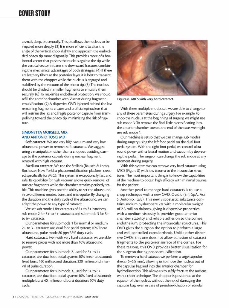
With this system we can remove very hard cataract usingMICS (Figure 8) with low trauma to the intraocular struc-tures. The most important thing is to know the capabilitiesof the machine to obtain high efficacy with minimal traumafor the patient.
Another pearl to manage hard cataracts is to use achop technique with a new OVD, Ovidio (Sifi, SpA, AciS. Antonio, Italy). This new viscoelastic substance con-tains sodium hyaluronate 2% with a molecular weightof 2.3 million daltons, giving it dispersive propertieswith a medium viscosity. It provides good anteriorchamber stability and reliable adhesion to the cornealendothelium, protecting the intraocular structures. ThisOVD gives the surgeon the option to perform a largeand well-controlled capsulorrhexis. Unlike other disper-sive OVDs, this one does not allow adhesion of cataractfragments to the posterior surface of the cornea. Forthese reasons, this OVD provides better visualization forthe surgeon during phacoemulsification.
To remove a hard cataract we perform a large capsulor-rhexis (6–6.5 mm), allowing us to move the nucleus out ofthe capsular bag and into the anterior chamber forhydrodissection. This allows us to safely fracture the nucleuswith a chop technique. The chopper is positioned at theequator of the nucleus without the risk of damaging thecapsular bag, even in case of pseudoexfoliation or zonular
8 I CATARACT & REFRACTIVE SURGERY TODAY EUROPE I MAY 2009
COVER STORY
Figure 8. MICS with very hard cataract.
weakness. A small amount of OVD is injected under thenucleus and over the capsular bag. The phaco tip, withbevel down, impales the nucleus as deeply into the center aspossible. The sleeve must be retracted to obtain as much as1.8 to 2 mm of tip length, thus penetrating the center of thenucleus. A deep nuclear fracture is then obtained with thechopper. After the first crack, the nucleus is divided intosmall slices and emulsified; using the manipulator avoidsdamage to the capsular bag. During these maneuvers, it isnecessary to refill the anterior chamber with OVD to pro-tect the endothelium and maintain stabilization of thenucleus.
MILIND V. PANDE, DO, FRCS, FRCOPHTH
Soft cataract. Adjust your flow and vacuum settingsdown when you want move the nucleus in one piece. If thevacuum or flow is too high, it will nibble away at the nucle-us rather than enable you to move the whole nucleus. It is abit like moving a blob of jelly without breaking it up; itrequires just the right amount of force.
Medium cataract. When you are chopping a mediumcataract, your angle of attack with the phaco probe must beacute. A shallow angle will drive the phaco tip superficialand into the peripheral (and thinner) aspect of the nucleus,increasing the risk of capsular rupture. An acute, almost ver-tical angle of approach will embed the phaco probe into thecentral part of the nucleus, engaging it securely on thephaco tip for a chop.
Hard cataract. The key in these cases is to be patient anddismantle the nucleus bit by bit. A combination of horizon-tal and vertical chop will allow segmental dismantling,much like removing the petals of a flower one by one, leav-ing the central posterior leathery core to be emulsified atthe end. Chipping safely in these nuclei means taking small-er bites; using lots of OVD protection; and adopting apatient, methodical approach.
ISABEL PRIETO, MDSoft cataract. Whether one faces a soft, medium, or hard
cataract, it is important to have a plan preoperatively. Underthe microscope light, many dense cataracts appear like softcataracts, so it is important to know ahead of time whatyou are facing for each case and have the right handpiece,the right phaco tip, the right technique. The goal in all casesis to customize cataract surgery depending on the needs ofthe patient.
My specialty is complicated cases, so often the cases I seeare not soft cataracts. However, it is possible for cataracts tobe very soft and yet still complicated. An example of a softbut difficult cataract could be, for instance, a posterior polarcataract, which may be more challenging than a hardernuclear cataract.
My preferred technique is microcoaxial phaco with a 2.2-mm incision. For soft cataracts, I like to use the AquaLasemode on the Infiniti phacoemulsification system (AlconLaboratories, Inc.). This is a safe mode because it has a lowerrisk of capsule rupture. Normally in soft lenses I use a modi-fied chip-and-flip technique with a Barraquer spatula(Moria, Antony, France) as my second instrument. Toremove cortical material, I use bimanual irrigation and aspi-ration because of improved cleaning, especially of the subin-cisional cortex. It is important in these patients to clean thecapsular bag thoroughly of cortical remnants to reduce thepostoperative inflammatory response.
Medium cataract. In both normal and hardcataracts, I favor a chopping technique. There are manynames for the various chopping techniques, but asDavid F. Chang, MD, of California, has pointed out,there are basically two types of chop: horizontal andvertical. I favor quick chop, which is a vertical choptechnique. This approach reduces stress on the zonulas,which can be important in complicated cases.Currently, I use torsional phaco with the OZil handpieceon the Infiniti, with a 12° reverse phaco tip. This workswell with the quick chop technique because the end ofthe tip is pointed down. To perform the quick chop, Iimpale the nucleus deeply using a little phaco energy. Ido not need much energy because I use high vacuum tosecure the nucleus. I chop the nucleus into severalpieces with a quick chopper, and then I emulsify andaspirate the nuclear pieces. This technique allows me towork most of the time in the center of the bag, which isuseful in nondilating pupils or difficult cases like sublux-ated cataract. I like to work fast because the less timespent inside the eye, the better. But with complicatedcases sometimes a slower approach is necessary. I amcareful to create a good incision, perform a good capsu-lorrhexis, and maintain the structures of the eye with asmuch balance of fluid dynamics as possible. It is impor-tant to avoid surge. Although I prefer a vertical chop,sometimes it is necessary to use a horizontal chopapproach. You must be flexible and respond to whatyou encounter in the eye.
Hard cataract. In hard cataracts, I use the same basictechniques as in medium cataracts, but in these cases it ismore difficult to break the nucleus. Sometimes there maybe problems with capsular bag. In more elastic cataracts, Iuse high-density OVDs to help crack the nucleus (ie, visco-crack). If there is no capsular bag instability, I still use highvacuum and high flow, and I combine longitudinal and tor-sional phaco energy to break the nucleus in pieces. Then, Iremove the pieces with torsional energy only.
For me, there is not a great difference between soft, medi-um, and hard cataracts, as long as I am prepared. Of course
MAY 2009 I CATARACT & REFRACTIVE SURGERY TODAY EUROPE I 9
COVER STORY
in soft cataracts usually the surgical time is shorter, and invery hard cataracts it is longer. Also, in softer cataracts wedo not need a great amount of phaco energy, using mostlymechanical force and aspiration. In hard cataracts we needmore energy to impale and secure the nucleus and to emul-sify the pieces. The surgical approach is decided preopera-tively, so that we know what we are facing.
One concern for me is the patient’s expectations. Someyoung patients with soft cataracts have great expectationsabout quick visual recovery and excellent postoperativevision, while older patients with very hard cataracts may nothave such high visual demands. They want to see well, ofcourse, but perhaps not 20/20 uncorrected, especially if theyare used to wearing glasses.
Every cataract is different, so the best approach is to beprepared. Know beforehand what you are doing and howyou are doing it. Know the platform well, know the patientwell, and know how to approach complications that youmay encounter.
MARIE-JOSE TASSIGNON, MDSoft cataract. Only in children or young adults one can
be sure that the cataract will be soft. Cataract surgery inmost patients in this age group is performed under generalanesthesia. In these cases, it is my preference to use two inci-sions of 1 mm each to remove the lens. Prior to performinganterior capsulorrhexis, a ring caliper is positioned on top ofthe lens capsule. Because of the elasticity of the lens capsulein this group of patients and the important regenerativepower of the lens epithelial cells (LECs), a 4.5 mm in diame-ter capsulorrhexis is performed to tightly seal the bag-in-the-lens (BIL; Morcher GmbH, Stuttgart, Germany), thestandard lens that I use. This lens can incorporate a toriccorrection when needed.
Hydrodissection is performed except in eyes with posteri-or polar cataract. In these cases, hydrodelineation is pre-ferred.
The principle of the BIL implantation is based on atwin capsulorrhexis. After protecting the anterior vitre-ous hyaloid by injecting a low molecular weight OVDinto Berger’s space, a posterior capsulorrhexis is per-formed of the same size as the anterior capsulorrhexis.Both capsules are glided into the groove on the edge ofthe IOL optic, defined by the elliptical anterior and pos-terior flanges (haptics). Anterior vitrectomy is not per-formed unless the anatomy of the anterior vitreouspresents abnormalities such as persistent hyperplasticprimary vitreous or in case of vitreous loss, as may hap-pen in posterior polar cataract.
Lens centration is performed based on the first andfourth Purkinje reflexes from the microscope light.
The BIL implantation technique requires perfect con-
trol of the pressure of both intraocular compartments:anterior chamber and vitreous body. The use of ade-quate OVD is therefore crucial. The posterior capsulor-rhexis can be performed only after the anterior cham-ber has been filled with high molecular weight cohesiveOVD and the capsular bag is in a horizontal position.The capsular bag is not inflated. Both the anterior andposterior capsules must stick to each other to allowthem to glide simultaneously into the lens groove.
Once the BIL is injected into the anterior chamber, itis positioned on top of the anterior capsule, and infront of both rhexis openings. The lens is kept in thisposition with the use of low molecular weight cohesiveviscoelastic material. It is then moved slightly laterally inorder to allow the posterior haptic to be positionedbehind the posterior capsule and to insert the peripher-al capsular bag in the lens groove by exerting small pres-sure on the four cardinal points of the lens optic at thelevel of the groove (the transition between haptic andoptic).
Medium cataract. In adult eyes with low densitycataracts of approximately 25% as measured with theScheimpflug lens densitometer, an incision of 2.5 to 2.8 mmis performed. Surgery is performed under topical anesthesiaunless patients have neurological problems, are mentallychallenged, or at the patient’s specific request. The incisionsize will depend of the power and thickness of the IOL to beinserted.
Anterior capsulorrhexis is calibrated at 5 mm diameterusing a ring caliper. Centration of the ring on the anteriorcapsular surface is based on the first and fourth Purkinjereflexes. The fourth Purkinje reflex corresponds to thereflection of the microscope light at the level of the posteri-or face of the lens. The first Purkinje reflex corresponds tothe reflection of the microscope light on the anterior sur-face of the cornea.
Hydrodissection is performed in all cases. Rotation of thelens within the capsular bag is mandatory before pha-coemulsification begins. Exception is made for posteriorpolar cataracts, in which case only hydrodelineation is per-formed.
A one-piece phaco and irrigation-aspiration technique isused. The lens pieces are aspirated out of the capsular bag,and ultrasound is applied at the iris plane. The fragmentsare aspirated with very little phacoemulsification.
A posterior capsulorrhexis of the same diameter as theanterior capsulorrhexis allows insertion of a monofocal BILwith or without toric component, depending on the resid-ual corneal astigmatism.
A high molecular weight cohesive OVD is used to fillthe anterior chamber and ensure proper counterpres-sure for the posterior segment. A light molecular weight
10 I CATARACT & REFRACTIVE SURGERY TODAY EUROPE I MAY 2009
COVER STORY
cohesive OVD is used to fill Berger’s space behind theposterior capsule.
Hard cataract. In very hard nuclei, the surgical steps aresimilar to those for medium cataracts. However, there areslight differences. The anterior capsulorrhexis is 5 mm indiameter, no smaller, and again measured with a ring caliper.Centration of the ring caliper on the Purkinje reflexes is notpossible because the lens is too dense. The microscope lightis absorbed by the lens, prohibiting the light to be reflectedfrom the posterior lens surface. As a result, it cannot beobserved by the surgeon. Centration is based on the pupilinstead.
Hydrodissection is performed, and rotation of the lensmaterial is again mandatory.
All cracking maneuvers and emulsification of lens frag-ments are performed at the lenticular plane, within the cap-sular bag. The reason is basically to avoid endothelial dam-age due to the prolonged and high power ultrasound thatmust be used.
Again a BIL with or without toric component dependingon the corneal astigmatism is used following the techniquespreviously described. Because in hypermature cataracts thecapsule is often very thin and large, a capsular tension ring isinserted to avoid capsular donesis.
The golden rule in cases with hard nuclei is, Take yourtime and be patient.
KHIUN F. TJIA, MDSoft cataract. Often inexperienced surgeons com-
plain about removal of very soft cataracts. All sideportinstruments to manipulate or crack the lens tend toslice through the very soft lens instead of moving it.Actually, these very soft lenses are quite easy to dealwith, but one should not try to manipulate the lenswithin the capsular bag.
Instead, after initial complete hydrodissection, thesurgeon should create several hydrodelineation planesfrom the periphery inward, like the layers of an onion.In a very soft lens, the volume of the injected balancedsaline solution will push the overlying material out ofthe bag. With subsequent hydrodelineation maneuvers,a significant part of the lens will be prolapsed into theanterior chamber, from which it can be aspirated easilywith any moderate fluidics settings.
Medium cataract. A yellow nuclear cataract is easiestto crack or chop. Careful manipulation will normallyresult in a successful outcome. A not-so-soft, but alsonot really nuclear cataract, however, can sometimes bebothersome. After hydrodissection with or withouthydrodelineation, and then nucleus removal, an epinu-clear bowl can remain in the capsular bag. Multipleattempts to engage the anterior edge of the epinucleus,
with potential risk for anterior capsule ruptures, mayonly result in nibbling away at the edge of the bowl cir-cumferentially, leaving a nuclear soup dish on top of theposterior capsule. It is important to have a specificepinucleus setting with moderate vacuum and aspira-tion flow in linear mode. In this way, you can engagethe edge of the epinucleus gently and gradually bypressing the footswitch down slowly (with therefore lowfluidics parameters). By slowly increasing the vacuum,you can pull at the epinucleus, subluxate it from thecapsular bag, and emulsify it with low power ultra-sound. High fixed vacuum and flow will only take directbites from the epinucleus edge and result in the frus-trating nibbling away of the edge.
If this strategy ultimately fails, do not try to attack thenuclear plate with the phaco tip within the capsule. Thiscarries a high risk for capsule rupture. It is almost alwayspossible to subluxate the epinucleus by hydroexpression—similar to hydrodissection—and then attempt emulsifica-tion with the epinucleus setting.
If this strategy also fails, viscoexpression is the ultimategateway to successfully finishing the case. I prefer a disper-sive OVD for this purpose because it tends to move moreeasily posterior to the epinucleus.
Hard cataract. Very dense nuclei require good surgicalskills. You will learn from all experienced surgeons that theleathery fibers in the posterior plate of the nucleus areextremely difficult to separate. Additionally, all maturecataracts, whether nuclear or cortical, also include the risksof weak zonules and fragile capsules. Referring the patient toa more experienced colleague is the best option for a begin-ning surgeon.
In this article, other surgeons have provided several tipsand tricks on how to completely crack or chop a hard andrubbery nucleus. It all comes down to carefully and meticu-lously moving forward step by step. Patience is the key tosuccess.
I would like to add one pearl: Repeated injection of a dis-persive OVD should be performed to protect the cornealendothelium. Emulsification of a very dense cataract alwaysinvolves elevated levels of dissipated ultrasound energy andprolonged high turbulence in the anterior chamber. Bothcan affect the endothelium significantly, and cornealdecompensation is not rare after phacoemulsification of avery hard cataract.
I also recommend a low aspiration flow rate setting(eg, 15 mL/minute) which will not aspirate the disper-sive OVD quickly, leaving the protective OVD layerintact. With torsional ultrasound, such a low aspira-tion flow does not affect the efficiency of emulsifica-tion efficiency.
My preferred phaco tip for a very dense nucleus and tor-
MAY 2009 I CATARACT & REFRACTIVE SURGERY TODAY EUROPE I 11
COVER STORY
sional phaco is the 45° Kelman Microtip, which has notapered lumen, eliminating potential tip obstruction.
Lastly, I also recommend injecting a dispersive OVDbehind the nucleus when you have succeeded in obtainingan initial crack in the periphery. This will provide a certainsafety zone for subsequent cracking or chopping maneu-vers. n
David Allen, FRCS, FRCOphth, is a ConsultantOphthalmologist specializing in cataract surgery at SunderlandEye Infirmary, England. Dr. Allen states that he no financialinterest in the products or companies mentioned. He may bereached at tel: +44 191 5699067; e-mail:[email protected].
Johan Blanckaert, MD, is the Director of the Eye& Refractive Center, Belgium. Dr. Blanckaert statesthat he is a paid consultant to Abbott MedicalOptics, Inc., and receives travel and research sup-port from Abbott Medical Optics, Inc., Novartis,Pfizer, and Alcon Laboratories, Inc. Dr. Blanckaert may bereached at tel: +32 57 202300; fax: +32 57 215616; e-mail: [email protected] [email protected].
Raja Datta, MS, practices at the Eye Care andLaser Centre, Jamshedpur, India. Dr. Datta statesthat he has no financial interest in the products orcompanies mentioned. He may be reached at tel:+916572224845; e-mail: [email protected].
I. Howard Fine, MD, is a Clinical Professor of Ophthalmologyat the Casey Eye Institute, Oregon Health & Science University,Portland, Oregon, and the cofounder of the Oregon Eye SurgeryCenter and the Oregon Eye Institute. Dr. Fine is also in privatepractice at Drs. Fine, Hoffman, & Packer LLC, Eugene, Oregon.Dr. Fine is a member of the CRST Europe Global AdvisoryBoard. He states that he is a paid consultant toAbbott Medical Optics, Inc., Bausch & Lomb,iScience, Carl Zeiss Meditec, and Omeros Corp. Dr.Fine also states that he receives research and travelsupport from Alcon Laboratories, Inc., STAARSurgican, and Rayner, Ltd. Dr. Fine may be reached at tel: +1541 687 2110; e-mail: [email protected].
Alessandro Franchini, MD, is Professor at the School ofOphthalmological Specialization, University of Florence, in Italy.Dr. Franchini states that he has no financial interest in thecompanies or products mentioned. He may be reached at e-mail: [email protected].
Mikio Inamura, MD, practices at InamuraGanka Clinic in Yokohama, Japan. Dr. Inamurastates that he has no financial relationshipsregarding products or companies mentioned. Hemay be reached at tel: +81-45-263-1771; fax: +81
45 263 1772; e-mail: [email protected]örn Johansson, MD, PhD, practices in the Department of
Ophthalmology, Linköping University Hospital, Sweden, and isthe Secretary of Swedish Ophthalmological Society. Dr.Johansson states that he has no financial interest in the prod-ucts or companies mentioned. He may be reached at tel: +4613 223068; fax: +46 13 223065; e-mail: [email protected].
Ramón Lorente Moore, MD, is the Chairman of theDepartment of Ophthalmology, ComplejoHospitalario Orense, Spain. Dr. Lorente Moore statesthat he has no financial interest in the companiesor products mentioned. He may be reached at e-mail: [email protected].
Simonetta Morselli, MD, is head of ophthalmology atBassano del Grappa City Hospital. Dr. Morselli is a member ofthe CRST Europe Editorial Board. She states that she has nofinancial interest in the products or companies mentioned. Shecan be reached at e-mail: [email protected].
Milind V. Pande, DO, FRCS, FRCOphth, is Headof the Vision Surgery & Research Centre, in EastYorkshire, United Kingdom. Dr. Pande states thathe has no financial interest in the products or com-panies mentioned. He may be reached at tel: +4401482 339515.
Isabel Prieto, MD, is Medical Chief of Ophthalmology,Department of the Professor Fernando Fonseca Hospital,Lisbon, Portugal. Dr. Prieto states that she receives travel grantsfrom Alcon Laboratories, Inc., but has no financial interest inthe products or companies mentioned. She may be reached attel: +35 1 217781991 or +35 214348290; e-mail:[email protected].
Marie-José Tassignon, MD, PhD, FEBO, is Head of theDepartment of Ophthalmology at the AntwerpUniversity Hospital, Belgium. She is immediate pastpresident of the European Board of Ophthalmologyand initiator of a network of educational pro-grams. Dr. Tassignon states that she has a patentownership with Morcher GmbH. She may be reached at tel:+32 3 821 33 77; fax +32 3 825 19 26; e-mail: [email protected].
Khiun F. Tjia, MD, is an Anterior Segment Specialist at theIsala Clinics, in Zwolle, Netherlands. Dr. Tjia is theCo-Chief Medical Editor of CRST Europe. He statesthat he is a research consultant to AlconLaboratories, Inc. Dr. Tjia may be reached at e-mail: [email protected].
Antonio Toso, MD, is a consultant in ophthalmology atBassano del Grappa City Hospital. Dr. Toso statesthat he has no financial interest in the products orcompanies mentioned. He can be reached via e-mail at: [email protected].
12 I CATARACT & REFRACTIVE SURGERY TODAY EUROPE I MAY 2009
COVER STORY

MAY 2009 I CATARACT & REFRACTIVE SURGERY TODAY EUROPE I 13
COVER STORY


![Overview of Congenital, Senile and Metabolic Cataractrelated cataract [7] and metabolic cataract [8]. Congenital & Senile Cataract Cataract is a clouding of the eye’s natural lens](https://img.pdfslide.net/doc/110x75/5f361b7a353bcc123d74d127/overview-of-congenital-senile-and-metabolic-cataract-related-cataract-7-and-metabolic.jpg)