Embed Size (px)
Citation preview
articles
918 nature structural biology • volume 7 number 10 • october 2000
β-Lactamases comprise the most widespread means by whichbacteria resist β-lactam antibiotics, including penicillins,cephalosporins, and monobactams1. These enzymes can be cate-gorized into four classes (termed A through D) based on theirsequence similarities and substrate profiles2. Class A, C and Denzymes are serine hydrolases while the class B β-lactamases aremetalloenzymes3,4. The serine β-lactamases and the D-Ala-D-Ala transpeptidases (DD-transpeptidases), whichare responsible for the biosynthesis of the bacterial cellwall and are targets of the β-lactam antibiotics, arethought to have a common evolutionary history5,6.Despite varying sizes and limited overall sequence identi-ties, comparison of the sequences and crystallographicstructures of the class A, class C and the DD-transpepti-dases has identified three common motifs (referred to asthe ‘active site elements’; Table 1). The corresponding ele-ments in class D enzymes have been identified only bysequence alignments (sequence identities between theclass D and the class A and C enzymes are on average16%7), as no structural work has been available for theclass D enzymes. Increasing numbers of class D enzymesare being found in the clinic, primarily located on plas-mids or integrons. This potential for wide dispersal, takentogether with the broad substrate specificity and lack of
clinically useful inhibitors, underlines the importance of investi-gating the molecular details of this fourth class of β-lactamase7–9.
Overall fold of OXA-10The 247 amino acids of OXA-10 fold into an α/β structure ofdimensions 43 Å × 50 Å × 47 Å (Fig. 1a,b). The structure can be
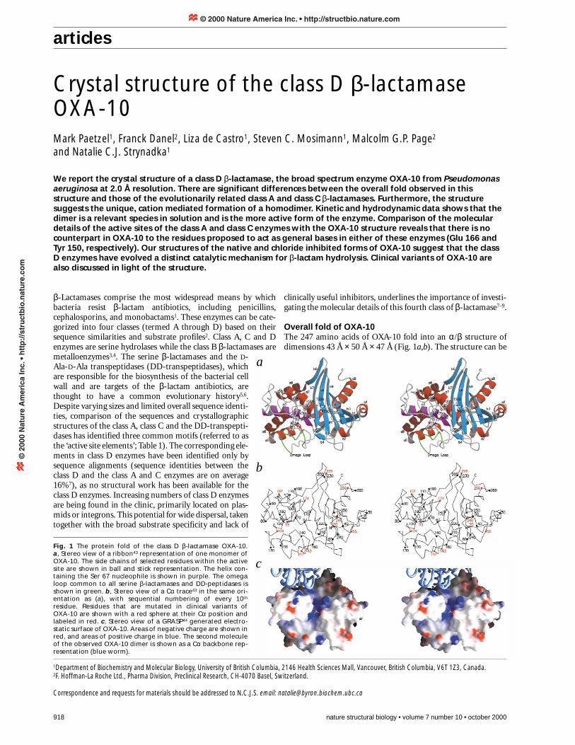
Crystal structure of the class D β-lactamaseOXA-10Mark Paetzel1, Franck Danel2, Liza de Castro1, Steven C. Mosimann1, Malcolm G.P. Page2
and Natalie C.J. Strynadka1
We report the crystal structure of a class D β-lactamase, the broad spectrum enzyme OXA-10 from Pseudomonasaeruginosa at 2.0 Å resolution. There are significant differences between the overall fold observed in thisstructure and those of the evolutionarily related class A and class C β-lactamases. Furthermore, the structuresuggests the unique, cation mediated formation of a homodimer. Kinetic and hydrodynamic data shows that thedimer is a relevant species in solution and is the more active form of the enzyme. Comparison of the moleculardetails of the active sites of the class A and class C enzymes with the OXA-10 structure reveals that there is nocounterpart in OXA-10 to the residues proposed to act as general bases in either of these enzymes (Glu 166 andTyr 150, respectively). Our structures of the native and chloride inhibited forms of OXA-10 suggest that the classD enzymes have evolved a distinct catalytic mechanism for β-lactam hydrolysis. Clinical variants of OXA-10 arealso discussed in light of the structure.
Fig. 1 The protein fold of the class D β-lactamase OXA-10. a, Stereo view of a ribbon43 representation of one monomer ofOXA-10. The side chains of selected residues within the activesite are shown in ball and stick representation. The helix con-taining the Ser 67 nucleophile is shown in purple. The omegaloop common to all serine β-lactamases and DD-peptidases isshown in green. b, Stereo view of a Cα trace43 in the same ori-entation as (a), with sequential numbering of every 10th
residue. Residues that are mutated in clinical variants of OXA-10 are shown with a red sphere at their Cα position andlabeled in red. c, Stereo view of a GRASP44 generated electro-static surface of OXA-10. Areas of negative charge are shown inred, and areas of positive charge in blue. The second moleculeof the observed OXA-10 dimer is shown as a Cα backbone rep-resentation (blue worm).
1Department of Biochemistry and Molecular Biology, University of British Columbia, 2146 Health Sciences Mall, Vancouver, British Columbia, V6T 1Z3, Canada. 2F. Hoffman-La Roche Ltd., Pharma Division, Preclinical Research, CH-4070 Basel, Switzerland.
Correspondence and requests for materials should be addressed to N.C.J.S. email: [email protected]
a
b
c
© 2000 Nature America Inc. • http://structbio.nature.com©
200
0 N
atu
re A
mer
ica
Inc.
• h
ttp
://s
tru
ctb
io.n
atu
re.c
om
articles
nature structural biology • volume 7 number 10 • october 2000 919
divided into two domains, one helical and one mixed α/β con-taining a six-stranded antiparallel β-sheet and the N-terminaland C-terminal α-helices. An alignment of class D sequences(Fig. 2) indicates that differences among these proteins occur aseither deletions or insertions at the termini, within the linkerbetween α-helices a3 and a4, the omega loop linking a6 and β-strand b5, and the linker between b7 and b8. The active siteresidues lie at the interface of the two domains and are containedwithin an extended cleft of overall positive charge (Fig. 1c) that iscomplementary to the negatively charged β-lactam substrate. Asingle disulfide bridge between Cys 44 and Cys 51 linking b2 andb3 is conserved in OXA-5, 7, 11, 13, 14, 16, and 17 (Fig. 2).
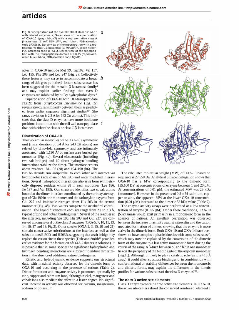
A comparison of class A, C and D enzyme folds reveals differ-ences that may contribute to their unique β-lactam specificities(Fig. 3). The root mean square (r.m.s.) deviations for superposi-tions of common Cα residues of the class D versus representativeclass A (Escherichia coli TEM-1; refs 10,11) and class C(Citrobacter freundii12) enzymes are 1.9 Å (153 residues) and 2.1Å (147 residues), respectively. Regions of structural similarityinclude helices 1, 3 and 5–9, and β-strands 2–5 and 7–9.However, there are a number of differences adjacent to the activesite. The first involves the omega loop region present in all classA and C β-lactamases as well as the cell wall transpeptidases5
(Fig. 3a–c). In OXA-10, the omega loop is shorter in sequenceand more compact in conformation than those of the other
classes and runs in the opposite direction to those of the class Cenzymes. Two additional regions of difference involve residueslinking a3 to a5 (residues 80–117) and a8 to b7 (residues193–199) in OXA-10 (Figs 1a, 3). In the class A and C enzymes,the corresponding regions are longer (class A, residues 86–132and 212–228; class C, residues 80–152 and 256–310) which limitthe size of the substrate binding cleft.
In addition to the extended substrate binding cleft, the class Denzymes have a significant hydrophobic character in the activesite region. The residues contributing to this hydrophobic char-
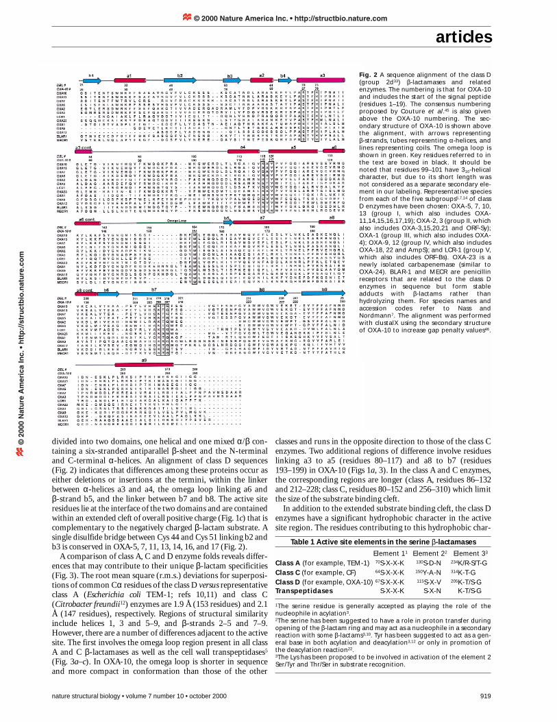
Fig. 2 A sequence alignment of the class D(group 2d33) β-lactamases and relatedenzymes. The numbering is that for OXA-10and includes the start of the signal peptide(residues 1–19). The consensus numberingproposed by Couture et al.45 is also givenabove the OXA-10 numbering. The sec-ondary structure of OXA-10 is shown abovethe alignment, with arrows representing β-strands, tubes representing α-helices, andlines representing coils. The omega loop isshown in green. Key residues referred to inthe text are boxed in black. It should benoted that residues 99–101 have 310-helicalcharacter, but due to its short length wasnot considered as a separate secondary ele-ment in our labeling. Representative speciesfrom each of the five subgroups5,7,14 of classD enzymes have been chosen: OXA-5, 7, 10,13 (group I, which also includes OXA-11,14,15,16,17,19); OXA-2, 3 (group II, whichalso includes OXA-3,15,20,21 and ORF-Sy);OXA-1 (group III, which also includes OXA-4); OXA-9, 12 (group IV, which also includesOXA-18, 22 and AmpS); and LCR-1 (group V,which also includes ORF-Bs). OXA-23 is anewly isolated carbapenemase (similar toOXA-24). BLAR-1 and MECR are penicillinreceptors that are related to the class Denzymes in sequence but form stableadducts with β-lactams rather thanhydrolyzing them. For species names andaccession codes refer to Nass andNordmann7. The alignment was performedwith clustalX using the secondary structureof OXA-10 to increase gap penalty values46.
Table 1 Active site elements in the serine β-lactamases
Element 11 Element 22 Element 33
Class A (for example, TEM-1) 70S-X-X-K 130S-D-N 234K/R-S/T-GClass C (for example, CF) 64S-X-X-K 150Y-A-N 314K-T-GClass D (for example, OXA-10) 67S-X-X-K 115S-X-V 205K-T/S-GTranspeptidases S-X-X-K S-X-N K-T/S-G
1The serine residue is generally accepted as playing the role of thenucleophile in acylation3.2The serine has been suggested to have a role in proton transfer duringopening of the β-lactam ring and may act as a nucleophile in a secondaryreaction with some β-lactams3,10. Tyr has been suggested to act as a gen-eral base in both acylation and deacylation3,12 or only in promotion ofthe deacylation reaction22.3The Lys has been proposed to be involved in activation of the element 2Ser/Tyr and Thr/Ser in substrate recognition.
© 2000 Nature America Inc. • http://structbio.nature.com©
200
0 N
atu
re A
mer
ica
Inc.
• h
ttp
://s
tru
ctb
io.n
atu
re.c
om
articles
920 nature structural biology • volume 7 number 10 • october 2000
acter in OXA-10 include Met 99, Trp102, Val 117,Leu 155, Phe 208 and Leu 247 (Fig. 2). Collectivelythese features may serve to accommodate a broadrange of side groups in the β-lactam substrates as hasbeen suggested for the metallo-β-lactamase family4
and may explain earlier findings that class Denzymes are inhibited by bulky hydrophobic dyes13.
Superposition of OXA-10 with DD-transpeptidasePBP2x from Streptococcus pneumoniae (Fig. 3c)reveals structural similarity between them as predict-ed from earlier sequence alignment studies2,14 (ther.m.s. deviation is 2.3 Å for 183 Cα atoms). This indi-cates that the class D enzymes have more backbonepositions in common with the cell wall transpeptidasethan with either the class A or class C β-lactamases.
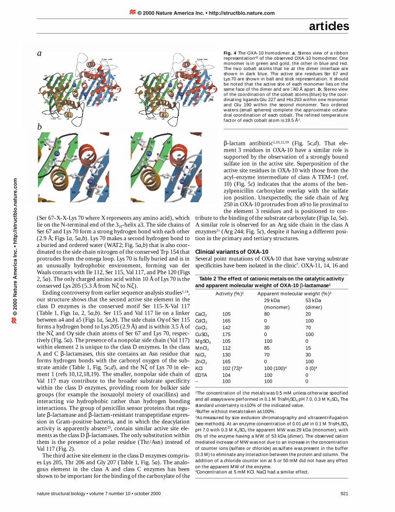
Dimerization of OXA-10The two similar molecules of the OXA-10 asymmetricunit (r.m.s. devation of 0.4 Å for 243 Cα atoms) arerelated by ∼ two-fold symmetry and are intimatelyassociated, with 1,130 Å2 of surface area buried permonomer (Fig. 4a). Several electrostatic (includingtwo salt bridges) and 10 direct hydrogen bondinginteractions stabilize the dimer. The interface centersabout residues 181–193 (a8) and 194–198 (b6). Thetwo b6 strands run antiparallel to each other and interact viahydrophobic (side chain of Ala 196) and water mediated interac-tions. Several hydrophobic interactions also arise from symmetri-cally disposed residues within a8 in each monomer (Leu 186, Ile 187 and Val 193). Our structure identifies two cobalt atomsbound at the dimer interface coordinated by the carboxylate oxy-gen of Glu 190 in one monomer and the carboxylate oxygens fromGlu 227 and imidazole nitrogen from His 203 in the secondmonomer (Fig. 4b). Two waters complete the octahedral coordi-nation. The ligand distances in each site range from 2.1 to 2.3 Å,typical of zinc and cobalt binding sites15. Several of the residues atthe interface, including Glu 190, His 203 and Glu 227, are con-served among several of the class D enzymes (OXA-5, 7, 10, 11, 13,14, 16, 17 and 19; Fig 2). Other species (OXA-2, 3, 15, 20 and 21)contain conservative substitutions at the interface as well as thesubstitutions E190D and H203R, suggesting that a salt bridge mayreplace the cation site in these species (Dale and Smith16 providedearlier evidence for the formation of OXA-2 dimers in solution). Itis possible that in some species the significant hydrophobic andhydrogen bonding interactions are sufficient to induce dimeriza-tion in the absence of additional cation binding sites.
Kinetic and hydrodynamic evidence supports our structuraldata, with maximal activity observed for the dimeric form ofOXA-10 and occurring in the presence of cations (Table 2).Dimer formation and enzyme activity is promoted optimally byzinc, copper and cadmium ions, although nickel, manganese andcobalt ions also mediate the effect to a lesser degree. No signifi-cant increase in activity was observed for calcium, magnesium,sodium or potassium.
The calculated molecular weight (MW) of OXA-10 based onsequence is 27,550 Da. Analytical ultracentrifugation shows thatOXA-10 has a MW corresponding to the dimeric form(55,100 Da) at concentrations of enzyme between 1 and 20 µM.At concentrations of 0.01 µM, the estimated MW was 29 kDa(monomer). However, in the presence of 0.5 mM cadmium, cop-per or zinc, the apparent MW at the lower OXA-10 concentra-tion (0.01 µM) increased to the dimeric 53 kDa value (Table 2).
The enzyme activity assays were performed at a low concen-tration of enzyme (0.025 µM). Under these conditions, OXA-10β-lactamase would exist primarily in a monomeric form in theabsence of cations. An excellent correlation was observedbetween the increase in activity against nitrocefin and the cationmediated formation of dimers, showing that the enzyme is moreactive in the dimeric form. Both OXA-10 and OXA-14 have beenshown to have complex biphasic kinetics with some substrates17,which may now be explained by the conversion of the dimericform of the enzyme to a less active monomeric form during thecourse of the assay. A β-turn between b6 and b7 in one monomerlies on the periphery of the binding site of the adjacent monomer(Fig.1c). Although unlikely to play a catalytic role (as it is >18 Åaway), it could affect substrate binding and, in combination withconformational or stability differences between the monomericand dimeric forms, may explain the differences in the kineticprofiles for various substrates of the class D enzymes7,17.
The class D active site elementsClass D enzymes contain three active site elements. In OXA-10,the active site centers about the conserved residues of element 1
Fig. 3 Superpositions of the overall fold of class D OXA-10with related enzymes. a, Stereo view of the superpositionof OXA-10 (gray ribbon43) with a representative class A β-lactamase (E. coli TEM-110,11, red ribbon, PDB accessioncode 1FQG). b, Stereo view of the superposition with a rep-resentative class C β-lactamase (C. freundii12, green ribbon,PDB accession code 1FR6). c, Stereo view of the superposi-tion with the transpeptidase domain of PBP2x (S. pneumo-niae6, blue ribbon, PDB accession code 1QME).
c
b
a
© 2000 Nature America Inc. • http://structbio.nature.com©
200
0 N
atu
re A
mer
ica
Inc.
• h
ttp
://s
tru
ctb
io.n
atu
re.c
om
articles
nature structural biology • volume 7 number 10 • october 2000 921
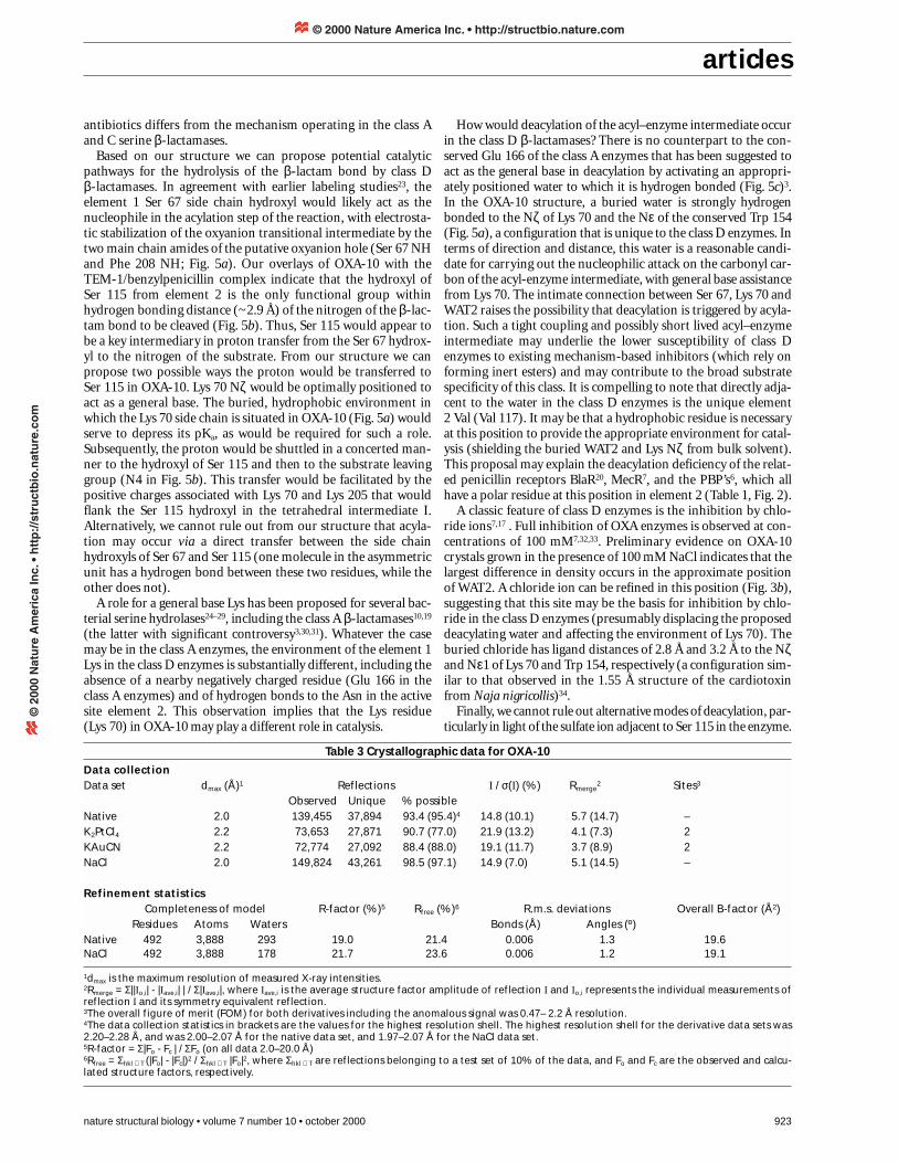
(Ser 67-X-X-Lys 70 where X represents any amino acid), whichlie on the N-terminal end of the 310-helix a3. The side chains ofSer 67 and Lys 70 form a strong hydrogen bond with each other(2.9 Å; Figs 1a, 5a,b). Lys 70 makes a second hydrogen bond toa buried and ordered water (WAT2; Fig. 5a,b) that is also coor-dinated to the side chain nitrogen of the conserved Trp 154 thatprotrudes from the omega loop. Lys 70 is fully buried and is inan unusually hydrophobic environment, forming van derWaals contacts with Ile 112, Ser 115, Val 117, and Phe 120 (Figs2, 5a). The only charged amino acid within 10 Å of Lys 70 is theconserved Lys 205 (5.3 Å from Nζ to Nζ).
Ending controversy from earlier sequence analysis studies1,14,our structure shows that the second active site element in theclass D enzymes is the conserved motif Ser 115-X-Val 117(Table 1, Figs 1a, 2, 5a,b). Ser 115 and Val 117 lie on a linkerbetween a4 and a5 (Figs 1a, 5a,b). The side chain Oγ of Ser 115forms a hydrogen bond to Lys 205 (2.9 Å) and is within 3.5 Å ofthe Nζ and Oγ side chain atoms of Ser 67 and Lys 70, respec-tively (Fig. 5a). The presence of a nonpolar side chain (Val 117)within element 2 is unique to the class D enzymes. In the classA and C β-lactamases, this site contains an Asn residue thatforms hydrogen bonds with the carbonyl oxygen of the sub-strate amide (Table 1, Fig. 5c,d), and the Nζ of Lys 70 in ele-ment 1 (refs 10,12,18,19). The smaller, nonpolar side chain ofVal 117 may contribute to the broader substrate specificitywithin the class D enzymes, providing room for bulkier sidegroups (for example the isoxazolyl moiety of oxacillins) andinteracting via hydrophobic rather than hydrogen bondinginteractions. The group of penicillin sensor proteins that regu-late β-lactamase and β-lactam-resistant transpeptidase expres-sion in Gram-positive bacteria, and in which the deacylationactivity is apparently absent20, contain similar active site ele-ments as the class D β-lactamases. The only substitution withinthem is the presence of a polar residue (Thr/Asn) instead ofVal 117 (Fig. 2).
The third active site element in the class D enzymes compris-es Lys 205, Thr 206 and Gly 207 (Table 1, Fig. 5a). The analo-gous element in the class A and class C enzymes has beenshown to be important for the binding of the carboxylate of the
β-lactam antibiotic5,10,12,19 (Fig. 5c,d). That ele-ment 3 residues in OXA-10 have a similar role issupported by the observation of a strongly boundsulfate ion in the active site. Superposition of theactive site residues in OXA-10 with those from theacyl–enzyme intermediate of class A TEM-1 (ref.10) (Fig. 5c) indicates that the atoms of the ben-zylpenicillin carboxylate overlap with the sulfateion position. Unexpectedly, the side chain of Arg250 in OXA-10 protrudes from a9 to lie proximal tothe element 3 residues and is positioned to con-
tribute to the binding of the substrate carboxylate (Figs 1a, 5a).A similar role is observed for an Arg side chain in the class Aenzymes21 (Arg 244; Fig. 5c), despite it having a different posi-tion in the primary and tertiary structures.
Clinical variants of OXA-10Several point mutations of OXA-10 that have varying substratespecificities have been isolated in the clinic7. OXA-11, 14, 16 and
a
b
Fig. 4 The OXA-10 homodimer. a, Stereo view of a ribbonrepresentation43 of the observed OXA-10 homodimer. Onemonomer is in green and gold, the other in blue and red.The two cobalt atoms that lie at the dimer interface areshown in dark blue. The active site residues Ser 67 andLys 70 are shown in ball and stick representation. It shouldbe noted that the active site of each monomer lies on thesame face of the dimer and are ∼ 40 Å apart. b, Stereo viewof the coordination of the cobalt atoms (blue) by the coor-dinating ligands Glu 227 and His 203 within one monomerand Glu 190 within the second monomer. Two orderedwaters (small spheres) complete the approximate octahe-dral coordination of each cobalt. The refined temperaturefactor of each cobalt atom is 19.5 Å2.
Table 2 The effect of cationic metals on the catalytic activityand apparent molecular weight of OXA-10 β-lactamase1
Activity (%)2 Apparent molecular weight (%)3
29 kDa 53 kDa (monomer) (dimer)
CaCl2 105 80 20CdCl2 165 0 100CoCl2 142 30 70CuSO4 175 0 100MgSO4 105 100 0MnCl2 112 85 15NiCl2 130 70 30ZnCl2 165 0 100KCl 102 (73)4 100 (100)4 0 (0)4
EDTA 104 100 0– 100 100 0
1The concentration of the metals was 0.5 mM unless otherwise specifiedand all assays were performed in 0.1 M Tris/H2SO4 pH 7.0, 0.3 M K2SO4. Thestandard uncertainty is ±10% of the indicated value.2Buffer without metals taken as 100%.3As measured by size exclusion chromatography and ultracentrifugation(see methods). At an enzyme concentration of 0.01 µM in 0.1 M Tris/H2SO4
pH 7.0 with 0.3 M K2SO4 the apparent MW was 29 kDa (monomer), with0% of the enzyme having a MW of 53 kDa (dimer). The observed cationmediated increase of MW was not due to an increase in the concentrationof counter ions (sulfate or chloride) as sulfate was present in the buffer(0.3 M) to eliminate any interaction between the protein and column. Theaddition of a chloride counter ion at 5 or 50 mM did not have any effecton the apparent MW of the enzyme.4Concentration at 5 mM KCl. NaCl had a similar effect.
© 2000 Nature America Inc. • http://structbio.nature.com©
200
0 N
atu
re A
mer
ica
Inc.
• h
ttp
://s
tru
ctb
io.n
atu
re.c
om

articles
922 nature structural biology • volume 7 number 10 • october 2000
17 are natural mutants of OXA-10 β-lactamase thatconfer resistance to an extended spectrum ofcephalosporins7. These mutations can be mappedonto the OXA-10 structure. We find that most of themutations lie not within, but adjacent to the active siteelements and are likely to exert their effects indirectlyby altering the positions of the adjacent active siteresidues (Fig. 1b). For example, OXA-11 contains twosubstitutions (N143S and G157D) relative to OXA-10.Asn 143 is part of the Tyr-Gly-Asn type II′ β-hairpinthat directly follows helix a6 and precedes the omegaloop (Figs 1a,b, 2). In OXA-10, the Asn side chainforms a hydrogen bond to the carbonyl oxygen ofGln 158 that lies in a 310-helix directly following theomega loop. The N143S and G157D substitutionscannot be accommodated by the conformation of theomega loop observed in our structure. One can pre-dict that the rearrangements necessitated by thesemutations will affect the orientation of the adjacentactive site residues (Trp 154 and Leu 155; Fig. 5a).OXA-14 and OXA-16 also contain the G157D muta-tion, with the latter having a second substitution,A124T. Ala 124 lies on helix a5 and is tightly packedand buried within the molecule. Its side chain formsvan der Waals contacts with the side chain of Trp 154,which would be altered upon substitution to thebulkier Val. In OXA-17, the substitution N73S (helix a3) isobserved. The side chain of Asn 73 forms van der Waals contactswith the side chains of Phe 69, Phe 120, Lys 70 and Trp 154 (Fig.5a), which again would be altered upon substitution by the small-er Ser side chain. OXA-13, a narrow spectrum enzyme with a sub-strate preference for cefotaxime and aztreonam, also contains theN73S substitution. Interestingly, of the nine substitutions in OXA-13, only N73S is adjacent to the active site cleft. The majority ofother changes are substitutions of negatively charged, solvatedresidues more than 30 Å away from the active site (D55N, E229Gand E259A). Other changes appear to be relatively conservative interms of structure: the N-terminal G20S, T107S (the helix cappingresidue of helix a4 that terminates with Ser 115) and Y174F.
Implications for the mechanism of class D β-lactamases Despite the many site-directed mutagenesis, crystallographic,inhibitory and kinetic investigations, there is still significant con-troversy regarding many of the mechanistic details for each of theclass A, B and C β-lactamases3. In the serine hydrolase classes,perhaps the most experimentally supported proposals includethe role of the serine nucleophile in acylation1,3, the role ofGlu 166 as the general base in deacylation in the class A enzymes3
(Fig. 5c), and an essential role of Tyr 150 in the class C enzymes(Fig. 5d)3,12,18,22. As comparisons of the active site region showthat class D enzymes contain no counterparts to Glu 166 orTyr 150, our structure would thus suggest that the catalyticmechanism by which class D enzymes hydrolyze β-lactam
a
b
c
d
Fig. 5 The active site of OXA-10. a, Stereo view of a ball andstick representation43 of the active site residues in OXA-10.Selected water molecules are shown as cyan spheres.Hydrogen bonds are shown as dotted green lines. For clari-ty, the sulfate ion observed in our structure is removed inthis view. b, Stereo view of the final refined 2Fo - Fc electrondensity (1.5 σ) around key active site residues and watermolecules in OXA-10 β-lactamase. 2Fo - Fc electron density(1.5 σ) for a refined chloride ion in data obtained from crys-tals grown in 100 mM NaCl is shown in green (refined tem-perature factor = 16 Å2). It is acknowledged that theposition of the water in the native structure may also bepartially occupied by chloride given the presence of 10 mMCoCl2 in the crystallization solution. Our refinement statis-tics suggest that it is primarily a water molecule. c, Stereoview of a stick representation of the superposition of theactive site of OXA-10 (black) with the class A E. coli TEM-1–penicillin G (PenG) acyl-enzyme complex (red)10. ThePenG substrate is shown in light gray ball and stick repre-sentation. A deacylation deficient mutation on the omegaloop of TEM-1, Glu166Asn, was used to trap the acyl-enzyme. The proposed deacylating water of TEM-1 isshown as a red sphere. d, Stereo view of the superpositionof the active site of OXA-10 (black) with the class C C. fre-undii–azeotrenam (MBF) acyl-enzyme complex (green)12.The monobactam MBF is shown in light grey ball and stickrepresentation.
© 2000 Nature America Inc. • http://structbio.nature.com©
200
0 N
atu
re A
mer
ica
Inc.
• h
ttp
://s
tru
ctb
io.n
atu
re.c
om
articles
nature structural biology • volume 7 number 10 • october 2000 923
antibiotics differs from the mechanism operating in the class Aand C serine β-lactamases.
Based on our structure we can propose potential catalyticpathways for the hydrolysis of the β-lactam bond by class D β-lactamases. In agreement with earlier labeling studies23, theelement 1 Ser 67 side chain hydroxyl would likely act as thenucleophile in the acylation step of the reaction, with electrosta-tic stabilization of the oxyanion transitional intermediate by thetwo main chain amides of the putative oxyanion hole (Ser 67 NHand Phe 208 NH; Fig. 5a). Our overlays of OXA-10 with theTEM-1/benzylpenicillin complex indicate that the hydroxyl ofSer 115 from element 2 is the only functional group withinhydrogen bonding distance (~2.9 Å) of the nitrogen of the β-lac-tam bond to be cleaved (Fig. 5b). Thus, Ser 115 would appear tobe a key intermediary in proton transfer from the Ser 67 hydrox-yl to the nitrogen of the substrate. From our structure we canpropose two possible ways the proton would be transferred toSer 115 in OXA-10. Lys 70 Nζ would be optimally positioned toact as a general base. The buried, hydrophobic environment inwhich the Lys 70 side chain is situated in OXA-10 (Fig. 5a) wouldserve to depress its pKa, as would be required for such a role.Subsequently, the proton would be shuttled in a concerted man-ner to the hydroxyl of Ser 115 and then to the substrate leavinggroup (N4 in Fig. 5b). This transfer would be facilitated by thepositive charges associated with Lys 70 and Lys 205 that wouldflank the Ser 115 hydroxyl in the tetrahedral intermediate I.Alternatively, we cannot rule out from our structure that acyla-tion may occur via a direct transfer between the side chainhydroxyls of Ser 67 and Ser 115 (one molecule in the asymmetricunit has a hydrogen bond between these two residues, while theother does not).
A role for a general base Lys has been proposed for several bac-terial serine hydrolases24–29, including the class A β-lactamases10,19
(the latter with significant controversy3,30,31). Whatever the casemay be in the class A enzymes, the environment of the element 1Lys in the class D enzymes is substantially different, including theabsence of a nearby negatively charged residue (Glu 166 in theclass A enzymes) and of hydrogen bonds to the Asn in the activesite element 2. This observation implies that the Lys residue(Lys 70) in OXA-10 may play a different role in catalysis.
How would deacylation of the acyl–enzyme intermediate occurin the class D β-lactamases? There is no counterpart to the con-served Glu 166 of the class A enzymes that has been suggested toact as the general base in deacylation by activating an appropri-ately positioned water to which it is hydrogen bonded (Fig. 5c)3.In the OXA-10 structure, a buried water is strongly hydrogenbonded to the Nζ of Lys 70 and the Nε of the conserved Trp 154(Fig. 5a), a configuration that is unique to the class D enzymes. Interms of direction and distance, this water is a reasonable candi-date for carrying out the nucleophilic attack on the carbonyl car-bon of the acyl-enzyme intermediate, with general base assistancefrom Lys 70. The intimate connection between Ser 67, Lys 70 andWAT2 raises the possibility that deacylation is triggered by acyla-tion. Such a tight coupling and possibly short lived acyl–enzymeintermediate may underlie the lower susceptibility of class Denzymes to existing mechanism-based inhibitors (which rely onforming inert esters) and may contribute to the broad substratespecificity of this class. It is compelling to note that directly adja-cent to the water in the class D enzymes is the unique element2 Val (Val 117). It may be that a hydrophobic residue is necessaryat this position to provide the appropriate environment for catal-ysis (shielding the buried WAT2 and Lys Nζ from bulk solvent).This proposal may explain the deacylation deficiency of the relat-ed penicillin receptors BlaR20, MecR7, and the PBP’s6, which allhave a polar residue at this position in element 2 (Table 1, Fig. 2).
A classic feature of class D enzymes is the inhibition by chlo-ride ions7,17 . Full inhibition of OXA enzymes is observed at con-centrations of 100 mM7,32,33. Preliminary evidence on OXA-10crystals grown in the presence of 100 mM NaCl indicates that thelargest difference in density occurs in the approximate positionof WAT2. A chloride ion can be refined in this position (Fig. 3b),suggesting that this site may be the basis for inhibition by chlo-ride in the class D enzymes (presumably displacing the proposeddeacylating water and affecting the environment of Lys 70). Theburied chloride has ligand distances of 2.8 Å and 3.2 Å to the Nζand Nε1 of Lys 70 and Trp 154, respectively (a configuration sim-ilar to that observed in the 1.55 Å structure of the cardiotoxinfrom Naja nigricollis)34.
Finally, we cannot rule out alternative modes of deacylation, par-ticularly in light of the sulfate ion adjacent to Ser 115 in the enzyme.
Table 3 Crystallographic data for OXA-10
Data collectionData set dmax (Å)1 Reflections I / σ(I) (%) Rmerge
2 Sites3
Observed Unique % possibleNative 2.0 139,455 37,894 93.4 (95.4)4 14.8 (10.1) 5.7 (14.7) –K2PtCl4 2.2 73,653 27,871 90.7 (77.0) 21.9 (13.2) 4.1 (7.3) 2KAuCN 2.2 72,774 27,092 88.4 (88.0) 19.1 (11.7) 3.7 (8.9) 2NaCl 2.0 149,824 43,261 98.5 (97.1) 14.9 (7.0) 5.1 (14.5) –
Refinement statisticsCompleteness of model R-factor (%)5 Rfree (%)6 R.m.s. deviations Overall B-factor (Å2)
Residues Atoms Waters Bonds (Å) Angles (º)Native 492 3,888 293 19.0 21.4 0.006 1.3 19.6NaCl 492 3,888 178 21.7 23.6 0.006 1.2 19.1
1dmax is the maximum resolution of measured X-ray intensities.2Rmerge = Σ||Io,i| - |Iave,i| | / Σ|Iave,i|, where Iave,i is the average structure factor amplitude of reflection I and Io,i represents the individual measurements ofreflection I and its symmetry equivalent reflection.3The overall figure of merit (FOM) for both derivatives including the anomalous signal was 0.47– 2.2 Å resolution.4The data collection statistics in brackets are the values for the highest resolution shell. The highest resolution shell for the derivative data sets was2.20–2.28 Å, and was 2.00–2.07 Å for the native data set, and 1.97–2.07 Å for the NaCl data set.5R-factor = Σ|Fo - Fc | / ΣFo (on all data 2.0–20.0 Å)6Rfree = Σhkl ⊂ T (|Fo| - |Fc|)2 / Σhkl ⊂ T |Fo|2, where Σhkl ⊂ T are reflections belonging to a test set of 10% of the data, and Fo and Fc are the observed and calcu-lated structure factors, respectively.
© 2000 Nature America Inc. • http://structbio.nature.com©
200
0 N
atu
re A
mer
ica
Inc.
• h
ttp
://s
tru
ctb
io.n
atu
re.c
om
articles
924 nature structural biology • volume 7 number 10 • october 2000
Although the direction would be less favorable, it may be that thepresence of the sulfate in the active site of our structure displaces apotential nucleophilic water (with Ser 115 acting as general base).
MethodsData collection. OXA-10 was expressed and purified asdescribed17. Crystals were grown by vapor diffusion using 1.8 M(NH4)2SO4, 0.1 M 2-(N-Morpholino)ethanesulfonic acid (MES), 10 mM CoCl2 at pH 6.5. The crystals are in space group P212121 withunit cell dimensions of a = 48.4 Å, b = 96.2 Å, c = 125.7 Å. Data forK2PtCl4 and KAuCN soaks (1 and 0.5 mM, respectively) were collect-ed at 1.07225 Å and 1.04031 Å, respectively, at the StanfordSynchrotron Radiation Laboratory (SSRL), beamline 1-5, using anADSC CCD detector, and processed with DENZO35.
Phase determination and refinement. Heavy atom sites andparameters were determined using SOLVE36 and SHARP37. Solventflattening was performed with SOLOMON37. Model building wasperformed with O38 and refinement done using CNS39 (Table 3).
NaCl inhibition. Crystals were grown in 1.6 M (NH4)2SO4, 0.1 MHepes, 0.1 M NaCl, 10 mM CoCl2 at pH 7.5. Crystals were isomor-phous to those described above. Data were collected at theNational Synchrotron Light Source (NSLS), beamline X12C, using aBrandeis Q4 CCD detector.
Structural analysis. Superpositions were performed with thealgorithm LSQ within O38 (cutoff 3.8 Å). The dimer interface wasanalyzed using the protein–protein interaction server40. Hydrogenbond distances quoted in the text are an average of the two mole-cules in the asymmetric unit and are within 0.2 Å of each other.
Analytical centrifugation. Sedimentation equilibrium experi-ments were performed by centrifugation overnight at 18,000 r.p.m.using a Beckman Optima XA analytical centrifuge at 20 °C. Theequilibrium between sedimentation and diffusion was followed byradial scanning of the 100 µl tube at 280 nm until the equilibrium
condition was reached. The MW and partial specific volume ofOXA-10 deduced from the amino acid sequence corresponded to28.5 kDa and 0.740 cm3 g-1 (ref. 41). The absorbance profile at equi-librium was fitted to the MW distribution of the monomer andoligomers using Discreek42.
Size exclusion chromatography. Due to limitations of detectionin the analytical ultracentrifugation method, at protein concentra-tions <1 µM the apparent MW was estimated by size exclusion chro-matography connected to a fluorescence detector. Measurementswere performed using a Jasco HPLC connected to a PharmaciaSuperdex 200 PC 3.2/30 gel filtration column. The column was equili-brated with buffer (three times the column volume) used for proteindilution in the enzyme assay. The flow rate was 0.1 ml min-1 and 5 µlof protein solution was injected. Elution of the protein was moni-tored by measuring its fluorescence (excitation at 280 nm, emissionat 330 nm, FP920 intelligent Fluorescent detector, Jasco). Calibrationof the column was performed using MW standards (Bio-Rad).
Kinetic analysis. OXA-10 (0.025 µM) was incubated for 20 min atroom temperature in 20 mM Tris (adjusted to pH 7.0 with H2SO4), andcontaining 0.3 M K2SO4 and 0.5 mM metal salt (chloride or sulfate)when appropriate. The reaction was initiated by addition of 0.1 mMnitrocefin (final concentration). The change in absorbance was mon-itored at 490 nm using a Bio-Rad Model 3550 microtitreplate reader.
Coordinates. The coordinates have been deposited in the ProteinData Bank (accession code 1FOF).
AcknowledgmentsWe thank the Medical Research Council of Canada (M.P. is a MRC fellow, N.C.J.S. isa MRC scholar), the Burroughs Wellcome Foundation (New Investigator Award toN.C.J.S.) and Hoffman-La Roche Pharmaceuticals (to F.D. and M.G.P.P.) for support.We thank H. Bellamy of beamline 1-5 at the SSRL for data collection access and R.Sweet for access to beamline X12C at the NSLS (Brookhaven National Laboratory).
Received 13 April, 2000; accepted 15 August, 2000.
© 2000 Nature America Inc. • http://structbio.nature.com©
200
0 N
atu
re A
mer
ica
Inc.
• h
ttp
://s
tru
ctb
io.n
atu
re.c
om

articles
nature structural biology • volume 7 number 10 • october 2000 925
1. Frère, J.M. Beta-lactamases and bacterial resistance to antibiotics. Mol.Microbiol. 16, 385–395 (1995).
2. Joris, B., et al. Comparison of the sequences of class A beta-lactamases and of thesecondary structure elements of penicillin-recognizing proteins. Antimicrob.Agents Chemother. 35, 2294–2301 (1991).
3. Frère, J.M., Dubus, A., Galleni, M., Matagne, A. & Amicosante, G. Mechanisticdiversity of beta-lactamases. Biochem. Soc. Trans. 27, 58–63 (1999).
4. Bush, K. Metallo-beta-lactamases: a class apart. Clin. Infect. Dis. 27 (Suppl 1),S48–53 (1998).
5. Knox, J.R., Moews, P.C. & Frère, J.M. Molecular evolution of bacterial beta-lactamresistance. Chem. Biol. 3, 937–947 (1996).
6. Gordon, E., Mouz, N., Duée, E. & Dideberg, O. The crystal structure of thepenicillin-binding protein 2x from Streptoccus pneumoniae and its acyl-enzymeform: implication in drug resistance. J. Mol. Biol. 299, 477–485 (2000).
7. Nass, T. & Nordmann, P. OXA-type beta-lactamases. Curr. Pharm. Design 5,865–879 (1999).
8. Mugnier, P., Casin, I., Bouthers, A.T. & Collatz, E. Novel OXA-10-derived extended-spectrum beta-lactamases selected in vivo or in vitro. Antimicrob. AgentsChemother. 42, 3113–3116 (1998).
9. Bush, K. The evolution of beta-lactamases. Ciba Found. Symp. 207, 152–163(1997).
10. Strynadka, N.C.J., et al. Molecular structure of the acyl-enzyme intermediate inbeta-lactam hydrolysis at 1.7 Å resolution. Nature 359, 700–705 (1992).
11. Jelsch, C., Mourey, L., Masson, J-M., & Samama, J-P. Crystal structure ofEscherichia coli TEM1 beta-lactamase at 1.8 Å resolution. Proteins 16, 364–383(1993).
12. Oefner, C. et al. Refined crystal structure of beta-lactamase from Citrobacterfreundii indicates a mechanism for beta-lactam hydrolysis. Nature 343, 284–289(1990).
13. Monaghan, C., Holland, S. & Dale, J.W. The interaction of anthraquinone dyeswith the plasmid-mediated OXA-2 beta-lactamase. Biochem. J. 205, 413–417(1982).
14. Sanschagrin, F., Couture, F. & Levesque, R.C. Primary structure of OXA-3 andphylogeny of oxacillin-hydrolyzing class D beta-lactamases. Antimicrob. AgentsChemother. 39, 887–893 (1995).
15. Glusker, J.P. Structural aspects of metal liganding to functional groups inproteins. Adv. Protein Chem. 42, 1–76 (1991).
16. Dale J.W., & Smith, J.T. The dimeric nature of an R-factor mediated beta-lactamase. Biochem. Biophys. Res. Commun. 68, 1000–1005 (1976).
17. Danel, F., Hall, L.M., Gur, D. & Livermore, D.M. OXA-16, a further extended-spectrum variant of OXA-10 beta-lactamase, from two Pseudomonas aeruginosaisolates. Antimicrob. Agents Chemother. 42, 3117–3122 (1998).
18. Lobkovsky, E., et al. Crystallographic structure of a phosphonate derivative of theEnterobacter cloacae P99 cephalosporinase: mechanistic interpretation of abeta-lactamase transition-state analog. Biochemistry 33, 6762–6772 (1994).
19. Maveyraud, L., Pratt, R.F. & Samama, J.P. Crystal structure of an acylationtransition-state analog of the TEM-1 beta-lactamase. Mechanistic implicationsfor class A beta-lactamases. Biochemistry 37, 2622–2628 (1998).
20. Zhu, Y., et al. Structure, function, and fate of the BlaR signal transducer involvedin induction of beta-lactamase in Bacillus licheniformis. J. Bacteriol.. 174,6171–6178 (1992).
21. Jacob-Dubuisson, F., Lamotte-Brasseur J., Dideberg, O., Joris, B. & Frere, J.M.Arginine 220 is a critical residue for the catalytic mechanism of the Streptomycesalbus G beta-lactamase. Protein Eng. 4, 811–819 (1991).
22. Dubus, A., Normark, S., Kania, K. & Page, M.G.P. The role of tyrosine 150 incatalysis of β-lactam hydrolysis by AmpC β-lactamase from Escherichia coliinvestigated by site-directed mutagenesis. Biochemistry 33, 8577–8586 (1994).
23. Ledent, P. Raquet, X., Joris, B., Van Beeumen, J. & Frere J.M. A comparative studyof class-D beta-lactamases. Biochem. J. 292, 555–562 (1993).
24. Little, J.W., et al. Cleavage of LexA repressor. Methods Enzymol. 244, 266–284(1994).
25. Paetzel, M., et al. Use of site-directed chemical modification to study an essentiallysine in Escherichia coli leader peptidase. J. Biol. Chem. 272, 9994–10003 (1997).
26. Patricelli, M.P. & Cravatt, B.F. Fatty acid amide hydrolase competitively degradesbioactive amides and esters through a nonconventional catalytic mechanism.Biochemistry 38, 14125–14130 (1999).
27. Birghan,C., Mundt, E. & Gorbalenya, A.E. A non-canonical lon proteinase lackingthe ATPase domain employs the Ser-Lys catalytic dyad to exercise broad controlover the life cycle of a double-stranded RNA virus. EMBO J. 19, 114–123 (2000).
28. Keiler, K.C. & Sauer, R.T. Identification of active site residues of the Tsp protease.J. Biol. Chem. 270, 28864–28868 (1995).
29. Haase, J. & Lanka, E. A specific protease encoded by the conjugative DNA transfersystems of IncP and Ti plasmids is essential for pilus synthesis. J. Bacteriol. 179,5728–5735 (1997).
30. Lietz, E.J., Truher, H., Kahn, D., Hokenson, M.J. & Fink, A.L. Lysine-73 is involved inthe acylation and deacylation of beta-lactamase. Biochemistry 39, 4971–4981(2000).
31. Damblon, C., et al. The catalytic mechanism of beta-lactamases: NMR titration ofan active-site lysine residue of the TEM-1 enzyme. Proc. Natl. Acad. Sci. USA 93,1747–1752 (1996).
32. Philippon, A.M., Paul G. & Jacoby, G.A. Properties of PSE-2 beta-lactamase andgenetic basis for its production in Pseudomonas aeruginosa. Antimicrob. AgentsChemother. 24, 362–369 (1983).
33. Bush, K., Jacoby G.A., Medeiros, A.A. A functional classification scheme for beta-lactamases and its correlation with molecular structure. Antimicrob. AgentsChemother. 39, 1211–1233 (1995).
34. Bilwes, A., Rees, B., Moras, D., Menez, R., & Menez, A. X-ray structure at 1.55 Å oftoxin gamma, a cardiotoxin from Naja nigricollis venom. Crystal packing reveals amodel for insertion into membranes. J. Mol. Biol. 239,122–136 (1994).
35. Otwinowski, Z. In Denzo (eds, Sawyer, L., Isaacs, N. & Baily, S.) 56–62 (SERCDaresbury Laboratory, Warrington, UK; 1993).
36. Terwilliger, T.C. & Berendzen, J. Automated structure solution for MIR and MAD.Acta Crystallogr. D 55, 849–861 (1999).
37. La Fortelle, E. & Bricogne, G. Maximum-likelihood heavy-atom parameterrefinement in the MIR and MAD methods Methods Enzymol. 276, 472–494 (1997).
38. Jones, T.A., Zou, J.-Y., Cowan, S.W. & Kieldgaard, M. Improved methods forbuilding protein models in electron density maps and the location of errors inthese models. Acta Crystallogr. A 47, 110–119 (1991).
39. Brünger, A.T. et al. Crystallography & NMR system: a new software suite formacromolecular structure determination. Acta Crystallogr. D 54, 905–921 (1998).
40. Jones, S. & Thornton, J.M. Protein-protein interactions: a review of protein dimerstructure. Prog. Biophys. Mol. Biol. 63, 31–165 (1995).
41. Cohn, E.J. & Edsall, J.T. Density and apparent specific volume of proteins. InProteins, amino acids and peptides as ions and dipolar ions (ed. E. J. Cohn),370–381 (Reinhold, New York; 1943).
42. Schuck, P. Simultaneous radial and wavelength analysis with the Optima XL-Aanalytical ultracentrifuge. Prog. Colloid Polymer Sci. 94, 1–13 (1994).
43. Kraulis, P. G. Molscript: a program to produce both detailed and schematic plotsof protein structures. J. Appl. Crystallog. 24, 946–950 (1991).
44. Nicholls, A., Sharp, K.A. & Honig, B. Protein folding and association: insights fromthe interfacial and thermodynamic properties of hydrocarbons. Proteins 11,281–296 (1991).
45. Couture, F., Lachapelle, J. & Levesque, R.C. Phylogeny of LCR-1 and OXA-5 withclass A and class D beta-lactamases. Mol. Microbiol. 6, 1693–1705 (1992).
46. Thompson, J.D., Higgins, D.G. & Gibson, T.J. CLUSTAL W: improving the sensitivityof progressive multiple sequence alignment through sequence weighting,positions-specific gap penalties and weight matrix choice. Nucleic Acids Res. 22,4673–4680 (1994).
© 2000 Nature America Inc. • http://structbio.nature.com©
200
0 N
atu
re A
mer
ica
Inc.
• h
ttp
://s
tru
ctb
io.n
atu
re.c
om