Embed Size (px)
Citation preview
HAL Id: pasteur-01499013https://hal-riip.archives-ouvertes.fr/pasteur-01499013
Submitted on 21 Sep 2018
HAL is a multi-disciplinary open accessarchive for the deposit and dissemination of sci-entific research documents, whether they are pub-lished or not. The documents may come fromteaching and research institutions in France orabroad, or from public or private research centers.
L’archive ouverte pluridisciplinaire HAL, estdestinée au dépôt et à la diffusion de documentsscientifiques de niveau recherche, publiés ou non,émanant des établissements d’enseignement et derecherche français ou étrangers, des laboratoirespublics ou privés.
Crystal Structure of the Metallo-β-Lactamase GOB inthe Periplasmic Dizinc Form Reveals an Unusual Metal
Site.Jorgelina Morán-Barrio, María-Natalia Lisa, Nicole Larrieux, Salvador I
Drusin, Alejandro Viale, Diego M Moreno, Alejandro Buschiazzo, Alejandro JVila
To cite this version:Jorgelina Morán-Barrio, María-Natalia Lisa, Nicole Larrieux, Salvador I Drusin, Alejandro Viale, etal.. Crystal Structure of the Metallo-β-Lactamase GOB in the Periplasmic Dizinc Form Reveals anUnusual Metal Site.. Antimicrobial Agents and Chemotherapy, American Society for Microbiology,2016, 60 (10), pp.6013-22. �10.1128/aac.01067-16�. �pasteur-01499013�
1
CRYSTAL STRUCTURE OF THE METALLO-BETA-LACTAMASE GOB IN THE 1
PERIPLASMIC DI-ZINC FORM REVEALS AN UNUSUAL METAL SITE 2
Jorgelina Morán-Barrio1#, María-Natalia Lisa1,2#, Nicole Larrieux2, Salvador I. Drusin3,4, 3
Alejandro M. Viale1, Diego M. Moreno3,5, Alejandro Buschiazzo2,6* and Alejandro J. Vila1* 4
5
1 Departamento de Química Biológica and Instituto de Biología Molecular y Celular de Rosario 6
(IBR, CONICET-UNR), Facultad de Ciencias Bioquímicas y Farmacéuticas, Universidad Nacional 7
de Rosario, S2002LRK Rosario, Argentina. 8
2 Laboratory of Molecular & Structural Microbiology, Institut Pasteur de Montevideo, 9
Montevideo 11400, Uruguay. 10
3 Departamento de Química-Física, Facultad de Ciencias Bioquímicas y Farmacéuticas, 11
Universidad Nacional de Rosario, Suipacha 531, S2002LRK Rosario, Santa Fe, Argentina. 12
4 Instituto de Biología Molecular y Celular de Rosario (CONICET-UNR). Ocampo y Esmeralda, 13
predio CCT, S2002LRK Rosario, Argentina. 14
5 Instituto de Química de Rosario (IQUIR, CONICET-UNR), Suipacha 570, S2002LRK Rosario, 15
Santa Fe, Argentina. 16
6 Département de Biologie Structurale et Chimie, Institut Pasteur, Paris, 75015, France. 17
#JMB and MNL contributed equally to this work. 18
*Address correspondence to Alejandro J. Vila ([email protected]) and Alejandro 19
Buschiazzo ([email protected]). 20
21
AAC Accepted Manuscript Posted Online 25 July 2016Antimicrob. Agents Chemother. doi:10.1128/AAC.01067-16Copyright © 2016, American Society for Microbiology. All Rights Reserved.
on Septem
ber 21, 2018 by guesthttp://aac.asm
.org/D
ownloaded from
2
ABSTRACT 22
23
Metallo-beta-lactamases (MBLs) are broad spectrum, Zn(II) dependent lactamases able 24
to confer resistance to virtually every β-lactam antibiotic currently available. The large diversity 25
of active site structures and metal content among MBLs from different sources has limited the 26
design of a pan-MBL inhibitor. GOB-18 is a divergent MBL from subclass B3, expressed by the 27
opportunistic Gram-negative pathogen Elizabethkingia meningoseptica. This MBL is atypical 28
since several residues conserved in B3 enzymes (such as a metal ligand His) are substituted in 29
GOB enzymes. Here we report the crystal structure of the periplasmic di-Zn(II) form of GOB-18. 30
This enzyme displays a unique active site structure, with residue Gln116 coordinating the Zn1 31
ion through its terminal amide moiety, replacing a ubiquitous His residue. This situation 32
contrasts with that of B2 MBLs, where an equivalent His116Asn substitution leads to a di-Zn(II) 33
inactive species. Instead, both the mono- and di-Zn(II) forms of GOB-18 are active against 34
penicillins, cephalosporins and carbapenems. In silico docking and molecular dynamics 35
simulations indicate that residue Met221 is not involved in substrate binding, in contrast with 36
Ser221, otherwise conserved in most B3 enzymes. These distinctive features are conserved in 37
recently reported GOB orthologues in environmental bacteria. These findings provide valuable 38
information for inhibitor design, and also posit that GOB enzymes might have alternative 39
functions. 40
41
on Septem
ber 21, 2018 by guesthttp://aac.asm
.org/D
ownloaded from
3
INTRODUCTION 42
43
The expression of β-lactamases is the main mechanism of bacterial resistance against β-44
lactam antibiotics. These enzymes catalyze the hydrolysis of the amide bond in the β-lactam 45
ring characteristic of this family of drugs (1-5). MBLs are metal-dependent hydrolases which 46
generally use Zn(II) as a Lewis acid to activate a water molecule for the nucleophilic attack. 47
These enzymes are refractive to clinically employed lactamase inhibitors (1) and have a 48
particular relevance in the clinical setting as they can hydrolyze a broad spectrum of β-lactam 49
substrates, being able to inactivate carbapenems, the “last resort” antibiotics in antibacterial 50
therapy (6). 51
MBLs have been classified into subclasses B1, B2 and B3, based on sequence identity (7). 52
Crystal structures of MBLs from the three subclasses have revealed that these enzymes present 53
a common αβ/βα sandwich fold, with the active site located within a groove at the interface 54
between these two halves (1-6). The Zn(II)-binding residues vary among different subclasses, 55
giving rise to diverse metal site architectures and metal contents required for activity (1-6). B1 56
and B3 MBLs are broad-spectrum enzymes that hydrolyze penicillins, cephalosporins, and 57
carbapenems with a wide variety of in vitro catalytic efficiencies, displaying a broad range of 58
resistance profiles in vivo (1-5,8). The di-Zn(II) form of B1 MBLs has been shown to be the active 59
form in the bacterial periplasm, despite contradictory data obtained from in vitro studies (8-10). 60
These enzymes display a conserved metal binding motif, where the coordination sphere of the 61
metal ion in the Zn1 site involves three His residues (3-H site), His116, His118 and His196, and a 62
water/hydroxide molecule (the active nucleophile) in a tetrahedral arrangement, while the 63
on Septem
ber 21, 2018 by guesthttp://aac.asm
.org/D
ownloaded from
4
metal ion in the Zn2 site adopts a trigonal bipyramidal coordination sphere, with Asp120, 64
Cys221, His263 (DCH site), a water molecule, and the formerly mentioned water/hydroxide 65
molecule as ligands (11-15) (Figure 1). Instead, B2 MBLs are essentially exclusive 66
carbapenemases and are active with one Zn(II) ion bound to the Zn2 site (superimposable to 67
the B1 DCH site), while binding of a second metal equivalent to the Zn1 site results in enzyme 68
inhibition (16,17). Substitution His116Asn (present in all B2 enzymes) impairs metal binding to 69
the Zn1 site by removing a metal ligand. Consequently, nucleophile activation does not involve 70
a metal ion (16,18,19). B3 lactamases, distantly related to the B1 and B2 subclasses, are mostly 71
di-Zn(II) enzymes (20-24). The Zn1 site of B3 enzymes is a 3-H site similar to that of B1 enzymes, 72
whereas two mutations (Cys221Ser and Arg121His) affect the Zn2 coordination geometry (20-73
24), and the metal ion is bonded to Asp120, His121 and His263. Ultimately, Ser221 (equivalent 74
to Cys221 in the DCH site) is no longer a metal ligand, so that the Zn2 site is a DHH site in B3 75
MBLs. Besides, a third water molecule participates as a fifth ligand, giving rise to a square 76
pyramidal coordination sphere of Zn2 (20-24). Similarly to B1 enzymes, the Zn1 ion in B3 77
lactamases is regarded as responsible of nucleophile activation (20-24). 78
The deepest branching member of the MBL B3 subclass, GOB from Elizabethkingia 79
meningoseptica (formerly Chryseobacterium meningosepticum) presents several unusual 80
features. Despite being related to B3 lactamases based on sequence homology, GOB enzymes 81
present a His116Gln substitution that resembles the active site mutation found in B2 enzymes 82
(25). In addition, Ser221 is replaced by a Met residue, suggesting a more divergent metal 83
binding site, or a B2-B3 hybrid enzyme. GOB-type enzymes include 18 allelic variants, all of 84
them expressed by E. meningoseptica, a pathogen responsible for neonatal meningitis and 85
on Septem
ber 21, 2018 by guesthttp://aac.asm
.org/D
ownloaded from
5
opportunistic infections in immunocompromised patients (26-28). So far, two GOB variants 86
have been characterized biochemically: GOB-1 and GOB-18 (29-32), which differ by three 87
residues located far from the active site (29). Expression of GOB-1 in the periplasm of E. coli led 88
to a di-Zn(II) enzyme showing a broad substrate profile, similar to most B3 MBLs (30). Instead, 89
cytoplasmic expression of GOB-18 resulted in accumulation of a non-active Fe(III)-substituted 90
form. This variant, when metal depleted and reconstituted with Zn(II), was able to bind only 91
one metal equivalent in the putative DHH site, as confirmed by mutagenesis and spectroscopic 92
studies (29). Besides, mono-Zn(II)-GOB-18 behaved as a fully active, broad spectrum B3 93
enzyme. These data, together with the unusual residues present in positions 116 and 221, 94
suggested that GOB enzymes might have novel structural and functional features. 95
Here we report the crystal structure of the periplasmic form of GOB-18, which shows 96
that GOB enzymes are unique among MBLs. GOB-18 contains a dinuclear Zn(II) site, revealing 97
that replacement of His116 does not preclude these lactamases to bind two metal ions. Indeed, 98
Gln116 is a metal-ligand in the Zn1 site, resulting in a novel HQH site instead of the canonical 3-99
H site. Met221 (yet another singularity of GOB enzymes) is not a metal ligand and is not 100
involved in substrate binding, further confirmed by docking and molecular dynamics 101
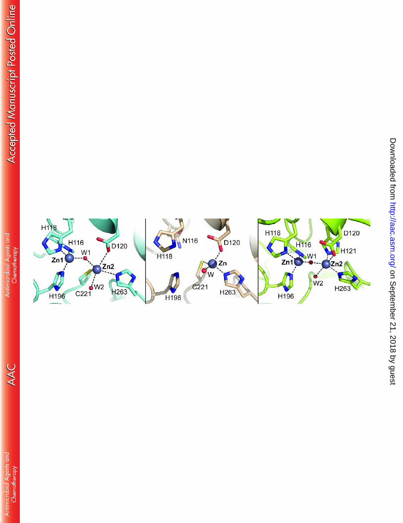
simulations. Thus, GOB enzymes present a divergent metal binding site with novel substrate 102
binding features, which make them unique among B3 MBLs. This observation, together with the 103
finding that the resistance profile of E. meningoseptica is elicited mostly by the B1 enzyme BlaB 104
(33), discloses a functional redundancy, and suggests that GOB enzymes may play alternative 105
roles in this microorganism yet to be disclosed. 106
107
on Septem
ber 21, 2018 by guesthttp://aac.asm
.org/D
ownloaded from
6
MATERIALS AND METHODS 108
109
Chemicals. Biochemical reagents were purchased from Sigma-Aldrich except when specified. 110
Molecular biology reagents were purchased from Promega, Invitrogen or New England Biolabs. 111
Metal-free buffers were prepared adding Chelex 100 to normal buffers and stirring for 0.5 h. 112
113
Production of recombinant GOB-18 in the periplasmic space of E. coli. GOB-18 was produced in 114
the periplasmic space of E. coli C41 (DE3) cells harboring the plasmid pKP-GOB-18 (29,31,32). 115
This vector allows the production of recombinant GOB-18 as a C-terminal fusion to the leader 116
peptide pelB (29,31). Secretion via the Sec pathway allows processing of the precursor and 117
targeting the mature protein to the periplasmic space of the bacterial host. E. coli C41 (DE3) 118
cells harboring plasmid pKP-GOB-18 were grown aerobically at 30ºC in LB broth supplemented 119
with kanamycin (50 µg/ml) until reaching 0.6 units of absorbance at 600 nm. Protein production 120
was induced adding isopropyl β-D-1-thiogalactopyranoside (IPTG) to a final concentration of 1 121
mM, the incubation was continued for 16 h at the same temperature. Mature GOB-18 was 122
extracted from the cellular periplasm by a two-step osmotic shock procedure without lysozyme 123
nor EDTA. Briefly, cells were harvested and resuspended with 1 ml/g (wet weight) of 20 mM 124
Tris-HCl pH 8, 50% w/v sucrose, 0.1 mM phenylmethylsulfonyl fluoride, and incubated for 2 h at 125
4ºC under orbital shaking. 10 ml of ice-cold water were added per ml of suspension, and further 126
incubated for 2 h in the same conditions. After centrifugation at 15,000 g for 8 min at 4ºC, the 127
supernatant (first extract), was collected and stored at 4ºC. The pellet was further resuspended 128
in ice-cold water in half the volume used for the first extract, and incubated in the same 129
on Septem
ber 21, 2018 by guesthttp://aac.asm
.org/D
ownloaded from
7
conditions for 1.5 h at 4ºC. The supernatant (second extract) was collected and pooled with the 130
first one rendering the periplasmic fraction. 131
132
Purification of GOB-18 from the periplasmic fraction of E. coli. Mature GOB-18 was purified to 133
homogeneity with two steps of ionic exchange chromatography (Figure 2). The periplasmic 134
extract was adjusted to pH 7 and loaded onto a Sephadex CM-50 column (Sigma) pre-135
equilibrated with buffer E (20 mM Tris-HCl, pH 7). The resin was washed with 5 column volumes 136
of buffer E, and eluted with buffer E supplemented with 400 mM NaCl. The elution peak was 137
concentrated, dialyzed against buffer E, and loaded onto a Mono S column (GE Healthcare) pre-138
equilibrated with buffer E. Elution was achieved with buffer E with added NaCl (gradient 0 to 1 139
M). Samples containing GOB-18 were pooled and concentrated to 20 mg/ml, frozen in liquid 140
nitrogen and stored at -70ºC until crystallogenesis assays. An aliquot of the enzyme was stored 141
at 4ºC for kinetic analyses. The average purification yields obtained were ca. 1 mg of GOB-18 142
per liter of E. coli culture, resulting in the expected 31 kDa polypeptide, as estimated by SDS-143
PAGE. 144
145
Metal content determination. The metal content of purified GOB-18 was measured by atomic 146
absorption spectroscopy in a Metrolab 250 instrument operating in the flame mode. 147
148
Circular dichroism spectroscopy. The circular dichroism spectrum of periplasmic GOB-18 was 149
obtained using a JASCO J-810 spectropolarimeter at 25ºC. 150
151
on Septem
ber 21, 2018 by guesthttp://aac.asm
.org/D
ownloaded from
8
Determination of the kinetic parameters. Antibiotic hydrolysis was monitored by absorbance 152
variation resulting from the scission of the β-lactam ring. Reactions were performed in 15 mM 153
Hepes pH 7.5, 200 mM NaCl at 30ºC. The kinetic parameters KM and kcat were derived from 154
initial rate measurements, as recorded with a Jasco V-550 spectrophotometer, and were 155
estimated by nonlinear data fitting to the integrated form of the Michaelis-Menten equation. 156
157
Crystallization and data collection. Crystals grew from a 9.8 mg/ml solution of GOB-18, by 158
adding 0.5 µl of protein to 1.5 µl mother liquor (100 mM Tris-HCl pH 8.5, 1.7 M NaCl, 1.7 M 159
(NH4)2SO4) in a hanging-drop setup with 1 ml mother liquor in the reservoir. These were not 160
single crystals, which were eventually obtained by microseeding fresh drops with identical 161
conditions except for using a slightly modified mother liquor (2 M NaCl/ (NH4)2SO4 and avoiding 162
Tris-HCl). Single crystals reached a size of ca. 150 µm, and were cryoprotected in mother liquor 163
containing 25% glycerol and flash frozen in liquid nitrogen. X-ray diffraction data were collected 164
in house (Unit of Protein Crystallography, Institut Pasteur de Montevideo, Montevideo, 165
Uruguay), from a single crystal, with a MicroMax-007 HF X-ray source (Rigaku) and a MAR345 166
image plate detector (Mar Research), employing radiation of 1.5418 Å. The diffraction data 167
were processed using XDS (34) and scaled with Aimless (35) from the CCP4 program suite. 168
169
Structure determination and refinement. The crystal structure of GOB-18 was solved by 170
molecular replacement using the program Phaser (36) and the model of native FEZ-1 from 171
Legionella gormanii (PDB 1K07) as search probe. The asymmetric unit contains two monomers 172
of GOB-18 (chains A and B) with ca. 44% of the volume occupied by solvent. SigmaA-weighted 173
on Septem
ber 21, 2018 by guesthttp://aac.asm
.org/D
ownloaded from

9
Fourier maps (2mFo-DFc) allowed rebuilding the initial model reliably through iterative cycles of 174
manual model building with COOT (37) and refinement with BUSTER (38). To minimize model-175
bias, initial refinement cycles were carried out including high-temperature (7,500K) slow-176
cooling simulated annealing procedures in torsional space (as implemented in the program 177
PHENIX (39)). The final model revealed two outlier residues in the Ramachandran plot. Figures 178
were generated and rendered with Pymol 1.5.0.2. (Schrödinger, LLC). Volume calculations for 179
automatically identified cavities were done using the CASTp algorithm (40) and Q-site finder 180
(41), giving similar results. 181
Atomic coordinates and structure factors were deposited in the Protein Data Bank under 182
the accession code 5K0W. 183
184
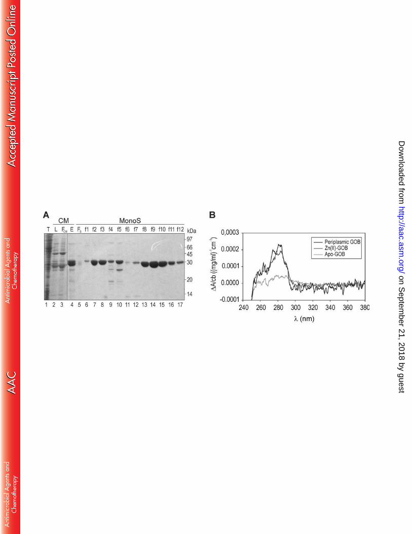
Substrate docking and molecular dynamics simulations. Molecular docking was performed 185
using Autodock 4.2 (42). Deprotonated imipenem was docked setting up a docking box of 40 × 186
40 × 40 Å (grid-point spacing of 0.375 Å) centered in the enzyme active site. The model of GOB-187
imipenem was built in silico by making a structural alignment with the structure of L1-imipenem 188
using the Multiseq plug-in of VMD (43). Molecular dynamics simulations were performed 189
starting from complexes GOB-18-imipenem and L1-imipenem. Each complex was immersed in a 190
truncated octahedral periodic box with a minimum solute-wall distance of 8 Å, filled with 191
explicit TIP3P water molecules (44). Molecular dynamic simulations were performed with the 192
AMBER14 package (45,46), using ff14SB (47). Particle-mesh Ewald (PME) was implemented for 193
long range interactions with a cutoff distance of 12 Å (48). Temperature and pressure were 194
regulated with the Berendsen thermostat and barostat (49). All bonds involving hydrogen were 195
on Septem
ber 21, 2018 by guesthttp://aac.asm
.org/D
ownloaded from
10
fixed using the SHAKE algorithm (50). Each initial system was equilibrated at 300K using a 196
conventional protocol, and then subjected to 50 ns of simulation in the NVT ensemble. To 197
maintain the coordination environment around zinc atoms we used harmonic bonds between 198
the metal centers and the residues of the coordination sphere. We applied a restraint to keep 199
the imipenem bound to the active site. Root mean squared deviation (rmsd) and root mean 200
square fluctuations (rmsf) calculations of the molecular dynamics simulations were performed 201
using the cpptraj module of AMBER (51). 202
203
on Septem
ber 21, 2018 by guesthttp://aac.asm
.org/D
ownloaded from
11
RESULTS 204
205
Expression and purification of GOB-18 for crystallogenesis 206
Production of GOB-18 in E. coli gives rise to different metallated species depending on 207
the cellular compartment where the protein accumulates. Overexpression of GOB-18 in the 208
cytoplasm results in an inactive Fe(III)-bound form, from which a mononuclear fully-active 209
Zn(II)-variant can be prepared by metal chelation and remetallation in vitro (29). Instead, GOB 210
binds exclusively Zn(II) when it is secreted into the bacterial periplasm (29,31). 211
GOB-18 produced in the cytoplasm of E. coli, both the Fe(III)-containing variant or the 212
reconstituted mono-Zn(II) form, proved recalcitrant after extensive crystallization trials. An 213
alternative protocol was thus optimized allowing to produce GOB-18 in the bacterial periplasm, 214
which is the physiological cellular compartment for MBLs’ expression in Gram-negative species 215
(Figure 2A). This strategy yielded GOB-18 containing exclusively Zn(II), with a maximum metal 216
content of (1.4± 0.1) Zn(II) equivalents/ GOB-18 molecule as determined by atomic absorption 217
spectroscopy. This metal-content figure indicates the presence of a dinuclear GOB-18 species, 218
which is consistent with previous reports for GOB-1 (30). Circular dichroism of periplasmic GOB-219
18 indicates it has similar tertiary structure as the in vitro reconstituted Zn(II) form (29) (Figure 220
2B). Periplasmic GOB-18 was able to catalyze the hydrolysis of a broad spectrum of β-lactam 221
substrates with kcat and KM values similar to those formerly reported for mono-Zn(II) and di-222
Zn(II)-GOB-1, with no changes in the catalytic efficiencies upon addition of 20 μM Zn(II) to the 223
reaction medium (Table 1). Nevertheless, in contrast to reconstituted Zn(II)-GOB-18, 224
on Septem
ber 21, 2018 by guesthttp://aac.asm
.org/D
ownloaded from
12
periplasmic GOB-18 crystallized in different conditions containing high concentrations of NaCl 225
and (NH4)2SO4 as precipitants. 226
227
Crystal structure of periplasmic GOB-18 228
Periplasmic GOB-18 crystallized in space group P21, with crystals diffracting X rays to 2.6 229
Å resolution (Table 2). The final refined atomic model contains two protein molecules per 230
asymmetric unit, 4 zinc atoms, 8 chlorides, 77 waters and one glycerol molecule. The two 231
protein chains are very similar, with 0.26 Å root mean squared deviation (rmsd) among 266 232
aligned α-carbons. Chain A includes residues Ser17 to Asp286 (the side chains of residues Asn26 233
and Asp286 were not included in the model due to weak electron density), while monomer B 234
spans residues Val20 to Lys290 (similarly, Val20 side chain was not modeled), with a continuous 235
main chain trace throughout. GOB-18 displays the expected αβ/βα sandwich fold, with two 236
core β-sheets composed of 7 (β1-β7) and 5 (β8-β12) β-strands, respectively, and 6 α-helices 237
(Figure 3A). Helices α1-α4 cover the 7-stranded β-sheet, with helix α4 closing one side of the 238
metal-binding groove. On the other hand, helices α5 and α6 wrap around the 5-stranded β-239
sheet, with the intervening region β12-α6, including three short helical elements, closing up 240
the other side of the catalytic site. As already described for the MBL fold (1-6), the whole 241
domain of GOB-18 can be depicted as a duplication of two structurally similar α/β hemi-242
domains. 243
GOB-18 is very similar to other B3 lactamases (20-24), despite low sequence identity. 244
Structural alignment of GOB-18 (chain A) with available B3 lactamase models allows for 245
similarity quantification (Figure 3B): FEZ-1 (PDB 1K07, 1.27 Å rmsd for 243 aligned residues); 246
on Septem
ber 21, 2018 by guesthttp://aac.asm
.org/D
ownloaded from
13
BJP-1 (PDB 3LVZ, 1.50 Å rmsd for 239 residues); and, L1 (PDB 1SML, 1.55 Å rmsd for 236 247
residues). When calculated per residue, the largest local rmsd values are observed for two 248
loops flanking the active site, which are expected to be involved in substrate specificity: loop 1 249
(residues 130-152 in GOB-18, equivalent to 148-172 according to BBL numbering; to facilitate 250
comparative analyses, residue numbering will hereafter be stated for GOB-18 followed by BBL 251
numbering in parenthesis) and loop 2 (198-218(219-239)). Structural differences are also 252
noticeable in the β12-α6 loop (residues 239-265(261-287)) as well as in the N-terminus of the 253
protein. 254
All B3 lactamases crystallized so far are characterized by at least one disulfide bridge, in 255
most cases between Cys residues located in the C-terminal helix and the loop linking elements 256
α5 and β12 (20-24). GOB-18 is an exception in this regard within B3 enzymes, with only one Cys 257
residue in its primary structure, i.e. Cys180(201) (in strand β10), which is buried within the 258
protein core (Figure 3A) forming hydrogen bonds in the base of the metal binding site (see 259
below). 260
261
The active site of di-Zn(II)-GOB-18 262
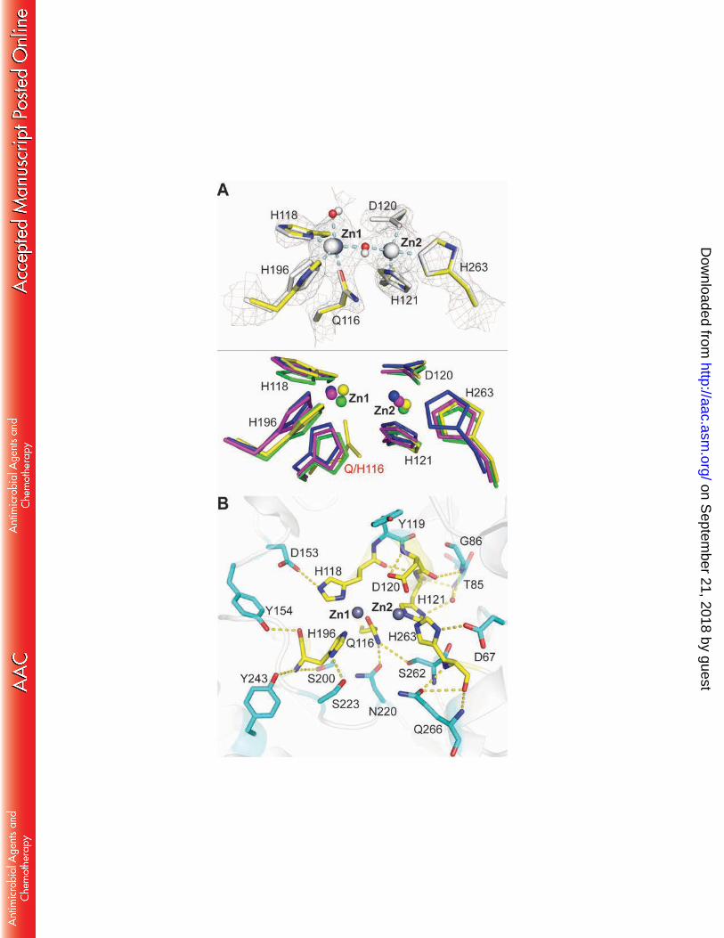
The electron density maps revealed the presence of two heavy atoms in the active sites 263
of each GOB-18 monomer in the asymmetric unit (Figure 4A). They occupy the metal binding 264
sites found in other dinuclear MBLs, and can be recognized as the Zn1 and Zn2 ions. The Zn-Zn 265
distance (3.5 and 3.8 Å in chains A and B, respectively) is similar to that found in other dinuclear 266
B3 lactamases (20-24). Zn1 is coordinated by the Nδ1 atom of residue His100(118), His175(196) 267
Nε2, Gln98(116) Oε1 and a Zn-Zn bridging water molecule. The Zn1-Oε1(Gln) distance is 2.0 Å, 268
on Septem
ber 21, 2018 by guesthttp://aac.asm
.org/D
ownloaded from
14
pinpointing Gln98(116) as a novel metal-binding residue in MBLs. On the other hand, Zn2 is 269
coordinated to His103(121) and His241(263) Nε2, Asp102(120) Oδ2 and the bridging water 270
molecule, adopting a distorted tetrahedral geometry. However, in all B3 lactamases crystallized 271
so far the coordination sphere in the Zn2 site adopts a trigonal bipyramidal geometry with two 272
water ligands (20-24). The presence of additional non-protein ligands in the GOB-18 site cannot 273
be ruled out, given our data’s resolution limit. In the same line of thought, an axial water 274
molecule with high mobility was modeled in the coordination sphere of the Zn1 ion in both 275
GOB-18 monomers, but a larger species (such as glycerol from the cryo-protection solution) 276
cannot be excluded. 277
The conformation of the zinc ligands in GOB-18 active site is stabilized by a network of 278
hydrogen bonds (Figure 4B), involving outer sphere ligands. His100(118) Nε2 and His175(196) 279
Nδ1 interact respectively with Asp135(153) and Ser202(223) side chains, and Gln98(116) Nε2 280
establishes contacts with Asn199(220) Oδ1. Additionally, the Nδ1 atom of His103(121) is H-281
bonded with the main chain N of residues Thr68(85) and Gly69(86) through a bridging water, 282
His241(263) Nε2 interacts with the side chain of Asp51(67), and the carboxylate of Asp102(120) 283
contacts its own amide nitrogen. Most of these outer sphere ligands are conserved in B3 284
enzymes (20-24), despite the divergence introduced by the His116Gln replacement. 285
A novel feature in GOB-18 can be observed at residue Cys180(201): its thiol and amide 286
nitrogen make H-bonds with the backbone carbonyls of Leu96(114) and Thr97(115), 287
respectively (Figure 3A). These residues are located in the β5-α3 loop, which contains the 288
metal-binding motif Q116XH118XD120H121 that replaces the HXHXDH signature present in the rest 289
of B3 enzymes. Thus, Cys180(201), within the β-strand just downstream from the loop including 290
on Septem
ber 21, 2018 by guesthttp://aac.asm
.org/D
ownloaded from
15
metal-binding His175(196), connects the bases of two loops that comprise five out of six metal 291
ligands, and all the Zn1-coordinating residues. 292
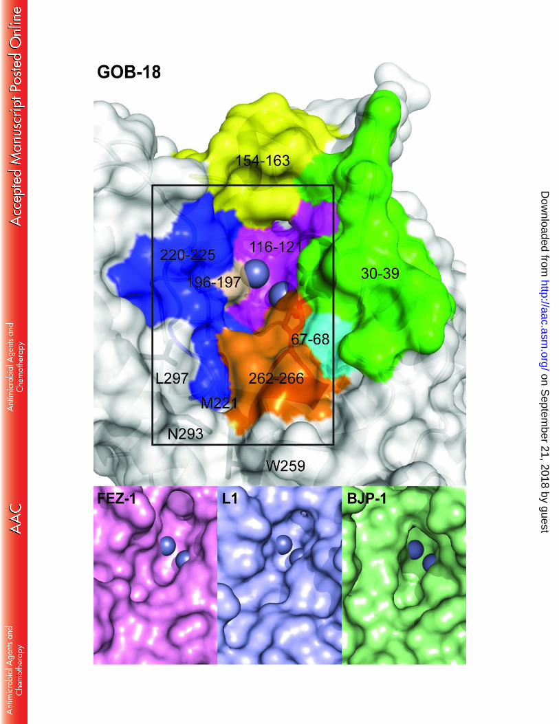
The residues that define the substrate binding cleft in GOB-18 are mainly found in loops. 293
The catalytic pocket (Figure 5) is delimited by segments 23-32(30-39) (spanning a non-294
structured coil and a short, kinked helix), 51-52(67-68), 98-103(116-121) (including the QXHXDH 295
motif), 135-144(154-163) (encompassing a short helix), 175-176(196-197) (containing metal-296
binding His175(196)), 199-204(220-225) (harboring residue Met 200(221)) and 240-244(262-297
266) (spanning a short helix and including metal-binding His241(263)). This cleft, albeit shallow, 298
is one of the top ranking cavities, comprising 150-200 Å3 with approximate 8 Å x 15 Å on the 299
opening and 10 Å deep. A second, smaller pocket, with residues Ser202(223), Gln244(266), 300
Asn271(293) and Leu275(297) conforming the side limits and Met200(221) and Trp237(259) on 301
the floor, is immediately adjacent to the first one, thus constituting a discontinuous groove. 302
Figure 5 compares the catalytic groove of GOB-18 with those from the B3 lactamases FEZ-1, L1 303
and BJP-1. The wide substrate-binding groove in GOB enzymes correlates with high catalytic 304
efficiencies for a broad spectrum of substrates, in contrast to BJP-1, in which an N-terminal 305
helix partially covers the active site (22). 306
307
Substrate docking and molecular dynamics simulations 308
We attempted to obtain a model for di-Zn(II) GOB-18 complexed with imipenem by in 309
silico docking calculations. However, none of the resulting models reproduced binding modes 310
consistent with a productive Michaelis complex. Similar docking simulations with L1 were 311
instead successful, yielding a model which reproduced most of the binding features reported in 312
on Septem
ber 21, 2018 by guesthttp://aac.asm
.org/D
ownloaded from
16
the crystal structure of L1 in complex with hydrolyzed moxalactam (PDB 2AIO) (52) (Figure S1). 313
In our docking model, the carbonyl C7 from imipenem is positioned close to the bridging OH- 314
enabling the nucleophilic attack, and the carboxylate C9 interacts with the Zn2 ion, anchoring 315
the substrate, in agreement with the generally accepted productive binding mode of β-lactam 316
compounds to dinuclear MBLs. Based on these results, we generated a model of GOB-18 in 317
complex with imipenem, by structural alignment of GOB-18 onto the L1-imipenem complex. 318
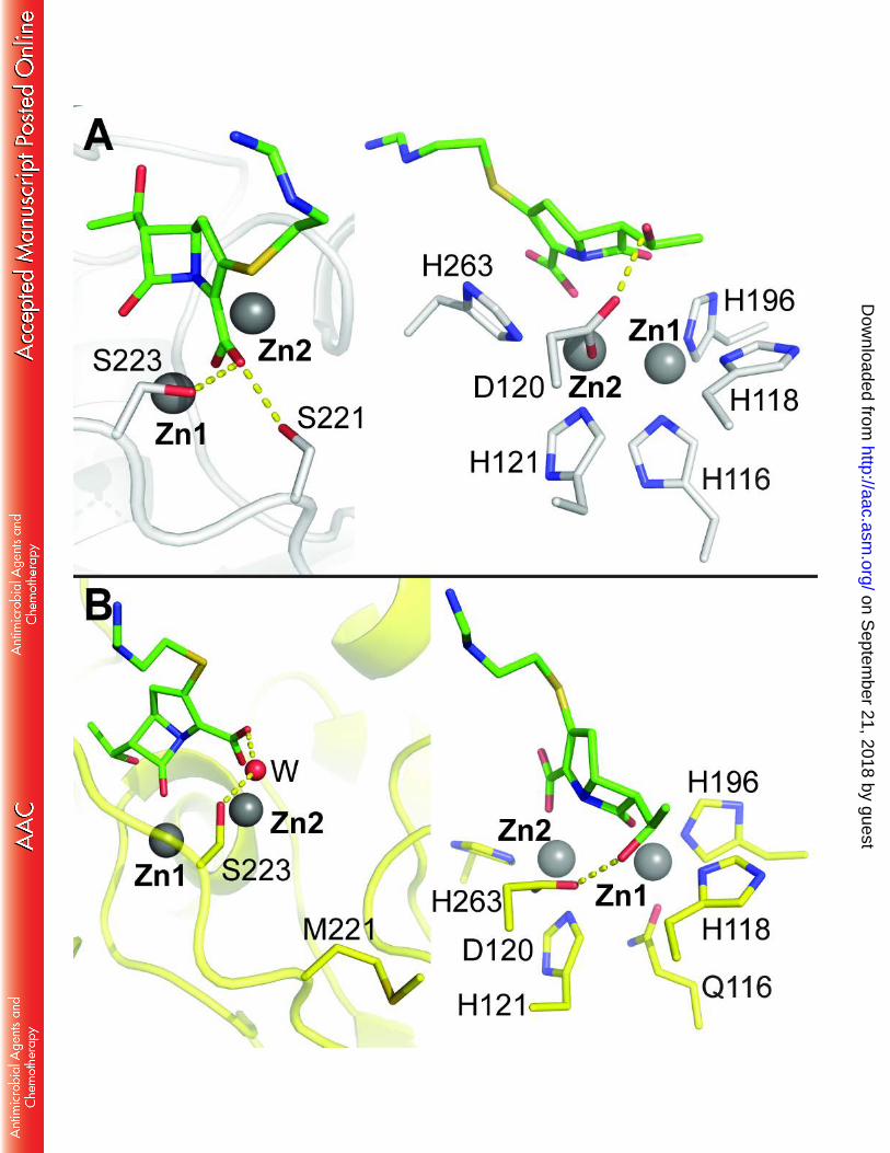
The L1-imipenem and GOB-18-imipenem models were used as starting geometries for 319
molecular dynamics simulations, ran for 50 ns. In the L1-imipenem complex, residues Ser221 320
and Ser223 interact with the carboxylate C9 of imipenem throughout the simulation (Figure 321
6A). Additionally, the OH group C10 of imipenem established a hydrogen bond with the non-322
chelating oxygen of the Zn2-ligand Asp120. In GOB-18, Ser221 is replaced by a Met residue 323
whose side chain points out of the active site, buried in the hydrophobic core of the protein, 324
and hence unable to interact with the substrate (Figure 6B). This orientation is conserved 325
throughout the dynamics. On the other hand, the imipenem carboxylate contacts residue 326
Ser202(223) through a water molecule, and the interaction of the OH group C10 with 327
Asp102(120) is also preserved in the GOB-18-imipenem complex. Taken together, these data 328
strongly suggest that the Ser221Met substitution may have an impact on substrate binding by 329
GOB enzymes. 330
The mobility of active site loops in MBLs has been related to substrate specificity. 331
Indeed, residues 221 and 223 are located in a loop flanking the active site (Figure S2). We 332
calculated the fluctuations of residues within this loop in GOB-18 and L1, both in the resting 333
state and in the Michaelis complex with imipenem. The rmsf (root mean square fluctuation) of 334
on Septem
ber 21, 2018 by guesthttp://aac.asm
.org/D
ownloaded from
17
this loop is substantially larger in GOB-18 than in L1 (Figure S2). We propose that the 335
Ser221Met substitution present in GOB enzymes is compensated by a larger flexibility in this 336
loop, which might assist substrate binding within the catalytic groove. 337
338
on Septem
ber 21, 2018 by guesthttp://aac.asm
.org/D
ownloaded from
18
DISCUSSION 339
340
Here we report the crystal structure and biochemical characterization of the periplasmic 341
di-Zn(II) form of GOB-18 from E. meningoseptica. The structural data reveal that a Gln residue 342
replaces the ubiquitous His116 in the coordination sphere of the Zn1 ion (Figure 4). Periplasmic 343
GOB-18 is a fully active broad spectrum lactamase in the di-Zn(II) form (Table 1), in agreement 344
with a previous report that studied GOB-1 (30). These results contrast our previous study with 345
recombinant GOB-18 obtained from the cytoplasm of E. coli cells (29). In that case, 346
accumulation of the protein in the bacterial cytoplasm resulted in an inactive Fe(III)-bound 347
form, which could be demetallated and subsequently loaded with Zn(II) in vitro to obtain an 348
active mono-Zn(II) variant. Indeed, the preferential binding of a given divalent cation over 349
others is influenced by the cellular localization of proteins (53), and mismetallation upon 350
protein overproduction in the cytoplasm of E. coli has been documented for a number of 351
systems (54-56). Thus, these separate preparations give us the opportunity to compare mono- 352
and di-Zn(II) variants of GOB-18. 353
We now show that when secreted into the bacterial periplasm, GOB exclusively binds 354
Zn(II), which is relevant given that this is the physiological cellular compartment of MBLs in 355
Gram-negative bacteria (53). Thus, in order to avoid chelation and remetallation steps, we 356
optimized a protocol for the production of GOB-18 in the periplasm of E. coli and the 357
preparation of high amounts of pure mature protein in the absence of affinity tags. Indeed, 358
periplasmic GOB-18 preparations containing exclusively Zn(II) were successfully obtained and, 359
while all efforts aimed at crystallizing the cytoplasmic Fe(III)-containing variant or the 360
on Septem
ber 21, 2018 by guesthttp://aac.asm
.org/D
ownloaded from
19
reconstituted mono-Zn(II) form of GOB-18 were unsuccessful, diffraction quality crystals readily 361
grew from solutions of periplasmic di-Zn(II) GOB-18. These results clearly show that the 362
differences in metal content previously noted between GOB-1 and GOB-18 were due to the 363
procedures employed to produce the recombinant proteins in each case and not due to the few 364
residues differing in their primary structures. Taking all these data into account, it is safe to 365
extrapolate several of the present conclusions to all GOB enzymes. 366
Periplasmic di-Zn(II) GOB-18 catalyzed hydrolysis of β-lactam substrates with kcat and KM 367
values similar to those reported for mono-Zn(II)-GOB-18 and di-Zn(II)-GOB-1 (Table 1). Besides, 368
previous analyses of mutant GOB-18 Asp120Ser and metal-substituted GOB-18 derivatives, 369
showed that the Zn2 site is essential for catalysis and for the stabilization of an anionic 370
intermediate in the hydrolysis of nitrocefin (29,57). On the other hand, even though 371
substitution of Gln116 by an isosteric His residue was somewhat detrimental for the resistance 372
profile conferred by GOB-18 (29,31), it had little effect both on metal content as well as on the 373
in vitro activity of dinuclear GOB-1 (30) and remetallated mono-Zn(II) GOB-18 (29). Indeed, 374
mutations Gln116Asn and Gln116Ala, which are predicted to impact on the structure of the Zn2 375
site through perturbations on the second coordination sphere (Figure 4B), reduced the catalytic 376
efficiency of GOB-1 (30). Overall, the available data indicate that the Zn1 site in GOB enzymes 377
contributes only marginally to activity, whereas it may be critical for the enzyme function in 378
vivo. This would depend on the zinc availability, which can vary widely depending on 379
environmental conditions (8). 380
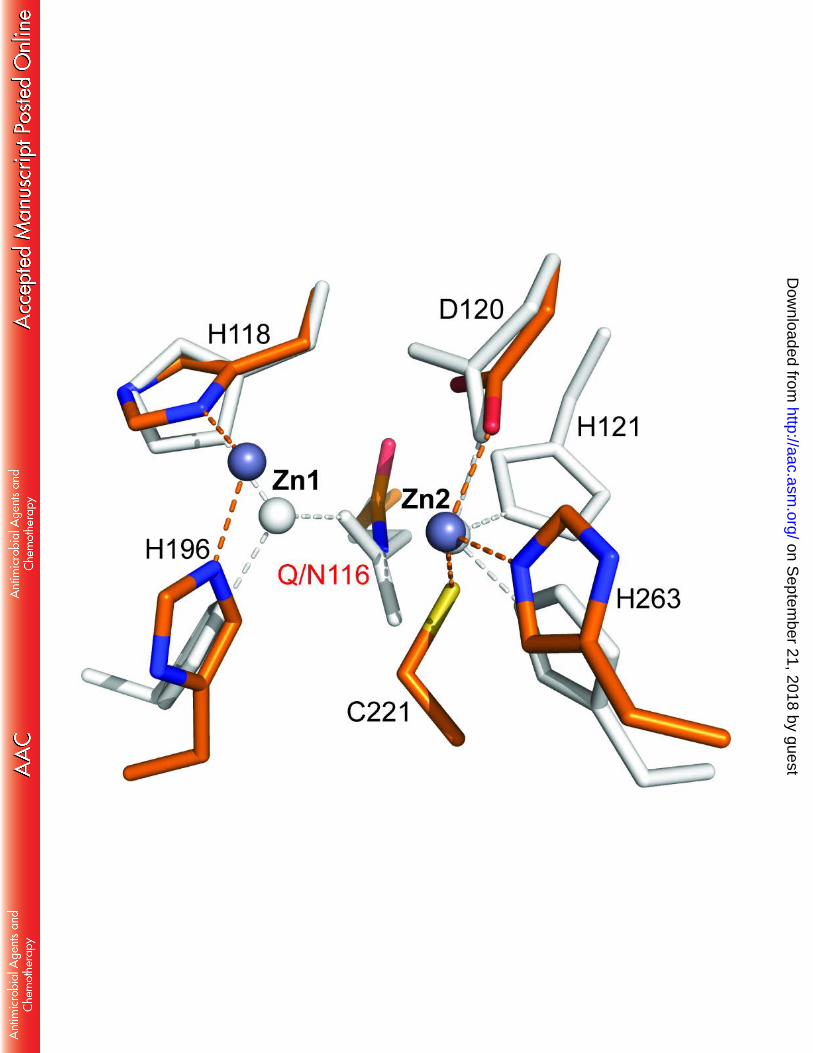
The finding of a dinuclear Zn(II) site where Gln116 acts as a metal ligand of the Zn1 ion 381
contrasts with that found in B2 MBLs, where substitution of His116 by an Asn residue leads to a 382
on Septem
ber 21, 2018 by guesthttp://aac.asm
.org/D
ownloaded from
20
dinuclear non-active species (18,19). In the case of di-Zn(II)-CphA, residue Asn116 does not act 383
as a metal ligand and the Zn1 ion is coordinated by only two protein residues, adopting a non-384
productive position (16). Instead, the longer side chain of Gln116 (isosteric to a His residue) in 385
GOB enzymes makes it a good metal ligand, so that the Zn1 ion adopts a position similar to that 386
held in other di-Zn MBLs (Figure 7). The finding of a conserved network of hydrogen bonds 387
among second sphere ligands confirms the requirement of a similar geometric arrangement in 388
the active site. We have also found that a unique Cys residue may help anchoring the two loops 389
that provide protein ligands for the Zn1 ion (Figure 3A). A conservative mutation of this Cys 390
residue to a Ser (29) preserves the enzyme activity, supporting this hypothesis. The role of the 391
Zn1 site in MBLs is to facilitate deprotonation of the bound water to provide an active 392
nucleophile (1). This function cannot be properly fulfilled by the Zn1 ion in B2 enzymes, thus 393
resulting in enzyme inhibition (16), but instead is fully preserved in GOB enzymes. Therefore, 394
the His116Gln mutation is conservative in terms of both structure and function. The role of the 395
Zn2 site is two-fold: to provide an anchoring electrostatic point for substrate binding and to 396
stabilize the development of negative charge in the bridgehead nitrogen. Both functions also 397
depend on the adequate positioning of this metal ion, which is indeed maintained in GOB-18. 398
Position 221 is essential in B1 and B2 MBLs, where a Cys residue acts as a ligand of the 399
Zn2 ion. Instead, in most B3 enzymes a Ser is found in this position. The report of a Met residue 400
in position 221 in GOB enzymes led us to speculate that the thioether moiety could act as a 401
weak metal ligand. However, mutagenesis experiments in GOB-18 showed that Met221 is not 402
involved in metal binding nor in catalysis, but that, instead, has a structural role (31,32). The 403
crystal structure that we are now disclosing, confirms that Met221 is not a metal ligand, 404
on Septem
ber 21, 2018 by guesthttp://aac.asm
.org/D
ownloaded from
21
actually its side chain does not point toward the substrate-binding site (Figure 6). Besides, 405
docking and molecular dynamics simulations confirm that this residue is not involved in 406
substrate binding, in sharp contrast to the role of the conserved Ser221 in most B3 enzymes. 407
Therefore, these results provide the first available evidence for an MBL where residue 221 is 408
not involved in metal chelation nor in substrate binding, and highlight the diversity of roles 409
fulfilled by this position among enzymes within the family. Additionally, a wide substrate-410
binding groove in GOB-18 (Figure 5) correlates with a high catalytic efficiency against a diverse 411
range of β–lactam substrates (Table 1) as compared to other B3 MBLs, in agreement with 412
previous observations. 413
The metal binding site of GOB enzymes is unusual not only among MBLs. Indeed, 414
Asn/Gln ligands in protein zinc sites are very rare (0.5 % of the cases, according to Dudev and 415
Lim) (58). Surprisingly, the His116Gln substitution does not have a direct impact on the broad 416
spectrum profile of GOB-18. GOB is also peculiar in that its native organism, E. meningoseptica, 417
is the only known bacterium expressing two MBLs: the B1 enzyme BlaB, and the B3 enzyme 418
GOB. We have shown that, even if both genes are actively expressed, the higher levels of BlaB 419
make this enzyme the one responsible for carbapenemase resistance (33). Thus, GOB could be 420
redundant in this organism despite its efficient catalytic performance. GOB-like alleles have also 421
been found in metagenomics studies of a remote Alaskan soil with minimal human-induced 422
selective pressure (59). More recently, new MBLs closely homologous to GOB enzymes have 423
been found in Pedobacter roseus (PEDO-1), Pedobacter borealis (PEDO-2), Chrisobacterium 424
piscium (CPS-1) and Epilithonimonas tenax (ESP-1) (60,61). All these MBLs feature the 425
characteristic Gln116 residue in the active site, as well as the Cys residue in second sphere 426
on Septem
ber 21, 2018 by guesthttp://aac.asm
.org/D
ownloaded from
22
position, and a hydrophobic residue in position 221 (Met or Leu), discussed in the present 427
crystal structure, confirming that GOB-like alleles are ubiquitous in environmental bacteria. We 428
can speculate that GOB genes could serve as a rather ancient resistance reservoir, or maybe 429
could give rise to a different, yet unknown function, elicited by the presence of the Gln residue, 430
providing at the same time an independent source of antibiotic resistance. In any case, these 431
findings further highlight the large structural diversity of MBLs in current microorganisms, the 432
details of which constitute valuable information in conceiving better antibiotic design 433
strategies. 434
on Septem
ber 21, 2018 by guesthttp://aac.asm
.org/D
ownloaded from
23
ACKNOWLEDGEMENTS 435
436
We thank Dr. R. Girolami for atomic absorption measurements. MNL is a postdoctoral 437
fellow from ANII and was a recipient of a doctoral fellowship from CONICET. SID is recipient of a 438
doctoral fellowship from CONICET. JMB, DMM, AMV and AJV are staff members from CONICET. 439
This work was supported by grants from ANPCyT and the US National Institutes of Health 440
(1R01AI100560) to AJV; from ANPCyT, CONICET, UNR, and Ministerio de Salud, Provincia de 441
Santa Fe, Argentina to AMV; from ANPCyT and CONICET to DMM. 442 443 444
REFERENCES 445
446
1. Meini MR, Llarrull LI, and Vila AJ. 2015. Overcoming differences: The catalytic 447
mechanism of metallo-beta-lactamases. FEBS Lett. 589:3419-3432. 448
2. Meini MR, Llarrull LI, and Vila AJ. 2014. Evolution of Metallo-beta-lactamases: Trends 449
Revealed by Natural Diversity and in vitro Evolution. Antibiotics.(Basel) 3:285-316. 450
3. Palzkill T. 2013. Metallo-beta-lactamase structure and function. Ann.N.Y.Acad.Sci. 451
1277:91-104. 452
4. Llarrull LI, Testero SA, Fisher JF, and Mobashery S. 2010. The future of the beta-453
lactams. Curr.Opin.Microbiol. 13:551-557. 454
on Septem
ber 21, 2018 by guesthttp://aac.asm
.org/D
ownloaded from
24
5. Fisher JF, Meroueh SO, and Mobashery S. 2005. Bacterial resistance to beta-lactam 455
antibiotics: compelling opportunism, compelling opportunity. Chem.Rev. 105:395-424. 456
6. Crowder MW, Spencer J, and Vila AJ. 2006. Metallo-beta-lactamases: Novel Weaponry 457
for Antibiotic Resistance in Bacteria. Acc.Chem.Res. 39:721-728. 458
7. Galleni M, Lamotte-Brasseur J, Rossolini GM, Spencer J, Dideberg O, and Frere JM. 459
2001. Standard numbering scheme for class B beta-lactamases. Antimicrob.Agents 460
Chemother. 45:660-663. 461
8. Gonzalez JM, Meini MR, Tomatis PE, Martin FJ, Cricco JA, and Vila AJ. 2012. Metallo-462
beta-lactamases withstand low Zn(II) conditions by tuning metal-ligand interactions. 463
Nat.Chem.Biol. 464
9. Llarrull LI, Tioni MF, and Vila AJ. 2008. Metal content and localization during turnover in 465
B. cereus metallo-beta-lactamase. J Am.Chem Soc. 130:15842-15851. 466
10. Hawk MJ, Breece RM, Hajdin CE, Bender KM, Hu Z, Costello AL, Bennett B, Tierney DL, 467
and Crowder MW. 2009. Differential binding of Co(II) and Zn(II) to metallo-beta-468
lactamase Bla2 from Bacillus anthracis. J Am.Chem Soc. 131:10753-10762. 469
11. Fabiane SM, Sohi MK, Wan T, Payne DJ, Bateson JH, Mitchell T, and Sutton BJ. 1998. 470
Crystal structure of the zinc-dependent beta lactamase from Bacillus cereus at 1.9 A 471
resolution: binuclear active site with features of a mononuclear enzime. Biochemistry 472
37:12404-12411. 473
on Septem
ber 21, 2018 by guesthttp://aac.asm
.org/D
ownloaded from
25
12. Garcia-Saez I, Docquier JD, Rossolini GM, and Dideberg O. 2008. The three-dimensional 474
structure of VIM-2, a Zn-beta-lactamase from Pseudomonas aeruginosa in its reduced 475
and oxidised form. J.Mol.Biol. 375:604-611. 476
13. Concha N, Rasmussen BA, Bush K, and Herzberg O. 1996. Crystal structure of the wide-477
spectrum binuclear zinc beta-lactamase from Bacteroides fragilis. Structure 4:823-836. 478
14. King DT, Worrall LJ, Gruninger R, and Strynadka NC. 2012. New Delhi metallo-beta-479
lactamase: structural insights into beta-lactam recognition and inhibition. 480
J.Am.Chem.Soc. 134:11362-11365. 481
15. Gonzalez LJ, Moreno DM, Bonomo RA, and Vila AJ. 2014. Host-specific enzyme-482
substrate interactions in SPM-1 metallo-beta-lactamase are modulated by second 483
sphere residues. PLoS.Pathog. 10:e1003817. 484
16. Bebrone C, Delbruck H, Kupper MB, Schlomer P, Willmann C, Frere JM, Fischer R, 485
Galleni M, and Hoffmann KM. 2009. The structure of the dizinc subclass B2 metallo-486
beta-lactamase CphA reveals that the second inhibitory zinc ion binds in the histidine 487
site. Antimicrob.Agents Chemother. 53:4464-4471. 488
17. Hernandez VM, Felici A, Weber G, Adolph HW, Zeppezauer M, Rossolini GM, 489
Amicosante G, Frere JM, and Galleni M. 1997. Zn(II) dependence of the Aeromonas 490
hydrophila AE036 metallo-beta-lactamase activity and stability. Biochemistry 36:11534-491
11541. 492
on Septem
ber 21, 2018 by guesthttp://aac.asm
.org/D
ownloaded from
26
18. Fonseca F, Bromley EH, Saavedra MJ, Correia A, and Spencer J. 2011. Crystal structure 493
of Serratia fonticola Sfh-I: activation of the nucleophile in mono-zinc metallo-beta-494
lactamases. J.Mol.Biol. 411:951-959. 495
19. Garau G, Bebrone C, Anne C, Galleni M, Frere JM, and Dideberg O. 2005. A metallo-496
beta-lactamase enzyme in action: crystal structures of the monozinc carbapenemase 497
CphA and its complex with biapenem. J.Mol.Biol. 345:785-795. 498
20. Ullah JH, Walsh TR, Taylor IA, Emery DC, Verma CS, Gamblin SJ, and Spencer J. 1998. 499
The crystal strucuture of the L1 metallo-beta-lactamase from Stenotrophomonas 500
maltophilia at 1.7 A resolution. J.Mol.Biol. 284:125-136. 501
21. Garcia-Saez I, Mercuri PS, Papamicael C, Kahn R, Frere JM, Galleni M, Rossolini GM, 502
and Dideberg O. 2003. Three-dimensional structure of FEZ-1, a monomeric subclass B3 503
metallo-beta-lactamase from Fluoribacter gormanii, in native form and in complex with 504
D-captopril. J.Mol.Biol. 325:651-660. 505
22. Docquier JD, Benvenuti M, Calderone V, Stoczko M, Menciassi N, Rossolini GM, and 506
Mangani S. 2010. High-resolution crystal structure of the subclass B3 metallo-beta-507
lactamase BJP-1: rational basis for substrate specificity and interaction with 508
sulfonamides. Antimicrob.Agents Chemother. 54:4343-4351. 509
23. Wachino J, Yamaguchi Y, Mori S, Kurosaki H, Arakawa Y, and Shibayama K. 2013. 510
Structural insights into the subclass B3 metallo-beta-lactamase SMB-1 and the mode of 511
on Septem
ber 21, 2018 by guesthttp://aac.asm
.org/D
ownloaded from
27
inhibition by the common metallo-beta-lactamase inhibitor mercaptoacetate. 512
Antimicrob.Agents Chemother. 57:101-109. 513
24. Leiros HK, Borra PS, Brandsdal BO, Edvardsen KS, Spencer J, Walsh TR, and Samuelsen 514
O. 2012. Crystal structure of the mobile metallo-beta-lactamase AIM-1 from 515
Pseudomonas aeruginosa: insights into antibiotic binding and the role of Gln157. 516
Antimicrob.Agents Chemother. 56:4341-4353. 517
25. Bellais S, Aubert D, Naas T, and Nordmann P. 2000. Molecular and biochemical 518
heterogeneity of class B carbapenem- hydrolyzing beta-lactamases in Chryseobacterium 519
meningosepticum. Antimicrob.Agents Chemother. 44:1878-1886. 520
26. Bloch KC, Nadarajah R, and Jacobs R. 1997. Chryseobacterium meningosepticum: an 521
emerging pathogen among immunocompromised adults. Report of 6 cases and 522
literature review. Medicine (Baltimore) 76:30-41. 523
27. Lee SW, Tsai CA, and Lee BJ. 2008. Chryseobacterium meningosepticum sepsis 524
complicated with retroperitoneal hematoma and pleural effusion in a diabetic patient. 525
J.Chin Med.Assoc. 71:473-476. 526
28. Shinha T and Ahuja R. 2015. Bacteremia due to Elizabethkingia meningoseptica. 527
IDCases. 2:13-15. 528
29. Moran-Barrio J, Gonzalez JM, Lisa MN, Costello AL, Peraro MD, Carloni P, Bennett B, 529
Tierney DL, Limansky AS, Viale AM, and Vila AJ. 2007. The metallo-beta-lactamase GOB 530
Is a mono-Zn(II) enzyme with a novel active site. J Biol.Chem 282:18286-18293. 531
on Septem
ber 21, 2018 by guesthttp://aac.asm
.org/D
ownloaded from
28
30. Horsfall LE, Izougarhane Y, Lassaux P, Selevsek N, Lienard BM, Poirel L, Kupper MB, 532
Hoffmann KM, Frere JM, Galleni M, and Bebrone C. 2011. Broad antibiotic resistance 533
profile of the subclass B3 metallo-beta-lactamase GOB-1, a di-zinc enzyme. FEBS J 534
278:1252-1263. 535
31. Moran-Barrio J, Lisa MN, and Vila AJ. 2012. In vivo impact of Met221 substitution in 536
GOB metallo-beta-lactamase. Antimicrob.Agents Chemother. 56:1769-1773. 537
32. Lisa MN, Moran-Barrio J, Guindon MF, and Vila AJ. 2012. Probing the Role of Met221 in 538
the Unusual Metallo-beta-lactamase GOB-18. Inorg.Chem. 539
33. Gonzalez LJ and Vila AJ. 2012. Carbapenem resistance in Elizabethkingia 540
meningoseptica is mediated by metallo-beta-lactamase BlaB. Antimicrob.Agents 541
Chemother. 56:1686-1692. 542
34. Kabsch W. 2010. XDS. Acta Crystallogr.D.Biol.Crystallogr. 66:125-132. 543
35. Winn MD, Ballard CC, Cowtan KD, Dodson EJ, Emsley P, Evans PR, Keegan RM, Krissinel 544
EB, Leslie AGW, McCoy A, McNicholas SJ, Murshudov GN, Pannu NS, Potterton EA, 545
Powell HR, Read RJ, Vagin A, and Wilson KS. 2011. Overview of the CCP4 suite and 546
current developments. Acta Crystallographica Section D 67:235-242. 547
36. McCoy AJ, Grosse-Kunstleve RW, Adams PD, Winn MD, Storoni LC, and Read RJ. 2007. 548
Phaser crystallographic software. J.Appl.Crystallogr. 40:658-674. 549
on Septem
ber 21, 2018 by guesthttp://aac.asm
.org/D
ownloaded from
29
37. Emsley P, Lohkamp B, Scott WG, and Cowtan K. 2010. Features and development of 550
Coot. Acta Crystallographica Section D 66:486-501. 551
38. Bricogne G, Blanc E, Brandl M, Flensburg C, Keller P, Paciorek W, Roversi P, Sharff A, 552
Smart OS, Vonrhein C, and Womack TO. 2011. BUSTER version 2.11.4. Cambridge, 553
United Kingdom: Global Phasing Ltd. 554
39. Adams PD, Grosse-Kunstleve RW, Hung LW, Ioerger TR, McCoy AJ, Moriarty NW, Read 555
RJ, Sacchettini JC, Sauter NK, and Terwilliger TC. 2002. PHENIX: building new software 556
for automated crystallographic structure determination. Acta Crystallographica Section 557
D-Biological Crystallography 58:1948-1954. 558
40. Dundas J, Ouyang Z, Tseng J, Binkowski A, Turpaz Y, and Liang J. 2006. CASTp: 559
computed atlas of surface topography of proteins with structural and topographical 560
mapping of functionally annotated residues. Nucleic Acids Res. 34:W116-W118. 561
41. Laurie AT and Jackson RM. 2005. Q-SiteFinder: an energy-based method for the 562
prediction of protein-ligand binding sites. Bioinformatics. 21:1908-1916. 563
42. Morris GM, Huey R, Lindstrom W, Sanner MF, Belew RK, Goodsell DS, and Olson AJ. 564
2009. AutoDock4 and AutoDockTools4: Automated docking with selective receptor 565
flexibility. J.Comput.Chem. 30:2785-2791. 566
43. Humphrey W, Dalke A, and Schulten K. 1996. VMD: visual molecular dynamics. 567
J.Mol.Graph. 14:33-38. 568
on Septem
ber 21, 2018 by guesthttp://aac.asm
.org/D
ownloaded from
30
44. Jorgensen WL, Chandrasekhar J, Madura JD, Impey RW, and Klein ML. 1983. 569
Comparison of simple potential functions for simulationg liquid water. J Chem Phys 570
79:926. 571
45. Pearlman DA, Case DA, Caldwell JW, Ross WS, Cheatham TE, DeBolt S, Ferguson D, 572
Seiben g, and Kollman P. 1995. AMBER, a package of computer programs for applying 573
molecular mechanics, normal mode analysis, molecular dynamics and free energy 574
calculations to simulate the structural and energetic properties of molecules. Computer 575
Physics Communications 91:1-41. 576
46. Case DA, Babin V, Berryman JT, Berz RM, Cai Q, Cerutti DS, Cheatham III TE, Darden 577
TA, Duke RE, Gohlke H, Goetz AW, Gusarov S, Homeyer N, Janowski P, Kaus J, 578
Kolossvary I, Kovalenko A, Lee TS, LeGrand S, Luchko T, Luo R, Madej B, Merz KM, 579
Paesani F, Roe DR, Roitberg A, Sagui C, Salomon-Ferrer R, Seabra G, Simmerling CL, 580
Smith W, Swails J, Walker RC, Wang J, Wolf RM, Wu X, and Kollman PA. 2014. AMBER 581
14. University of California, San Francisco. 582
47. Maier JA, Martinez C, Kasavajhala K, Wickstrom L, Hauser KE, and Simmerling C. 2015. 583
ff14SB: Improving the Accuracy of Protein Side Chain and Backbone Parameters from 584
ff99SB. J Chem Theory.Comput. 11:3696-3713. 585
48. Luty BA, Tironi IG, and van Gunsteren WF. 1995. Lattice-sum methods for calculating 586
electrostatic interactions in molecular simulation. J Chem Phys 103:3014. 587
on Septem
ber 21, 2018 by guesthttp://aac.asm
.org/D
ownloaded from
31
49. Berendsen HJC, Postma JPM, van Gunsteren WF, Dinola A, and Haak JR. 1984. 588
Molecular dynamics with coupling to an external bath. J Chem Phys 81:3684-3690. 589
50. Ryckaert JP, Ciccotti G, and Berendsen HJC. 1977. Numerical-Integration of Cartesian 590
Equations of Motion of A System with Constraints - Molecular-Dynamics of N-Alkanes. 591
Journal of Computational Physics 23:327-341. 592
51. Roe DR and Cheatham III TE. 2013. PTRAJ and CPPTRAJ: software for processing and 593
analysis of molecular synamics trajectory data. J Chem Theory Com3084-3095. 594
52. Spencer J, Read J, Sessions RB, Howell S, Blackburn GM, and Gamblin SJ. 2005. 595
Antibiotic recognition by binuclear metallo-beta-lactamases revealed by X-ray 596
crystallography. J.Am.Chem.Soc. 127:14439-14444. 597
53. Moran-Barrio J, Limansky AS, and Viale AM. 2009. Secretion of GOB metallo-beta-598
lactamase in Escherichia coli depends strictly on the cooperation between the 599
cytoplasmic DnaK chaperone system and the Sec machinery: completion of folding and 600
Zn(II) ion acquisition occur in the bacterial periplasm. Antimicrob.Agents Chemother. 601
53:2908-2917. 602
54. Tottey S, Waldron KJ, Firbank SJ, Reale B, Bessant C, Sato K, Cheek TR, Gray J, Banfield 603
MJ, Dennison C, and Robinson NJ. 2008. Protein-folding location can regulate 604
manganese-binding versus copper- or zinc-binding. Nature 455:1138-1142. 605
55. Nar H, Huber R, Messerschmidt A, Filippou AC, Barth M, Jaquinod M, van de Kamp M, 606
and Canters GW. 1992. Characterization and crystal structure of zinc azurin, a by- 607
on Septem
ber 21, 2018 by guesthttp://aac.asm
.org/D
ownloaded from
32
product of heterologous expression in Escherichia coli of Pseudomonas aeruginosa 608
copper azurin. Eur.J.Biochem. 205:1123-1129. 609
56. Cotruvo JA, Jr. and Stubbe J. 2012. Metallation and mismetallation of iron and 610
manganese proteins in vitro and in vivo: the class I ribonucleotide reductases as a case 611
study. Metallomics. 4:1020-1036. 612
57. Lisa MN, Hemmingsen L, and Vila AJ. 2010. Catalytic role of the metal ion in the 613
metallo-beta-lactamase GOB. J Biol.Chem 285:4570-4577. 614
58. Dudev T, Lin YL, Dudev M, and Lim C. 2003. First-second shell interactions in metal 615
binding sites in proteins: a PDB survey and DFT/CDM calculations. J.Am.Chem.Soc. 616
125:3168-3180. 617
59. Allen HK, Moe LA, Rodbumrer J, Gaarder A, and Handelsman J. 2009. Functional 618
metagenomics reveals diverse beta-lactamases in a remote Alaskan soil. ISME.J. 3:243-619
251. 620
60. Gudeta DD, Pollini S, Docquier JD, Bortolaia V, Rossolini GM, and Guardabassi L. 2015. 621
Biochemical Characterization of CPS-1, a Subclass B3 Metallo-beta-Lactamase from a 622
Chryseobacterium piscium Soil Isolate. Antimicrob.Agents Chemother. 60:1869-1873. 623
61. Gudeta DD, Bortolaia V, Amos G, Wellington EM, Brandt KK, Poirel L, Nielsen JB, 624
Westh H, and Guardabassi L. 2016. The Soil Microbiota Harbors a Diversity of 625
Carbapenem-Hydrolyzing beta-Lactamases of Potential Clinical Relevance. 626
Antimicrob.Agents Chemother. 60:151-160. 627
on Septem
ber 21, 2018 by guesthttp://aac.asm
.org/D
ownloaded from
33
62. Chen VB, Arendall WB, III, Headd JJ, Keedy DA, Immormino RM, Kapral GJ, Murray LW, 628
Richardson JS, and Richardson DC. 2010. MolProbity: all-atom structure validation for 629
macromolecular crystallography. Acta Crystallogr.D.Biol.Crystallogr. 66:12-21. 630
631
632
633
on Septem
ber 21, 2018 by guesthttp://aac.asm
.org/D
ownloaded from
34
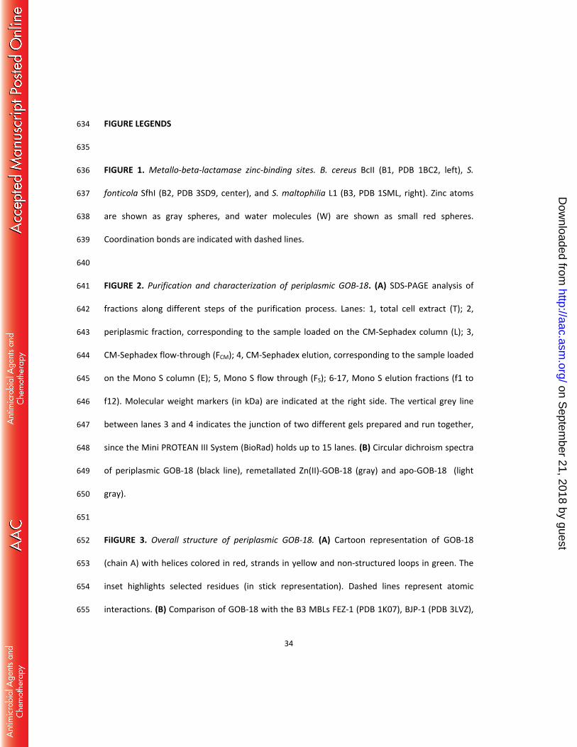
FIGURE LEGENDS 634
635
FIGURE 1. Metallo-beta-lactamase zinc-binding sites. B. cereus BcII (B1, PDB 1BC2, left), S. 636
fonticola SfhI (B2, PDB 3SD9, center), and S. maltophilia L1 (B3, PDB 1SML, right). Zinc atoms 637
are shown as gray spheres, and water molecules (W) are shown as small red spheres. 638
Coordination bonds are indicated with dashed lines. 639
640
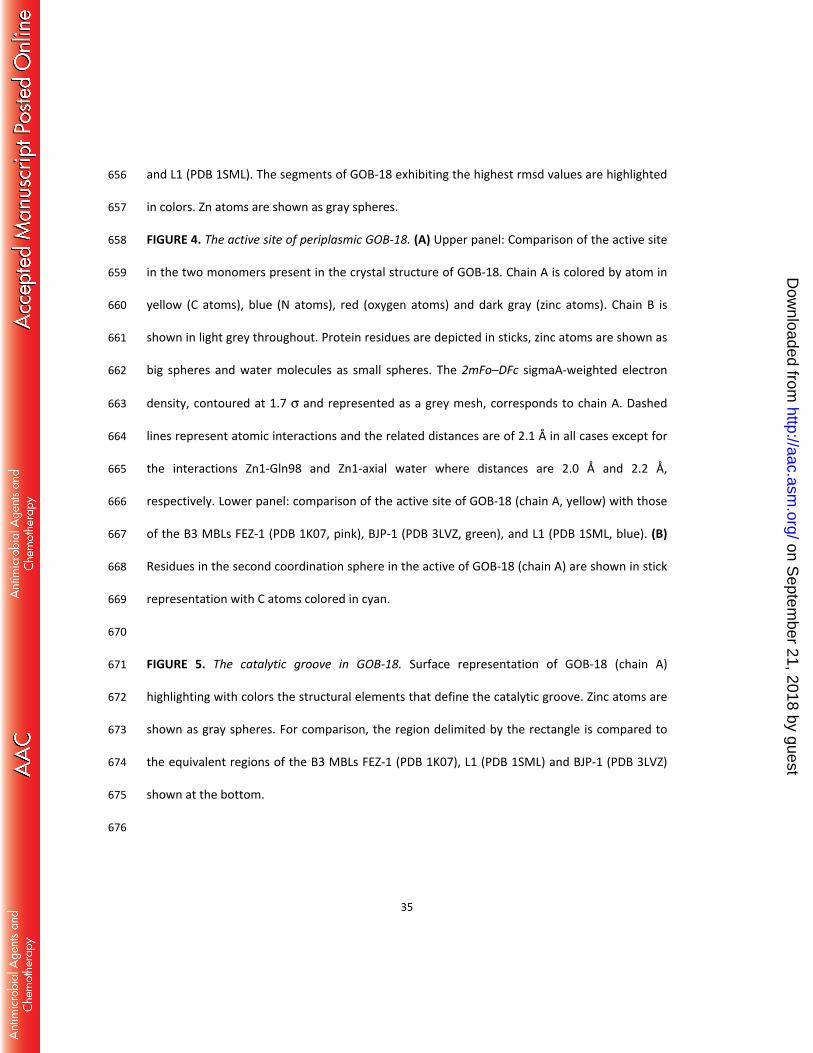
FIGURE 2. Purification and characterization of periplasmic GOB-18. (A) SDS-PAGE analysis of 641
fractions along different steps of the purification process. Lanes: 1, total cell extract (T); 2, 642
periplasmic fraction, corresponding to the sample loaded on the CM-Sephadex column (L); 3, 643
CM-Sephadex flow-through (FCM); 4, CM-Sephadex elution, corresponding to the sample loaded 644
on the Mono S column (E); 5, Mono S flow through (FS); 6-17, Mono S elution fractions (f1 to 645
f12). Molecular weight markers (in kDa) are indicated at the right side. The vertical grey line 646
between lanes 3 and 4 indicates the junction of two different gels prepared and run together, 647
since the Mini PROTEAN III System (BioRad) holds up to 15 lanes. (B) Circular dichroism spectra 648
of periplasmic GOB-18 (black line), remetallated Zn(II)-GOB-18 (gray) and apo-GOB-18 (light 649
gray). 650
651
FiIGURE 3. Overall structure of periplasmic GOB-18. (A) Cartoon representation of GOB-18 652
(chain A) with helices colored in red, strands in yellow and non-structured loops in green. The 653
inset highlights selected residues (in stick representation). Dashed lines represent atomic 654
interactions. (B) Comparison of GOB-18 with the B3 MBLs FEZ-1 (PDB 1K07), BJP-1 (PDB 3LVZ), 655
on Septem
ber 21, 2018 by guesthttp://aac.asm
.org/D
ownloaded from
35
and L1 (PDB 1SML). The segments of GOB-18 exhibiting the highest rmsd values are highlighted 656
in colors. Zn atoms are shown as gray spheres. 657
FIGURE 4. The active site of periplasmic GOB-18. (A) Upper panel: Comparison of the active site 658
in the two monomers present in the crystal structure of GOB-18. Chain A is colored by atom in 659
yellow (C atoms), blue (N atoms), red (oxygen atoms) and dark gray (zinc atoms). Chain B is 660
shown in light grey throughout. Protein residues are depicted in sticks, zinc atoms are shown as 661
big spheres and water molecules as small spheres. The 2mFo–DFc sigmaA-weighted electron 662
density, contoured at 1.7 σ and represented as a grey mesh, corresponds to chain A. Dashed 663
lines represent atomic interactions and the related distances are of 2.1 Å in all cases except for 664
the interactions Zn1-Gln98 and Zn1-axial water where distances are 2.0 Å and 2.2 Å, 665
respectively. Lower panel: comparison of the active site of GOB-18 (chain A, yellow) with those 666
of the B3 MBLs FEZ-1 (PDB 1K07, pink), BJP-1 (PDB 3LVZ, green), and L1 (PDB 1SML, blue). (B) 667
Residues in the second coordination sphere in the active of GOB-18 (chain A) are shown in stick 668
representation with C atoms colored in cyan. 669
670
FIGURE 5. The catalytic groove in GOB-18. Surface representation of GOB-18 (chain A) 671
highlighting with colors the structural elements that define the catalytic groove. Zinc atoms are 672
shown as gray spheres. For comparison, the region delimited by the rectangle is compared to 673
the equivalent regions of the B3 MBLs FEZ-1 (PDB 1K07), L1 (PDB 1SML) and BJP-1 (PDB 3LVZ) 674
shown at the bottom. 675
676
on Septem
ber 21, 2018 by guesthttp://aac.asm
.org/D
ownloaded from
36
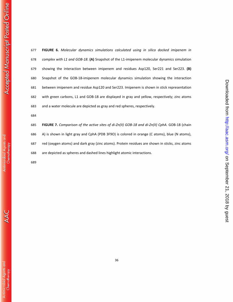
FIGURE 6. Molecular dynamics simulations calculated using in silico docked imipenem in 677
complex with L1 and GOB-18. (A) Snapshot of the L1-imipenem molecular dynamics simulation 678
showing the interaction between imipenem and residues Asp120, Ser221 and Ser223. (B) 679
Snapshot of the GOB-18-imipenem molecular dynamics simulation showing the interaction 680
between imipenem and residue Asp120 and Ser223. Imipenem is shown in stick representation 681
with green carbons, L1 and GOB-18 are displayed in gray and yellow, respectively; zinc atoms 682
and a water molecule are depicted as gray and red spheres, respectively. 683
684
FIGURE 7. Comparison of the active sites of di-Zn(II) GOB-18 and di-Zn(II) CphA. GOB-18 (chain 685
A) is shown in light gray and CphA (PDB 3F9O) is colored in orange (C atoms), blue (N atoms), 686
red (oxygen atoms) and dark gray (zinc atoms). Protein residues are shown in sticks, zinc atoms 687
are depicted as spheres and dashed lines highlight atomic interactions. 688
689
on Septem
ber 21, 2018 by guesthttp://aac.asm
.org/D
ownloaded from
37
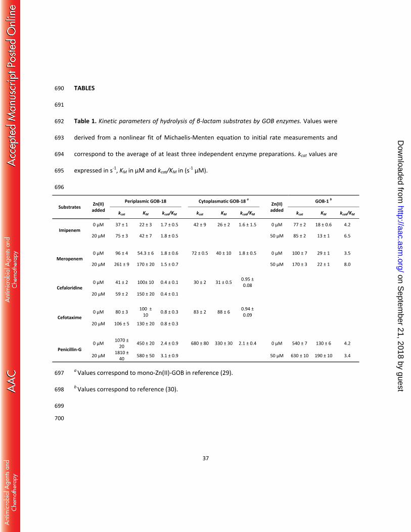
TABLES 690
691
Table 1. Kinetic parameters of hydrolysis of β-lactam substrates by GOB enzymes. Values were 692
derived from a nonlinear fit of Michaelis-Menten equation to initial rate measurements and 693
correspond to the average of at least three independent enzyme preparations. kcat values are 694
expressed in s-1, KM in µM and kcat/KM in (s-1 µM). 695
696
Substrates Zn(II) added
Periplasmic GOB-18 Cytoplasmatic GOB-18 a Zn(II) added
GOB-1 b
kcat KM kcat/KM kcat KM kcat/KM kcat KM kcat/KM
Imipenem 0 μM 37 ± 1 22 ± 3 1.7 ± 0.5 42 ± 9 26 ± 2 1.6 ± 1.5 0 μM 77 ± 2 18 ± 0.6 4.2
20 μM 75 ± 3 42 ± 7 1.8 ± 0.5 50 μM 85 ± 2 13 ± 1 6.5
Meropenem 0 μM 96 ± 4 54.3 ± 6 1.8 ± 0.6 72 ± 0.5 40 ± 10 1.8 ± 0.5 0 μM 100 ± 7 29 ± 1 3.5
20 μM 261 ± 9 170 ± 20 1.5 ± 0.7 50 μM 170 ± 3 22 ± 1 8.0
Cefaloridine 0 μM 41 ± 2 100± 10 0.4 ± 0.1 30 ± 2 31 ± 0.5 0.95 ±
0.08
20 μM 59 ± 2 150 ± 20 0.4 ± 0.1
Cefotaxime 0 μM 80 ± 3 100 ±
10 0.8 ± 0.3 83 ± 2 88 ± 6 0.94 ± 0.09
20 μM 106 ± 5 130 ± 20 0.8 ± 0.3
Penicillin-G 0 μM 1070 ±
20 450 ± 20 2.4 ± 0.9 680 ± 80 330 ± 30 2.1 ± 0.4 0 μM 540 ± 7 130 ± 6 4.2
20 μM 1810 ± 40 580 ± 50 3.1 ± 0.9 50 μM 630 ± 10 190 ± 10 3.4
a Values correspond to mono-Zn(II)-GOB in reference (29). 697
b Values correspond to reference (30). 698
699
700
on Septem
ber 21, 2018 by guesthttp://aac.asm
.org/D
ownloaded from
38
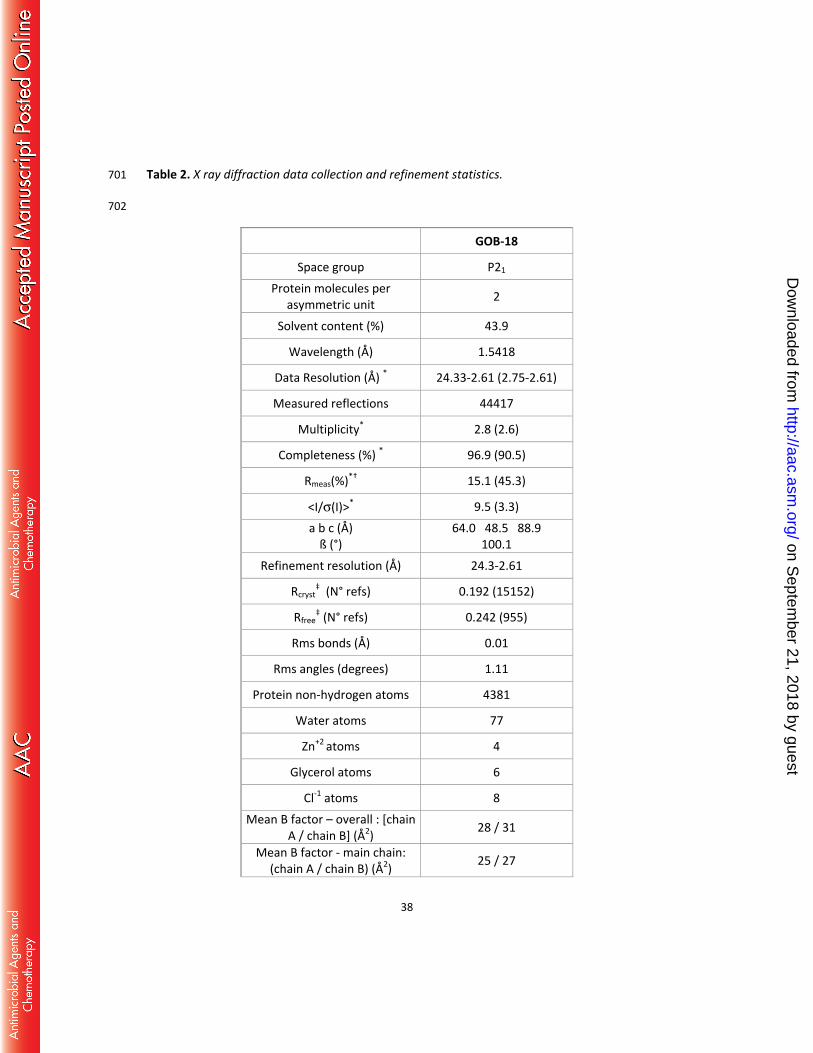
Table 2. X ray diffraction data collection and refinement statistics. 701 702
GOB-18
Space group P21
Protein molecules per asymmetric unit 2
Solvent content (%) 43.9
Wavelength (Å) 1.5418
Data Resolution (Å) * 24.33-2.61 (2.75-2.61)
Measured reflections 44417
Multiplicity* 2.8 (2.6)
Completeness (%) * 96.9 (90.5)
Rmeas(%)*† 15.1 (45.3)
<I/σ(I)>* 9.5 (3.3) a b c (Å)
ß (°) 64.0 48.5 88.9
100.1 Refinement resolution (Å) 24.3-2.61
Rcryst‡ (N° refs) 0.192 (15152)
Rfree‡ (N° refs) 0.242 (955)
Rms bonds (Å) 0.01
Rms angles (degrees) 1.11
Protein non-hydrogen atoms 4381
Water atoms 77
Zn+2 atoms 4
Glycerol atoms 6
Cl-1 atoms 8 Mean B factor – overall : [chain
A / chain B] (Å2) 28 / 31
Mean B factor - main chain: (chain A / chain B) (Å2) 25 / 27
on Septem
ber 21, 2018 by guesthttp://aac.asm
.org/D
ownloaded from
39
Mean B factor – side chains: (chain A / chain B) (Å2) 32 / 35
Mean B factor – waters (Å2) 18 Mean B factor – liganded Zn+2
(Å2) 28
Mean B factor – liganded Cl-1 (Å2) 47
Mean B factor – liganded glycerol (Å2) 41
Map vs model correlation coefficient (overall/local) ‡ 0.839 / 0.881
N° residues in Ramachandran plot regions §
(allowed/favored/outliers) 535/524/2
PDB ID 5K0W 703 704
* Values in parentheses apply to the high-resolution shell. 705
† meas = ∑ √ h/( h−1) ∑ | i−< ±>|ℎ ∑ ∑ ±ℎ ; Nh, multiplicity for each reflection; Ii, the intensity of the ith 706
observation of reflection h; <I>, the mean of the intensity of all observations of reflection h, 707
with ± = h ∑ ( (−) (+)); ∑ is taken over all reflections; ∑ is taken over all 708
observations of each reflection. 709
‡ = ∑ | (ℎ) − (ℎ) |/ ∑ | (ℎ) |; Rcryst and Rfree were calculated using the 710
working and test hkl reflection sets, respectively. 711
‡‡ Calculated with Phenix get_cc_mtz_pdb (39). 712
§ Calculated with Mol Probity (62). 713
on Septem
ber 21, 2018 by guesthttp://aac.asm
.org/D
ownloaded from