Embed Size (px)
Citation preview
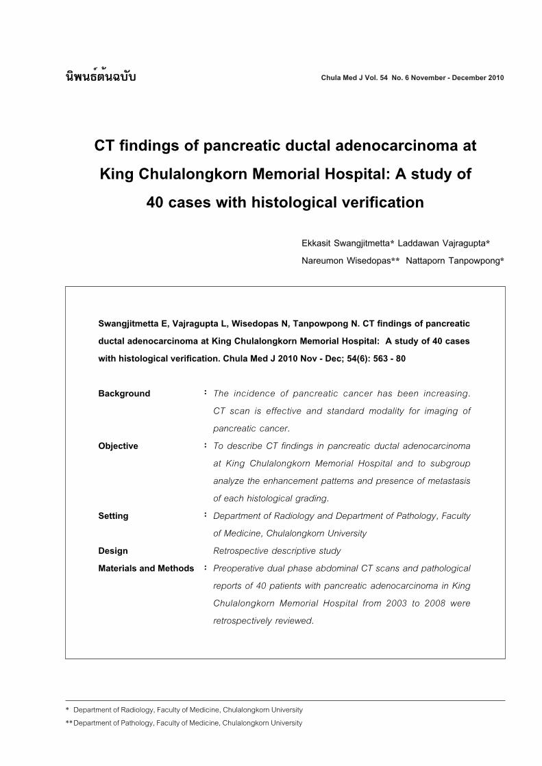
Chula Med J Vol. 54 No. 6 November - December 2010
Swangjitmetta E, Vajragupta L, Wisedopas N, Tanpowpong N. CT findings of pancreatic
ductal adenocarcinoma at King Chulalongkorn Memorial Hospital: A study of 40 cases
with histological verification. Chula Med J 2010 Nov - Dec; 54(6): 563 - 80
Background The incidence of pancreatic cancer has been increasing.
CT scan is effective and standard modality for imaging of
pancreatic cancer.
Objective To describe CT findings in pancreatic ductal adenocarcinoma
at King Chulalongkorn Memorial Hospital and to subgroup
analyze the enhancement patterns and presence of metastasis
of each histological grading.
Setting Department of Radiology and Department of Pathology, Faculty
of Medicine, Chulalongkorn University
Design Retrospective descriptive study
Materials and Methods Preoperative dual phase abdominal CT scans and pathological
reports of 40 patients with pancreatic adenocarcinoma in King
Chulalongkorn Memorial Hospital from 2003 to 2008 were
retrospectively reviewed.
นพินธต์น้ฉบบั
:
:
:
:
CT findings of pancreatic ductal adenocarcinoma at
King Chulalongkorn Memorial Hospital: A study of
40 cases with histological verification
Ekkasit Swangjitmetta* Laddawan Vajragupta*
Nareumon Wisedopas** Nattaporn Tanpowpong*
* Department of Radiology, Faculty of Medicine, Chulalongkorn University
**Department of Pathology, Faculty of Medicine, Chulalongkorn University
564 Chula Med Jเอกสทิธิ ์สวา่งจติเมตตา และคณะ
Results In 40 patients, 13 were male and 27 were female with their mean
age of 61.9 ± 2.36 years old. Most common location was
pancreatic head in 29 patients. The tumor size ranged from
1.4-10.9 cm with the mean of 3.9 cm. On the precontrast study,
28 tumors were isodense and 12 tumors were hypodense. On
arterial phase, all were hypodense. On portovenous phase,
36 tumors were hypodense and 4 tumors were isodense.
Pancreatic duct dilatation was seen in12 patients (30%) and
bile duct dilatation was seen in13patients (32.5%). Arterial
involvement was seen in 22 patients (55%); the splenic artery
was the most commonly involved. Venous involvement was seen
in 27 patients (67.5%); the splenic vein and SMV were the most
commonly involved. Adjacent organ invasion was seen in
17 patients (42.5%); the duodenum was the most commonly
involved. Regional node involvement was seen in 12 patients
(30%); the aortocaval node was the most commonly involved.
Metastasis was seen in 15 patients (37.5%); liver metastasis was
the most common. There was no statistically significant correlation
of enhancement pattern and the presence of metastasis with
tumor grading.
Conclusion The most common CT findings of pancreatic ductal
adenocarcinoma was ill-defined mass with hypodensity on arterial
phase. The most common location was pancreatic head. There
was no statistically significant correlation of enhancement pattern
and the presence of metastasis with tumor grading.
Keywords CT, enhancement pattern, metastases, pancreas, pancreatic
ductal adenocarcinoma, tumor grading.
Reprint request: Wisedopas N. Department of Pathology, Faculty of Medicine, Chulalongkorn
University, Bangkok 10330, Thailand.
Received for publication. January 22, 2010.
:
:
:
565Vol. 54 No. 6
November - December 2010
ลักษณะภาพเอกซเรยค์อมพวิเตอรข์องมะเรง็ตับอ่อนชนดิดกัตลัอะดโีนคารซิ์โนมา
ในโรงพยาบาลจฬุาลงกรณ ์: การศกึษาในผูป้ว่ย 40 ราย
ทีม่ผีลยนืยนัทางพยาธวิทิยา
เอกสิทธิ์ สว่างจิตเมตตา, ลัดดาวัลย์ วัชระคุปต์, นฤมล วิเศษโอภาส, ณัฐพร ตั่นเผ่าพงษ์.
ลักษณะภาพเอกซเรย์คอมพิวเตอร์ของมะเร็งตับอ่อนชนิดดักตัลอะดีโนคาร์ซิโนมาใน
โรงพยาบาลจุฬาลงกรณ์ : การศึกษาในผู ้ป่วย 40 รายที ่มีผลยืนยันทางพยาธิวิทยา.
จุฬาลงกรณเ์วชสาร 2553 พ.ย. - ธ.ค.; 54(6): 563 - 80
เหตุผลของการทำวิจัย อุบัติการณ์ของมะเร็งตับอ่อนในปัจจุบันเพิ ่มขึ ้นเรื ่อย ๆ เอกซเรย์
คอมพิวเตอร์เป็นการตรวจที่เป็นมาตรฐานและมีประสิทธิภาพ
วัตถุประสงค์ เพื่อบรรยายลักษณะภาพเอกซเรย์คอมพิวเตอร์ของมะเร็งตับอ่อนชนิด
ดักตัลอะดีโนคาร์ซิโนมาในโรงพยาบาลจฬุาลงกรณ์ และหาความสัมพันธ์
ระหว่าง Histological Grading กับรูปแบบ Enhancement และการ
กระจายไปอวัยวะอื่น
สถานที่ทำการศึกษา ภาควิชารังสีวิทยาและพยาธิวิทยา คณะแพทยศาสตร์ จุฬาลงกรณ์-
มหาวทิยาลยั
รูปแบบการวิจัย การศึกษาย้อนหลังเชิงพรรณนา
ตัวอย่างและวิธีการศึกษา โดยการวิเคราะห์ภาพเอกซเรย์คอมพิวเตอร์ก่อนการผ่าตัดและรายงาน
ผลทางพยาธิวิทยาของผู้ป่วยมะเร็งตับอ่อน 40 ราย ในโรงพยาบาล
จุฬาลงกรณ ์ต้ังแตพ่.ศ. 2546 - 2551
ผลการศึกษา จากผู้ป่วย 40 ราย เป็นชาย 13 ราย และหญิง 27 ราย อายุเฉลี่ย
61.9 ± 2.36 ปี ตำแหนง่ ทีพ่บรอยโรคมากสดุคอื pancreatic head
(29 ราย, 72.5%) ขนาดกอ้นอยูร่ะหวา่ง1.4 -10.9 ซม. (เฉล่ีย 3.9 ซม.)
ก้อนใน 28 รายให้ลักษณะ isodense และ 12 รายให้ลักษณะ
hypodense ใน precontrast study ก้อนในทุกรายให้ลักษณะ
hypodense ใน arterial phase ก้อนใน 36 รายให้ลักษณะ hypodense
ใน portovenous phase ท่อตับอ่อนขยายตัวพบใน 12 ราย (30%)
ท่อน้ำดขียายตวัพบใน 13 ราย (32.5%) Arterial involvement พบใน
22 ราย (55%) พบมากสดุที ่splenic artery และ venous involvement
พบใน 27 ราย (67.5%) พบมากสุดที่ splenic vein และ SMV
การลุกลามอวัยวะข้างเคียงพบใน17 ราย (42.5%) พบมากสุดท่ีดูโอดีนัม
การกระจายไปต่อมน้ำเหลืองพบใน12ราย (30%) พบมากสุดที ่
aortocaval node การกระจายไปอวัยวะอื่น พบใน15 ราย (37.5%)
พบมากสดุทีต่บั ไม่มีความสมัพนัธอ์ยา่งมนียัสำคญัทางสถติ ิ ระหวา่ง
histological grading กับรูปแบบ enhancement และการกระจายไป
อวัยวะอื่น
:
:
:
:
:
:
566 Chula Med Jเอกสทิธิ ์สวา่งจติเมตตา และคณะ
สรุป ล ักษณะภาพเอกซเรย ์คอมพ ิว เตอร ์ของมะเร ็ งต ับอ ่อนชน ิด
ดักตัลอะดีโนคาร์ซิโนมาท่ีพบมากสุดคือก้อนขอบไม่ชัดเจน, hypodense
ใน arterial phase ตำแหน่งที่พบมากที่สุด คือ pancreatic head
การลุกลามเส้นเลือดมีผลต่อการวางแผนการผ่าตัด ไม่มีความสัมพันธ์
อย่างมีนัยสำคัญทางสถิติ ระหว่าง histological grading กับรูปแบบ
enhancement และการกระจายไปอวยัวะอืน่
คำสำคัญ เอกซเรยค์อมพวิเตอร,์ รูปแบบ enhancement, การกระจายไปอวยัวะ
อ่ืน, ตบัอ่อน, มะเรง็ตบัอ่อน, tumor grading.
:
:
567Vol. 54 No. 6
November - December 2010
ลักษณะภาพเอกซเรยค์อมพวิเตอรข์องมะเรง็ตับอ่อนชนดิดกัตลัอะดโีนคารซิ์โนมา
ในโรงพยาบาลจฬุาลงกรณ ์: การศกึษาในผูป้ว่ย 40 ราย
ทีม่ผีลยนืยนัทางพยาธวิทิยา
The incidence of pancreatic cancer has
been increasing over the past 40 years.(1) In the United
States, pancreatic cancer is the fourth leading cause
of cancer deaths.(2) More than 90% of patients present
in the late stage of the disease. This observation
emphasizes the role of radiology in early detection
and determination of the resectability of the tumor.
CT scan is the standard imaging modality for
the diagnosis and determination of the resectability
of pancreatic cancer.(3) The objectives of this study
are to describe CT findings in pancreatic ductal
adenocarcinoma at King Chulalongkorn Memorial
Hospital and to subgroup analyze the enhancement
patterns and presence of metastasis of each
histological grading.
Materials and Methods
There were 66 patients with definite
pathological diagnosis of pancreatic ductal adeno
carcinoma at King Chulalongkorn Memorial
Hospital from January 1, 2003 to December 31, 2008.
Forty of them were recruited in this study. We
excluded 22 patients: eight of them were due to no
available CT scans in PACS (Picture Archiving and
Communication System); four had no dual phase
contrast CT study; and, 14 only had available CT scan
of post surgery or post intervention. Preoperative dual
phase abdominal CT scan and pathological reports
of all the 40 cases were retrospectively reviewed. We
analyzed CT findings, enhancement pattern and
presence of metastasis of each histological grading.
Pathological criteria(4)
Well-differentiated adenocarcinomas are
defined as the tumors form well-defined glands. The
glands are complete, and the neoplastic cells are
cuboidal to columnar with basally oriented uniform
round to oval nuclei with evenly dispersed chromatin.
Intensive mucin production is present. Only minimal
nuclear pleomorphism is seen. Mitoses are not more
than five per high power field. The gland formation
in moderately differentiated adenocarcinoma is less
well-defined with incomplete glandular lumina and
nuclear pleomorphism. The nucleoli are larger and
more irregular. Mitoses are 6-10 per high power
field and may be atypical. Poorly differentiated
adenocarcinomas are composed of poorly formed
glands, with individual infiltrating cells and solid
areas. Mucin production is abortive. Nuclear
pleomorphism is prominent with large bizarre nuclei
and the nucleoli are large, multiple and more
irregular. Mitoses, including atypical mitoses, are seen
more than 10 per high power field.
CT Protocol
Siemens Somatom Sensation Plus 4 is used
with 4 mm slice width, 2.5 mm collimator, 12.5 mm
feed per rotation and 0.5 sec rotation time. The
Somatom Sensation Plus 16 is used with 16x1.5 mm
collimator, 24.0 mm feed per rotation and 0.5 sec
rotation time (140 mAs, 120 kVp and pitch = 1).
A bolus injection 3-4 ml/sec of 100 ml
non-ionic contrast medium was done with the bolus
tracking placed at the abdominal aorta and the
threshold is 100 HU. Then arterial and portovenous
phases were obtained by the location of monitoring in
mid hepatic level for upper abdomen studies or just
above the level of the pancreas for pancreatic
protocol. The images of arterial phase were obtained
at about 30-35 seconds and portovenous phase were
568 Chula Med Jเอกสทิธิ ์สวา่งจติเมตตา และคณะ
obtained at 65-70 seconds after contrast injection.
CT Findings Review
Two gastrointestinal radiologists retro-
spectively reviewed the CT findings from PACS
independently in aspect of the followings: (In case of
different opinion, the images were interpreted by
consensus.)
1. Location: head, uncinate, body, tail, more than
one compartment
2. Tumor size: 2 cm or less, more than 2 cm but not
more than 4 cm, more than 4 cm
3. Margin: well-defined, ill-defined
4. Density and enhancement (relative to normal
pancreatic tissue in precontrast, arterial and
portovenous phases): hyperdense, isodense,
hypodense
5. Homogeneity: homogeneous, heterogeneous
6. Main pancreatic duct dilatation: absent,
present
7. Bile duct dilatation: absent, present
8. Vascular involvement: absent, arterial
involvement, venous involvement
9. Adjacent organ invasion: absent, present
10. Regional lymph nodes enlargement: absent,
present
11. Metastasis: absent, present
Image Analysis and Operational Definitions
Tumor size was measured at its longest
diameter by the caliper tool on PACS.
Tumor density was measured in Hounsfield
unit (HU) by drawing a region of interest (ROI) of each
lesion and computed by CT software and was
compared with a normal pancreas in the same phase.
The ROI of 0.5 cm2 was used in normal pancreas and
the lesions smaller than 4 cm and ROI of 1 cm 2 was
used in the lesions of 4 cm or more. Hypodense
lesion was defined as density of the lesion lower than
10 HU as compared to a normal pancreas. Density of
the lesion less than or equal to 10 HU different from
of a normal pancreas was defined as isodense.
Hyperdense lesion was defined as density of the
lesion greater than 10 HU of normal pancreas.(5 - 8)
Main pancreatic duct dilatation was defined
as the diameter of dilated portion was greater than
5 mm in the head and 3 mm in the tail. Bile duct
dilatation was defined when the largest diameter of
the common bile duct that exceeded 8 mm and/or of
common hepatic duct that exceeded 6 mm.(9,10)
CT findings of major arterial involvement
included obliteration of the normal fat between the
pancreatic margin and the adjacent vessel, greater
than 180-degree contact between the tumor and
the vessel, and morphologic changes of the artery
including narrowing or encasement of the affected
artery.(9,10)
Criteria for venous invasion included greater
than 180-degree contact with a soft tissue mass and
the vein. Collateral venous channels and another
characteristics such as “tear drop” configuration
of the superior mesenteric vein were also be
assessed.(10)
Adjacent organ invasion was defined as
interrupted, obliterated or loss of fat plane between
the pancreas and the adjacent structures.(10)
Regional lymph nodes involvement(11) was
evaluated by short axis diameter. The criteria were
as followings: retrocrural and porta hepatis nodes
exceeded 6 mm; gastrohepatic ligament nodes
569Vol. 54 No. 6
November - December 2010
ลักษณะภาพเอกซเรยค์อมพวิเตอรข์องมะเรง็ตับอ่อนชนดิดกัตลัอะดโีนคารซิ์โนมา
ในโรงพยาบาลจฬุาลงกรณ ์: การศกึษาในผูป้ว่ย 40 ราย
ทีม่ผีลยนืยนัทางพยาธวิทิยา
exceeded 8 mm; and, pancreaticoduodenal,
perisplenic, retropenitoneal, celiac axis, mesenteric
nodes exceeded 10 mm or multiple slightly smaller
(8-10 mm) in these regions containing necrotic area.
Correlations of enhancement pattern and
presence of metastases with tumor grading were
evaluated by Fisher’s exact test using SPSS program.
Interobserver agreement was evaluated by Kappa
analysis.
Results
In 40 patients, 13 were male and 27 were
female. The age of the patients ranged from 29 to 83
years old with the mean of 61.9 +/- 2.36 years and
median of 63 years. Pathological diagnoses were ob-
tained from surgical specimens (Whipple
operation and en bloc resection) in 22 patients and
from FNA, biopsy under ERCP or core needle biopsy
in 18 patients.
The histological grading was well-
differentiated in 14 patients (35%), moderately-
differentiated in 12 patients (30%), poorly-
differentiated in six patients (15%) and not available
in eight patients (20%).
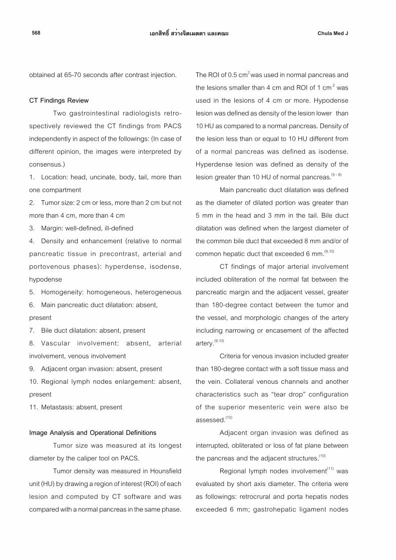
All patients revealed a single pancreatic mass
with tumor location shown in Figure 1. The most
common location was the pancreatic head in 29 cases
(72.5%), followed by pancreatic body in 15 cases
(37.5%).
Tumor size ranged from 1.4 to 10.9 cm with
the mean of 3.9 cm. Six tumors (15%) were less than
2 cm, 18 (45%) were 2-4 cm and 16 (40%) were more
than 4 cm in size.
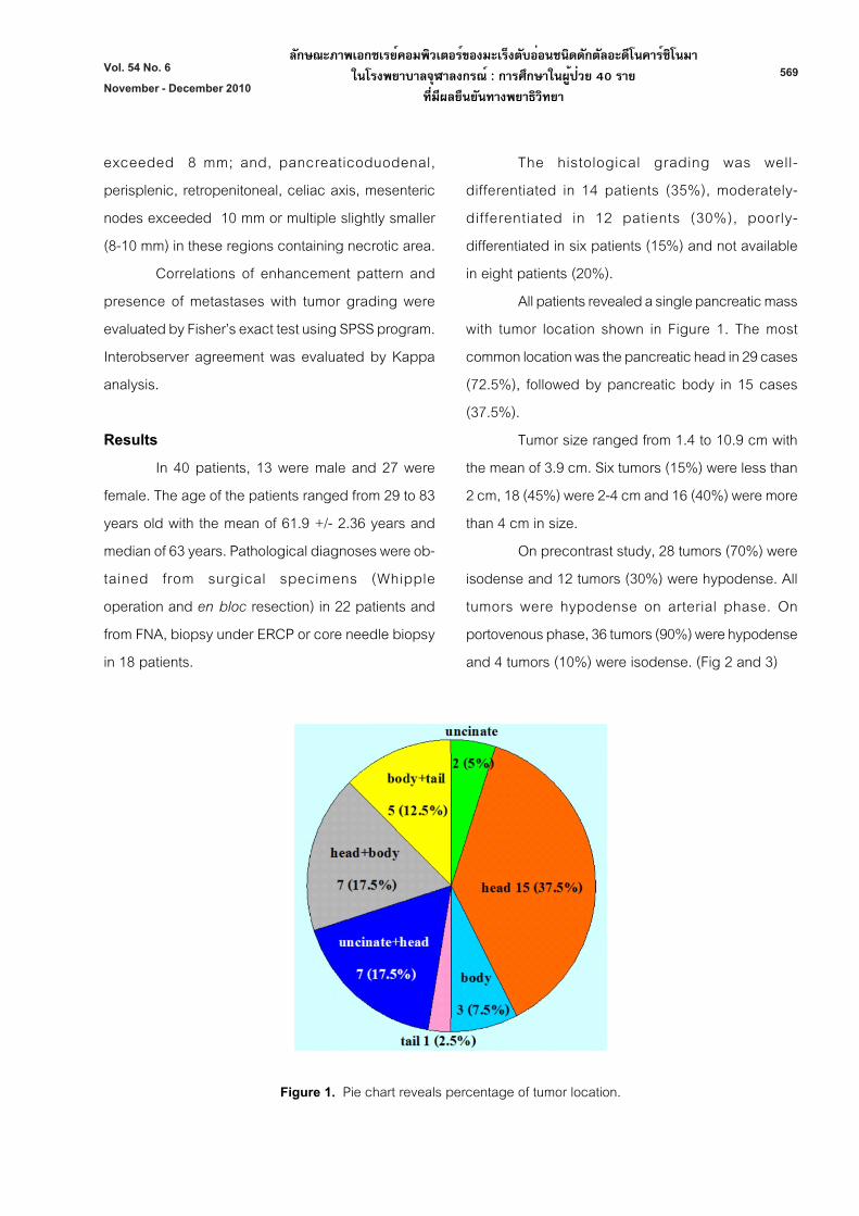
On precontrast study, 28 tumors (70%) were
isodense and 12 tumors (30%) were hypodense. All
tumors were hypodense on arterial phase. On
portovenous phase, 36 tumors (90%) were hypodense
and 4 tumors (10%) were isodense. (Fig 2 and 3)
Figure 1. Pie chart reveals percentage of tumor location.
570 Chula Med Jเอกสทิธิ ์สวา่งจติเมตตา และคณะ
There was no statistically significant
correlation between histological grading and
enhancement pattern (P-value = 1 in precontrast
study and P-value = 0.498 in portovenous phase) as
shown in Table 1.
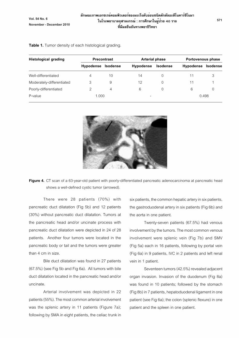
Tumors with ill-defined margin (see Fig 3)
were seen in 38 patients (95%). Tumor with well-
defined margin was found in two patients (5%).
Cystic degeneration within the tumor was seen in
the two cases with well-defined margin. (Fig 4)
Tumors with heterogeneous were detected
in 33 patients (82.5%). (see Fig 3) Tumor with
homogeneous density on pre and post contrast
enhanced studies were seen in seven patients
(17.5%).
Figure 2. Histogram demonstrates number of tumor in each density on precontrast study, arterial phase (A-phase)
and portovenous phase (V-phase).
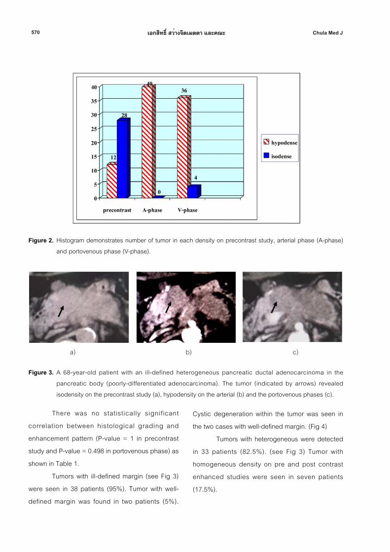
a) b) c)
Figure 3. A 68-year-old patient with an ill-defined heterogeneous pancreatic ductal adenocarcinoma in the
pancreatic body (poorly-differentiated adenocarcinoma). The tumor (indicated by arrows) revealed
isodensity on the precontrast study (a), hypodensity on the arterial (b) and the portovenous phases (c).
571Vol. 54 No. 6
November - December 2010
ลักษณะภาพเอกซเรยค์อมพวิเตอรข์องมะเรง็ตับอ่อนชนดิดกัตลัอะดโีนคารซิ์โนมา
ในโรงพยาบาลจฬุาลงกรณ ์: การศกึษาในผูป้ว่ย 40 ราย
ทีม่ผีลยนืยนัทางพยาธวิทิยา
There were 28 patients (70%) with
pancreatic duct dilatation (Fig 5b) and 12 patients
(30%) without pancreatic duct dilatation. Tumors at
the pancreatic head and/or uncinate process with
pancreatic duct dilatation were depicted in 24 of 28
patients. Another four tumors were located in the
pancreatic body or tail and the tumors were greater
than 4 cm in size.
Bile duct dilatation was found in 27 patients
(67.5%) (see Fig 5b and Fig 6a). All tumors with bile
duct dilatation located in the pancreatic head and/or
uncinate.
Arterial involvement was depicted in 22
patients (55%). The most common arterial involvement
was the splenic artery in 11 patients (Figure 7a);
following by SMA in eight patients, the celiac trunk in
six patients, the common hepatic artery in six patients,
the gastroduodenal artery in six patients (Fig 6b) and
the aorta in one patient.
Twenty-seven patients (67.5%) had venous
involvement by the tumors. The most common venous
involvement were splenic vein (Fig 7b) and SMV
(Fig 5a) each in 16 patients, following by portal vein
(Fig 6a) in 9 patients, IVC in 2 patients and left renal
vein in 1 patient.
Seventeen tumors (42.5%) revealed adjacent
organ invasion. Invasion of the duodenum (Fig 8a)
was found in 10 patients; followed by the stomach
(Fig 8b) in 7 patients, hepatoduodenal ligament in one
patient (see Fig 6a); the colon (splenic flexure) in one
patient and the spleen in one patient.
Table 1. Tumor density of each histological grading.
Histological grading Precontrast Arterial phase Portovenous phase
Hypodense Isodense Hypodense Isodense Hypodense Isodense
Well-differentiated 4 10 14 0 11 3
Moderately-differentiated 3 9 12 0 11 1
Poorly-differentiated 2 4 6 0 6 0
P-value 1.000 - 0.498
Figure 4. CT scan of a 63-year-old patient with poorly-differentiated pancreatic adenocarcinoma at pancreatic head
shows a well-defined cystic tumor (arrowed).
572 Chula Med Jเอกสทิธิ ์สวา่งจติเมตตา และคณะ
a) b)
a) b)
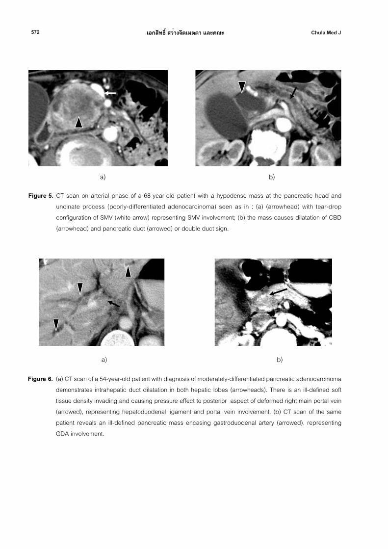
Figure 5. CT scan on arterial phase of a 68-year-old patient with a hypodense mass at the pancreatic head and
uncinate process (poorly-differentiated adenocarcinoma) seen as in : (a) (arrowhead) with tear-drop
configuration of SMV (white arrow) representing SMV involvement; (b) the mass causes dilatation of CBD
(arrowhead) and pancreatic duct (arrowed) or double duct sign.
Figure 6. (a) CT scan of a 54-year-old patient with diagnosis of moderately-differentiated pancreatic adenocarcinoma
demonstrates intrahepatic duct dilatation in both hepatic lobes (arrowheads). There is an ill-defined soft
tissue density invading and causing pressure effect to posterior aspect of deformed right main portal vein
(arrowed), representing hepatoduodenal ligament and portal vein involvement. (b) CT scan of the same
patient reveals an ill-defined pancreatic mass encasing gastroduodenal artery (arrowed), representing
GDA involvement.
573Vol. 54 No. 6
November - December 2010
ลักษณะภาพเอกซเรยค์อมพวิเตอรข์องมะเรง็ตับอ่อนชนดิดกัตลัอะดโีนคารซิ์โนมา
ในโรงพยาบาลจฬุาลงกรณ ์: การศกึษาในผูป้ว่ย 40 ราย
ทีม่ผีลยนืยนัทางพยาธวิทิยา
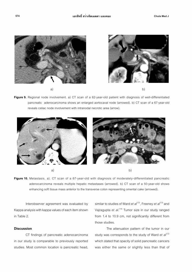
Regional node involvement was seen in 12
patients (30%). The most common nodal involvement
was the aortocaval node (Fig 9a) in five patients;
followed by the celiac node (Fig 9b) in three patients;
the peripancreatic node in three patients,
hepatoduodenal ligament node in three patients and
the peripancreatic node in two patients.
Fifteen patients (37.5%) had metastases.
Liver metastases were noted in 14 of 15 patients
(Fig 10a); peritoneal and omental metastases in two
patients (Fig 10b). Metastasis was detected in five of
fourteen of well-diffentiated tumors, five of twelve of
moderately-differentiated tumors and one of six of
poorly-differentiated tumors. There was no statistically
significant relationship between the histological
grading and the presence of metastasis (P-value =
0.639).
a) b)
a) b)
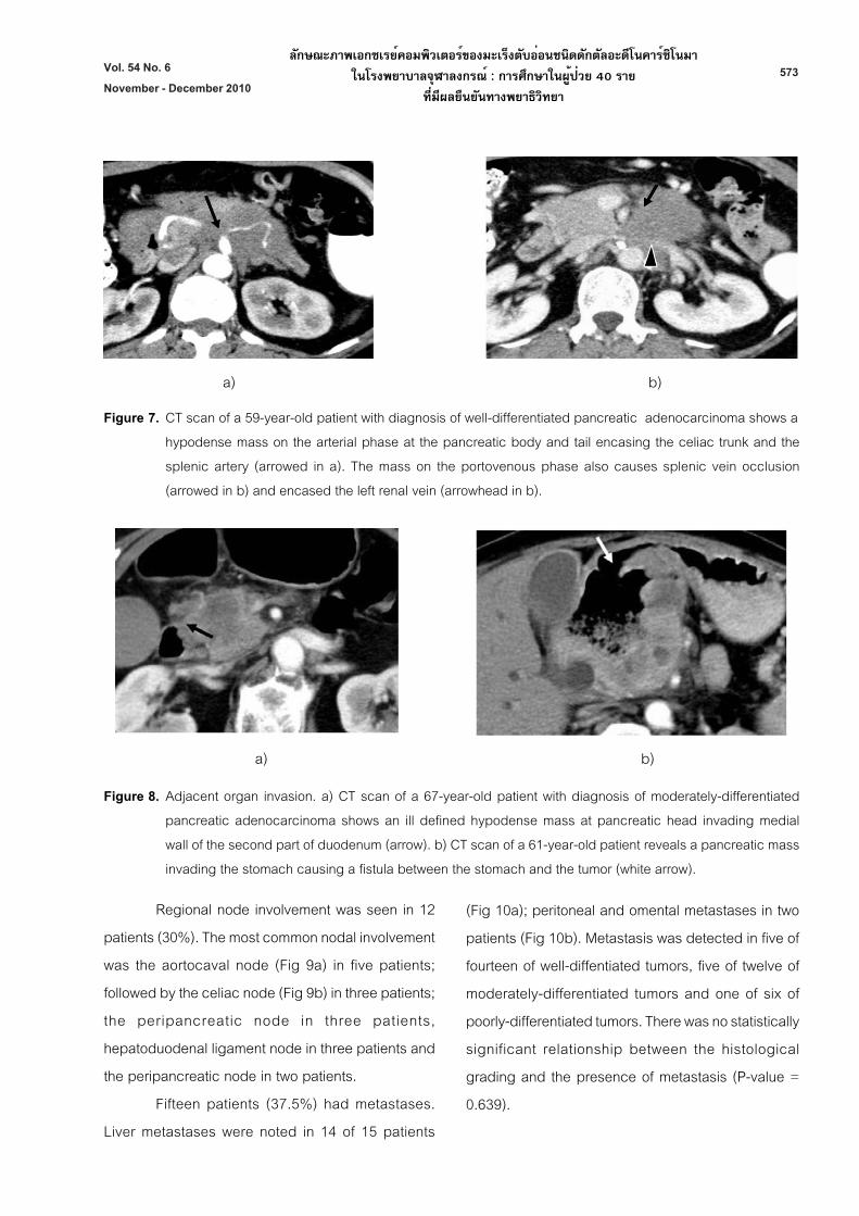
Figure 7. CT scan of a 59-year-old patient with diagnosis of well-differentiated pancreatic adenocarcinoma shows a
hypodense mass on the arterial phase at the pancreatic body and tail encasing the celiac trunk and the
splenic artery (arrowed in a). The mass on the portovenous phase also causes splenic vein occlusion
(arrowed in b) and encased the left renal vein (arrowhead in b).
Figure 8. Adjacent organ invasion. a) CT scan of a 67-year-old patient with diagnosis of moderately-differentiated
pancreatic adenocarcinoma shows an ill defined hypodense mass at pancreatic head invading medial
wall of the second part of duodenum (arrow). b) CT scan of a 61-year-old patient reveals a pancreatic mass
invading the stomach causing a fistula between the stomach and the tumor (white arrow).
574 Chula Med Jเอกสทิธิ ์สวา่งจติเมตตา และคณะ
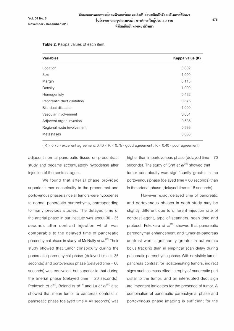
Interobserver agreement was evaluated by
Kappa analysis with kappa values of each item shown
in Table 2.
Discussion
CT findings of pancreatic adenocarcinoma
in our study is comparable to previously reported
studies. Most common location is pancreatic head,
similar to studies of Ward et al(12), Freeney et al(13) and
Vajragupta et al.(14) Tumor size in our study ranged
from 1.4 to 10.9 cm, not significantly different from
those studies.
The attenuation pattern of the tumor in our
study was corresponds to the study of Ward et al(12)
which stated that opacity of solid pancreatic cancers
was either the same or slightly less than that of
Figure 9. Regional node involvement. a) CT scan of a 62-year-old patient with diagnosis of well-differentiated
pancreatic adenocarcinoma shows an enlarged aortocaval node (arrowed). b) CT scan of a 67-year-old
reveals celiac node involvement with intranodal necrotic area (arrow).
a) b)
a) b)
Figure 10. Metastasis. a). CT scan of a 67-year-old with diagnosis of moderately-differentiated pancreatic
adenocarcinoma reveals multiple hepatic metastases (arrowed). b) CT scan of a 50-year-old shows
enhancing soft tissue mass anterior to the transverse colon representing omental cake (arrowed).
575Vol. 54 No. 6
November - December 2010
ลักษณะภาพเอกซเรยค์อมพวิเตอรข์องมะเรง็ตับอ่อนชนดิดกัตลัอะดโีนคารซิ์โนมา
ในโรงพยาบาลจฬุาลงกรณ ์: การศกึษาในผูป้ว่ย 40 ราย
ทีม่ผีลยนืยนัทางพยาธวิทิยา
adjacent normal pancreatic tissue on precontrast
study and became accentuatedly hypodense after
injection of the contrast agent.
We found that arterial phase provided
superior tumor conspicuity to the precontrast and
portovenous phases since all tumors were hypodense
to normal pancreatic parenchyma, corresponding
to many previous studies. The delayed time of
the arterial phase in our institute was about 30 - 35
seconds after contrast injection which was
comparable to the delayed time of pancreatic
parenchymal phase in study of McNulty et al.(15) Their
study showed that tumor conspicuity during the
pancreatic parenchymal phase (delayed time = 35
seconds) and portovenous phase (delayed time = 60
seconds) was equivalent but superior to that during
the arterial phase (delayed time = 20 seconds).
Prokesch et al(7), Boland et al(16) and Lu et al(17) also
showed that mean tumor to pancreas contrast in
pancreatic phase (delayed time = 40 seconds) was
higher than in portovenous phase (delayed time = 70
seconds). The study of Graf et al(18) showed that
tumor conspicuity was significantly greater in the
portovenous phase (delayed time = 60 seconds) than
in the arterial phase (delayed time = 18 seconds).
However, exact delayed time of pancreatic
and portovenous phases in each study may be
slightly different due to different injection rate of
contrast agent, type of scanners, scan time and
protocol. Fukukura et al(19) showed that pancreatic
parenchymal enhancement and tumor-to-pancreas
contrast were significantly greater in autonomic
bolus tracking than in empirical scan delay during
pancreatic parenchymal phase. With no visible tumor-
pancreas contrast for isoattenuating tumors, indirect
signs such as mass effect, atrophy of pancreatic part
distal to the tumor, and an interrupted duct sign
are important indicators for the presence of tumor. A
combination of pancreatic parenchymal phase and
portovenous phase imaging is sufficient for the
Table 2. Kappa values of each item.
Variables Kappa value (K)
Location 0.802
Size 1.000
Margin 0.113
Density 1.000
Homogeniety 0.432
Pancreatic duct dilatation 0.875
Bile duct dilatation 1.000
Vascular involvement 0.651
Adjacent organ invasion 0.536
Regional node involvement 0.536
Metastases 0.838
( K > 0.75 - excellent agreement, 0.40 < K < 0.75 - good agreement , K < 0.40 - poor agreement)
576 Chula Med Jเอกสทิธิ ์สวา่งจติเมตตา และคณะ
detection of pancreatic adenocarcinoma and
vascular involvement.
Our study (32 patients with available
histological grading) revealed no significant
correlation between enhancement pattern of the
tumor and histological grading. The result contradicts
to the study of Wang et al(20) (n = 34), which showed
significant correlation between CT enhancement and
histological grading. The extent of CT enhancement
is inversely proportional to the degree of histological
grading. Assessment of the correlation in both
studies was different. Our study assessed
attenuation on precontrast, arterial and portovenous
phases with 2 degrees of density (isodense and
hypodense) while their study assessed only
pancreatic phase (arterial phase) with 4 degrees of
enhancement. Statistical methods, therefore, were
also different. Additionally, apart from tumor grading,
enhancement of the tumor was influenced by many
pathological factors. Hiroshi et al(21) showed that
enhancement pattern was influenced by cellularity of
tumor cells, coexisting acinar tissues, fibrosis, mucin
and extent of necrosis. Hattori et al(22) proved that
histological features affecting the enhancement
pattern were angiogenesis (determined by vascular
endothelial growth factor (VEGF) and microvessel
density) and the extent of fibrosis.
The percentage of pancreatic duct and bile
duct dilatation in our study is higher than those of
Ward et al(12), Freeney et al(13) and Vajragupta et al.(14)
Most tumors with pancreatic duct dilatation in our
study were located at pancreatic head and/or
uncinate. All tumors in the pancreatic body or tail with
pancreatic duct dilatation were greater than 4 cm in
size. All cases with bile duct dilatation in our study
also revealed tumor located in pancreatic head
and/or uncinate. This reflects that main pancreatic
duct dilatation is mainly determined by the
tumor location and tumor size. Tumor location also
determines the presence of bile duct dilatation.
In light of vascular involvement, most
common arterial involvement in the study of Ward
et al(12) were the celiac axis and SMA while in our
study was the splenic artery. The most common
arterial and venous involvements in our study were
similar to the study of Li et al (23), Lee et al(24) and
Vajragupta et al.(14)
Most common adjacent organ invasion in
our study was duodenum, also similar to studies of
Freeney et al(13) and Vajragupta et al.(14)
In aspect of regional lymph nodes
enlargement, the percentage in our study was higher
than those of Ward et al, Freeney et al(13) and Diehl
et al(25) but lower than that of Vajragupta et al.(14) The
most common node involvement in our study was
the aortocaval node while that in the study of Ward
et al(12) was left paraaortic node.
The percentage of metastasis in our study
was not significantly different from that of Ward
et al(12), Freeney et al(13) and Vajragupta et al.(14) The
liver is the most common organ of metastasis in all
studies. We found that there was no significant
correlation between histological grading and the
presence of metastasis. There has been no study
that proves the significant correlation between
histological grading and metastasis.
Interobserver agreement in our study was
excellent in most radiological features. Tumor
margin was the only one showing poor agreement.
This was probably due to varied subjective
577Vol. 54 No. 6
November - December 2010
ลักษณะภาพเอกซเรยค์อมพวิเตอรข์องมะเรง็ตับอ่อนชนดิดกัตลัอะดโีนคารซิ์โนมา
ในโรงพยาบาลจฬุาลงกรณ ์: การศกึษาในผูป้ว่ย 40 ราย
ทีม่ผีลยนืยนัทางพยาธวิทิยา
perception of individuals to evaluate whether each
tumor was ill-defined or poorly-defined. However, this
feature does not significantly influence making
diagnosis, determining tumor resectability or tumor
staging. Cystic degeneration was the feature that
shows obviously well-defined margin.
There were two cases with cystic
degeneration which is uncommon condition
of pancreatic adenocarcinoma and can be
misdiagnosed as other cystic neoplasms of the
pancreas. Cystic degeneration may be seen when
the tumor increases in size and the majority tend
to be large and poorly-differentiated tumors, with
a single large cavity lined by tumor cells and
containing hemorrhagic debris.(26) In our cases, one
was well-differentiated and the other was poorly-
differentiated. Cystic change in pancreatic
adenocarcinoma should be considered in the
differential diagnosis of a cystic pancreatic lesion,
apart from the more common pseudocysts and true
cystic pancreatic neoplasms.
We also found a 29-year-old male patient with
pancreatic adenocarcinoma which is rare under the
age of 40 years.(27) He had no underlying disease nor
family history of pancreatic cancer. The presenting
symptoms were abdominal pain and jaundice.
CT scan showed a 1.6-cm ill-defined tumor at
pancreatic head and uncinate. Whipple’s operation
was performed and the pathological findings revealed
moderately-differentiated pancreatic ductal
adenocarcinoma.
Luttges et al(28) reviewed of the literature from
1818 to 2001 and found 71 cases diagnosed as
ductal adenocarcinoma in patients under 40 years of
age. According to the WHO classification system of
1996 and data available in the reports, only 20 of these
were thought by the authors to represent true ductal
adenocarcinomas. Most were male patients. They and
some other studies(29,30) noted that young patients with
ductal adenocarcinoma of the pancreas tend to have
special circumstances or familial predisposition.
Genetic conditions associated with increased
risk of pancreatic carcinoma include hereditary
pancreatitis, hereditary pancreatic cancer syndrome,
hereditary non-polyposis colon carcinoma,
Peutz-Jeghers syndrome, familial atypical multiple
mole melanoma, and the BRCA2 gene. Imaging
findings of pancreatic adenocarcinoma in younger
patients is similar to those of the older age group.
Some limitations in our study should be
considered. First, the number of cases was small.
Assessment of statistically significant correlation of
histological grading, enhancement pattern and
presence of metastasis could be limited. Second, this
study was performed retrospectively. Some cases
did not have thin slices images for pancreatic
region (pancreatic protocol) or sagittal or coronal
reconstructions. These could make some difficulties
in the evaluation of small structures especially
vascular or adjacent organ involvements. However,
the available images of those cases were satisfying
and interobserver agreement was excellent and good
in almost all the features to review.
Conclusion
The most common CT findings of pancreatic
ductal adenocarcinoma are ill-defined heterogeneous
mass with isodensity on precontrast phase and
hypodensity on the arterial and portovenous
phases. Most of the tumors are best depicted on the
578 Chula Med Jเอกสทิธิ ์สวา่งจติเมตตา และคณะ
arterial phase in which normal pancreas reveals
homogeneous enhancement while the tumor reveals
less enhancement. The most common location is
pancreatic head. Pancreatic duct and bile duct
dilatation mainly depend on the tumor location. The
most common arterial and venous involvements
are the splenic artery, splenic vein and SMV. Most
common adjacent organ involvement, regional node
involvement and metastatic site are duodenum, the
aortocaval node and the liver, respectively. There is
no statistically significant correlation between tumor
grading and enhancement pattern or the presence
of metastasis.
References
1. Office for National Statistics. Cancer Statistics
Registrations: Registrations of cancer
diagnosed in 2004, England. Series MB1
no.35. 2007
2. American Cancer Society. Cancer Facts & Figures
2005. Cancer, New York: American Cancer
Society 2005:1–60.
3. Paspulati RM. Multidetector CT of the pancreas.
Radiol Clin N Am. 2005 Nov;43(6):999-1020
4. Hruban RH, Fugushima N. Pancreatic adenocar-
cinoma: update on the surgical pathology
of carcinomas of ductal origin and PanINs.
Modern Pathology 2007; 20: S61–S70
5. Hollett MD, Jorgensen MJ, Jeffrey RB.
Quantitative evaluation of pancreatic
enhancement during dual-phase helical CT.
Radiology 1995 May;195(2):359-61
6. Fletcher JG, Wiersema MJ, Farrell MA, Fidler JL,
Burgart LJ, Koyama T, Johnson CD, Stephens
DH, Ward EM, Harmsen WS.Pancreatic
malignancy: Value of arterial, pancreatic and
hepatic phase imaging with multi–detector
row CT. Radiology 2003 Oct;229(1):81-90
7. Prokesch RW, Chow LC, Beaulieu CF, Bammer R,
Jeffrey RB. Isoattenuating pancreatic
adenocarcinoma at multi–detector row CT:
Secondary signs.Radiology 2002 Sep;224(3):
764-8
8. Baron RL. Understanding and optimizing use of
contrast material for CT of the liver. AJR
1994 Aug;163(2):323-31
9. Berland LL, Lawson TL, Foley WD, Geenen JE,
Stewart ET. Computed tomography of the
pancreatic duct: Correlation with pancreatic
ductography. Radiology 1981 Dec;141(3):
715-24
10. Megibow AJ. Pancreatic neoplasms. In: Gore RM,
Levine MS. Textbook of Gastrointestinal
Radiology. 3th ed. Philadelphia: Saunder,
2008: 1915-31
11. Einstein DM, Singer AA, Cilcote WA, Desai RK.
Abdominal lymphadenopathy: spectrum of
CT findings. RadioGraphics 1991 May;11(3):
457-72
12. Ward EM, Stephens DH, Sheedy PF. Computed
tomographic characteristics of pancreatic
carcinoma: An analysis of 100 cases.
Radiographics 1983 Nov;3(4): 547-65
13. Freeny PC, Marks WM, Ryan JA, Traverso LW.
Pancreatic ductal adenocarcinoma:
Diagnosis and staging with dynamic CT.
Radiology 1988 Jan;166(1 Pt 1):125-33
14. Vajragupta L, Jirappapa B. CT Findings of
pancreatic adenocarcinoma. Asean J Radiol
2002;3(1):21-8
579Vol. 54 No. 6
November - December 2010
ลักษณะภาพเอกซเรยค์อมพวิเตอรข์องมะเรง็ตับอ่อนชนดิดกัตลัอะดโีนคารซิ์โนมา
ในโรงพยาบาลจฬุาลงกรณ ์: การศกึษาในผูป้ว่ย 40 ราย
ทีม่ผีลยนืยนัทางพยาธวิทิยา
15. McNulty NJ, Francis IR, Platt JF, Cohan RH,
Korobkin M, Gebremariam A.Multi–detector
row helical CT of the pancreas: Effect of
contrast-enhanced multiphasic imaging on
enhancement of the pancreas, peripancreatic
vasculature, and pancreatic adenocarci-
noma. Radiology 2001 Jul;220(1):97-102
16. Boland GW, O’ Malley ME, Saez M, Fernandez-
del-Castillo C, Warshaw AL, Mueller PR.
Pancreatic-phase versus portal vein-phase
helical CT of the pancreas: Optimal temporal
window for evaluation of pancreatic adeno-
carcinoma. AJR 1999 Mar;172(3):605-8
17. Lu DS, Vedantham S, Krasny RM, Kadell B,
Berger WL, Reber HA. Two-phase helical CT
for pancreatic tumors: Pancreatic versus
hepatic phase enhancement of tumor,
pancreas, and vascular structures. Radiology
1996 Jun;199(3):697-701
18. Graf O, Boland GW, Warshaw AL, Fernandez-
del-Castillo C, Hahn PF, Mueller PR. Arterial
versus portal venous helical CT for revealing
pancreatic adenocarcinoma: Conspicuity of
tumor and critical vascular anatomy. AJR
1997 Jul;169(1):119-23
19. Fukukura Y, Takumi K, Kamiyama T, Shindo T,
Higashi R, Nakajo M. Pancreatic adenocar-
cinoma: a comparison of automatic bolus
tracking and empirical scan delay. Abdom
Imaging [online] 2009 Jul 9. [Epub ahead
of print] [cited 2009 Jul 9]:[8 screens].
Available from: http//www.springerlink.com/
content/a572255v6j865768/fulltext.html
20. Wang ZQ, Li JS, Lu GM, Zhang XH,Chen ZQ,
Meng K. Correlation of CT enhancement,
tumor angiogenesis and pathologic
grading of pancreatic carcinoma. World J
Gastroenterol 2003 Sep;9(9):2100-4
21. Demachi H, Matsui O, Kobayashi S, Akakura Y,
Konishi K, Tsuji M, Miwa A, Miyata S.
Histological influence on contrast-enhanced
CT of pancreatic ductal adenocarcinoma. J
Comput Assist Tomogr 1997 Nov-Dec;21(6):
980-5
22. Hattori Y, Gabata T, Matsui O, Mochizuki K,
Kitagawa H, Kayahara M, Ohta T, Nakanuma
Y. Enhancement patterns of pancreatic
adenocarcinoma on conventional dynamic
multi-detector row CT: Correlation with
angiogenesis and fibrosis. World J
Gastroenterol 2009 Jul 7;15(25):3114-21
23. Li H, Zeng MS, Zhou KR, Jin DY, Lou WH.
Pancreatic adenocarcinoma: the different
CT criteria for peripancreatic major arterial
and venous invasion. J Comput Assist Tomogr
2005 Mar-Apr;29(2):170-5
24. Lee JK, Kim AY, Kim PN, Lee MG, Ha HK.
Prediction of vascular involvement and
resectability by multidetector-row CT versus
MR imaging with MR angiography in patients
who underwent surgery for resection of
pancreatic ductal adenocarcinoma. Eur J
Radiol [online] 2008 Dec 12. [Epub ahead of
print] [cited 2008 Dec 13]: 1-7. Available from:
http://www.sciencedirect.com/
25. Diehl SJ, Lehmann KJ, Sadick M, Lachmann M,
Georgi M. Pancreatic cancer: Value of
dual-phase helical CT in assessing
resectability. Radiology 1998 Feb;206(2):
373-8
580 Chula Med Jเอกสทิธิ ์สวา่งจติเมตตา และคณะ
26. Kosmahl M, Pauser U, Anlauf M, Kl ppel G.
Pancreatic ductal adenocarcinomas with
cystic features: neither rare nor uniform. Mod
Pathol 2005 Sep;18(9):1157-64
27. Chung EM, Travis MD, Conran RM. Pancreatic
Tumors in Children: Radiologic-Pathologic
Correlation. RadioGraphics 2006 Jul-Aug;
26(4):1211-38
28. L ttges J, Stigge C, Pacena M, Kloppel G. Rare
ductal adenocarcinoma of the pancreas in
patients younger than age 40 years: an
analysis of its features and a literature review.
Cancer 2004 Jan 1;100(1):173-82
29. IvyEJ, Sarr MG, Reiman HM. Nonendocrine
cancer of the pancreas in patients under
age forty years. Surgery 1990 Sep;108(3):
481-7
30. Tersmette AC, Petersen GM, Offerhaus GJ,
Falatko FC, Brune KA, Goggins M,
Rozenblum E, Wilentz RE, Yeo CJ, Cameron
JL, et al. Increased risk of incident
pancreatic cancer among first-degree
relatives of patients with familial pancreatic
cancer. Clin Cancer Res 2001 Mar;7(3):
738-44