Embed Size (px)
Citation preview
Mark J. Popovich1
Robert H. Arthur Edward Helmer
Received April 4, 1989; revision requested May 16, 1989; revision received June 22, 1989; accepted July 3. 1989.
'All authors: Department of Diagnostic Imaging, Kaiser Foundation Hospital, 1505 N. Edgemont St., Los Angeles , CA 90027 . Address reprint requests toM. J. Popovich.
0195-6108/90/1101-0139 © American Society of Neuroradiology
139
CT of Intracranial Cryptococcosis
CT scans of 35 patients with intracranial cryptococcal infection were reviewed retrospectively. Studies were normal in 43% of the patients. Positive findings in others included diffuse atrophy in 34%, mass lesions (cryptococcoma) in 11%, hydrocephalus in 9%, and diffuse cerebral edema in 3%. Two unusual types of cryptococcoma were encountered, namely gelatinous pseudocysts and an intraventricular cryptococcal cyst. All findings were nonspecific for CNS cryptococcosis.
The results suggest that CNS cryptococcosis should be considered in all patients at risk for the disease who have these abnormal CT findings, no matter what their initial clinical presentation. In addition, MR demonstration of gelantinous pseudocysts in one patient indicates that this technique may be helpful in locating cryptococcal mass lesions not visualized on CT.
AJNR 11:139-142, January/February 1990; AJR 154: March 1990
Infection with Cryptococcus neoformans is the most common fungal disease of the central nervous system. Rarely seen in otherwise healthy individuals, it more commonly occurs as an opportunistic infection in immunosuppressed patients, debilitated patients with cancer or diabetes, those treated with steroids or chemotherapy, and , most commonly, AIDS patients. The increase in AIDS cases over the past several years has resulted in an increase in CNS cryptococcal infections, with approximately 5% of AIDS patients developing CNS involvement [1, 2] . Among the infectious agents causing CNS disease in AIDS , Cryptococcus ranks third in frequency behind the human immunodeficiency virus (H IV) and Toxoplasma gondii [3]. Because many AIDS patients do not exhibit specific symptoms of CNS infection , routine evClJuation of these patients with cranial CT has been suggested, regardless of their presenting symptoms.
Intracranial infection with Cryptococcus may assume a variety of CT appearances, none of which are specific for the disease. Previous reports have documented the various CNS manifestations, including hydrocephalus, gyral enhancement, and multiple nodules (both enhancing and nonenhancing) [4 , 5] . Prior to this study, the largest series involved 20 patients from Malaysia, none of whom were reported to have AIDS [6].
This study examines the cranial CT findings of 35 patients with proved cryptococcosis of the CNS. Two unusual CT manifestations of CNS cryptococcosis were encountered , each of which has been described only once previously in the Englishlanguage radiologic literature. In addition, an unusual manifestation of cryptococcal meningoencephalitis demonstrated on MR is reviewed.
Materials and Methods
We undertook a retrospective study of 35 diagnosed cases of cryptococcal infection of the CNS occurring at our institution during the period August 1984 through August 1988.
140 POPOVICH ET AL. AJNR:11 , January/February 1990
Patients' ages ranged from 26 to 84 years, with a mean age of 43.2 years. Thirty-one patients were men and four were women. Of the total, 28 patients (80%) had AIDS. Twenty-six of the AIDS patients were homosexual males, one was female with a bisexual husband, and one patient had contracted the disease through a blood transfusion . Of the seven non-AIDS patients, two were on high-dose steroid therapy (one for sarcoidosis and another for immune thrombocytopenic purpura), one was diabetic, one had systemic lupus erythematosus, one was an alcoholic, and two had lymphoma (one of whom was on concurrent chemotherapy for the disease). The overall mortality from cryptococcal infection was 31%. In all cases, CNS cryptococcosis was diagnosed by positive CSF cultures, and in one patient an open brain biopsy was also confirmatory.
A cranial CT study done with a fourth-generation scanner was obtained for each patient at the time of initial presentation and before the diagnosis of cryptococcosis was known. Five-millimeter-thick axial images were obtained through the posterior fossa, and 1 0-mmthick axial images were obtained through the remainder of the brain. Twenty-eight patients received IV contrast infusion, 26 patients (7 4%) at the time of the initial study and two (6%) on subsequent follow-up studies . Seven patients (20%) did not receive IV contrast medium because of a history of contrast allergy or renal insufficiency. An MR imaging study of one patient was obtained on a 0.35-T superconductive system. Spin-echo images at 1 0-mm intervals were obtained through the brain , including single-echo T1-weighted coronal images and double-echo T2-weighted axial images.
Results
The initial clinical presentations of the patients studied are listed in Table 1. Several patients had more than one significant presenting symptom. One AIDS patient had no symptoms referable to the CNS, but a cranial CT was ordered prior to lumbar puncture. There was no correlation between presenting symptoms and the presence of intracranial mass lesions on CT, and, in addition , no correlation between CT findings and the severity of the disease or its eventual outcome.
The cranial CT findings are listed in Table 2. No abnormalities were seen in 43% of the patients. Significant atrophy was seen in about a third (12/35) of the patients. Nine of these 12 patients were generally much younger and had AIDS; the atrophy was probably due to direct HIV infection. One of these patients was 7 4 years old and not an AIDS patient; the atrophy was probably related to age. Age was possibly a factor in a second patient who was 84 and had contracted AIDS from a blood transfusion. Hydrocephalus was present in three patients (9%). Generalized cerebral edema was the only finding in one patient with AIDS. Two patients had nonspecific findings that could not definitely be
TABLE 1: Major Presenting Symptoms in 35 Patients with CNS Cryptococcal Infection
Symptom No. of Cases (%)
Headache 23 (66) Altered mental status 10 (29) Fever 9 (26) Generalized weakness 4 (11) Visual disturbances 3 (9) Seizures 2 (6) Nuchal rigidity 2 (6) No CNS symptoms 1 (3)
attributed to cryptococcal infection, even though they subsequently had positive CSF cultures. These included a nonenhancing low-density lesion in the genu of the corpus callosum and several nonenhancing low densities in the occipital lobes that may have represented infarcts. These two patients were not included in the group of cases of cryptococcal mass lesions.
Cryptococcal mass lesions were present in four patients (11 %). The CT appearances of these lesions are listed in Table 3. Following are abbreviated case histories on these four patients.
Case 1
A 52-year-old man with AIDS-related complex was in relatively good health until the night prior to admission when he awoke with a headache and altered mental status , associated with photophobia, nausea, and vomiting . Upon admission his vital signs were normal and he was afebrile. Blood studies and electrolytes were normal.
A CT scan was obtained prior to a lumbar puncture and revealed multiple ring-enhancing lesions with surrounding edema (Fig . 1). CSF analysis resulted in a positive India ink stain for Cryptococcus . The patient was started on amphotericin B. Subsequent CSF cultures grew Cryptococcus . During the hospitalization the patient's mental status returned to normal and his headaches resolved. Follow-up CT studies showed gradual resolution of the intracerebral lesions.
Case 2
A 47-year-old woman presented with a 3-day history of acute onset of worsening headache, nausea, vomiting, and somnolence. Upon admission, she could not give a coherent history. Physical examination demonstrated a left pronator drift, bilaterally upgoing toes, left-sided sensory extinction, and a left hemianopsia. CT showed a large cystic mass within the temporal horn of the right lateral ventricle, with significant mass effect and midline shift to the left (Fig. 2).
Because the patient did not respond to medical management, a craniotomy was performed. An incision into the right temporal lobe allowed entry into the di lated temporal horn and revealed a viscous yellow-green material surrounding the choroid plexus (cultures of this material confirmed the diagnosis of cryptococcosis). Multiple loculated areas of CSF separated by fibrous septations were also noted
TABLE 2: Cranial CT Findings in 35 Patients with CNS Cryptococcal Infection
Finding No. of Cases (%)
Normal 15 (43) Diffuse atrophy 12 (34) Mass lesions 4 (11) Hydrocephalus 3 (9) Diffuse cerebral edema 1 (3)
TABLE 3: Appearance of Intracranial Cryptococcal Mass Lesions on CT Scans in Four Patients
Finding No. of Cases (%)
Multiple gelatinous pseudocysts 2 (6) Intraventricular cryptococcoma 1 (3) Multiple ring-enhancing lesions 1 (3)
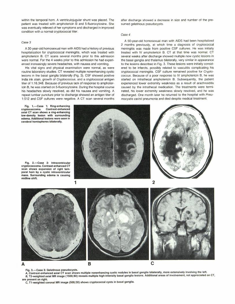
within the temporal horn . A ventriculojugular shunt was placed. The patient was treated with amphotericin B and 5-fluorocytosine. She was eventually relieved of her symptoms and discharged in improved condition with a normal cryptococcal titer.
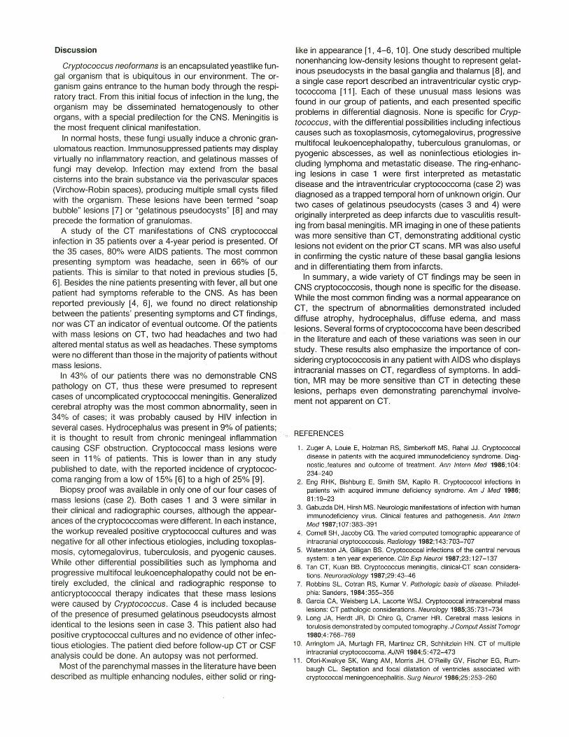
Case 3
A 30-year-old homosexual man with AIDS had a history of previous hospitalization for cryptococcal meningitis, which was treated with amphotericin B. CT scans several months prior to this admission were normal. For the 4 weeks prior to this admission he had experienced increasingly severe headaches, with nausea and vomiting.
His vital signs and physical examination were normal , as were routine laboratory studies. CT revealed multiple nonenhancing cystic lesions in the basal ganglia bilaterally (Fig. 3). CSF showed positive India ink stain , growth of Cryptococcus, and a cryptococcal antigen titer of 1:16,348. Because of previous lack of response to amphotericin B, he was started on 5-fluorocytosine. During the hospital course his headaches slowly resolved , as did his nausea and vomiting. A repeat lumbar puncture prior to discharge showed an antigen titer of 1:512 and CSF cultures were negative. A CT scan several months
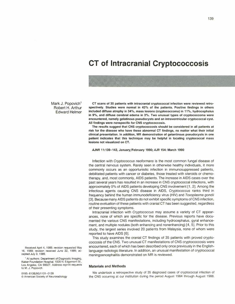
Fig. 1.-Case 1: Ring-enhancing cryptococcoma. Contrast-enhanced axial CT scan shows a ring-enhancing low-density lesion with surrounding edema. Additional lesions were seen in cerebral hemispheres bilaterally.
Fig. 2.-Case 2: Intraventricular cryptococcoma. Contrast-enhanced CT scan shows expansion of right temporal horn by a cystic intraventricular mass. Surrounding edema is causing midline shift.
1
after discharge showed a decrease in size and number of the presumed gelatinous pseudocysts.
Case 4
A 50-year-old homosexual man with AIDS had been hospitalized 2 months previously, at which time a diagnosis of cryptococcal meningitis was made from positive CSF cultures . He was initially treated with IV amphotericin B. CT at that time was normal. CT several weeks after discharge showed multiple new cystic lesions in the basal ganglia and thalamus bilaterally, very similar in appearance to the lesions described in Fig . 3. These lesions were initially considered to be infarcts, possibly related to vasculitis complicating his cryptococcal meningitis. CSF culture remained positive for Cryptococcus. Because of a poor response to IV amphotericin B, he was started on intrathecal amphotericin B. Subsequently, the patient experienced lower extremity weakness as a result of arachnoiditis caused by the intrathecal medication. The treatments were terminated , his lower extremity weakness slowly resolved , and he was discharged. One month later he returned to the hospital with Pneumocystis carinii pneumonia and died despite medical treatment.
2
Fig. 3.-Case 3: Gelatinous pseudocysts. . . . . . A Contrast-enhanced axial CT scan shows multiple nonenhancing cystic nodules in basal ganglia bilaterally, more extensively 1nvolvmg the left. a: T2-weighted axial MR image (1500/80) reveals multiple high-intensity basal ganglia lesions. Additional areas of involvement, not appreciated on CT,
are present on right. C, T1-weighted coronal MR image (500/30) shows cryptococcal cysts in basal ganglia.
Discussion
Cryptococcus neoformans is an encapsulated yeastlike fungal organism that is ubiquitous in our environment. The organism gains entrance to the human body through the respiratory tract. From this initial focus of infection in the lung, the organism may be disseminated hematogenously to other organs, with a special predilection for the CNS. Meningitis is the most frequent clinical manifestation.
In normal hosts, these fungi usually induce a chronic granulomatous reaction. Immunosuppressed patients may display virtually no inflammatory reaction, and gelatinous masses of fungi may develop. Infection may extend from the basal cisterns into the brain substance via the perivascular spaces (Virchow-Robin spaces), producing multiple small cysts filled with the organism. These lesions have been termed "soap bubble" lesions [7] or "gelatinous pseudocysts" [8] and may precede the formation of granulomas.
A study of the CT manifestations of CNS cryptococcal infection in 35 patients over a 4-year period is presented. Of the 35 cases, 80% were AIDS patients. The most common presenting symptom was headache, seen in 66% of our patients. This is similar to that noted in previous studies [5, 6]. Besides the nine patients presenting with fever, all but one patient had symptoms referable to the CNS. As has been reported previously [4, 6], we found no direct relationship between the patients' presenting symptoms and CT findings, nor was CT an indicator of eventual outcome. Of the patients with mass lesions on CT, two had headaches and two had altered mental status as well as headaches. These symptoms were no different than those in the majority of patients without mass lesions.
In 43% of our patients there was no demonstrable CNS pathology on CT, thus these were presumed to represent cases of uncomplicated cryptococcal meningitis. Generalized cerebral atrophy was the most common abnormality, seen in 34% of cases; it was probably caused by HIV infection in several cases. Hydrocephalus was present in 9% of patients; it is thought to result from chronic meningeal inflammation causing CSF obstruction. Cryptococcal mass lesions were seen in 11 % of patients. This is lower than in any study published to date, with the reported incidence of cryptococcoma ranging from a low of 15% [6] to a high of 25% [9].
Biopsy proof was available in only one of our four cases of mass lesions (case 2). Both cases 1 and 3 were similar in their clinical and radiographic courses, although the appearances of the cryptococcomas were different. In each instance, the workup revealed positive cryptococcal cultures and was negative for all other infectious etiologies, including toxoplasmosis, cytomegalovirus, tuberculosis, and pyogenic causes. While other differential possibilities such as lymphoma and progressive multifocal leukoencephalopathy could not be entirely excluded , the clinical and radiographic response to anticryptococcal therapy indicates that these mass lesions were caused by Cryptococcus. Case 4 is included because of the presence of presumed gelatinous pseudocysts almost identical to the lesions seen in case ~ . This patient also had positive cryptococcal cultures and no evidence of other infectious etiologies. The patient died before follow-up CT or CSF analysis could be done. An autopsy was not performed.
Most of the parenchymal masses in the literature have been described as multiple enhancing nodules, either solid or ring-
like in appearance [1 , 4-6, 1 OJ . One study described multiple nonenhancing low-density lesions thought to represent gelatinous pseudocysts in the basal ganglia and thalamus [8] , and a single case report described an intraventricular cystic cryptococcoma [11]. Each of these unusual mass lesions was found in our group of patients, and each presented specific problems in differential diagnosis. None is specific for Cryptococcus, with the differential possibilities including infectious causes such as toxoplasmosis, cytomegalovirus, progressive multifocal leukoencephalopathy, tuberculous granulomas, or pyogenic abscesses, as well as noninfectious etiologies including lymphoma and metastatic disease. The ring-enhancing lesions in case 1 were first interpreted as metastatic disease and the intraventricular cryptococcoma (case 2) was diagnosed as a trapped temporal horn of unknown origin . Our two cases of gelatinous pseudocysts (cases 3 and 4) were originally interpreted as deep infarcts due to vasculitis r~sulting from basal meningitis. MR imaging in one of these pat1ents was more sensitive than CT, demonstrating additional cystic lesions not evident on the prior CT scans. MR was also useful in confirming the cystic nature of these basal ganglia lesions and in differentiating them from infarcts.
In summary, a wide variety of CT findings may be seen in CNS cryptococcosis, though none is specific for the disease. While the most common finding was a normal appearance on CT, the spectrum of abnormalities demonstrated included diffuse atrophy, hydrocephalus, diffuse edema, and mass lesions. Several forms of cryptococcoma have been described in the literature and each of these variations was seen in our study. These results also emphasize the importance of considering cryptococcosis in any patient with AIDS who displays intracranial masses on CT, regardless of symptoms. In addition, MR may be more sensitive than CT in detecting these lesions, perhaps even demonstrating parenchymal involvement not apparent on CT.
REFERENCES
1. Zuger A, Louie E, Holzman RS, Simberkoff MS, Rahal JJ. Cryptococcal disease in patients with the acquired immunodeficiency syndrome. Diagnostic Jeatures and outcome of treatment. Ann Intern Med 1986;104: 234-240
2. Eng RHK, Bishburg E, Smith SM, Kapilo R. Cryptococcol infections in patients ·with acquired immune deficiency syndrome. Am J Med 1986; 81 :19-23
3. Gabuzda DH , Hirsh MS. Neurologic manifestations of infection with human immunodeficiency virus. Clinical features and pathogenesis. Ann Intern Med 1987;107:383-391
4. Cornell SH , Jacoby CG. The varied computed tomographic appearance of intracranial cryptococcosis. Radiology 1982; 143: 703-707
5. Waterston JA, Gilligan BS. Cryptococcal infections of the central nervous system: a ten year experience. Clin Exp Neurol1987;23 :127-137 .
6. Tan CT, Kuan BB. Cryptococcus meningitis, clinicai-CT scan considerations. Neuroradiology 1987;29 :43-46
7. Robbins SL, Cotran RS, Kumar V. Pathologic basis of disease. Philadelphia: Sanders, 1984 :355-356
8. Garcia CA, Weisberg LA, Lacorte WSJ. Cryptococcal intracerebral mass lesions: CT pathologic considerations. Neurology 1985;35:731-734
9. Long JA, Herdt JR, Di Chiro G, Cramer HR. Cerebral mass lesions in torulosis demonstrated by computed tomography. J Comput Assist Tomogr 1980;4 :766-769
10. Arringtom JA, Murtagh FR , Martinez CR , Schhitzlein HN. CT of multiple intracranial cryptococcoma. AJNR 1984;5 :472-473
11 . Ofori-Kwakye SK, Wang AM, Morris JH, O'Reilly GV, Fischer EG, Rumbaugh CL. Septation and focal dilatation of ventricles associated with cryptococcal meningoencephalitis. Surg Neurol1986;25 :253-260