Embed Size (px)
Citation preview
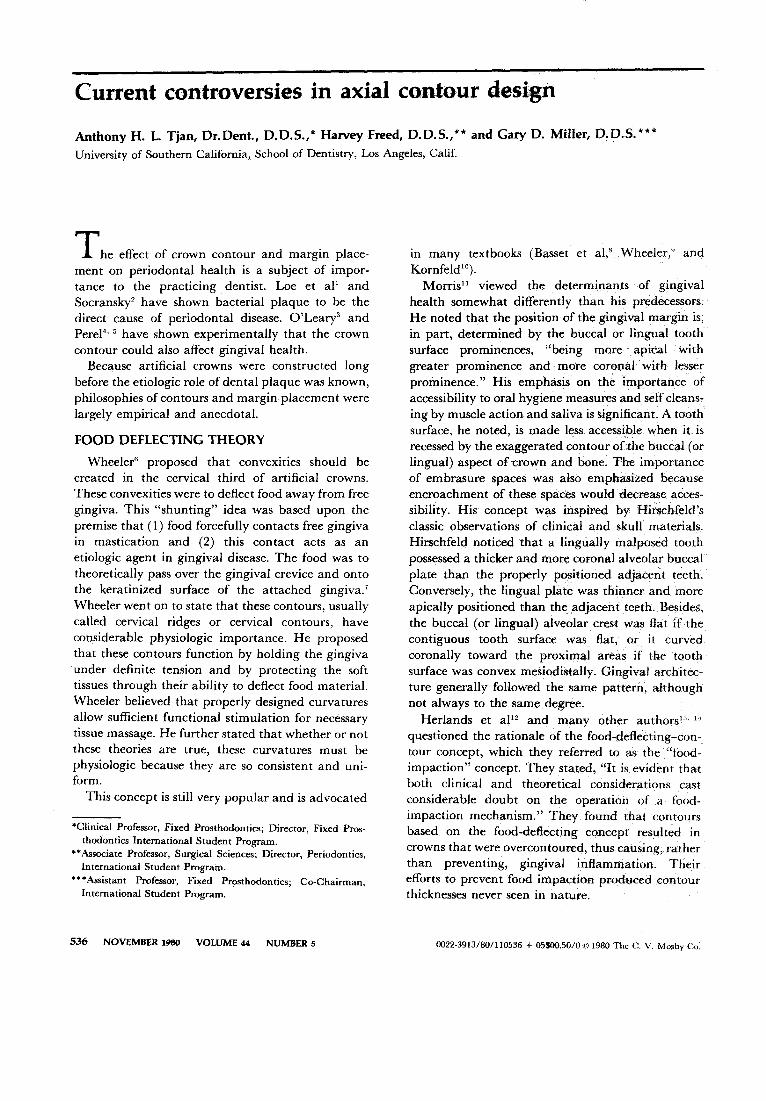
Current controversies in axial contour design
Anthony H. L. Tjan, Dr. Dent., 0.0.5.,* Harvey Freed, 0.0.5.,** and Gary O. Miller, 0.0.5. *** University of Southem Califomia, School of Dentistry, Los Angeles, Calif.
The effect of crown contour and margin placement on periodontal health is a subject of importance to the practicing dentist. Loe et aP and Socransky2 have shown bacterial plaque to be the direct cause of periodontal disease. O'Leary" and Perel4
•5 have shown experimentally that the crown
contour could also affect gingival health. Because artificial crowns were constructed long
before the etiologic role of dental plaque was known, philosophies of contours and margin placement were largely empirical and anecdotal.
FOOD DE.FLECTING THEORY
Wheeler6 proposed that convexities should be created in the cervical third of artificial crowns. These convexities were to deflect food away from free gingiva. This "shunting" idea was based upon the premise that (1) food forcefully contacts free gingiva in mastication and (2) this contact acts as an etiologic agent in gingival disease. The food was to theoretically pass over the gingival crevice and onto the keratinized surface of the attached gingiva. 7
Wheeler went on to state that these contours, usually called cervical ridges or cervical contours, have considerable physiologic importance. He proposed that these contours function by holding the gingiva under definite tension and by protecting the soft tissues through their ability to deflect food material. Wheeler believed that properly designed curvatures allow sufficient functional stimulation for necessary tissue massage. He further stated that whether or not these theories are true, these curvatures must be physiologic because they are so consistent and uniform.
This concept is still very popular and is advocated
*Clinical Prof essor, Fixed Prosthodontics; Director, Fixed Prosthodontics International Student Program.
** Associate Prof essor, Surgical Sciences; Director, Periodontics, International Student Program.
'-'Assistant Prof essor, Fixed Prosthodonticsj Co-Chairman, International Student Program.
536 NOVEMBER 1980 VOLUME 44 NUMBER 5
in many textbooks (Basset et al,' Wheeler," and Kornfeld10
).
Morris" viewed the determinants of gingival health somewhat differently than his predecessof!t He noted that the position of the gingival margin is, in part, determined by the buccal or lingual tooth surface prominences, "being more apical with greater prominence and more coronal with lesser prominence." His emphasis on the importance of accessibility to oral hygiene measures and self cleansing by muscle action and saliva is significant. A tooth surface, he noted, is made less accessible when it is recessed by the exaggerated contour of the buccal (or lingual) aspect of crown and bone. The importance of embrasure spaces was also emphasized because encroachment of these spaces would decrease accessibility. His concept was inspired by Hirschfeld's classic observations of dinical and skull materials. Hirschfeld noticed that a lingually malposed tooth possessed a thicker and more corona! alveolar buccal plate than the properly positioned adjacent teeth. Conversely, the lingual plate was thinner and more apically positioned than the adjacent teeth. Besides, the buccal (or lingual) alveolar crest was flat if the contiguous tooth surface was flat, or it curved coronally toward the proximal areas if the tooth surface was convex mesiodistally. Gingival architecture generally followed the same pattern, although not always to the same degree.
Herlands et al'2 and many other authors1:l. l,'
questioned the rationale of the food-deflecting-contour concept, which they referred to as the "foodimpaction" concept. They stated, "It is evidentthat both clinical and theoretical considerations cast considerable doubt on the operation of a foodimpaction mechanism." They found that contours based on the food-deflecting concept resulted in crowns that were overcontoured, th us causing, rather than preventing, gingival inflammation. Their efforts to prevent food impaction produced contour thicknesses never seen in nature.
0022·3913/80/110536 + 05$00.50/0<01980 The C. V. Mosby Co.
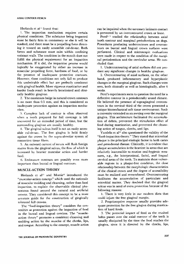
AXIAL CONTOUR DESIGN
Herlands et ap2 found that: 1. The impaction mechanism requires certain
physical conditions. The substance being impacted must be fairly firm in consistency or else it will be mashed, and there must be a propelling force directing it toward an easily accessible cul-de-sac. Both forces and substance must exist within confinÎng resistant walls. The condition in the mouth does not fulfill the physical requirement for an impaction mechanism. If it did, the impaction process would logically be exaggerated by either an increase in muscular propelling force, hardness of food, or by the presence of inadequate protective contours. However, these conditions not only fail to produce this undesirable effect but are perfectly consistent with gingival health. More vigorous mastication and harder foods result in heavily keratinized and clinically healthy gingivae.
2. The maximum bulge in natural crown contours is no more th an 0.5 mm, and this is considered as inadequate protection against an impaction mechamsm.
3. Complete lack of contour is often observed when a tooth prepared for full coverage is left uncovered for an extended period of time, but the surrounding gingivae are usually healthy.
4. The gingival sulcus itself is not an easily accessible cul-de-sac. The free gingiva is held firmly against the crown by the supra-alveolar system of connective tissue fibers.
5. An outward current of serum will flush foreign matter from the gingival sulcus, the flow of which is increased by heavier muscular action and harder food.
6. Embrasure contours are possibly even more important than buccal or lingual contours.
MUSCLE-ACTION THEORY
Herlands et a{12 and Morris13 introduced the "muscular-action concept" which used the rationale of muscular molding and cleansing, rather than food impaction, to explain the observable clinical phenomena found around the natural and artificial crowns. They considered this concept to be a more accurate guide for the construction of gingivally tolerated full crowns.
The "food-impaction theory" considers the convexities as protection against the impaction of food in the buccal and lingual crevices. The "muscleaction theory" promotes a consistent cleansing and molding action by the muscles of the cheeks, lips, and tongue. According to the concept, muscle action
THE JOURNAL OF PROSTHETIC DENTISTRY
can be impaired when the necessary intimate contact is prevented by an overeontoured erown or bone.
Perel4, 5 studied the relationship between axial
tooth contour and marginal periodontium on dogs, Procedures producing undercontours and overcontours on buccal and lingual crown surfaces were performed. Clinical and microscopic evaluations were made in respect to the condition of the marginal periodontium and the crevicular areas. He concluded that:
1. Undercontouring of axial surfaces did not produce any significant changes in healthy gingivae.
2. Overcontouring of axial surfaces, on the other hand, produced inflammatory and hyperplastic changes in the marginal gingivae. Such changes were seen, both clinically as weil as histologically, after 4 weeks.
Perel's experiments seem to question the need for a deflective contour in a periodontally sound climate. He believed the presence of supragingival overcontours in the cervical third of the crown presented a unique biomechanical soft tissue environment, Such a convexity extended as an awning over the marginal gingiva, This architecture facilitated the accumulation of debris, prevented the stimulation effect of food during mastication, and prevented the cleansing action of tongue, cheeks, and lips,
Yuodelis et al'5 also questioned the validity of the "food-impaction theory." They stated that microbial plaque is the principal etiologic factor in both caries and periodontal disease. Clinically, it is evident that plaque accumulation is the heaviest in areas that are relatively inaccessible to routine oral-hygienic measures, e.g., the interproximal, facial, and lingual cervical areas of the teeth. To main tain these vulnerable regions in a plaque-free condition, the close relationship between the morphologie eharaeteristies of the clinical erown and the degree of accessibility must be realized and remembered. Overcontouring facilitates the accumulation of particulate and microbial matter. They doubted that the gingival sulcus was in need of extra protection because of the following reasons:
1. There is very little in our modern diets that could injure the free gingival margin.
2. Proprioceptive response usually provides adequate protection for the free gingiva during mastication of hard foods,
3. The potential impact of food as the crushed bolus passes over the axial contour of the teeth is usually dissipated by the time the food reaches the gingiva, since it is directed by the cheeks, lips,
537
tongue, and other parts of the mouth into a position for deglutition.
4. Most human dentitions have little, if any, clinical bulge and yet show no deleterious effects of mastication.
S. The dentitions of lower species of animais do not provide this theoretic protection, since any buccal and lingual bulges are usually subgingival. This also demonstrates the importance of the proprioceptive system to protect the gingiva from trauma tic effect of food that is much coarser.
PLAQUE-RETENTION THE ORY
Hazen and Osborne,H Herland et al,'2 Morris,':l Perel,4. j Yuodelis et al," Eissmann et al,'6 Schluger et al," Baer and Morris,'" Vogan,l" and Veldkamp21l
advocated an axial contour design based on the "muscle-action theory," which has recently been called the "plaque-retention theory."
The proponents of this concept prefer axial contours of artificial crowns which facilitate oralhygiene measurements and promote self-cleaning by muscle action of the tongue, cheeks, and lips. Furthermore, they stated that crown contour should not harbor any plaque traps.
ANATOMIC THEORY
Kraus et al,"' Burch and MiIIer,22 Beaudreau,"'l Farer and Isaacson,24 and Burch2
,;, 26 introduced the anatomic or biologic concept of tooth contour, a contour which simulated natural, healthy teeth. They considered that a biologic contour was a self-protective contour to the supporting tissues and defended the gingival unit, attachment apparatus, and protected bone from trauma and irritation. Improper contour often induced early breakdown of the supporting structures and tooth tissue, resulting in premature loss of teeth."' They stated that facial and lingual convexities form the height of contour of tooth crowns, which are located at the gingival third of each tooth and are approximately one-half millimeter wider than the adjoining cementoenamel junction (CE]). The exception to this general rule is on the lingual surface of the lower molars and second premolars. There, the convexities measure approximately one millimeter and are located halfway between the occlusal plane and the gingival margin.
MAR GIN PLACEMENT
So far, aIl the previous authors have focused their attention on the supragingival aspect of an artificial crown. However, recent data have suggested that
538
TIAN, fREED, AND MILLER
margin placement plays a significant role in gingival health. The majority of data indicate that subgingival margins can be conducive to plaque accumulation and to periodontal disease. 27
•12 There are, of
course, conditions which preclude the use of supragingival margins (i.e., esthetics, caries). When subgingival margins are created, the question of subgingival crown contours must be approached.
Wagman33 began to observe the function of subgingival contours pertinent to gingival health. He emphasized the importance of establishing the proper contour to maintain the "knife-like" shape of the free gingival margin. This was to facilitate the removal of microbial plaque.
Wagman believed that subgingival contour should be made convex facially and lingually. This was to protect the gingival suleus and to promo te a knife-like, free gingival margin. The degree of these convexities, he stated, should not exceed one-half of the thickness of the gingiva at the height of its attachment. So far as the supragingival contour was concerned, graduai curvatures in aIl directions were advocated to facilitate self-cleansing by the function of the lips, cheeks, and tongue. Proper interproximal contour was also suggested, and undercontour was preferred to overcontour.
Wagman's concept was mostly inspired by Ross's" statement that subgingival contour has a considerable effect upon the free marginal gingiva and the gingival suleus. Ross stated that if the subgingival contour is made flat, and does not support the gingiva, the free marginal gingiva will tend to form a "roll" around the tooth. This will provide a ledge upon which the plaque accumulates with easy access into the gingival crevice. When the subgingivaI contour of the crown is made overly thick, the free marginal gingiva will become congested. circumferential fibers will be tom'" and the gingival tissues pushed beyond their physiologie limit of accommodation. The gingiva will then become infiamed. The subsequent swelling and loss of tissue tone will cause bleeding and increase plaque accumulation.
Like Wagman, Weisgold36 also emphasized the importance of proper contouring of the subgingival portion on artificial crowns. Weisgold stated, however, that subgingival contour should be dictated by the level of the free gingival margin.
Wh en the gingival margin is placed coron al to the CE], the subgingival contour should be made convex. On the other hand, when gingival margin is located below the CE] (on the root surface) due to recession, the subgingival contour should be made flat. His clinical experiences have shawn that the
NOVEM8ER 1980 VOLUME 44 NUM8ER 5
AXIAL CONTOUR DESIGN
artificial crown portion placed subgingivaUy should generaUy imitate the original shape of the tooth.
Spurow and Lytle37 realized the importance of interproximal embrasure as a "yardstick" of periodontal health in patients with virgin teeth. In restorative dentistry, this was an even more sensitive gauge. Spurow and Lytle, as well as many others, have emphasized the importance of creating a proper embrasure space for the health of the interdental papiUae.
Stein and Kuwata"" and Presswood39 emphasized the importance of "the emergence of profile," i.e., the axial contour that emerges from the gingival sulcus, which was generally considered as straight or concave. Stein and Kuwata"" stated, "Ignorance of this characteristic of human tooth morphology is directly responsible for much of the marginal gingival disease around the artificial crowns. If the emergence profile of the finished crown is incorrectly formed, aU interrelated contours will be incorrect and, accordingly, the deflective mechanism will be impaired."
SUMMARY AND CONCLUSION
The subject of axial contour of artificial crowns is highly controversial. After years of dispute, there is still no definite agreement on what the role of crown contour is and what effect, if any, alterations in contour have on the health of marginal periodontium. Some of the controversies surrounding this subject appear to center around whether the gingival sulcus is really in need of protection from buccal and/or lingual deflecting convexities. Other considerations are whether a flatter form of axial contour (which facilitates the self-cleansing muscle action of cheeks, lips, and tongue) is preferable. Except for very few histologie facts, most of the discussions are based on clinical evidences and investigators' experiences.
Three concepts or theories can be differentiated. They are (1) the "food-impaction theory," (2) the "plaque-retention theory," and (3) the "anatomic theory." Each concept represents a specifie era. Unfortunately, none of these concepts provides a satisfactory solution to the problem of maintaining the health of the marginal periodontium.
The most frustrating problem confronting the dentist is an artificial crown which demands subgingival placement of its margin. This is most problematic, primarily because a portion of the crown is placed in a gingival sulcus which is extremely vulnerable to periodontal disease.
The clinical experience of one of the authors (A. H. L. T.) showed that, besides the design of
THE JOURNAL OF PROSTHETIC DENTISTRY
profile contour, it is essential to carefully evaluate the morphology or architecture of the marginal periodontium. The henefit of any kind of contour will he easily negated if the operator ignores the significance of the proper architecture of marginal periodontium and proper oral hygiene instruction. It is sometimes advisable to perform corrective gingival surgery on the marginal periodontium and/ or orthodontic therapy to correct a malaligned tooth prior to restoration to assure the ultimate success of a restoration.
We would like to extend our appreciation to Dr. Josephine G. The for her help in the preparation of this manuscript.
REFERENCES
1. Loe, H., Theilade, E., and Jensen, S. B.: Experimental gingivitis in man. J Peridontol 36:177,1965.
2. Socransky, S. S.: Relationships of bacteria to the etiology of periodontal disease. J Dent Res 49:203, 1970.
3. O'Leary, T. J.: Oral hygiene agents and procedures. J Periodontal 41:625, 1970.
4. Perel, M. L.: Axial crown contours. J PROSTHET DENT 25:642, 1971.
5. Perel, M. L.: Periodontal considerations of crown contours. J PROSTHET DENT 28:627, 1971.
6. Wheeler, R. C.: Complete crown form and the periodontium. J PROSTHET DENT 11:722, 1961.
7. Amsterdam, M., and Fox, L.: Provisional splinting-Principies and technics. Dent Clin North Am 3:73, 1959.
8. Bassett, R. W., lngraham, R., and Koser, J. R.: An Atlas of Cast Gold Procedures, ed 1. West Orange Cou nt y Publishing Co., Buena Park, Calif., 1964.
9. Wheeler, R. C.: Dental Anatomy, Physiology, and Occlusion, ed 3. Philadelphia, 1958, W. B. Saunders Co.
10. Kornfeld, M.: Mouth Rehabilitation, Vol 1. St. Louis, 1967, The C. V. Mosby Co., pp 95-100.
11. Morris, M. L.: The position of the margin of the gingiva. Oral Surg 11:969, 1958.
12. Herlands, R. E., Lucca, J. J., and Morris, H. L.: Forms, contours, and extensions of full coverage restorations in occlusal reconstruction. Dent Clin North Am 6:147, 1962.
13. Morris, M. L.: Artificial crown contours and gingival health. J PROSTHET DENT 12:\146, 1962.
14. Hagen, S. P., and Osborne, J. W.: Relationship of operative dentistry to periodontal health. Dent Clin North Am Il :245, 1967.
15. Yuodelis, R. A., Weaver, J D., and Sapkos, S.: Facial and lingual contours of artificial complete crown restorations and their effects on the periodontium. J PROSTHET DENT 29:61, 1973.
16. Eissmann, H. F., Radke, R. A., and Noble, W. H.: Physiologie design criteria for fixed dental restorations. Dent Clin North Am 14:543, 1971.
17. Schluger, S., Yuodelis, R. A., and Page, R. C.: Periodontal Disease. Philadelphia, 1977, Lea and Febiger, pp 586-608.
18. Baer, P. N., and Morris, M. L.: Textbook of Periodontics. Philadelphia, 1977, J. B. Lippincott Co., pp 80-86.
19. Vogan, W. 1.: The effects ofbucco-lingual crown contours on gingival health. Prey Dent 3:30, 1976.
539
20. Veldkamp, D. F.: The relationship between tooth form and gingival health. Dent Pract 14:158, 1973.
21. Kraus, B. S., Jordan, R. E., and Abrams, L.: Dental Anatomy and Occlusion, ed 1. Baltimore, 1969, WîlIiams and Wilkins Co.
22. Burch,j. G., and MîlIer,]. B.: Evaluating crown contours of wax pattern. j PROSTHET DENT 30:454, 1973.
23. Beaudreau, D. E.: Tooth form and contour. Prey Dent, july-Aug., pp 36-47, 1973.
24. Farer,]. W., and Isaacson, D.: Biologie contours. Prey Dent 1:4, 1974.
25. Burch, j. G.: Ten rules for developing crown contours in restorations. Dent Clin North Am 15:611,1971.
26. Burch,]. G.: Periodontal considerations in operative dentistry. J PROSTHET DENT 34:165, 1975.
27. Waerhaug,].: Tissue reactions around artificial crowns. j PROSTHET DENT 24: 172, 1953.
28. Waerhaug,j.: Historical considerations which govern where the margins of restorations should be located in relation to the gingiva. Dent Clin North Am 4:161, 1960.
29. Marcum, j. S.: The etfect of crown marginal depth upon gingival tissue. j PROSTHET DENT 17:479, 1967.
30. Loe, H.: Reactions of marginal periodontal tissues to restorative procedures. Int Dent J 18:759, 1968.
31. Karlsen, K.: Gingival reactions to dental restorations. Acta Odon toI Scand 28:895, 1970.
32. Newcomb, G. M.: The relationship between the location of subgingival crown margins and gingival inflammation. J Periodontal 45:151, 1974.
TJAN. FREED. AND MILLER
33. Wagman, S.: The role of coronal contours in gingival health. J PROSTHET DENT 37:280, 1977.
34. Ross, 1. F.: The relation between periodontaJ therapy and fixed restorative care. J Periodontal 42: 13, 1971.
35. Arnim, S. S., and Hangerman, D. A.: The connective tissue fibers of the marginal gingiva. J Am Dent Assoc 47:271, 1953.
36. Weisgold, A.: Coronal forms of the full crown restoration-Their c1incial applications. Continuing Dental Educa· tion, UnÎv. of Penn., School of Dental Medicine, Vol. 1, No. 6, 1977, pp 1-17.
37. Spurow, H. M., and Lytle,]. D.: The interproximal embrasure. Dent Clin North Am 15:641, 1971.
38. Stein, R. S., and Kuwata, M.: A dentist and a dental technologist analyze current ceramo-mètal procedures. Dent Clin North Am 21:729, 1977.
39. Presswood, R. G.: EsthetÎcs and color: Perceiving the problem. Dent Clin N Am 21:823, 1977.
40. Wilcox, C.E., and Everett, F. G.: Friction on the teeth and the gingiva during mastication. j Am Dent Assoc 66;513, 1963.
Reprint requests toc DR. ANTHONY H. L. TJAN UNIVERSITY OF SOUTHERN CALIFORNJA SCHOOL OF DEN'11STRY 925 WEST THiRTY-FOURTH ST. Los ANGELES, CAUF. 90007
JOURNAL announces new feature-Tips from our readers
540
The JOURNAL OF PROSTHETIC DENTISTRY is pleased to announce a new feature. Tips From Our Readers will consist of brief reports describing a sUllcessfuI laboratory or c1inical procedure, or timesaving step in a procedure, that authors would like to share with our readers.
Tips From Our Readers will be published on a space-available basis throughout the JOURNAL and will appear in a manner similar to the IADR Prosthodontic Abstracts.
Manuscripts must he Iimited to approxima tel y 250 words, typed double-spaced, with no more than two black and white illustrations. Color illustrations will not be accepted. The format for tide, author(s), institution, and dt y will be the same as that used for full-length articles. References will not he accepted.
Footnotes for commercial products will be used, but they should he held to a minimum. The TiPs must descdbe a helpfuI procedure or timesaving step, and of course, they must not be used to promote a commercial product.
Reprints will he avaIJable .to authors at the usual cost. Tips From Our Readers will be accepted immediately for evaluation.
NOVEMBER 1980 VOLUME 44 NUMBER 5