Embed Size (px)
Citation preview
Current Topics in Pediatric Surgery for the
Primary Care Provider
SCAAP 2016 Annual Meeting
Robert L. Gates MD FACS FAAP Clinical Associate Professor
Division of Pediatric Surgery
University of South Carolina School of Medicine Greenville
Speakers Disclosure Statement
Dr. Gates discloses that he has no relevant financial relationships with commercial interests.
Dr. Gates does not anticipate discussing unlabeled uses of any commercial products or any investigational
products
Introduction
• Vascular Malformations
• Common Skin Lesions
• The Coccygeal Dimple
• Breast masses in adolescents
• The latest in Inguinal Hernia Repair
• Intussusception
• The Spectrum of Malrotation
• Pyloric Stenosis
• Pectus Deformities
Vascular
Malformations
Vascular
Malformations
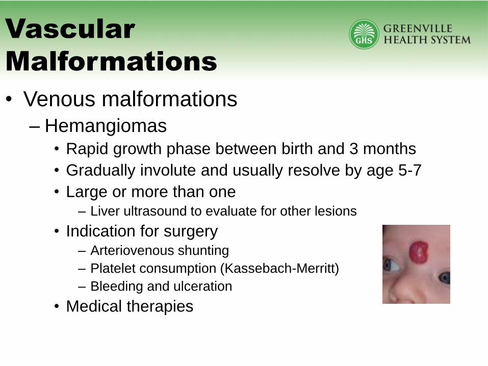
• Venous malformations
– Hemangiomas • Rapid growth phase between birth and 3 months
• Gradually involute and usually resolve by age 5-7
• Large or more than one – Liver ultrasound to evaluate for other lesions
• Indication for surgery – Arteriovenous shunting
– Platelet consumption (Kassebach-Merritt)
– Bleeding and ulceration
• Medical therapies
Vascular
Malformations
• Venous Malformations
Vascular
Malformations
• Lymphatic Malformations
– Usually a collection of abnormal lymphatic channels
with a vascular component
– Microcystic and macrocystic
– Role of ultrasound and MRI
• Characterization
• Deep tissue extent
Vascular
Maformations
• Lymphatic Malformations – Complications
• Infection and cellulitis
• Anatomic dysfunctions – Tracheal occlusion
– GI tract
» Protein loss
» Pancreatitis
» Obstruction
» Chylous ascites
– Chest
» Chylothorax
» Venous Occlusion
Vascular
Malformations
• Lymphatic Malformations
– Treatment
• Surgery
• Sclerotherapy – Ethanol, Picibanil
• Laser – Integrated with other therapies and spaced over
months
• Radiofrequency ablation
• Chemotherapy - Rapamycin
Vasuclar
Malformations
• Lymphatic malformations

Common Skin Lesions
Common Skin Lesions
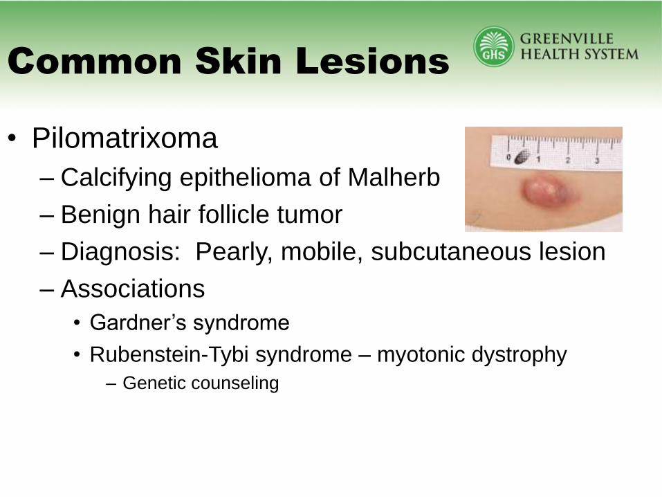
• Pilomatrixoma
– Calcifying epithelioma of Malherb
– Benign hair follicle tumor
– Diagnosis: Pearly, mobile, subcutaneous lesion
– Associations
• Gardner’s syndrome
• Rubenstein-Tybi syndrome – myotonic dystrophy
– Genetic counseling
Common Skin Lesions
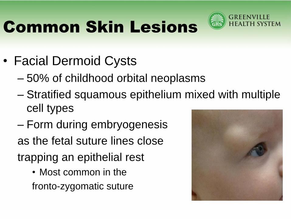
• Facial Dermoid Cysts
– 50% of childhood orbital neoplasms
– Stratified squamous epithelium mixed with multiple
cell types
– Form during embryogenesis
as the fetal suture lines close
trapping an epithelial rest
• Most common in the
fronto-zygomatic suture
Common Skin Lesions
• Lymph Nodes
– Differentiate between acute, subacute, chronic
– Determine possible infectious causes
• Chronic otitis, pharyngitis
• Cat scratch disease
• Mycobacterium
Common Skin Lesions
• Lymph Nodes
– Signs suggesting malignancy
• Size > 2.5 cm
• Firm/fixed
• Supraclavicular or axillary
• Non-tender
• Progressive
• “B”Symptoms
– Role of ultrasound
Coccygeal Dimples
Coccygeal Dimples
• Incidence – 4.8% of children
– Problematic lesions
• Spinal dysraphism – 1/2500
• Spinal lipoma – 1/4000
• Dermal sinus – 1/2500
Coccygeal Dimples
• Indications of High Risk
– Larger than 0.5 cm
– Located more than 2.5 cm above the anal verge
– Overlying cutaneous lesions • True hypertrichosis
• Skin tags
• Telangectasia
• Subcutaneous mass
• Aplasia cutis
• Abnormal pigmentation
– Asymmetric gluteal crease
Coccygeal Dimples
• Further evaluation
– Avoid probing
– Ultrasound imaging
• Prior to 4 months of age
• Operator sensitive
– MRI
• Requires sedation
– Neurosurgical consultation

Coccygeal Dimples
Breast Masses in
Adolescents
Breast Masses in
Adolescents
• Infections
– Treatment
• Asymmetric breast bud development
• Gynecomastia
– Usually unilateral
– Usually resolves as puberty progresses
– Don’t forget the testicular exam!
Breast Masses in
Adolescents
• Tumors – Incidence: 3.2% teenage girls
– 95% will be a benign fibroadenoma • 10% regress spontaneously
• < 0.02% are malignant
• Juvenille fibroadenoma or Giant fibroadenoma
– Other tumors • Phyllodes
• PASH
• Intraductal papilloma
• Lymphangiomas
• Malignancy

Breast Masses in
Adolescents

Inguinal Hernias
Inguinal Hernias
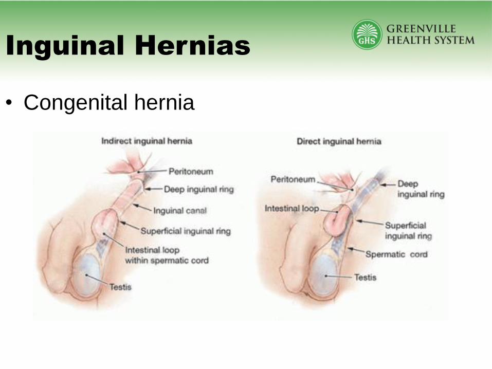
• Congenital hernia
Inguinal Hernias
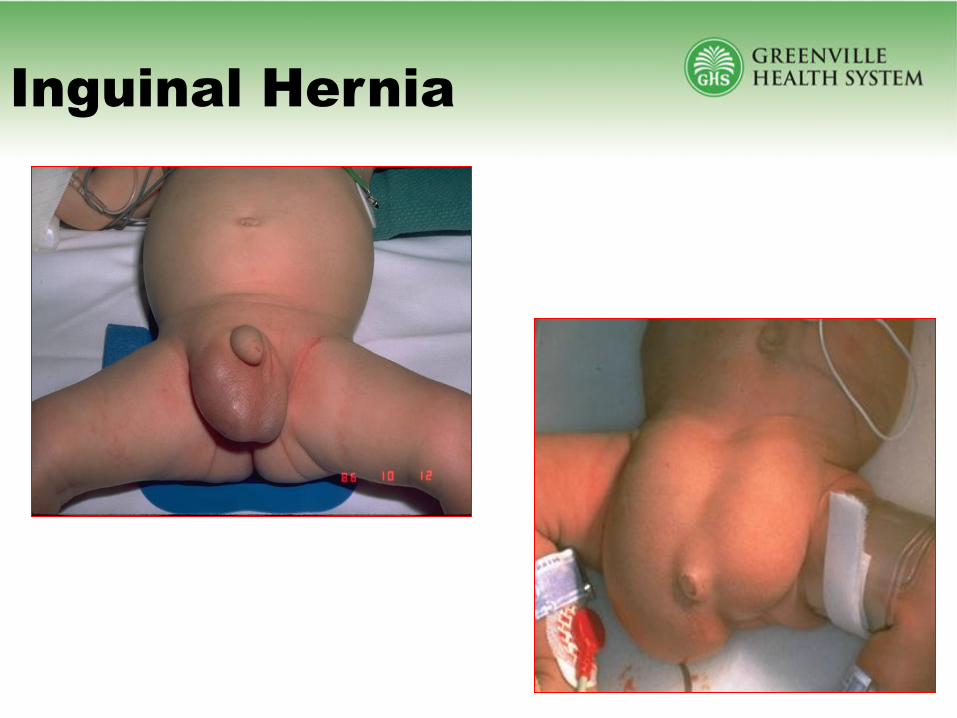
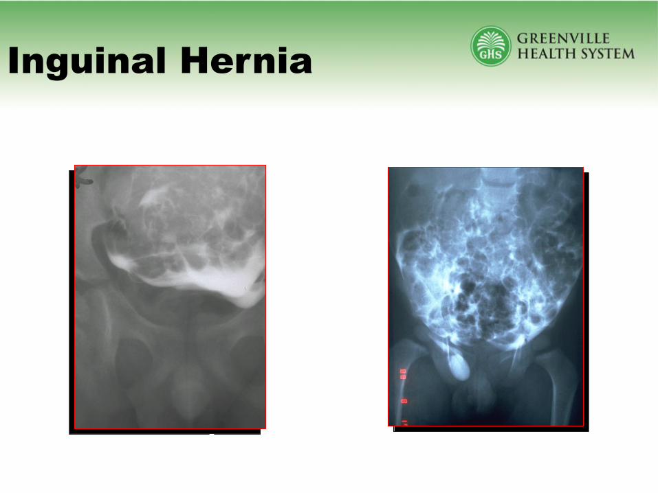
Inguinal Hernia
Inguinal Hernia
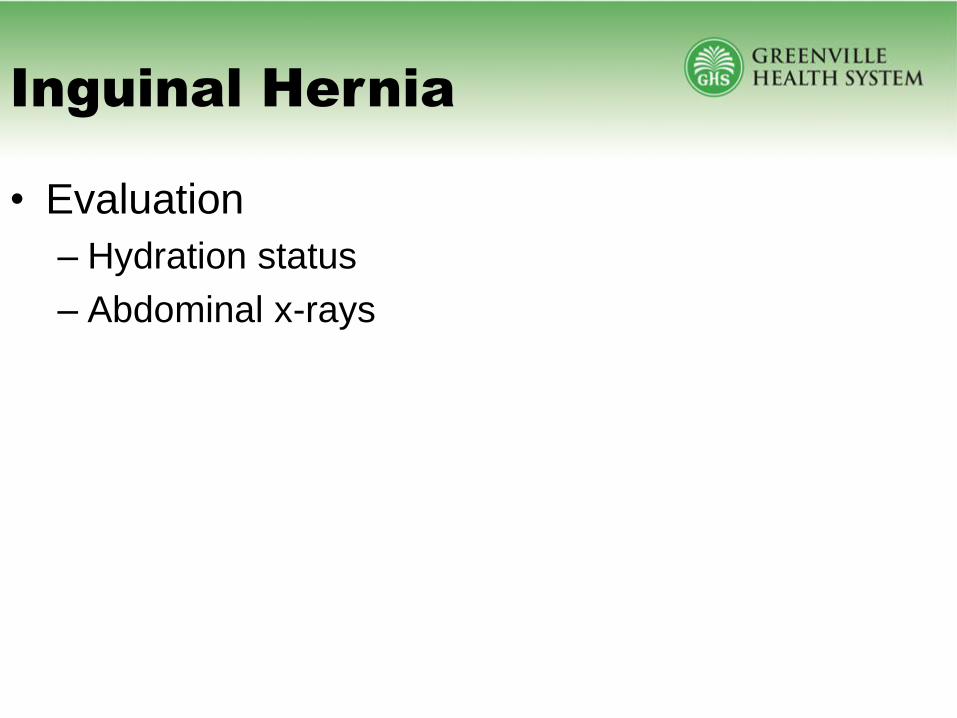
• Evaluation
– Hydration status
– Abdominal x-rays
Inguinal Hernia
Normal RIH
Inguinal Hernia
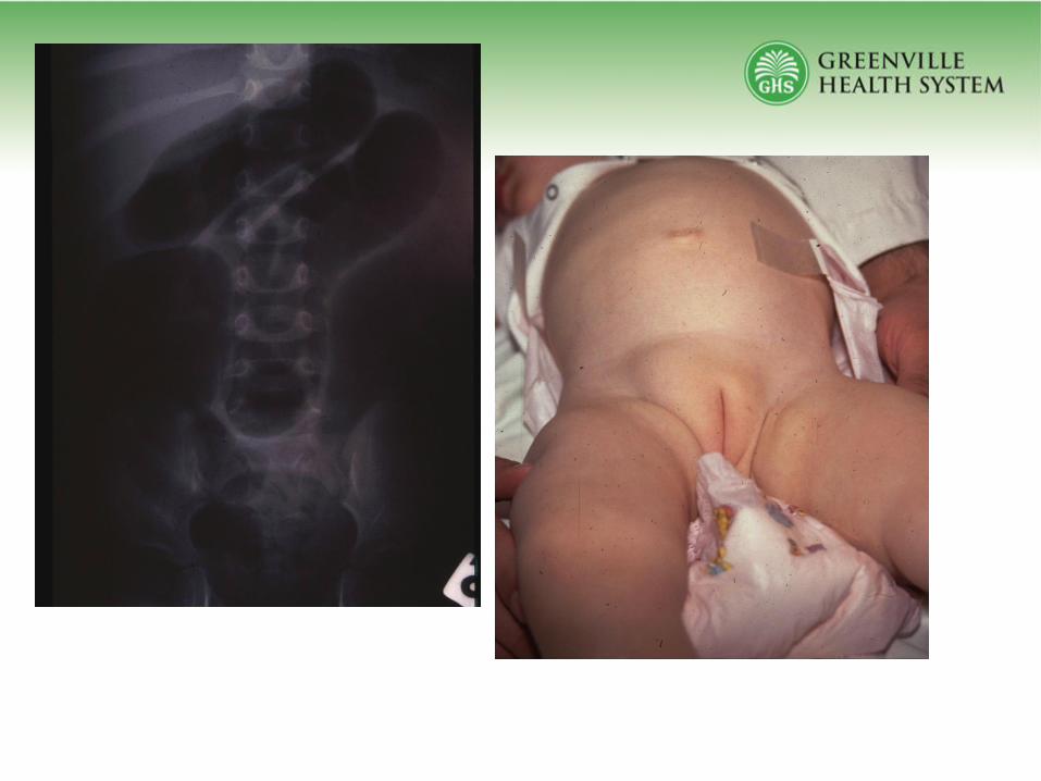
• Evaluation
– Hydration status
– Abdominal x-rays
• Reduction
– Dark, quiet room
– Hydration
– Consider conscious sedation
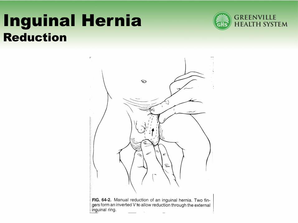
Inguinal Hernia
Reduction
Inguinal Hernia
Evaluation
Hydration status
Abdominal x-rays
Reduction
Dark, quiet room
Hydration
Conscious sedation
Timing of Repair
Allow time for edema to resolve
Within 1-2 weeks
Parent education

Inguinal Hernia
Inguinal Hernia
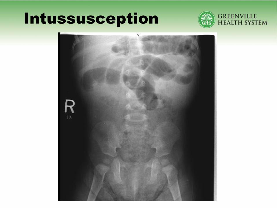
Intussusception
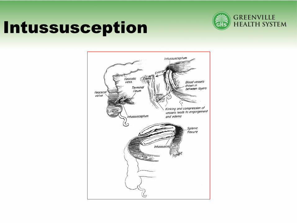
Intussusception
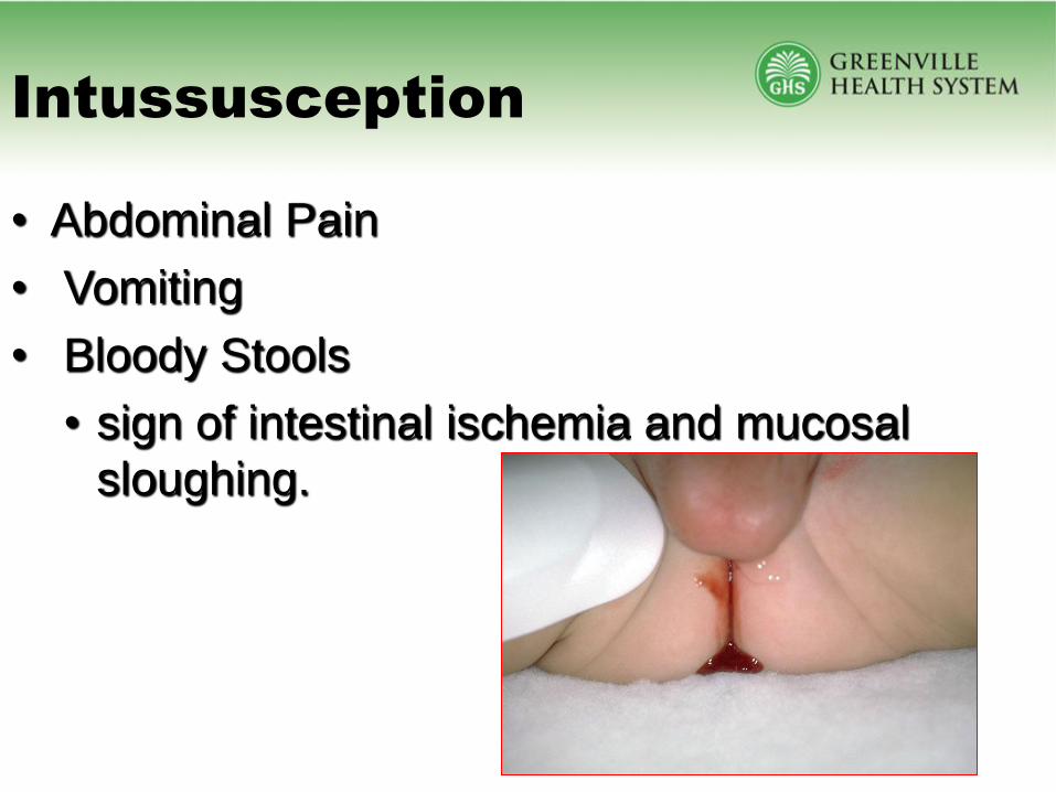
Intussusception
• Abdominal Pain
• Vomiting
• Bloody Stools
• sign of intestinal ischemia and mucosal
sloughing.
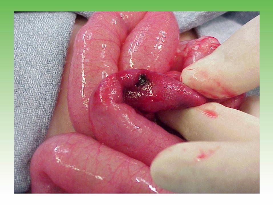
Intussusception
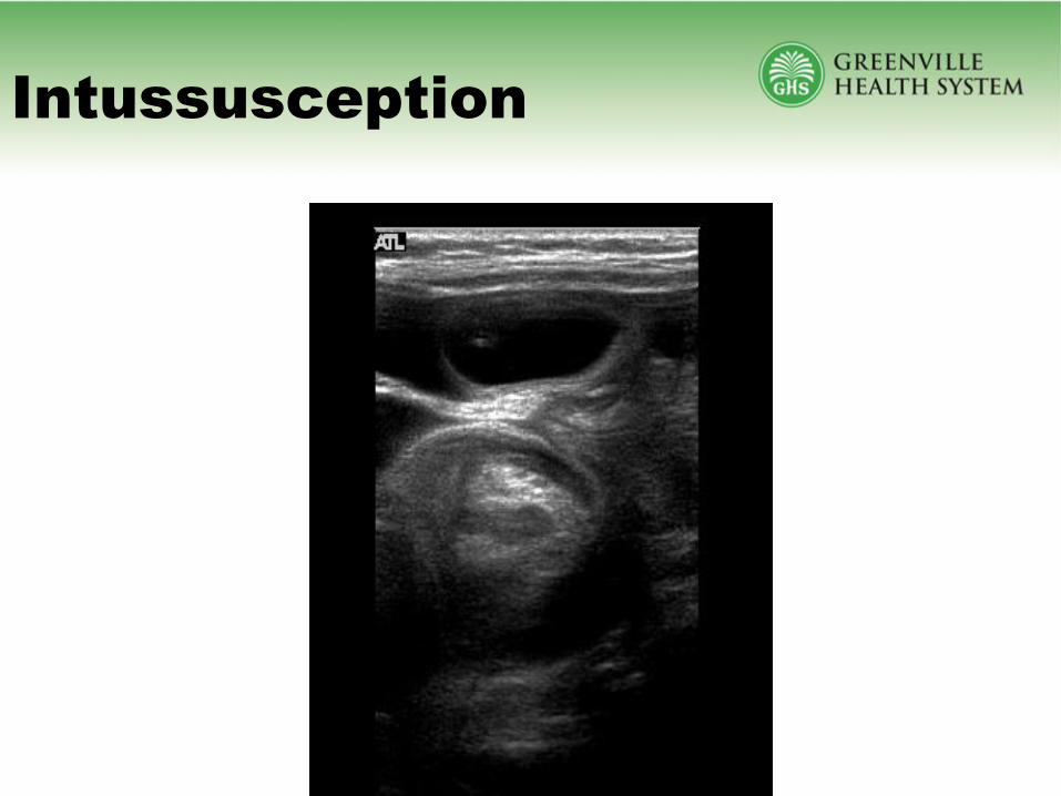
Intussusception
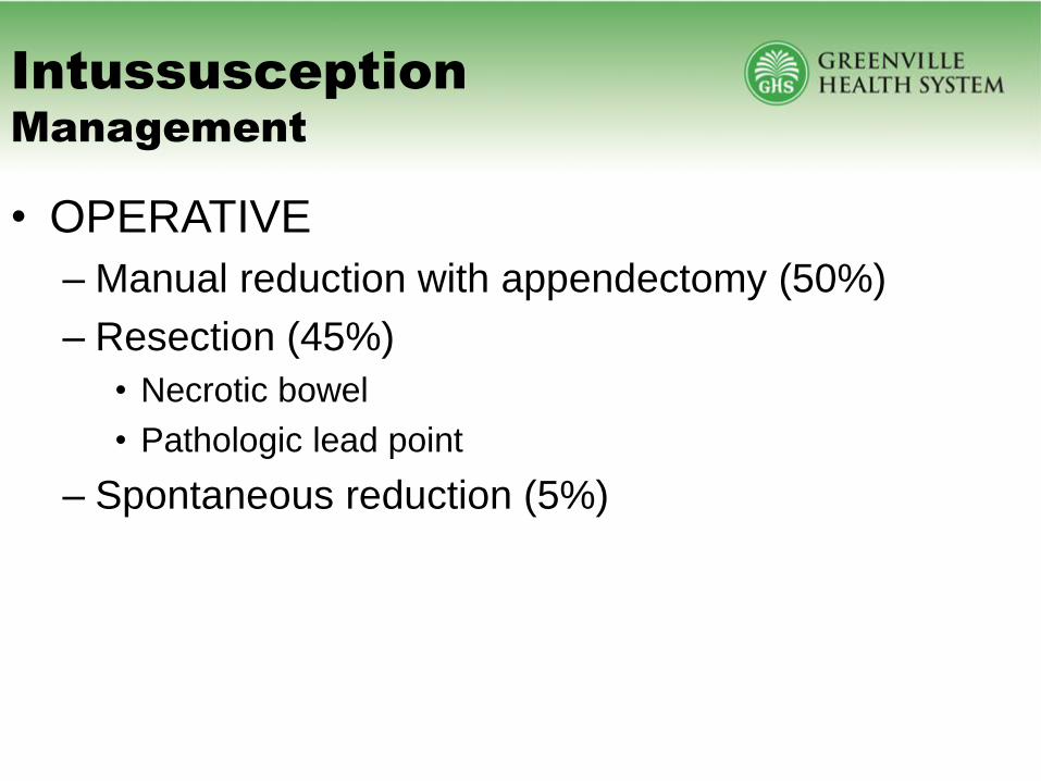
Intussusception
Management
• OPERATIVE
– Manual reduction with appendectomy (50%)
– Resection (45%)
• Necrotic bowel
• Pathologic lead point
– Spontaneous reduction (5%)
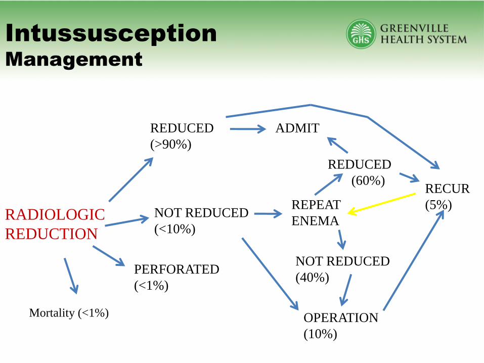
Intussusception
Management
RADIOLOGIC
REDUCTION
Mortality (<1%)
REDUCED
(>90%)
NOT REDUCED
(<10%)
PERFORATED
(<1%)
ADMIT
RECUR
(5%)
OPERATION
(10%)
REDUCED
(60%)
NOT REDUCED
(40%)
REPEAT
ENEMA
Intussusception
• Lead points
• more common in older children
• nearly always found in adults with
intussusception
Intussusception
Lead Points: • Meckel’s diverticulum (most common) • polyps • duplications • lymphomas • submucosal hemorrhage with Henoch Schoenlein
purpura • hemangiomas • lymphosarcomas • inspissated meconium in the terminal ileum
(Cystic Fibrosis)
Intussusception
Post-operative Causes
Unique to pediatric population
Prolonged surgery with extensive bowel retraction
Wilm’s tumor
Nissen fundoplication
Retroperitoneal surgery
Often confused with post-operative ileus
Symptoms develop 1-2 weeks after surgery
Distension, nausea/vomiting, cramping Suspect if marked increased volume of gastric drainage
Ultrasound for diagnosis
Surgical exploration required
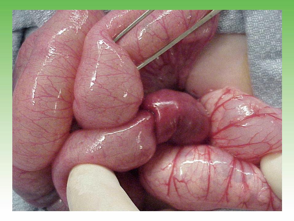
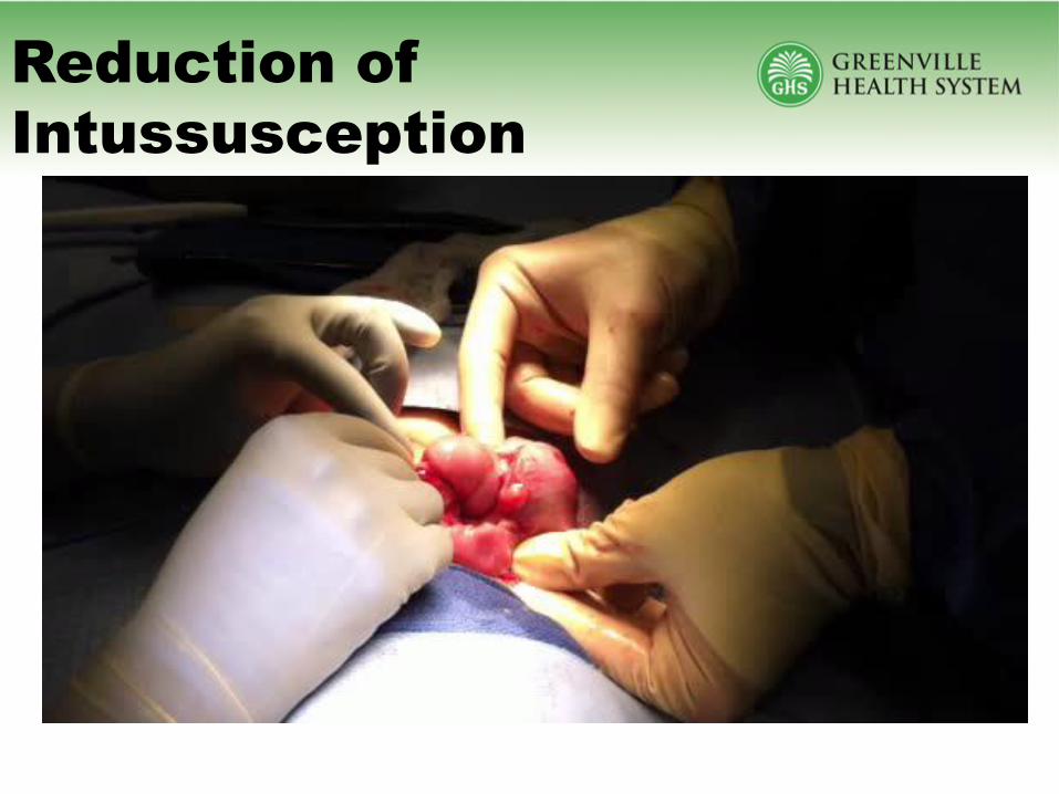
Reduction of
Intussusception
Malrotation
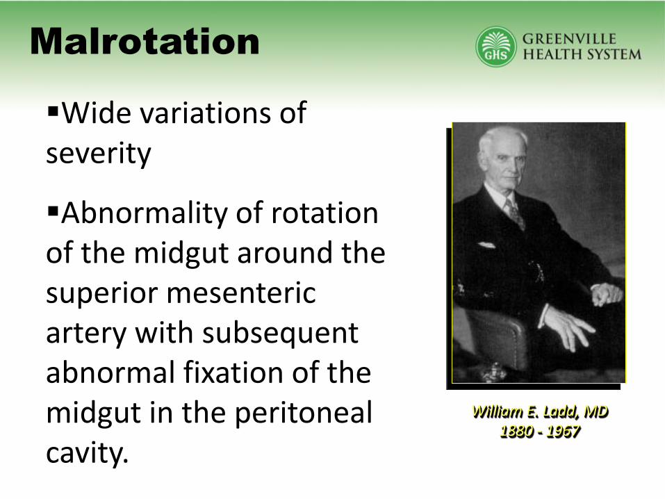
Malrotation
William E. Ladd, MD 1880 - 1967
Wide variations of severity
Abnormality of rotation of the midgut around the superior mesenteric artery with subsequent abnormal fixation of the midgut in the peritoneal cavity.
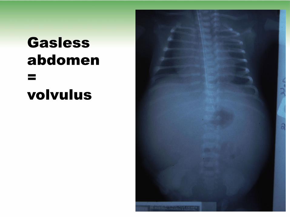
Gasless
abdomen
=
volvulus
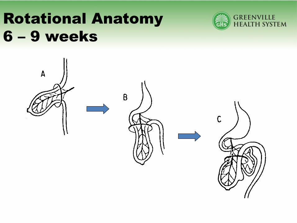
Rotational Anatomy
6 – 9 weeks
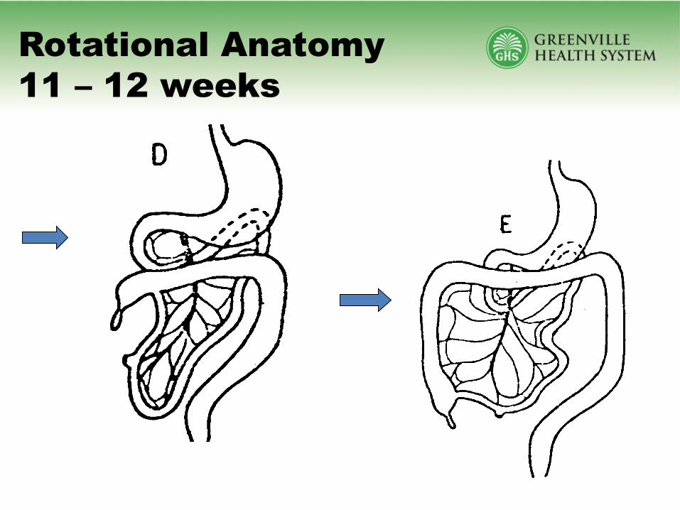
Rotational Anatomy
11 – 12 weeks
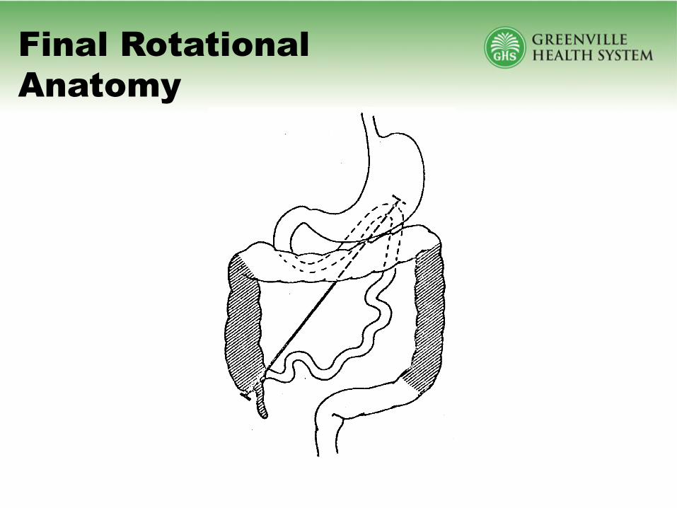
Final Rotational
Anatomy
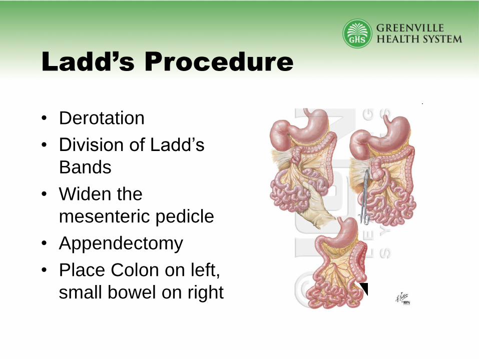
Ladd’s Procedure
• Derotation
• Division of Ladd’s
Bands
• Widen the
mesenteric pedicle
• Appendectomy
• Place Colon on left,
small bowel on right
Malrotation
• Late Presentation
– Chronic, intermittent abdominal pain
– Incidental finding for other abdominal surgery
Pyloric Stenosis
Pyloric Stenosis
• Incidence – 4/1000 live births
– 5:1 boys to girls
• Presentation
– Non-bilious projectile vomiting
– Visible peristaltic waves in LUQ
– Hypochloremic metabolic alkalosis
– 75% will have a palpable “olive”
Pyloric Stenosis
• Ultrasound
– Operator dependent
– Pyloric dimensions • Thickness >3 mm
• Length >14 mm
• Upper GI
– Elongated pyloric channel
– Antral shouldering
– May be confused with pyloric spasm
Pyloric Stenosis
• Repair
– Open
• RUQ or umbilical incision
– Laparoscopic
• Minimal scar
• Faster recovery
• Faster return to full feeds
• Less post-operative emesis
• Decreased pain

Pyloric Stenosis
Chest Wall Deformities
Chest Wall Deformities
• Pectus Excavatum
• Pectus Carinatum
• Poland Syndrome
• Failure of Sternal Fusion
• Asphyxiating Thoracic Dystrophy
– Cerebrocostomandibular Syndrome
– Jarcho-Levin Syndrome
– Jeume Syndrome
Chest Wall Deformities
• Pectus Excavatum
• Pectus Carinatum
• Poland Syndrome
• Failure of Sternal Fusion
• Asphyxiating Thoracic Dystrophy
– Cerebrocostomandibular Syndrome
– Jarcho-Levin Syndrome
– Jeume Syndrome
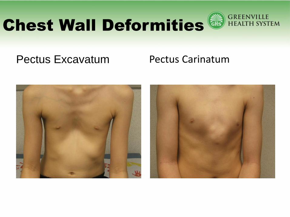
Chest Wall Deformities
Pectus Excavatum
Pectus Carinatum
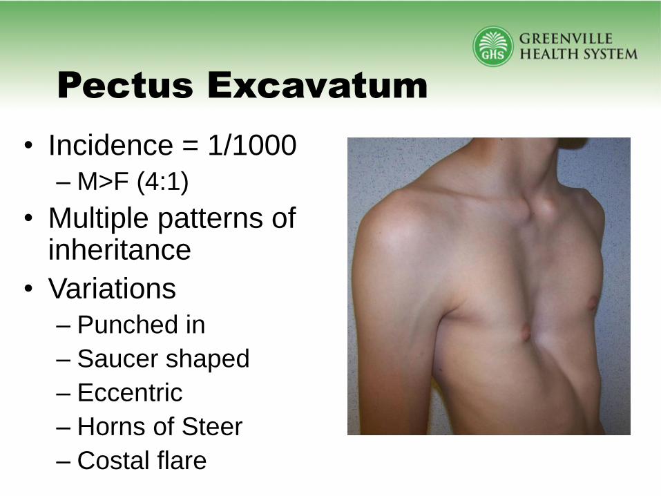
Pectus Excavatum
• Incidence = 1/1000
– M>F (4:1)
• Multiple patterns of inheritance
• Variations
– Punched in
– Saucer shaped
– Eccentric
– Horns of Steer
– Costal flare
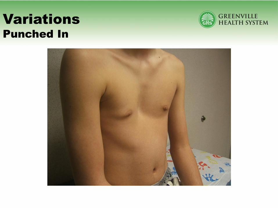
Variations
Punched In
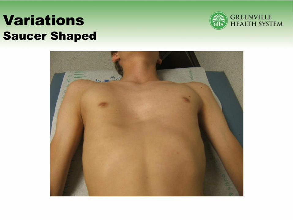
Variations
Saucer Shaped
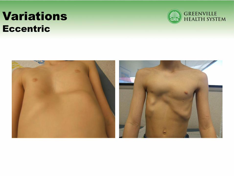
Variations
Eccentric
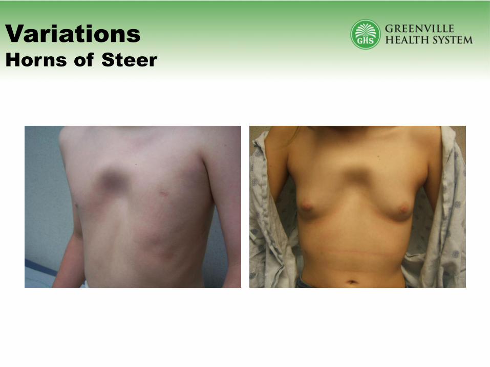
Variations
Horns of Steer
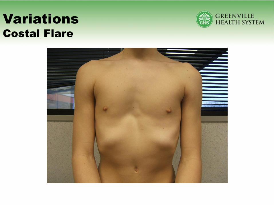
Variations
Costal Flare
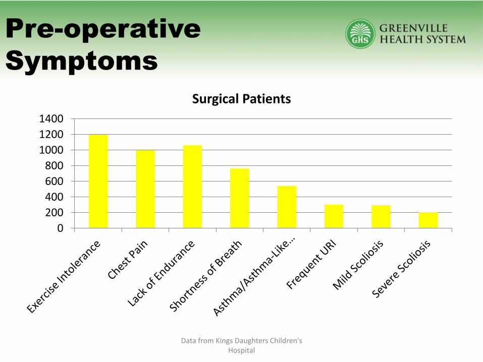
Pre-operative
Symptoms
0
200
400
600
800
1000
1200
1400
Surgical Patients
Data from Kings Daughters Children's Hospital
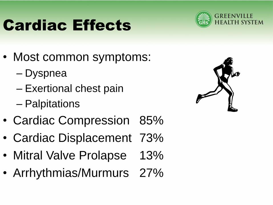
Cardiac Effects
• Most common symptoms:
– Dyspnea
– Exertional chest pain
– Palpitations
• Cardiac Compression 85%
• Cardiac Displacement 73%
• Mitral Valve Prolapse 13%
• Arrhythmias/Murmurs 27%
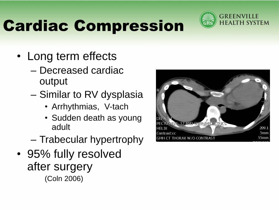
Cardiac Compression
• Long term effects
– Decreased cardiac output
– Similar to RV dysplasia • Arrhythmias, V-tach
• Sudden death as young adult
– Trabecular hypertrophy
• 95% fully resolved after surgery
(Coln 2006)
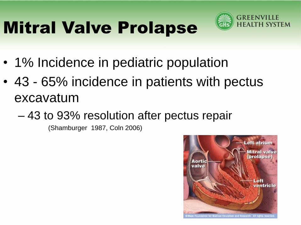
Mitral Valve Prolapse
• 1% Incidence in pediatric population
• 43 - 65% incidence in patients with pectus
excavatum
– 43 to 93% resolution after pectus repair (Shamburger 1987, Coln 2006)
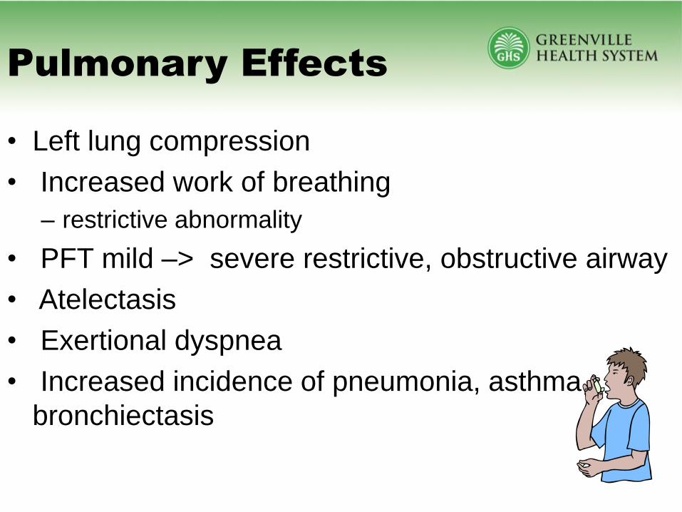
Pulmonary Effects
• Left lung compression
• Increased work of breathing
– restrictive abnormality
• PFT mild –> severe restrictive, obstructive airway
• Atelectasis
• Exertional dyspnea
• Increased incidence of pneumonia, asthma,
bronchiectasis
Pulmonary Effects
• After placement and subsequent removal of the
pectus bar, patients have significant
improvement in pulmonary function
• Children over age 11 have a greater degree of
improvement
(Goretsky 2005)
Psychosocial Effects
• 8 of 9 psychosocial indicators showed
statistically significant improvement, including:
– Body-image satisfaction
– Feelings of sadness, frustration, restlessness, and
isolation
– Experience of social ridicule
(Lawson 2003)
Workup Prior to
Surgical Referral
• Complete History and Physical Exam
• Radiographic studies
– PA/Lateral chest x-ray
• Other radiographic and functional studies are
patient specific and will be obtained AFTER the
surgical consult
History
• When did the deformity manifest – Infancy vs. older child
– Increase in depth often occurs during periods of rapid growth
– Patients with pectus excavatum from infancy will have fewer symptoms due to adaptation
• Type of physical activity and change
• History of allergies – Jewelry, metal
Physical Exam
• Height / weight percentiles
• Cardiac exam - displacement of PMI
• Type of Pectus
• Symmetry / costal flaring
• Scoliosis / joint laxity / stria
Criteria for Referral
• Age over 8
• Cardiopulmonary symptoms
• Significant increase in deformity
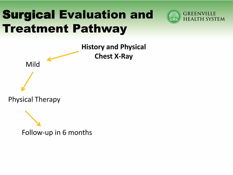
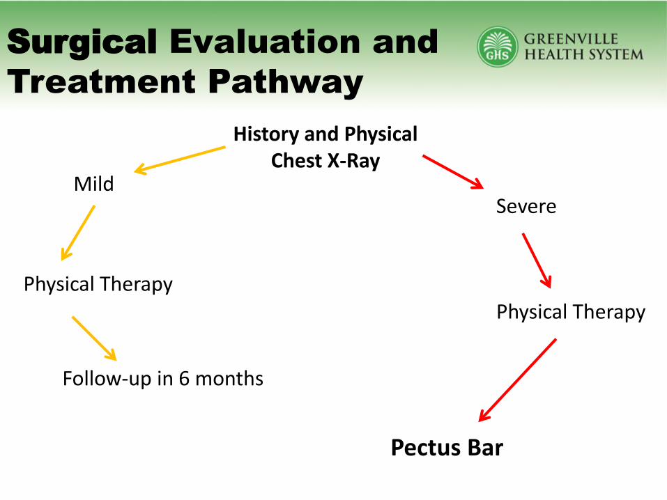
Surgical Evaluation and
Treatment Pathway
History and Physical Chest X-Ray
Mild
Physical Therapy
Follow-up in 6 months
Surgical Evaluation and
Treatment Pathway
History and Physical Chest X-Ray
Mild
Physical Therapy
Follow-up in 6 months
Severe
Physical Therapy
Pectus Bar
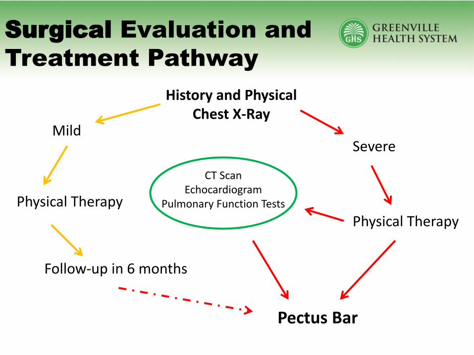
Surgical Evaluation and
Treatment Pathway
History and Physical Chest X-Ray
Mild
Physical Therapy
Follow-up in 6 months
Severe
Physical Therapy
CT Scan Echocardiogram
Pulmonary Function Tests
Pectus Bar
So… what is a severe defect?
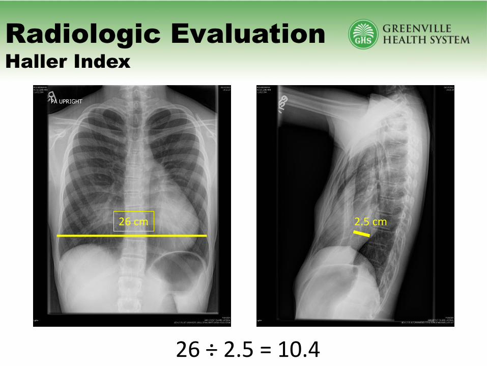
Radiologic Evaluation
Haller Index
26 cm 2.5 cm
26 ÷ 2.5 = 10.4
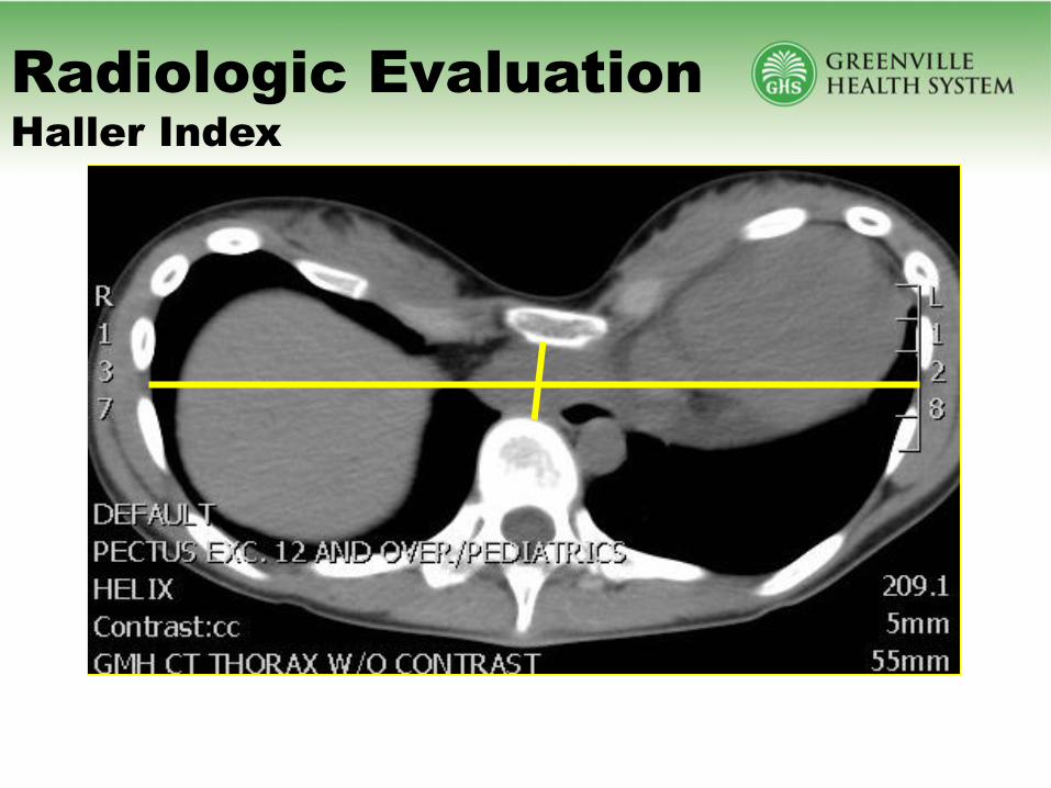
Radiologic Evaluation
Haller Index
Severe Pectus
Excavatum
• Haller index greater than 3.2
• Cardiac and pulmonary symptoms
– Exercise intolerance
– Chest pain with exertion
– Cardiac murmur
Goals of Physical
Therapy
• Strengthen chest wall musculature
• Improve chest wall flexibility
• Increase cardiovascular endurance
• Improve posture
• Deep breathing – chest expansion
Pectus Excavatum
• Effectiveness of Physical Therapy Program
• Decreased post-operative length of stay
• Earlier ambulation
• Transferred supine to sit independently earlier
• Improved post-operative pain scores compared
to those children who received epidural catheters
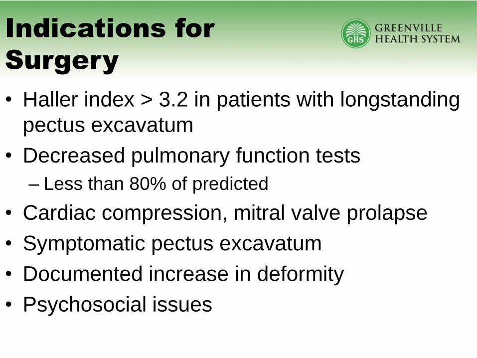
Indications for
Surgery
• Haller index > 3.2 in patients with longstanding
pectus excavatum
• Decreased pulmonary function tests
– Less than 80% of predicted
• Cardiac compression, mitral valve prolapse
• Symptomatic pectus excavatum
• Documented increase in deformity
• Psychosocial issues
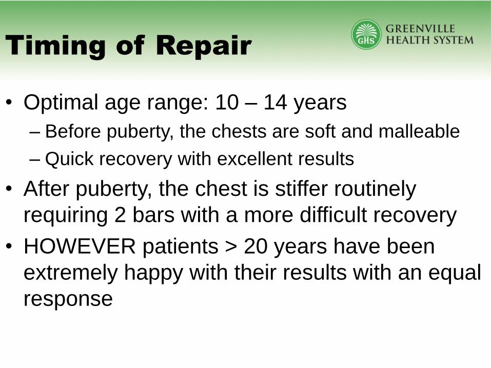
Timing of Repair
• Optimal age range: 10 – 14 years
– Before puberty, the chests are soft and malleable
– Quick recovery with excellent results
• After puberty, the chest is stiffer routinely
requiring 2 bars with a more difficult recovery
• HOWEVER patients > 20 years have been
extremely happy with their results with an equal
response
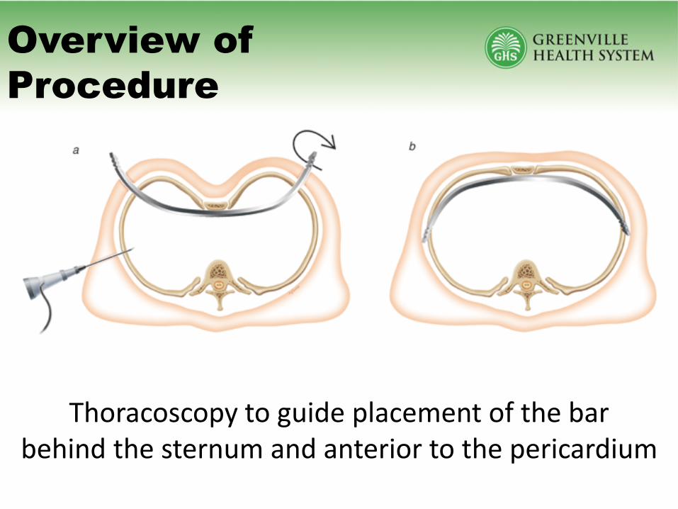
Overview of
Procedure
Thoracoscopy to guide placement of the bar behind the sternum and anterior to the pericardium
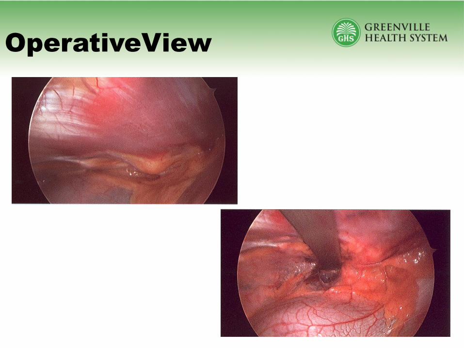
OperativeView
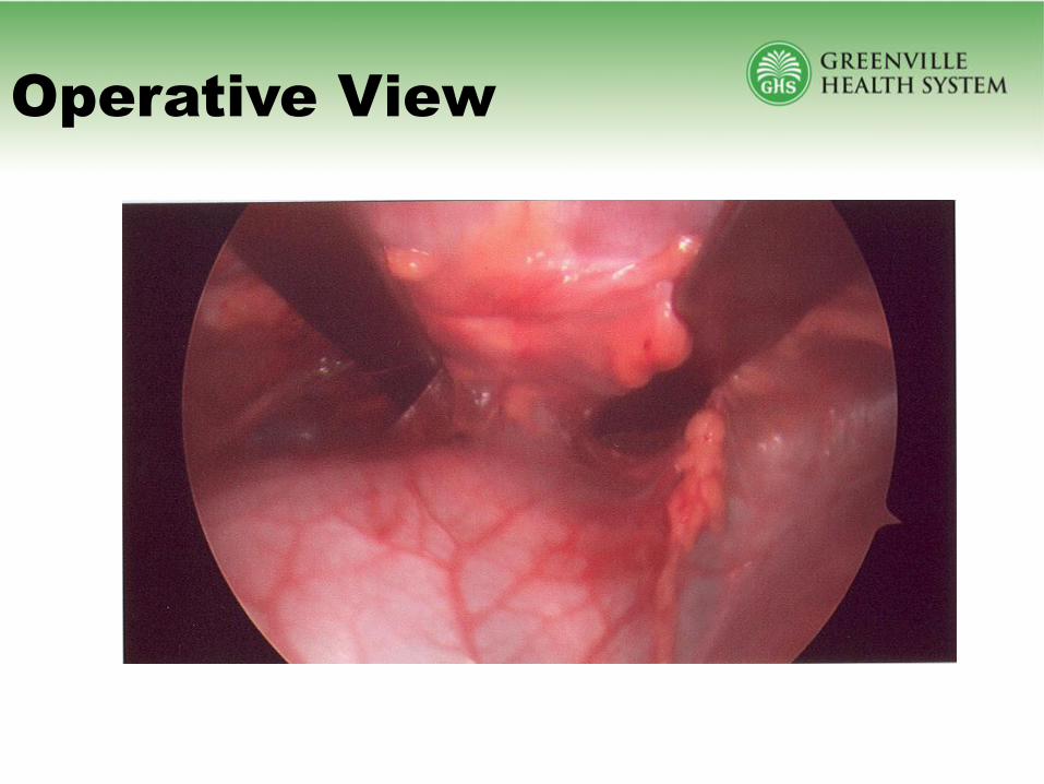
Operative View
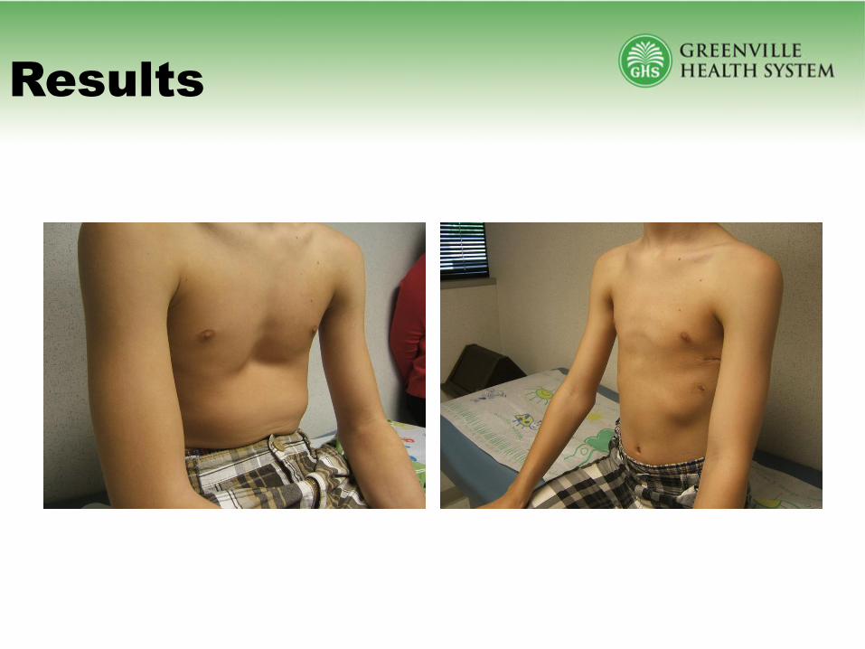
Results
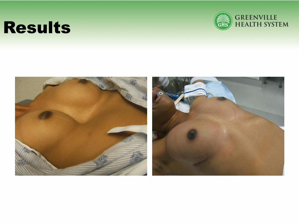
Results
Post-operative
Considerations
• Significant post-operative pain
– Lessened by pre-operative physical therapy
– Usually a 1-2 week convalescence
– Minimal pain at 6 weeks
• Plan operation for summer or winter break
• Restart physical therapy at 6 weeks
• Return to previous activities in 6 months
• Follow up at 6 month intervals
Long Term Results
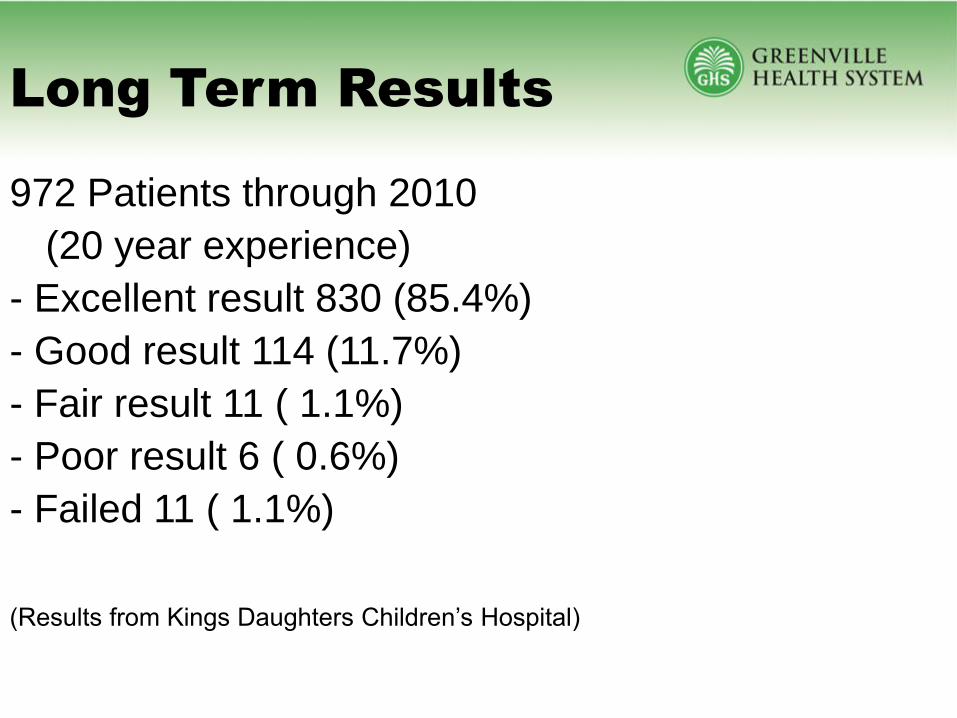
972 Patients through 2010
(20 year experience)
- Excellent result 830 (85.4%)
- Good result 114 (11.7%)
- Fair result 11 ( 1.1%)
- Poor result 6 ( 0.6%)
- Failed 11 ( 1.1%)
(Results from Kings Daughters Children’s Hospital)
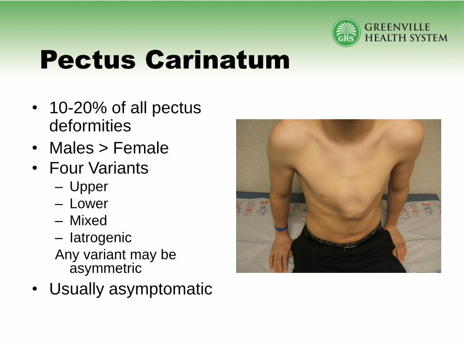
Pectus Carinatum
• 10-20% of all pectus deformities
• Males > Female
• Four Variants – Upper
– Lower
– Mixed
– Iatrogenic
Any variant may be asymmetric
• Usually asymptomatic
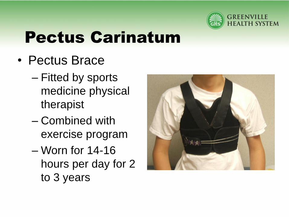
Pectus Carinatum
• Pectus Brace
– Fitted by sports
medicine physical
therapist
– Combined with
exercise program
– Worn for 14-16
hours per day for 2
to 3 years
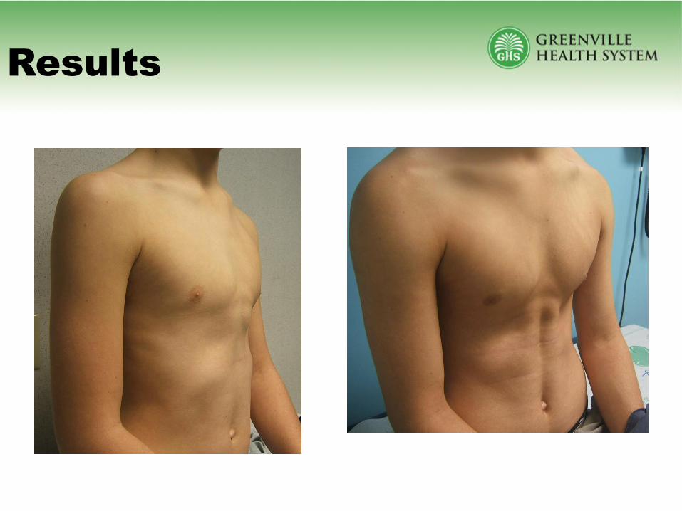
Results
Pectus Carinatum
• Pectus brace effectiveness – 75% Compliance
– 85% Good or Excellent result (Stephenson 2008)
• Pectus brace complications – Pain or discomfort
– Fit problems
– Skin breakdown
– Overcorrection
– Shortness of breath
Pectus Carinatum
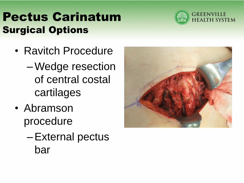
Surgical Options
• Ravitch Procedure
– Wedge resection
of central costal
cartilages
• Abramson
procedure
– External pectus
bar

Introduction
• Vascular Malformations
• Common Skin Lesions
• The Coccygeal Dimple
• Breast masses in adolescents
• The latest in Inguinal Hernia Repair
• Intussusception
• The Spectrum of Malrotation
• Pyloric Stenosis
• Pectus Deformities

QUESTIONS?