Embed Size (px)
Citation preview

Cutaneous Lymphoid Hyperplasia, Cutaneous T-Cell Lymphoma, Other Malignant Lymphomas, and Allied Diseases
Rick Lin, DO, MPHMarch 18, 2003
Cutaneous Lymphoid Hyperplasia
Collection of lymphocytes with other inflammatory cells on the skin
Maybe monoclonal or mixed with both T or B cells
Caused by unknown stimuliMedications, infections, insect bites
Cutaneous Lymphoid Hyperplasia
AKA Pseudolymphoma May progress to lymphoma Immunosuppression may aggravate the
infiltrate and may regress with immunosuppression is removed
Cutaneous B-Cell Lymphoid Hyperplasias Knowns as Speigler-Fendt sarcoid Caused by Borrelia, infections, herpes zosters
scars, tatoo, drugs Appears as discrete firm of doughy cutaneous
papules or nodules Most lesions are asymptomatic, treatment not
required If caused by medication, medication should be
removed
Cutaneous T-Cell Lymphoid Hyperplasias Maybe idiopathic Aka actinic reticuloid and chronic actinic
dermatitis Patient resembles mycosis fungoides Histologically, dermal infiltrate that is
band-like with no grenz zone.
Cutaneous lymphoid hyperplasia
Pseudolymphoma has to be distinguished from cutaneous lymphomas by the combination of clinicopathological correlation, histochemical studies, and, in selected cases, gene rearrangement studies
T cell lymhoma can be usually distinguished from T cell pseudolymphoma by the presence of prominent epidermotropism, large and atypical lymphocytes, and T cell gene rearrangements up to 90%
Cutaneous lymphoid hyperplasia
The lack of acanthosis, "bottom-heavy" infiltrates, light-chain expression of monotypical B-cells, and immunoglobulin gene rearrangements (75%) provide strong evidence for the diagnosis of B-cell lymphoma
A careful monitoring of these patients for the development of lymphoma is necessary
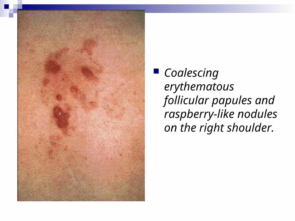
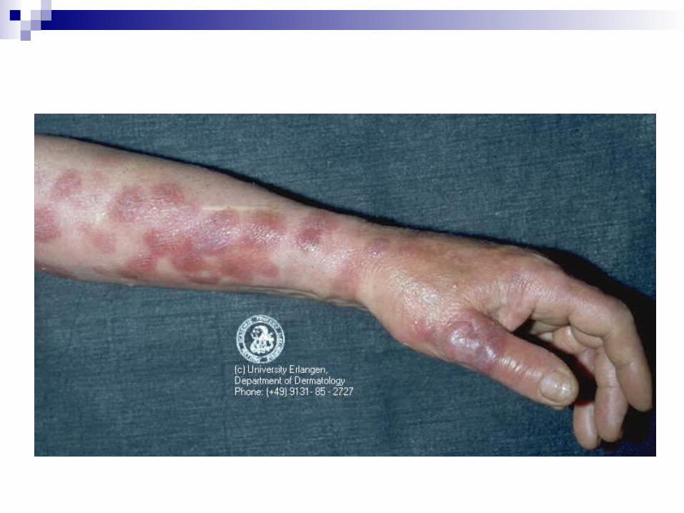
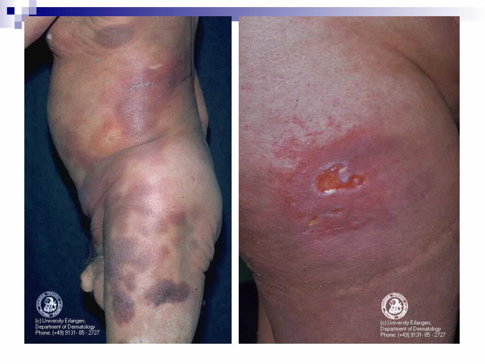
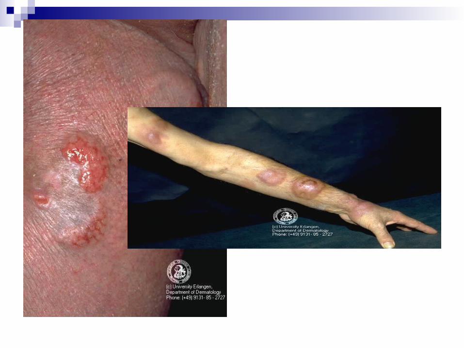
Coalescing erythematous follicular papules and raspberry-like nodules on the right shoulder.
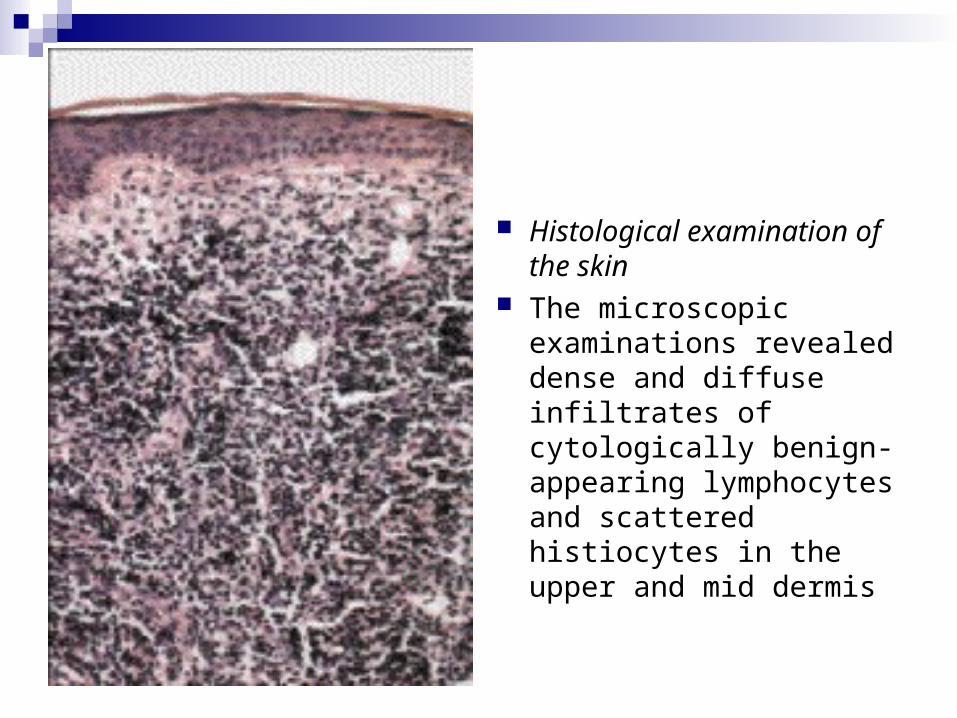
Histological examination of the skin
The microscopic examinations revealed dense and diffuse infiltrates of cytologically benign-appearing lymphocytes and scattered histiocytes in the upper and mid dermis
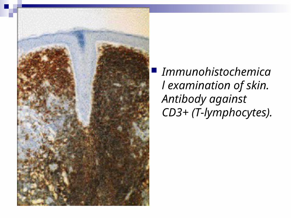
Immunohistochemical examination of skin. Antibody against CD3+ (T-lymphocytes).
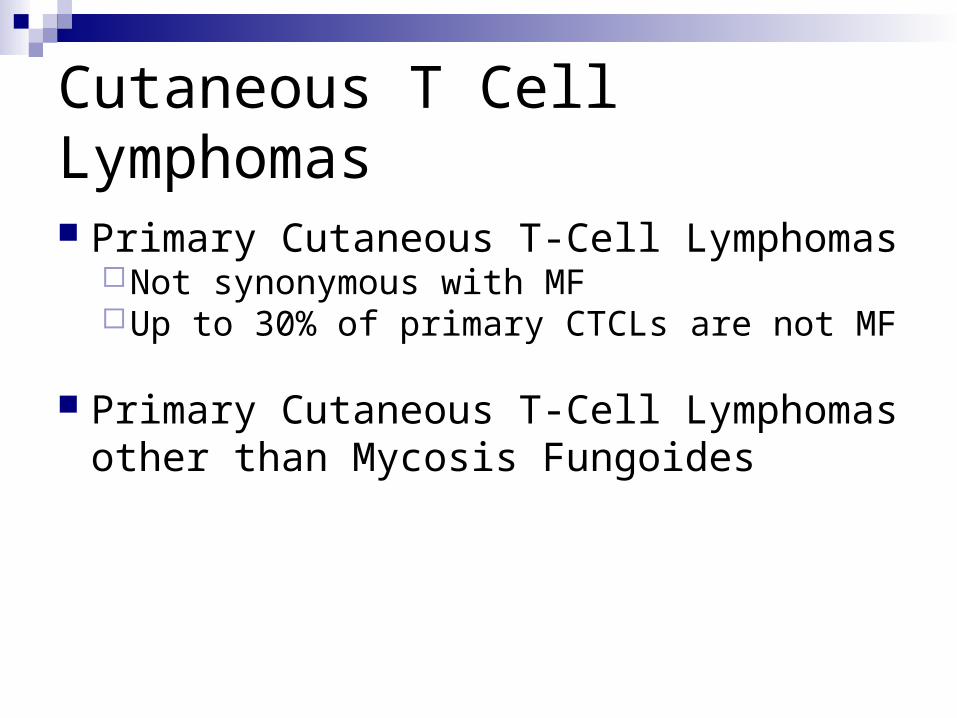
Cutaneous T Cell Lymphomas
Primary Cutaneous T-Cell LymphomasNot synonymous with MFUp to 30% of primary CTCLs are not MF
Primary Cutaneous T-Cell Lymphomas other than Mycosis Fungoides
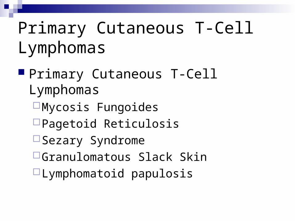
Primary Cutaneous T-Cell Lymphomas Primary Cutaneous T-Cell Lymphomas
Mycosis FungoidesPagetoid ReticulosisSezary SyndromeGranulomatous Slack SkinLymphomatoid papulosis
Mycosis Fungoides
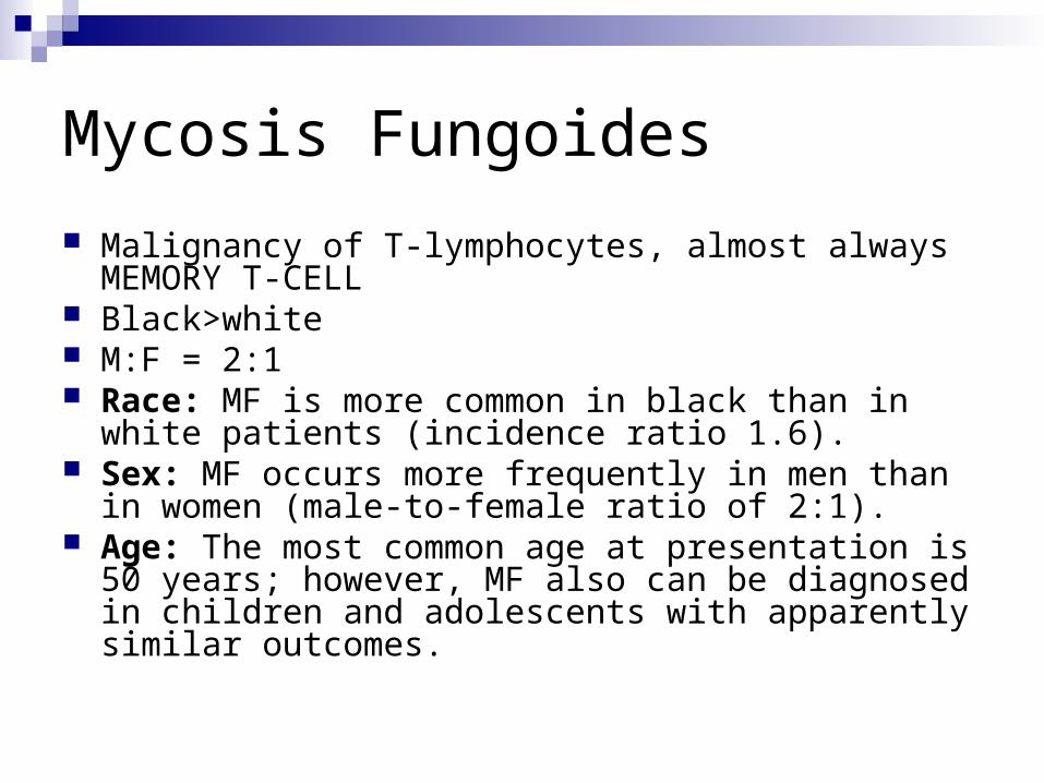
Malignancy of T-lymphocytes, almost always MEMORY T-CELL
Black>white M:F = 2:1 Race: MF is more common in black than in white
patients (incidence ratio 1.6). Sex: MF occurs more frequently in men than in women
(male-to-female ratio of 2:1). Age: The most common age at presentation is 50 years;
however, MF also can be diagnosed in children and adolescents with apparently similar outcomes.
Mycosis Fungoides
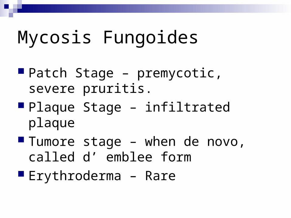
Patch Stage – premycotic, severe pruritis. Plaque Stage – infiltrated plaque Tumore stage – when de novo, called d’
emblee form Erythroderma – Rare
MF Staging
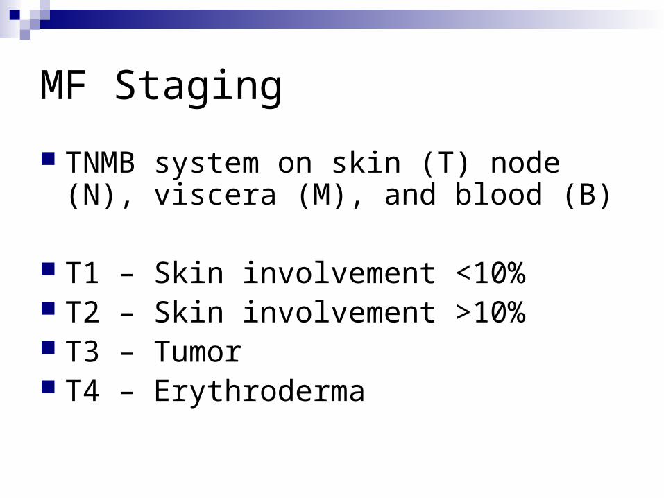
TNMB system on skin (T) node (N), viscera (M), and blood (B)
T1 – Skin involvement <10% T2 – Skin involvement >10% T3 – Tumor T4 – Erythroderma
MF Staging
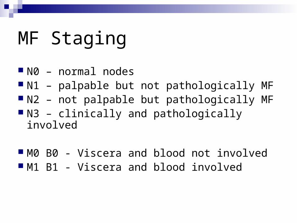
N0 – normal nodes N1 – palpable but not pathologically MF N2 – not palpable but pathologically MF N3 – clinically and pathologically involved
M0 B0 - Viscera and blood not involved M1 B1 - Viscera and blood involved
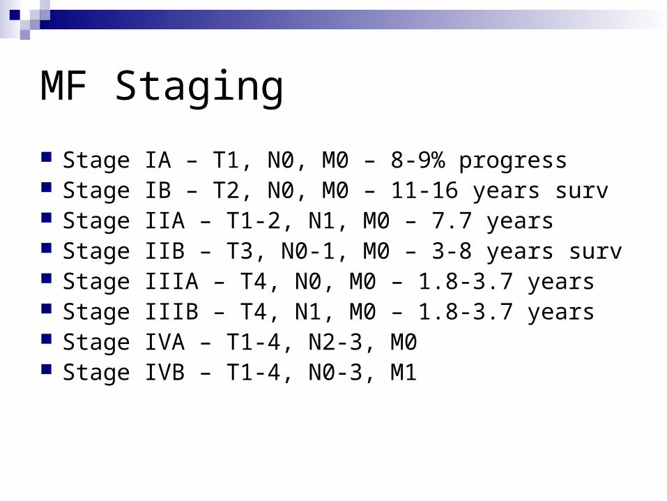
MF Staging
Stage IA – T1, N0, M0 – 8-9% progress Stage IB – T2, N0, M0 – 11-16 years surv Stage IIA – T1-2, N1, M0 – 7.7 years Stage IIB – T3, N0-1, M0 – 3-8 years surv Stage IIIA – T4, N0, M0 – 1.8-3.7 years Stage IIIB – T4, N1, M0 – 1.8-3.7 years Stage IVA – T1-4, N2-3, M0 Stage IVB – T1-4, N0-3, M1
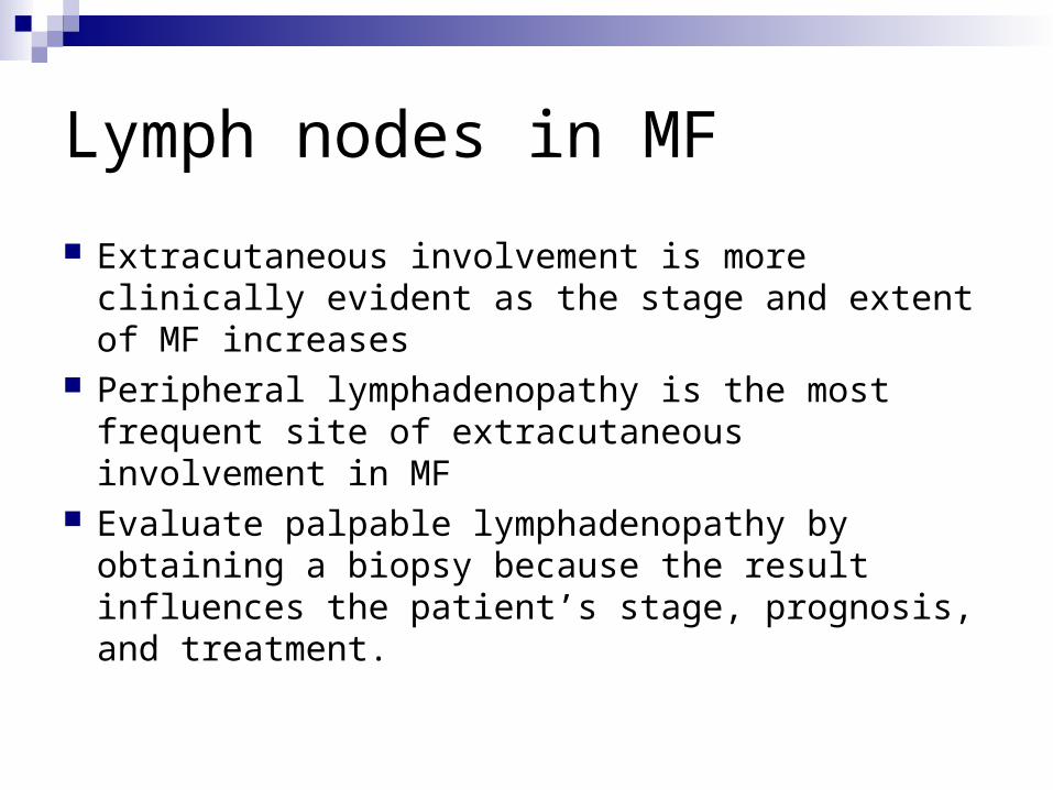
Lymph nodes in MF
Extracutaneous involvement is more clinically evident as the stage and extent of MF increases
Peripheral lymphadenopathy is the most frequent site of extracutaneous involvement in MF
Evaluate palpable lymphadenopathy by obtaining a biopsy because the result influences the patient’s stage, prognosis, and treatment.
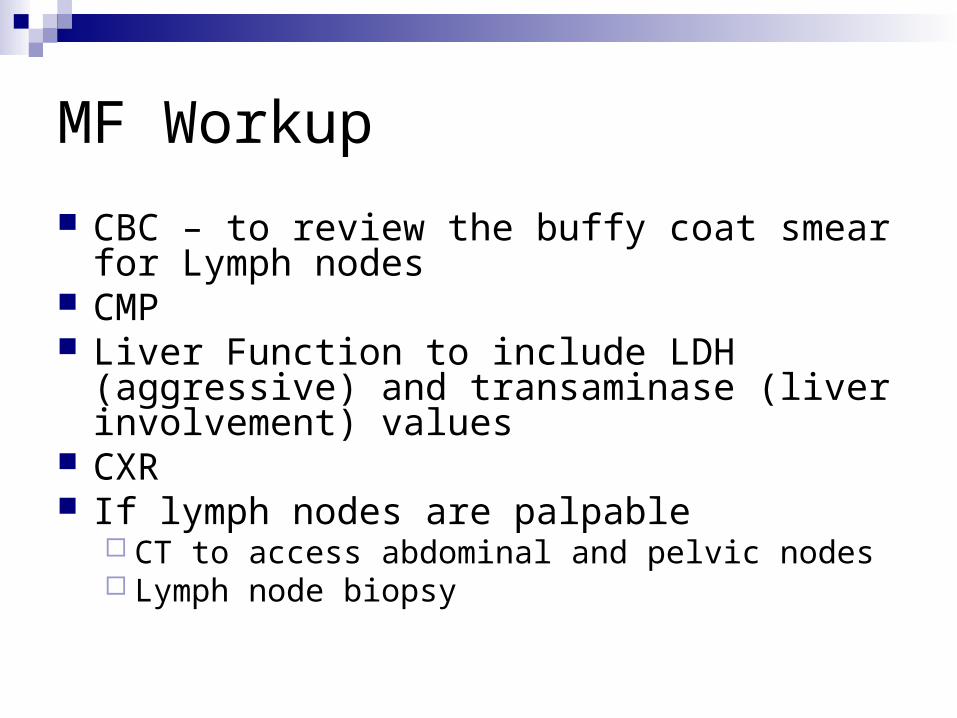
MF Workup
CBC – to review the buffy coat smear for Lymph nodes
CMP Liver Function to include LDH (aggressive) and
transaminase (liver involvement) values CXR If lymph nodes are palpable
CT to access abdominal and pelvic nodes Lymph node biopsy
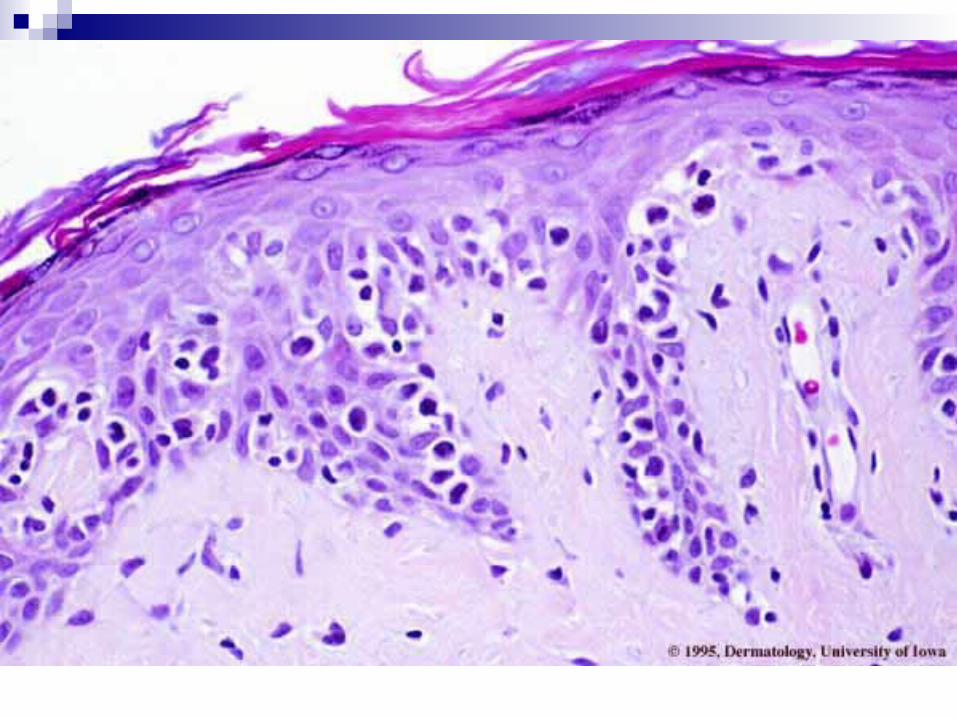
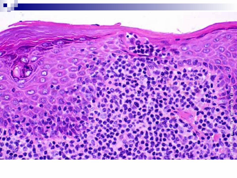
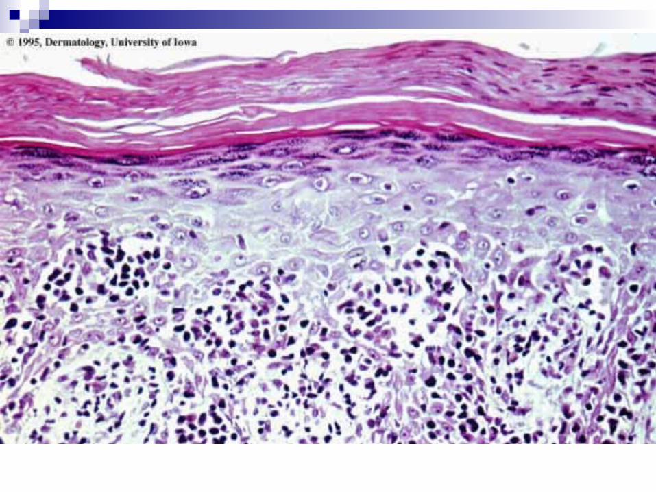
Histologic Findings
The criteria for diagnosis include the following: A bandlike upper dermal infiltrate of lymphocytes and
other inflammatory cells, with no grenz zone, is present. Epidermotropism of mononuclear cells occurs. When a clear halo surrounds an intraepidermal
mononuclear cell singly or in clumps, this is called a Pautrier microabscess. Its presence is suggestive of MF, but it is not necessary for diagnosis.
Little spongiosis of the epidermis is found. Lymphocytes have nuclei that are hyperchromatic and
convoluted or cerebriform.
Pagetoid Reticulosis
Localized epidermotropic reticulosis Woringer-Kolopp disease Acral mycoses fungoides Mycosis fungoides palmaris et plantaris

Pagetoid Reticulosis
0.6% of all MF cases Woringer-Kolopp variant: solitary lesion Ketron-Goodman variant: multiple lesions Long durantion without progression to frank
lymphoma is the clincal hallmark of Woringer-Kolopp
Local excision and radiation therapy maybe curative.
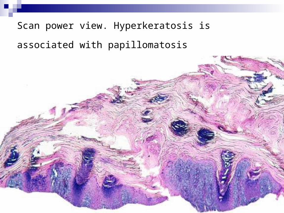
Scan power view. Hyperkeratosis is associated with
papillomatosis
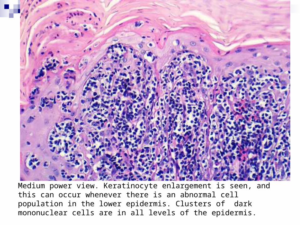
Medium power view. Keratinocyte enlargement is seen, and this can occur whenever there is an abnormal cell population in the lower epidermis. Clusters of dark mononuclear cells are in all levels of the epidermis.
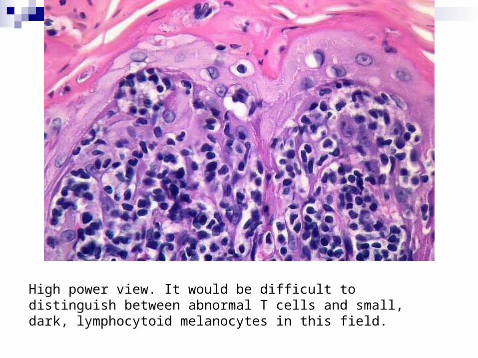
High power view. It would be difficult to distinguish between abnormal T cells and small, dark, lymphocytoid melanocytes in this field.
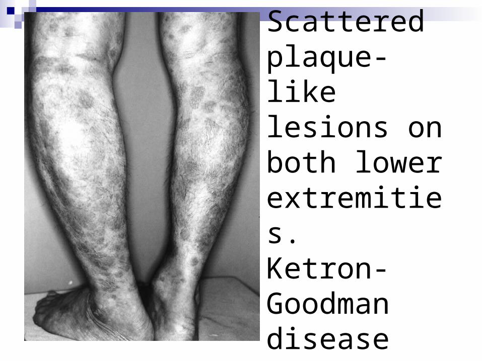
Scattered plaque-like lesions on both lower extremities.Ketron-Goodman disease
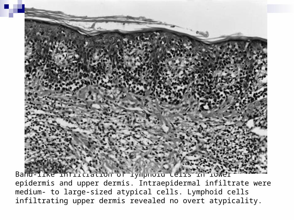
Band-like infiltration of lymphoid cells in lower epidermis and upper dermis. Intraepidermal infiltrate were medium- to large-sized atypical cells. Lymphoid cells infiltrating upper dermis revealed no overt atypicality.
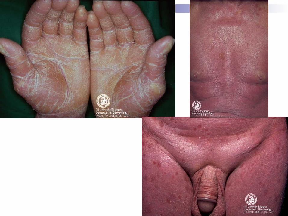
Sezary Syndrome
Leukemic phase of mycosis fungoides Generalized erythroderma, superficial
lymphadenopathy, atypical cells in circulating blood
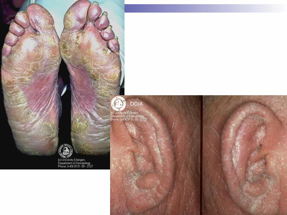
Erythroderma from onset with leonine facies, eyelid edema, ectropion, alopecia, palm and sole hyperkeratosis
Pruritis, burning, chill and profuse sweating
Prognosis
Difficult to treat Median survival is 3 years Low dose methotrexate has reasonable
response rate of 50% Photophoresis Retinoid, interferon alfa, lowdose
chlorabucil, prednisone, systemic chemo
Granulomatous Slack Skin
Rare variant of CTCL Middle-age adult and gradually progress Erythematous atrophic pendulous
redundant plaque Multinucleated giant cells replaces fat
lobules histologically.
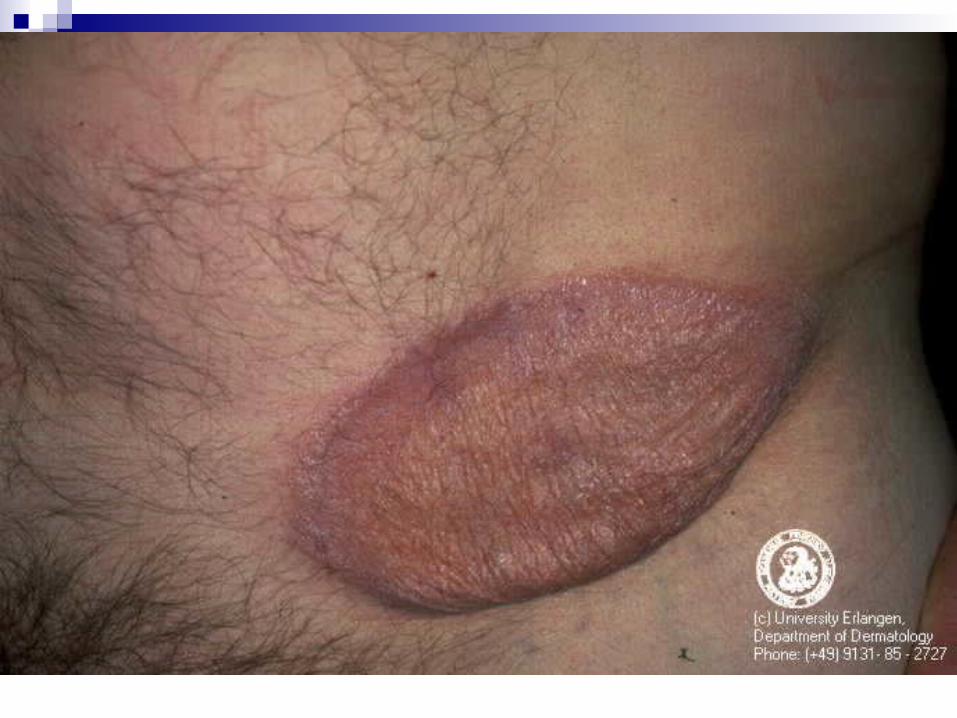
Lymphomatoid Papulosis
LyP has a chronic indolent course in most patients; estimates indicate that as many as 10-20% of LyP
patients have a history of associated malignant lymphoma (ALCL, HD, or mycosis fungoides [MF]) prior to, concurrent with, or subsequent to the diagnosis of LyP.
Race: Black persons may be less affected than other racial groups.
Sex: No consistent sex predominance is found in studies.
Age: LyP may develop at any age, usually in the third to fourth decade

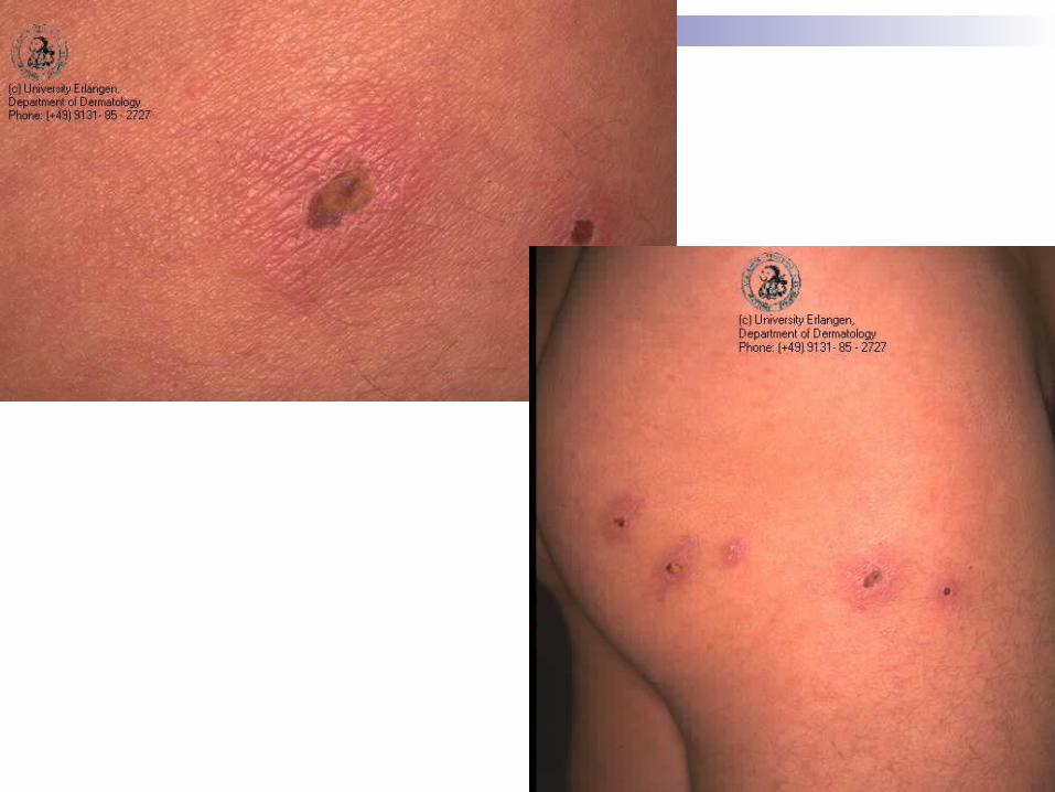
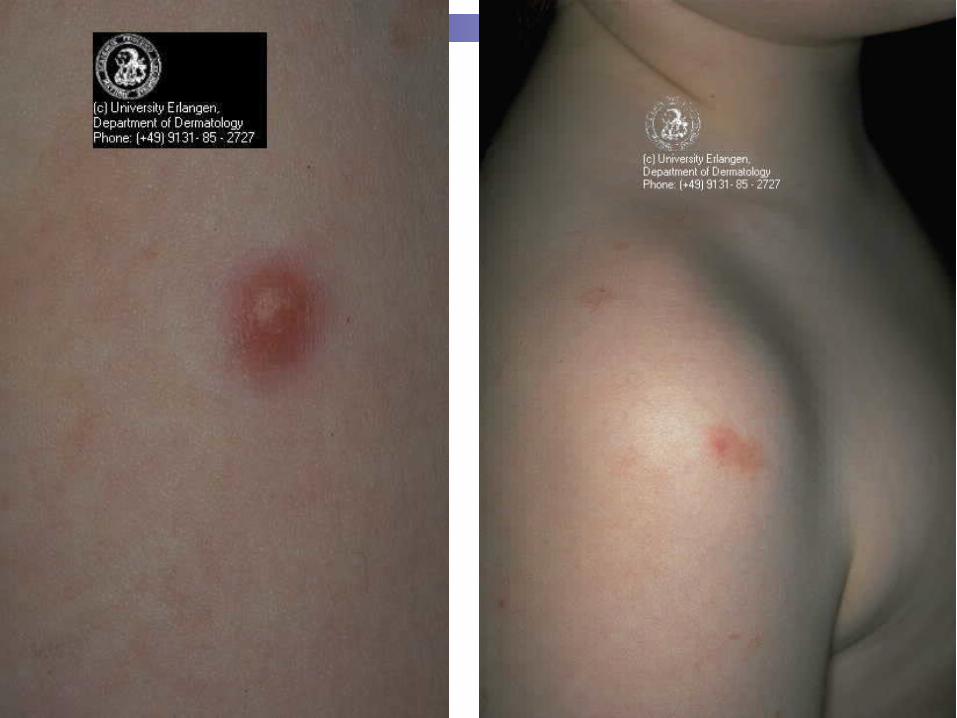
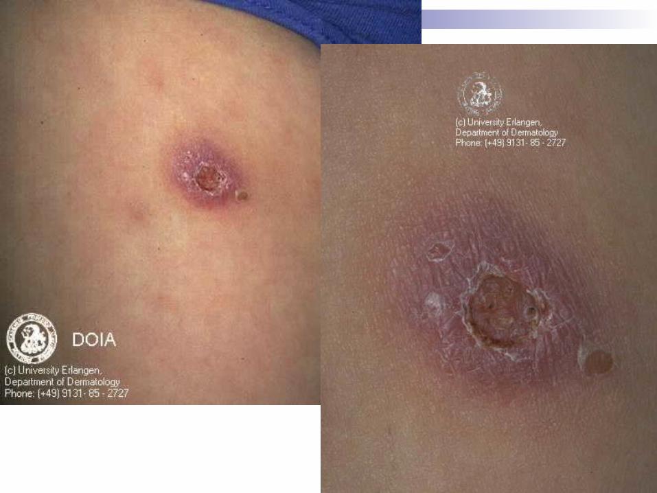
Presentation
Primary lesion: Each erythematous papule evolves into a red-brown, often hemorrhagic, papulovesicular or papulopustular lesion over days to weeks.
Some lesions develop a necrotic eschar before healing spontaneously. Occasionally, noduloulcerative lesions may be present
Each papule heals within 2-8 weeks, leaving a hypopigmented or hyperpigmented depressed oval varioliform scar.
Large nodules and plaques may take months to resolve. Distribution: Characteristically, lesions appear on the trunk
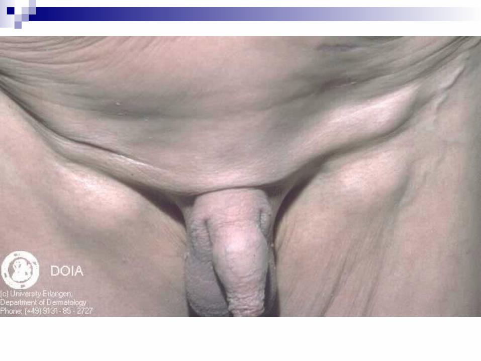
and extremities, although the palms and/or soles, face, scalp, and anogenital area also may be involved.
Lymphomatoid Papulosis
Type A: Characterized by large (25-40 mm) CD30+ atypical cells with polymorphic convoluted nuclei and a minimum of 1 prominent nucleolus. These large cells resemble Reed-Sternberg cells when binucleate. Type A LyP is the most common histologic variant.
Type B: Characterized by smaller (8-15 mm) atypical cells with hyperchromatic cerebriform nuclei resembling the atypical lymphocytes in MF. CD30+ large cells are rare, but epidermotropism is more common in this variant.
Lymphomatoid Papulosis
Type C (diffuse large cell type): Characterized by sheets of CD30+ anaplastic large cells
Treatment of LyP
mid-to-high potency topical steroids to hasten resolution. Low-dose weekly methotrexate is a safe and effective
treatment for suppressing LyP; however, the disease recurs within 1-2 weeks after ending medication.
Oral psoralen plus UV-A phototherapy (PUVA) also effectively treats and suppresses the disease.
carmustine, topical nitrogen mustard, intralesional interferon, low-dose cyclophosphamide, chlorambucil, and dapsone help disease suppression.
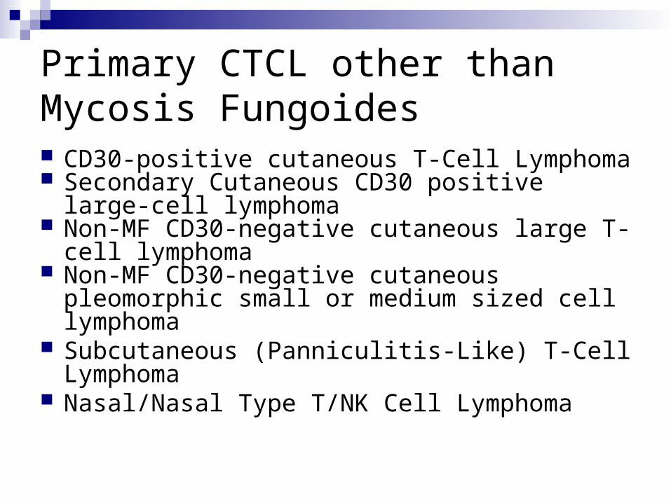
Primary CTCL other than Mycosis Fungoides CD30-positive cutaneous T-Cell Lymphoma Secondary Cutaneous CD30 positive large-cell
lymphoma Non-MF CD30-negative cutaneous large T-cell
lymphoma Non-MF CD30-negative cutaneous pleomorphic
small or medium sized cell lymphoma Subcutaneous (Panniculitis-Like) T-Cell
Lymphoma Nasal/Nasal Type T/NK Cell Lymphoma
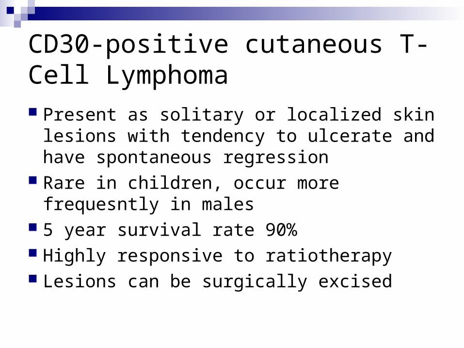
CD30-positive cutaneous T-Cell Lymphoma Present as solitary or localized skin lesions
with tendency to ulcerate and have spontaneous regression
Rare in children, occur more frequesntly in males
5 year survival rate 90% Highly responsive to ratiotherapy Lesions can be surgically excised
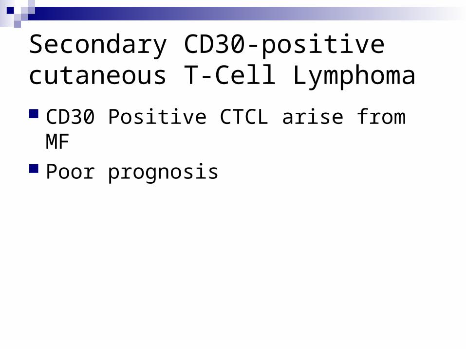
Secondary CD30-positive cutaneous T-Cell Lymphoma CD30 Positive CTCL arise from MF Poor prognosis
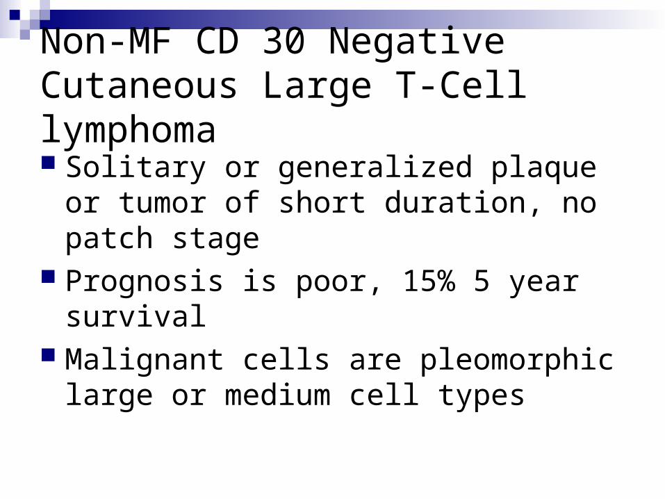
Non-MF CD 30 Negative Cutaneous Large T-Cell lymphoma Solitary or generalized plaque or tumor of
short duration, no patch stage Prognosis is poor, 15% 5 year survival Malignant cells are pleomorphic large or
medium cell types
Non-MF CD 30 Negative Cutaneous Small or Medium Size Cell T-Cell lymphoma
Differentiate from large-cell type by having less than 30% large pleomorphic celll.
Similar to large type clinically Prognosis is better than large cell type.
50% 5 year survival. Radiation tx, interferon alfa, or
cyclophosphamide are effective
Subcutaneous (Panniculitis-Like) T-Cell Lymphoma Clinically presents with subcutaneous
nodules Usually on lower extremities Frequently diagnosed as having erythema
nodosum or other forms of panniculitis Poor prognosis
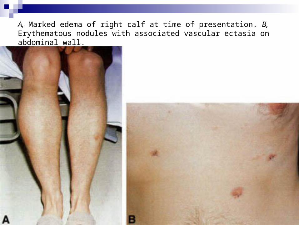
A, Marked edema of right calf at time of presentation. B, Erythematous nodules with associated vascular ectasia on abdominal wall.
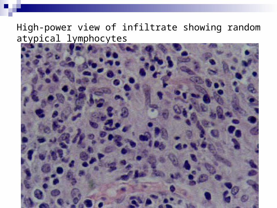
High-power view of infiltrate showing random atypical lymphocytes
Cutaneous B-Cell Lymphoma
Primary Cutaneous Follicular Center Cell Lymphoma
Primary Cutaneous Immunocytoma Intravascluar Large B-Cell Lymphoma Plasmacytoma (Multiple Myeoloma)
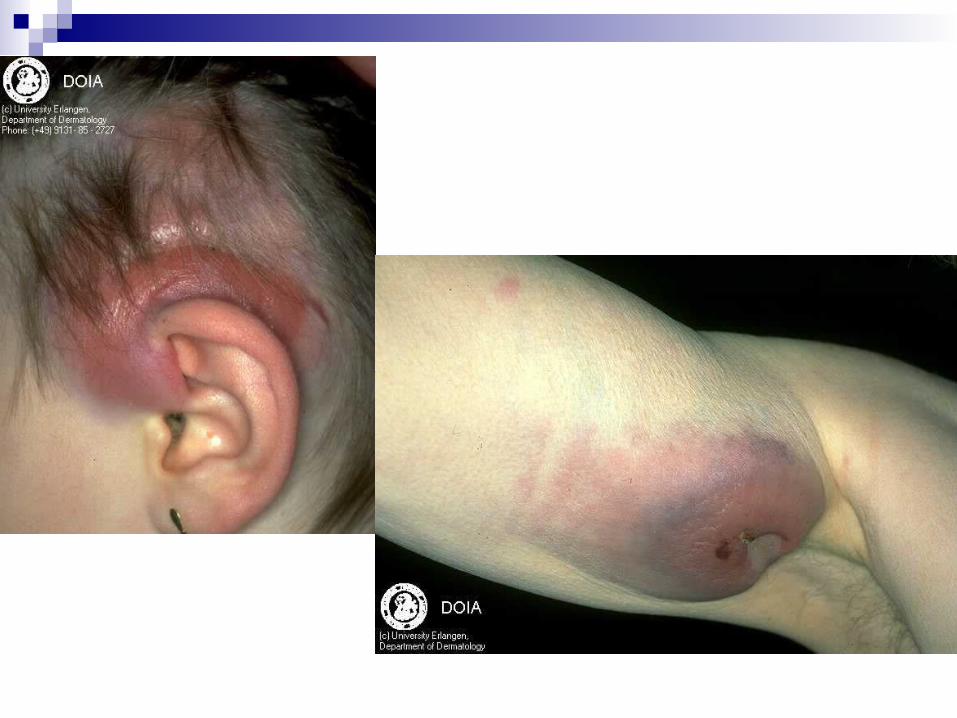
Primary Cutaneous Follicular Center Cell Lymphoma AKA B-Cell lymphoma of follicular center cell
origin AKA Reticulohistiocytoma of the dorsum Multiple papules and nodules in one anatomic
region. 2/3 of case on the trunk, 1/5 on the head and
neck 15% on the leg
Primary Cutaneous Follicular Center Cell Lymphoma M:F = 2:1 Prognosis: Head and neck 100% 5 yr surv.
Leg lesion of people over 70, 50% 5 yr surv.
Stains with B-Cell marker CD 20, monotypic for immunoglobulin production of kappa or lambda chain, not both
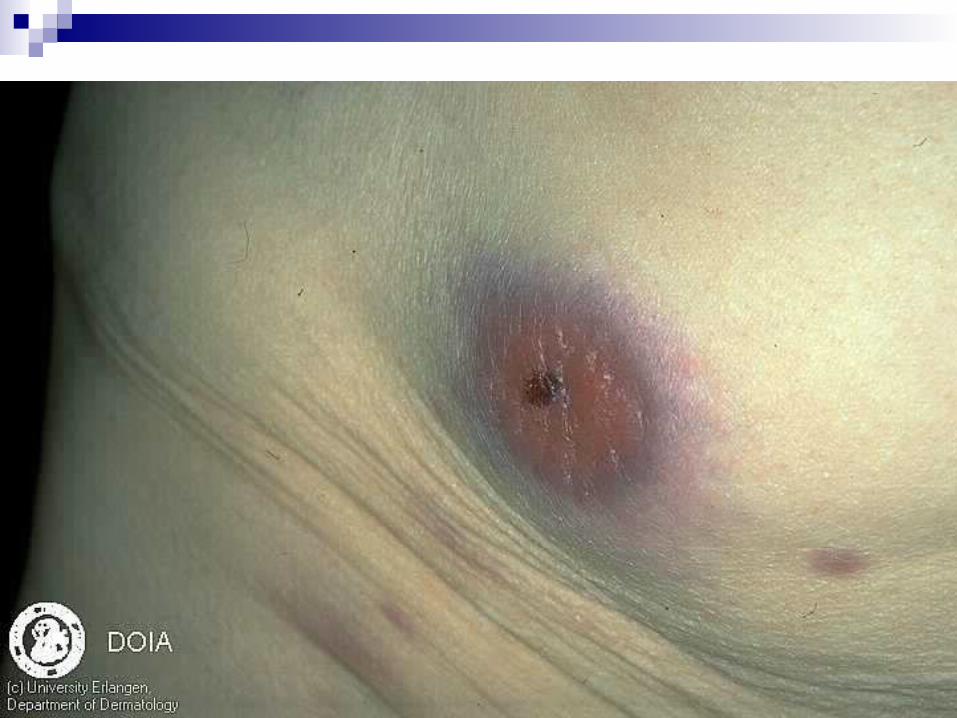
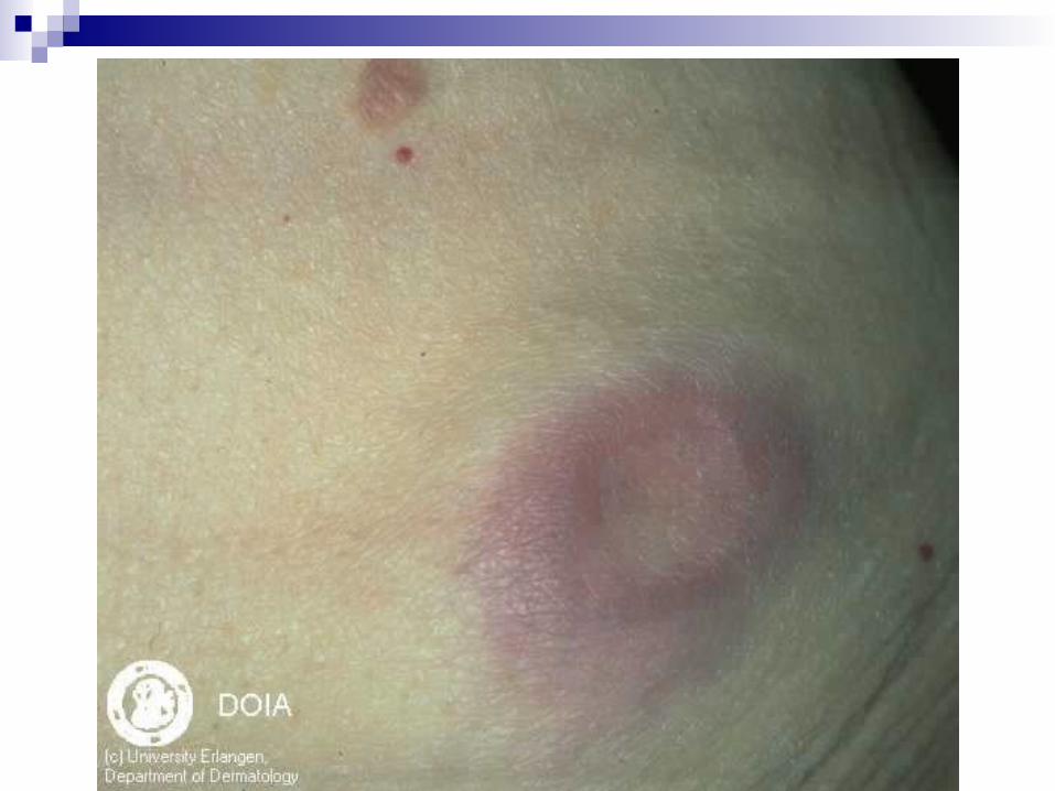
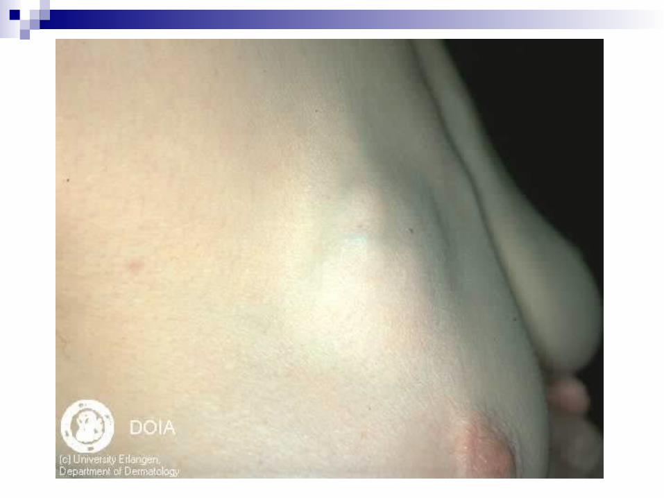
Primary Cutaneous Immunocytoma
AKA Marginal Zone B-Cell Lymphoma AKA MALT Type Lymphoma SubQ nodule or tumor primaroily of the
extremities or trunk CD79, CD 20, and bcl-2 positive 5 years survival near 100%
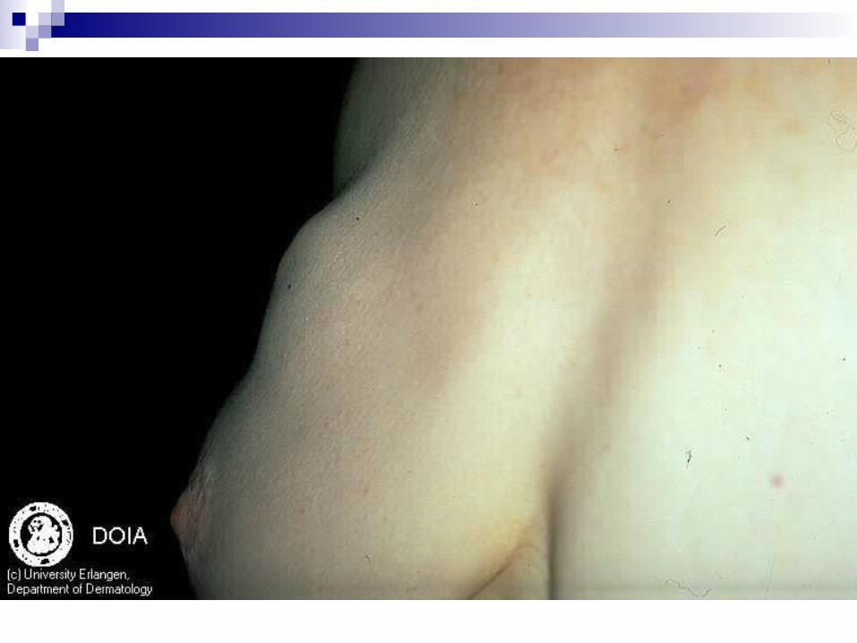
Plasmacytoma Multiple Myeloma Spectrum of solitary plasmacytoma to
multiple plasmacytoma to multiple myeloma.
Neoplasm of B lymphcytes Multiple myeloma is most common
characterized by lytic bone lesions and infiltration of bone marry by plasma cells
Plasmacytoma Multiple Myeloma Cutaneous plasmacytomas seen most
commonly a secondary lesion in the setting of primary myeloma. Prognosis is poor.
When bone film and bone biopsy are normal but cutaneous lesions present, these are primary cutaneous plasmacytoma. Excellent prognosis.
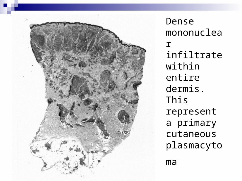
Dense mononuclear infiltrate withinentire dermis.This represent a primary cutaneous
plasmacytoma

Neoplastic plasma cells, some with atypical
features, are visible J Am Acad Dermatol 2000;43:962-5.)
Plasmacytoma Multiple Myeloma Numerous nonspecific skin lesions occurs
in patient with multiple myeloma.AmyloidosisCutaneous vasculitisalopeciaRaynaud’sPyoderma gangrenosum

Hodgkin’s Disease
Vast majority of cutaneous Hodgkin’s disease report are type A lymphomatoid papulosis with Reed-Sternberg
Difficult to prove cutaneous disease
Malignant Histiocytosis
Rare, fatal occur in men in second to fourth decade of life
Solitary lesion or wide spread papule occur in 10% of cases
Onset of acute fever, hepatosplenomegly, and painful lymphadenopathy
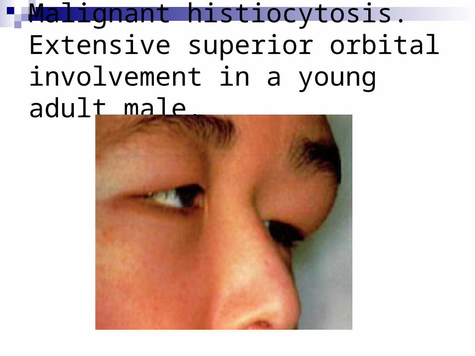
Malignant histiocytosis. Extensive superior orbital involvement in a young adult male.
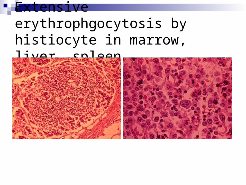
Extensive erythrophgocytosis by histiocyte in marrow, liver, spleen
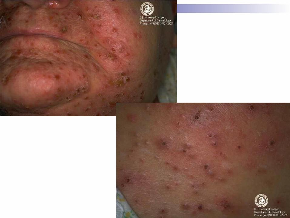
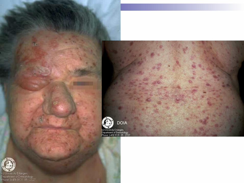
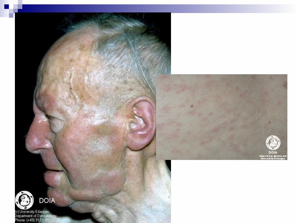
Leukemia Cutis
30% of Leukemia patient will have leukemia cutis
Vast majority of derm manifestation are from AML
Morphology: 60% multiple papules and nodules, 26% of infiltrated plaques
Subtypes and variants: Granulocytic Sarcoma Hairy-cell Leukemia Nonspecific Condition associated with Leukemia
Cutaneous Myelofibrosis
Overproduction and premature death of atypical megakaryocytes in bone marrow
Inrease in platelet-derived growth factor Extramedullary hematopoesis is the hallmark Blast cells escape marrow and enter circulation
and form tumor Cutaneous EMH reported in 20 cases
Hypereosinophilic Syndrome
Icrease Eos with more than 1500 eos per cubit millimeter for 6 month or more.
Cardiac disease most frequent complication
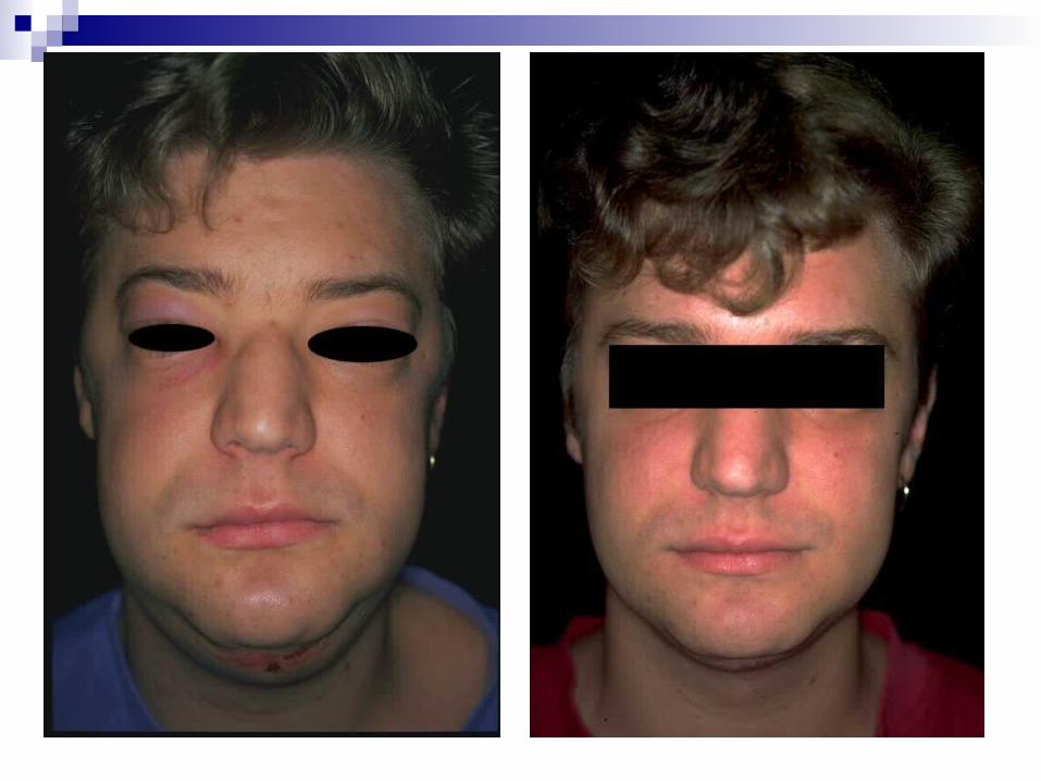
90% patient are men between 20 to 50 Angioedema and urticaria lesions most
common. Sometimes papules.
Angioimmunoblastic T-Cell Lymphoma AKA Angioimmunoblastic T-Cell
Lymphoma Uncommon, affect middle age and elderly Unspecific skin finding (pruritis, skin rash) Unspecific histology finding (patchy
perivascular dermal infiltrate) Lymph node biopsy required for diagnosis

Polycythemia Vera
Increase hermatocrite to 55% to 80% Associated with aquagenic pruritis in 50%
of the patients. Elevation of blood and skin histamine Tx – control of pruritis by antihistamin or
PUVA. Referral to HemOnc