Embed Size (px)
Citation preview
Cytopathology of Pediatric Malignancies
Where Are We Today With Fine-Needle Aspiration Biopsies in Pediatric Oncology?
Sara E. Monaco, MD, Lisa A. Teot, MD
Pediatric malignancies are uncommon and many have overlapping morphologic features, which together present diagnos-
tic challenges. Cytopathology is recognized as an accurate and cost-effective modality for the diagnosis of pediatric
malignancies in resource-limited countries, but is underused for this purpose in the United States. This review focuses on
the cytopathology of pediatric malignancies with the goal of demystifying cytologic diagnoses of these entities. Differen-
ces between malignancies in young patients and adults are discussed, and key epidemiological features of childhood
malignancies are highlighted. In addition, the use of cytopathology in different geographical settings is contrasted to illus-
trate the impact of variable usage on the incidence of malignancy and the types of tumors observed in cytologic speci-
mens. A review of the pattern-based approach to differential diagnosis is also incorporated, including the
cytomorphologic features and ancillary studies that help to distinguish between various malignancies within each pattern.
Cancer (Cancer Cytopathol) 2014;122:322-36. VC 2014 American Cancer Society.
KEY WORDS: children; cytopathology; fine-needle aspiration; pediatric; malignancy; tumor; young.
INTRODUCTION
Pediatric malignancies often present diagnostic challenges, particularly in small biopsies and cytologic speci-
mens. This is partly attributed to the overall rarity of these tumors, the different risk factors and associations,
and the different spectrum of entities in comparison with tumors arising in adults, and is compounded by mor-
phologic similarities between various tumors.1 This challenge is accentuated on a global level, because 86% of
the world’s pediatric population lives in developing countries and this percentage is expected to rise over time.2
In developing countries, pediatric malignancies are disproportionately represented and comprise up to 2% of all
cancers, in contrast to Europe and the United States in which they represent approximately 0.5% of all cancers.2
Furthermore, although infectious diseases are far more prevalent than cancer in developing countries, the mor-
tality rate associated with cancer is greater, largely due to delayed diagnosis, comorbidities, and a lack of access
to modern treatments in resource-limited settings. Fine-needle aspiration biopsy (FNAB) is routinely used for
the evaluation of suspected pediatric malignancies in resource-limited settings and has proven to be an accurate
diagnostic tool.3,4 In contrast, physicians in the United States have been slow to embrace this modality, despite
its being less costly and less invasive than either core needle or open biopsy. The objective of the current review
is to provide a framework for approaching the cytologic diagnosis of pediatric malignancies and to raise aware-
ness and acceptance of the value of this modality in resource-rich settings.
Corresponding author: Lisa A. Teot, MD, Division of Cytopathology, Department of Pathology, Boston Children’s Hospital, Bader 110, 300 Long-
wood Ave, Boston, MA 02115; Fax: (617) 730-0207; [email protected]
Division of Cytopathology, Department of Pathology, Boston Children’s Hospital, Boston, Massachusetts
Childhood malignancies are infrequent and differ from adult malignancies clinically and pathologically. This review discusses childhood malignan-
cies, focusing on their frequency in different age groups, representation in cytologic specimens in different geographic regions, the use of cytopa-
thology as a diagnostic tool in the United States, and cytological features of selected tumors using a pattern-based approach.
Received: November 4, 2013; Revised: December 31, 2013; Accepted: January 2, 2014
Published online March 6, 2014 in Wiley Online Library (wileyonlinelibrary.com)
DOI: 10.1002/cncy.21401, wileyonlinelibrary.com
322 Cancer Cytopathology May 2014
Review Article

CHILDHOOD MALIGNANCIES: TUMORTYPES, FREQUENCY, ANDCORRELATION WITH AGE
The incidence of malignant neoplasms in children and
adolescents is difficult to estimate in individual regions
due to the rarity of these tumors, and thus population-
based cancer registries provide unique and important
data. The Automated Childhood Cancer Information
System (ACCIS) project represents> 60 registries in 19
European countries and uses the International Classifica-
tion of Childhood Cancer (ICCC), which is designed to
allow robust comparisons across geographic regions and
time. The third edition of the ICCC (ICCC-3) classifies
tumors in 12 main groups (Table 1).1 Based on the
ACCIS data, children and adolescents aged< 20 years
comprise� 1% of all patients with cancers; however, the
incidence rate has steadily increased over the past 25
years.1,5,6 The increase involves most groups of tumors,
with the exception of bone tumors, hepatic tumors, and
retinoblastoma, in which little or no change in overall
incidence was observed.6 This increase is partly attribut-
able to better diagnostic capabilities and more precise
characterization of tumors, as evidenced by a decrease in
unclassifiable lesions over time. However, the increase
may also reflect changes in risk factors, such as prenatal
factors related to advanced maternal age, changing expo-
sure to sex hormones, and environmental factors.6,7 Fur-
thermore, although the incidence of cancer has increased
in all age groups, survival rates increased over the same
time period in developed countries with access to cutting-
edge resources.5,6
The ACCIS provides additional important demo-
graphic and epidemiologic data regarding cancer in chil-
dren (aged birth-14 years) and adolescents (aged 15-19
years). Based on ACCIS data, the incidence of malignan-
cies is higher in boys at all ages, with the exception of renal
tumors, thyroid carcinomas, and melanomas, which are
more common in girls.5,6 Environmental factors (eg,
exposure to radiation) and genetic factors (eg, familial
syndromes) also play a role in determining the risk of can-
cer. The incidence and spectrum of specific pediatric
malignancies also vary with small changes in age, and
there are important differences in the patterns of malig-
nancies observed at different ages (Table 2).5 For example,
TABLE 1. The 12 Main Diagnostic Groups in theInternational Classification of Childhood Cancer,Third Edition1
I. Leukemias, myeloproliferative diseases, and myelodysplastic diseases
II. Lymphomas and reticuloendothelial neoplasms
III. Central nervous system and miscellaneous intracranial and intraspinal
neoplasms
IV. Neuroblastoma and other peripheral nervous system tumors
V. Retinoblastomas
VI. Renal tumors
VII. Hepatic tumors
VIII. Malignant bone tumors
IX. Soft tissue and extraosseous sarcomas
X. Germ cell tumors, trophoblastic tumors, and neoplasms of gonads
XI. Other malignant epithelial tumors and malignant melanoma
XII. Other and not otherwise specified malignant neoplasms
TABLE 2. Cancer Incidence by Age Group in Chil-dren Based on Data From the Automated Child-hood Cancer Information System5
Age Group Tumor Category
Infants (<1 y) 1) Sympathetic nervous system tumors
2) Leukemias
3) CNS tumors
Others:
Renal tumors
Retinoblastomas
Germ cell tumors
Soft tissue sarcomas
Lymphomas (rare)
Bone tumors (rare)
Young children (1-4 y) 1) Leukemias
2) CNS tumors
3) Renal tumors
Others:
Sympathetic nervous system tumors
Soft tissue sarcomas
Lymphomas
Hepatic tumors (rare)
Bone tumors (rare)
School-age children (5-9 y) 1) CNS tumors
2) Leukemias
3) Lymphomas
Others:
Soft tissue sarcomas
Bone tumors
Hepatic tumors
Older school-age children
or young adolescents (10-14 y)
1) Lymphomas
2) Leukemias
3) CNS tumors
Others:
Bone tumors
Carcinomas
Soft tissue sarcomas
Germ cell tumors
Retinoblastoma (rare)
Renal tumors (rare)
Liver tumors (rare)
Older adolescents (15-19 y) 1) Lymphomas
2) Carcinomas
3) Germ cell tumors
Others:
Leukemias
CNS tumors
Bone tumors
Retinoblastoma (rare)
Renal tumors (rare)
Liver tumors (rare)
Abbreviation: CNS, central nervous system.
Cytopathology of Pediatric Malignancies/Monaco and Teot
Cancer Cytopathology May 2014 323

lymphoid leukemias predominate between the ages of 2
and 3 years when they peak in occurrence, whereas the
spectrum in adolescents more closely approximates that
noted in adults, with a greater predominance of lympho-
mas and carcinomas. Lymphomas increase in incidence in
the adolescent population due to the peak of Hodgkin
lymphoma (HL) in this age group, whereas in adults non-
Hodgkin lymphomas are more common. The carcinomas
that predominate in children and adolescents include thy-
roid carcinomas, adrenocortical carcinomas, and nasopha-
ryngeal carcinomas. It is interesting to note that
approximately 40% of all malignancies in children and
adolescents occur in children aged< 5 years, and as a con-
sequence, these tumors are the most common overall in
the pediatric population (Tables 2 and 3).5,7
Although to our knowledge few recent data regard-
ing the incidence of specific subtypes of childhood cancer
in developing countries exist, earlier GLOBOCAN data
and data from smaller or disease-specific studies have sug-
gested that geographic variability is considerable.8 In con-
trast to developed countries, Burkitt lymphoma accounts
for nearly one-half of all childhood cancers in equatorial
Africa, whereas acute lymphoblastic leukemia is uncom-
mon. Human immunodeficiency virus-related Kaposi sar-
coma has increased in African children and in some
countries, such as Uganda and Zimbabwe, is now more
common than endemic Burkitt lymphoma. The incidence
of hepatocellular carcinoma, which is associated with hep-
atitis B and C viral infections and is rare in developed
countries, is higher in children in some Asian countries.
Retinoblastoma is the most common solid tumor in some
countries such as India, and there, as well as in countries
such as Thailand, Uganda, Zimbabwe, and Costa Rica,
exceeds neuroblastoma, which is more common than reti-
noblastoma in developed countries.
Based on ACCIS data, the overall 5-year survival
rate for children with malignancies is approximately 65%
to 75%, which represents a significant improvement over
the 5-year survival rate of 50% reported during 1978 to
1982. Not surprisingly, the most frequent tumors (leuke-
mias, central nervous system [CNS] tumors, sympathetic
nervous system tumors, and soft tissue sarcomas) account
for the majority of deaths.5-7 Cancers with the greatest
reduction in mortality include leukemias and lymphomas,
in addition to retinoblastomas, hepatic tumors, and germ
cell tumors, whereas CNS tumors and soft tissue sarcomas
have demonstrated the lowest rate of change in mortality
rates.6 Based on data from the Surveillance, Epidemiol-
ogy, and End Results program in the United States,
advances in treatment have occurred at a faster pace for
childhood malignancies than for those in adults and con-
sequently have had a greater impact on prognosis in the
pediatric age group.7
PEDIATRIC CYTOPATHOLOGY INDIFFERENT PRACTICE SETTINGS
The types of malignancies that are represented in the
cytology literature regarding pediatric tumors differ from
the population-based statistics. The 2 most common
childhood tumors are rarely represented in aspirates eval-
uated by cytopathologists. This includes leukemias that
are typically evaluated by hematopathologists and CNS
tumors that are rarely amenable to FNAB and moreover
are usually examined by neuropathologists. Several other
variables affect the types of pediatric tumors evaluated by
FNAB. These include the practice setting (eg, academic vs
community; resource-rich or resource-limited), type of
hospital (eg, free-standing children’s hospital or pediatric
service within a university or community hospital), the
types of clinicians that comprise the referral base (eg,
pediatricians, oncologists, otolaryngologists, interven-
tional radiologists), institutional access to cytologic evalu-
ation of pediatric mass lesions (eg, involvement of
cytopathologists, the presence of a pediatric FNAB serv-
ice), and the overriding opinion of clinicians regarding
diagnostic modality (eg, advocate of small biopsies or pro-
ponents of excisional biopsies). Thus, the percentage and
types of malignancies evaluated by FNAB vary dramati-
cally in different geographical regions and between
institutions.
The majority of the world’s pediatric population
lives in resource-limited areas, in which the lack of access
to major medical centers and the high incidence of
TABLE 3. Overall Frequency of Most CommonTumors in Children5,7
Tumor CategoryRelative Frequency (% of All
Childhood Malignancies)
Leukemias 31.3-44.8
CNS tumors 20.9-29.8
Lymphomas 10.1-15.5
Sympathetic nervous system tumors 8.0-10.0
Abbreviation: CNS, central nervous system.
Review Article
324 Cancer Cytopathology May 2014
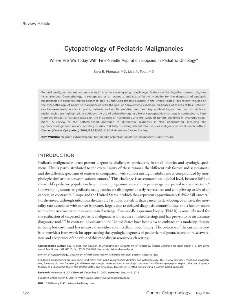
infectious diseases that can be quickly diagnosed by cytol-
ogy to expedite treatment promote the use of FNAB. In
these regions, acceptance of FNAB as a primary diagnostic
modality and willingness to treat based on the cytologic
diagnosis are more common than in resource-rich coun-
tries. This stands in stark contrast to the United States, in
which there is underuse of FNAB in pediatric populations
and trepidation with regard to treatment based on a cyto-
logic diagnosis, which are compounded and perpetuated
by Children’s Oncology Group (COG) protocols that
emphasize the need for histological sampling. Differences
in the approach to the diagnosis of Wilms tumor illustrate
this dichotomy.9 The International Society of Paediatric
Oncology protocols for the treatment of Wilms tumor use
preoperative chemotherapy as the initial therapy and thus
staging is performed on the posttreatment resection speci-
men. In this setting, FNAB has proven to be accurate for
the diagnosis of renal tumors and as a basis for directing
treatment.10 In contrast, the COG protocols base the stag-
ing of Wilms tumor on the primary prechemotherapy
resection specimen and emphasize the need for histologi-
cal evaluation before treatment.9
In the limited publications on cytologic diagnosis of
pediatric tumors available in the United States, the major-
ity of FNABs are from lesions of the head and neck,
lymph nodes, and soft tissue.11-15 Benign diagnoses out-
number malignant diagnoses, with only 10% to 40% of
cases being malignant. The majority of malignancies
reported include lymphomas, sarcomas, thyroid carcino-
mas, and small round cell tumors. Abdominal malignan-
cies represent a small minority of the tumors in these
series. In contrast, in a study that included 290 FNABs
from resource-poor areas, 77% of the diagnoses were
malignant.4 In this study, the most common malignancies
were Wilms tumor, lymphomas, rhabdomyosarcomas,
and neuroblastoma. FNAB resulted in the precise subtyp-
ing of the tumor that allowed for the initiation of treat-
ment in 76% of cases, including neoadjuvant treatment
before surgery.4 Furthermore, this and another large study
reported that the sensitivity and specificity of FNA were
97% to 98% and 93% to 97%, respectively.3,4 A study
from Argentina that examined 899 FNA specimens from
patients aged< 20 years and a smaller study from Spain
demonstrated that> 50% of the cases were malignant.3,16
The majority of these malignancies were from abdominal
masses. Differences between aspirates from the United
States and those from resource-limited settings are sum-
marized in Tables 4 and 5.
In the United States, the National Cancer Institute
supports the COG, a cooperative group that conducts
clinical trials, as well as research on the biology, risk fac-
tors, and outcomes of childhood cancers. Given that 90%
to 95% of children aged< 15 years with a newly diag-
nosed malignancy are seen at COG institutions, patholo-
gists and cytopathologists at these institutions typically
have more experience in diagnosing these tumors and
greater resources with which to perform the necessary
ancillary studies compared with institutions that are not
affiliated with COG.17 However, COG protocols are
based on histologic diagnosis and associated biologic
TABLE 4. Comparison of Pediatric Cytopathology in Resource-Limited Countries Versus the United States
Resource-Limited Countries United States
Percentage of worldwide pediatric population Majority (>85%) Minority
No. of pediatric FNABs More Fewer (variable)
Percentage of abdominal/pelvic FNABs More abdominal/pelvic FNABs Fewer abdominal/pelvic FNABs
Clinical acceptance Greater Less
Abbreviation: FNAB, fine-needle aspiration biopsy.
TABLE 5. Comparison of Pediatric Lesions Sampled in Head and Neck Versus Abdomen and Pelvis
Head and Neck Abdomen and Pelvis
Location Superficial Deep-seated
Type of FNAB Palpation-guided FNAB or image-guided FNAB Image-guided FNAB
Types of tissue specimens Mostly lymph nodes Mostly solid organ masses
Benign vs malignant Benign >> malignant Malignant>>benign
Abbreviation: FNAB, fine-needle aspiration biopsy.
Cytopathology of Pediatric Malignancies/Monaco and Teot
Cancer Cytopathology May 2014 325
studies require frozen or formalin-fixed tissue from the
primary tumor and, when available, metastatic and recur-
rent tumors. These requirements serve as powerful deter-
rents to the use of FNAB as a primary diagnostic
modality. However, as the use of small biopsies increases
and rising numbers of clinically validated molecular tests
shift the focus from the precise classification of disease to
the identification of biomarkers for targeted therapies, the
use of FNAB for the diagnosis of suspected pediatric
malignancies may increase.18
DIFFERENTIAL DIAGNOSES
Childhood tumors are frequently classified based on mor-
phology, rather than site of origin, as is typically used for
adult tumors.1,15,19 This reflects the differences in the
types of tumors noted in these populations and, from a
practical perspective, provides a useful framework for the
pathologic evaluation of mass lesions in children and ado-
lescents. Common morphologic patterns include inflam-
matory, epithelial/epithelioid, cystic, spindle cell, clear
cell, small round cell, and large cell/pleomorphic. Differ-
ential diagnostic considerations using this pattern-based
approach are summarized in Table 6 and the cytologic
features of the more commonly encountered entities in
each category are discussed (Fig. 1)
Inflammatory Pattern
An inflammatory pattern most frequently represents a
benign process, such as reactive lymphoid hyperplasia,
infection, or inflammation involving the aspirated tissue
or organ, but is also characteristic of Langerhans cell his-
tiocytosis (LCH) and HL, and may rarely mask other
malignancies (Figs. 1A-1D).15,19 Aspirates from reactive
lymphoid hyperplasia are highly cellular and comprised of
a polymorphous population of lymphocytes, dominated
by small mature lymphocytes but spanning a spectrum
from small lymphocytes to immunoblasts, with varying
numbers of interspersed plasma cells and tingible body
macrophages (Fig. 1A). A high-grade lymphoma should
be excluded, particularly in the setting of numerous tin-
gible body macrophages or a monomorphic intermediate-
to-large cell population. Depending on the inciting stimu-
lus, eosinophils may also be present. These features are
also characteristic of HL, in which the malignant cells
occur in a heterogeneous background with varying num-
bers of interspersed histiocytes and eosinophils. Classic
and/or variant Reed-Sternberg cells in the appropriate
background are diagnostic of HL, but may comprise a
minority of cells and be overlooked or underrepresented
on cell blocks (Fig. 1D). Fragments of metachromatic
stroma are characteristic of the nodular sclerosis subtype
of classic HL, which is the most common variant in the
pediatric population, and sarcoid-like granulomas and/or
necrosis may also be present. Although these features pro-
vide clues to the diagnosis, they are nonspecific. Granu-
lomatous lymphadenitis is characteristic of infections due
to atypical mycobacteria, Mycobacterium tuberculosis, Bar-
tonella, and fungal and other organisms, as well as other
processes encountered in the pediatric population, such as
chronic granulomatous disease, sarcoidosis, and rheuma-
toid arthritis. Similarly, necrosis is encountered in aspi-
rates from abscesses or acute suppurative infectious
processes, as well as other tumors. Immunohistochemical
(IHC) stains are useful for confirming the diagnosis of
HL. Classic and variant Reed-Sternberg cells are positive
for CD30, CD15, MUM-1, and fascin, and are negative
for CD45 and ALK-1. L and H (so-called popcorn) cells,
which are characteristic of nodular lymphocyte-
predominant HL, a rare variant that is infrequently
encountered in the pediatric population, are positive for
CD45, CD20, and CD79a and negative for CD30 and
CD15. In specimens of HL, flow cytometry reveals a reac-
tive pattern, and thus does not contribute to the diagnosis,
aside from excluding the possibility of a non-Hodgkin
lymphoma in difficult cases. It is interesting to note that
inflammatory cells can also be present in other malignan-
cies, such as a lymphohistiocytic or granulomatous infil-
trate in seminoma, and thus the presence of inflammation
alone does not exclude the need to thoroughly search for
neoplastic cells. Furthermore, certain leukemias, such as
myeloid leukemias and myeloid sarcomas, can mimic an
acute inflammatory process and should be considered,
particularly in the appropriate clinical scenario.
Aspirates from LCH, a monoclonal neoplastic pro-
liferation of Langerhans cells that is classified among the
histiocytic neoplasms in the 2008 World Health Organi-
zation classification,20 also have an inflammatory pattern.
The Langerhans cells are present in a background of
mixed inflammation composed of variable numbers of
lymphocytes, eosinophils, neutrophils, and mononuclear
and multinucleated non-Langerhans histiocytes. Langer-
hans cells are characterized by pale moderately abundant
cytoplasm and folded ovoid nuclei with inconspicuous
Review Article
326 Cancer Cytopathology May 2014
nucleoli (Fig. 1C). The presence of eosinophils is a useful
clue to the diagnosis, particularly in aspirates with few
Langerhans cells. Ultrastructurally, Langerhans cells have
Birbeck granules; however, IHC stains are more often
used to confirm the diagnosis. Cells are positive for
CD1a, S-100, and langerin (CD207), and may aberrantly
express CD68. Although activated macrophages may
express S-100, in contrast to Langerhans cells they are pos-
itive for CD68 and CD163, and negative for CD1a and
langerin.
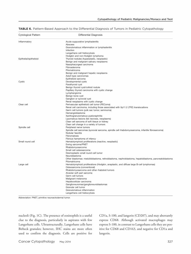
TABLE 6. Pattern-Based Approach to the Differential Diagnosis of Tumors in Pediatric Cytopathology
Cytological Pattern Differential Diagnosis
Inflammatory Acute suppurative lymphadenitis
Abscess
Granulomatous inflammation or lymphadenitis
Infection
Langerhans cell histiocytosis
Hodgkin and non-Hodgkin lymphoma
Epithelial/epithelioid Thyroid nodules (hyperplastic, neoplastic)
Benign and malignant salivary neoplasms
Nasopharyngeal carcinoma
Fibroadenoma
Pilomatricoma
Benign and malignant hepatic neoplasms
Adult-type carcinomas
Epithelioid sarcoma
Cystic Developmental cysts
Parathyroid cyst
Benign thyroid cyst/colloid nodule
Papillary thyroid carcinoma with cystic change
Lymphangioma
Benign bone cyst
Ganglion or synovial cyst
Renal neoplasms with cystic change
Clear cell Perivascular epithelioid cell tumor (PEComa)
Renal cell carcinoma, including those associated with Xp11.2 (TFE) translocations
Germ cell tumors (yolk sac tumor, seminoma)
Hemangioblastoma
Xanthogranulomatous pyelonephritis
Lipomatous lesions (fat necrosis, neoplasms)
Clear cell sarcoma of soft tissue or kidney
Clear cell change in a variety of tumors
Spindle cell Peripheral nerve tumors
Spindle cell sarcomas (synovial sarcoma, spindle cell rhabdomyosarcoma, infantile fibrosarcoma)
Nodular fasciitis
Fibromatosis
Fibrous hamartoma of infancy
Small round cell Hematolymphoid proliferations (reactive, neoplastic)
Ewing sarcoma/PNET
Rhabdomyosarcoma
Small cell osteosarcoma
Desmoplastic small round cell tumor
Neuroblastoma
Other blastomas: medulloblastoma, retinoblastoma, nephroblastoma, hepatoblastoma, pancreatoblastoma
Pilomatricoma
Large cell Hematolymphoid proliferations (Hodgkin, anaplastic, and diffuse large B-cell lymphomas)
Osteosarcoma (conventional)
Rhabdomyosarcoma and other rhabdoid tumors
Alveolar soft part sarcoma
Germ cell tumors
Malignant melanoma
Hepatocellular carcinoma
Ganglioneuromas/ganglioneuroblastomas
Granular cell tumor
Granulomatous inflammation
Langerhans cell histiocytosis
Abbreviation: PNET, primitive neuroectodermal tumor.
Cytopathology of Pediatric Malignancies/Monaco and Teot
Cancer Cytopathology May 2014 327

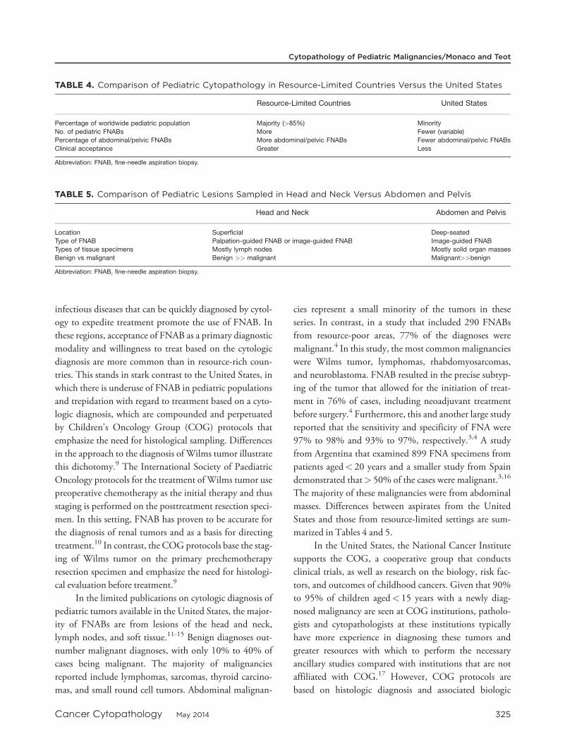
Figure 1.
Review Article
328 Cancer Cytopathology May 2014

Epithelial/Epithelioid Pattern
A minority of pediatric malignancies present with an epi-
thelial/epithelioid pattern, reflecting the relative rarity of
carcinomas in this population compared with
adults.3,4,15,19 An epithelial pattern is observed in thyroid,
salivary, and hepatic neoplasms; nasopharyngeal carcino-
mas; and the rare, adult-type carcinomas arising in chil-
dren and adolescents. Some of these tumors, such as
thyroid carcinomas, may be associated with a particular
genetic association or familial syndrome and therefore be
more likely to be diagnosed in young patients. This
includes the occurrence of the cribriform morular variant
of papillary thyroid carcinoma in the setting of familial
adenomatous polyposis syndrome, and medullary thyroid
carcinoma in the setting of multiple endocrine neoplasia
syndromes.21,22 However, in general, the cytologic fea-
tures of these epithelial malignancies are identical irre-
spective of age and are well characterized in the standard
textbooks on cytopathology. Therefore, with the excep-
tion of hepatoblastoma, a rare tumor predominantly of
young children, these entities will not be discussed
further.
Hepatoblastoma is subdivided into epithelial and
mixed epithelial-mesenchymal subtypes, and aspirates
tend to be cellular. Fetal and/or embryonal cells comprise
the predominant epithelial elements, whereas a minority
have small cell or macrotrabecular patterns. Fetal cells are
smaller than hepatocytes, and have moderate amounts of
granular or clear cytoplasm and round central nuclei (Fig.
1E). Embryonal cells are smaller and more primitive with
scant amphophilic cytoplasm, and irregular nuclei with
coarse chromatin and prominent nucleoli. Mesenchymal
elements are present in nearly one-half of the tumors, and
in some of these, teratoid elements are also observed.23-26
Extramedullary hematopoiesis is common. Using IHC,
fetal and embryonal epithelial cells are positive for a-
fetoprotein, glypican-3, glutamine synthetase, and nuclear
b-catenin. Fetal and, to a lesser extent, embryonal cells are
positive for hepatocyte paraffin 1, cytokeratins 8 and 18
(Cam 5.2), and carcinoembryonic antigen. Distinction
between the fetal pattern of hepatoblastoma and well-
differentiated hepatocellular carcinoma may be difficult
in cytologic preparations. The cells of fetal hepatoblas-
toma are typically smaller and more uniform than those of
hepatocellular carcinoma and are more likely to demon-
strate nuclear staining for b-catenin. If present, embry-
onal or mesenchymal elements also provide a clue to the
correct diagnosis. However, in young children, clinical
correlation is often the most useful tool for distinguishing
these entities, given the extreme rarity of hepatocellular
carcinoma in this age group in the absence of a predispos-
ing metabolic disorder.
Cystic Pattern
The vast majority of pediatric masses resulting in a cystic
pattern on FNAB are benign processes, such as develop-
mental and other cysts, colloid nodules, and lymphatic
malformations. However, cystic changes may occur in
Figure 1. Representative examples in the pattern-based approach to pediatric cytopathology are shown, specifically (A-C)
inflammatory, (D) large cell, (E) epithelial, (F) cystic, (G) spindle cell, (H) clear cell, and (I-J) small round cell patterns. (A) Reac-
tive lymphoid hyperplasia is shown. Aspirates demonstrate a heterogeneous lymphoid population with scattered tingible body
macrophages and an absence of eosinophilia or Reed-Sternberg cells (Diff-Quik, 3 400). (B) Burkitt lymphoma is shown. These
aspirates reveal a monomorphic population of intermediate-sized lymphocytes with mitoses and tangible body macrophages.
There is a lack of heterogeneity and a shift toward more intermediate-sized cells, in comparison with panel A (Diff-Quik, 3 400).
(C) Langerhans cell histiocytosis is shown. Aspirates reveal an inflammatory background with increased eosinophils, in addition
to intermediate-sized histiocytic cells with abundant cytoplasm and cleaved nuclei (Diff-Quik, 3 400). (D) Classic Hodgkin lym-
phoma is shown. Classic Hodgkin lymphomas have a heterogeneous lymphoid population and an increase in eosinophils, with
Reed-Sternberg cells that are usually binucleated with prominent nucleoli (Diff-Quik, 3 400). (E) Hepatoblastoma is shown. Aspi-
rates show fetal cells, which tend to be epithelioid cells that are smaller and more uniform than hepatocytes, in addition to
embryonal cells (H & E, 3 400). (F) Papillary thyroid carcinoma is shown. Aspirates from primary or metastatic papillary thyroid
carcinomas can demonstrate cystic changes with histiocytes and/or colloid. This example shows follicular epithelial cells with
intranuclear cytoplasmic inclusions (arrow) and grooves in a background of watery colloid (Diff-Quik, 3 400). (G) Synovial sar-
coma is shown. This spindle cell tumor demonstrates cellular aspirates with monomorphic-appearing spindle cells and interlacing
metachromatic material (Diff-Quik, 3 400). (H) Renal cell carcinoma associated with the transcription factor E3 (TFE3) gene
translocation is shown. This clear cell tumor shows abundant pale cytoplasm and round nuclei, in addition to stripped nuclei
(Diff-Quik, 3 400). (I) Neuroblastoma is shown. Neuroblastoma typically yields cellular aspirates with cells that have dark round
nuclei, scant cytoplasm, nuclear molding, and clustering with occasional rosette formation. No lymphoglandular bodies are noted
(Diff-Quik, 3 400). (J) Small cell osteosarcoma is shown. Aspirates from this bone lesion show a small round cell tumor with
occasional metachromatic osteoid material (arrow) (Diff-Quik, ;3 400).
Cytopathology of Pediatric Malignancies/Monaco and Teot
Cancer Cytopathology May 2014 329

papillary thyroid carcinoma and, less frequently, other pedi-
atric malignancies, and may result in a false-negative diag-
nosis unless these entities are considered (Fig. 1F). Aspirates
comprised of cyst contents or fluid may have obscuring
inflammatory elements or harbor few or no malignant cells,
thereby making an accurate diagnosis difficult or impossi-
ble. When the clinical presentation and/or imaging studies
suggest a malignant neoplasm, the processing of any resid-
ual cyst fluid is essential before rendering a cytologic diag-
nosis. Failure to sample lesional tissue surrounding a cyst
cavity is a well-recognized source of false-negative diagnoses
and thus correlation with the radiographic findings is essen-
tial. In addition, cystic lesions with a predominance of
squamous cells should be approached differently in children
compared with adults. In children, these findings likely rep-
resent developmental cysts with squamous lining or squa-
mous metaplasia and carcinoma is highly unlikely, whereas
in adults the possibility of metastatic squamous cell carci-
noma with cystic changes must be excluded. In addition,
germ cell tumors with a teratomatous component contain-
ing squamous epithelium may also give rise to a cystic neck
mass in adolescents, and should be considered in the appro-
priate clinical setting.
Spindle Cell Pattern
A spindle cell pattern is characteristic of FNAB specimens
from a variety of common and uncommon pediatric mes-
enchymal proliferations. These include nonneoplastic
tumefactions, such as nodular fasciitis and traumatic neu-
roma; benign neoplasms, such as fibroma and schwan-
noma; and malignancies, such as synovial sarcoma,
malignant peripheral nerve sheath tumor (MPNST), and
spindle cell rhabdomyosarcoma. Furthermore, some neo-
plasms, such as hepatoblastomas and Wilms tumors, may
be morphologically heterogeneous and therefore a spindle
cell pattern may predominate in tumors that are not typi-
cally associated with a pure spindle cell pattern. Overall,
reactive and benign neoplastic proliferations comprise the
majority of pediatric spindle cell lesions; however, this dis-
cussion will focus on the cytologic features and differential
diagnosis of selected malignant tumors.
Considerable overlap exists in the cytomorphologic
features of pediatric spindle cell sarcomas. Aspirates vary
in cellularity, often reflecting the grade of the tumor and
the presence or absence of collagenized stroma. Thus,
FNAB specimens from high-grade tumors without
fibrotic stroma are typically highly cellular, whereas those
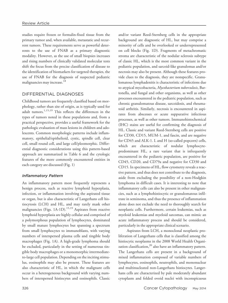
TABLE 7. Distinguishing Features of Selected Pediatric Small Blue Cell Tumors
Tumor Cytomorphology Immunophenotype Genetics
Wilms tumor Blastema 1/-epithelial component
1/- stroma, rarely, anaplasia
1 WT1; 1 EMA, cytokeratin
(epithelial component); - Syn-
aptophysin, chromogranin
Mutations of WT1, WT2
Neuroblastoma Neuropil, rosettes, 1/- ganglion cells,
1/- schwannian stroma, 1/-
calcification
1 Synaptophysin, chromo-
granin, CD56, PgP9.5;
- S-100, CD99, desmin,
myogenin, lymphoid markers
1/- N-MYC amplification
Rhabdomyosarcoma Rhabdomyoblastic differentiation
subtle to obvious1/- floret cells,
1/- strap cells
1 Myogenin, myoD1, desmin;
- TLE1; 1/2 aberrant CD99,
cytokeratin, EMA, neural
markers
Alveolar subtype:
t(2;13)(q35;q14),
t(1;13)(p36;q14)
Ewing sarcoma/PNET 1/- Rosettes, 1/-neuropil,1/- Tigroid
background
1CD99, FLI-1;1/- synapto-
physin, PgP9.5, CD56;- Des-
min, myogenin, CD45, TLE-1,
EMA, cytokeratins
t(11;22)(q24;q12) in >90%;
t(21;22)(q12;q12),
t(2;22)(q33;q12), others rare
Synovial sarcoma (round cell) 1/- Metachromatic stroma, 1/-
calcifications
1 TLE-1, EMA; 1/- cytokera-
tin, CD99; - myogenin,
myoD1, desmin
t(X;18)
Small cell osteosarcoma Osteoid (often scant or absent) 1/- S-100, osteonectin,
osteocalcin, CD99;- FLI-1,
myogenin, neural markers
EWS rearrangements absent
Lymphoid malignancies Morphology varies with type;
lymphoglandular bodies
Varies with lineage and type
(B-cell, T-cell);1TdT (lym-
phoblastic lymphoma)
Burkitt: MYC translocations,
t(8;14)(q24;q32) and less
commonly, t(2;8)(p12;q24),
t(8;22)(q24;q11)
Abbreviations: 1, positive; -, negative; EMA, epithelial membrane antigen; EWS, Ewing sarcoma gene; FLI-1, friend leukemia integration 1 transcription factor;
PNET, primitive neuroectodermal tumor; TdT, terminal deoxynucleotidyl transferase; TLE-1, transducin-like enhancer of split 1; WT1, Wilms tumor 1; WT2,
Wilms tumor 2.
Review Article
330 Cancer Cytopathology May 2014
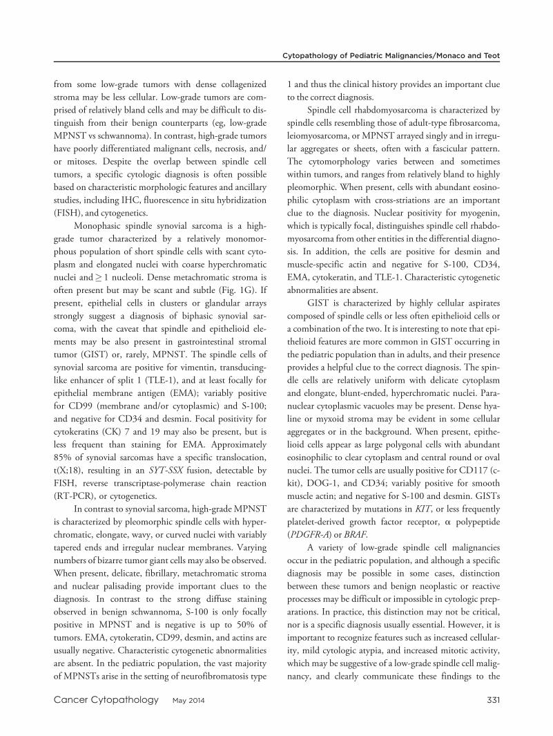
from some low-grade tumors with dense collagenized
stroma may be less cellular. Low-grade tumors are com-
prised of relatively bland cells and may be difficult to dis-
tinguish from their benign counterparts (eg, low-grade
MPNST vs schwannoma). In contrast, high-grade tumors
have poorly differentiated malignant cells, necrosis, and/
or mitoses. Despite the overlap between spindle cell
tumors, a specific cytologic diagnosis is often possible
based on characteristic morphologic features and ancillary
studies, including IHC, fluorescence in situ hybridization
(FISH), and cytogenetics.
Monophasic spindle synovial sarcoma is a high-
grade tumor characterized by a relatively monomor-
phous population of short spindle cells with scant cyto-
plasm and elongated nuclei with coarse hyperchromatic
nuclei and� 1 nucleoli. Dense metachromatic stroma is
often present but may be scant and subtle (Fig. 1G). If
present, epithelial cells in clusters or glandular arrays
strongly suggest a diagnosis of biphasic synovial sar-
coma, with the caveat that spindle and epithelioid ele-
ments may be also present in gastrointestinal stromal
tumor (GIST) or, rarely, MPNST. The spindle cells of
synovial sarcoma are positive for vimentin, transducing-
like enhancer of split 1 (TLE-1), and at least focally for
epithelial membrane antigen (EMA); variably positive
for CD99 (membrane and/or cytoplasmic) and S-100;
and negative for CD34 and desmin. Focal positivity for
cytokeratins (CK) 7 and 19 may also be present, but is
less frequent than staining for EMA. Approximately
85% of synovial sarcomas have a specific translocation,
t(X;18), resulting in an SYT-SSX fusion, detectable by
FISH, reverse transcriptase-polymerase chain reaction
(RT-PCR), or cytogenetics.
In contrast to synovial sarcoma, high-grade MPNST
is characterized by pleomorphic spindle cells with hyper-
chromatic, elongate, wavy, or curved nuclei with variably
tapered ends and irregular nuclear membranes. Varying
numbers of bizarre tumor giant cells may also be observed.
When present, delicate, fibrillary, metachromatic stroma
and nuclear palisading provide important clues to the
diagnosis. In contrast to the strong diffuse staining
observed in benign schwannoma, S-100 is only focally
positive in MPNST and is negative is up to 50% of
tumors. EMA, cytokeratin, CD99, desmin, and actins are
usually negative. Characteristic cytogenetic abnormalities
are absent. In the pediatric population, the vast majority
of MPNSTs arise in the setting of neurofibromatosis type
1 and thus the clinical history provides an important clue
to the correct diagnosis.
Spindle cell rhabdomyosarcoma is characterized by
spindle cells resembling those of adult-type fibrosarcoma,
leiomyosarcoma, or MPNST arrayed singly and in irregu-
lar aggregates or sheets, often with a fascicular pattern.
The cytomorphology varies between and sometimes
within tumors, and ranges from relatively bland to highly
pleomorphic. When present, cells with abundant eosino-
philic cytoplasm with cross-striations are an important
clue to the diagnosis. Nuclear positivity for myogenin,
which is typically focal, distinguishes spindle cell rhabdo-
myosarcoma from other entities in the differential diagno-
sis. In addition, the cells are positive for desmin and
muscle-specific actin and negative for S-100, CD34,
EMA, cytokeratin, and TLE-1. Characteristic cytogenetic
abnormalities are absent.
GIST is characterized by highly cellular aspirates
composed of spindle cells or less often epithelioid cells or
a combination of the two. It is interesting to note that epi-
thelioid features are more common in GIST occurring in
the pediatric population than in adults, and their presence
provides a helpful clue to the correct diagnosis. The spin-
dle cells are relatively uniform with delicate cytoplasm
and elongate, blunt-ended, hyperchromatic nuclei. Para-
nuclear cytoplasmic vacuoles may be present. Dense hya-
line or myxoid stroma may be evident in some cellular
aggregates or in the background. When present, epithe-
lioid cells appear as large polygonal cells with abundant
eosinophilic to clear cytoplasm and central round or oval
nuclei. The tumor cells are usually positive for CD117 (c-
kit), DOG-1, and CD34; variably positive for smooth
muscle actin; and negative for S-100 and desmin. GISTs
are characterized by mutations in KIT, or less frequently
platelet-derived growth factor receptor, a polypeptide
(PDGFR-A) or BRAF.
A variety of low-grade spindle cell malignancies
occur in the pediatric population, and although a specific
diagnosis may be possible in some cases, distinction
between these tumors and benign neoplastic or reactive
processes may be difficult or impossible in cytologic prep-
arations. In practice, this distinction may not be critical,
nor is a specific diagnosis usually essential. However, it is
important to recognize features such as increased cellular-
ity, mild cytologic atypia, and increased mitotic activity,
which may be suggestive of a low-grade spindle cell malig-
nancy, and clearly communicate these findings to the
Cytopathology of Pediatric Malignancies/Monaco and Teot
Cancer Cytopathology May 2014 331

clinician. When a definitive cytologic diagnosis is not pos-
sible and the differential diagnosis includes low-grade
malignancies and benign processes, a biopsy may help to
clarify the diagnosis and thereby ensure adequate resection
of a malignant tumor, while avoiding overly aggressive
treatment of a benign lesion.
Clear Cell Pattern
Pediatric tumors with a predominance of clear cells may
be diagnostically challenging due to the morphologic
overlap with benign histiocytic lesions, and the low
nuclear-to-cytoplasmic ratio in the tumor cells. This
group includes perivascular epithelial tumors (PEComas),
germ cell tumors (yolk sac tumors, seminomas), clear cell
sarcomas, and carcinomas with clear cell change (eg, renal
cell carcinoma [RCC]). Benign entities in the differential
diagnosis include xanthogranulomatous proliferations
and adipocytic lesions.
The PEComa family of neoplasms, characterized by
cells with myomelanocytic differentiation, includes angio-
myolipomas, lymphangiomyomatosis, and clear cell
“sugar” tumor of the lung. Many of these lesions are ini-
tially misdiagnosed, and have been reported in a wide
variety of locations. In children, these may occur sporadi-
cally or in association with tuberous sclerosis. Cytological
features include epithelioid cells with clear cytoplasm,
usually near vessels, with more discohesive spindle cells
and multinucleated giant cells. The cells are typically posi-
tive for melanocytic markers (HMB-45, MelanA) and
muscle markers (smooth muscle actin), but are negative
for cytokeratin, CD117/C-kit, and CD34. PEComa must
be distinguished from clear cell sarcoma of soft tissue,
which is also composed of epithelioid and spindle cells
with clear or granular cytoplasm that is positive for HMB-
45 and MelanA but negative for muscle markers.
Germ cell tumors, particularly yolk sac tumors and
seminomas, may also have abundant clear cytoplasm.
Yolk sac tumors have a variety of histological patterns,
and therefore may vary from cohesive to discohesive on
FNAB, but generally have moderately abundant clear
cytoplasm. Cytoplasmic and/or extracellular hyaline glob-
ules are an important clue to the diagnosis. In seminoma,
the tumor cells appear discohesive with round central
nuclei, prominent nucleoli, and pale cytoplasm. Due to
the presence of glycogen, the disrupted cytoplasm imparts
a “tigroid” appearance to the background on air-dried
slides stained with modified Romanowsky stain. Lympho-
cytes and granulomas are often present. Yolk sac tumors
typically are positive for a-fetoprotein, cytokeratin, and
glypican-3, and negative for CD117/C-kit and Oct3/4,
whereas the opposite pattern is seen in seminomas.
Clear cell sarcoma of the kidney (CCSK) is a rare,
deceptively bland tumor that typically occurs in children
aged< 5 years. In aspirates, CCSK appears as small spin-
dle or epithelioid cells arrayed singly or in aggregates with
transgressing blood vessels. The cells have clear cytoplasm
and nuclei with distinct grooves and fine chromatin.
CCSKs are positive for vimentin, but typically negative
for nearly all other stains, including cytokeratin, S-100,
CD99, desmin, WT-1, and synaptophysin.
A clear cell phenotype is often observed in both con-
ventional and Xp11.2 (transcription factor E3 [TFE]
gene) translocation-associated RCC. The latter variant is
characterized by translocations of TFE3 on chromosome
Xp11.2, resulting in overexpression of TFE3, which can
be detected by IHC.27 These tumors are typically
observed in children and young adults, and although only
approximately 5% of pediatric renal tumors are RCC,
greater than one-third of these are Xp11.2 translocation
RCC.28 The key cytological features of these tumors
include clear cells with loose cohesion or in papillary frag-
ments and numerous stripped nuclei (Fig. 1H). Cells have
voluminous cytoplasm with punched-out, discrete cyto-
plasmic vacuoles and central nuclei. Hyalinized metachro-
matic globules, psammoma bodies, and thin transgressing
vessels may also be noted. RCCs associated with TFE3
translocations tend to be negative or only focally positive
for epithelial markers, such as cytokeratins and EMA, and
are usually negative for vimentin, in contrast to conven-
tional RCC. Nuclear staining for TFE3 by IHC is a help-
ful marker for RCC associated with TFE3 translocations,
and a FISH break-apart probe for the TFE3 gene can also
confirm the presence of a translocation in these
tumors.27,28
Small Round Cell Tumors
Small round cell tumors (SRCTs) are the most common
group of pediatric malignancies and encompass a broad
spectrum of tumors, arising from hematolymphoid, mes-
enchymal, neuroepithelial, neural crest, epithelial, and
primitive blastemal cells. As with spindle cell tumors, con-
siderable overlap exists in the cytomorphologic features of
SRCTs. FNAB specimens are typically highly cellular and
are composed either of predominantly single cells (eg,
Review Article
332 Cancer Cytopathology May 2014

lymphomas, Ewing sarcoma) or discohesive cellular aggre-
gates with single cells in the background (eg, neuroblas-
toma, Wilms tumor). Necrosis and mitotic figures,
including abnormal forms, are often present. Despite the
overlap between SRCTs, a specific cytologic diagnosis is
often possible based on characteristic morphologic fea-
tures and ancillary studies, including IHC, FISH, RT-
PCR, and cytogenetics.
Wilms tumors (nephroblastomas) typically have a
blastemal component (nearly 100% of cases) and an epi-
thelial component (approximately 70% of cases), whereas
a mesenchymal component is less frequent (approxi-
mately 20% of cases).25 In FNABs, the blastemal compo-
nent is characterized by small cells with scant cytoplasm
and round or irregular, often molded nuclei with coarse
chromatin, which are arrayed as discohesive aggregates
and single cells. In contrast, the epithelial elements appear
as clusters or tubules composed of cells with moderate to
abundant cytoplasm. When present, the mesenchymal
component is composed of bland spindle cells in a back-
ground of metachromatic collagenous or myxoid matrix.
Anaplasia, defined by the triad of nuclei at least 3 times
the size of adjacent tumor nuclei, hyperchromasia, and
multipolar mitotic figures, is observed in< 5% of Wilms
tumors. Anaplasia may be focal and, as a consequence, not
sampled in FNAB. However, its presence correlates with
poor prognosis and therefore, it is important to recognize,
particularly in the setting of preoperative chemotherapy.
Wilms tumors are usually positive for WT1 and negative
for chromogranin and synaptophysin, and retain INI-1.
The blastemal component is variably positive for cytoker-
atin and desmin, and the epithelial component is positive
for EMA and cytokeratin. When rhabdomyoblastic differ-
entiation is present, the mesenchymal component is posi-
tive for desmin and myogenin. It is important to note that
nephrogenic rests and nephroblastomatosis are indistin-
guishable from Wilms tumor in cytologic preparations,
and therefore correlation with imaging studies is impor-
tant to confirm the diagnosis of Wilms tumor.
In contrast to Wilms tumors, neuroblastomas
appear more monomorphic and are composed of cells
with round nuclei with fine stippled chromatin and small
nucleoli (Fig. 1I). Neuropil, which appears as metachro-
matic fibrillary matrix; rosettes; and immature to mature
ganglion cells are variably present, depending on the
degree of differentiation, and are diagnostic of neuroblas-
toma.25 Ancillary studies are essential for the diagnosis of
undifferentiated neuroblastoma and help to confirm the
diagnosis when neuropil and/or ganglionic differentiation
are present. The tumor cells are usually positive for synap-
tophysin, chromogranin, and CD56, and negative for S-
100, CD99, desmin, myogenin, and lymphoid markers.
FISH is useful for the assessment of N-MYC amplifica-
tion, which is used for risk stratification and treatment.
The mitotic-karyorrhectic index is important for deter-
mining favorable or unfavorable histology, another fea-
ture used in risk stratification, but cannot be assessed in
cytologic preparations.
Aspirates from rhabdomyosarcoma are characterized
by relatively monotonous round cells. In alveolar rhabdo-
myosarcoma (ARMS), the cells are often larger than those
of embryonal rhabdomyosarcoma (ERMS), Ewing sar-
coma/primitive neuroectodermal tumor (PNET), or
synovial sarcoma, and have round nuclei with coarse chro-
matin and� 1 conspicuous nucleoli. Small cells with
eccentric eosinophilic cytoplasm and occasional large
multinucleated cells with eosinophilic cytoplasm and
peripherally placed nuclei (floret or wreath cells) may be
identified in ARMS, but cells with cross-striations are
uncommon. Fragments of fibrous stroma may be present.
In contrast, ERMS is composed of smaller round cells
with round or oval hyperchromatic nuclei with fine chro-
matin and inconspicuous nucleoli. Elongate cells with
cross-striations may be present, and in some cases are
numerous, whereas floret cells are usually rare. Fragments
of myxoid stroma may be present. Nuclear positivity for
myogenin, which is typically strong and diffuse in ARMS
and focal in ERMS, distinguishes rhabdomyosarcomas
from other entities in the differential diagnosis. In addi-
tion, the cells are positive for desmin and muscle-specific
actin, and usually negative for chromogranin, synapto-
physin, CD99, EMA, cytokeratin, and TLE-1. It is inter-
esting to note that aberrant staining for cytokeratins and
neural markers is observed in a significant minority of
ARMS cases. Translocations involving FOXO1 (forkhead
box protein O1) on chromosome 13 and either PAX3
(paired box 3) or PAX7 on chromosomes 2 and 1, respec-
tively, are present in approximately 80% of ARMS but
not ERMS, and can be detected by FISH, RT-PCR or
cytogenetics. In the absence of a translocation, FNAB
does not reliably distinguish between ARMS and ERMS.
Ewing sarcoma/PNET is composed of cells with
scant pale cytoplasm and round to oval nuclei with fine
pale chromatin and inconspicuous nucleoli. Homer-
Cytopathology of Pediatric Malignancies/Monaco and Teot
Cancer Cytopathology May 2014 333
Wright rosettes may be present, depending on the degree
of differentiation of the tumor. Crush artifact is often
present. A tigroid background may be evident in air-dried
smears stained with modified Romanowsky stains, but
matrix is absent. Ewing sarcoma/PNET is usually positive
for CD99 and FLI-1; variably positive for PgP9.5, synap-
tophysin, and CD56; and negative for desmin, myogenin,
CD45, TLE-1, EMA, and cytokeratins. Greater than
90% of Ewing sarcomas/PNETs have a characteristic
translocation, t(11;22)(q24;q12), which results in fusion
of FLI-1 and EWSR1. Other translocations involving
EWSR1 or ERG occur in another 5% of cases.
Round cell, poorly differentiated synovial sarcoma is
composed of cells with round to ovoid hyperchromatic
nuclei with irregular membranes, coarse chromatin, and
prominent nucleoli. As with other synovial sarcomas,
metachromatic fibrous stroma and calcifications may be
present. Round cell, poorly differentiated synovial sar-
coma has the same immunophenotype and specific trans-
location, t(X;18), as other variants.
Small cell osteosarcoma is an extremely rare variant
of osteosarcoma, which may be confused with Ewing sar-
coma/PNET. The cells are the same size as or somewhat
larger than those of Ewing sarcoma/PNET and have scant
cytoplasm and round or oval nuclei with granular chro-
matin and small nucleoli. Short spindle cells may also be
present. The presence of osteoid is diagnostic, but may be
scant or absent in cytologic specimens (Fig. 1J). Small cell
osteosarcomas are often positive for CD99, but have no
characteristic cytogenetic abnormalities.
Desmoplastic small round cell tumor is another
round cell tumor affecting young patients, and typically
presents with abdominal pain and distention in adolescent
and young men. These are typically aggressive mesenteric
or pelvic masses that may undergo image-guided FNA or
core needle biopsy, and demonstrate uniform oval-to-
round nuclei without conspicuous nucleoli and scant cyto-
plasm. Mitoses and spindled or rhabdoid-like cells are fre-
quently noted. Necrosis and fragments of metachromatic
desmoplastic stromal material can also be observed. These
tumors are unique in that they are positive for epithelial
(eg, cytokeratin and EMA), neural (eg, neuron-specific eno-
lase), and muscle (eg, desmin) markers. The desmin stain-
ing is also typically perinuclear and dot-like. FISH studies
have shown a t(11;22)(p13;q12) translocation between
EWS and WT1, which is similar but not identical to that
noted in Ewing sarcoma/PNET.
Lymphoblastic and Burkitt lymphomas comprise
the vast majority of pediatric lymphomas in the differen-
tial diagnosis of SRCTs. Lymphoblastic lymphoma is
comprised of small to medium-sized cells with scant cyto-
plasm and round or convoluted nuclei with immature
fine chromatin and nucleoli that vary from inconspicuous
to multiple. Both T- and B-lymphoblastic lymphomas are
typically positive for terminal deoxynucleotidyl transfer-
ase (TdT), which helps to distinguish them from other
lymphomas. Flow cytometry and/or immunocytochemis-
try are used to further classify the cells as T cell or B cell in
origin. Burkitt lymphoma is composed of medium-sized
cells with scant cytoplasm with multiple small vacuoles
and round nuclei with clumped chromatin and multiple
small nucleoli. Mitoses and apoptoses are usually readily
apparent, and numerous tingible body macrophages are
present in the background (Fig. 1B). Nuclear positivity
for Ki-67 is observed in nearly 100% of cells. Burkitt lym-
phoma has a mature B-cell phenotype by flow cytometry
and immunocytochemistry and, in contrast to lympho-
blastic lymphoma, is negative for TdT. Translocation of
C-MYC, located on chromosome 8, is almost always pres-
ent and is detected by FISH or cytogenetics.
Large Cell/Pleomorphic Lesions
The large cell pattern encompasses a wide variety of enti-
ties, including HL and non-Hodgkin lymphomas, germ
cell tumors, melanoma, sarcomas, and some carcinomas,
as summarized in Table 6. In children, this category also
includes granular cell tumors, tumors with gangliocytic
differentiation, and lesions with multinucleated giant cells
(eg, granulomatous inflammation, pilomatricoma). The
morphology of these lesions is similar to that noted in
adults. However, ganglion cells are infrequently encoun-
tered in adult tumors, but are more likely to be observed
in children, given the higher incidence of sympathetic
nervous system tumors. Ganglion cells appear as large cells
with abundant cytoplasm with variably conspicuous
darkly stained Nissl substance, and large round nuclei
with prominent nucleoli. In contrast to melanoma, binu-
cleation and intranuclear cytoplasmic inclusions are not
common and the ganglion cells are negative for HMB-45,
MelanA, and microphthalmia-associated transcription
factor. The identification of ganglion cells is important
because ganglioneuromas and intermixed ganglioneuro-
blastomas generally have a better prognosis than neuro-
blastoma. However, these lesions are typically excised for
Review Article
334 Cancer Cytopathology May 2014
definitive classification, which depends in part on the per-
centage of neuroblastic cells and the histological
architecture.
PITFALLS AND LIMITATIONS
The cytologic diagnosis of pediatric malignancies presents
several challenges, due in part to the overlapping features
of these tumors. This is particularly true for SRCTs and
spindle cell tumors, in which extensive morphologic over-
lap exists and accurate diagnoses rely heavily on ancillary
studies.3,4,12,19,25 In addition, the cytologic features of
some pediatric malignancies overlap with those of benign
lesions, leading to both false-negative and false-positive
diagnoses. Benign lymphoid proliferations, such as infec-
tious mononucleosis, may be misdiagnosed as lymphoma
in the absence of ancillary studies, and pilomatricomas
may be confused with rhabdomyosarcomas. LCH may
mimic granulomatous inflammation, and reactive or
other benign myofibroblastic and fibroblastic prolifera-
tions may be confused with low-grade sarcomas.19 Other
factors that impact the ability to make a definitive diagno-
sis include sampling error, heterogeneous tumor popula-
tions, predominant obscuring necrosis or cyst contents,
and equivocal or problematic IHC results.4 A further
limitation of FNAB is that, even with an adequate sample,
the precise classification of a malignancy within a diagnos-
tic category is not always possible. Examples include the
distinction between embryonal and alveolar rhabdomyo-
sarcomas in the absence of a translocation, and differentia-
tion between favorable and unfavorable histology
neuroblastomas based on the mitotic karyorrhectic index.
When treatment differs based on the precise classification
of a tumor, as in these examples, a subsequent biopsy is
required for further subtyping.
Conclusions
In the United States, pathologists and pediatric oncolo-
gists have been slow to embrace the use of cytopathology
for the evaluation of suspected malignancies in children
and adolescents, despite evidence that this modality is
highly sensitive and specific, accurate, less expensive, and
less invasive than either core needle or open biopsy. The
widespread influence of COG protocols, which are based
on histological findings, is a major deterrent to the use of
FNAB, but pathologists also play a role in the underuse of
this modality. Pediatric pathologists are familiar with the
histologic features of malignancies arising in children and
adolescents, but the majority do not perform or interpret
FNABs. Conversely, cytopathologists routinely evaluate
FNABs, but may rarely encounter pediatric malignancies.
As a consequence, apprehension exists among a cadre of
pediatric pathologists and cytopathologists regarding the
diagnosis of these lesions in cytologic specimens. Thus,
advocates of FNAB for the evaluation of suspected malig-
nancies in children and adolescents often encounter resist-
ance from clinicians, pathologists, or both. In contrast, in
resource-limited regions, cytopathology is more widely
accepted as a valuable diagnostic modality for pediatric
malignancies. Use of a pattern-based approach, such as
that described in the current review and used previously in
the literature,19 can be helpful in determining a differen-
tial diagnosis.
Over the past several decades, the incidence of can-
cer in children and adolescents has increased; however,
the survival rate has also increased in developed countries
due to improvements in diagnosis and treatment. These
trends are expected to continue, with a consequent
increase in the number of pediatric malignancies encoun-
tered by pathologists. The increasing use of minimally
invasive diagnostic modalities and the availability of tech-
niques that allow for the detection of important diagnos-
tic, prognostic, and therapeutic markers with small
amounts of tissue provide an opportunity to reevaluate
the role of FNAB in the diagnosis of these tumors.18 In an
era of decreasing reimbursements and rising pressures to
contain the costs of health care, FNAB offers a highly sen-
sitive, specific, and accurate alternative to more costly and
invasive biopsies.
FUNDING SUPPORT
No specific funding was disclosed.
CONFLICT OF INTEREST DISCLOSURES
The authors made no disclosures.
REFERENCES
1. Steliarova-Foucher E, Stiller C, Lacour B, Kaatsch P. InternationalClassification of Childhood Cancer, third edition. Cancer. 2005;103:1457-1467.
2. Sullivan R, Kowalczyk JR, Agarwal B, et al. New policies toaddress the global burden of childhood cancers. Lancet Oncol.2013;14:e125-e135.
3. Drut R, Drut RM, Pollono D, et al. Fine-needle aspiration biopsyin pediatric oncology patients: a review of experience with 829patients (899 biopsies). J Pediatr Hematol Oncol. 2005;27:370-376.
Cytopathology of Pediatric Malignancies/Monaco and Teot
Cancer Cytopathology May 2014 335
4. Razack R, Michelow P, Leiman G, et al. An interinstitutionalreview of the value of FNAB in pediatric oncology in resource-limited countries. Diagn Cytopathol. 2012;40:770-776.
5. Steliarova-Foucher E, Stiller C, Kaatsch P, et al. Geographical pat-terns and time trends of cancer incidence and survival among chil-dren and adolescents in Europe since the 1970s (theACCISproject): an epidemiological study. Lancet. 2004;364:2097-2105.
6. Pritchard-Jones K, Kaatsch P, Steliarova-Foucher E, Stiller CA,Coebergh JW. Cancer in children and adolescents in Europe:developments over 20 years and future challenges. Eur J Cancer.2006;42:2183-2190.
7. Brenner H. Up-to-date survival curves of children with cancer byperiod analysis. Br J Cancer. 2003;88:1693-1697.
8. Magrath I, Steliarova-Foucher E, Epelman S, et al. Pediatric cancerin low-income and middle-income countries. Lancet Oncol. 2013;14:e104-e116.
9. Ko EY, Ritchey ML. Current management of Wilms’ tumor inchildren. J Pediatr Urol. 2009;5:56-65.
10. Fernandez-Pineda I, Cabello R, Garcia-Canton JA, et al. Fine-nee-dle aspiration cytopathology in the diagnosis of Wilms; tumor.Clin Transl Oncol. 2011;13:809-811.
11. Anne S, Teot LA, Mandell DL. Fine needle aspiration biopsy: rolein diagnosis of pediatric head and neck masses. Int J Pediatr Oto-rhinolaryngol. 2008;72:1547-1553.
12. Cohen MB, Bottles K, Ablin AR, Miller TR. The use of fine-needle aspiration biopsy in children. West J Med. 1989;150:665-667.
13. Silverman JF, Gurley AM, Holbrook CT, Joshi VV. Pediatric fine-needle aspiration biopsy. Am J Clin Pathol. 1991;95:653-659.
14. Ponder TB, Smith D, Ramzy I. Lymphadenopathy in children andadolescents: role of fine-needle aspiration in management. CancerDetect Prev. 2000;24:228-233.
15. Howell LP. Changing role of fine-needle aspiration on the evalua-tion of pediatric masses. Diagn Cytopathol. 2001;24:65-70.
16. Verdeguer A, Castel V, Torres V, et al. Fine-needle aspirationbiopsy in children: experience in 70 cases. Med Pediatr Oncol.1988;16:98-100.
17. O’Leary M, Krailo M, Anderson JR, Reaman GH; Children’sOncology Group. Progress in childhood cancer: 50 years of
research collaboration, a report from the Children’s OncologyGroup. Semin Oncol. 2008;35:484-493.
18. Kanagal-Shamanna R, Portier BP, Singh RR, et al. Next-generationsequencing-based multi-gene mutation profiling of solid tumorsusing fine needle aspiration samples: promises and challenges forroutine clinical diagnostics [published online ahead of print August2, 2013]. Mod Pathol. doi: 10.1038/modpathol. 2013.122.
19. Howell LP, Russell LA, Howard PH, Teplitz RL. The cytology ofpediatric masses: a differential diagnostic approach. Diagn Cytopa-thol. 1992;8:107-115.
20. Abla O, Egeler RM, Weitzman S. Langerhans cell histiocytosis:current concepts and treatments. Cancer Treat Rev. 2010;36:354-359.
21. Boonyaarunnate T, Olson MT, Bishop JA, Yang GC, Ali SZ. Cri-briform morular variant of papillary thyroid carcinoma: clinicaland cytomorphological features on fine-needle aspiration. ActaCytol. 2013;57:127-133.
22. Wohllk N, Schweizer H, Erlic Z, et al. Multiple endocrine neopla-sia type 2. Best Pract Res Clin Endocrinol Metab. 2010;24:371-387.
23. Wakely PE Jr, Silverman JF, Geisinger KR, Frable WJ. Fine needleaspiration biopsy cytology of hepatoblastoma. Mod Pathol. 1990;3:688-693.
24. Iyer VK, Kapila K, Agarwala S, Verma K. Fine needle aspirationcytology of hepatoblastoma. Recognition of subtypes on cytomor-phology. Acta Cytol. 2005;49:355-364.
25. Viswanathan S, George S, Ramadwar M, Medhi S, Arora B,Kurkure P. Evaluation of pediatric abdominal masses by fine-needle aspiration cytology: a clinicoradiologic approach. DiagnCytopathol. 2010;38:15-27.
26. Barwad A, Gupta N, Gupta K, et al. Hepatoblastoma-an attemptof histologic subtyping on fine-needle aspiration material. DiagnCytopathol. 2013;41:95-101.
27. Klatte T, Streubel B, Wrba F, et al. Renal cell carcinoma associatedwith transcription factor E3 expression and Xp11.2 translocationincidence characteristics and prognosis. Am J Clin Pathol. 2012;137:761-768.
28. Srigley JR, Delahunt B. Uncommon and recently described renalcarcinomas. Mod Pathol. 2009;22(suppl 2):S2-S23.
Review Article
336 Cancer Cytopathology May 2014