Embed Size (px)
Citation preview
385
Ann. N.Y. Acad. Sci. 1000: 385–388 (2003). © 2003 New York Academy of Sciences.doi: 10.1196/annals.1280.036
Damage to the Right Hippocampal-Amygdala Formation during Early Infancy and Recognition of Fearful Faces
Neuropsychological and fMRI Evidence inSubjects with Temporal Lobe Epilepsy
STEFANO MELETTI,a FRANCESCA BENUZZI,b PAOLO NICHELLI,bAND CARLO ALBERTO TASSINARIa
a Division of Neurology, Department of Neurosciences, Bellaria Hospital, University of Bologna, Bologna, ItalybDepartment of Neurosciences, University of Modena and Reggio Emilia, Modena, Italy
KEYWORDS: amygdala; fear; temporal lobe epilepsy; early damage
In the human the processing of facial expressions relies upon a distributedneural network involving the amygdala and strictly connected subcorticaland cortical regions, especially the superior temporal sulcus and the orbito-frontal and right frontoparietal cortices.1–3 Human subjects with bilateralamygdala damage typically fail in recognizing facial expressions and espe-cially fear.4 Emotional processing, and particularly the recognition of facialexpressions of emotions, has not been investigated after unilateral medialtemporal lobe damage occurring in childhood. To address this issue we stud-ied the recognition of emotional facial expressions in temporal lobe epilepsy(TLE) subjects. TLE is often associated with childhood febrile convulsionsfollowed by drug-resistant seizures during adolescence, and the amygdalacomplex may be damaged unilaterally in children and adults with TLE.5 Re-cent studies have hypothesized that hippocampal-amygdala atrophy (HAA)might be caused by progressive seizure-induced damage to the medial tem-poral lobe structures.6,7 We evaluated patients with symptomatic TLE (n =63; 25 men, 38 women; mean age 35.9 years) and extra-TLE (n = 33;
Address for correspondence: Stefano Meletti, Division of Neurology, Bellaria Hospital, Uni-versity of Bologna, Via Altura no. 2, Bologna 40139, Italy. Voice: 039 051 6225369.
386 ANNALS NEW YORK ACADEMY OF SCIENCES
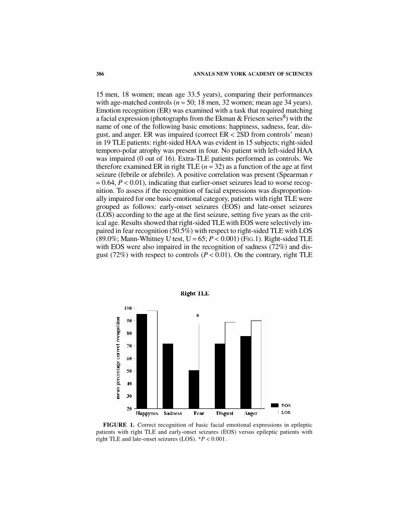
15 men, 18 women; mean age 33.5 years), comparing their performanceswith age-matched controls (n = 50; 18 men, 32 women; mean age 34 years).Emotion recognition (ER) was examined with a task that required matchinga facial expression (photographs from the Ekman & Friesen series8) with thename of one of the following basic emotions: happiness, sadness, fear, dis-gust, and anger. ER was impaired (correct ER < 2SD from controls’ mean)in 19 TLE patients: right-sided HAA was evident in 15 subjects; right-sidedtemporo-polar atrophy was present in four. No patient with left-sided HAAwas impaired (0 out of 16). Extra-TLE patients performed as controls. Wetherefore examined ER in right TLE (n = 32) as a function of the age at firstseizure (febrile or afebrile). A positive correlation was present (Spearman r= 0.64, P < 0.01), indicating that earlier-onset seizures lead to worse recog-nition. To assess if the recognition of facial expressions was disproportion-ally impaired for one basic emotional category, patients with right TLE weregrouped as follows: early-onset seizures (EOS) and late-onset seizures(LOS) according to the age at the first seizure, setting five years as the crit-ical age. Results showed that right-sided TLE with EOS were selectively im-paired in fear recognition (50.5%) with respect to right-sided TLE with LOS(89.0%; Mann-Whitney U test, U = 65; P < 0.001) (FIG.1). Right-sided TLEwith EOS were also impaired in the recognition of sadness (72%) and dis-gust (72%) with respect to controls (P < 0.01). On the contrary, right TLE
FIGURE 1. Correct recognition of basic facial emotional expressions in epilepticpatients with right TLE and early-onset seizures (EOS) versus epileptic patients withright TLE and late-onset seizures (LOS). *P < 0.001.
387MELETTI et al.: FEAR RECOGNITION AND TEMPORAL LOBE EPILEPSY
subjects with LOS and controls did not differ in the recognition of any emo-tional category.
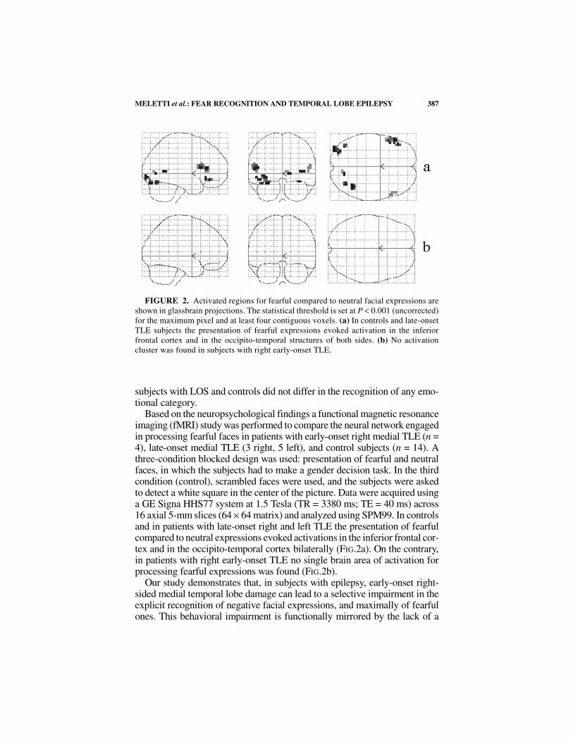
Based on the neuropsychological findings a functional magnetic resonanceimaging (fMRI) study was performed to compare the neural network engagedin processing fearful faces in patients with early-onset right medial TLE (n =4), late-onset medial TLE (3 right, 5 left), and control subjects (n = 14). Athree-condition blocked design was used: presentation of fearful and neutralfaces, in which the subjects had to make a gender decision task. In the thirdcondition (control), scrambled faces were used, and the subjects were askedto detect a white square in the center of the picture. Data were acquired usinga GE Signa HHS77 system at 1.5 Tesla (TR = 3380 ms; TE = 40 ms) across16 axial 5-mm slices (64 × 64 matrix) and analyzed using SPM99. In controlsand in patients with late-onset right and left TLE the presentation of fearfulcompared to neutral expressions evoked activations in the inferior frontal cor-tex and in the occipito-temporal cortex bilaterally (FIG.2a). On the contrary,in patients with right early-onset TLE no single brain area of activation forprocessing fearful expressions was found (FIG.2b).
Our study demonstrates that, in subjects with epilepsy, early-onset right-sided medial temporal lobe damage can lead to a selective impairment in theexplicit recognition of negative facial expressions, and maximally of fearfulones. This behavioral impairment is functionally mirrored by the lack of a
FIGURE 2. Activated regions for fearful compared to neutral facial expressions areshown in glassbrain projections. The statistical threshold is set at P < 0.001 (uncorrected)for the maximum pixel and at least four contiguous voxels. (a) In controls and late-onsetTLE subjects the presentation of fearful expressions evoked activation in the inferiorfrontal cortex and in the occipito-temporal structures of both sides. (b) No activationcluster was found in subjects with right early-onset TLE.
388 ANNALS NEW YORK ACADEMY OF SCIENCES
distinctive neural network underlying the processing of fearful facial expres-sions. Several studies conducted on human subjects showed the presence ofa crucial period for the development of emotion-recognition ability betweenthe fifth and the seventh year of age.9 It has been proposed that the amygdalais part of a system that has evolved for rapid detection of threatening stimuliand social signals such as facial expressions.10 Our data suggest that integrityof the right amygdala and related limbic structures during early infancy areessential for the processing and appropriate interpretation of social signalsconveyed by negative facial expressions later in life.
REFERENCES
1. ADOLPHS, R. 2002. Neural systems for recognizing emotions. Curr. OpinionNeurobiol. 12: 169–177.
2. MORRIS, J.S., et al. 1998. A neuromodulatory role for the human amygdala inprocessing emotional facial expressions. Brain 121: 47–57.
3. IIDAKA, T., et al. 2001. Neural interaction of the amygdala with the prefrontaland temporal cortices in the processing of facial expressions as revealed byfMRI. J. Cogn. Neurosci. 13: 1035–1047.
4. ADOLPHS, R., et al. 1994. Impaired recognition of emotion in facial expressionfollowing bilateral damage of the human amygdala. Nature 372: 669–672.
5. GLOOR, P. 1992. Role of the amygdala in temporal lobe epilepsy. In TheAmygdala. Neurobiological Aspects of Emotion, Memory, and Mental Dys-function. J. Aggleton, Ed.: 507–538. Wyley and Liss. New York.
6. TASCH, E., et al. 1999. Neuroimaging evidence of progressive neuronal lossand dysfunction in temporal lobe epilepsy. Ann. Neurol. 45: 568–576.
7. FUERST, D., et al. 2001. Volumetric MRI, pathological, and neuropsychologicalprogression in hippocampal sclerosis. Neurology 57: 184–188.
8. EKMAN, P. & W.V. FRIESEN. 1976. Pictures of Facial Affect. Consulting Psy-chologist Press. Palo Alto, CA.
9. TREMBLAY, C., et al. 2001. The recognition of adults’ and children's facialexpressions of emotions. J. Psychol. 121: 341–350.
10. ROLLS, E.T. 1992. Neurophysiology and functions of the primate amygdala. InThe Amygdala: Neurobiological Aspects of Emotion, Memory and MentalDysfunction. J.P. Aggleton, Ed. Wyley-Liss. New York.


![Self-Regulation of Amygdala Activation Using Real-Time ...€¦ · amygdala participates in more detailed and elaborate stimulus evaluation [20,26,27]. The involvement of the amygdala](https://img.pdfslide.net/doc/110x75/5fa8a495e8acaa50d8405bd2/self-regulation-of-amygdala-activation-using-real-time-amygdala-participates.jpg)