Embed Size (px)
Citation preview
.................... ................................................ ARTICLE
Charles B. Hermesch, DMD, John T. Cody, PhD, Jannine D. Cody, PhD
Dental caries history in nine children with chromosome 18p deletion syndrome
Chromosome 18p deletion syn- drome is caused by the deletion of a pottlon of genetlc material on the short (p) arm of chromosome 18. Many of 100 prior case reports In the medlcal lkrature descrlbing the dental health of subjects wlth thls syndrome reported multlple carles associated wlth the syn- drome. At the thlrd annual lnternatlonal conference of The Chromosome 18 Reglstry & Research Society, dental examinatlons were carried out on nine children wlth chromosome l 8 p deletion syndrome and flve of thelr unaffected siblings. The dental examlnation includ- ed an intra-oral evaluation of coronal decay and fllled permanent teeth sur- faces (DFS) and decayed and fllled prl- mary tooth surfaces (dfs) using a mouth mirror, explorer, and a high-lntenslty flber optlc light. An evaluation of the data revealed that five of nine chlldren wlth l 8 p deletlon synt'rome (56%) were free of tooth decay or a hlstory of tooth decay. Four of the nlne (44%) had tooth dewy or a history of tooth decay. The prevalence of decay was quite simllar in the genetlcally unaffected slbllngs. Three of the fhre (60%) unaffected sib- lings of the children with 18p were free of tooth decay, whereas two of the flve (40%) had tooth decay. One of the affected chlldren had a mlsslng mandibular left central lnclsor. None of the children had abnormally shaped teeth. The car/es pattern seems to be slmllar to that reported In the NHANES 111 data collected In the United States from l98&1991. Analysls of these p m llminary data suggests that the risk for carles In chromosome 18p deletlon syn- drome may be lower than previously reported.
hromosome 18p deletion syn- drome (18p-) is caused by the C deletion of a portion of genetic
material on the short (p) arm of chro- mosome 18. This condition was first reported in 1963 by de Grouchy et al.' The female-to-male ratio for individu- als with this syndrome is 2:l. Affected individuals have an average birth weight of 2600 grams (the low end of the normal range) and present with a variety of clinical findings, which can vary greatly in number and severity. Over 100 cases of this syndrome have been reported in the literature. The clinical characteristics can vary consid- erably among individuals. Several authors have reported multiple defects (> 10%) in persons with 18p deletion. These findings include men- tal retardation, short stature (< 5th percentile), abnormal external ears, cleft lip and/or palate, hypertelorism, flat nasal bridge, IgA deficiency, small mandible, epicanthic folds, short neck, microcephaly, ptosis, hypotonia, stra- bismus, broad trunk, and webbed neck? Several recent case reports have also observed dystonia3z4. The dental findings in individuals with chromosome 18p deletion syndrome are reported to be: dental hypoplasia,5 downturned corners of the mouth, late eruption of teeth, and irregular and often carious teeth.6-'3 It is unknown to what extent the chromo- somal anomaly contributed to the dental caries, since dental caries prevalence was high in the years dur- ing which most of these case reports were published.
Given the large number of reports of dental caries associated with 18p- syndrome in the literature, a survey of
the dental caries was undertaken. Clinical dental examinations were offered to children of parents attending the third annual international confer- ence of The Chromosome 18 Registry & Research Society, in Memphis, Tennessee. Some of the parents of chil- dren with chromosome 18p- expressed concerns that their children experi- enced more dental problems than their karotypically normal siblings or other children. The purpose of this prelimi- nary communication is to report data on the prevalence of dental caries (or dental caries history) in children with deletions of 18p and their unaffected siblings (normal chromosome 18).
Materials and methods
Fourteen children from nine families attending the conference voluntarily participated in a dental examination in which data related to their dental and chromosome status were collected. This convenience sample of children with 18p- syndrome (n = 9) ranged in age from 3 to 9 years. The sample of unaffected siblings (n = 5) ranged in age from 6 to 13 years. All the children were non-institutionalized. Children who were unable to cooperate with the examination procedure were excluded. The dental examination included an intra-oral soft tissue examination, and an evaluation of coronal decay and filled permanent teeth surfaces (DFS), and of decayed and filled primary tooth surfaces (dfs). One dentist (CBH) carried out the examinations using a mouth mirror, #23 explorer, and a high-intensity light. A second individual (JTC)
SCD Speclal Care In Dentistry, Vol20 No 2 2000 53
recorded the dental findings and perti- nent demographic data. All examina- tions were made on the same day and without the benefit of radiographs.
The 18p- diagnosis had been previ- ously made by karotyping. The sub- jects reported here are limited strictly to known 18p- deletions. Several chil- dren were examined who, in addition to 18p- deletion, had other chromoso- mal abnormalities, such as 18q+, 18q-, 15p+, and 8q+, but they are excluded from this report.
Results
The DE/dfs data from each of the subjects are shown in the Table. The dental examination revealed that five of nine children with 18p- (56%) were free of tooth decay or a history of tooth decay. Four of nine (44%) children had tooth decay or a history of tooth decay. The prevalence of decay was quite similar to that in the unaffected sib- lings. Three of five (60%) unaffected siblings of the children with 18p- were free of tooth decay, whereas two of the five (40%) had tooth decay. One of the affected children had a congenitally missing mandibular left central prima- ry incisor. None of the children had
abnormally shaped teeth or cleft palate or cleft lip.
Discussion
The prevalence of caries-free children with 18p- and their unaffected siblings compares favorably with that of chil- dren in the United States overall. In the third National Health and Nutrition Examination Survey (NHANES 111),14 data were collected by the National Institutes of Health from 1988 to 1991. The survey report- ed on weighted estimates for over 58 million children from ages 2 to 17. Of children aged 2 to 9 years, 62.1 % were decay-free in their primary dentition. In children aged 5 to 17 years, 54.7% were decay-free in their permanent dentition.
Canes, or the history of caries, was clustered in 44% of the children with 18p- syndrome and 40% of their siblings. This pattern is similar to that reported in the NHANES I11 data, in which 80% of the decay in permanent teeth was found in 25% of children ages 5 to 17.
Since these data are from clinical examinations of a small convenience sample of individuals volunteering for a dental examination, the findings
must be considered preliminary and are not based on a randomized sur- vey. Therefore, caution is advised in the generalization of these data. However, analysis of the data sug- gests that, for nine children with chromosome 18p-, their caries pattern is similar to that in unaffected chil- dren. Since the majority of children with 18p deletion syndrome were caries-free (56%), the majority of the decay was found in a small percent- age of individuals (44%). These find- ings are in conflict with previous case reports of a high rate of caries in chil- dren with chromosome 18p deletion syndrome. We speculate that previ- ous case reports reflected the wide- spread caries present in the popula- tion during past decades and not a specific genetic influence of chromo- some 18p deletion.
The majority of the individuals examined had a dentist of record and received regular dental examinations and preventive care. This care might ameliorate a genetic risk for dental caries if, in fact, chromosome 18p deletion results in increased caries risk. The interventions believed to be responsible for the decline in caries over the past several decades in the United States are appropriate for the
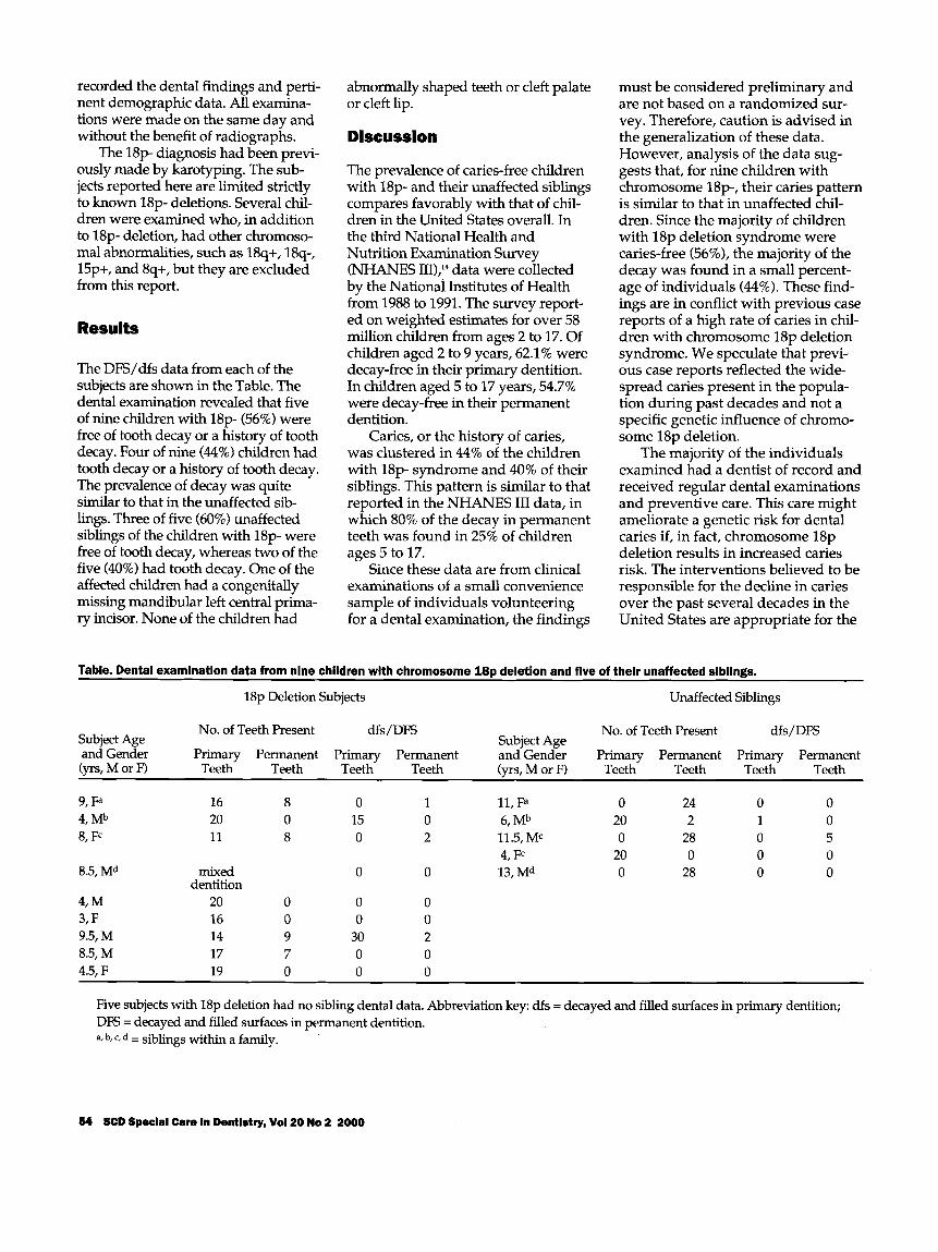
Table. Dental examination data from nine children with chromosome l 8 p deletlon and five of their unaffected slbllngs.
18p Deletion Subjects Unaffected Siblings
No. of Teeth Present dfs/DFS No. of Teeth Present dfs/DFS Subject Age Subject Age and Gender Primary Permanent Primary Permanent and’GendGr Primary Permanent Primary Permanent (yrs, M or F) Teeth Teeth Teeth Teeth (yrs, M or F) Teeth Teeth Teeth Teeth
9, Fa 4, Mb 8, Fc
8.5,Md
4, M 3, F 9.5, M 8.5, M 4.5, F
16 8 20 0 11 8
mixed dentition
20 0 16 0 14 9 17 7 19 0
0 15 0
0
0 0
30 0 0
1 11, Fa 0 24 0 0 0 6, I@ 20 2 1 0 2 11.5, ME 0 28 0 5
4, Fc 20 0 0 0 0 13, Md 0 28 0 0
Five subjects with 18p deletion had no sibling dental data. Abbreviation key: dfs = decayed and filled surfaces in primary dentition; DFS = decayed and filled surfaces in permanent dentition. a, b, c, d = siblings within a family.
54 SCD Special Care In Dentistry, Voi20 No 2 2000
dental health of patients with 18p-. Fluoride, sealants, oral hygiene, appropriate sucrose consumption, and regular dental care are all indi- cated to maximize their dental health. As with all children, the early identification of caries-susceptible children with chromosome 18p dele- tion is important.
Dr. Hermesch is Assistant Professor, Department of General Dentistry, The University of Texas Health Science Center, Mail Code 7914,7703 Floyd Curl Drive, San Antonio, Texas 78229-3900. Dr. John Cody is with the Interservice Physician Assistant Program, AMEDD C & S , MCCS-HMP PA Branch, 3151 Scott Road, Ft. Sam Houston, Texas. Dr. Jannine Cody is Assistant Professor, Department of Pediatrics, The University of Texas Health Science Center, San Antonio, Texas. Please address correspon- dence to Dr. Hermesch.
The Chromosome 18 Registry & Research Society is a non-profit educational/research organization that provides educational, refer- ral, and family support for persons with chro- mosome 18 anomalies as well as research and
treatment-related services. Its address is 6302 Fox Head, San Antonio, Texas 78247. The e-mail address is: [email protected].
The views expressed in this article are those of the authors and do not reflect the official poli- cy of the Department of Defense or other Departments of the US Government.
1.
2.
3.
4.
5.
6.
de Grouchy J, Lamy M, Thieffry S, et al. Dysmorphie complexe avec oligophrenie: deletion des bras courts d'un chromosome 17-18. CR'Acad Sci (Paris) 2561028-9,1963. Schinzel A, Schmid W, Luscher U, et al. Structural aberrations of chromosome 18. I. The 18p- syndrome. Arch fiir Genetik 471- 15,1974. Awaad Y, Munoz S, Nigro M. Progressive dystonia in a child with chromosome 18p deletion treated with intrathecal baclofen. J Child Neurol14:75-7,1999. Klein C, Page CE, LeWitt P, et al. Genetic analysis of three patients with an 18p- syn- drome and dystonia. Neurology 52:649-51, 1999. Surh LC, Ledbetter DH, Greenberg R. Interstitial deletion of chromosome 18. Am J Med Genet 41:15-7,1991. Uchida IA, McRae KN, Wang HC, et al. Familial short arm deficiency of chromo- some 18 concomitant with arhinencephaly
and alopecia1 congenita. Am J Hum Genet
de Grouchy J. The 18p-, 18q- and 18r syn- dromes. Birth Defects: Orig Art Ser 5(5):74- 87,1969. Levenson JE, Crandall BF, Sparkes RS. Partial deletion syndromes of chromosome 18. Ann Ophthalmol3:756-60,1971. Taylor KM, Wolfinger HL, Brown MG, et al. Origin of a small metacentric chromo- some: familial and cytogenetic evidence. Clin Genet 8:364-9,1975.
10. Funderburk SJ. 18p- syndrome resulting from translocation (13q;18p) in a mildly affected adult male. J Med Genet 16399- 402,1979.
11. Schinzel A. Catalogue of unbalanced chro- mosome aberrations in man. Berlin: Walter de Gruyter & Co., pp. 604-5,1983.
Intellectual, behavioral, and linguistic characteristics of three children with 18p- syndrome. J Dev Behav Pediatr 7(1):1-7, 1986.
13. Aughton DJ, Alsaadi AA, Transue DJ. Single maxillary central incisor in a girl with del(18p) syndrome. J Med Genet
14. Brown LJ, Kaste LM, Selwitz RH, et al.
17410-9,1965. 7.
8.
9.
12. Thompson RW, Peters JE, Smith SD.
28530-2,1991.
Dental caries & sealant usage in U.S. chil- dren, 1988-1991. J Am Dent Assoc 127335- 43,1996.
SCD Special Care In Dentistry, Vol20 No 2 2000 55