Embed Size (px)
Citation preview
337
Abstract: Oculodentodigital dysplasia is anextremely rare autosomal dominant pleiotropicdisorder. The syndrome is characterized by abnormalfacial features, central nervous system involvement,syndactyly and clinodactyly of fourth and fifth fingers,dry and lusterless hair, generalized enamel hypoplasiaand odontodysplasia. Combination of odontodysplasia,poor oral hygiene, and parental neglect can lead toextensive destruction of tooth structure and thetreatment options become limited. Early diagnosiswith a proper treatment plan and meticulous oralhygiene program helps eliminate the necessity ofmultiple tooth extractions. This case report describesthe comprehensive dental treatment aimed atrehabilitation of function and aesthetics of the dentitionin an 8-year-old boy with oculodentodigital dysplasia.(J Oral Sci 52, 337-342, 2010)
Keywords: dental enamel hypoplasia; mouthrehabilitation; odontodysplasia.
IntroductionOculodentodigital dysplasia (ODDD) is a highly
penetrant autosomal dominant disorder, with variableexpression (1). ODDD is caused by mutations in the GJA1gene located on human chromosomes 6q22-q23, encodingthe gap junction protein Connexin 43 (Cx43) (2,3). Oral
and dental manifestations of ODDD consist of oval palate(4), coronoid hypoplasia (5), mandibular retrognathism (6),enamel hypoplasia of the deciduous as well as permanentdentition and delayed tooth eruption (6). Histologicalexamination shows enamel dysplasia, dentin hypocal-cification, pulp denticles and hypercementosis (5).
Dental manifestations of the syndrome includinggeneralized enamel hypoplasia, thin dentinal walls,extensive caries leading to early pulp involvement, and oftenopen apices present challenges in providing dental treatmentfor these patients. A conservative approach should betaken to maintain the integrity and aesthetics of the patient’spermanent dentition. The conservative treatment optionsrequire knowledge of the best choices for the presentingclinical situation, i.e., functional requirements and theinherent aesthetic properties of each material. The reportedcase describes the comprehensive dental treatment of an8-year-old white boy diagnosed with oculodentodigitaldysplasia (7).
Case ReportAn 8-year-old boy was referred to the Department of
Paediatric Dentistry, Tabriz University of Medical Sciencesfor treatment of a dental abscess related to the left lowerfirst premolar.
Dental findings included yellowish color of the teeth withextensive carious lesions, hypoplastic appearance of theatypically-shaped teeth which were smaller than normal,and premature eruption of permanent canines andpremolars. The parents reported that the lower right andleft central incisors and lower left lateral incisor weremissing and there was no history of extraction (Fig. 1).Panoramic radiograph revealed shortened teeth with ‘ghost’appearance, unclear lamina dura, decreased thickness of
Journal of Oral Science, Vol. 52, No. 2, 337-342, 2010
Correspondence to Dr. Naser Asl Aminabadi, Department ofPediatric Dentistry, School of Dentistry, Tabriz University(Medical Sciences), Daneshgah St., Tabriz, IranTel: +98-411-3340310, +98-914-415-7200Fax: +98-411-3346977E-mail: [email protected] & [email protected]
Dental management of oculodentodigital dysplasia: a case report
Naser A. Aminabadi1), Maryam Pourkazemi1), Sina G. Oskouei2) and Zahra Jamali3)
1)Department of Pediatric Dentistry, School of Dentistry, Tabriz University (Medical Sciences), Tabriz, Iran2) School of Dentistry, Tabriz University (Medical Sciences), Tabriz, Iran
3)Department of Oral Medicine, School of Dentistry, Tabriz University (Medical Sciences), Tabriz, Iran
(Received 13 October 2009 and accepted 10 March 2010)
Case Report

338
dentin, and lack of contrast between enamel and dentine(Fig. 2). The existing permanent teeth were found to havetaurodontism. Periapical radiographs of the teeth revealedirregular calcification and pulp denticles in most of the teethevaluated (Figs. 3a and b). The permanent upper caninesreached full eruption during the course of the treatmentwhich lasted for two months, indicating an accelerated rateof eruption. During the same period, the second permanentmolars also erupted.
The patient’s five year old brother had frontal bossa,clinodactyly, hyperkeratosis of hands, mild enamelhypoplasia of primary teeth, and a bi-rooted right upperprimary canine. The patient’s parents who were secondcousins also had clinodactyly. Considering all the signsobserved in the family, oculodentodigital dysplasia wassuspected, and therefore, the patient was referred to apediatric hospital for further evaluation.
The patient’s ophthalmologic examination revealedmicrophthalmia, microcornea, congenital cataract, chronicuveitis, glaucoma and reduced vision. Conventionalradiography of both hands revealed agenesis of secondphalanges of the fifth fingers and osteopenia of the wrists.No congenital heart disease was reported after examinationby the specialist.
Autosomal dominant trait of the syndrome was suggestedby genetic evaluation. To confirm the clinical diagnosis,a genome-wide search for the location of ODDD locus wasperformed using Short Tandem Repeat Polymorphisms(STRPs). Evidence of linkage between ODDD and markersfrom chromosome 6q22-q23 was detected. Missensemutation in GJA1 (Connexin 43) exon 2 was found.
Before visiting the Department of Pediatric Dentistry,left upper and lower first permanent molars, left uppercentral incisor, and right lower first permanent molar hadbeen extracted. On the first appointment, under localanesthesia (2% lidocaine with 1:100,000 epinephrine;Dentsply Pharmaceutical, York, PA, USA), pulp tissue ofthe abscessed left lower first premolar was extirpatedusing a Hedström-file and irrigated with 2.5% sodiumhypochlorite. A calcium hydroxide dressing (DentsplyHerpo, Petrópolis, RJ, Brazil) was then given for 14 daysand the tooth was restored with light-cured glass-ionomer(GC Fuji II LC, Tokyo, Japan) temporarily.
After resolution of the acute symptoms, in the nextappointment, diagnostic dental casts of both arches wereprepared for precise evaluation. The overall treatmentobjective, procedures involved, limitations, and alternativetreatments were explained to the parents, and they signedan informed consent form.
At a subsequent appointment, after administration of localanesthesia to the left upper and lower quadrants, caries were
Fig. 1 Dysplastic teeth and multiple caries seen beforetreatment.
Fig. 2 Panoramic radiograph of the patient showing shortenedteeth with ‘ghost’ appearance, generalized enlarged pulpchambers, and decreased thickness of dentin. Note theperiapical radiolucency of lower left first premolar.
Fig. 3 (a) Short length and diffused calcification of rightupper central incisor. Pulp calcification is seen inlateral incisor; (b) MTA plug is seen in the apex of rightupper second premolar. Also note the calcification ofcoronal pulp in canine and first premolar.
339
removed using a No. 1 round bur on a slow-speed hand-piece and an excavator. Pulp exposure was seen in the leftupper second premolar, which revealed a necrotic pulp.Considering the fragility of the teeth, a rubber dam couldnot be placed and, thus, isolation was accomplished usingcotton rolls. Due to thin dentinal walls of teeth, minimalfiling was performed, and after irrigation with 2.5% sodiumhypochlorite solution, root canals were dressed withcalcium hydroxide paste for 14 days and restored with light-cured glass-ionomer temporarily.
In teeth without pulpal involvement following cariesremoval, the pulp was protected with a liner (Dycal Ivory;Dentsply De Trey, Konstanz, Germany), and the preparedcavities were filled with light-cured glass-ionomer untila composite full-veneer was placed later.
In the following appointment, the same treatmentprocedures were performed in the right upper and lowerquadrants. Initial filing of the root canal revealed dispersedcalcification and non-vital pulp of the upper right centralincisor. After irrigation with sodium hypochlorite solution,the root canal was filled with calcium hydroxide paste andthe tooth was restored with light-cured glass-ionomertemporarily. Final endodontic therapy was performed onthe teeth after two weeks.
Continuing with the right side in the next appointment,the upper lateral incisor and second premolar were exposedduring caries removal, revealing a hyperemic pulp whichindicated irreversible pulpitis. Due to incomplete rootformation, apexification with Mineral Trioxide Aggregate(MTA; Dentsply, Tulsa Dental, OK, USA) was consideredfor these teeth. Minimal filing to the working length (14mm in the lateral incisor and 13 mm in the second premolar)was performed followed by irrigation. The apical 3-mmof root canals was filled with MTA and a periapicalradiograph was taken (Fig. 3b). Then, a moist cotton pelletwas placed in the canals and teeth were restored temporarilywith light-cured glass-ionomer. In a subsequent appoint-ment, teeth were reinforced with fibre post (D.T.Composipost RTD, France) and restored with light-curedcomposite. Later, the second premolar was covered withan SSC (Unitek Stainless Steel Crowns, 3M ESPE, St. Paul,MN, USA).
Upper right central incisor had a short working length(10 mm) and malformed root canal, which made the useof fibre-post impossible (Fig. 3a). Therefore, the total rootcanal length was filled with MTA, and the coronal part ofthe teeth was filled with light-cured composite. The toothwas subsequently restored with the direct full-veneercomposite technique.
Lower left first premolar also had a short working length(8 mm). Total canal length was filled with MTA, and
subsequently restored with light-cured composite untilfull-coverage with SSC.
For the upper left second premolar (working length, 13mm), the same procedures that were performed for theupper right second premolar (MTA plug and fibre-postplacement) were followed.
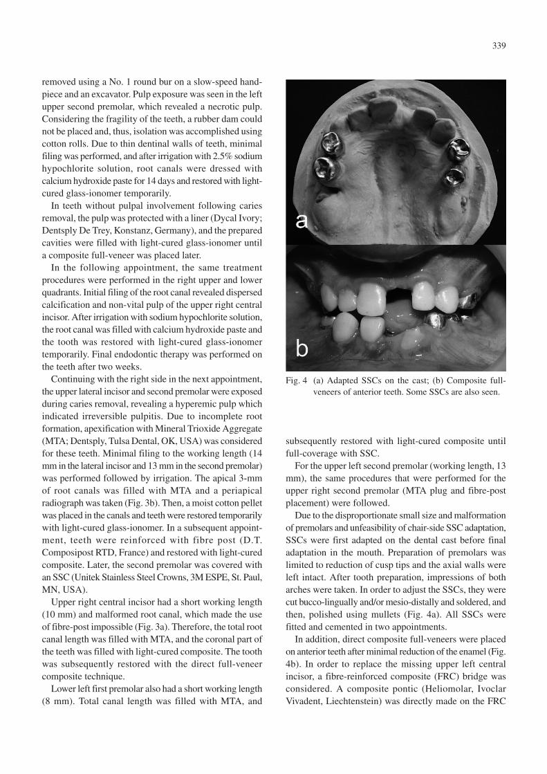
Due to the disproportionate small size and malformationof premolars and unfeasibility of chair-side SSC adaptation,SSCs were first adapted on the dental cast before finaladaptation in the mouth. Preparation of premolars waslimited to reduction of cusp tips and the axial walls wereleft intact. After tooth preparation, impressions of botharches were taken. In order to adjust the SSCs, they werecut bucco-lingually and/or mesio-distally and soldered, andthen, polished using mullets (Fig. 4a). All SSCs werefitted and cemented in two appointments.
In addition, direct composite full-veneers were placedon anterior teeth after minimal reduction of the enamel (Fig.4b). In order to replace the missing upper left centralincisor, a fibre-reinforced composite (FRC) bridge wasconsidered. A composite pontic (Heliomolar, IvoclarVivadent, Liechtenstein) was directly made on the FRC
Fig. 4 (a) Adapted SSCs on the cast; (b) Composite full-veneers of anterior teeth. Some SSCs are also seen.
340
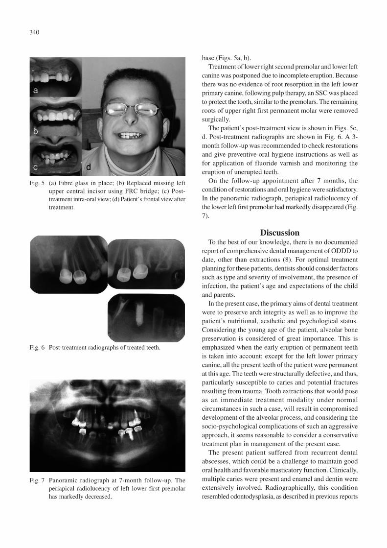
base (Figs. 5a, b).Treatment of lower right second premolar and lower left
canine was postponed due to incomplete eruption. Becausethere was no evidence of root resorption in the left lowerprimary canine, following pulp therapy, an SSC was placedto protect the tooth, similar to the premolars. The remainingroots of upper right first permanent molar were removedsurgically.
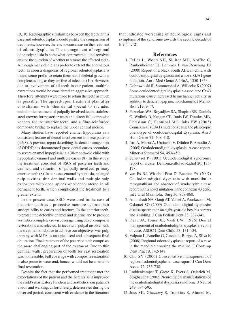
The patient’s post-treatment view is shown in Figs. 5c,d. Post-treatment radiographs are shown in Fig. 6. A 3-month follow-up was recommended to check restorationsand give preventive oral hygiene instructions as well asfor application of fluoride varnish and monitoring theeruption of unerupted teeth.
On the follow-up appointment after 7 months, thecondition of restorations and oral hygiene were satisfactory.In the panoramic radiograph, periapical radiolucency ofthe lower left first premolar had markedly disappeared (Fig.7).
DiscussionTo the best of our knowledge, there is no documented
report of comprehensive dental management of ODDD todate, other than extractions (8). For optimal treatmentplanning for these patients, dentists should consider factorssuch as type and severity of involvement, the presence ofinfection, the patient’s age and expectations of the childand parents.
In the present case, the primary aims of dental treatmentwere to preserve arch integrity as well as to improve thepatient’s nutritional, aesthetic and psychological status.Considering the young age of the patient, alveolar bonepreservation is considered of great importance. This isemphasized when the early eruption of permanent teethis taken into account; except for the left lower primarycanine, all the present teeth of the patient were permanentat this age. The teeth were structurally defective, and thus,particularly susceptible to caries and potential fracturesresulting from trauma. Tooth extractions that would poseas an immediate treatment modality under normalcircumstances in such a case, will result in compromiseddevelopment of the alveolar process, and considering thesocio-psychological complications of such an aggressiveapproach, it seems reasonable to consider a conservativetreatment plan in management of the present case.
The present patient suffered from recurrent dentalabscesses, which could be a challenge to maintain goodoral health and favorable masticatory function. Clinically,multiple caries were present and enamel and dentin wereextensively involved. Radiographically, this conditionresembled odontodysplasia, as described in previous reports
Fig. 5 (a) Fibre glass in place; (b) Replaced missing leftupper central incisor using FRC bridge; (c) Post-treatment intra-oral view; (d) Patient’s frontal view aftertreatment.
Fig. 6 Post-treatment radiographs of treated teeth.
Fig. 7 Panoramic radiograph at 7-month follow-up. Theperiapical radiolucency of left lower first premolarhas markedly decreased.
341
(9,10). Radiographic similarities between the teeth in thiscase and odontodysplasia could justify the comparison oftreatments; however, there is no consensus on the treatmentof odontodysplasia. The management of regionalodontodysplasia is somewhat controversial and revolvesaround the question of whether to remove the affected teeth.Although many clinicians prefer to extract the anomalousteeth as soon a diagnosis of regional odontodysplasia ismade, some prefer to retain them until skeletal growth iscomplete as long as they are free of infection (10). However,due to involvement of all teeth in our patient, multipleextractions would be considered an aggressive approach.Therefore, attempts were made to retain the teeth as muchas possible. The agreed-upon treatment plan afterconsultation with other dental specialists includedendodontic treatment of pulpally involved teeth, stainlesssteel crowns for posterior teeth and direct full compositeveneers for the anterior teeth, and a fibre-reinforcedcomposite bridge to replace the upper central incisor.
Many studies have reported enamel hypoplasia as aconsistent feature of dental involvement in these patients(4,6,8). A previous report describing the dental managementof ODDD has documented gross dental caries secondaryto severe enamel hypoplasia in a 30-month-old child withhypoplastic enamel and multiple caries (8). In this study,the treatment consisted of SSCs of posterior teeth andcanines, and extraction of pulpally involved primaryanterior teeth (8). In our case, enamel hypoplasia, enlargedpulp cavities, thin dentinal walls and multiple pulpexposures with open apices were encountered in allpermanent teeth, which complicated the treatment to agreater extent.
In the present case, SSCs were used in the case ofposterior teeth as a protective measure against theirsusceptibility to caries and fracture. In the anterior teeth,to protect the defective enamel and dentine and to provideaesthetics, complete crown coverage using direct compositerestorations was selected. In teeth with pulpal involvement,the treatment of choice to achieve our objectives was pulptherapy with MTA as an apical seal and subsequent finalobturation. Final treatment of the posterior teeth comprisesthe more challenging part of the treatment. Due to thindentinal walls, preparation of teeth for cast restorationwas not feasible. Full coverage with composite restorationis also prone to wear and, hence, would not be a suitablefinal restoration.
Despite the fact that the performed treatment met theexpectations of the patient and the parents as it improvedthe child’s masticatory function and aesthetics, our patient’svision and walking, unfortunately, deteriorated during theobserved period, consistent with evidence in the literature
that indicated worsening of neurological signs andsymptoms of the syndrome towards the second decade oflife (11,12).
References1. Feller L, Wood NH, Sluiter MD, Noffke C,
Raubenheimer EJ, Lemmer J, van Rensburg EJ(2008) Report of a black South African child withoculodentodigital dysplasia and a novel GJA1 genemutation. Am J Med Genet A 146A, 1350-1353.
2. Dobrowolski R, Sommershof A, Willecke K (2007)Some oculodentodigital dysplasia-associated Cx43mutations cause increased hemichannel activity inaddition to deficient gap junction channels. J MembrBiol 219, 9-17.
3. Paznekas WA, Boyadjiev SA, Shapiro RE, DanielsO, Wollnik B, Keegan CE, Innis JW, Dinulos MB,Christian C, Hannibal MC, Jabs EW (2003)Connexin 43 (GJA1) mutations cause the pleiotropicphenotype of oculodentodigital dysplasia. Am JHum Genet 72, 408-418.
4. Itro A, Marra A, Urciuolo V, Difalco P, Amodio A(2005) Oculodentodigital dysplasia. A case report.Minerva Stomatol 54, 453-459.
5. Scheutzel P (1991) Oculodentodigital syndrome:report of a case. Dentomaxillofac Radiol 20, 175-178.
6. van Es RJ, Wittebol-Post D, Beemer FA (2007)Oculodentodigital dysplasia with mandibularretrognathism and absence of syndactyly: a casereport with a novel mutation in the connexin 43 gene.Int J Oral Maxillofac Surg 36, 858-860.
7. Aminabadi NA, Ganji AT, Vafaei A, Pourkazemi M,Oskouei SG (2009) Oculodentodigital dysplasia:disease spectrum in an eight-year-old boy, his parentsand a sibling. J Clin Pediatr Dent 33, 337-341.
8. Dean JA, Jones JE, Vash BW (1986) Dentalmanagement of oculodentodigital dysplasia: reportof case. ASDC J Dent Child 53, 131-134.
9. Volpato L, Botelho G, Casela L, Borges A, Silva K(2008) Regional odontodysplasia: report of a casein the mandible crossing the midline. J ContempDent Pract 9, 142-148.
10. Cho SY (2006) Conservative management ofregional odontodysplasia: case report. J Can DentAssoc 72, 735-738.
11. Loddenkemper T, Grote K, Evers S, Oelerich M,Stögbauer F (2002) Neurological manifestations ofthe oculodentodigital dysplasia syndrome. J Neurol249, 584-595.
12. Joss SK, Ghazawy S, Tomkins S, Ahmed M,

342
Bradbury J, Sheridan E (2008) Variable expressionof neurological phenotype in autosomal recessive
oculodentodigital dysplasia of two sibs and reviewof the literature. Eur J Pediatr 167, 341-345.