Embed Size (px)
Citation preview
CASE REPORT
Dentinogenesis imperfecta associated with short stature,hearing loss and mental retardation: a new syndrome withautosomal recessive inheritance?
R. G. E. C. Cauwels1, P. J. De Coster
1, G. R. Mortier
2, L. A. M. Marks
1, L. C. Martens
1
1Department of Paediatric Dentistry, Centre for Special Care, Paecamed Research, Ghent University, Belgium; 2Department ofMedical Genetics, Ghent University Hospital, Belgium
The follow-up history and oral findings in two brothers
from consanguineous parents suggest that the association
of dentinogenesis imperfecta (DI), delayed tooth erup-
tion, mild mental retardation, proportionate short stat-
ure, sensorineural hearing loss and dysmorphic facies
may represent a new syndrome with autosomal recessive
inheritance. Histological examination of the dentin mat-
rix of a permanent molar from one of the siblings reveals
morphological similarities with defective dentinogenesis
as presenting in patients affected with Osteogenesis
Imperfecta (OI), a condition caused by deficiency of
type I collagen. A number of radiographic and histological
characteristics, however, are inconsistent with classical
features of DI. These findings suggest that DI may imply
greater genetical heterogeneity than currently assumed.
J Oral Pathol Med (2005) 34: 444–6
Keywords: autosomal recessive; collagen I; consanguinity; den-
tinogenesis imperfecta
Cases
Two brothers with a similar association of congenitalanomalies were referred for oral examination to theCentre for Special Care, Ghent University Hospital, atthe ages of 3 and 7 years. Their parents were of Turkishorigin and were consanguineous (first cousins). Theolder sister as well as both parents were phenotypicallynormal. Both boys presented at birth with a low weightand showed delayed psychomotor development onfollow-up. They currently have a mild mental retarda-tion with normal karyotype and normal brain MRI. Inaddition they suffer from sensorineural hearing loss.Physical examination revealed proportionate short
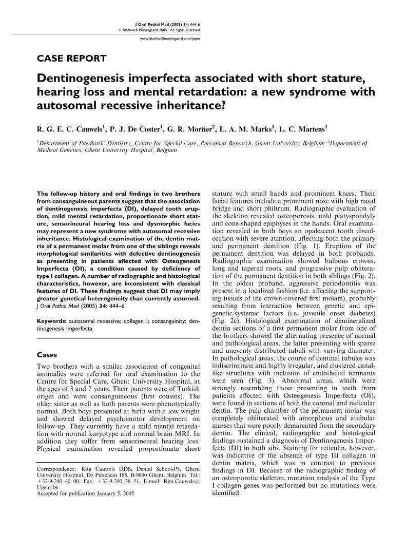
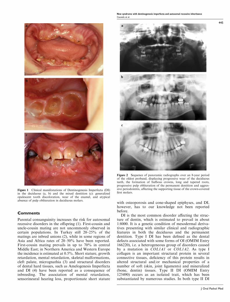
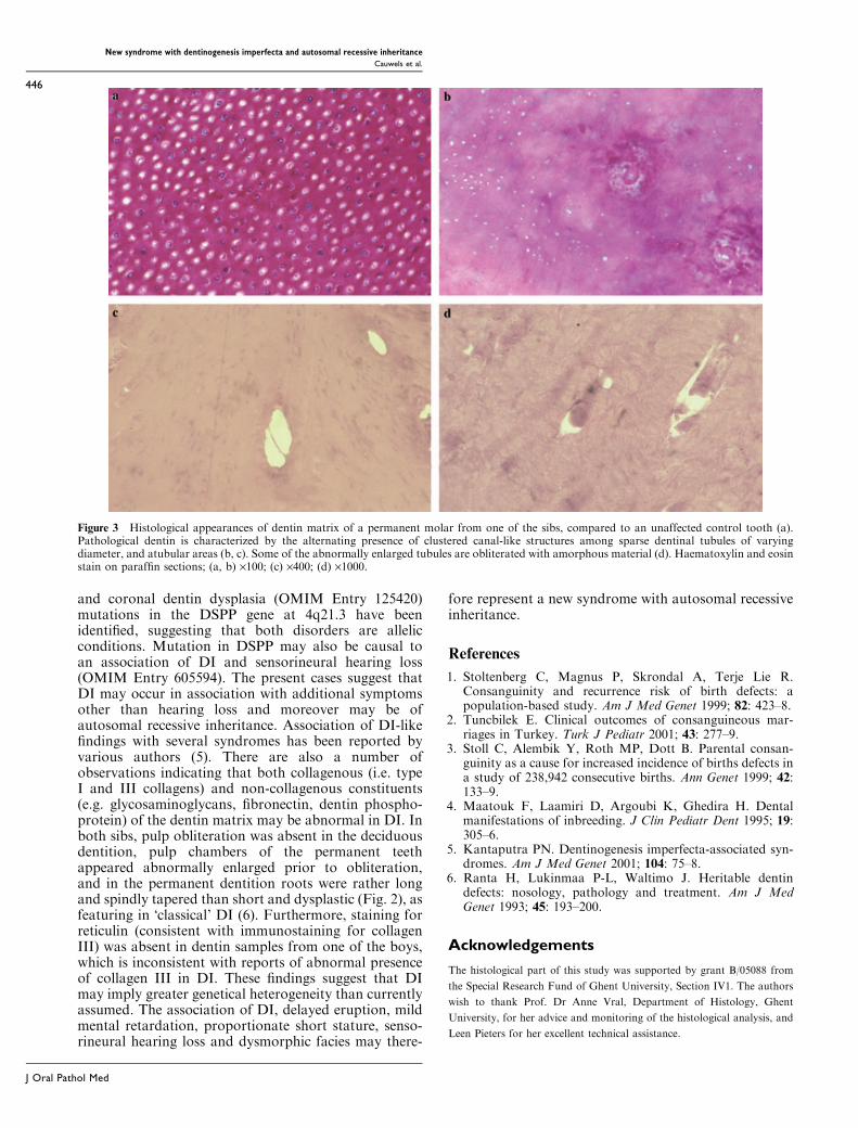
stature with small hands and prominent knees. Theirfacial features include a prominent nose with high nasalbridge and short philtrum. Radiographic evaluation ofthe skeleton revealed osteoporosis, mild platyspondylyand cone-shaped epiphyses in the hands. Oral examina-tion revealed in both boys an opalescent tooth discol-oration with severe attrition, affecting both the primaryand permanent dentition (Fig. 1). Eruption of thepermanent dentition was delayed in both probands.Radiographic examination showed bulbous crowns,long and tapered roots, and progressive pulp oblitera-tion of the permanent dentition in both siblings (Fig. 2).In the oldest proband, aggressive periodontitis waspresent in a localized fashion (i.e. affecting the support-ing tissues of the crown-covered first molars), probablyresulting from interaction between genetic and epi-genetic/systemic factors (i.e. juvenile onset diabetes)(Fig. 2c). Histological examination of demineralizeddentin sections of a first permanent molar from one ofthe brothers showed the alternating presence of normaland pathological areas, the latter presenting with sparseand unevenly distributed tubuli with varying diameter.In pathological areas, the course of dentinal tubules wasindiscriminate and highly irregular, and clustered canal-like structures with inclusion of endothelial remnantswere seen (Fig. 3). Abnormal areas, which werestrongly resembling those presenting in teeth frompatients affected with Osteogenesis Imperfecta (OI),were found in sections of both the coronal and radiculardentin. The pulp chamber of the permanent molar wascompletely obliterated with amorphous and atubularmasses that were poorly demarcated from the secondarydentin. The clinical, radiographic and histologicalfindings sustained a diagnosis of Dentinogenesis Imper-fecta (DI) in both sibs. Staining for reticulin, however,was indicative of the absence of type III collagen indentin matrix, which was in contrast to previousfindings in DI. Because of the radiographic finding ofan osteoporotic skeleton, mutation analysis of the TypeI collagen genes was performed but no mutations wereidentified.
Correspondence: Rita Cauwels DDS, Dental School-P8, GhentUniversity Hospital, De Pintelaan 185, B-9000 Ghent, Belgium. Tel.:+32-9-240 40 00, Fax: +32-9-240 38 51, E-mail: [email protected] for publication January 5, 2005
J Oral Pathol Med (2005) 34: 444–6
ª Blackwell Munksgaard 2005 Æ All rights reserved
www.blackwellmunksgaard.com/jopm
Comments
Parental consanguinity increases the risk for autosomalrecessive disorders in the offspring (1). First-cousin anduncle-cousin mating are not uncommonly observed incertain populations. In Turkey still 20–25% of thematings are inbred unions (2), while in some regions ofAsia and Africa rates of 20–50% have been reported.First-cousin mating prevails in up to 70% in centralMiddle East; in Northern America and Western Europethe incidence is estimated at 0.5%. Short stature, growthretardation, mental retardation, skeletal malformations,cleft palate, micrognathia (3) and structural disordersof dental hard tissues, such as Amelogenesis Imperfectaand DI (4) have been reported as a consequence ofinbreeding. The association of mental retardation,sensorineural hearing loss, proportionate short stature
with osteoporosis and cone-shaped epiphyses, and DI,however, has to our knowledge not been reportedbefore.
DI is the most common disorder affecting the struc-ture of dentin, which is estimated to prevail in about1:8000. It is a genetic condition of mesodermal deriva-tives presenting with similar clinical and radiographicfeatures in both the deciduous and the permanentdentition. Type I DI has been defined as the dentaldefects associated with some forms of OI (OMIM Entry166220), i.e. a heterogeneous group of disorders causedby a mutation in COL1A1 or COL1A2. As type Icollagen is an important structural protein in severalconnective tissues, deficiency of this protein results inaltered structural and/or mechanical properties of anumber of soft (skin, joint ligaments) and mineralized(bone, dentin) tissues. Type II DI (OMIM Entry125490) occurs as an isolated trait, which has beensubstantiated by numerous studies. In both type II DI
Figure 1 Clinical manifestations of Dentinogenesis Imperfecta (DI)in the deciduous (a, b) and the mixed dentition (c): generalizedopalescent tooth discoloration, wear of the enamel, and atypicalabsence of pulp obliteration in deciduous molars.
Figure 2 Sequence of panoramic radiographs over an 8-year periodof the oldest proband, displaying progressive wear of the deciduousteeth, the formation of bulbous crowns, long and tapered roots,progressive pulp obliteration of the permanent dentition and aggres-sive periodontitis, affecting the supporting tissue of the crown-coveredfirst molars.
New syndrome with dentinogenesis imperfecta and autosomal recessive inheritance
Cauwels et al.
445
J Oral Pathol Med
and coronal dentin dysplasia (OMIM Entry 125420)mutations in the DSPP gene at 4q21.3 have beenidentified, suggesting that both disorders are allelicconditions. Mutation in DSPP may also be causal toan association of DI and sensorineural hearing loss(OMIM Entry 605594). The present cases suggest thatDI may occur in association with additional symptomsother than hearing loss and moreover may be ofautosomal recessive inheritance. Association of DI-likefindings with several syndromes has been reported byvarious authors (5). There are also a number ofobservations indicating that both collagenous (i.e. typeI and III collagens) and non-collagenous constituents(e.g. glycosaminoglycans, fibronectin, dentin phospho-protein) of the dentin matrix may be abnormal in DI. Inboth sibs, pulp obliteration was absent in the deciduousdentition, pulp chambers of the permanent teethappeared abnormally enlarged prior to obliteration,and in the permanent dentition roots were rather longand spindly tapered than short and dysplastic (Fig. 2), asfeaturing in �classical’ DI (6). Furthermore, staining forreticulin (consistent with immunostaining for collagenIII) was absent in dentin samples from one of the boys,which is inconsistent with reports of abnormal presenceof collagen III in DI. These findings suggest that DImay imply greater genetical heterogeneity than currentlyassumed. The association of DI, delayed eruption, mildmental retardation, proportionate short stature, senso-rineural hearing loss and dysmorphic facies may there-
fore represent a new syndrome with autosomal recessiveinheritance.
References
1. Stoltenberg C, Magnus P, Skrondal A, Terje Lie R.Consanguinity and recurrence risk of birth defects: apopulation-based study. Am J Med Genet 1999; 82: 423–8.
2. Tuncbilek E. Clinical outcomes of consanguineous mar-riages in Turkey. Turk J Pediatr 2001; 43: 277–9.
3. Stoll C, Alembik Y, Roth MP, Dott B. Parental consan-guinity as a cause for increased incidence of births defects ina study of 238,942 consecutive births. Ann Genet 1999; 42:133–9.
4. Maatouk F, Laamiri D, Argoubi K, Ghedira H. Dentalmanifestations of inbreeding. J Clin Pediatr Dent 1995; 19:305–6.
5. Kantaputra PN. Dentinogenesis imperfecta-associated syn-dromes. Am J Med Genet 2001; 104: 75–8.
6. Ranta H, Lukinmaa P-L, Waltimo J. Heritable dentindefects: nosology, pathology and treatment. Am J MedGenet 1993; 45: 193–200.
Acknowledgements
The histological part of this study was supported by grant B/05088 from
the Special Research Fund of Ghent University, Section IV1. The authors
wish to thank Prof. Dr Anne Vral, Department of Histology, Ghent
University, for her advice and monitoring of the histological analysis, and
Leen Pieters for her excellent technical assistance.
Figure 3 Histological appearances of dentin matrix of a permanent molar from one of the sibs, compared to an unaffected control tooth (a).Pathological dentin is characterized by the alternating presence of clustered canal-like structures among sparse dentinal tubules of varyingdiameter, and atubular areas (b, c). Some of the abnormally enlarged tubules are obliterated with amorphous material (d). Haematoxylin and eosinstain on paraffin sections; (a, b) ·100; (c) ·400; (d) ·1000.
New syndrome with dentinogenesis imperfecta and autosomal recessive inheritance
Cauwels et al.
446
J Oral Pathol Med


![Successful management of a type II Dens invaginatus with an … · 2018-11-28 · of permanent tooth germs, taurodontism, supernumerary tooth, and dentinogenesis imperfecta [3]. Clinically,](https://img.pdfslide.net/doc/110x75/5e9529423a2cec077d2f9125/successful-management-of-a-type-ii-dens-invaginatus-with-an-2018-11-28-of-permanent.jpg)