Embed Size (px)
Citation preview
Department of Dermatology, InselspitalUniversity Hospital BernActivity Report 2015–2016

2
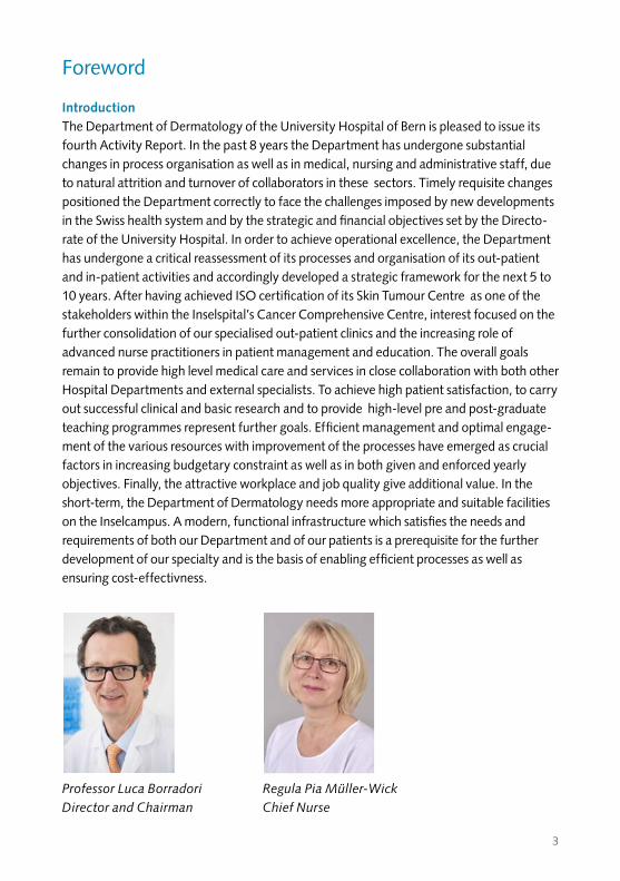
Professor Luca Borradori Regula Pia Müller-Wick Director and Chairman Chief Nurse
3
Foreword
IntroductionThe Department of Dermatology of the University Hospital of Bern is pleased to issue its fourth Activity Report. In the past 8 years the Department has undergone substantial changes in process organisation as well as in medical, nursing and administrative staff, due to natural attrition and turnover of collaborators in these sectors. Timely requisite changes positioned the Department correctly to face the challenges imposed by new developments in the Swiss health system and by the strategic and financial objectives set by the Directo-rate of the University Hospital. In order to achieve operational excellence, the Department has undergone a critical reassessment of its processes and organisation of its out-patient and in-patient activities and accordingly developed a strategic framework for the next 5 to 10 years. After having achieved ISO certification of its Skin Tumour Centre as one of the stakeholders within the Inselspital’s Cancer Comprehensive Centre, interest focused on the further consolidation of our specialised out-patient clinics and the increasing role of advanced nurse practitioners in patient management and education. The overall goals remain to provide high level medical care and services in close collaboration with both other Hospital Departments and external specialists. To achieve high patient satisfaction, to carry out successful clinical and basic research and to provide high-level pre and post-graduate teaching programmes represent further goals. Efficient management and optimal engage-ment of the various resources with improvement of the processes have emerged as crucial factors in increasing budgetary constraint as well as in both given and enforced yearly objectives. Finally, the attractive workplace and job quality give additional value. In the short-term, the Department of Dermatology needs more appropriate and suitable facilities on the Inselcampus. A modern, functional infrastructure which satisfies the needs and requirements of both our Department and of our patients is a prerequisite for the further development of our specialty and is the basis of enabling efficient processes as well as ensuring cost-effectivness.
Professor Luca Borradori Regula Pia Müller-Wick Director and Chairman Chief Nurse

4
Director and Chairman Professor L. BorradoriVice-Chairman Professor N. YawalkarChief Nurse R. Müller-WickChief of service Professor R. Hunger / Professor D. Simon / PD Dr H. BeltraminelliResearch Dr B. Favre / Professor E. Müller / Professor Ch. SchlapbachChief physician clinical ward Dr H.-W. KlötgenChief surgical unit Dr R. della TorreDermatopathology sector PD Dr H. Beltraminelli / Dr R. Blum / Dr. L. FeldmeyerChief administration officer J. Peissard-AubersonAdministration officer J. SprecherDirector assistants L. Bohm, S. Nyffenegger
Attending physiciansDr N. Dietrich, Dr. K. Heidemeyer, Dr. C. Gouveia, Dr M. Stieger, Dr P. Weber
Consultants and lectorsDr M. Adatto (Laser), Dr M. Baumgartner (Phlebology), Dr P. de Viragh(Trichology), Professor E. Haneke (Nail diseases), Professor Th. Hunziker(Vitiligo), Dr H. Nievergelt (Histopathology), Dr Dr A.-A. Ramelet (Phlebology),Dr A. Skaria (Mohs Surgery), Professor L. Naldi (Epidemiology), Professor Y. Barrandon (Epidermolysis bullosa)
ResidentsDr. Ch. Bürgler, Dr. S. Bossart, Dr. M. Gabutti, Dr. Dr K. Gadaldi, Dr S. Häfliger, Dr C. Houriet, Dr I. Joggi, B. Page, Dr I. Räber, Dr. S. Radonjic, Dr. F. Schibler
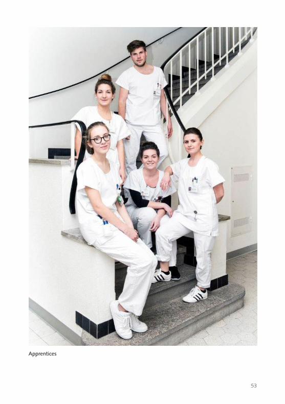
Staff and collaborators (as at 31.12.2016)
5
Background• The Department of Dermatology is part of the Inselspital, University Hospital of Bern.
The Inselspital belongs to the Insel Gruppe AG. The latter is under agreement with the Canton of Bern to serve as a tertiary health care centre for the population of the Canton of Bern and as a University teaching hospital.
• The Department of Dermatology within the Inselspital is a tertiary referral centre for skin and sexually-transmitted diseases. Patients are referred from all over Switzer-land. More than 30% of the treated patients come from outside the Canton of Bern.
Structural organisation, medical staff and nurse team• Our Department comprises an in-patient section with 10 to 14 beds, an out-patient
section with several specialised clinics, a day hospital for the management of complex cases with emphasis on chronic wounds, a phototherapy unit, a surgical unit, a laser platform, as well as a dermatopathology unit.
• The medical staff comprises 13 senior and 11 junior physicians. There are one full Professor, three associate Professors and one senior lecturer. In addition, there are 9 part-time board-certified consultants and lecturers. The Department regularly hosts a number of guest physicians in training as well as board-certified dermatologists from European and non-European countries for elective periods.
• The medical staff works in close collaboration with the specialised out-patient and in-patient nursing teams. The latter take particular care in ensuring best education and practice quality in nursing. High quality levels and standards have been accredit-ed for activities both in the in-patients ward and as well as for distinct out-patient activities.
6
Activity and areas of expertise Our current medical offer is focused on the evaluation and management of:• Skin cancers including clinical evaluation, management and systematic follow-up of
melanoma and non-melanoma skin cancer patients using video-microscopy and digital imaging devices
• Dermatosurgery, including Mohs micrographic surgery and correcting skin procedures
• Inflammatory skin diseases, psoriasis and atopic dermatitis• Autoimmune blistering disorders of the skin and mucous membranes• Skin allergies and environmental dermatology• Paediatric dermatology, inherited blistering and fragility syndromes of the skin• Acne and hidradenitis suppurativa• Medical lasers• Hair and nail diseases with specific conservative and surgical treatment• Phlebology as well as chronic wound care• Dermatopathology
Certification of the Skin Cancer CenterThe best and comprehensive care of our patients is one of the priorities of our Depart-ment. In November 2014 we were able to successfully complete the certification of our Skin Cancer Centre through the «Deutsche Krebsgesellschaft (DKG)». We are now able to offer our patients with skin cancers, comprehensive medical care with high stand-ards of quality and weekly tumour board meetings . The certification process has allowed us to further improve our collaboration both with other Departments (Plastic and Reconstructive Surgery, Medical Oncology, Radiotherapy, Nuclear Medicine) in our Hospital as well as with specialist physicians and general practitioners in private medical practice to ensure the best possible management of patients.
7
Current challenges The structure of the Swiss health system continues to undergo substantial reorganisa-tion. The Swiss health system is one of the most expensive in the world. Measures are being taken in an effort to reduce expenditure for both in-patient and out-patient services. This situation has put hospitals under pressure with budget cuts and rigorous budgetary control, making the implementation of cost-containment measures and the reassessment of the running of the Department including patient management and medical services essential. Yearly since 2012, the hospital Directorate has set a number of medical and financial objectives to be met by each Department in the hospital. The goals invariably encompass a defined budget, a given number of hospital admissions, out-patient cases, medical treatments, as well as management improvement processes and a campaign of quality projects. This new context has unfortunately led to unpleas-ant consequences impacting teamwork and inter-departmental collaboration.
Lean and Kaizen principles: process reorganisation in our DepartmentTo ensure high quality in medical and nursing care, better process organisation and, last but not least, continuous improvement of the financial results, systematic analysis of work flow since 2012 and 2013 has been performed according to the «Lean» manage-ment philosophy with constant assessment of the activities in the various out-patient units. «Lean» management focuses on the elimination of waste (overburden and unevenness) and on putting patients and referring physicians (the costumers) at the centre of interest.Our goal is to identify and eliminate waste and to cut out unnecessary steps, to simplify pathways, patient flow and administrative processes, to improve or adapt the available infrastructure to the new simplified steps, to better allocate human resources to the newly-defined tasks. The basic idea is to seize this challenging and critical time as the opportunity to begin a transformation of working methods with the ultimate goal of continuous improvement (Kaizen: Kai = change, Zen = good). Therefore we have now implemented a «lean» and «Kaizen» board to maintain and encourage the new philosophy, to control the performance and results and to continuously improve workflow.
8
ObjectivesThe main goals of the Department of Dermatology are
1. to provide high quality medical care and service by offering a spectrum of special-ised out-patient consultations as well as appropriate evaluation and management of patients requiring hospitalisation. Furthermore, the Department aims at developing teledermatology as a new diagnostic tool to facilitate and improve patient manage-ment and follow up as an alternative to live patient appointments. Respect of patient dignity and overall ethical issues are of constant concern in patient treat-ment,
2. to carry out clinical, translational and basic research. The ultimate objective is to ensure best patient care, state of the art therapy and to have a better understanding of disease mechanisms,
3. to provide intensive teaching for pre-graduate and post-graduate students in medicine and biomedical sciences, residents, physicians and specialists of dermato-logy and venereology, as well as other specialisations. Overall, the Department is very enthusiastic and highly motivated to share experiences in clinical evaluation, management and treatment of dermatological patients through interface with other partners within the Canton of Bern and in the rest of Switzerland.
Numerous joint ventures with focus on consultative and training activities for dermato-logical patients have been established with one of the largest emergency and primary referral out-patient centres in Bern (City Notfall) , with two major public hospitals in neighbouring Cantons (Fribourg, Solothurn) , with the Hirslanden Private Hospital Group as well as with a number of private dermatology practices. Finally, the Depart-ment is keen on developing and carrying out clinically oriented and basic investigative research projects in collaboration with either other researchers or with pharmaceutical companies or both.

9
Patients and technical statistics The number of dermatological in-patients has decreased to up to 581 new cases per year. The average length of stay in hospital has decreased from 6 days to 4.6 days in the last 6 years. There are up to 22,900 out-patient consultations per year, 3,800 patients are cared for at the wound care clinic, up to 6,400 phototherapy treatments and 2,386 surgical procedures are routinely performed. The dermatohistopathology sector processes and analyses 20,000 skin biopsy specimens per year and is one of the largest dermatopathology units still fully integrated within a University Hospital in Switzerland.
Specialised nursing team• The nursing team is headed by Mrs R. Müller-Wick, a member of the department’s
directorate. The nursing team constitutes the largest group among medical personnel working in the Department of Dermatology and represents the critical backbone requisite to providing specialised high level patient medical care. Mrs Müller-Wick and her closest collaborators ( Mrs A. Egli-Ryser, clinical nurse specialist, Mrs K. Thormann, advanced practice nurse,) have been faced with a number of significant organisational changes and new goals over recent years.
• The Department has a certified Wound Care Centre providing the highest quality patient care for the treatment of problem wounds. Furthermore, there is specialist experience in working with people affected by epidermolysis bullosa with large patient caseloads. Finally, we are establishing a number of clinics, in which an advanced practice nurse will be directly implicated as medical provider and for patient follow up. The most significant challenges have been to deal with a new management pathway of in-patients with a significant reduction in the length of the hospital stay and a rapid turnover of patients in the clinical ward, an increased number of out-patient surgical cases related amongst others, to the introduction of the micrographic Mohs surgery, as well as the implementation of new therapeutic approaches with related training, including UVA1 phototherapy and lasers. Further-more, in the light of increasing budgetary restriction, the personnel pool has to be constantly adapted to needs with always the ultimate goal of increased efficiency. Finally, the nursing team is keen to maintain a high standard of care. These efforts are reflected by the regular internal and external controls meetings which attest to the quality management standards required to maintain certification.
10
In-patient area and wound treatment out-patient facility/day clinicRegula Müller-Wick, Head of Nursing Service
Anyone working in a nursing environment on a day-to-day basis will be familiar with the concepts of cost-effectiveness, transformation, change and process optimisation. Any attempt to bring these various concepts together in some kind of meaningful way represents a considerable challenge.
With a view to increasing efficiency and cost-effectiveness, we have been looking after orthopaedic patients, as well as dermatology patients, on the ward since 2015. Besides the complex medical histories and the fact that hospital stays are getting ever shorter, this represents not only an additional challenge for nursing personnel, but also an enhancement and expansion of the service we offer. In order to ensure specialist expertise is available, internal training is provided on a regular basis across both specialist areas, and new employees are recruited in line with the relevant priorities.
The aim of the project to create a department-specific competence profile is to use healthcare professionals on the ward in a logical manner based on the competencies they have learned and acquired and through the experience they have gained in the relevant field.
The education on offer to patients with chronic skin conditions across all age groups, and their relatives, is being further developed and expanded. The aim is to ensure they are able to cope adequately with their chronic skin condition on a day-to-day basis, so their quality of life improves and they can enjoy longer periods with few or no symp-toms.An Advanced Nursing Practice (ANP) Team – to be led by an Advanced Practice Nurse (APN) – is being put together with this in mind.
Our success in securing certification from Concret last year and the wealth of positive feedback from patients have confirmed we are achieving outstanding quality in nursing terms. Our highly motivated and committed team does all it can to deliver this every day.
11
Out-patient areas at the Dermatology ClinicRita Stähli, Head of out-patient areas
The role of Head of outpatient areas was created in September 2014. I took over a well-managed polyclinic, which had been reorganised as part of a process optimisation initiative.
A total of seven clinical assistants and two healthcare professionals work in the out- patient sector. Some 26 different doctors run a surgery. Scheduling of surgeries represents a major challenge.
The field of light therapy currently enjoys a position of some importance within dermatology. We have two clinical assistants working in this area. With medica-tion-based therapy options developing very considerably within dermatology in recent years, the impact on light therapy remains unknown.
Dermatosurgery is an important area for our clinic. Patients really appreciate the fact that dermatology interventions can be performed at our clinic. Another specific possibility is Mohs surgery. In this area, there is one qualified expert surgical nurse, one qualified specialist nurse, and one nurse certified by the Swiss Red Cross. Various senior physicians and assistant physicians also still perform surgery. The out-patient sector also serves as a placement for healthcare professionals to complete a six-month term during their vocational training.
Since September 2015, we have been working with the I-pdos electronic patient file system, which eliminates paper medical records. Prior to this, more than 100 sets of medical records were first retrieved then archived, daily.
In future, it is certainly going to be important for us to find capable employees to fill vacant positions thereby ensuring exacting demands can be met.It is an exciting time, and I relish the challenges the next few years will bring.
12
The out-patient sectorThe out-patient clinics deal with the largest volume of patients in the Department. Therefore, the presence of a professional and efficient staff performing accurate and rapid patient registration and scheduling combined with friendly patient admittance is essential. The medical administrative staff led by Mrs Stähli-Hofer and coadjuvated by Mrs J. Egli has made substantial efforts to further improve the administrative processes as well as to provide the required nurse support to a high level in the out-patient service. Waiting time has been reduced and cycle time with better patient-centered care achieved. Online registration is available. One of the most important innovations was the implementation of the electronic medical record system which allowed us to simplify processes with faster and easier access to information. Finally, the introduction of a new phone system replacing the old pagers has greatly facilitated the internal reachability.
Strategy development: setting the goals for 2020–2025The Department undertook strategy development performed by Porsche Consulting GmbH. Vision and Missions have been defined. Overall, we have defined 4 major objectives for the next 5 to 10 years: 1) strengthening and further improving distinct medical services, which represent our areas of medical expertise and strategically important domains. The latter include allergology, melanoma and non-melanoma skin tumours, surgery and Mohs micrographic surgery, autoimmune and immune mediated skin diseases, dermatopathology and patient education with specialised nurses; increased use of teledermatology as a novel tool for diagnosis and disease manage-ment; 2) financial profitability as an essential prerogative for the further development of the speciality; 3) attractive workplace with quality employment and quality jobs; 4) greater patient satisfaction with the provided services.
Detailed clinical servicesIn the following section the specialised out-patient clinics available in our Department and specific areas of clinical and research interests are both presented.
13
Autoimmune skin diseases clinic (responsible: Professor L. Borradori, Dr. L. Feldmeyer) • Our Department has a specific interest and expertise in the evaluation, diagnosis and
management of patients with severe autoimmune blistering diseases of the skin and mucosa, such as bullous pemphigoid, mucous membrane pemphigoid, epidermolysis bullosa, and pemphigus which is unique in Switzerland. Furthermore, we are interest-ed in the evaluation and management of patients with cutaneous manifestations of systemic diseases, such as systemic lupus erythematosus, dermatomyositis, scleroder-ma, vasculitis and Adamiantades-Behçet disease. In this context, our clinic is involved as tertiary referral centre for patients from the Canton of Bern as well as from all over the country.
• Besides the clinical management of patients with autoimmune diseases, we have been directly involved : 1) as experts in international consensus conferences recom-mending definitions and outcome measures for various autoimmune bullous diseases as well as in the generation of management guideline for autoimmune bullous and other immune-mediated diseases; 2) in clinical multicentre European studies to characterise the clinical course, severity and prognostic markers in affected patients; 3) in the development of diagnostic tools such as ELISA and immunohistochemical approaches for improved diagnosis and better follow-up; 4) in basic investigative studies to assess the immunological humoral and cellular response in affected patients (see research part); 5) in assessing the impact of novel targeted therapies which specifically block mediators ( such as IL-5) critically involved in the inflammato-ry response involved in tissue damage in bullous pemphigoid.
• For correct evaluation of patients and classification of autoimmune diseases, we provide the required immunopathological analyses, such as direct and indirect immunofluorescence microscopy techniques, specific ELISAs as well as immunoblot and immune-precipitation techniques using recombinant proteins. These immuno-pathological studies are carried out in close collaboration with the immunopathology unit (Dr. M. Horn) of the Centre of Laboratory Medicine of the Inselspital. Finally, in the past we have contributed to the development of several different novel diagnostic ELISAs.
Chronic wound care centre(responsible: Dr H.W. Klötgen)The management of chronic skin wounds is organised in close collaboration with thechronic wound care centre. Our wound care centre is the largest out-patient clinicspecialised in the evaluation and management of recalcitrant vascular, neuropathic,tumoral and inflammatory skin wounds in the county of Bern.
14
We provide the entire conservative (e.g. use of novel wound dressings and matrix products) and surgical (conventional skin grafting, complex grafting procedures) spectrum of wound therapy. For the management of recalcitrant chronic wounds our team frequently takes advantage of tissue-engineered skin equivalents,some of which have been developed in our clinic. The treatment and long-term management of patients is professionally supported by wound specialists.
Dermatopathology unit(responsible: PD Dr H. Beltraminelli, Dr R. Blum, Dr. Dr. L. Feldmeyer, Dr H. Niev-ergelt, Professor E. Haneke)At our clinic, dermatopathology diagnostics are currently taken care of by three dermatology specialists who have undergone further training and specialisation in dermatopathology and by two consultants who have also had specialist training and experience in this area. This arrangement reflects the critical importance of sound clinical expertise to ensure correct appraisal of skin specimens within the discipline of dermatopathology – an integral part of clinical dermatology. The fact the dermatopa-thology laboratory is very much part of the clinic (in both organisational and physical terms) must also be viewed as a good thing in this respect.The dermatopathology facility processes some 20,000 tissue samples a year (with >50,000 sections). Incoming samples consist of our own specimens from the Universi-ty Hospital (30-40%) and material sent in by dermatologists across the whole of Switzerland (60-70%). Diagnostics cover the full spectrum of dermatopathology and particularly inflammatory, autoimmune, and neoplastic diseases. Conventional histological investigations are conducted in most cases, and immunohistochemistry is also used as required, with a wide range of immunohistochemistry dyes (>70 antibo dies) being stocked. These approaches cover over 95% of the diagnostics associated with dermatopathology scenarios. Molecular pathology analyses are offered as part of an interdisciplinary collaboration with the Institute of Pathology at the University of Bern. The entire field of immunofluorescence diagnostics is also covered by a collaborative arrangement with the University Hospital’s Immunoserology Department. Difficult cases are discussed within the team on a daily basis and sometimes also, as required, with colleagues from the Institute of Pathology at the University of Bern. Occasionally, these are referred to specialists outside the hospital acting on a consultancy basis (often based abroad). Our main areas of interest are diagnostics for cutaneous lymphomas, melanocytic lesions, and inflammatory diseases. Our team of dermatopa-thologists is actively involved in providing training and development in dermatopathol-ogy to postgraduate students, while the correlation between clinical and pathological aspects is highlighted each week during joint case discussions amongst personnel.

15
In our capacity as dermatopathologists, we take part in the regular meetings of the certified skin tumour centre at the University Hospital (certified in accordance with ISO 9001:2008 and the German Cancer Society in 2015): Lymphoma Board (Helmut Beltraminelli), Tumour Board (Roland Blum), Sarcoma Board (Laurence Feldmeyer).
Our laboratory has been accredited since 2012 (SN EN ISO/IEC 17025:2005), and since as long ago as 2010 our Dermatopathology Department has been officially recognised as a training centre by the International Committee of Dermatopathology ICDP-UEMS (www.icdermpath.org). Since 2009, many doctors from abroad have been invited to visit our department, including representatives from China, Egypt, Ethiopia, India, Kenya, Rwanda, Saudi Arabia, Tanzania, and Turkey.
Since 2010, we have been active members of the Groupe Francais pour l’Étude des Lymphomes Cutanés (French Group for the Study of Cutaneous Lymphomas – GFELC), during whose regular meetings held quarterly in Paris, histological diagnostics of skin lymphoma cases from our laboratory are discussed.Our research projects are focused on skin lymphomas (in collaboration with the GFELC), stromal changes in tumours, inflammatory dermatoses, and the development of dermatopathology in Africa. Some of the research projects also involve collaboration with the University of Bern as part of students’ doctoral theses.2011 saw the launch of a project to develop dermatopathology in Africa. Since then, there has been intensive collaboration with the Regional Dermatology Training Centre (RDTC) in Moshi, Tanzania. We provide manpower and technical support to promote the development of dermatopathology and the training of African colleagues on the ground. We have also enabled several African dermatologists from various countries (Egypt, Ethiopia, Kenya, Malawi, Rwanda, Tanzania) to take up scholarships, with three of these doctors having already passed the International Dermatopathology Examina-tion in Frankfurt. This was possible thanks to a number of grants (mainly from the European Academy of Dermatology and Venereology (EADV), as well as grants from the International Office at the University of Basel, the International Society of Derma-tology (ISD), and the International Foundation for Dermatology (IFD) and private donations.
Since 2015, Helmut Beltraminelli has organised the annual African Dermatopathology Conference (ADPC), a two-day event held at the RDTC in Moshi, Tanzania.
In 2016, Helmut Beltraminelli founded the African Dermatopathology Society (ADPS) with a number of committed African dermatologists and pathologists.
16
Again in 2016, Helmut Beltraminelli and Laurence Feldmeyer were elected President and Secretary respectively of the Swiss Group of Dermatopathology (SGDP), a working group of the Schweizerische Gesellschaft für Dermatologie und Venerologie (Swiss Society for Dermatology and Venereology – SGDV).Helmut Beltraminelli has also been a member of the Executive Committee of the International Society of Dermatopathology (ISDP) again since 2016.
Dermatosurgery clinic: Micrographic surgery(responsible: Dr R. Della Torre, Dr A.M. Skaria, Dr M. Stieger)The dermatosurgery offered by Inselspital focuses on tumour surgery, which includes Mohs and Slow Mohs micrographic surgery. This efficient surgical technique whereby the entire tumour area can be assessed in frozen sections (Mohs) and formalin-fixed sections (Slow Mohs) was introduced to the Inselspital Dermatology clinic by consult-ant Dr André Skaria in 2008. Under his supervision, senior physicians Dr Rocco della Torre and Dr Marco Stieger have been able to spend the last seven years learning and developing the Mohs technique and reconstructive tumour surgical techniques. Head of dermatosurgery Dr Rocco della Torre spent three months in the spring of 2016 exchanging surgical skills and gaining an insight into international treatment approaches at Mohs surgery specialist centres in Rotterdam, Bordeaux and Madrid. We are also constantly striving to improve the quality of surgery, train young medical assistants and instruct them in Mohs surgery at our Skin Cancer Centre, which is accredited by the German Cancer Society and is ISO certified. By working together with the Head of the Skin Cancer Centre, Professor Robert Hunger, further and advanced training in skin tumours and their treatment is held on a regular basis. Focus also remains on nail surgery, led by Professor Eckart Haneke and Dr Marco Stieger, and phlebological procedures, with Dr Marc Baumgartner and Dr Albert-Adrien Ramelet as consultants.
Eczema and atopic dermatitis clinic(responsible: Professor D. Simon)Eczematous skin diseases are very common and concern over 20% of dermatologic patients. Atopic dermatitis often starts in infancy and predisposes children to develop atopic airway diseases and food allergy. Irritant and allergic contact dermatitis inclu-ding occupational eczema are common in adults. These groups of cutaneous diseases are characterised by an acute and/or chronic inflammation of the skin and intense itch. Therefore, they have an enormous impact on the quality of life and are of medical as well as socioeconomic importance. Recently, we could demonstrate these associations in a Swiss cohort of patients with severe hand eczema. We have actively been involved in publishing Swiss and European guidelines for the management of hand eczema and atopic dermatitis, respectively.
17
Our specialised clinic provides all diagnostic and therapy facilities for patients with eczematous skin diseases. Diagnostic tests comprise blood and skin tests (patch test, skin prick test, atopy patch test, and provocation tests) to identify exogenous and endogenous pathogenic factors. Comprehensive management of affected patients encompasses installation of adequate anti-inflammatory topical and systemic thera-pies, patient education for skin care and skin protection including practical instructions, avoidance of triggers in daily life and occupational activities as well as psychological advice and support. In order to intensify patient care, we have started a project on advanced nurse practice in our eczema clinic. Currently, our team is involved in several clinical trials investigating novel therapies for atopic dermatitis.Furthermore, we offer specific education courses for paediatric and adult atopic dermatitis patients provided by dermatologists, allergists, psychologists, and nutritio-nists as well as train-the-trainer programmes. Our on-going clinical and translational research projects focus on pathogenic mecha-nisms of allergic skin diseases such as atopic dermatitis and contact dermatitis, as well as eosinophilic diseases of the skin and other organs in particular eosinophilic esophagi-tis. Furthermore, we are involved in epidemiologic studies on hand eczema, contact allergy and atopic dermatitis.
Hidradenitis suppurativa clinic(responsible: Professor R. Hunger)Hidradentis suppurativa (also called acne inversa) is a chronic inflammatory disease affecting mainly the intertriginous areas such as axillary, inguinal and perianal regions. The clinical course can be devastating. End-stage hidradenitis suppurativa is disabling and has a profound impact on the quality of life. Despite these facts there is still a lack of efficient treatments. To better help these patients we have a special hidradenitis suppurativa clinic in our department. At present we can offer patients many different therapeutic options including the participation in clinical studies (i.e. biologicals, laser treatment, extracorporal shock wave).
Laser clinic(responsible: Dr K. Heidemeyer, Dr N. Dietrich, Dr M. Adatto,)The laser clinic manages both medical and aesthetic skin problems by using various laser types and light sources. Established and innovative therapies are provided under the supervision of experienced laser specialists to provide high clinical care. Dr Adatto has been directly involved in the development of laser devices in collaboration with various American and European companies.Our laser centre is equipped with state of the art laser devices such as vascular, ablative
18
fractional, pigmented and excimer lasers. A close collaboration with specialists from other Departments (angiology, paediatrics, and paediatric surgery) allows interdiscipli-nary clinical management especially in the treatment of vascular malformations.Efforts are made to improve therapy algorithms with the use of lasers for a number of skin disorders, such vascular malformations, pigmentary disorders and traumatic tattoo, acantholytic disorders psoriasis, and inflammatory skin conditions. We are carrying out several clinical research studies to assess the effectiveness and safety of laser in rare vascular and pigmentary lesions. The clinical staff consists of Dr M. Adatto, past-President and Honorary Member of the European Society of Laser Dermatology as well as founder and medical director of Skinpulse Dermatology and Laser Centre Geneva, Dr N.Dietrich and Dr K. Heidemeyer, both dermatologists with specific interest in lasers.
Specialised consultations for melanoma and pigmented skin tumours(responsible: Professor R. Hunger)Melanoma incidence is still increasing at a rapid rate. Current figures in Switzerland show around 24 new cases per 100,000 people each year. More than 200 melanoma patients die every year in Switzerland from this disease. It is important to diagnose and treat melanoma early because only patients in the early stages have a good prognosis. In order to be able to treat melanoma patients as well as possible, we have special consultations for melanoma and pigmented skin tumours at our clinic where medical professionals examine and treat melanomas and patients receive regular aftercare. For patients with early-stage melanoma, there is the opportunity to first discuss the necessary steps to be taken and then they are put into effect. Afterwards patients are examined regularly so as to diagnose metastasis or new melanomas as early as possible. This is why it is important to examine all areas of skin on a regular basis. There are various ways in which we can detect new and even inconspicuous-looking melano-ma: 1. Dermatoscopy: a procedure whereby epiluminescence microscopy is used to better assess pigmented skin tumours. 2. Digital dermatoscopy: photographs are taken of suspected pigmented tumours over time so that the smallest change can be detected early. 3.Digital total body photography: standard photographs are taken of the entire skin area, which can be compared later to detect the smallest changes on the skin. 4. In vivo confocal microscopy: a non-surgical method for analysing any abnormal pigmented moles in microscopic resolution.
19
Nail diseases clinic(responsible: Professor E. Haneke)In dermatological practices, up to 15% of the patients present with nail disorders. The current knowledge in this area is not always satisfactory and management of many nail diseases has been limited due to lack of specific and effective therapeutic modalities. For example, onychomycoses that affect up to a third of the elderly still represent a problem with a complete cure rate remaining far below 50%. Furthermore, treatment of nail psoriasis is still challenging despite the availability of systemic biological treatments. Evaluation of nail diseases and their diagnoses are further hampered by the fact that few dermatopathologists and pathologists have the required experience with the interpretation of nail biopsies. Most physicians are reluctant to biopsy the nail organ. Finally, some nail diseases require a specific surgical approach, and few experts are familiar with nail surgery.In our Nail Clinic, we offer advanced diagnostic procedures and both conservative as well as surgical management for inflammatory, infectious, tumoral and congenital nail diseases. Our clinic provides a unique expertise in primary and revision nail surgery and nail pathology throughout Europe.
Non-melanoma skin cancer consultation(responsible: Dr. P. Edelmann-Weber)Non-melanoma skin cancers (NMSC) such as basal cell carcinoma and squamous cell carcinoma are one of the challenges facing Western medicine.This is due in part to a yearly increase in skin cancer incidence, which is linked to lifestyle (sun exposure) and can also be explained by the ageing population.At Inselspital, we are seeing rising numbers of patients with skin cancer or skin cancer precursors.Skin cancer often affects organ transplant patients. Inselspital Bern is one of the leading centres for organ transplantation.In the last few years, the general understanding of the increased risk of skin cancer among organ transplant patients has begun to change which means that patients at a high risk are presented to us regularly.We work closely with other disciplines such as plastic surgery and radiation oncology, as well as working on complex systemic treatments with the oncology team at Inselspital.We became a certified skin cancer centre in 2014 and we are recertified yearly.We hold an interdisciplinary ‘tumour board’ once a week to ensure the best possible treatment for our patients with complex tumours.In recent years we have developed our operational activity towards tumour patients. We also offer Mohs surgery for complex tumour locations.
20
Our operational activity is supported by the active contribution of external consultants.Non-invasive therapy for skin cancer precursors includes topical therapies and cryosur-gery. Alongside traditional PDT (photodynamic therapy), more and more frequently we are using daylight PDT. This is a treatment that patients appreciate in particular since it is less painful and requires less time in the clinic.We are also particularly pleased about the increasingly close collaboration with the Nephrology Clinic, which is where weekly dermatology consultations for transplant patients are held with Dr Patrizia Weber.By compiling a nephrology database over the next years, we hope to gain further information on new immunosuppressants and skin tumours.The general awareness of the increased risk of tumours in transplant patients is also leading us to work more closely with the cardiology, hepatology and pneumology disciplines.
Paediatric dermatology clinic(responsible: Dr C. Gouveia)Dr Gouveia, consultant in paediatric dermatology, ensures a comprehensive approach to a wide range of skin diseases in infants, children and adolescents, which includes diagnosis, treatment, follow-up and patient education. Besides inflammatory, infectious, allergic, tumoral, endocrine and immunological diseases, she is particularly interested in rare genetic skin diseases (genodermatoses), especially epidermolysis bullosa and ichthyosis.Patients are evaluated in close collaboration with other specialists in the University Children’s Hospital of Bern as well as with the human genetics department and the other subspecialty experts from the dermatology department to provide the most adequate care to the paediatric population. The consultation takes place in a children-friendly environment with time to address both the children’s and the parents’ questions.
Our multidisciplinary approach includes, namely:• Management of atopic dermatitis and allergy investigation in collaboration with
Prof. Simon• Diagnose and treatment of nail diseases in collaboration with Prof. Haneke• Diagnose and treatment of hair disorders in collaboration with Dr. De Viragh• Treatment of hemangiomas, in the interdisciplinary consultation at the Children’s
Hospital• Laser treatment of vascular malformations in collaboration with Dr. Heidemeyer and
Dr. Adatto
21
• Evaluation of complex vascular lesions in collaboration with the Angiodisplasia Board of the Inselspital
• Treatment of Psoriasis in collaboration with Prof. Yawalkar• Patient and parents’ education regarding the skin care made by our specialist nurses.
Furthermore, molecular diagnosis for a variety of congenital diseases is provided in close collaboration with the department of human genetics and various leading European laboratories. A long-standing cooperation with Professor Peter Itin, Head of Dermatology, University of Basel, has been established to ensure the best-possible approach for diagnosis and management in patients with unsolved complex syndromal skin disease.
Phlebology clinic(responsible: Dr M. Baumgartner, Dr. F. Schibler, Dr Dr A.A. Ramelet)More than 50% of the adult population in Western countries suffers from chronic venous disorders (CVD). Besides varicose veins, the most severe form of CVD, chronic venous insufficiency (CVI), occurs in up to 10% of people. CVI is responsible for and may lead to acute and/or chronic eczematous diseases, pigmentation, leg oedema, der-matoliposclerosis, atrophie blanche, and leg ulcers, resulting in high morbidity and health costs. In addition to classic vein pathologies and cosmetic impairments play a major role in the phlebology.Our Department is specialised in the clinical evaluation, clinical investigation (cw-Doppler and colour duplex) as well as in both the conservative (compression, venoac-tive drugs, physiotherapy) and surgical (sclerotherapy, echo-guided sclerotherapy, surgery) treatment of CVD. There is close collaboration with the Department of Angiology in Bern (Professor I. Baumgartner), and the Department of vascular surgery (PD Dr M.K. Widmer) which is of fundamental importance for comprehensive and multidisciplinary evaluation of patients. The dermatology residency program includes a 6-month elective in the Department of Angiology.Management of chronic wounds comprises special therapeutic procedures which are provided by nurses specifically trained in wound care. Management of small vessel problems also cosmetic aspects are dealt with through sclerotherapy and vascular lasers.
Psoriasis clinic(responsible: Professor N. Yawalkar)Psoriasis is a common inflammatory skin disease of variable severity with significant morbidity and impact on quality of life. Evidence exists indicating that psoriasis may be associated with serious comorbidities such as cardiovascular and metabolic diseases.The underlying pathomechanisms are not yet fully understood. Recent advances in our
22
understanding of the pathomechanisms of psoriasis have opened the way for new therapeutic strategies in psoriasis, such as the use of targeted therapies with biologic treatments.
In our specialised psoriasis clinic the following services are provided:• topical and systemic treatments including phototherapy, traditional immunosuppres-
sive agents (methotrexate, retinoids, ciclosporin, apremilast) and biologics such as tumour necrosis factor inhibitors (etanercept, infliximab, adalimumab) and anti-IL-12/23p40 monoclonal antibodies (ustekinumab) and anti-IL-17 monoclonal antibodies (secukinumab) are routinely used
• interdisciplinary medical education courses provided by dermatologists, psycholo-gists and nutritionists are offered to affected patients and their families, an opportu-nity exclusive to Switzerland
• clinical research studies aimed at testing novel biologic treatments are carried out. The clinic participates in phase 2 and phase 3 trials.
The clinic is also directly involved in basic investigative studies focused in the characteri-sation of the immune and inflammatory response in psoriasis (see research part).
Rare skin disease and genodermatoses(responsible: Dr C. Gouveia 12.2015)Dr Gouveia, consultant in paediatric dermatology, focuses on rare skin disease, mainly in children, such as the field of genodermatoses. In particular, this includes the diagno-sis and management of paediatric and adult patients with inherited skin blistering disorders, such as epidermolysis bullosa (EB).We offer a specialised and comprehensive interdisciplinary evaluation of affected patients by the EB network at the Inselspital. Our specialised team ensures an appropri-ate care of wounds and addresses further problems occurring in this rare and complex disease, namely social issues, dental/oral complications, nutritional, gastroenterologi-cal, bone/joints and oncological complications. Several specific treatments are provided, namely hand surgery, esophagus dilatation and insertion of PEG tubing, production and fitting of hand braces, occupational therapy and physiotherapy as well as diagnostics and surgical removal of squamous cell carcinoma (SCC).We focus also on information and support of affected people, as well as of their family members and institutions involved, namely home nursing teams (Spitex), school teams and patients associations. Our hospital team is completed by the EB-Expert that supports and counsels the patients at home ensuring the best possible liaison between the care at home and the hospital. Furthermore, immunofluorescence and molecular diagnosis is provided in collaboration with the department of human genetics and various leading European laboratories. We offer genetic counseling to all the families
23
affected. Although a cure for this group of genetic skin diseases is still not available, several advanced research projects are being done in different European centres with which we cooperate whenever possible. We also work in close collaboration with the EB consultation at Zurich Children’s University Hospital and with Professor Peter Itin, Head of Dermatology, University of Basel, who ensures the best possible approach for diagnosis and management of the squamous cell carcinomas in the adult patients.
Trichology clinic(responsible: Dr. P. A. de Viragh)We provide a specialised evaluation and care of patients with complex and severe diseases of the scalp and hair. Expert evaluation and individualised therapy are manda-tory in cases of hair loss, hair structure alteration, and scalp inflammation or scarring.To properly evaluate patients, the following exams may be carried out:• microscopical analysis of hair samples (trichogram) to assess structural abnormali-
ties, establish the rate of hair loss, or identify genetic influences• scanning electron microscopy for definitive diagnosis of hereditary hair abnormalities• stereotactic photography and computer-assisted digital imaging (trichoscan or
trichoscale) is used to evaluate treatment efficacy objectively• dermoscopic scalp evaluation (trichoscopy)• scalp biopsies for examination by light microscopy and immunohistochemistry studies.
The scientific experience in the field of hair physiology and diseases of the pilosebaceus follicle is documented in numerous presentations at international meetings and publica-tions by the staff involved, as well as by the ongoing research activities such as treatment protocols for alopecia areata and lichen planopilaris, respectively histological studies to refine diagnostic accuracy for hair diseases.
Urticaria clinic(responsible: Professor D. Simon)Urticaria is a common mast cell-driven skin disease presenting with an acute onset of wheals and/or angioedema. Acute urticarial is often associated with acute infections, drugs, and allergic reactions. The diagnostic of chronic spontaneous urticarial is complex and based on the patient’s history, clinical examination and laboratory tests. The triggers of inducible urticarial should be identified by the provocation test. We provide all diagnostic work-up and therapy, including anti-IgE antibody and other immunomodula-ting therapies for those patients not responding to antihistamines. Since the approval of omalizumab for chronic spontaneous urticaria in Switzerland, the number of patients treated in our clinic has significantly increased. Furthermore, we have actively supported the Swiss guidelines for the management of urticaria in general practice.
24
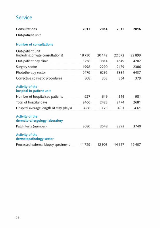
Service
Consultations 2013 2014 2015 2016
Out-patient unit
Number of consultations
Out-patient unit (including private consultations) 18 730 20 142 22 072 22 899
Out-patient day clinic 3256 3814 4549 4702
Surgery sector 1998 2290 2479 2386
Phototherapy sector 5475 6292 6834 6437
Corrective cosmetic procedures 808 353 364 379
Activity of the hospital In-patient unit
Number of hospitalised patients 527 649 616 581
Total of hospital days 2466 2423 2474 2681
Hospital average length of stay (days) 4.68 3.73 4.01 4.61
Activity of the dermato-allergology laboratory
Patch tests (number) 3080 3548 3893 3740
Activity of the dermatopathology sector
Processed external biopsy specimens 11 725 12 903 14 617 15 407
25
Research and development projects
1. Hand eczemaIn 2011, the CARPE registry on chronic hand eczema (CHE) started involving seven study centres in Switzerland. This investigator-initiated study was sponsored by the Department of Dermatology Inselspital, Bern University Hospital. Overall, 200 patients were enrolled and followed over two years. Currently, we are in the process of evaluat-ing the follow-up data. Previous analyses of registry data were mainly descriptive or focused on selected risk factors, and thus might not fully reflect the complexity of causes, triggers and consequences of CHE. To overcome this gap, we aim at investigating the associations among numerous variables and to provide a semantic connectivity map. Using this novel statistical approach, we will analyse baseline data of the registries of Germany and Switzerland.
2. Atopic diseasesAtopic dermatitis is characterised by epidermal barrier defects and a T helper 2 immune response and thus shares many similarities with other atopic diseases, e.g. eosinophilic esophagitis (EoE). There is increasing evidence that barrier defects play a crucial role in the pathogenesis of EoE, however, it is not clear whether the barrier dysfunction is associated with the disease per se or subsequent to corticosteroid therapy. We aim at investigating the effects of corticosteroid therapy on the epithelial barrier and subse-quent sensitisation to Candida albicans in EoE patients.
3. Bullous pemphigoidBullous pemphigoid (BP) is the most common autoimmune blistering skin disease affecting the elderly. The histology shows a characteristic subepidermal blister formation and an inflammatory infiltrate rich of eosinophils. There is increasing evidence that eosinophils play a key pathogenic role in BP, however the exact mecha-nisms have poorly been understood. We aim at investigating whether eosinophils are involved in dermal-epidermal splitting and the conditions required using an ex vivo BP skin model. Further, we will investigate the interaction of monocytes and neutrophils in blister formation in BP. Corticosteroids that are used as first line therapy for BP, might cause severe adverse events, in particular when used for long-term treatment and in elderly patients. Based on the hypothesis that eosinophils play a pathogenic role in BP, a targeted therapy against interleukin-5 with mepolizumab has been applied to BP patients in an investi-gator initiated study.
26
4. Autoimmune blistering diseases of the skinIn the past decade, one of the major interests of our group has been the characterisa-tion of the immune response in the pemphigoids and pemphigus, a group of severe autoimmune blistering diseases of the skin and mucosae. Better knowledge of the pathogenesis of pemphigus and pemphigoid may provide crucial insights into mecha-nisms of autoimmunity in general and may help to design more specific therapeutic strategies. The pemphigoids include bullous pemphigoid, gestational pemphigoid and mucous membrane pemphigoid. They are a relatively common group of autoimmune blistering disorders associated with autoantibodies directed against two proteins of the cutaneous basement membrane zone, BP180/BPAG2 and BP230/BPAG1e. In the past years, we have been implicated in projects aimed at: 1) characterising the humoral and autoreactive T cell response to BP180 and BP230 in the disease course of the pemphig-oids; 2) identifying laboratory markers predicting disease activity and outcome; 3) developing innovative diagnostic tools such as ELISAs and microarrays for the detec-tion of patients’ autoantibodies with high sensitivity and specificity.
There is another related autoimmune bullous disease called paraneoplastic pemphigus (PNP) sharing some overlap with pemphigus. Paraneoplastic pemphigus autoantibodies target the transmembrane adhesion proteins desmogleins and desmocollins, several intracellular plakins, and a novel protein identified in our laboratory, called alpha-2 macroglobulin-like 1 protein, which is an extracellular broad range protease inhibitor. Our ongoing project represents a joint effort by several European groups with the following long term goals: 1) analysis of the autoantibody-driven effector phase frequently involving «epitope spreading»; 2) characterisation of the molecular events leading to tissue damage and skin blistering. Specifically, we are assessing the impact of autoantibodies directed against alpha-2 macroglobulin-like 1 protein on epidermal homeostasis; 3) analysis of the impact of therapeutic strategies such as the monoclonal antibody anti-CD20 (rituximab) on the cellular and humoral autoimmune response in pemphigus, and 4) definition and establishment of clinical parameters as valid measure ments for the extent and activity of the disease and life quality assessment in pemphigus.
5. Characterisation of the interactions between plakins and the cytoskeletonOur group is primarily implicated in basic investigative studies aimed at characterising 1. the association of several intracellular linkers of the cytoskeleton, plectin, desmo-
plakin and bullous pemphigoid antigen 1 (BPAG1), which belong to the plakin family, with the cytoskeleton in epithelia and striated muscle cells;
2. the regulation of these interactions by posttranslational modifications (such as phosphorylation), and
3. the contribution of the plakins to the overall organisation of the cytoskeleton since
27
plakins can usually interact with at least two components of the cytoskeleton, the microfilaments, the microtubules and/or the intermediate filaments, and specifically anchor these structural networks to various membrane complexes.
All these connections are critical for the maintenance of the cell architecture and tissue resilience to mechanical forces. In fact, mutations in the genes coding for plectin, desmoplakin, BPAG1, and intermediate filaments cause a variety of devastating human diseases, attesting to the importance of these proteins for tissue integrity.
6. Research on molecular mechanisms of skin diseases (Prof. Eliane J. Müller, PhD)In 2015-16 the molecular dermatology group of the Vetsuisse Faculty joined the Department of Dermatology. The group has three research foci: studies on molecular mechanisms leading to the autoimmune blistering disease pemphigus vulgaris, analyses of skin homeostasis including tissue stem cells and studies on inherited skin disease affecting the homeostatic process.
We had the fortunate opportunity to move into new premises in Murtenstrasse 40 (Mu40). The laboratories at this address, come under the jurisdiction of the Department of Clinical Research (DCR/DKF) and their contemporary open-concept design with generous glass work facilitate communication, collaboration and easy interface.
1. Pemphigus vulgaris: how do autoantibodies disrupt skin architecture? Pemphigus vulgaris autoantibodies bind to structures which glue skin cells together:
these are adhesion molecules. By focusing on adhesion-mediated signaling in skin renewal and disease, the group had introduced a paradigm shift. They have demon-strated that adhesion molecules are sensors of cells overlooking cell fate by fine-tun-ing intercellular signaling processes depending on the adhesive state of adhesion.1-4 Pemphigus antibodies interfere with these processes by simply binding to the extracellular sensors. The group is currently performing pre-clinical experiments using pharmacologic targeting of antibody-mis-activated signals aiming at novel adjuvant therapies for pemphigus vulgaris patients.
2. Stem Cell Biology and skin homeostasis: how do stem cells contribute to tissue architecture?
Cyclical skin and hair follicle renewal takes place throughout a life-time and are driven by epidermal and hair follicle stem cells. This field of regenerative research is of particular interest to the Molecular Dermatology group at Murtenstrasse 40 and serves their studies linking the autoimmune disease pemphigus vulgaris to skin homeostasis. Pemphigus vulgaris autoantibodies target the adhesion molecules of the skin´s stem cell reservoirs thereby affecting stem cell homeostasis.1, 5
28
The genuine interest in stem cell biology, the known potential of stem cells for regenerative medicine and, at the same time, the ill-defined understanding of how to exploit these cells clinically, prompted the molecular dermatology group to join efforts with Bernese stem cell researchers to found the Stem Cell Research and Regenerative Medicine (SCRM) Platform in Bern (www.stemcellsbern.ch) and very recently a platform in cell therapies and regenerative medicine at national level .
3. Genodermatoses, the inherited skin diseases In a recently funded SNF Sinergia project of «One Health» genodermatoses (main
applicant Prof. T. Leeb, Institute of Genetics) the group contributed to the assembly of three DermFocus laboratories from the Institutes of Genetics and Pathology and Clinical Dermatology at the Vetsuisse Faculty as well as of national and international collaborators thus strengthening the Dermatology Network at the University of Bern and Inselspital, Bern University Hospital. 6,7
Beyond dermatology & stemc biology: translational medicine The Molecular Dermatology group and the SCRM platform are both closely involved in the Sitem-Insel AG project – the Swiss Institute for translational and entrepreneurial medicine – and in the non-profit organisation planned around Inselspital.
The implementation of a research laboratory at Murtenstrasse 40 opens new exciting opportunities to collaborate on stem-cell mediated disease towards a fast track into clinical translational medicine.
References1. Di Zenzo G, Amber KT, Sayar BS, Muller EJ, Borradori L. Immune response in
pemphigus and beyond: progresses and emerging concepts. Seminars in Immuno-pathology 2016: 38: 57-74.
2. Müller EJ, Williamson L, Kolly C, Suter MM. Outside-in signaling through integrins and cadherins: a central mechanism to control epidermal growth and differentia-tion? J Invest Dermatol 2008: 128: 501-516.
3. Sayar BS, Ruegg S, Schmidt E, Sibilia M, Siffert M, Suter MM, Galichet A, Muller EJ. EGFR inhibitors erlotinib and lapatinib ameliorate epidermal blistering in pemphigus vulgaris in a non-linear, V-shaped relationship. Experimental Dermatology 2014: 23: 33-38.
4. Luyet C, Schulze K, Sayar BS, Howald D, Muller EJ, Galichet A. Preclinical studies identify non-apoptotic low-level 13. caspase-3 as therapeutic target in pemphigus vulgaris. PLoS ONE 2015: 10: e0119809.
29
5. Schulze K, Galichet A, Sayar BS, Scothern A, Howald D, Zymann H, Siffert M, Zenhausern D, Bolli R, Koch PJ, Garrod D, Suter MM, Muller EJ. An adult passive transfer mouse model to study desmoglein 3 signaling in pemphigus vulgaris. The Journal of Investigative Dermatology 2012: 132: 346-355. e
6. Drogemuller M, Jagannathan V, Becker D, Drogemuller C, Schelling C, Plassais J, Kaerle C, Dufaure d
Citres C, Thomas A, Muller EJ, Welle MM, Roosje P, Leeb T. A Mutation in the FAM83G Gene in Dogs with Hereditary Footpad Hyperkeratosis (HFH). PLoS genetics 2014: 10: e1004370.
7. Jagannathan V, Bannoehr J, Plattet P, Hauswirth R, Drogemuller C, Drogemuller M, Wiener DJ, Doherr M, Owczarek-Lipska M, Galichet A, Welle MM, Tengvall K, Bergvall K, Lohi H, Rufenacht S, Linek M, Paradis M, Muller EJ, Roosje P, Leeb T. A mutation in the SUV39H2 gene in Labrador Retrievers with hereditary nasal parakeratosis (HNPK) provides insights into the epigenetics of keratinocyte differen-tiation. PLoS genetics 2013: 9: e1003848.
7. Cutaneous drug reactionsThe main research goals are 1) to improve the understanding of the molecular interac-tions of drugs/chemicals with immune cells, i.e. T cells, dendritic cells and 2) to dissect the mechanisms by which these interactions stimulate and affect the immune system. These studies are planned to pave the way for improved methods to diagnose adverse drug reaction and to improve risk assessment of chemicals/drugs. The pathogenic role of cytokines in acute generalised exanthematous pustulosis (AGEP) and psoriasiform reactions induced by TNF-Inhibitors is currently being investigated in collaboration with colleagues at the Department of Dermatology in Zurich.
8. Cutaneous T cell lymphomaPrimary cutaneous T-cell lymphomas (CTCL) represent a heterogeneous group of extranodal non-Hodgkin lymphomas, where mycosis fungoides (MF) is the most common type. Our specialists are well-connected internationally, especially with the Groupe Francais pour l’ Étude des Lymphomes Cutanés (GFELC) in Paris, where we are collaborating in research projects. In the Dermatology clinic in Graz, we studied the characteristics of a rare cutaneous CD4+ pleomorphic lymphoprolipherative disease and other related conditions.
9. Stroma characteristics in skin lesionsMost tumours have a tumour and a stromal component, in some entities pathological changes within the stroma are characteristic for one peculiar tumour. We are studying stromal characteristic as the distribution of elastic fibers in selected skin conditions.
30
10. Hidradenitis suppurativaHidradenitis suppurativa (also called acne inversa) is a chronic inflammatory disorder ofthe apocrine gland-bearing skin. At present, the pathophysiology of this condition is still poorly understood. To better understand its mechanisms, we are currently performing studies using immunohistochemical and molecular biology methods to better comprehend the mechanisms leading to chronic inflammation. In the last 2 years we were able to demonstrate a critical role of the IL32 in the disease development. IL32 seems to be relatively specifically expressed in lesions of HS whereas it is not found in active lesions of other inflammatory skin disease such as psoriasis and atopic dermatitis. Currently we are analysing the role of further cytokines such as IL36 but also the pro-inflammatory role of the antimicrobial peptide cathelicidine.
11. Human interleukin 9-producing T helper memory cells and their role in antitumour immune response in malignant skin diseaseHuman T helper (TH) cells are crucial mediators of the adaptive immune system. To respond to the myriad of infectious and non-infectious challenges, they have evolved into distinct subsets such as TH1, TH2, or TH17 cells. IL-9 producing TH9 cells have recently been proposed as a novel subset of TH cells and studies in animal models suggest a protective role for these cells in tumour immunity. However, studies of TH9 cells thus far have largely been limited to TH cells differentiated in vitro. Studies of human in vivo differentiated TH9 cells are lacking. Therefore, the existence of TH9 cells as an authentic cell type has been called into question. Our data now indicates for the first time, the existence of human in vivo differentiated TH9 cells, thus raising the possibility to investigate their true identity and functional properties. In addition, we find large numbers IL-9 expressing cells in the immune infiltrate of human melanoma, thus warranting further investigation of the role of TH9 cells in the human anti-mela-noma immune response.The overarching aim of our research is to investigate the identity and properties of human TH9 cells and their role in the anti-melanoma immune response. Based on our preliminary data, we hypothesise that TH9 cells are in fact a distinct subset and that they can be identified by a specific set of skin-homing receptors. Due to robust tumour immunity mediated by TH9 cells in mice, we further hypothesise that they can be found at higher numbers in the immune infiltrate of primary melanomas compared to metastasised melanoma, since metastasis requires immune evasion of the tumour, and that the number of TH9 cells correlates with disease prognosis.Established immunological methods for the analysis of human T cell biology (cell culture, intracellular FACS staining, FACS sorting, ELISA, Luminex cytokine multiplex assay, RT-PCR, immunohistochemistry, immunofluorescence double staining) are used in combination with a novel method for ex vivo analysis of human tissue-resident T cells.
31
Answering the questions raised in this project will increase our understanding of TH cell biology, lead to a thorough characterisation of human TH9 cells and shed light on their contribution to anti-melanoma immune response. Based on surprising recent findings that TH9 cells mediate superior tumour immunity in mice, it would appear highly promising to investigate the biology of TH9 cells in humans; a better understanding of these cells may lead to the development of innovative T cell based immunotherapies for malignant melanoma.
12. Non-melanoma skin cancersOur research is focused on epidemiological studies on NMS,• We are collecting UV-exposure behavioral data of the general population• We aim to identify patients that are at high risk for developing NMSC (and melanoma)in the setting of immune suppression e.g. due to organ transplantation. Our collabora-tions with the different departments of the Transplant Board of the Inselspital are being constantly developed.
13. Psoriasis research Our studies are aimed at investigating immunological mechanisms and their regulation through therapeutic interventions like apremilast and biologics in psoriasis. As control, findings are compared to those obtained in different forms of eczema. Previous reports indicate that serum IgE levels are increased in psoriasis. We investigated the presence and distribution of IgE and FcεRI in psoriatic lesions and their alteration after successful systemic treatment with ustekinumab. Elevated total serum IgE levels were found in 39% of patients with psoriasis. A positive correlation between IgE+ and FcεRI+ cells and a significant increase of these cells was found in psoriatic lesions when compared with normal skin. IgE+ and FcεRI+ cells decreased significantly after successful therapy with ustekinumab. IgE and FcεRI were coexpressed on mast cells, epidermal Langer-hans cells, dermal dendritic cells, macrophages and a small number of neutrophils. Our data suggests that IgE might participate in psoriatic inflammation by activating FcεRI-bearing cells in a subset of patients.
14. Development of Dermatopathology In AfricaSince 2009 , we have been running a project to develop dermatopathology in Africa, in collaboration with some European partners and starting from the prominent reference centre, namely the Regional Dermatology Training Centre (RDTC) in Moshi, Tanzania. The main goal is to train dermatopathology expertise to several motivated and talented African specialists (dermatologists or pathologists) in Europe and to prepare them to participate to the International Board Examination in Der-matopathology (IBED) in Frankfurt, Germany. These experts will then spread their
32
knowledge to other African colleagues, which ensures the sustainability of the project.Thanks to many hard-working experts in Africa and in Europe and with the financial support of the European Academy of Dermatology and Venerology (EADV) and the International Society of dermatology (ISD) for the last 5 years, some goals have already been met: the dermatopathology activities at the RDTC are continuing intensively, and are in development in some other African countries, 4 African fellows successfully passed the IBED in Frankfurt and are now working and teaching in their respective African country.We have several scientific collaborations with African specialists; 3 studies have been published.
Results
AllergyEosinophilic esophagitis (EoE) was described in the early 1990s. Intensive research is going on in order to elucidate EoE pathogenesis, clinical features and therapeutic approaches. Recent-ly, we characterised an EoE-like disease that shares clinical and also immunological features of EoE but lacks eosinophil infiltration of the esophagus.
• Straumann A, Blanchard C, Radonjic-Hoesli S, Bussmann C, Hruz P, Safroneeva E, Simon D, Schoepfer AM, Simon HU. A new eosinophilic esophagitis (EoE)-like disease without tissue eosinophilia found in EoE families. Allergy 2016; 71:889-900.
In most patients, EoE has been associated with atopic diseases, food allergy and increased IgE levels. Moreover, elimination diets were shown to improve EoE symptoms and signs. Ho-wever, based on recent research results, the pathogenesis of EoE is complex and might not simply be explained by an IgE-mediated food allergy. In a comprehensive review, we report on the potential mechanisms underlying food hypersensitivity in EoE.
• Simon D, Cianferoni A, Spergel JM, Aceves S, Holbreich M, Venter C, Rothenberg ME, Terreehorst I, Muraro A, Lucendo AJ, Schoepfer A, Straumann A, Simon HU. Eosinophilic esophagitis is characterized by a non-IgE-mediated food hypersensitivity. Allergy 2016;71:611-620.
Eosinophils are innate immune cells and play an important role in host defense. By releasing extracellular DNA traps (EET), they are able to kill bacteria. In a recent study, we showed the formation of EET in eosinophil esophagitis. The number of eosinophils and EET formation correlated with a reduced expression of the protease inhibitor LEKTI and filaggrin, and were associated with Th2 cytokine expression, e.g. TSLP.
33
• Simon D, Radonjic-Hösli S, Straumann A, Yousefi S, Simon HU. Active eosinophilic esophagitis is characterised by epithelial barrier defects and eosinophil extracellular trap formation. Allergy 2015;70:443-52.
Hand eczema has gained increasing interest over the last years when new therapeutic options became available. Since data on the epidemiology of hand eczema in Switzer-land was scarce, a registry study on chronic hand eczema (CHE) was initiated. By analysing the baseline data providing information on the last twelve months prior to enrollment in the study, we demonstrated the high impact of CHE on medical well-being, patient quality of life and work ability. CHE was associated with an intense use of health care services, high rate of sick leave as well as job loss and change.
• Cazzaniga S, Ballmer-Weber BK, Gräni N, Spring P, Bircher A, Anliker M, Sonntag AK, Piletta P, Huber C, Borradori L, Diepgen T, Apfelbacher C, Simon D. Medical, psychological and socio-economic implications of chronic hand eczema: a cross-sec-tional study. J Eur Acad Dermatol Venereol 2016;30:628-637.
Hand eczema is triggered by endogenous and exogenous factors. Allergen identifica-tion by patch testing and avoidance are essential for the treatment of allergic contact dermatitis. For over 10 years, we have provided our patch test results to the European networks on contact allergy and thus contribute to the surveillance of contact aller-gens. Recently, a report on contact allergy to rubber chemicals and the impact of testing special rubber series was published.
• Uter W, Warburton K, Weisshaar E, Simon D, Ballmer-Weber B, Mahler V, Fuchs T, Geier J, Wilkinson M. Patch test results with rubber series in the European Surveil-lance System on Contact Allergies (ESSCA), 2013/14. Contact Dermatitis 2016;75:345-352.
The pathogenesis of lichen planus is not completely understood. In a recent study, we aimed at investigating the inflammatory infiltrate and cytokine expression of cutaneous and oral lichen planus. We observed a distinct expression of interferon-gamma and interleukin-9. The cell and cytokine pattern of lichen planus resembled those reported for autoimmune diseases.
• Weber B, Schlapbach C, Stuck M, Simon HU, Borradori L, Beltraminelli H, Simon D. Distinct interferon-gamma and interleukin-9 expression in cutaneous and oral lichen planus. JEADV in press.

34
MelanomaMalignant melanoma is the deadliest of all skin cancers. Sentinel lymph node dissection is a very important procedure in assessing the risk of disease progression. However, the impact of this staging analysis is not clear for thick melanoma. In an epidemiological study we could demonstrate the positive value of this procedure on the outcome. In another study we analysed the value of sentinellymph node dissec-tion in patients treated in the last 13 years. Both studies could confirm the prognostic value of this procedure.
Plakins and the cytoskeletonThe organisation and dynamics of the cytoskeleton, as well as its anchorage to specific sites in the plasma membrane and organelles, are regulated by the plakins. These structurally related proteins anchor different cytoskeletal networks to each other and/or to other cellular structures. The association of several plakins with intermediate filaments (IFs) is critical for maintenance of the cytoarchitectureWe have developed a protein-protein fluorescence binding assay, which is based on the production of recombinant proteins tagged with green fluorescent protein (GFP) and their use as fluid-phase fluorescent ligands on immobilised proteins of interest. This simple and sensitive technique, which is expected to facilitate further studies in this area, can also be potentially employed for any kind of protein-protein interaction studies. Using this method, we have been able to assess the ability of C-terminal regions of GFP-tagged plakin proteins, such as plectin, desmoplakin or BPAG1e to bind to distinct IF proteins and to map the involved binding sites. We have also studied the effect of several pathogenic amino acid substitutions in desmoplakin causing complex inherited diseases associated with skin fragility, hair abnormalities and/or cardiomyopathy on the interaction of desmoplakin with the epidermal keratins and the muscle-specific IF protein desmin. Finally, we have assessed the diagnostic value of immunohistochemistry on formalin-fixed, paraffin-embedded skin biopsy specimens for bullous pemphigoid.
35
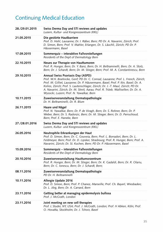
Continuing Medical Education
28./29.01.2015 Swiss Derma Day and STI reviews and updates Luzern, Kultur- und Kongresszentrum (KKL)
21.05.2015 Die gestörte Hautbarriere Prof. D. Hohl, Lausanne; Dr. I. Räber, Bern; PD Dr. A. Navarini, Zürich; Prof. D. Simon, Bern; Prof. V. Mahler, Erlangen, Dr. S. Läuchli, Zürich; PD Dr. P. Häusermann, Basel
17.09.2015 Sommerquiz – interaktive Fallvorstellungen Residents of the Dept of Dermatology Bern
22.10.2015 Neues zur Therapie von Hauttumoren Prof. R. Hunger, Bern; Dr. Y. Banz, Bern; Dr. H. Beltraminelli, Bern; Dr. A. Stolz, Bern; Dr. J. Schardt, Bern; Dr. M. Stieger, Bern; Prof. M. A. Constantinescu, Bern
29.10.2015 Annual Swiss Psoriasis Day (ASPD) Prof. W.H. Boehncke, Genf; PD Dr. C. Conrad, Lausanne; Prof. L. French, Zürich; Prof. M. Gilliet, Lausanne; Dr. P. Häusermann, Basel; Prof. P. Itin, Basel; Dr. A. Kolios, Zürich; Prof. S. Lautenschlager, Zürich; Dr. J.-T. Maul, Zürich; PD Dr. A. Navarini, Zürich; Dr. M. Streit, Aarau; Prof. R. Trüeb, Wallisellen; Dr. Dr. A. Wysocki, Luzern; Prof. N. Yawalkar, Bern
10.11.2015 Zuweiserveranstaltung Dermatopathologie Dr. H. Beltraminelli, Dr. R. Blum
26.11.2015 Haare und Nägel Prof. N. Yawalkar, Bern; Dr. P. de Viragh, Bern; Dr. S. Rohner, Bern; Dr. P. Weber, Bern; Dr. S. Radonjic, Bern; Dr. M. Stieger, Bern; Dr. D. Perruchoud, Bern; Prof. E. Haneke, Bern
27./28.01.2016 Swiss Derma Day and STI reviews and updates Luzern, Kultur- und Kongresszentrum (KKL)
26.05.2016 Neutrophile Erkrankungen der Haut Prof. D. Simon, Bern; Dr. C. Gouveia, Bern; Prof. L. Borradori, Bern; Dr. L. Feldmeyer, Bern; Prof. Dr. D. Lipsker, Strasbourg; Prof. R. Hunger, Bern; Prof. A. Navarini, Zürich; Dr. St. Kuchen, Bern; PD Dr. P. Häusermann, Basel
15.09.2016 Sommerquiz – interaktive Fallvorstellungen Residents of the Dept of Dermatology Bern
20.10.2016 Zuweiserveranstaltung Hauttumorzentrum Prof. R. Hunger, Bern; Dr. M. Stieger, Bern; Dr. K. Gadaldi, Bern; Dr. R. Olariu, Bern; Dr. C. Ionescu, Bern; Dr. J. Schardt, Bern;
08.11.2016 Zuweiserveranstaltung Dermatopathologie PD Dr. H. Beltraminelli
10.11.2016 Allergie Update 2016 Prof. D. Simon, Bern; Prof. P. Chanez, Marseille; Prof. Ch. Bayerl, Wiesbaden; Dr. L. Jörg, Bern; Dr. A. Carrard, Bern
23.11.2016 Getting better at managing epidermolysis bullosa Prof. J. McGrath, London
23.11.2016 Joint meeting on new cell therapies Prof. L Studer, NY, USA; Prof. J. McGrath, London; Prof. H Abken, Köln; Prof. O. Hovatta, Stockholm; Dr. J. Tchorz, Basel
36
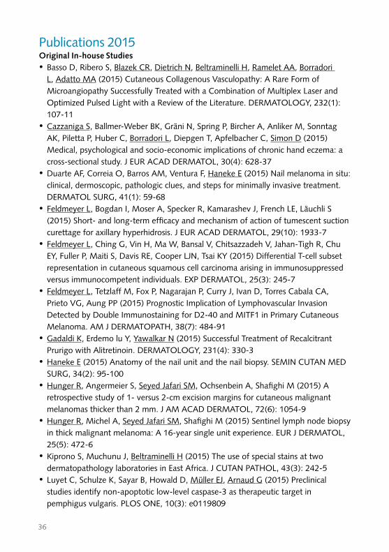
Publications 2015 Original In-house Studies •Basso D, Ribero S, Blazek CR, Dietrich N, Beltraminelli H, Ramelet AA, Borradori
L, Adatto MA (2015) Cutaneous Collagenous Vasculopathy: A Rare Form of Microangiopathy Successfully Treated with a Combination of Multiplex Laser and Optimized Pulsed Light with a Review of the Literature. DERMATOLOGY, 232(1): 107-11
•Cazzaniga S, Ballmer-Weber BK, Gräni N, Spring P, Bircher A, Anliker M, Sonntag AK, Piletta P, Huber C, Borradori L, Diepgen T, Apfelbacher C, Simon D (2015) Medical, psychological and socio-economic implications of chronic hand eczema: a cross-sectional study. J EUR ACAD DERMATOL, 30(4): 628-37
•Duarte AF, Correia O, Barros AM, Ventura F, Haneke E (2015) Nail melanoma in situ: clinical, dermoscopic, pathologic clues, and steps for minimally invasive treatment. DERMATOL SURG, 41(1): 59-68
• Feldmeyer L, Bogdan I, Moser A, Specker R, Kamarashev J, French LE, Läuchli S (2015) Short- and long-term efficacy and mechanism of action of tumescent suction curettage for axillary hyperhidrosis. J EUR ACAD DERMATOL, 29(10): 1933-7
• Feldmeyer L, Ching G, Vin H, Ma W, Bansal V, Chitsazzadeh V, Jahan-Tigh R, Chu EY, Fuller P, Maiti S, Davis RE, Cooper LJN, Tsai KY (2015) Differential T-cell subset representation in cutaneous squamous cell carcinoma arising in immunosuppressed versus immunocompetent individuals. EXP DERMATOL, 25(3): 245-7
• Feldmeyer L, Tetzlaff M, Fox P, Nagarajan P, Curry J, Ivan D, Torres Cabala CA, Prieto VG, Aung PP (2015) Prognostic Implication of Lymphovascular Invasion Detected by Double Immunostaining for D2-40 and MITF1 in Primary Cutaneous Melanoma. AM J DERMATOPATH, 38(7): 484-91
•Gadaldi K, Erdemoεlu Y, Yawalkar N (2015) Successful Treatment of Recalcitrant Prurigo with Alitretinoin. DERMATOLOGY, 231(4): 330-3
•Haneke E (2015) Anatomy of the nail unit and the nail biopsy. SEMIN CUTAN MED SURG, 34(2): 95-100
•Hunger R, Angermeier S, Seyed Jafari SM, Ochsenbein A, Shafighi M (2015) A retrospective study of 1- versus 2-cm excision margins for cutaneous malignant melanomas thicker than 2 mm. J AM ACAD DERMATOL, 72(6): 1054-9
•Hunger R, Michel A, Seyed Jafari SM, Shafighi M (2015) Sentinel lymph node biopsy in thick malignant melanoma: A 16-year single unit experience. EUR J DERMATOL, 25(5): 472-6
•Kiprono S, Muchunu J, Beltraminelli H (2015) The use of special stains at two dermatopathology laboratories in East Africa. J CUTAN PATHOL, 43(3): 242-5
• Luyet C, Schulze K, Sayar B, Howald D, Müller EJ, Arnaud G (2015) Preclinical studies identify non-apoptotic low-level caspase-3 as therapeutic target in pemphigus vulgaris. PLOS ONE, 10(3): e0119809
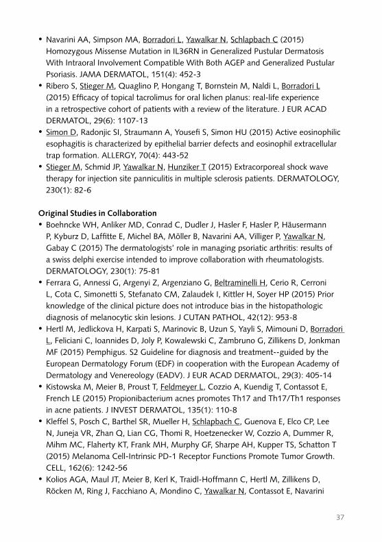
37
•Navarini AA, Simpson MA, Borradori L, Yawalkar N, Schlapbach C (2015) Homozygous Missense Mutation in IL36RN in Generalized Pustular Dermatosis With Intraoral Involvement Compatible With Both AGEP and Generalized Pustular Psoriasis. JAMA DERMATOL, 151(4): 452-3
•Ribero S, Stieger M, Quaglino P, Hongang T, Bornstein M, Naldi L, Borradori L (2015) Efficacy of topical tacrolimus for oral lichen planus: real-life experience in a retrospective cohort of patients with a review of the literature. J EUR ACAD DERMATOL, 29(6): 1107-13
• Simon D, Radonjic SI, Straumann A, Yousefi S, Simon HU (2015) Active eosinophilic esophagitis is characterized by epithelial barrier defects and eosinophil extracellular trap formation. ALLERGY, 70(4): 443-52
• Stieger M, Schmid JP, Yawalkar N, Hunziker T (2015) Extracorporeal shock wave therapy for injection site panniculitis in multiple sclerosis patients. DERMATOLOGY, 230(1): 82-6
Original Studies in Collaboration •Boehncke WH, Anliker MD, Conrad C, Dudler J, Hasler F, Hasler P, Häusermann
P, Kyburz D, Laffitte E, Michel BA, Möller B, Navarini AA, Villiger P, Yawalkar N, Gabay C (2015) The dermatologists’ role in managing psoriatic arthritis: results of a swiss delphi exercise intended to improve collaboration with rheumatologists. DERMATOLOGY, 230(1): 75-81
• Ferrara G, Annessi G, Argenyi Z, Argenziano G, Beltraminelli H, Cerio R, Cerroni L, Cota C, Simonetti S, Stefanato CM, Zalaudek I, Kittler H, Soyer HP (2015) Prior knowledge of the clinical picture does not introduce bias in the histopathologic diagnosis of melanocytic skin lesions. J CUTAN PATHOL, 42(12): 953-8
•Hertl M, Jedlickova H, Karpati S, Marinovic B, Uzun S, Yayli S, Mimouni D, Borradori L, Feliciani C, Ioannides D, Joly P, Kowalewski C, Zambruno G, Zillikens D, Jonkman MF (2015) Pemphigus. S2 Guideline for diagnosis and treatment--guided by the European Dermatology Forum (EDF) in cooperation with the European Academy of Dermatology and Venereology (EADV). J EUR ACAD DERMATOL, 29(3): 405-14
•Kistowska M, Meier B, Proust T, Feldmeyer L, Cozzio A, Kuendig T, Contassot E, French LE (2015) Propionibacterium acnes promotes Th17 and Th17/Th1 responses in acne patients. J INVEST DERMATOL, 135(1): 110-8
•Kleffel S, Posch C, Barthel SR, Mueller H, Schlapbach C, Guenova E, Elco CP, Lee N, Juneja VR, Zhan Q, Lian CG, Thomi R, Hoetzenecker W, Cozzio A, Dummer R, Mihm MC, Flaherty KT, Frank MH, Murphy GF, Sharpe AH, Kupper TS, Schatton T (2015) Melanoma Cell-Intrinsic PD-1 Receptor Functions Promote Tumor Growth. CELL, 162(6): 1242-56
•Kolios AGA, Maul JT, Meier B, Kerl K, Traidl-Hoffmann C, Hertl M, Zillikens D, Röcken M, Ring J, Facchiano A, Mondino C, Yawalkar N, Contassot E, Navarini
38
AA, French LE (2015) Canakinumab in adults with steroid-refractory pyoderma gangrenosum. BRIT J DERMATOL, 173(5): 1216-23
•Moalli F, Proulx ST, Schwendener R, Detmar M, Schlapbach C, Stein JV (2015) Intravital and whole-organ imaging reveals capture of melanoma-derived antigen by lymph node subcapsular macrophages leading to widespread deposition on follicular dendritic cells. FRONT IMMUNOL, 6(114): 114
•Morton CA, Wulf HC, Szeimies RM, Gilaberte Y, Basset-Seguin N, Sotiriou E, Piaserico S, Hunger R, Baharlou S, Sidoroff A, Braathen LR (2015) Practical approach to the use of daylight photodynamic therapy with topical methyl aminolevulinate for actinic keratosis: a European consensus. J EUR ACAD DERMATOL, 29(9): 1718-23
•Murrell DF, Marinovic B, Caux F, Prost C, Ahmed R, Wozniak K, Amagai M, Bauer J, Beissert S, Borradori L, Culton D, Fairley JA, Fivenson D, Jonkman MF, Marinkovich MP, Woodley D, Zone J, Aoki V, Bernard P, Bruckner-Tuderman L, Cianchini G, Venning V, Diaz L, Eming R, Grando SA, Hall RP, Hashimoto T, Herrero-González JE, Hertl M, Joly P, Karpati S, Kim J, Chan Kim S, Korman NJ, Kowalewski C, Lee SE, Rubenstein DR, Sprecher E, Yancey K, Zambruno G, Zillikens D, Doan S, Daniel BS, Werth VP (2015) Definitions and outcome measures for mucous membrane pemphigoid: recommendations of an international panel of experts. J AM ACAD DERMATOL, 72(1): 168-74
•Philipp-Dormston WG, Karrer S, Petering H, Ulrich C, Dirschka T, Berking C, Lonsdorf AS, Gerber PA, Radakovic S, Hunger R, Szeimies RM (2015) MAL-PDT mit Tageslicht - Aktuelle Datenlage und praxisorientierte Empfehlungen eines Expertentreffens. J DTSCH DERMATOL GES, 13(12): 1240-9
• Saunte DM, Boer J, Stratigos A, Szepietowski JC, Hamzavi I, Kim KH, Zarchi K, Antoniou C, Matusiak L, Lim HW, Williams M, Kwon HH, Gürer MA, Mammadova F, Kaminsky A, Prens E, van der Zee HH, Bettoli V, Zauli S, Hafner J, Lauchli S, French LE, Riad H, El-Domyati M, Abdel-Wahab H, Kirby B, Kelly G, Calderon P, Del Marmol V, Benhadou F, Revuz J, Zouboulis CC, Karagiannidis I, Sartorius K, Hagströmer L, McMeniman E, Ong N, Dolenc-Voljc M, Mokos ZB, Borradori L, Hunger R, Sladden C, Scheinfeld N, Moftah N, Emtestam L, Lapins J, Doss N, Kurokawa I, Jemec GBE (2015) Diagnostic delay in hidradenitis suppurativa is a global problem. BRIT J DERMATOL, 173(6): 1546-9
• Schatton T, Yang J, Kleffel S, Uehara M, Barthel SR, Schlapbach C, Zhan Q, Dudeney S, Mueller H, Lee N, de Vries JC, Meier B, Vander Beken S, Kluth MA, Ganss C, Sharpe AH, Waaga-Gasser AM, Sayegh MH, Abdi R, Scharffetter-Kochanek K, Murphy GF, Kupper TS, Frank NY, Frank MH (2015) ABCB5 Identifies Immunoregulatory Dermal Cells. CELL REP, 12(10): 1564-74
•Watanabe R, Gehad A, Yang C, Scott LL, Teague JE, Schlapbach C, Elco CP, Huang V, Matos TR, Kupper TS, Clark RA (2015) Human skin is protected by four
39
functionally and phenotypically discrete populations of resident and recirculating memory T cells. SCI TRANSL MED, 7(279): 279ra39
•Wouters J, Hunger R, Garrod T, Dubuis B, Hunziker T, van den Oord JJ, Lahav-le Coutre R (2015) First-in-Human Proof-of-Concept Study: Intralesional Administration of BQ788, an Endothelin Receptor B Antagonist, to Melanoma Skin Metastases. ONCOLOGIST, 20(10): 1121-2
•Yousefi S, Morshed MM, Amini P, Stojkov D, Simon D, von Gunten S, Kaufmann T, Simon HU (2015) Basophils exhibit antibacterial activity through extracellular trap formation. ALLERGY, 70(9): 1184-8
•Zecca C, Mainetti C, Blum R, Gobbi C (2015) Recurrent Nicolau syndrome associated with subcutaneous glatiramer acetate injection--a case report. BMC NEUROL, 15(249): 249
Others•Ana Filipa OC, Haneke E (2015) Melanonychia and Blue lunulae with the
Multikinase Inhibitor Sorafenib. J Pharmacovigilance, s2•Cogrel O, Haneke E (2015) [Tangential excision for a longitudinal melanonychia].
ANN DERMATOL VENER, 142(6-7): 450-1•dchtee Graauw EL, Beltraminelli H, Simon HU, Simon D (2015) Eosinophilia in
Dermatologic Disorders. IMMUNOL ALLERGY CLIN, 35(3): 545-60• Feliciani C, Joly P, Jonkman MF, Zambruno G, Zillikens D, Ioannides D, Kowalewski
C, Jedlickova H, Kárpáti S, Marinovic B, Mimouni D, Uzun S, Yayli S, Hertl M, Borradori L (2015) Management of bullous pemphigoid: the European Dermatology Forum consensus in collaboration with the European Academy of Dermatology and Venereology. BRIT J DERMATOL, 172(4): 867-77
•Göktay F, Altan ZM, Haras ZB, GüneεP, Yaεar, Aytekin S, Haneke E (2015) Multibranched acquired periungual fibrokeratomas with confounding histopathologic findings resembling papillomavirus infection: a report of two cases. J CUTAN PATHOL, 42(9): 652-6
•Häfliger S, Klötgen HW, Horn MP, Beltraminelli H, Borradori L (2015) Erythrodermic Epidermolysis Bullosa Acquisita. JAMA DERMATOL, 151(6): 678-80
•Haneke E (2015) Managing Complications of Fillers: Rare and Not-So-Rare. J Cutan Aesthet Surg, 8(4): 198-210
•Haneke E, Chang P, Leon Alvarado A, Vasquez Acabojon BMV (2015) Síndrome de Iso Kikuchi. Reporte de dos casos. Dermatologia CMQ, 13(3): 237-9
•Haneke E, Rammlmair A, Perruchoud DL (2015) Nagelpilz-Infektion - Was ist bei Onychomykose in der Praxis zu beachten? Dermatologie Praxis, 26(1): 10-3
• Lande R, Botti E, Jandus C, Dojcinovic D, Fanelli G, Conrad C, Chamilos G, Feldmeyer L, Marinari B, Chon S, Vence L, Riccieri V, Guillaume P, Navarini AA,
40
Romero P, Costanzo A, Piccolella E, Gilliet M, Frasca L (2015) Corrigendum: the antimicrobial peptide LL37 is a T-cell autoantigen in psoriasis. NAT COMMUN, 6: 6595
• Löser C, Dippel E, Wolter M, Haneke E (2015) Kasuistik: Melanonychie im Kindesalter. Akt Dermatol, 41(08/09)
•Morton C, Szeimies RM, Sidoroff A, Wennberg AM, Basset-Seguin N, Calzavara-Pinton P, Gilaberte Y, Hofbauer G, Hunger R, Karrer S, Lehmann P, Piaserico S, Ulrich C, Braathen L (2015) European Dermatology Forum Guidelines on topical photodynamic therapy. EUR J DERMATOL, 25(4): 296-311
•Nast A, Gisondi P, Ormerod AD, Saiag P, Smith C, Spuls PI, Arenberger P, Bachelez H, Barker J, Dauden E, de Jong EM, Feist E, Jacobs A, Jobling R, Kemény L, Maccarone M, Mrowietz U, Papp KA, Paul C, Reich K, Rosumeck S, Talme T, Thio HB, van de Kerkhof P, Werner RN, Yawalkar N (2015) European S3-Guidelines on the systemic treatment of psoriasis vulgaris - Update 2015 - Short version - EDF in cooperation with EADV and IPC. J EUR ACAD DERMATOL, 29(12): 2277-94
• Skaria A, (2015) The median forehead flap reviewed: a histologic study on vascular anatomy. EUR ARCH OTO-RHINO-L, 272(5): 1231-7
•Tschumi C, Seyed Jafari SM, Rothenberger J, Van de Ville D, Keel M, Krause F, Shafighi M (2015) Post-operative monitoring of free muscle transfers by Laser Doppler Imaging: A prospective study. MICROSURG, 35(7): 528-35
•Zouboulis CC, Desai N, Emtestam L, Hunger R, Ioannides D, Juhász I, Lapins J, Matusiak L, Prens EP, Revuz J, Schneider-Burrus S, Szepietowski JC, van der Zee HH, Jemec GBE (2015) European S1 guideline for the treatment of hidradenitis suppurativa/acne inversa. J EUR ACAD DERMATOL, 29(4): 619-44
Publications 2016Original in-house Studies•Christoph S, Cazzaniga S, Hunger R, Naldi L, Borradori L, Oberholzer PA (2016)
Ultraviolet radiation protection and skin cancer awareness in recreational athletes: a survey among participants in a running event. SWISS MED WKLY, 146: w14297
• Favre B, Begré N, Bouameur JE, Borradori L (2016) Development of a Novel Green Fluorescent Protein-Based Binding Assay to Study the Association of Plakins with Intermediate Filament Proteins. METHOD ENZYMOL, 569: 117-37
• Feldmeyer L, Hudgens CW, Ray-Lyons G, Nagarajan P, Aung PP, Curry JL, Torres-Cabala CA, Mino B, Rodriguez-Canales J, Reuben A, Chen PL, Ko JS, Billings SD, Bassett RL, Wistuba II, Cooper ZA, Prieto VG, Wargo JA, Tetzlaff MT (2016) Density, Distribution, and Composition of Immune Infiltrates Correlate with Survival in Merkel Cell Carcinoma. CLIN CANCER RES, 22(22): 5553-63
•Gautschi M, Oberholzer PA, Baumgartner M, Gadaldi K, Yawalkar N, Hunger R
41
(2016) Prognostic markers in lentigo maligna patients treated with imiquimod cream: A long-term follow-up study. J AM ACAD DERMATOL, 74(1): 81-87.e1
•Glauser S, Rutz M, Cazzaniga S, Hegyi I, Borradori L, Beltraminelli H (2016) Diagnostic value of immunohistochemistry on formalin-fixed, paraffin-embedded skin biopsy specimens for bullous pemphigoid. BRIT J DERMATOL, 175(5): 988-93
•Häfliger S, Horn MP, Suter VGA, Bornstein MM, Borradori L (2016) Rituximab for the Treatment of Isolated Refractory Desquamative Gingivitis Due to Mucous Membrane Pemphigoid. JAMA DERMATOL, 152(12): 1396-8
•Kaufmann RA, Oberholzer PA, Cazzaniga S, Hunger R (2016) Epithelial skin cancers after kidney transplantation: a retrospective single-centre study of 376 recipients. EUR J DERMATOL, 26(3): 265-70
•Perruchoud DL, Varonier C, Haneke E, Hunger R, Beltraminelli H, Borradori L, Ehnis Pérez A (2016) Bowen disease of the nail unit: a retrospective study of 12 cases and their association with human papillomaviruses. J EUR ACAD DERMATOL, 30(9): 1503-6
• Seyed Jafari SM, Timchik T, Hunger R (2016) In vivo confocal microscopy efficacy assessment of daylight photodynamic therapy in actinic keratosis patients. BRIT J DERMATOL, 175(2): 375-81
• Simon D, Cianferoni A, Spergel JM, Aceves S, Holbreich M, Venter C, Rothenberg ME, Terreehorst I, Muraro A, Lucendo AJ, Schoepfer A, Straumann A, Simon HU (2016) Eosinophilic esophagitis is characterized by a non-IgE-mediated food hypersensitivity. ALLERGY, 71(5): 611-20
•Ventura F, Correia O, Duarte AF, Barros AM, Haneke E (2016) «Retronychia – clinical and pathophysiological aspects». J EUR ACAD DERMATOL, 30(1): 16-9
•Yan KX, Huang Q, Fang X, Zhang ZH, Han L, Gadaldi K, Kang KF, Zheng ZZ, Xu JH, Yawalkar N (2016) IgE and FcεRI are highly expressed on innate cells in psoriasis. BRIT J DERMATOL, 175(1): 122-33
Original Studies in Collaboration•Dhayade S, Kaesler S, Sinnberg T, Dobrowinski H, Peters S, Naumann U, Liu H,
Hunger RE, Thunemann M, Biedermann T, Schittek B, Simon HU, Feil S, Feil R (2016) Sildenafil Potentiates a cGMP-Dependent Pathway to Promote Melanoma Growth. CELL REP, 14(11): 2599-610
•Di Landro A, Cazzaniga S, Cusano F, Bonci A, Carla C, Musumeci ML, Patrizi A, Bettoli V, Pezzarossa E, Caproni M, Fortina AB, Campione E, Ingordo V, Naldi L (2016) Adult female acne and associated risk factors: Results of a multicenter case-control study in Italy. J AM ACAD DERMATOL, 75(6): 1134-1141.e1
•Diphoorn J, Cazzaniga S, Gamba C, Schroeder J, Citterio A, Rivolta AL, Vighi GD, Naldi L (2016) Incidence, causative factors and mortality rates of Stevens-Johnson syndrome (SJS) and toxic epidermal necrolysis (TEN) in northern Italy: data from the
42
REACT registry. PHARMACOEPIDEM DR S, 25(2): 196-203•Gerhards NM, Sayar B, Origgi F, Galichet A, Müller EJ, Welle MM, Wiener DJ (2016)
Stem Cell-Associated Marker Expression in Canine Hair Follicles. J HISTOCHEM CYTOCHEM, 64(3): 190-204
•Kolios AGA, Yawalkar N, Anliker M, Boehncke WH, Borradori L, Conrad C, Gilliet M, Häusermann P, Itin P, Laffitte E, Mainetti C, French LE, Navarini AA (2016) Swiss S1 Guidelines on the Systemic Treatment of Psoriasis Vulgaris. DERMATOLOGY, 232(4): 385-406
•Murgiano L, Waluk DP, Towers R, Wiedemar N, Dietrich SJ, Jagannathan V, Drögemüller M, Balmer P, Druet T, Galichet A, Penedo MC, Müller EJ, Roosje P, Welle MM, Leeb T (2016) An Intronic MBTPS2 Variant Results in a Splicing Defect in Horses with Brindle Coat Texture. G3-GENES GENOM GENET, 6(9): 2963-70
•Protic M, Schoepfer A, Yawalkar N, Vavricka S, Seibold F (2016) Development of psoriasis in IBD patients under TNF-antagonist therapy is associated neither with anti-TNF-antagonist antibodies nor trough levels. SCAND J GASTROENTERO, 51(12): 1482-8
• Straumann A, Blanchard C, Radonjic-Hoesli S, Bussmann C, Hruz P, Safroneeva E, Simon D, Schoepfer AM, Simon HU (2016) A new eosinophilic esophagitis (EoE)-like disease without tissue eosinophilia found in EoE Families. ALLERGY, 71(6): 889-900
•Uter W, Warburton K, Weisshaar E, Simon D, Ballmer-Weber B, Mahler V, Fuchs T, Geier J, Wilkinson M (2016) Patch test results with rubber series in the European Surveillance System on Contact Allergies (ESSCA), 2013/14. CONTACT DERMATITIS, 75(6): 345-52
•Waluk DP, Zur G, Kaufmann R, Welle MM, Jagannathan V, Drögemüller C, Müller EJ, Leeb T, Galichet A (2016) A Splice Defect in the EDA Gene in Dogs with an X-Linked Hypohidrotic Ectodermal Dysplasia (XLHED) Phenotype. G3-GENES GENOM GENET, 6(9): 2949-54
•Wiener DJ, Doherr M, Müller EJ, Welle MM (2016) Spatial Distribution of Stem Cell-Like Keratinocytes in Dissected Compound Hair Follicles of the Dog. PLOS ONE, 11(1): e0146937
•Wollenberg A, Oranje A, Deleuran M, Simon D, Szalai Z, Kunz B, Svensson A, Barbarot S, von Kobyletzki L, Taieb A, de Bruin-Weller M, Werfel T, Trzeciak M, Vestergard C, Ring J, Darsow U (2016) ETFAD/EADV Eczema task force 2015 position paper on diagnosis and treatment of atopic dermatitis in adult and paediatric patients. J EUR ACAD DERMATOL, 30(5): 729-47
•Zeeh F, Suter M, Müller EJ, Brünisholz N, Zimmermann W, Ricklin M, Rüfenacht S, Olivry T, Roosje P (2016) Chronological clinical and pathological documentation of porcine ulcerative dermatitis. VET DERMATOL, 27(4): 315-e78
43
Others•Andre P, Haneke E (2016) Nicolau syndrome due to hyaluronic acid injections. J
COSMET LASER THER, 18(4): 239-44•Bieber T, Akdis C, Lauener R, Traidl-Hoffmann C, Schmid-Grendelmeier P, Schäppi
G, Allam JP, Apfelbacher C, Augustin M, Beck L, Biedermann T, Braun-Fahrländer C, Chew FT, Clavel T, Crameri R, Darsow U, Deleuran M, Dittlein D, Duchna HW, Eichenfeld L, Eyerich K, Frei R, Gelmetti C, Gieler U, Gilles S, Glatz M, Grando K, Green J, Gutermuth J, Guttman-Yassky E, Hanifin J, Hijnen D, Hoetzenecker W, Irvine A, Kalweit A, Katoh N, Knol E, Koren H, Möhrenschlager M, Münch D, Novak N, O’Mahony L, Paller AS, Rhyner C, Roduit C, Schiesser K, Schröder J, Simon D, Simon HU, Sokolowska M, Spuls P, Stalder JF, Straub D, Szalai Z, Taieb A, Takaoka R, Todd G, Todorova A, Vestergaard C, Werfel T, Wollenberg A, Ring J (2016) Global Allergy Forum and 3rd Davos Declaration 2015: Atopic dermatitis/Eczema: challenges and opportunities toward precision medicine. ALLERGY, 71(5): 588-92
•Cazzaniga S, Castelli E, Di Landro A, Zucchi A, Naldi L (2016) [Development of a teledermatology system for the melanoma diagnosis. The pilot experience of the project Clicca il neo]. Recenti progressi in medicina, 107(8): 440-3
•Cazzaniga S, Naldi L, Lecchi S, Kürzinger ML, Auclert L, Gori M, Chosidow O, Hercogova J (2016) A pilot study on the incidence of severe photosensitivity reactions leading to hospitalization linked to topical ketoprofen and other medications in selected European regions. Pharmacology research & perspectives, 4(3): e00225
•Di Zenzo G, Amber KT, Sayar B, Müller EJ, Borradori L (2016) Immune response in pemphigus and beyond: progresses and emerging concepts. SEMIN IMMUNOPATHOL, 38(1): 57-74
•Dummer R, Siano M, Hunger R, Lindenblatt N, Braun R, Michielin O, Mihic-Probst D, von Moos R, Najafi Y, Guckenberger M, Arnold A (2016) The updated Swiss guidelines 2016 for the treatment and follow-up of cutaneous melanoma. SWISS MED WKLY, 146: w14279
• Feldmeyer L, Heidemeyer K, Yawalkar N (2016) Acute Generalized Exanthematous Pustulosis: Pathogenesis, Genetic Background, Clinical Variants and Therapy. INT J MOL SCI, 17(8)
• Feldmeyer L, Prieto VG, Ivan D, Nagarajan P, Tetzlaff MT, Curry JL, Bell D, Moon BS, Torres-Cabala CA, Aung PP (2016) Role of Radiotherapy in Aggressive Digital Papillary Adenocarcinoma. ANN CLIN LAB SCI, 46(2): 222-4
•Gadaldi K, Feldmeyer L, Yawalkar N, Hunger R (2016) Ingenol Mebutate for Lentigo Maligna: A Case Report. DERMATOLOGY, 232 Suppl 1: 24-8
•Gritti P, Cazzaniga S, Castioni CA, Lorini FL, Velly LJ, Bilotta F (2016) Variability
44
by country in the European Neuroanaesthesia and Critical Care Interest Group subarachnoid haemorrhage survey. EUR J ANAESTH, 33(5): 387-90
•Haneke E (2016) Double Nail of the Little Toe. Skin Appendage Disord, 1(4): 163-7•Haneke E (2016) Perlen der Nagelchirurgie. Jatros. Dermatologie & Chirurgie,
2016(2): 20-3•Haneke E (2016) Hot Tipps bei Nageloperationen. Jatros. Dermatologie & Chirurgie,
2016(2): 20-1•Haneke E, Nakamura RC, Papaiordanou F, Machado EA, D’Almeida LF (2016)
Conservative surgery of subungual melanoma in situ. Surgical & Cosmetic Dermatology, 8(1): 70-2
•Kim M, Borradori L, Murrell DF (2016) Autoimmune Blistering Diseases in the Elderly: Clinical Presentations and Management. DRUG AGING, 33(10): 711-23
•Künzli K, Favre B, Chofflon M, Borradori L (2016) One gene but different proteins and diseases: the complexity of dystonin and bullous pemphigoid antigen 1. EXP DERMATOL, 25(1): 10-6
•Naldi L, Cazzaniga S (2016) Prediction of high total naevus count to estimate melanoma risk. We need more, don’t we? BRIT J DERMATOL, 174(2): 261-2
• Schlapbach C, Navarini AA (2016) The continuing evolution of targeted therapy for inflammatory skin disease. SEMIN IMMUNOPATHOL, 38(1): 123-33
• Simon HU, Radonjic SI (2016) Eosinophile Granulozyten. In: Allergologie. Biedermann,Tilo;Heppt,Werner;Renz,Harald;Röcken,Martin (Hg.): Springer, S. 77-85
• Simon HU, Yousefi S, Simon D (2016) Basophils can form extracellular DNA traps independent of a functional NADPH oxidase. In: Allergies: Current challenges and solutions. Proceedings of the 30th Symposium of the Collegium Internationale Allergologicum (CIA). Maurer,Markus;Behrendt,Heidrum (eds.): Pacini editore Medicina, S. 67-70
• Stieger M, Hunger R (2016) Ingenol Mebutate Treatment in a Patient with Gorlin Syndrome. DERMATOLOGY, 232 Suppl 1: 29-31
Master thesis and PhD thesis• Andrea Stillhard: Tumor-stroma characteristics in melanocytic lesions - Analysis of
elastic fibres in regressing/fibrosing/scarring zones (Professor L. Borradori, Dr. H. Beltraminelli); Master thesis
– Master abgeschlossen.• Elisabeth de Graauw: The role of eosinophils in bullous pemphigoid. Dissertation
(PhD thesis), 2012-2016. • Lea Bützberger: Allergic reactions after intravitreal injections. Masterarbeit (Medizin),
2015
45
• Debora Krähenbühl: Epithelial barrier in EoE patients wit/without corticosteroid therapy. Masterarbeit (Pharmazie), 2016
• Danijela Kostresevic: Is sensitization to Candida albicans in patients with eosinophilic esophagitis owing to disease progress or corticosteroid therapy? Masterarbeit (Pharmazie), 2015
• Claire Micossé: Characterization of human Th9 cells (PhD thesis, advisor: C. Schlap-bach), 2014-ongoing
• Leonhard von Meyenn: Cell-fate analysis of human IL-9 producing T cells (PhD thesis, advisor: C. Schlapbach), 2016-ongoing
• Martino Daniele Bianchi: Immune checkpoint blockade in advanced Merkel cell carcinoma. (Master Thesis, Advisor: C. Schlapbach), 2016 – ongoing
• Stefano Scanzio: Immune checkpoint blockade in advanced basal cell carcinoma and squamous
• cell carcinoma of the skin. (Master Thesis, Advisor: C. Schlapbach), 2016 – ongoing
Guest doctors• Mohammad Munshi from Saudi Arabia (from 01.05.2015)• Amal Wagih from Cairo, Egypt (30.03.2015 – 29.06.2015)• Alice Amani from Kigali, Republic of Rwanda (15.05.2015 – 14.08.2015 /
12.09.2016 – 03.12.2016)• Michael Joseph Benzaquen from Marseille, France (01.05.2016 – 31.10.2016)• Adane Ayele Legesse from Addis Abeba, Ethiopia (10.05.2016 – 03.08.2016)• Alex Richard Mremi from Moshi, Tanzania (10.05.2016 – 03.08.2016)• Carolyn Jean Chua-Aguilera from Taguig City, Philippines (05.09.2016 – 03.12.2016)• Workalemahu Alemu Belachew from Mekele, Ethiopia (03.10.2016 – 03.12.2016)• Jessica Kehdy from Lebanon (regularly during 2016)
Professional achievementsIn January, 2016, upon successful post-doctoral qualifications, Helmut Beltraminelli MD was promoted to Lecturer (Habilitation) in Dermatology at the University of Bern. His main research areas and grants in recent years, have centred on cutaneous lymphoma and on the development of dermatopathology in Africa.On 1.2.2016 Professor Christoph Schlapbach MD., PhD., was conferred the Peter Hans Hofschneider Professorship Award for molecular medicine over 3 years. He also became Lecturer after successful post-doctoral qualifications in Dermatology at the University of Bern. In recent years, his research and grants have centred on‚ Interleukin 9-producing T hleper memory cells and their role in anti-tumour immune response in malignant disease‘.
46
International recognitions and awardsProf. Dr. L. Borradori: • President, EADV (European Academy of Dermatology and Venereology) 2016-2018• Honorary Member,Société française de Dermatologie 2016• Honorary Member of the Polish Society of Dermatology 2016
PD Dr. med. H. Beltraminelli: • President Elect of the Swiss Group of Dermatopathology (SGDP) der Schweizerische
Gesellschaft für Dermatologie und Venerologie (SGDV) 2016• Elected Member of the Executive Committee of the International Society of Derma-
topathology (ISDP) 2016
GrantsProf. Dr. Ch. Schlapbach
Career Funding:Peter Hans Hofschneider Endowed Professorship for Molecular Medicine 2015Financing for 3 to 5 years:• Personal salary equivalent to that of an assistant professor at a Swiss University• One graduate student position• CHF 50,000/year for running costs
Project Funding as Principal Investigator:• Promedica Stiftung: Investigating the existence and function of a human TH9 cell
subset. CHF 165’000; 2016• W. & H. Berger-Janser Stiftung: The role of human TH9 cells in the anti-tumour
immune response in melanoma. CHF 60’000; 2016• Krummenacher Foundation: The role of human TH9 cells in the anti-tumour immune
response in melanoma. ε 5’000; 2016• Olga Mayenfisch Stiftung: Role of TH9 cells in initiation of cutaneous inflammation.
CHF 59’260; 2015
47
Sponsors 2016A. Menarini AGAbbVie AGAlmirall AGBiotest (Schweiz) AGCelgene GmbHDermapharm AGGalderma - Spirig IBSA Institut Biochimique SAJanssen Cilag AGLa Roche-Posay; Cosmetique Avtive(Suisse SA)Leo Pharmaceutical Products Sarath Ltd.Louis Widmer AGMEDA Pharma GmbHMerz Pharma (Schweiz) AGNovartis Pharma Schweiz AGPermamed AGPierre Fabre (Suisse) SAPro Farma AGRoche Pharma (Schweiz) AG
Sponsors 2015AbbVie AGAlmirall AGBiotest (Schweiz) AGCSL Behring AGDermapharm AGGalderma - Spirig GlaxoSmithKline AGIBSA Institut Biochimique SAJanssen Cilag AGLa Roche-Posay; Cosmetique Avtive(Suisse SA)Leo Pharmaceutical Products Sarath Ltd.Louis Widmer AGMEDA Pharma GmbHMerz Pharma Schweiz AGMölnlycke Health Care AGNovartis Pharma Schweiz AGPermamed AGPfizer AGPhadia AG (ThermoFisher Scientific)Pierre Fabre (Suisse) SARoche Pharma (Schweiz) AG
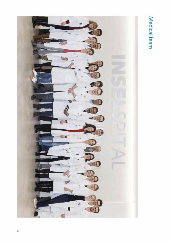
48
Medical team
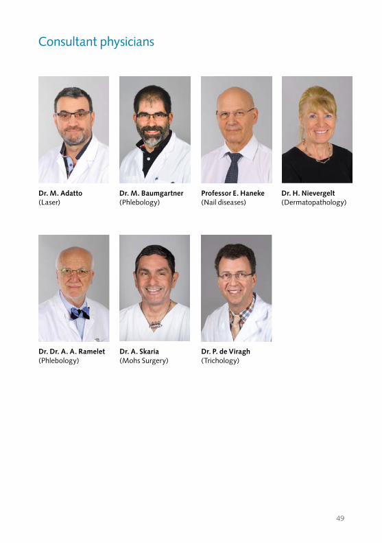
49
Consultant physicians
Dr. A. Skaria (Mohs Surgery)
Dr. Dr. A. A. Ramelet (Phlebology)
Dr. H. Nievergelt (Dermatopathology)
Professor E. Haneke (Nail diseases)
Dr. M. Baumgartner(Phlebology)
Dr. P. de Viragh (Trichology)
Dr. M. Adatto (Laser)

50
Dermatopathology team
Research team
51
Dermatosurgery nursing team
Out-patient clinic team
52
In-patient nursing team
54
Clinical nurse leaders
55
Allergy laboratory
Administration team

170
053
_201
7_10
_11_
KFG
/mm
InselspitalUniversitätsklinik für DermatologieFreiburgstrasse 34CH-3010 BernTel. +41 31 632 22 88www.insel.ch