Embed Size (px)
Citation preview
Brazilian Journal of Microbiology (2008) 39:508-510ISSN 1517-8382
508
DERMATOPHYTOSIS CAUSED BY MICROSPORUM CANIS AND MICROSPORUM GYPSEUMIN FREE-LIVING BRADYPUS VARIEGATUS (SCHIZ, 1825) IN THE STATE OF PERNAMBUCO,
BRAZIL
Gileno Antônio Araújo Xavier1; Leonildo Bento Galiza da Silva2; Davi Rubem da Silva3;Rodolfo de Moraes Peixoto4#; Gileno Câmara Lino4#; Rinaldo Aparecido Mota2*
1Departamento de Morfologia e Fisiologia Animal, Universidade Federal Rural de Pernambuco, Recife, PE, Brasil;2Departamento de Medicina Veterinária, Universidade Federal Rural de Pernambuco, Recife, PE, Brasil; 3Programa de
Pós-Graduação em Ciência Veterinária, Universidade Federal Rural de Pernambuco, Recife, PE, Brasil; 4Recife, PE, Brasil.
Submitted: October 03, 2007; Returned to authors for corrections: January 08, 2008; Approved: July 13, 2008.
ABSTRACT
Three cases of dermatophytosis in free living brown-throated three-toed sloths (Bradypus variegatus) in theZona da Mata, North of Pernambuco State, Brazil, were studied. Two animals presented areas of alopecia onthe pelvic member and thorax and one animal on the pelvic member only. The three animals presented scabs.Hair and scabs samples were submitted to microscopical examination after treatment with a 30 % KOH andcultivated in Mycosel Agar. The direct examination indicated the presence of arthrospores in the hair. Coloniesgrown after seven days of culture were confirmed as Microsporum based on examination of the structure ofthe macroconidia. This is the first observation of dermatophytosis caused by Microsporum canis andMicrosporum gypseum in free living sloths in the State of Pernambuco.
Key-words: Microsporum canis, Microsporum gypseum, Bradypus variegatus, dermatophytosis, sloths.
*Corresponding Author. Mailing address: Departamento de Medicina Veterinária, Universidade Federal Rural de Pernambuco, R. Dom Manoel deMedeiros, s/n - Dois Irmãos, Recife, PE, Brasil. 52171-900. E-mail: [email protected]# Médico Veterinário, autônomo.
INTRODUCTION
Microorganisms present in the wild, following anevolutionary process of millions of years, are well adapted tothe environment, rarely representing a threat to the animalpopulation. The infectious agents can be considered normaland essential components of the environment, and should bepreserved along with other elements of the ecosystem (1,2). Inthe absence of environmental changes, therefore, the role ofinfectious diseases as a native population-controlling factor isprobably significant.
In recent years, the expansion of the human population andthe increase of domesticated animals have created a straightpath for infecting organisms uncommon to the wild environment(1). When discussing disease transmission among differentspecies, it is convenient take three groups into consideration: a)wild fauna; b) domestic fauna; and c) human population. In this
context, the involvement of different interests - conservationists,economists, and public health officials, becomes evident (3).
Sloths are arboricole mammals that live, feed and reproduceon the top of trees and can hold in feces and urine for a week.They descend the trees to eliminate the feces, using their tailsto dig a hole in the ground; afterwards they urinate on the topof the hold and cover it with leaves, and with a typical legmovement climb back up the tree. The whole process ofdescent, defecation and return to the tree takes approximately30 minutes (4).
Dermatitis occurs often in these animals, and the diagnosis,treatment and prophylactic orientation vary according to theetiology of the process (5).
Dermatophytosis is an infection of keratinized tissues, nails,fur and cornea extract, caused by different species from thegenera Microsporum, Trichophyton and Epidermophyton.Those dermatophytes are the only fungi capable of invading
Dermatophytosis in B. variegatus
509
and residing in keratinized tissues. They are transmitted bycontact with fur and dandruff, either infected or containingfungal particles, originating either from the animals, theenvironment or fomites. Geophilic dermatophytes, such as M.gypseum, usually inhabit the soil, where they decompose intokeratinized debris. The zoophilics, such as M. canis, haveadapted to animals and are found in the soil only rarely (6).
Microsporum canis is the etiologic agent most frequentlyassociated with dog and cat dermatophytosis. Human infectionthrough contact with animals is fairly common. Microsporumcanis has also been found associated with other species:ruminant, equine, swine, primate, large felines, and others. It isconsidered highly pathogenic to canines, rodents and mustelids(7,8). In Brazil, the only report of this type of infection in slothsB. tridactylus was obtained in the city of Belém, PA by Silva etal. (9). M. gypseum produces lesions that are generally scabbyand almost always isolated. It can be found on dogs, cats, horses,steers and wild animals (7).
Due to the lack of studies involving the sanitizing aspectsof free-living sloths (Bradypus variegatus) in Brazil, the mainobjective of this study was to report the incidence ofdermatophytosis caused by M. canis or M. gypseum in theaforementioned species in the state of Pernambuco, Brasil.
MATERIAL AND METHODS
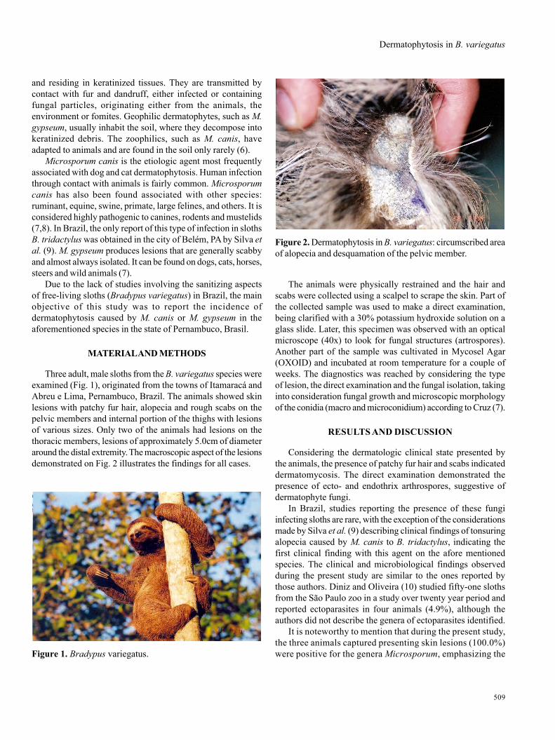
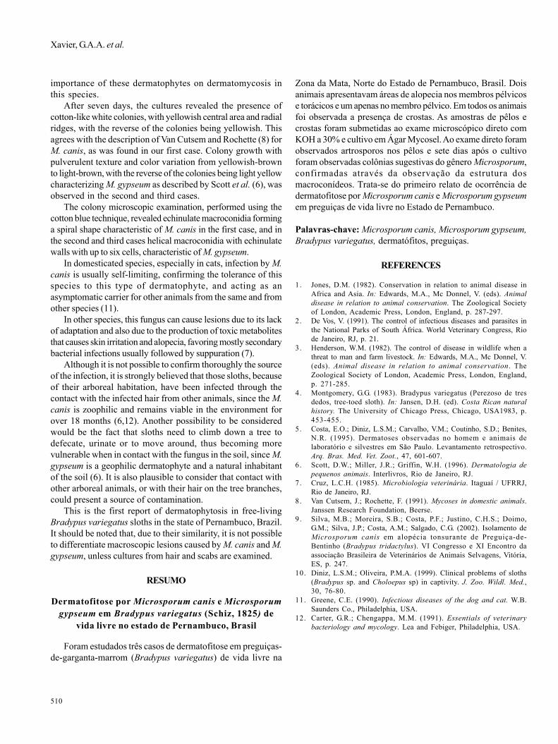
Three adult, male sloths from the B. variegatus species wereexamined (Fig. 1), originated from the towns of Itamaracá andAbreu e Lima, Pernambuco, Brazil. The animals showed skinlesions with patchy fur hair, alopecia and rough scabs on thepelvic members and internal portion of the thighs with lesionsof various sizes. Only two of the animals had lesions on thethoracic members, lesions of approximately 5.0cm of diameteraround the distal extremity. The macroscopic aspect of the lesionsdemonstrated on Fig. 2 illustrates the findings for all cases.
The animals were physically restrained and the hair andscabs were collected using a scalpel to scrape the skin. Part ofthe collected sample was used to make a direct examination,being clarified with a 30% potassium hydroxide solution on aglass slide. Later, this specimen was observed with an opticalmicroscope (40x) to look for fungal structures (artrospores).Another part of the sample was cultivated in Mycosel Agar(OXOID) and incubated at room temperature for a couple ofweeks. The diagnostics was reached by considering the typeof lesion, the direct examination and the fungal isolation, takinginto consideration fungal growth and microscopic morphologyof the conidia (macro and microconidium) according to Cruz (7).
RESULTS AND DISCUSSION
Considering the dermatologic clinical state presented bythe animals, the presence of patchy fur hair and scabs indicateddermatomycosis. The direct examination demonstrated thepresence of ecto- and endothrix arthrospores, suggestive ofdermatophyte fungi.
In Brazil, studies reporting the presence of these fungiinfecting sloths are rare, with the exception of the considerationsmade by Silva et al. (9) describing clinical findings of tonsuringalopecia caused by M. canis to B. tridactylus, indicating thefirst clinical finding with this agent on the afore mentionedspecies. The clinical and microbiological findings observedduring the present study are similar to the ones reported bythose authors. Diniz and Oliveira (10) studied fifty-one slothsfrom the São Paulo zoo in a study over twenty year period andreported ectoparasites in four animals (4.9%), although theauthors did not describe the genera of ectoparasites identified.
It is noteworthy to mention that during the present study,the three animals captured presenting skin lesions (100.0%)were positive for the genera Microsporum, emphasizing the
Figure 2. Dermatophytosis in B. variegatus: circumscribed areaof alopecia and desquamation of the pelvic member.
Figure 1. Bradypus variegatus.
510
Xavier, G.A.A. et al.
importance of these dermatophytes on dermatomycosis inthis species.
After seven days, the cultures revealed the presence ofcotton-like white colonies, with yellowish central area and radialridges, with the reverse of the colonies being yellowish. Thisagrees with the description of Van Cutsem and Rochette (8) forM. canis, as was found in our first case. Colony growth withpulverulent texture and color variation from yellowish-brownto light-brown, with the reverse of the colonies being light yellowcharacterizing M. gypseum as described by Scott et al. (6), wasobserved in the second and third cases.
The colony microscopic examination, performed using thecotton blue technique, revealed echinulate macroconidia forminga spiral shape characteristic of M. canis in the first case, and inthe second and third cases helical macroconidia with echinulatewalls with up to six cells, characteristic of M. gypseum.
In domesticated species, especially in cats, infection by M.canis is usually self-limiting, confirming the tolerance of thisspecies to this type of dermatophyte, and acting as anasymptomatic carrier for other animals from the same and fromother species (11).
In other species, this fungus can cause lesions due to its lackof adaptation and also due to the production of toxic metabolitesthat causes skin irritation and alopecia, favoring mostly secondarybacterial infections usually followed by suppuration (7).
Although it is not possible to confirm thoroughly the sourceof the infection, it is strongly believed that those sloths, becauseof their arboreal habitation, have been infected through thecontact with the infected hair from other animals, since the M.canis is zoophilic and remains viable in the environment forover 18 months (6,12). Another possibility to be consideredwould be the fact that sloths need to climb down a tree todefecate, urinate or to move around, thus becoming morevulnerable when in contact with the fungus in the soil, since M.gypseum is a geophilic dermatophyte and a natural inhabitantof the soil (6). It is also plausible to consider that contact withother arboreal animals, or with their hair on the tree branches,could present a source of contamination.
This is the first report of dermatophytosis in free-livingBradypus variegatus sloths in the state of Pernambuco, Brazil.It should be noted that, due to their similarity, it is not possibleto differentiate macroscopic lesions caused by M. canis and M.gypseum, unless cultures from hair and scabs are examined.
RESUMO
Dermatofitose por Microsporum canis e Microsporumgypseum em Bradypus variegatus (Schiz, 1825) de
vida livre no estado de Pernambuco, Brasil
Foram estudados três casos de dermatofitose em preguiças-de-garganta-marrom (Bradypus variegatus) de vida livre na
Zona da Mata, Norte do Estado de Pernambuco, Brasil. Doisanimais apresentavam áreas de alopecia nos membros pélvicose torácicos e um apenas no membro pélvico. Em todos os animaisfoi observada a presença de crostas. As amostras de pêlos ecrostas foram submetidas ao exame microscópico direto comKOH a 30% e cultivo em Ágar Mycosel. Ao exame direto foramobservados artrosporos nos pêlos e sete dias após o cultivoforam observadas colônias sugestivas do gênero Microsporum,confirmadas através da observação da estrutura dosmacroconídeos. Trata-se do primeiro relato de ocorrência dedermatofitose por Microsporum canis e Microsporum gypseumem preguiças de vida livre no Estado de Pernambuco.
Palavras-chave: Microsporum canis, Microsporum gypseum,Bradypus variegatus, dermatófitos, preguiças.
REFERENCES
1. Jones, D.M. (1982). Conservation in relation to animal disease inAfrica and Asia. In: Edwards, M.A., Mc Donnel, V. (eds). Animaldisease in relation to animal conservation. The Zoological Societyof London, Academic Press, London, England, p. 287-297.
2. De Vos, V. (1991). The control of infectious diseases and parasites inthe National Parks of South África. World Veterinary Congress, Riode Janeiro, RJ, p. 21.
3. Henderson, W.M. (1982). The control of disease in wildlife when athreat to man and farm livestock. In: Edwards, M.A., Mc Donnel, V.(eds). Animal disease in relation to animal conservation. TheZoological Society of London, Academic Press, London, England,p. 271-285.
4. Montgomery, G.G. (1983). Bradypus variegatus (Perezoso de tresdedos, tree-toed sloth). In: Jansen, D.H. (ed). Costa Rican naturalhistory. The University of Chicago Press, Chicago, USA1983, p.453-455.
5. Costa, E.O.; Diniz, L.S.M.; Carvalho, V.M.; Coutinho, S.D.; Benites,N.R. (1995). Dermatoses observadas no homem e animais delaboratório e silvestres em São Paulo. Levantamento retrospectivo.Arq. Bras. Med. Vet. Zoot., 47, 601-607.
6. Scott, D.W.; Miller, J.R.; Griffin, W.H. (1996). Dermatologia depequenos animais. Interlivros, Rio de Janeiro, RJ.
7. Cruz, L.C.H. (1985). Microbiologia veterinária. Itaguaí / UFRRJ,Rio de Janeiro, RJ.
8. Van Cutsem, J.; Rochette, F. (1991). Mycoses in domestic animals.Janssen Research Foundation, Beerse.
9. Silva, M.B.; Moreira, S.B.; Costa, P.F.; Justino, C.H.S.; Doimo,G.M.; Silva, J.P.; Costa, A.M.; Salgado, C.G. (2002). Isolamento deMicrosporum canis em alopécia tonsurante de Preguiça-de-Bentinho (Bradypus tridactylus). VI Congresso e XI Encontro daassociação Brasileira de Veterinários de Animais Selvagens, Vitória,ES, p. 247.
10. Diniz, L.S.M.; Oliveira, P.M.A. (1999). Clinical problems of sloths(Bradypus sp. and Choloepus sp) in captivity. J. Zoo. Wildl. Med.,30, 76-80.
11. Greene, C.E. (1990). Infectious diseases of the dog and cat. W.B.Saunders Co., Philadelphia, USA.
12. Carter, G.R.; Chengappa, M.M. (1991). Essentials of veterinarybacteriology and mycology. Lea and Febiger, Philadelphia, USA.