Embed Size (px)
Citation preview
Poster presentations
SESSION I: Novel opioid peptide agonists and antagonists
PS I – 1
Design and synthesis of opioid peptide analogues and mimetics
Rossella De Marco1, Luca Gentilucci1, Alessandra Tolomelli1, Sören Feddersen1,Santi Spampinato2, Andrea Bedini2, Roberto Artali3
����������� � ���� ��� ����������� ������ ��� � �������� �������� ����������� � ������������� ������ ��� � ���������������� ����������� � ��������������� ������� ��� ����� �� ������ ��� � ������� ������� ������
Background and Aim: Native opioid peptides pos-
sess enormous promise for the treatment of pain.
However, despite this potential, such peptides have
seen no use as clinically viable drugs for their low sta-
bility against proteolysis, resulting in a short duration
of in vivo activity, and for their poor ability to cross
the blood-brain barrier. One way to overcome these
disadvantages is the use of modified peptides, the so
called peptidomimetics [Gentilucci et al., 2010].
Methods: Starting from the structure of Endo-
morphin-1 (Tyr-Pro-Trp-PheNH2), the endogenous
ligand of µ-opioid receptor (MOR), we prepared modi-
fied peptides and peptidomimetics by introduction of
unnatural amino acids, halogenation of the Trp, intro-
duction of Pro-analogues, lipidization, and size reduc-
tion. The syntheses of (S)- or (R)-halo-tryphtophans
and other Trp-analogues have been performed by tan-
dem 1,4-addition and stereoselective enolate protona-
tion of DHA-containing peptides. Otherwise, substi-
tuted tryphtophans have been prepared in optically
pure form by enzymatic resolution. Oxazolidin-2-
one4-carboxylate has been introduced in position 2 of
endomorphin-1 as a proline mimetic, to give a trans
conformation of the Tyr1-Xaa2 peptide bond. Lipidiza-
tion and a rationale search for focused structure modifi-
cations lead to the endomorphin mimetic N-Ac-D-
Trp-Phe-Gly-NH2 and halo-substituted derivatives.
Results: The modified peptides and mimetics have
been bio-assayed in vitro by binding experiments, to
determine the affinity for MORs, and in vivo, to verify
the analgesic activity; the bioactivity has been ration-
alized by molecular docking computations.
Conclusion: The new endomorphin analogues and mi-
metics described here constitute promising leads for the
development of potentially useful, lipophilic analgesics.
�������������
���� ���� !"#� �$��%�&���� %�� ����� %� ������� � ������������ ������� ���� ��� �� $������ '��� �(�������� ���)� �����*��+���#�
226 �������������� ���� �� ����� ��� �������
�������� ��� �������� � ����� ������� ����������� �����������
PS I – 2
Novel opioid peptides from genomic data banks: phylogenetic
and bioactivity studies
Engin Bojnik1, Anna Magyar2, Fruzsina Babos2, Anna Borsodi2, Sandor Benyhe1
��� ���,�� � �������� ���� ���������� �� ����� ������ -,������� '��%��� � ������� � �&���%� -,������ ��� ����� .��,��� �����%� ���� ���� /01/*-'�� �,%��� �� -,�����
Background and Aim: Endogenous opioid peptides
are processed from large molecular mass precursor
polypeptides encoded by distinct genes. Proopiomela-
nocortin (POMC), Proenkephalin (PENK), Prodynor-
phin (PDYN) and Pronociceptin (PNOC) are the four
ubiquitous precursors, and their respective genes are
located in different chromosomes. In lower verte-
brates, mainly in fish species, additional copies of
such precursor propeptide genes exist. Our aim was to
collect and biochemically characterize novel opioid
sequences identified by data mining.
Methods and Results: Our systematic database sur-
vey reveals that individual peptides identified in ge-
nomic or cDNA databases of various species exhibit
remarkable polymorphism due to mutational changes.
However, the purifying selection during evolution
well restored those sequence motifs which are respon-
sible for the biological activites. Variable regions can
therefore be found mainly in the C-termini of the
opioid oligopeptides. The YGGF tetrapeptide seg-
ment is the most conserved region even in the case of
a number of non-mammalian nociceptin, indicating
that the ancestral forms of nociceptin contained tyro-
sine in the first position. Some bioactivity studies, in-
cluding radioreceptor binding and G-protein activa-
tion, with the chemically synthesized forms of the
newly identified peptides will be presented.
Conclusion: The entire set of opioid peptides from
different taxa constitutes natural- (NPL) or phyloge-
netic- (PPL) peptide libraries. NPL for endogenous
opioids seems to be important for phylogenetic, com-
parative bioactivity and systems biology studies. The
rapidly increasing content of NPLs displays amazing
structural variegation, herewith it is a component of
the chemical biodiversity.
�������������
�,������% (� 213'*3* 4"566�
PS I – 3
A putative pharmacophore model of µ-opioid receptor activity
Attila Borics1, Jayapal Reddy Mallareddy1, Katalin E. Kövér2, Géza Tóth1
��� ���,�� � �������� ���� ���������� �� ����� ������ -,������� '��%��� � ������� � �&���%� -,������ ����������� � ���� ���������� ��� � ��(������ -,�����
Background and Aim: Many natural and synthetic
peptides and peptidomimetics were shown previously
to bind to the µ-opioid receptor (MOR) but there is no
consensus about the structure responsible for biological
activity. A putative pharmacophore model was estab-
lished by our recent structure-activity study in which
ten µ-opioid receptor ligands and their analogues, pos-
sessing different µ-opioid receptor affinity and selec-
tivity were examined using molecular dynamics.
Results: Here we present the structural analysis of
a small pool of endomorphin derivatives containing
Dmt1 (2’,6’-dimethyltyrosine), Achc2 (2-aminocyclo-
hexanecarboxylic acid), pFPhe4 (para-fluorophenyl-
alanine) or �MePhe4 (�-methyl-phenylalanine) sub-
stitutions. These µ-opioid ligands were shown to have
similar or better pharmacological properties than
those of the parent compounds. Our present NMR
spectroscopic analysis and molecular modeling data
�������������� ���� �� ����� ��� ������� 227
�������� ��� �������� � ����� ������� ����������� �����������
indicated that the solution structures of these high-
affinity MOR ligands are in perfect agreement with
our previously proposed bioactive structure model, in
which the trans �1 rotamer of Tyr1/Dmt1, the gauche
conformer of Phe3 and the bent backbone structure
were assumed to be responsible for the high µ-opioid
activity.
Conclusions: Information obtained here could possi-
bly resolve many contradictions in the past literature
of opioid peptide structures and may serve as a guid-
ing principle in the pre-screening of possible struc-
tural candidates in the design of novel µ-opioid ago-
nists.
�������������
1�� 7��8 7� ,������% (� ��� -,������� ������� �� ����� $,�%213' ��*49!": �'� ����� # ��% 213' 3*6"54" �3� /� 3;�<�#��,����� ��� 1=�2�*>�?�:�@�*!A@:@32 B*?!:!*!!!4 ��% ���/� �* �� ���)��� �� ??">6: �3� /� 3;�<�# � �� � ��8��7��%��%�
PS I – 4
Cardiorespiratory effects of morphine and chimeric peptide
in anaesthetized rats
Piotr Wojciechowski1, Ma³gorzata Szereda-Przestaszewska1, Andrzej W. Lipkowski2
�0�(������� � �� �������� ����+� � ����������� � �,�������%� � �� �8�7 8� ��%���� �� ����� ����� �'�� C�� �7� �����%
Background and Aim: The use of opioid analgesics
is limited by a variety of side effects. AWL3106 – re-
cently synthesized chimeric peptide comprising two
pharmacophores with agonistic activity towards
µ-opioid (dermorphin) and tachykinin NK1 (sub-
stance P) receptors presents 30 folds stronger analge-
sic activity than morphine. The aim of this study was
to compare cardiorespiratory effects induced by
AWL3106 and a model opioid analgesic – morphine.
Methods: We measured cardiorespiratory parameters
in 16 anaesthetized rats, breathing spontaneously
room air, subdue to an intravenous (iv) injection of
0.3 µmol/kg of either AWL3106 (n = 9) or morphine
hydrochloride (n = 7).
Results: Bolus injection of AWL3106 evoked an ap-
noea of mean duration of 5.1 ± 0.7 s in all rats, fol-
lowed by breathing of 19% reduced frequency (f) and
augmented by 23% tidal volume (VT), which resulted
in no change in minute ventilation. Morphine admin-
istered at the same dose, in five out of seven rats in-
duced apnoea lasting on average 3.8 ± 1.1 s, which
was not statistically different from that provoked by
AWL3106. There was no effect on the frequency of
breathing, but 17% transient decrease in VT resulted
in short-lived decline in post-apnoeic ventilation.
Both substances lowered mean arterial pressure
(MAP). Morphine transiently reduced MAP by 24%
at 15 s after drug challenge. Hypotension evoked by
AWL3106 was biphasic and reached the lowest value
immediately after injection at a rate of 63% and at
43% between the first and second minute, compared
with control values.
Conclusion: This study showed that iv morphine in-
duces respiratory depression due to the lack of com-
pensatory mechanism. AWL3106 presents an additive
effect on respiratory pattern which is beneficial to
ventilation though strongly affects MAP.
������������
1�� 7��8 �� (��� ��������� ,������% (� / .���� � ���������0�-*1*?!!6*!94499#
228 �������������� ���� �� ����� ��� �������
�������� ��� �������� � ����� ������� ����������� �����������
PS I – 5
Evaluation of constrained amino acids in the design of
neurokinin 1 receptor- and bifunctional opioid – neurokinin 1
receptor ligands
Steven Ballet1, Lukasz Frankiewicz1, Karel Guillemyn1, Isabelle van den Eynde1,Nga N. Chung2, Attila Keresztes3, Joost Van Duppen4, Josephine Lai3, Eva Varga3,Frank Porreca3, Peter W. Schiller2, Jozef Vanden Broeck4, Dirk Tourwé1
����������� � 2������ ���� ���� B��)� ������ ����� ��, ��� ��, �� � �����,�� ����������� � ������� ������� ��% �����%��� ������ ������� �� ����� �� ���,��� ��������� ���%�� ����������� � ������������� ������ ��� � '��&���� 1,� ��� ��'���'����� ��� ������ ��% �,��(������� D��������� �� ���,��� 3�������8� ������ ����� 0�,���� 0�,���� �����,�
Background and Aim: Polypharmacology is gener-
ally possible in three ways: 1) drug cocktails, 2) multi-
component drugs and 3) multiple ligands. The first two
methods show disadvantages like poor patient compli-
ance and drug-drug interactions. Multiple ligands how-
ever, consisting of a single chemical entity that modu-
lates multiple targets simultaneously, potentially over-
come these drawbacks [Morphy et al., 2005; 2007].
Methods: Earlier research by Lipkowski and Hruby
demonstrated that bifunctional peptides possessing
both NK1R antagonist properties and opioid agonist
potency show advantages over current analgesic
drugs, such as a potent analgesic effect in both acute
and neuropathic pain states, suppression of the devel-
opment of tolerance and the presentation of antiallo-
dynic and antihyperalgesic effects [Bonney et al.,
2004; Hruby et al., 2009; Yamamoto et al., 2010].
Results and Conclusions: Different types of con-
strained aromatic amino acids were derivatized to pre-
pare new neurokinin 1 receptor (NK1R) antagonists.
Subsequent identification of the most potent antago-
nist allowed the preparation a chimeric structure pos-
sessing the desired dual opioid-NK1 activity (NK1 Ki
= 0.5 nM, pA2 = 7.8; MOR Ki = 0.4 nM) Our latest
efforts in the construction of this type of ligands will
be presented.
PS I – 6
Opioid and anti-opioid peptides derived from plant proteins
Masaaki Yoshikawa1,2, Soushi Sonoda1, Yuko Yamada2, Kousaku Ohinata2,Andrzej W. Lipkowski3
��� ����� �� ���,�� �� ���%,����� ������������ 3����� E����� �3���� ������ ���� 3����� E����� ��� �8�7 8� ��%���� �� ����������� C�� �7� �����%
Background and Aim: We have found various types
of opioid and anti-opioid peptides derived from plant
proteins. Rubiscolin-6 (YPLDLF) has been isolated as
a � agonist from an enzymatic digest of spinach ribulose
bisphosphate carboxylase/oxygenase (Rubisco), which
is the most abundant protein on the earth existing
ubiquitously in photosynthetic organisms. Although
the antinociceptive activity of rubiscolin-6 was small,
it exerted anxiolytic-like and memory-consolidating
effects in the elevated plus-maze and the step-through
type passive avoidance experiments, respectively. Inter-
estingly, the �1, 5HT�� and D� receptors were involved
in the anxiolytic effect, while the �1, 5HT�� and D� re-
ceptors were involved in the memory-consolidating
effect, respectively, downstream the � receptor.
Results: Rapakinin (RIY) has been isolated as a in-
hibitory peptide for the angiotensin I-converting en-
zyme. However, its antihypertensive effect was mainly
due to the CCK� receptor-dependent vasorelaxation.
Rapakinin exerted an anti-opioid effect in the tail-pinch
�������������� ���� �� ����� ��� ������� 229
�������� ��� �������� � ����� ������� ����������� �����������
test in the CCK� receptor-dependent manner. It also
showed a memory-consolidating effect in the CCK�
receptor-dependent manner. Rapakinin might stimulate
CCK release downstream of an unidentified receptor
since it showed no affinities for the CCK receptors.
Conclusions: It should be noted that these plant-
derived peptides might have been on the earth far much
earlier than the evolution of their receptors in animals.
PS I – 7
Design, synthesis and pharmacological evaluation of novel
endomorphin analogues with multiple structural modifications
Jayapal Reddy Mallareddy, Attila Borics, Attila Keresztes, Géza Tóth
�� ���,�� � �������� ���� ���������� �� ����� ������ -,������� '��%��� � ������� � �&���%� -,�����
Background and Aim: A major goal in opioid pep-
tide research is the development of novel analgesics
that could substitute morphine without its well-known
side-effects of dependence, tolerance, respiratory de-
pression and reward-seeking behavior [Keresztes et
al., 2010]. Endomorphins (EM-1, YPWF-NH2,
EM-2, YPFF-NH2) are high-affinity, µ-selective en-
dogenous opioids which inhibit pain without some of
the undesired side-effects of plant opiates [Zadina et
al., 1997]. These properties render endmorphins
(EMs) the object of continuous investigations and a
promising target for the development of new opioid
analgesics. Although EMs suffer from serious limita-
tions, including short duration of action, lack of activ-
ity after oral administration, relative inability to cross
the blood-brain barrier (BBB) into the central nervous
system (CNS) and poor metabolic stability.
Results: Herein, we report on the synthesis and phar-
macological evaluation of a small pool of endomor-
phin derivatives containing unnatural amino acids
(Dmt1, Achc2, pFPhe4 and �MePhe4). The multiple
structural modifications resulted in proteolytically
stable and pharmacologically active analogues. De-
pending upon the chiralities of incorporated amino
acid, analogues have exhibited moderate to high bind-
ing potencies, selectivities and efficacies.
Conclusions: In the present investigation we obtained
the stable analogues with improved pharmacological
properties.
�������������
1�� 7��8 7� ,������% (� ��� -,������� 213'*49!": ,�%�����% $�*6 /,������ ������� ����� �0�-*1*?!!6*;94499#
230 �������������� ���� �� ����� ��� �������
�������� ��� �������� � ����� ������� ����������� �����������
PS I – 8
Structural modification and biological evaluation
of Dmt1-DALDA analogues
Alexandre Novoa1, Sylvia Van Dorpe2, Evelien Wynendaele2, Nga Chung N3,Carole Lemieux3, Peter W Schiller3, Dirk Tourwé1, Bart De Spiegeleer2, Steven Ballet1
�2�.� ���������� � 2������ ���� ���� B��)� ������ ����� ��, ��� ��������� ?� �*:!5!� ��, �� � �����,�� ���,� F,����� G���� ������� ���,F,��# ���,�� ���������� � ��������,����� '���� � � $��,��� � ��������,����� ������� � .���� ������ ����-����(�8� ����� 4?� �*A!!! .���� �����,�� �0�(������� � ������� ������� ��% �����%� �� ������ ������� �� ����� �� ���,�� ���������� ::! ���� '���,� C� �� ��������� F,��� ���%� -?C :�4
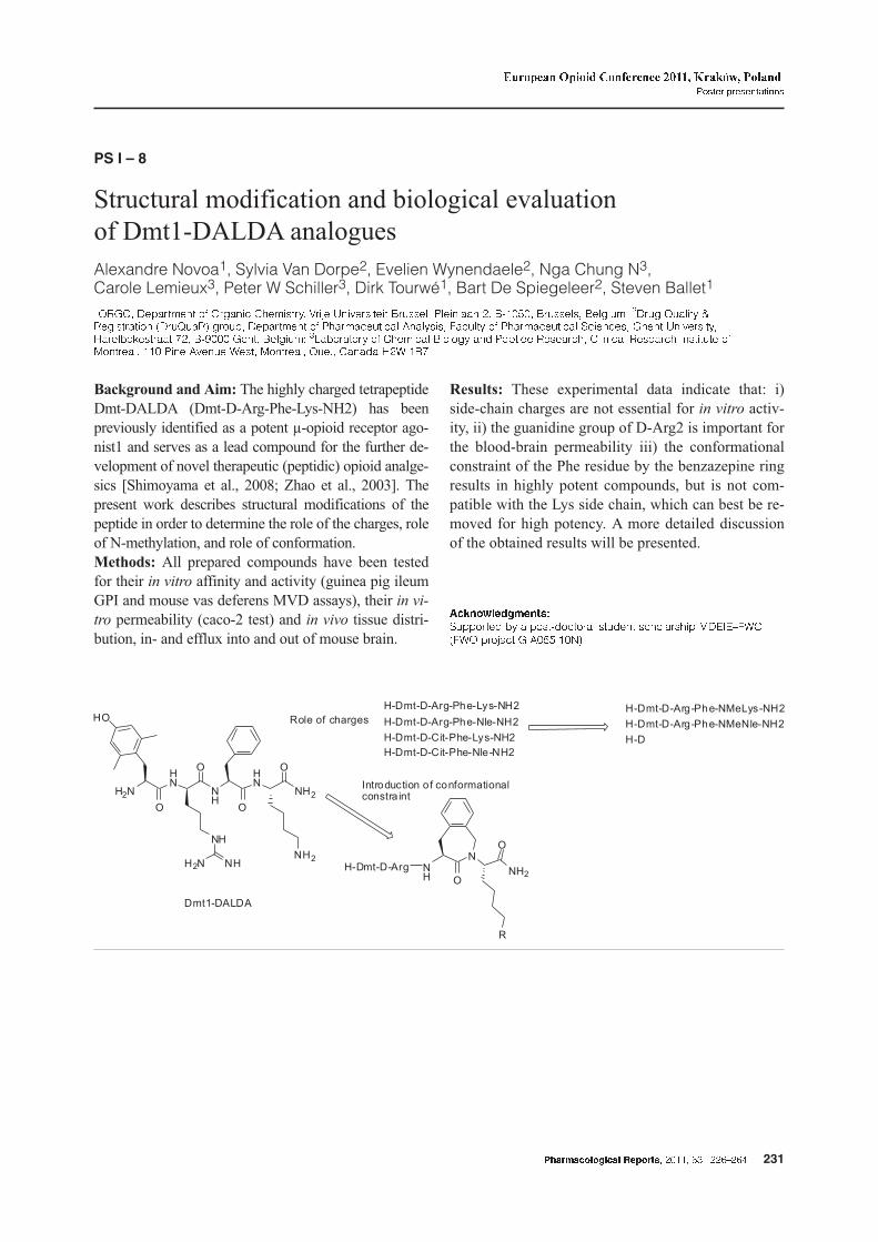
Background and Aim: The highly charged tetrapeptide
Dmt-DALDA (Dmt-D-Arg-Phe-Lys-NH2) has been
previously identified as a potent µ-opioid receptor ago-
nist1 and serves as a lead compound for the further de-
velopment of novel therapeutic (peptidic) opioid analge-
sics [Shimoyama et al., 2008; Zhao et al., 2003]. The
present work describes structural modifications of the
peptide in order to determine the role of the charges, role
of N-methylation, and role of conformation.
Methods: All prepared compounds have been tested
for their in vitro affinity and activity (guinea pig ileum
GPI and mouse vas deferens MVD assays), their in vi-
tro permeability (caco-2 test) and in vivo tissue distri-
bution, in- and efflux into and out of mouse brain.
Results: These experimental data indicate that: i)
side-chain charges are not essential for in vitro activ-
ity, ii) the guanidine group of D-Arg2 is important for
the blood-brain permeability iii) the conformational
constraint of the Phe residue by the benzazepine ring
results in highly potent compounds, but is not com-
patible with the Lys side chain, which can best be re-
moved for high potency. A more detailed discussion
of the obtained results will be presented.
�������������
�,������% (� � �� �*%������� �,%��� ������ ��� ��/�/H$C2�$C2 ���)��� .�'!55�:! #
�������������� ���� �� ����� ��� ������� 231
�������� ��� �������� � ����� ������� ����������� �����������
PS I – 9
Development of new opioid agonist tachykinin antagonist
chimeric compounds
Jolanta Dyniewicz1, Marta Bochynska1, Anna Lesniak1, Slawomir Ostrowski2,Jan Dobrowolski2, Agnieszka Kalicka2, Andrzej W.Lipkowski1
��� �8�7 8� ��%���� �� ����� ����� ���� � '��%��� � ������� � ���%, ����� ���� ��� �� ����� �� ���,��� C�� �7� �����%
Background and Aim: Our group proposed to de-
velop new chimeric analgesics in which opioid phar-
macophores are covalently hybridized with other
types of pharmacophores that positively modulate ef-
fects of the opioid part. Synergistic enhancement of
opioid analgesia and/or decrease of unwanted side-
effects should result from such hybridization. It is
generally accepted, that opioids and tachykinins are
classified as functional antagonists. Therefore, hy-
bridization of opioid agonist with tachykinin antago-
nist should result with very effective analgesics.
Melanoma cancer cells overexpress tachykinin recep-
tors. It has been already documented that tachykinin
antagonists express antiproliferative properties of
melanoma cells. Therefore chimeras of opioid ago-
nists and tachykinin antagonists should express both
analgesic and anticancer properties that make them
ideal for cancer pain treatment. Series of new opioid
agonist-tachykinin antagonists conjugates have been
developed, synthesized and tested.
Methods: Series of new opioid agonist-tachykinin
antagonists conjugates have been developed and syn-
thesized. The affinities to opioid receptors µ and � has
been evaluated. The antiproliferative properties of
new compounds has been evaluated in vitro in com-
parison to substance P antagonist aprepitant
Results: New compounds express high affinity to
opioid receptors as well as antiproliferative properties
of melanoma cancer cells in vitro.
Conclusion: New compounds are good candidates for
further studies as analgesics for cancer pain treatment.
������������
1�� �,%� �� (��� ,������% (� /,������ 6�� �1��� .���� ������� �0�-*1*?!!6*!94499#
PS I – 10
Synthesis and in vitro characterization of new salvinorin
a analogs incorporating natural amino acids
Jakub Fichna1,2, Kevin Lewellyn2, Feng Yan3, Bryan L. Roth3, Jordan K. Zjawiony2
����������� � ��������,��� ���� ���� ��%���� ������ ��� � 0�%&� 0�%&� �����%� ����������� � ����������� � ��% �� ������� ���,�� � ��������,����� ������� � ������ � ��������� ������ ��� � �� � ����� ������ ���� ��� ��'� ����������� �������������� ������ ��� � ���� ������� ��%���� ������� ����� -���� � ��'
Background and Aim: A series of new derivatives of
salvinorin A (SA), substituted at the C(2) position
with natural non-aromatic and aromatic amino acids,
was synthesized. The binding properties of SA ana-
logs were characterized in the in vitro studies.
Methods: SA was isolated from S. divinorum, puri-
fied and converted to salvinorin B (SB) according to
the methods established in our laboratory. New ana-
logs were synthesized from SB and N-Fmoc-protected
amino acids using N-methylmorpholine as a catalyst
and TBTU as a coupling reagent, followed by depro-
tection with piperidine. For all the compounds spec-
tral data were obtained. The �-opioid receptor (KOR)
and [��S]GTP�S binding assays were conducted as de-
scribed previously.
Results: The introduction of N-Fmoc-protected
amino acids resulted in loss of affinity at KOR. The
removal of Fmoc from Val-substituted analog in-
232 �������������� ���� �� ����� ��� �������
�������� ��� �������� � ����� ������� ����������� �����������
creased its affinity at KOR binding sites, making it al-
most equipotent with the parent compound, SA (K�
values of 42.0 ± 2.05 and 0.75 ± 0.62 nM for Val-SA
vs. SA, respectively). As shown in the [��S]GTP�S
binding assays, Val-SA was a full agonist at KOR.
Conclusion: Using a novel approach, a series of new
amino acid derivatives of SA was synthesized. One of
the obtained analogs, Val-SA, displayed high affinity
and full agonist activity at KOR, comparable to that
of the parent compound.
�������������
1�� 7��8 7� ,������% (� ��� �,����, ��, ������� � ������� � ���� ��� � ������� ��% -����� /%,������ �I�� ?!:!!!"A4!@5!>*!6*!!9 �� E$#� ��� �- .���� �!: �'!:4?!> ��% ��� ��- � ���������� ��,� ��������� ��������
�������������� ���� �� ����� ��� ������� 233
�������� ��� �������� � ����� ������� ����������� �����������
SESSION II: Opioid agonist specific signaling and regulation
PS II – 1
Opioids and the apoptotic pathway in MCF-7 breast cancer cell line
Katarzyna Gach1, Janusz Szemraj2, Anna Wyrêbska1, Anna Janecka1
����������� � ��������,��� ���� ���� ��%���� ������ ��� � 0�%&� �����%� ����������� � �������� ���� ��%���� ������ ���� 0�%&� �����%
Background and Aim: Apoptosis is an active pro-
cess of controlled cell death in the development and
maintenance of tissue homeostasis. There are two
main apoptotic pathways: the intrinsic or
mitochondrial-mediated pathway and the extrinsic or
death receptor-mediated pathway Mitochondrial-
mediated apoptosis is controlled by Bcl-2 family of
proteins. The death receptor-mediated pathway is ini-
tiated by the ligation of cell death ligands with their
death receptors. Caspase-3 is activated in both apop-
totic pathways and plays central role in the execution-
phase of cell apoptosis. Apoptosis is usually deregu-
lated in cancer cells and this deregulation can contrib-
ute to uncontrollable proliferation and tumor growth.
Opioids, especially morphine, are the most effective
drugs available clinically for the management of se-
vere pain associated with cancer. However, opioids
can also affect proliferation, migration and apoptosis
of tumor cells. This study was designed to test the ef-
fect of different opioids such as: morphine,
endomorphin-2, and morphiceptin on some crucial
apoptotic gene expression in MCF-7 breast adenocar-
cinoma cells.
Methods: The expression of studied genes was quan-
tified using real-time RT-PCR
Results: All tested opioids increase the expression
levels of pro-apoptotic genes, bax and caspase-3, and
decrease anti-apoptotic gene bcl-2 expression.
Conclusion: The findings reported in this study may
be useful in designing further experiments aimed at
elucidating the role of the opioids in apoptosis ma-
chinery. The ability to modulate the life or death of
a cell by opioids makes them potential targets for drug
development.
�������������
1�� 7��8 7� ,������% (� ��� ����� ��� ���� � ���� ��� �������� J�,����, ��, � � � ��?!:!!!A>4!#�
234 �������������� ���� �� ����� ��� �������
�������� ��� �������� � ����� ������� ����������� �����������
PS II – 2
Lack of agonist-induced desensitization of µ-opioid receptors
located at nerve terminals
Janet Denise Lowe, Chris Philip Bailey
���������� � �������� ��% ������������� ������ ��� � ����� ����� �3
Background and Aim: Agonist-induced desensitiza-
tion of µ opioid receptors (MOPrs) is one of the lead-
ing mechanisms thought to underlie the development
of tolerance to opioid drugs such as morphine and
heroin. Although many studies have investigated de-
sensitization of MOPrs located on cell bodies, few
have examined MOPrs located at nerve terminals. In
the ventral tegmental area (VTA), a brain region im-
plicated in the rewarding properties of numerous
drugs of abuse, MOPrs are located on GABAergic in-
terneurones both somatodendritically (ie. on cell bod-
ies and dendrites) and on nerve terminals that inner-
vate dopaminergic neurones.
Methods: Using whole-cell patch-clamp electro-
physiological methods, we have investigated agonist-
induced desensitization of both populations of MOPrs
in mouse VTA slices. Actions of MOPrs at nerve ter-
minals were assessed by recording miniature and
evoked inhibitory postsynaptic currents (IPSCs) from
the postsynaptic dopaminergic neurones. Cell body
MOPr activity was measured by recording G-protein
activated inwardly rectifying potassium channel
(GIRK) currents directly from GABAergic neurons.
Results: Both morphine (30 µM) and DAMGO
(10 µM) inhibited the frequency of miniature IPSCs
and decreased the amplitude of evoked IPSCs. These
effects were sustained during 10-minute applications,
even in conditions where there was no receptor re-
serve. In contrast, MOPrs at the cell bodies of GA-
BAergic neurones rapidly desensitized; DAMGO
(10 µM) induced a GIRK current that declined by ap-
proximately 50% during a 10-minute application.
Conclusions: In the mouse ventral tegmental area,
MOPrs located at nerve terminals do not readily de-
sensitize, or do so by different mechanisms to those
located at cell bodies.
�������������
$,�%�% (� �� �.!4!:>"6#�
PS II – 3
The effect of sodium ion on µ-opioid receptor G-protein
activation
Ferenc Zádor, Sándor Benyhe
�� ���,�� � �������� ���� ���������� �� ����� ������ -,������� '��%��� � ������� � �&���%� -,�����
Background and Aim: During functional [��S]GTP�S
binding assays, in which the G-protein activation is
measured, the used buffer contains 100 mM NaCl so-
lution, which probably decreases the agonist binding
in a high extent, thus the agonist induced G-protein
activation could also be highly inhibited. Our aim was
to investigate agonist induced G-protein responses in
the absence, and in the presence of different concen-
tration of Na� in µ-opioid receptors (MOR) in rat
brain membrane homogenates.
Methods: In our experiments we used functional
[��S]GTP�S binding assays to monitor G-protein acti-
vation. The MOR was activated by a highly selective
[d-Ala�,�Phe�,Gly�-ol]enkephalin (DAMGO) syn-
�������������� ���� �� ����� ��� ������� 235
�������� ��� �������� � ����� ������� ����������� �����������
thetic peptide. NaCl was added either only directly to
the tubes in the absence or presence of DAMGO
(10 µM), or either just in to the assay buffer.
Results: Sodium ion decreased the basal activity of
G-protein in a concentration dependent manner. When
the receptor was activated with DAMGO the highest
stimulation was in 10 and 25 mM concentrations of
NaCl. If the ion was added in the assay buffer the
strongest stimulation was in the 100 mM concentra-
tion, and at the same time the affinity of DAMGO was
the lowest.
Conclusion: We assume that the reason why the G-
protein activation culminates in 100 mM NaCl con-
centration is because it is the nearest to the physio-
logical NaCl concentration, which is 150 mM. Thus
in the future it is expedient to carry out experiments in
such concentration.
�������������
-,������� ��������� �� ����� $,�% � 31-*213'� ����� �,�(��3*4"566#�
PS II – 4
RGS2 and RGS4 proteins act as novel modulators of � and �
opioid receptors signaling
Maria-P. Papakonstantinou, Leonidas J. Leontiadis, Michalis Sarris, Fotis Nikolos,Zafiroula Georgoussi
0�(������� � ���,��� ��������� ��% �����,��� ������������� �� ���,�� � �������� ������� ����� �� ��������� �� ����������8���� �� '�� ���� 8���� '���� � .�����
Background and Aim: Regulators of G protein Sig-
naling (RGS) comprise a large multifunctional protein
family that accelerate GTP hydrolysis of G� subunits,
thus modulating G protein coupled receptor (GPCR)
signaling. RGS proteins also act as effector antago-
nists and serve as “platforms” where protein com-
plexes can be formed [Willars , 2006]. We have previ-
ously demonstrated that RGS4 directly interacts with
µ (µ-OR) and � (�-OR) opioid receptors to regulate
their signaling [Georgoussi et al., 2006; Leontiadis et
al. 2009]. To deduce whether selectivity in coupling
between members of RGS proteins and opioid recep-
tors exist we tested the ability of members of
B/R4-RGS family to interact with � opioid receptor
(�-OR).
Results: Pulldown experiments using GST fusion
peptides encompassing the �-third intracellular loop
(�-i3L) and the carboxyl terminal tails of all three
opioid receptors subtypes (µ, �, �) indicated that
RGS2 interacts within the �-i3L and the C-terminal
regions of �-OR and �-OR but not µ-OR. Co-
immunoprecipitation studies indicated that RGS2 as-
sociates with �-OR and �-OR constitutively and re-
tains upon agonist stimulation for both receptors.
Moreover subcellular localization of both RGS pro-
teins is altered upon receptor stimulation. RGS2 and
RGS4 display a differential regulatory effect as as-
sessed by a series of functional assays on �-OR and
�-OR upon receptor activation.
Conclusion: Collectively, our results suggest that al-
though �-OR and �-OR interact with the same subset
of RGS proteins each of them affects signaling in
a distinct manner.
�������������
1�� 7��8 7� ,������% (� ��� /� ����� K �������L�0�-*1?!!6*!94499# �� D�.�
236 �������������� ���� �� ����� ��� �������
�������� ��� �������� � ����� ������� ����������� �����������
PS II – 5
�-opioid receptor activation leads to neurite outgrowth and
neuronal survival via a STAT5B-G�i/o pathway
Eirini-Maria Georganta, Zafiroula Georgoussi
0�(������� � ���,��� ��������� ��% �����,��� ������������� �� ���,�� � �������� ������� ����� �� ��������� �� �����M����8���� N� '���� � .�����
Background and Aim: The opioid receptors partici-
pate in mechanisms controlling neural growth, differ-
entiation and synaptic plasticity [Christie, 2008]. We
have recently demonstrated that �- and µ- opioid re-
ceptors (�-OR and µ-OR) form multi-component sig-
naling complexes, consisting of STAT5A/B, c-Src ki-
nase and selective G protein subunits, leading to
STAT5A/B phosphorylation [Georganta et al., 2010;
Mazarakou and Georgoussi, 2005]. It seems, there-
fore, plausible to speculate that opioids receptors may
modulate differentiation of neuronal cells involving
members of the STAT family.
Result: To examine the effect of �-OR activation on
neuronal survival and neurite outgrowth, Neuro-2A
cells were treated with various ligands and a) the
number of live cells was visualized and counted under
a microscope in the presence of trypan blue, or b) the
length of the neurites was measured. Our results have
shown a higher percentage of surviving cells in the
presence of DSLET, an effect that was reversed by an-
tagonist co-treatment or expression of a dominant
negative STAT5B construct (DN-STAT5B). Similarly,
agonist administration resulted to increased neurite
outgrowth and this effect was blocked by pertussis
toxin pre-treatment and the presence of DN-STAT5B.
Conclusion: Taken together, our findings demon-
strate that �-OR activation leads to neuronal cell sur-
vival and neurite outgrowth via a signaling pathway
involving G�i/o proteins and STAT5B.
�������������
1�� 7��8 7� ,������% (� ��� /� ����� M �������N�0�-*1?!!6*!94499#�
PS II – 6
A novel mechanism of signal transmission: “prototypical”
interactions of neuropeptides with plasma membrane
Tatiana Yakovleva1, Volodymyr Khmyz2, Oleksandr Maximyuk2, Zoya Marinova3,Vladana Vukojeviæ3, Oleg Krishtal2, Georgy Bakalkin1
����������� ��������,����� ��� ������ � ��� ��� ������ ���� ��� ���� ����������� ���,��� ���(��������� ���������& �� ���,��� ��� ������� 3���� �8������ ����������� ���� �,�� ���� 3������ 8� �� ��� ����8����� �7�%��
Background and Aim: Several peptides including
penetratin and Tat are known to translocate across the
plasma membrane. We here demonstrate that dynorphin
A (DA) and big dynorphin (BD), consisting of dynor-
phins A and B, can translocate across the plasma
membrane of live neuronal and non-neuronal cells,
and form pores in this membrane.
Methods: Confocal fluorescence microscopy, fluorescence
correlation spectroscopy and patch-clamp techniques.
Results: DA and BD were found to be able to pene-
trate across the cell membrane into neurons and non-
neuronal cells. The peptide distribution was character-
ized by substantial cytoplasmic and plasma mem-
brane labeling with minimal signal in the cell nucleus.
The translocation potential of DA was comparable to
that of transportan-10, a prototypical cell penetrating
peptide. A central BD fragment, which retains all ba-
sic amino acid residues and dynorphin B did not enter
�������������� ���� �� ����� ��� ������� 237
�������� ��� �������� � ����� ������� ����������� �����������
the cells. The patch-clamp experiments demonstrated
that BD at low micromolar concentrations, and DA at
higher concentrations induced transient increases in
the cell membrane conductance in cultivated neurons
and in non-neuronal cells. The BD-induced conduc-
tance increases were voltage-dependent; their fre-
quency increased at the low negative potential inside
the cell. Translocation and pore formation were not
mediated via opioid receptors. The potential of dynor-
phins to penetrate into cells and to form pores corre-
lated with their ability to induce non-opioid effects in
animals including pathological pain.
Conclusions: Translocation across the plasma mem-
brane and pore formation may represent a hitherto un-
known mechanism by which dynorphins can signal
information across the plasma membrane.
�������������
$,�%��� * ��� �7�%� � B�� $'� ��% �7�%� � �� ���,�� B� (� ������
238 �������������� ���� �� ����� ��� �������
�������� ��� �������� � ����� ������� ����������� �����������
SESSION III: Opioids in pain
PS III – 1
Activation of opioid receptors in injured nerves is required for
efficient analgesia in neuropathic pain
Dominika Labuz, Yvonne Schmidt, Halina Machelska
���������� � '�� ��� ������� $��� ������ ��� ������� �����<*���, � ���)���� $���8���� ������� .������
Background and Aim: Pain arising from peripheral
nerve injuries is one of the most debilitating forms of
chronic painful conditions, and it is considered to be
weakly responsive to opioids. In this study we exam-
ined whether opioid delivery relative to the site of
nerve injury is important for efficient peripheral anal-
gesia.
Methods: Two and 14 days after chronic constriction
injury (CCI) we examined the expression of opioid re-
ceptors by immunofluorescence and mechanical allo-
dynia using von Frey test in mice. Opioid receptor
agonists and antagonists were injected at the CCI site
or into the paw (ipl) innervated by injured nerve.
Results: We found that the number of µ- and �-receptor
expressing neurons in dorsal root ganglia did not
change after CCI. However, in injured nerves, the in-
tesity of µ- and �-receptor staining was enhanced
proximally to ligatures. In hind paws innervated by
damaged nerves, the number of µ- and �-receptor
expressing fibers was unchanged or decreased, re-
spectively. µ-, �- and �-receptor selective agonists
(DAMGO, DPDPE and U50,488H, respectively) in-
jected at the CCI site or ipl produced dose-dependent
analgesia. Interestingly, the potency and efficacy of
opioids was substantially higher after their application
at the CCI site than ipl. The analgesic effects of ago-
nists were blocked by the respective selective receptor
antagonists applied locally.
Conclusions: Our studies suggest that activation of
opioid receptors directly at the site of nerve injury is
critical for efficient peripheral analgesia in neuro-
pathic conditions.
�������������
�,������% (� ��,� ��� $�� ��,�� ������ ���� ��' ?>94@:*>#� � ������� � ������ � �+� � �
�������������� ���� �� ����� ��� ������� 239
�������� ��� �������� � ����� ������� ����������� �����������
PS III – 2
The influence of minocycline on injury-induced changes
in dynorphin and nociceptin expression and effects of KOP
and NOP receptor ligands in a rat model of neuropathic pain
Ewelina Rojewska, Joanna Mika, Wioletta Makuch, Barbara Przewlocka
���������� � ���� ������������� �� ���,�� � ������������� ���� � '��%��� � ������� � 3��8�7� �����%
Background and Aim: A role of neuropeptides in
neuropathic pain development has been implicated;
however, the neuroimmune interactions that are in-
volved in the underlying mechanisms may be more
important than previously thought. The aim of our
study was to examine the effects of inhibition of glia
activation by minocycline on injury-induced dynorphin
and nociceptin changes and effects of KOP and NOP
receptor ligands in a rat model of neuropathic pain.
Methods: The chronic constriction injury (CCI) of
the rat sciatic nerve was performed. The opioid recep-
tor ligands and minocycline were injected intrathe-
cally or intraperitonealy. Two behavioral tests were
conducted to measure allodynia (von Frey test) and
hyperalgesia (cold plate test). For biochemical studies
the RT-PCR was used. The experiments were carried
out according to IASP rules (Zimmermann, 1983).
Results: The RT-PCR results indicated a strong
upregulation of prodynorphin mRNA with no changes
in the expression of pronociceptin in the spinal cord
after chronic constriction injury (CCI). Changes in
prodynorphin were parallel to higher expression of
microglia marker C1q mRNA. In the DRG, a very
strong upregulation of prodynorphin (1387%) and
pronociceptin (122%) mRNAs was observed. Inter-
estingly, preemptive and repeated intraperitoneal in-
jection of minocycline inhibits the activation of C1q
positive cells and upregulation of prodynorphin and
pronociceptin in the DRG. We demonstrate that anti-
allodynic effect of intrathecal administration of
U50,488H (25–100 µg), but not dynorphin 0.15–15
µg) and nociceptin (0.05–5 µg) were significantly po-
tentiated by preemptively and repeatedly injected
minocycline.
Conclusions: We present evidence that glial inhibi-
tion not only diminishes neuropathic pain-related be-
havior, but also reduces the injury-enhanced expres-
sion of prodynorphin and nociceptin in the DRG. The
behavioral studies underline that glia activation can
be an important factor especially in kappa-opioid me-
diated analgesia.
�������������
�,������% (� ����� >!5945A94 ��% ������� :�:�?� ���� ��&C�
240 �������������� ���� �� ����� ��� �������
�������� ��� �������� � ����� ������� ����������� �����������
PS III – 3
The possible role of endomorphin 2 biosynthesis and CART
peptide in the nociceptive information processing in rat spinal
dorsal horn: pharmacology and immunohistochemistry
Kornél Király1, Márk Kozsurek2, Zita Puskár2, Apolka Szentirmay1, Csaba Fekete3,András Z. Rónai1
����������� � ������������ ��% ���������������� ������7�� ������ ���� �,%��� � -,������ ����� � '�������� -� ��������% /�(�������� ������7�� ������ ���� �,%��� � -,������ ��� ���,�� � /+���������� ��%����� � 1�� -,������� '��%��� �������� � �,%��� �� -,�����
Background and Aim: While demonstrating the pos-
sibility of de novo biosynthesis of endomorphin 2 in
rat isolated L4,5 dorsal root ganglia from Tyr-Pro pre-
cursor in the presence of DPP4 inhibitor Ile-Pro-Ile,
besides the heretic idea itself, two further features had
to be considered. First, the biosynthesis took place at
the external surface of plasma membrane. Second,
while Tyr-Pro had to be provided exogenously, the
other co-substrate must have been generated endoge-
nously. Since similar endomorphin biosynthetic rules
operate in rat spinal dorsal horn in carrageenan induced
inflammation, we investigated these aspects as well.
Methods: Carrageenan-induced hyperalgesia was
studied in male Wistar rats. Drugs were given mainly
intrathecally, occasionally subcutaneously. Morpho-
logically multiple immunostaining was combined
with confocal microscopy or silver-intensified immu-
nogold staining combined with electron microscopy.
Results: Inrathecally injected rCART(55-76) peptide,
which carries the N-terminal Ile-Pro-Ile motif, exerted en-
domorphin 2- and opioid receptor-mediated antihyperal-
gesic effect. Using quadruple immunostaing in transverse
sections of rat spinal cord at L4,5 level followed by con-
focal microscopy, we have shown in laminae I-IIo the
neural co-localization of endomorphin 2, CART and sub-
stance P (possible Phe-Phe-Gly source) and contacts by
NPY-containing profiles (possible Tyr-Pro source). Fur-
thermore increased plasma membrane apposition with
scattered intracellular staining of endomorphin 2 was
found by electron microscopy in sections from plantarly
carrageenan-, intrathecally Ile-Pro-Ile-treated rats.
Conclusions: These morphological findings do not
prove either that endomorphin 2 biosynthesis takes
place the way we have proposed, or it happens at the
external plasma membrane surface, they are at least
suggestive for these possibilities.
PS III – 4
Dose-dependent potentiation by the � antagonist cha-TIPPpsi
on the spinal antinociceptive effect of the µ agonist DAMGO
in opioid-naive rats
Apolka K. Szentirmay, Kornél P. Király, Mahmoud Al-Khrasani, Erzsébet Lackó,Tamás Friedmann, Susanna Gyarmati, Júlia Timár, Susanna Fürst, Pál Riba
���������� � ������������� ��% ���������������� ������7�� ������ ���� �,%��� �� -,�����
Background and Aim: The role of � opioid receptors
in analgesia and the development of opioid tolerance
is not clear. We reported that the selective �-antag-
onist TIPPpsi eliminated the tolerance to the intrathe-
cal (it) analgesic (tail-flick) action of the selective µ
agonist DAMGO in mice and rats. This potentiating
�������������� ���� �� ����� ��� ������� 241
�������� ��� �������� � ����� ������� ����������� �����������
effect was observed in opioid-naive rats as well. We
asked whether the potentiating effect of cha-TIPPpsi
is dose-dependent.
Methods: MVD: Male NMRI mice (30–35 g) were used.
The inhibitory effects of DAMGO and the selective delta
agonist DPDPE were measured and the equilibrium con-
stants of CTAP and cha-TIPPpsi were calculated. Tail-
flick test: Male Wistar rats (150–200 g) were used. The
effect of DAMGO was measured with and without cha-
TIPPpsi given it. Antinociceptive ED50 values and their
95% confidence intervals were determined.
Results: In MVD both antagonists showed very high se-
lectivities for µ and � receptors, respectively. The Ke
value of CTAP for the µ receptor was 20-fold less than
that of cha-TIPPpsi for � receptors. In tail-flick test 1
nmole/rat cha-TIPPpsi potentiated the antinociceptive
effect of DAMGO but no potentiation was observed in
the presence of 0.1 nmole/rat cha-TIPPpsi.
Conclusion: The potentiating effect of cha-TIPPpsi is
dose-dependent. The required potentiating dose is sur-
prisingly high. We hypothesize that cha-TIPPpsi does
not bind to the � receptor monomers but possibly to
a µ-� heterodimer. We suggest that the basal density
of the µ-� heterodimer in naive rats can be higher than
in naive mice where no potentiation can be observed.
�������������
�,������% (� 213' 3*6!AAA� /11*94>@?!!A ��% ������ $����7 ���� ��� -'��
PS III – 5
The antihyperalgesic and acute antinociceptive effects
of intrathecally injected DAMGO, endomorphin 2 and the
endomorphin 2 biosynthesis inducers ILE-PRO-ILE
and vildagliptin in rats
Apolka Szentirmay1, Kornél Király1, Zita Puskár2, Márk Kozsurek2, András Z. Rónai1
����������� � ������������ ��% ���������������� ����������� � '�������� -� ������ ��% /�(�������� ������7�� ������ ���� �,%��� �� -,�����
Background and Aim: We have shown previously in
rat isolated L4,5 dorsal root ganglia that using depo-
larizing stimulus, the DPP4 inhibitor Ile-Pro-Ile (IPI)
stimulated the de novo biosynthesis of endomorphin 2
from Tyr-Pro precursor. We have presumed that
carrageenan-induced hindpaw inflammation creates
conditions favouring endomorphin 2 generation in
spinal dorsal horn. Indeed, intrathecally injected Ile-
Pro-Ile and vildagliptin (VIL) exerted potent, endo-
morphin 2- and opioid receptor-mediated antihyperal-
gesic effect in the rat Randall-Selitto test whereas
they had no analgesic action (at 30 nmol/rat) in the
tail-flick test. We compare the antihyperalgesic and
acute antinociceptive effects of DAMGO, endomor-
phin 2, Ile-Pro-Ile and vildagliptin
Methods: Radiant heat-induced tail flick was used to
characterize acute nociception. Carrageenan-induced
mechanical hyperalgesia was measured by the
Randall-Selitto method. Direct intrathecal injection
was used for drug delivery. Effects were tested
5-15-30-60 min after injection.
Results: Comparing the antihyperalgesic/acute antino-
ciceptive potency ratios of it injected DAMGO, E2,
IPI and VIL, the ratio for DAMGO was found 6.1-fold
(5.9 pmol/rat ED50 in acute antinociception vs
0.96 pmol/rat antihyperalgesic ED50), for E2 15.5-fold
(13.3 nmol/rat vs 0.86 nmol/rat) whereas the ratios for
IPI and VIL were well over 20-fold (antihyperalgesic
ED50s below 1.5 nmol/rat, no acute antinociception
at 30 nmol/rat).
Coclusion: The data for DAMGO and EM2 match
the tendencies reported by others previously and rea-
sonable explanations can be given, whereas the case
for IPI and VIL may be more complex.
������������
1�� 7��8 7� ,������% (� ��� (������ �� '��&� ���������7�� �������� �������
242 �������������� ���� �� ����� ��� �������
�������� ��� �������� � ����� ������� ����������� �����������
PS III – 6
Genetic ablation of � opioid receptors in nociceptive sensory
neurons increases chronic pain and abolishes analgesia
Raphael Weibel1, Claire Gaveriaux-Ruff1, Chihiro Nozaki1, Xavier Nadal2, Xavier C. Hever1,Audrey Matifas1, David Reiss1, Dominique Filliol1, John N. Wood3, Rafael Maldonado2,Brigitte L. Kieffer1
��.�� �� ���,� %� .<�<��O,� �� %� �������� ���<�,����� �� ���,������ �� ���� �%� ������ ��< %� ���� (�,��� $������ �0�(�������%� �,��������������� $��,���� %� ������ %� �� ���,� � %� �� ��%�� ������ ���� �����, $�(��� ���������� ������ ������,��� ����������� C�� �� �� ���,�� �� �����%���� �� ������ ������ ��� ������ 0��%��� �3
Background and Aim: Opioid receptors are major
actors in pain control and are broadly distributed
throughout the nervous system. A major challenge in
pain research is the identification of key opioid recep-
tor populations within nociceptive pathways, which
control physiological and pathological pain. In par-
ticular, the respective contribution of peripheral ver-
sus central receptors remains unclear, and has not
been addressed by genetic approaches.
Methods: To investigate the contribution of peripheral �
opioid receptors in pain control, we created conditional
knockout mice where � receptors are deleted specifically in
peripheral Nav1.8-positive primary nociceptive neurons.
Results: Mutant mice showed normal pain responses to
acute heat, mechanical and formalin stimuli. In contrast,
mutant animals showed a remarkable increase of mechani-
cal allodynia under both inflammatory pain induced by
Complete Freund’s Adjuvant (CFA) and neuropathic pain
induced by partial sciatic nerve ligation (SNL). In these
two models, heat hyperalgesia was virtually unchanged.
SNC80, a � agonist administered either systemically (CFA
and SNL) or intra-paw (SNL), reduced thermal hyper-
algesia and mechanical allodynia in control mice.
However, these analgesic effects were absent in con-
ditional mutant mice.
Conclusion: This study reveals the existence of �
opioid receptor-mediated mechanisms, which operate
at the level of Nav1.8-positive nociceptive neurons. �
receptors in these neurons tonically inhibit mechani-
cal hypersensitivity in both inflammatory and neuro-
pathic pain, and are essential to mediate � opioid anal-
gesia under conditions of persistent pain. This � re-
ceptor population represents a feasible therapeutic
target to alleviate chronic pain while avoiding adverse
central effects.
�������������
1�� 7��8 7� ,������% (� ������ ��< %� ���� (�,�� �%� ���� �/��� $��%����� ��,� �� ��������� �<%����� $��� ���� ����� %�/%,������ � ������ ��'$?!!4*6>!6?#� ��� ������� �� ���,�� �-������ ��' � �''' !:665"� ��' !!5!:!� ��' !:646"#� ��% ���/,������ ����� �./ '���1@$�6 !!5:66#� ��� C������� 1�, ���� ��% �����
�������������� ���� �� ����� ��� ������� 243
�������� ��� �������� � ����� ������� ����������� �����������
PS III – 7
The in vitro and in vivo pharmacology of c-14-substituted
agonist and antagonist opioids
Erzsébet Lackó1, András Váradi2, Pál Riba1, Attilio Lemolo1, Melinda Sobor1, Apolka Szentirmay1,Júlia Timár1, Sándor Hosztafi2, Béla Noszál2, Susanna Fürst1, Mahmoud Al-Khrasani1
����������� � ������������� $��,��� � ��%������ ������7�� ������ ���� ����������� � ��������,����� ���� ����������7�� ������ ���� �,%��� � -,�����
Background and Aim: Opioid agonists and antago-
nists are used to manage pain and as opioid antidote,
respectively. Morphine-6-sulfate (M6S) has been re-
ported to have analgesic effect similar to morphine-
6-glucuronide and much higher than morphine after
central administration. In vitro (vasa deferentia of
mouse, MVD and rat, RVD) and in vivo characteriza-
tion of 14-O-methoxy-morphine-6-sulfate (14-O-
MeM6S) and naltrexone-14-O-sulfate (NTX-14-O-S).
Results: In MVD, the IC50 (nM) was 18.74, 109.14
and 974.7 for 14-O-MeM6S, DAMGO (D-Ala2,N-
Me-Phe4,Gly5-ol-enkephalin) and morphine, respec-
tively. The dissociation constant (Ke) values of
naltrexone (NTX) indicate the selectivity of 14-O-
MeM6S for µ-opioid receptors (MOR). To assess the
efficacy of the test compounds, we used the RVD, dis-
playing low MOR pool and only drugs with higher ef-
ficacy inhibit RVD contractions. In RVD, 14-O-
MeM6S and DAMGO but neither M6S nor morphine
produced inhibitory effects. The NTX-14-O-S Ke
value (nM) was 6.88 against DAMGO (MOR selective
agonist), 77.47 against DADLE (selective �-opioid re-
ceptor agonist) and 87.78 against EKC (selective
�-opioid receptor agonist). In rat tail-flick test 14-O-
MeM6S was more potent than morphine and M6S.
Sc/icv ratio was 12887 for 14-O-MeM6S and 177 for
morphine. NTX-14-O-S reversed the antinociceptive
action of sc morphine in rat tail-flick.
Conclusion: Presence of methoxy group at C14 of
M6S increased the activity and the efficacy of M6S,
whereas the sulfate group at C14 in NTX retain the
selectivity but ameliorate the affinity for MOR. The
higher sc/icv ratio for 14-O-MeM6S may indicate the
limited access of the compound into the brain.
������������
1�� 7��8 ,������% (� -,������� �� ����� ����� 213' �3*6!AAA#�
PS III – 8
H-Dmt-DLys-Phe-Phe-OH, a tetrapeptide metabolite
of the opioid-neurotensin hybrid pepide PK20, expresses high
antinociception
Patrycja Kleczkowska1, Piotr Kosson1, Isabelle Van den Eynde1, Dirk Tourwé2, Tsuda Yuko3,Andrzej W. Lipkowski1
���%���� �� ����� ����� ���� � '��%��� � ������� � ���������� � �,�������%� � C�� �7� �����%� �B��)� ������ ����� ��, ������������� � 2������ ���� ���� ��, �� � �����,�� �3�(� .�8,�� ������ ���� $��,��� � ��������,����� ������� � 3�(�� E����
Background and Aim: The clinical treatment of
various types of pain relies upon opioid analgesic,
however most of them produce, in addition to the an-
algesic effect, several side effects such as develop-
ment of dependence and addiction as well as sedation
and dysphoria. One of the solutions to these problems
are chimeric compounds in which opioid pharma-
cophore is hybridized with other type of synergically
active antinociceptor. Neurotensin-induced antino-
ciception is not mediated through the opioid system.
244 �������������� ���� �� ����� ��� �������
�������� ��� �������� � ����� ������� ����������� �����������
Recently, we presented a novel highly antinociceptive
compound PK20, combining in one molecule both
opioid and neurotensin analogue pharmacophores.
The metabolic studies of PK20 indicated formation of
quite stable N-terminal tetrapeptide, named PK20M
which is also a novel endomorphine-2 analogue with
the sequence Dmt-Dlys-Phe-Phe-NH2.
The aim of presented study was to evaluate a pharma-
cological profile of PK20M which was therefore ex-
posed to both in vitro and in vivo studies.
Methods: The receptor binding data to opioid receptors (µ
and �) was performed using rat membrane homogenates.
Whereas measurements of antinociception was carried out
by intrathecally administration of PK20M into Wistar rats
and using tail-flick test, where role of nociceptor
agent was fulfilled by a light beam.
Results: PK20M, being N-terminal metabolite of the
opioid-neurotensin chimera PK20, expresses a high
dose- and time-dependent analgesic activity. Moreo-
ver, its central administration at a dose of 0.02 nM/rat
induced antinociceptive response stronger than mor-
phine, which concentration was 150-fold lower.
Conclusion: Opioid metabolite PK20M is characterized
by a very high affinity to mu opioid receptor thus gener-
ating a significantly intensified analgesic effect.
������������
1�� �,%� �� (��� ,������% (� /,������ 6�� ��1��� .���� ������� �0�-*1*?!!6*!94499#
PS III – 9
Antinociceptive effect induced by combination of opioid and
neurotensin pharmacophores and their hybrid peptide [Ile9]PK20
Patrycja Kleczkowska1, Piotr Kosson1, Dirk Tourwé2, Andrzej W. Lipkowski1
���%���� �� ����� ����� ���� � '��%��� � ������� � ���������� � �,�������%� � C�� �7� �����%� �B��)� ������ ����� ��, ������������� � 2������ ���� ���� ��, �� � �����,�
Background and Aim: Pain signals transmission and
their perception are regulated by large number of neu-
romediators and neuromodulators. Endogenous opioids
are major regulator of pain signal. Neurotensin involve-
ment in modulation of pain signal are much more com-
plicated. Recently, we developed peptide PK20 which
chimerized opioid and neurotensin pharmacophore ana-
logues. PK20 expressed high and prolong analgesic ef-
fects. The construction of chimeras are new chemical
molecule, structurally distinct from parent molecules.
Therefore, hybridization of two active components
raises the question to what extension of such hybridiza-
tion cross influence on activity of separated active com-
ponents. To perform such structure activity studies sepa-
rated opioid and neurotensin pharmacophores as well as
their chimera have been synthesized and comparison of
antinociceptive effects mediated by them were done.
Methods: Synthesis of a mixture of opioid and neuro-
tensin pharmacophores and their covalently hybridized
peptide [Ile9]PK20 were developed manually using a
standard procedure of solid-phase peptide synthesis
(SPPS).
To measure the analgesic activity drugs were adminis-
tered intrathecally at various doses into rats and antinocis-
ponsive action was evaluated using the tail-flick.
Results: Both opioid-neurotensin chimera [Ile9]PK20
and a conjunction of its pharmacophores exert time-
and dose-dependent analgesia, however administra-
tion of a hybrid peptide seems to induce antinocicep-
tive response not as strong as in case of the mixture of
opioid and neurotensin parts alone.
Conclusion: Presented results shows that analgesia
mediated by [Ile9]PK20 probably resulted from in-
hibitory interactions between both its hybridized
parts. Moreover, it may be concluded that there are
different pathways involved in [Ile9]PK20’s and com-
bination of its pharmacophores’ analgesic effect.
������������
1�� �,%� �� (��� ,������% (� /,������ 6�� �1��� .���� ������� �0�-*1*?!!6*!94499#
�������������� ���� �� ����� ��� ������� 245
�������� ��� �������� � ����� ������� ����������� �����������
PS III – 10
The antinociceptive effects of central versus peripheral µ-opioid
agonists in a rat model of visceral pain
Mahmoud Al-Khrasani1, Erzsébet Lackó1, Pál Riba1, Melinda Sobor1, Júlia Timár1,Zsuzsanna Gyarmati1, Shaaban Mousa2, Michael Schäfer2, Susanna Fürst1
����������� � ������������� $��,��� � ��%������ ������7�� ������ ���� �,%��� �� -,������ ����������� � '��� ��� ��������% ����� ��� ��� ��%������ �����< ������ ��� ������� ���, B�����7 3����8,� ��% ���, ������ ������ ������� .������
Background and Aim: The involvement of periph-
eral µ-opioid receptors in the peripheral antinocicep-
tion of the µ-opioid agonists D-Ala2, N-Me-Phe4,
Gly5-ol-enkephalin (DAMGO) and morphine is well
documented. However, the effects of local versus cen-
tral administration of these agonists in the rat writhing
test are scarce.
Method: 2% acetic acid was injected intraperitoneally
(ip) in Wistar rats to induce visceral nociceptive re-
sponses. At a time, when these nociceptive responses
became stable (after 50 min) DAMGO and morphine
were injected ip or icv and their antinociceptive effects
were investigated over the next 20 min.
Results: Both ip and icv DAMGO as well as mor-
phine inhibited writhing in a dose dependent manner.
The antinociceptive effects of DAMGO and morphine
were more potent after icv than ip administration.
Co-administration of the peripherally restricted opioid
antagonist naloxone methiodide (QNX) significantly
reversed the antinociceptive action of ip DAMGO and
morphine. On the other side, icv injections of QNX
partially antagonized the antinociceptive effects of ip
morphine, but not of DAMGO. Finally, ip injections
of opioid antagonist naloxone abolished completely
the antinociceptive effects of morphine or DAMGO.
Conclusion: The antinociceptive effects of morphine
on rat visceral nociceptive responses are based on the
activation of both central and peripheral opioid recep-
tors. However, the antinociceptive effects of DAMGO
are based mostly on the peripheral opioid receptors. In-
terestingly, central antinociceptive effects of icv
DAMGO and morphine were more pronounced than
peripheral effects resulting from ip administration.
������������
1�� ��� ��� 7��8 7� ,������% (� -,������� �� ����� �����213' �3*6!AAA#�
PS III – 11
Sadowski mice as a model of blood brain barrier variability
Anna Kosson1, Piotr Kosson1, Anna Lesniak1, Barbara Gajkowska1, Patrycja Kleczkowska1,Mariusz Sacharczuk2, Agnieszka Ragan2, Andrzej W. Lipkowski1
��� �8�7 8� ��%���� �� ����� ����� ���� � '��%��� � ������� � C�� �7� �����%� ��� ���,�� � .������ ��% '����� ����%����E� ��&P(���� �����%
Background and Aim: Bidirectional selection of
Swiss-Webster mice for High (HA) and low (LA) swim
stress-induced analgesia caused substancial differences
in endogenous pain inhibition mechanisms triggered
by stress. The developed HA/LA strains we named
“Sadowski mice” Years of searching for reason of dif-
ferences between LA and HA strains have not resulted in
final clear conclusion. We hypothesized that the dif-
ferences in leaking of the blood-brain barrier (BBB) is
main differences between the LA and HA of Sadowski
mice strains.
Methods: Complementary methods of BBB effec-
tiveness have been applied, including (a) electron mi-
croscopy analysis of morphological changes of BBB,
246 �������������� ���� �� ����� ��� �������
�������� ��� �������� � ����� ������� ����������� �����������

(b) in vivo studies of analgesic effectiveness low and
high permeable opioid analgesics, (c) effect of manni-
tol (“BBB opener”) on peripheral application of anal-
gesics with low and high BBB permeability.
Results:
- The electron microscopy analysis of BBB of LA and
HA indicated morphological changes mainly in HA strain;
- Peripheral application of compounds with high
BBB permeabilities resulted in central analgesia simi-
lar for both, HA and LA strains;
- Peripheral application of compound with lower
BBB permeabilities resulted in central analgesia
higher for HA than LA strain;
- Intravenous application of mannitol strongly in-
fluence LA strain, whereas in HA mice the effect of
mannitol was very small.
Conclusion: Our studies suggests that changes in
blood-brain barrier permeability are one of the major
factors of differences between LA and HA strains of
Sadowski mice.
PS III – 12
Hippocampal transcriptome associated with stress-induced
analgesia phenotype in mice – involvement of nts2 receptors
Pawel Lisowski1, Adrian Stankiewicz2, Grzegorz Juszczak2, Marek Wieczorek3,Artur H. Swiergiel2
����������� � �����,��� �������� �� ���,�� � .������ ��% '����� ����%���� ���� � '��%��� � ������� � E� ��&�(���� �����%������������ � '����� ��������� �� ���,�� � .������ ��% '����� ����%���� ���� � '��%��� � ������� � E� ��&�(���� �����%������������ � �,����� ������� ������ ��� � 0�%&� 0�%&� �����%�
Background and Aim: Stress decreases pain sensi-
tivity in laboratory rodents, a phenomenon known as
stress-induced analgesia (SIA). Pain perception, sen-
sitivity to analgesics and responses to stressors, in-
cluding SIA, strongly depend on genotype and, in
part, on hippocampus. The present study examined
basal gene expression in hippocampus in lines of mice
bred for high (HA) and low (LA) swim SIA. The lines
differ in opioid system and the opioid and non-opioid
types of analgesia.
Methods: To characterize between-the-lines differ-
ences in genetic correlates of hippocampal mecha-
nisms of swim SIA whole-genome expression mi-
croarrays were used.
Results: We found 1.5 fold or greater differences be-
tween the lines in expression of 205 genes in the hip-
pocampus. In contrast, in hypothalamus and raphe nu-
clei the transcriptome profiling revealed only 19 and 9
differentially expressed genes in naive LA and HA
animals. These data indicates that selective breeding
affected many aspects of hippocampal neurons physi-
ology, including metabolism, structural changes and
cellular signaling. Differentially expressed genes in-
volved in calcium signaling pathway, including
Slc8a1, Slc8a2, Prkcc, and Ptk2b, were up-regulated
in LA. In HA mice we found robust up-regulation of
genes coding receptors for neurotensin (Ntsr2) and
GABA (Gabard) or GC-box-binding transcription fac-
tors (Klf5).
Conclusion: Our data indicate that selection for a sin-
gle behavioral trait, swim SIA, results in alterations in
hippocampal gene expression networks underlying in-
volvement of hippocampus in SIA. Moreover, signifi-
cantly increased constitutive expression of Ntsr2 and
Gabard in HA mice suggests the genetic basis of the
non-opioid type of SIA in HA mice.
������������
1�� 7��8 7� ,������% (� ,�% ��� ��� /,������ $����7��8 6���������% ���)��� �7���%Q �7 �����,�� �� ���% �� ��%�� �����,���� .���� ��B��>� � ��C .���� 9:: 6!>A9"�
�������������� ���� �� ����� ��� ������� 247
�������� ��� �������� � ����� ������� ����������� �����������
SESSION IV: Opioid and cannabinoid interaction
PS IV – 1
�-opioid and cannabinoid-1 receptors mediate anti-inflammatory
and antinociceptive effect of salvinorin a on experimental colitis
in mice
Jakub Fichna1, Michael Dicay3, Christophe Altier2, Kevin Lewellyn4, Jordan K. Zjawiony4,Wallace K. MacNaughton3, Anna Janecka1, Martin A. Storr2
����������� � ��������,��� ���� ���� $��,��� � ��%������ ��%���� ������ ��� � 0�%&� �����%� ����%�� �� ���,�� � ������������,���� ��% ����������� ����#� ���� ��� � .� �������������� ���������� � ��%����� ��% ����������� � ��� ������ ��%������������� ������ ��� � ������� '�� ���%�� ����������� � ����������� � ��% �� ����� �� ���,�� � ��������,������������ � ������ � ��������� ������ ��� � �� � ����� ������ ���� ��� ��'
Background and Aim: Salvinorin A (SA), the pri-
mary active compound in the plant Salvia divinorum,
inhibits motility and epithelial ion transport in the
mouse gastrointestinal tract in a �-opioid (KOR)- and
cannabinoid receptor (CBR)-dependent manner. Our
aim was to investigate the effects of SA on experi-
mental colitis in mice.
Methods: Two experimental mouse models were
used throughout the study, TNBS-induced model of
Crohn’s disease and DSS-induced model of ulcerative
colitis. The anti-inflammatory action of SA was
evaluated based on macro- and microscopic damage
scores, and myeloperoxidase (MPO) levels. Changes
in KOR and CBR expression were analyzed by West-
ern Blot. The antinociceptive effect of SA was charac-
terized based on the behavioural response to ic ad-
ministration of oil of mustard (OM).
Results: SA (ip, po) dose-dependently reduced colo-
nic inflammation scores in TNBS- and DSS-treated
mice. The anti-inflammatory effect of SA was
blocked by KOR, but not CBR, antagonists. Interest-
ingly, a significant increase in KOR protein levels in-
duced by TNBS-treatment was normalized by ip SA.
In addition, SA (ip, ic) significantly decreased the
number of pain responses after ic OM in control and
TNBS-treated mice. The antinociceptive action of SA
was blocked by KOR and CB1 antagonists.
Conclusion: SA displays a potent anti-inflammatory
effect on experimental colitis in mice, mediated
through KOR. The antinociceptive action of SA is
KOR- and CB1-dependent.
�������������
1�� 7��8 7� ,������% (� ��� �,����, ��, ������� � ������� � ���� ��� � ������� ��% -����� /%,������ �I�� ?!:!!!"A4!@5!>*!6*!!9 �� E$# ��% � ����� ��� ��� ��. ��� ��#�
248 �������������� ���� �� ����� ��� �������
�������� ��� �������� � ����� ������� ����������� �����������
PS IV – 2
Cannabidiol affects body weight gain and adipose tissue
accumulation in rats
Bogna Ignatowska-Jankowska, Maciej Micha³ Jankowski, Agnieszka Torczynska,Alicja Gaffke, Artur Hugo Swiergiel
���������� � '����� ��� ������� ������ ��� � .%�� 8� .%�� 8� �����%
Background and Aim: Endocannabinoid and opioid
systems are tightly linked and play important roles in
the regulation of food intake and energy balance. Can-
nabidiol (CBD) is a major non-psychotropic constitu-
ent of Cannabis and has ability to act via both can-
nabinoid and opioid receptors. Our previous studies
revealed decreased body weight gain following re-
peated CBD administration in the rapidly growing
rats weighting approximately 280 g at the start of
study. Now, we evaluated the effects of repeated CBD
administration on body weight gain, food intake and
adipose tissue accumulation under standard (SD) and
high fat (HFD) diet.
Methods: Adult male Wistar rats (n = 36) weighing
approximately 400 g at the beginning of the experi-
ment, fed with SD only or with access to HFD (60%
kcal from fat, 10% kcal from sucrose) received CBD
for 14 consecutive days (5 mg/kg/day, intraperitoneal
injections). Body weight gain and intake of food and
water were measured daily. Total intra-abdominal fat
was assessed by a dissection method.
Results: In contrast to previous findings, CBD treat-
ment increased body weight gain in rats fed SD, but
no significant change in food or water intake was ob-
served. CBD did not affect weight gain in rats fed
HFD, but it lowered preference for HFD and de-
creased intra-abdominal fat accumulation.
Conclusions: The results suggest that CBD affects
regulation of body weight, adipose tissue accumula-
tion, and preference for HFD. CBD may produce bidi-
rectional effects depending on age or the metabolic
state of the animal.
�������������
���)��� �������% 7����� ��� $�,�%����� �� ���� � ������� B���,�� ��������� �B���,�� @?!!A*>@9# ��*������% (� ��� /� /,�������������� ����������� $,�%� C��8 ,������% (� ���� � ���� ��� �������� ��% -����� /%,������ ���%������� �� ����� .���� 9!9 9A>!96 ��% 9!9 >:4:94 �� �� ������7 8�*E��8�7 8���% ���� E��8�7 8�� ',���� %������ �� ������� � ������ ��
PS IV – 3
A prototype cannabinoid receptor 1 antagonist rimonabant
decreases µ-opiod receptor G-protein activation in mice
forebrain
Ferenc Zádor1, Eszter Páldy2, Tibor Wenger3, Sándor Benyhe1
��� ���,�� �������� ���� �� ��� -,�� '��% ����� �&���%� -,������ ������������� �� ������,��� ������ ���� � -��%��(����-��%��(���� .������� ������������ -,��� ���������� ��% ������������� �������� �,%��� �� -,�����
Background and Aim: Opioids, cannabinoids and
their receptors are well known for their overlapping
pharmacological properties. These two systems are in
tight interactions partly achieving their effects by al-
tering each others receptor activity via direct and/or
indirect mechanisms. A prototype cannabinoid-CB(1)
receptor antagonist SR141716 (Rimonabant) is known
as anorectic, anti-obesity drug. We investigated whether
acute, intraperitoneal (ip) treatment as well as direct
application of Rimonabant to a forebrain homogenate
�������������� ���� �� ����� ��� ������� 249
�������� ��� �������� � ����� ������� ����������� �����������
has an impact on µ-opioid receptor (MOR) activation.
We also tested the direct binding ability of Rimonabant
to MORs in CB(1) receptor knockout mice forebrain.
Methods: We have performed functional [35S]GTP�S
binding assay to study the changes on MORs G-
protein activation, and radioligand binding competi-
tion experiments to directly measure Rimonabant
binding to MORs. For both functional and receptor
binding assays, we have used [D-Ala2,NMePhe4,Gly5-
ol]enkephalin (DAMGO), a synthetic peptide agonist
ligand with excellent MOR specifity.
Results: Acute ip injection of Rimonabant caused
significant decrease in MOR-mediated G-protein acti-
vation both in CB(1)��� wild type and CB(1) � fore-
brain. Direct application of 1 microM Rimonabant
also significantly decreased the efficacy of DAMGO
in both types of forebrain tissues. In competition
binding assays we haven’t seen any direct effect of
Rimonabant on MOR binding sites both in CB(1)���
and in CB(1) � forebrain membrane fractions. Direct
application of Rimonabant in competition binding as-
says had only effect DAMGO binding in quite large
100 µM concentration.
Conclusion: Based on our data we assume that
Rimonabant causes attenuation on the level of MOR G-
protein activation without binding directly to the MORs.
�������������
-,������� ��������� �� ����� $,�% � 31-*213'� ����� �,�(��3 4"566#
PS IV – 4
Changes in the cannabinoid (CB1) receptor expression level
and G-protein activation in the kainic acid model of epilepsy
Engin Bojnik1,4, Ezgi Turunc2, Guliz Armagan2, Lutfiye Kanit3, Sandor Benyhe1, Ayfer Yalcin2,Anna Borsodi1
��� ���,�� � �������� ���� ���������� �� ����� ������ -,������� '��%��� � ������� � �&���%� -,������ ����������� ��������� ���� $��,��� � ��������� /�� ������ ���� �������� �&���� 1,�8��� ������ �� ����� ������ $��,��� � ��%������ /�������� ���� �������� �&���� 1,�8��� ������,��� -��������� 0�(�������� ������������� ����� �� .������ /���������� ��%�������������� � ���, � ����������%�� ����� �����
Background and Aim: It is known that exogenous
cannabinoids, such as tetrahydrocannabinol, the ma-
jor constituent of cannabis drugs, such as marijuana,
have anticonvulsant activity. Recent studies have ad-
vanced our understanding of the endogenous cannabi-
noid system and renewed the interest in cannabinoids
as potential pharmacological tools for treatment of
epilepsy. Endogenous cannabinoid system is becom-
ing rapidly activated after seizure activity but still lit-
tle is known about the molecular mechanisms under-
lining the role of the cannabinoid system in epilepsy.
Method: In the present study we have evaluated the
effects of the CB1 (cannabinoid receptor1) agonist, ACEA
(N-(2-Chloroethyl)-5Z,-8Z,11Z,14Z-eicosatetraenamide)
on G-protein signaling using the agonist-stimulated
[35S]GTP�S binding assay in membranes of the con-
trol and kainic acid treated rat hippocampus and cortex.
Results: Our results showed that ACEA displayed
high potency and efficacy in stimulating the G-
proteins and when compared to control animals, sig-
nificant enhancements were observed in tissues from
the kainic acid treated animals. Gene expression lev-
els of the (CB1) receptor and cannabinoid receptor in-
teracting protein 1 CRIP1 were also measured by
RT-PCR, and both CB1 and CRIP1 expressions were
found to be elevated in kainic acid treated animals.
Conclusion: For the first time, our present investiga-
tion demonstrates the increase in CB1 receptor ago-
nist mediated G-protein stimulation in hippocampus
and cortex in epileptic rat brain. The hippocampus
seems to be the most vulnerable structure in epilepsy,
in light of the observation of enhanced potency and
efficacy produced by the CB1 agonist ACEA.
�������������
�,�������� ����� Q /119A"*!9@?!!A ��% 1��*::@?!!6 -,������1���1'3 ��'.*:!6�?>A 1,�8���
250 �������������� ���� �� ����� ��� �������
�������� ��� �������� � ����� ������� ����������� �����������
PS IV – 5
Involvement of the endocannabinoid system and nitric oxide
pathway in opioid analgesia in a rat model of neuropathic pain
Wioletta Makuch, Katarzyna Starowicz, Magdalena Serafin, Barbara Przewlocka
����� � ���� ������������� �� ���,�� � ������������� ���� � '��%��� � ������� � 3��8�7� �����%
Background and Aim: A role of nitric oxide (NO) in
the mechanisms of pain generation and transmission
throughout the central and peripheral nervous systems
has been implicated. It has been earlier reported that
L-arginine/NO pathway is involved in antinociceptive
mechanisms induced by analgesics, opioids and can-
nabinoids. Endocannabinoid anandamide (AEA) has
been suggested to attenuate NO activity through the
CB1 receptor. It is known that neuropathic pain states
alter AEA level in the spinal cord which in conse-
quence may influence NO activity and nociceptive
processes in neuropathic pain.
Methods: Male Wistar rats were implanted with in-
trathecal catheters and 5 days later chronic constric-
tion injury (CCI) to the sciatic nerve in rats was per-
formed. Using the von Frey and cold plate tests, we
examined the influence of the selective inhibitor of
neuronal nitric oxide synthase (nNOS), TRIM (500 µg)
as well as fatty acid amide hydrolase (FAAH) inhibi-
tor, URB597 on nociceptive threshold and the effects
of their intrathecal coadministration with DAMGO
and morphine on analgesia in neuropathic rats on day
7 after CCI. Additionally, we examined also the effect
of TRIM on endomorphin-1-induced analgesia in CCI
rats. The experiments were carried out according to
IASP rules (Zimmermann, Pain 16; 1983).
Results: DAMGO and morphine administration leads
to a dose-dependent increase in the von Frey and cold
plate response latencies. Increased cannabinoid sys-
tem activity following injection of 10 ug URB597 po-
tentiate opioid effects in CCI-exposed rats. Similarly,
the inhibition of nNOS activity by TRIM increased
morphine and DAMGO-induced analgesia. However,
the influence of nNOS inhibition on endomorphin-1-
induced analgesia was much less pronounced in com-
parison with two drugs mentioned above. Generation
of NO via activation of nNOS in a spinal cord of neu-
ropathic rats was observed at both mRNA and protein
level and display a tendency to decrease after
URB597 and TRIM administration.
Conclusion: These results suggest that NO is in-
volved in the nociceptive processes and that activa-
tion of the NO pathway may diminish the efficiency
of opioid antinociception in the central nervous sys-
tem. Upregulated spinal endocannabinoid level in
CCI rats indicate that the increase in AEA level is an-
algesic in CCI rats and possibly inhibits pain also via
NOS pathway. The above data supports the impor-
tance of the postulated endogenous way of nNOS in-
hibition by increased spinal AEA levels and will be
a subject of a further study.
�������������
�,������% (� � ����� ��� � ��&C !:5?@�@?!!"@95 ��% (� ���,���� ,�% � ��� �� ���,�� � �������������
�������������� ���� �� ����� ��� ������� 251
�������� ��� �������� � ����� ������� ����������� �����������
SESSION V: Opioids in addiction
PS V – 1
Persistent brain region-specific upregulation of vasopressin
(V1ar) and oxytocin receptors in chronic intermittent escalating
dose morphine administration in mice
Panos Zanos, Majed Alshehri, Tatyana Sahabandu, Raphaelle Winsky-Sommerer, Ian Kitchen,Alexis Bailey
������ ��� � �,����� $��,��� � -����� ��% ��%���� ������� � .,��%��%� �,����� �����% 3���%��
Background and Aim: Arginine vasopressin (AVP)
and Oxytocin (OT) are two important hypothalamic
peptides playing a key role in stress processes. There
is evidence implicating the oxytocin and vasopressin
systems in opioid addiction. Two subtypes of vaso-
pressin receptors have been identified within the brain
(V1aR and V1bR) whereas one type of oxytocin re-
ceptor has been characterized. The present study aims
to further investigate the role of these systems in dif-
ferent phases of opioid addiction.
Methods: Quantitative autoradiographic mapping of
vasopressin ([�H]-AVP) and oxytocin ([���I]- OVTA)
receptors was carried out in chronically morphine-
treated (20–100 mg/kg, ip, twice daily, 8 days)
C57BL/6 male mice, and in mice withdrawn for 1 day
(acutely), or 7 days (chronically) from this paradigm.
To discriminate between V1aR and V1bR, a selective
V1bR antagonist (SSR149,514) was used for dis-
placement studies.
Results: Chronic morphine administration significantly
increased SSR149,514-resistant [�H]-AVP binding in
the piriform cortex, nucleus accumbens, lateral sep-
tum and amygdala. This upregulation persisted fol-
lowing acute and protracted withdrawal. No detect-
able SSR149,514-sensitive vasopressin binding sites
were observed in the brain regions analysed. Chronic
morphine treatment increased [���I]-OVTA binding in the
olfactory nuclei which persisted following acute with-
drawal, while long-term morphine withdrawal induced an
increase in the [���I]-OVTA binding in the piriform cor-
tex, septum, nucleus accumbens, amygdala, ventromedial
hypothalamus and periventricular thalamus.
Conclusions: These results indicate a profound dysregu-
lation of the vasopressin and oxytocin systems by chronic
morphine administration which persists after long-term
withdrawal, suggesting that these systems might play an
important role in the mechanisms underlying opioid ad-
diction and opioid craving after abstinence.
252 �������������� ���� �� ����� ��� �������
�������� ��� �������� � ����� ������� ����������� �����������
PS V – 2
CP154,526, a selective inhibitor of CRF 1 receptor, blocks the
acquisition of opioid withdrawal-induced conditioned place
aversion in mice
Ana González-Cuello1, Iván Gómez-Milanés2, Cristina Núñez2, Almela Pilar2,Maria Victoria Milanés2, Maria Luisa Laorden2
����������� � ,� ���� ������ ��� � �,����� ������ ����������� � ������������� $��,��� � ��%������ �,����� ������
Background and Aim: Conditioned place aversion
(CPA) is a highly sensitive index of the aversive moti-
vational consequences of withdrawal from a chronic
state of opioid dependence. Corticotropin-releasing
factor (CRF) system has been implicated in the nega-
tive affective symptoms of opioid withdrawal. The
CRF signal is transmitted by two distinct receptors,
CRF1 (CRF1R) and CRF2, located at multiple ana-
tomical sites. The purpose of the present study was to
evaluate the changes in tyrosine hydroxylase (TH) ex-
pression in the ventral tegmental area (VTA), one of
the most important anatomical substrates for drug re-
ward an aversion, and the role of CRF1R in the nega-
tive affective states of opioid withdrawal.
Methods: Male mice received chronic morphine at
increasing doses or saline, were conditioned by na-
loxone and killed immediately after the test.
CP154,526 was administered 30 min the naloxone
dose. The number of TH-positive neurons was deter-
mined by immunohistochemistry.
Results: The injection of naloxone to mice pretreated
with morphine+vehicle induced CPA versus the de-
pendent group treated with CP-154526 (a selective
CRF1R inhibitor) and versus the group pretreated
with saline instead of morphine. In addition, present
data showed no changes in the number of dopaminer-
gic neurons in the VTA when we compared the four
groups studied.
Conclusion: These results provide pharmacological
evidence for the importance of CRF1R in the negative
affective states of naloxone-precipitated withdrawal.
�������������
�,������% (� ���� ����� %� /%,����R� � ������ �.�����'$@$/�/� ?!!4*6?45"#� $,�%���R� �<����� ��,��%�%',�R���� %� �� ����R� %� �,���� �.���� !564A@��@!4# ��% �� ���,��%� ���,% ���� ��� ���!6@!!!:@:!!6#�
PS V – 3
Glucocorticoid (GC) receptor antagonist mifepristone attenuates
morphine withdrawal behaviors
Javier Navarro-Zaragoza, Natalia Meca, María Luisa Laorden, María Victoria Milanés
���������� � ������������� ������ � ��%������ ������ ��� � �,����� �����
Background and Aim: Several studies have shown
that glucocorticoids may contribute to the aversive
symptoms of withdrawal from exposure to drugs of
abuse. Previous data from our laboratory have shown
that the hypothalamus-pituitary-adrenal (HPA) axis
represent one of the main systems implicated in stress
adaptations. Different systems have been identified
and controversial data have been shown about their
role mediating the somatic signs of withdrawal from
drugs of abuse. The aim of this study was to investi-
gate the effect of the GC receptor blockade on the so-
matic symptoms of morphine withdrawal.
Methods: Rats were made dependent on morphine by
means of two morphine pellets implantation (sc). Six
�������������� ���� �� ����� ��� ������� 253
�������� ��� �������� � ����� ������� ����������� �����������
days later, rats were injected with the specific GC re-
ceptor antagonist, mifepristone (25 or 50 mg/kg, ip).
Morphine withdrawal was precipitated by naloxone
(1 mg/kg, sc) and the following opiate abstinence
signs were assessed during a period of 30 min: jump-
ing, wet-dog shakes, paw tremor, sniffing, writhing,
tremor, teeth chattering, piloerection, rinhorrhea and
chromodacryorrhea. Rats were also weighed before
and 60 min after naloxone injection.
Results: Pre-treatment with mifepristone signifi-
cantly attenuated the incidence of morphine with-
drawal signs, such as wet-dog shakes, sniffing, tremor,
ptosis, and piloerection (25 and 50 mg/kg). Also teeth
chattering and chromodacryorrhea (50 mg/kg).
Conclusions: These data might suggest a main role
for glucocorticoids on the expression of morphine ab-
stinence syndrome signs which is in agreement with
previous studies that have involved CRF receptors,
such as CRF1R and CRF2R, on opiate withdrawal.
�������������
�,������% (� �'$@$/�/�?!!A@!4:4" ��%�/1��*��!6@!!!:@:!!6*�� ������ ���# ������*D�����&� � � ��� ����7 ��� ��/�?!!"*!!??6A#
PS V – 4
Disposition of opiate medications by human placenta
Mahmoud S. Ahmed, Tatiana Nanovskaya, Gary D.V. Hankins
��������*$���� ������������ ��% ���*%���������� 0�(�������� � ���������� � 2( ������ ��% .���������� ������ ��� � 1�+� ��%���� ������� .���� ���� 1�+� � ��'
Background and Aim: Treatment of the pregnant
opiate addict with methadone (M) or buprenorphine
(BUP) improves maternal and neonatal outcomes but
is associated with neonatal abstinence syndrome
(NAS). The lack of correlation between opiate dose
and incidence and severity of NAS prompted us to the
following hypothesis; human placenta acts as a func-
tional barrier that regulates maternal to fetal transfer
of opiate medications. Accordingly, fetal dependence
on the opiate should relate to its concentration in the
fetal circulation. The aim of our investigations is to
determine placental disposition of the two opiate
medications.
Results: The hypothesis was validated by the identifi-
cation of the following; (1) Placental transfer, mater-
nal to fetal, of methadone is > BUP (dual perfusion of
placental lobule). (2) The enzyme CYP 19/aromatase
is responsible for the biotransformation of M and
BUP to EDDP and norBUP, respectively (placental
microsomes). (3) The activity of the enzyme increases
with gestational age (term is higher than preterm pla-
centas). (4) The efflux transporter P-gp extrudes M
but not BUP. The uptake of M by inside-out-vesicle
(IOV) preparations from placental apical membranes
exhibited saturation kinetics and a Kt of 300 nM. (5)
The expression of P-gp decreases with gestation. (6)
Single nucleotide polymorphisms (SNPs) in the gene
expressing P-gp affect its expression. (7) The activi-
ties of CYP 19 and P-gp vary widely between indi-
viduals.
Conclusions: Placental functions responsible for dispo-
sition of opiate medications include regulation of trans-
fer from maternal to fetal circulation, its biotransforma-
tion and its efflux from the feto-placental unit.
������������
�,������% (� � ����� ��'*:9>9:#��� ��� ������� �� ���,�� ����,� '(, � �� ��'�
254 �������������� ���� �� ����� ��� �������
�������� ��� �������� � ����� ������� ����������� �����������
PS V – 5
Role of CRFR1 in the activation of catecholaminergic neurons
from ventrolateral medulla during naloxone-induced morphine
withdrawal
Maria Luisa Laorden1, Juan Antonio García-Carmona1, Almela Pilar1, Alberto Baroja-Mazo2,Maria Victoria Milanés1
����������� � ������������� $��,��� � ��%������ �,����� ������ �.��,� � ������������ $$��*������ ��� -� ����� B� '�� �,����� �����
Background and Aim: Recent evidence suggests
that corticotropin-releasing factor (CRF) system is an
important mediator in the opioid withdrawal state,
contributing to the anxiogenesis and aversive symp-
toms. Catecholaminergic neurons of ventrolateral me-
dulla (VLM) have been related to alterations of
exploratory behavior and to an enhancement in stress
hormones. In this study we examined the impact of
CRF1 receptor (CRF1R) deficiency linked to the ex-
pression of tyrosine hydroxylase (TH), during
naloxone-induced morphine withdrawal in knockout
and wild-type mice. For this purpose, animals took part
of a conditioned place aversion (CPA) paradigm, as
a measure of the aversive effect of morphine withdrawal.
Methods: Animals were made dependent on mor-
phine and confined to a selected room after naloxone
injection.
Results: A double-labeled immunohistochemistry of
phosphorylated TH at serine 31 (Ser31 TH) with c-
Fos, a stressor marker, revealed that the number of
Ser31 TH positive neurons that coexpress c-Fos in
VLM is lower in the CRF1R-deficient mice versus the
wild-type group, after the conditioned place aversion
paradigm. In addition, the number of c-Fos positive
cells decreased in the knockout mice.
Conclusion: Our results suggest that CRF1R is par-
tially involved in the decreased anxiety/stress behav-
iour during morphine withdrawal.
�������������
�,������% (� ���� ����� %� /%,����R� � ������ �.�����'$@$/�/� ?!!4*6?45"#� $,�%���R� �<����� ��,��%�%',�R���� %� �� ����R� %� �,���� �.���� !564A@��@!4# ��% �� ���,��%� ���,% ���� ��� ���!6@!!!:@:!!6#�
PS V – 6
�FosB expression in the brain stress system from
adrenalectomized rats during morphine dependence
Maria Victoria Milanés, Daniel García-Pérez, Maria Luisa Laorden, Juana M. Hidalgo, Cristina Núñez
.��,� � ���,��� ��% �����,��� ������������� ������ ��� � �,����� �����
Background and Aim: �FosB is one transcription
factor of the Fos family. Because of its long half-life,
it persists for long periods after the cessation of drug
use. As a result, it gradually accumulates, suggesting
that �FosB could represent a mechanism by which
drugs of abuse produce lasting changes in gene ex-
pression pattern long after the drug is withdrawn. On
the other hand, it has been proposed that glucocorti-
coids may facilitate acquisition, maintenance and re-
lapse of drug self administration. This study was de-
signed to evaluate the possible modifications in
�FosB expression in the brain stress system during
morphine dependence in adrenalectomized rats.
Methods: Male Sprague-Dawley rats underwent
sham adrenalectomized (SHAM) or bilateral ad-
renalectomized (ADX) and were rendered dependent
�������������� ���� �� ����� ��� ������� 255
�������� ��� �������� � ����� ������� ����������� �����������
on morphine. Ten days after the surgery, rats were
anesthetized and transcardially perfused. Using an im-
munohistochemical approach, FosB/�FosB immuno-
reactivity was determined in the brain stress system.
Results: �FosB was induced after chronic morphine
administration in the hypothalamic paraventricular
nucleus (PVN), the shell of the nucleus accumbens
(NAc-shell), bed nucleus of the stria terminalis
(BNST), central amygdala (CeA) and nucleus tractus
solitarius (NTS) in both, SHAM and ADX rats. Nev-
ertheless, this enhancement in �FosB expression was
markedly reduced in the NAc-shell and in the CeA
from ADX rats.
Conclusion: These data indicate that glucocorticoids
may regulate at some extent the induction of �FosB in
the brain stress system and suggest that glucocorti-
coids would have a role in the alterations in gene ex-
pression during opiate dependence.
�������������
�,������% (� �'$@$/�/�?!!A@!4:4" ��% �1' ��� ���,�� ���� ���#��� .���S�*�<��& � � ��� ����7 ��� �'�?!!A*?94A#�
PS V – 7
Morphine withdrawal induces the activation of the orexinergic
neurons in the rat lateral hypothalamus
Cristina Núñez1, Laura Luz González-Martín1, Ferenczi Szilamer2, BernadettPintér-Kübler 2, Krisztina J. Kovács2, Maria Luisa Laorden1, Maria Victoria Milanés1
����������� � ������������� $��,��� � ��%������ ������ ��� � �,����� ������ �0�(������� � �����,��� �,����%������������� ���,�� � /+���������� ��%������ �,%��� �� -,�����
Background and Aim: The addiction process in-
volves the activation of the brain stress system, pro-
ducing the negative emotional state that becomes the
powerful motivation for drug-seeking associated with
compulsive use. Orexins are neuropeptides synthe-
sized exclusively in lateral hypothalamic neurons
(LH) that have been found to mediate some effects of
stress on addiction behavior. The aim of the present
study was to evaluate the possible activation of the
orexinergic neurons during morphine dependence and
withdrawal.
Methods: Rats were made dependent on morphine by
subcutaneous implantation of two 75 mg morphine
pellets under light ether anaesthesia. Control rats re-
ceived placebo pellets containing lactose instead of
morphine. On day 7, rats were given saline or na-
loxone (1 mg/kg) and killed by perfusion or decapita-
tion 30, 60, 90 and 120 min later.
Results: The effects of morphine withdrawal on
orexin-A (OX-A) neurons activity were determined
by RT-PCR and inmunohistochemistry techniques.
Our results show that naloxone-precipitated morphine
withdrawal induces an increase of OX-A synthesis (as
estimated by OX-A mRNA levels), which was not ac-
companied by modification in the number of orex-
inergic neurons in any of the three areas of the LH:
DMH (dorsomedial hypothalamus), PFA (perifornical
area) and LLH (lateral LH). However, we found an
enhancement in the number of orexinergic neurons
expressing c-Fos during morphine withdrawal.
Conclusions: These results indicate that OX-A neu-
rons are activated during naloxone-induced morphine
withdrawal and suggest that orexin projections to the
brain stress system could play a role in morphine de-
pendence.
�������������
�,������% (� �'$@$/�/� ?!!A@!4:4" ��% �1' ��� ���,�� ���� ������!6@!!!:@:!!6#
256 �������������� ���� �� ����� ��� �������
�������� ��� �������� � ����� ������� ����������� �����������
PS V – 8
Transdermal delivery of a buprenorphine/naltrexone
combination for the treatment of polydrug abuse
Alistair Taverner, Sarah Cordery, Stephen M. Husbands, Richard H. Guy,Begoña M. Delgado-Charro, Chris P. Bailey
���������� � �������� ��% ������������� ������ ��� � ����� ����� �����% 3���%��
Background and Aim: There is evidence that a bu-
prenorphine/naltrexone combination may be effective
as a relapse prevention not only for opioid abuse, but
for polydrug abuse, yet little work has been carried
out to determine the optimum ratio of the two drugs.
A significant problem with this treatment strategy
arises from the fact that buprenorphine and naltrexone
need to be administered by different routes. However,
both buprenorphine and naltrexone can be delivered
transdermally using iontophoresis, and we are cur-
rently using this technique to develop a buprenor-
phine/naltrexone combination therapy that is effective
in reducing relapse to polydrug abuse.
Method: Using conditioned place preference (CPP)
in male Sprague-Dawley rats, the rewarding proper-
ties of buprenorphine and naltrexone combinations
were assessed. The ability of buprenorphine/naltrex-
one to inhibit drug-primed reinstatement of morphine-
induced CPP was then tested.
Results: Buprenorphine alone (0.3 mg/kg) was re-
warding, whereas a buprenorphine to naltrexone ratio
of 1:10 (0.3 and 3 mg/kg) was aversive. However,
a ratio of 1:3 (0.3 and 1 mg/kg) was neither rewarding
nor aversive. A morphine priming dose of 2.5 mg/kg
reinstated morphine CPP (animals were trained to
demonstrate CPP with 10 mg/kg morphine followed
by extinction training), an effect that was inhibited by
prior administration of buprenorphine/naltrexone (0.3
and 1 mg/kg respectively). All drugs were adminis-
tered ip
Conclusions: We have demonstrated a combination
of buprenorphine/naltrexone with no rewarding or
aversive effects that appears to inhibit reinstatement
in an animal model of drug-seeking behavior. Ongo-
ing work is designed to further test the ability of
buprenorphine/naltrexone to prevent CPP reinstate-
ment, and to optimise conditions for their transdermal
delivery.
�������������
C� �����,��� ��8��7��%�� ,�%��� ��� ��� ��%���� �� ������,���� �.!"!?4?"#�
PS V – 9
Role of the corticotropin-releasing factor receptor-1 in the
conditioned preference place induced by morphine in mice
Carmen Lasheras1, Ana González-Cuello2, Maria Victoria Milanés1, Maria Luisa Laorden1
����������� � ������������� ������ ��� � �,����� ����������� � ,� ���� ������ ��� � �,����� �,����� �����
Background and Aim: Corticotropin-releasing fac-
tor (CRF) plays a major role in regulating behavioural
and hormonal responses to stress in animal models. It
has been proposed that CRF system contribute in an
important way at compulsive drug use. The purpose of
the present work is to evaluate the ability of the selec-
tive CRF1 receptor (CRF1R) antagonist, CP154,526,
in the conditioned preference place (CPP) induced by
morphine in male mice.
Methods: The rewarding effects of morphine were
evaluated using the CPP paradigm. We gave a dose of
CRF1R antagonist 30 minutes before morphine or sa-
�������������� ���� �� ����� ��� ������� 257
�������� ��� �������� � ����� ������� ����������� �����������
line. Animals were weighted every day during the ex-
periment. The CPP score was calculated for each
mouse as the difference between time spent in the
drug-paired compartment during the postconditioning
and the preconditioning phase.
Results: Mice treated with morphine showed a sig-
nificantly lower weight gain than animals receiving
saline injection, the administration of CRF1R antago-
nist didn’t modify the weight gained observed during
morphine-dependence. Moreover, a significant re-
warding effect of morphine was observed in the place
conditioning paradigm. The administration of CP154,526
prevent the CPP acquisition in animals treated chroni-
cally with morphine compared to animals which re-
ceived vehicle.
Conclusion: Data suggest that CRF1R is firmly con-
nected to the conditioned preference place induced by
morphine in mice. In contrast, the administration of
CP154,526 didn’t change the impact of morphine on
the weight gain observed.
�������������
�,������% (� ���� ����� %� /%,����R� � ������ �.�����'$@$/�/� ?!!4*6?45"#� $,�%���R� �<����� ��,��%�%',�R���� %� �� ����R� %� �,���� �.���� !564A@��@!4# ��% �� ���,��%� ���,% ���� ��� ���!6@!!!:@:!!6#�
PS V – 10
Cell-type specific regulation of SGK1 isoforms by morphine
and dexamethasone
Micha³ Œlêzak1, Agnieszka Gieryk1, Micha³ Korostyñski1, Marcin Piechota1, Eliza Wlaz³o2,Ryszard Przew³ocki1
����������� � �����,��� �,��������������� �� �� ������������ �'�� 3��8�7� ����������� �,���������� �� �� D�������E����������� ������ ���� 3��8�7� �����%
Background and Aim: Our previous research indi-
cated that morphine regulates the expression of
a number of genes in the mouse brain (Korostynski et
al. 2007). Interestingly, we identified a large group of
genes which expression is regulated by glucocorticoid
receptor (GR). Serum and glucocorticoid-regulated
kinase 1 (SGK1) belongs to this category and is one
of the most strongly upregulated transcripts in the
mouse striatum after single and chronic morphine ad-
ministration. Sgk1 is regulated by a variety of factors,
and was recently suggested to play important role in
the process of learning and memory. Since there are
few transcriptional isoforms of Sgk1 gene, we aimed
to investigate 1) the abundance of individual isoforms
in different time points after single administration of
morphine and other drugs of abuse, 2) their cell-type
specific regulation by morphine and GR agonist –
dexamethasone.
Methods: We used cell-type specific culture systems
and variant-specific probes in qRT-PCR and in situ
hybridization of brain slices.
Results: We found that:
– sgk1 and sgk1.2 were upregulated by all drugs of
abuse in vivo with the strongest effect 2 h after mor-
phine and heroin administration;
– sgk1 and sgk1.2 were expressed in glia-specific cell
cultures and were upregulated by dexamethasone in vitro;
– sgk1.1 was expressed almost exclusively in neu-
rons, but its level remained constant regardless of the
treatment both in vitro and in vivo.
Conclusions: In summary, we describe sgk1 and
sgk1.2 as glia-specific transcripts regulated by drugs of
abuse. The GR signaling pathway is the most probable
candidate to be responsible for the reported changes.
�������������
1�� 7��8 7� ,������% ��� ��-/ ����� >!: !6:495�
258 �������������� ���� �� ����� ��� �������
�������� ��� �������� � ����� ������� ����������� �����������
PS V – 11
Expression regulation of opioid propeptides and dopamine
receptors in the mesostriatal system by chronic intake
of palatable foods
Barbara Zió³kowska1, Agnieszka Gieryk1, Elena Martín-García2, Aurelijus Burokas2,Rafael Maldonado2, Jerome McDonald3, Mara Dierssen3, Ryszard Przew³ocki1
����������� � �����,��� �,��������������� �� ���,�� � ������������� ���� � '��%��� � ������� � 3��8R7� �����%������������ � /+���������� ��% -����� ������� � �����, $�(�� ������ ���� ���������� ������ �.��� ��% �� �� � ��������.������ ���,������ �����*�.� �����, $�(�� ������ ���� ���������� �����
Background and Aim: Chronic (self-)administration
of addictive drugs typically up-regulates the pro-
dynorphin (PDYN) gene expression in the striatum or
nucleus accumbens. Drug effects on other genes in the
mesostriatal system, including proenkephalin (PENK)
and dopamine receptors, have also been character-
ized. The aim of our study was to determine if pro-
longed intake of highly palatable foods, which may
produce addiction-like behaviours, affects the
mesostriatal gene expression in the same manner as
drugs of abuse.
Methods: Two models of chronic intake of palatable
foods by C57BL/6 mice were used: i) instrumental
self-administration of cocoa-flavoured pellets (daily
1-h sessions for 20 days) and ii) diet-induced obesity
(DIO) model, i.e. constant free-choice access to a choco-
late-based diet for 10 weeks. mRNA levels of PDYN,
PENK and the dopamine receptors D1 and D2 (D1R,
D2R) were assessed in the mesostriatal system by in
situ hybridization.
Results: The PDYN mRNA levels were down-
regulated in the striatum and nucleus accumbens in
the instrumental model, and in the central amygdala
of DIO mice. The D2R transcript was down-regulated
in the nucleus accumbens both in the instrumental
self-administration and DIO model. Expression of
PENK and D1R remained unchanged in both models.
Conclusions: Neuroadaptations in the mesostriatal
system produced by chronic intake of palatable foods
seem distinct from those produced by drugs of abuse.
Whereas tasty food self-administration fails to up-
regulate the PDYN gene (unlike addictive sub-
stances), its characteristic effect seems to be down-
regulation of the D2R in the nucleus accumbens, par-
ticularly in the shell. This suggests that drug and food
addictions involve different neuronal mechanisms.
�������������
�,������% (� ��� /� ����� 0�-�*1*?!!4* !9466A ��-/2��#���� ��-/ ,( �%���� ����� �� >4"@6� �� �/@?!!4@4 ��% ����2�.�!:�!:�!?*:?*!!>@!A ���*��*1�� :�:#�
�������������� ���� �� ����� ��� ������� 259
�������� ��� �������� � ����� ������� ����������� �����������
Opioid peptides
PS VI – 1
Naloxone influences the morphine-induced enhancement
of plasma �-endorphin in stress-resistant pigs
Ziemowit Ciepielewski, Wojciech Stojek, Danuta Wrona
���������� � '����� ��� ������� ������ ��� � .%�T 8� �����%
Background and Aim: The experiments were per-
formed on 18 chronically catheterised pigs of com-
mercial line 990 divided in DNA diagnostic assay
(PCR) for mutation of the ryanodine receptor gene
(RYR1) into 3 groups: dominant homozygous (NN)
and heterozygous (Nn) – stress resistant, and reces-
sive homozygous (nn) – stress susceptible. Plasma
concentration of �-endorphin (BEND) and cortisol
(COR) (RIA) were measured during 8 hours after
a single intravenous dose of 1 mg/kg of morphine
(MF) or 1 mg/kg of naloxone (NX) or MF pretreated
with NX (MF+NX).
Results: The highest concentrations of BEND after
MF were observed in NN and Nn pigs with maximal
effect at 15 min (NN-190.38 ± 10.33, p < 0.01; Nn –
229.38 ± 7.89 pg/ml, p < 0.001, in comparison with
the baseline and NX-treated pigs). The MF-induced
elevation of BEND concentration was significantly (p
< 0.001) decreased by NX in NN and Nn genotypes
(at 15 min: NN – by 51.4%; Nn – by 59.6%). In nn
pigs injection of MF+NX did not evoke significant
changes in BEND concentration. The increase in
plasma COR concentration after MF was observed in
all genotypes with maximal effect at 180 min (NN –
189.03 ± 7.63; Nn – 137.28 ± 8.26; nn – 145.09 ±
7.56 ng/ml). These MF-induced effects were signifi-
cantly (p < 0.01) decreased by NX in NN and Nn pigs
(by 47.3 and 50.0%, respectively).
Conclusion: The obtained results suggest that
morphine-induced increase in plasma BEND concen-
tration concerns phenotypically “stress resistant” pigs
(NN and Nn) only. It suggests that these effects may
depend not only on the opioid receptor system (NX-
sensitive), but also on genetically based differences in
the release of opioids in RYR1 genotype pigs.
260 �������������� ���� �� ����� ��� �������
�������� ��� �������� � ����� ������� ����������� �����������
PS VI – 2
Autocrine regulation of human sperm motility
by met-enkephalin
Nerea Subiran1,2, Francisco M Pinto2, Luz Candenas 2, Jon Irazusta1
����������� � ��� ������� $��,��� � ��%����� ��% ����� ���� ������ ��� � ��� �� O,� �,����� 0����� ��&8���� ������ ���������� ����� �� ���,��� �� ������ ��� � �������� �������� �����
Bakground and Aim: Sperm motility appears to be
essential for natural reproduction and is an important
feature and currently the most reliable predictor of
male factor infertility. Enkephalins are opioid pep-
tides present in human semen which are involved in
the regulation of sperm cell function, but the action of
these bioactive peptides is not completely understood.
Although spermatogenic cells are a major site of pre-
cursor RNA expression, proenkephalin (PENK, pro-
tein precursor of met-enkephalin) is not efficiently
translated in mouse testis. In spite of that, some of
PENK-derived peptides are stored in the mature sper-
matozoa but the role of its products is not clearly in ma-
ture spermatozoa. Therefore, the aim of this study was
to study the presence of PENK and met-enkephalin in
human spermatozoa and to clarify the effects of endoge-
nous met-enkephalin on sperm motility.
Methods: The expression and localization of PENK
and met-enkephalin was analyzed by RT-PCR and in-
munofluorescence techniques. Met-enkephalin secre-
tion was analyzed by flow cytometry. The motility
analysis was conducted by computer-assisted sperm
analysis (SCA)
Results: We found transcript of PENK in mature
spermatozoa and its protein as well as met-enkephalin
were present in mature spermatozoa its inmunoreac-
tivity decreased during the time. The inhibition of en-
kephalin metabolism increased the sperm motility, be-
ing this effect reversed by naloxone.
Conclusion: The present study demonstrates a new
possible endogenous mediator of sperm motility, be-
cause we found that human ejaculated spermatozoa
are able to secrete met-enkephalin and improve sperm
motility. The autocrine regulation of sperm motility
by enkephalins opens a new area of study in male fer-
tility.
������������
1�� �����7 �� (��� ,������% (� � ����� ��� ��� �� O,�.��������� �.�:!@A5#�
PS VI – 3
Nigral dynorphin peptide levels are associated with
L-DOPA-induced dyskinesia
Anna Karlsson1, Jörg Hanrieder2, Maria Fälth3, Jonas Bergquist2, Malin Andersson1
����������� � ��������,����� ��� ������� ��,� ���� ��% 1�+�������� ��� ��� ������ ���� ��� ���� �7�%��� ����������� ���� ���� ��% '��������� ���� ���� '��������� ���� ���� ��� ��� ������ ���� ��� ���� �7�%��� ����� ��� � �����,��� .������ ����� ����� .����� �� ������ .����� ����� �� ����� ������ -��%��(���� .������
Background and Aim: Striatal prodynorphin mRNA
levels correlate well with the severity of L-DOPA-
induced dyskinesia in animal models of Parkinson’s
disease (PD), but the actual neuroactive opioid pep-
tides in the target basal ganglia output structure sub-
stantia nigra have not yet been determined.
Methods: Here, we use MALDI-TOF Imaging mass
spectrometry (IMS) for the topographical elucidation
of neuropeptides with focus on opioid peptides and
their changing concentrations in the substantia nigra
in an experimental model of PD and L-DOPA-
induced dyskinesia. Endogenous dynorphin peptides
�������������� ���� �� ����� ��� ������� 261
�������� ��� �������� � ����� ������� ����������� �����������
were identified by Fourier transform ion cyclotron
resonance (FT-ICR) mass spectrometry.
Results: Dyskinesia rating revealed two separate
groups of animals; one high-dyskinetic group and one
low-dyskinetic group. Masses corresponding to pro-
dynorphin (PDYN) derived peptides were of focus,
there among dynorphin A (1–17), dynorphin A (1–8),
alpha-neoendorphin (aNeo), dynorphin B (DynB) and
their metabolites. DynB and aNeo were significantly
upregulated in the lesioned side of the brain in the
high-dyskinetic group (DynB; 75% and aNeo; 54%).
Furthermore the peak intensity of DynB and aNeo
were positively correlated to the dyskinesia score
(DynB; p < 0.05, R = 0.72, aNeo; p < 0.01, R = 0.82).
The metabolite Leu-Enk-Arg displayed a similar pat-
tern as DynB and aNeo. Multiplexing DynB and
Leu-Enk-Arg revealed areas of low DynB peak inten-
sity and higher Leu-Enk-Arg peak intensity, suggest-
ing that dynorphin B has been released and metabo-
lized to Leu-Enk-Arg.
Conclusion: The MALDI IMS data from this study
provides information in an unbiased way that would
be difficult to find using other methods without prior
hypothesis.
�������������
1�� 7��8 7� ,�%�% (� ��� (� �7�%� � �� ����� �,���������I?!!6*6>:6 ��'#� ?!!6*6>:> ��'#� ?!!4*5>!4 ��'#� �7�%� ����8�� �� $�,�%����� ��'#� �7�%� � ����� '��%��� � ������� ��'#� �7�%� � �� ����� �,���� ����� I9>?*?!!>*9A>>�E�#�6?:*?!!"*956?�E�#�
PS VI – 4
Opioid-like immunoreactivity in the mamillary body of the
guinea pig in fetal and postnatal stages of development
Witold ¯akowski1, Anna Robak1, Krystyna Bogus-Nowakowska1, Maciej Równiak1,Barbara Wasilewska1, Janusz Najdzion1, Joanna Wojtkiewicz2
����������� � ���������� '������� ���� ��� � �������� $��,��� � �������� ����������� � �,������ ��% �,�� ,���������� ��� � �,�� ,������ $��,��� � ��%������ ������ ��� � C����� ��% ��&,�� �� 2� &���� �����%
Background and Aim: The mamillary body (MB) is
associated with memory, learning and various physio-
logical processes. Our previous studies have sug-
gested that two systems of opioid precursors (proen-
kephalin and prodynorphin genes) are expressed in
that region in the pig. The aim of this study was to ex-
amine immunoreactivity (ir) of products of these pre-
cursors in the development of the mamillary body in
the guinea pig.
Methods: Brains from fetal stages (E30, E60), new-
born (P0) and adult (PD) animals were fixed in 4%
paraformaldehyde in phosphate buffer and then cryo-
protected. Frozen sections were immunostained with
the fluorescent technique using the primary antibodies
against leu-enkephalin (LENK), �-neo-endorphin
(END) and dynorphin A (DYN). The specific secon-
dary antibodies were conjugated with fluorochromes.
Results: In E30 stage there was a low level of
LENK-ir structures, observed in the differentiating
perimamillary area as single, fine fibres, and in the
mamillary area as dots in weak immunoreactive neu-
ropil. In stage E60 LENK-ir fibres were located in
whole area of the MB, especially in the most ventral
region of it, close to the mamillary capsule. A level of
density of LENK-ir structures in P0 stage was higher
than in the prenatal stages. In E60, P0 and PD stages
the patterns of DYN and END immunoreactivity were
very similar. Single long fibres were located within
the MB, whereas fibres of various morphology in the
perimamillary region.
Conclusions: It seems that leu-enkephalin system innervates
the mamillary body as the first from the studied opioids in
fetal period and probably acts in non-opioid way.
262 �������������� ���� �� ����� ��� �������
�������� ��� �������� � ����� ������� ����������� �����������
PS VI – 5
Opioids involvement in the regulation of metabolic syndrome
Krystyna Pierzchala-Koziec1, Joanna Zubel1, Ewa Oclon1, Andrzej Ciesla2
����������� � '����� ��� ������ ��% /�%����������� ������ ��� � '����,��,��� 3��8�7� �����%� ����������� � .� ��������������-���������� ��% �������, �� �� � � � �E� 3��8�7� �����%
Background and Aim: Endogenous opioid peptides
are involved into many different metabolic processes ,
also in the regulation of blood pressure and cardiovas-
cular diseases as well as inflammation. It is suggested
that some of the peripheral and central pathophysio-
logical symptoms associated with diabetes may be at-
tributed, in part, to altered activity of enkephalinergic
systems. The aim of the study was to measure the
changes of the mRNA proenkephalin expression and
Met-enkephalin concentration in the brain of diabetic
piglets.
Results: Diabetes, DM I and DM II, were induced in
the piglets by the streptozotocin and glucocorticoids,
respectively. Expressions of mRNA proenkephalin in
the hypothalamus and pituitary were measured by in
situ hybridization, the concentration of native and
cryptic Met-enkephalin was estimated by RIA
method. As a proof of metabolic syndrome the vis-
fatin and resistin mRNA expressions in the epicar-
dium adipose tissue (EAT) were tested by Q-PCR
method. Both, DM I and DM II significantly in-
creased the concentration of native and cryptic Met-
enkephalin in the hypothalamus and pituitary (p <
0.01). The expression of proghrelin mRNA in the
brain fragments was changed by diabetes (p < 0.01).
The resistin mRNA expression in the EAT was in-
creased by 240% in the DM I and by 196% in the DM
II (p < 0.01). In contrast, the visfatin mRNA expres-
sion was decreased from 1.00 to 0.04 (DM I) and
from 1.00 to 0.316 RQ/18sRNA (DM II).
Conclusion: Thus, the obtained results clearly
showed the involvement of opioids in the regulation
of metabolic syndrome in the diabetic piglets.
�������������
�,������% (� ��� ��� ����� �Q:?!!6>@?!!A�
PS VI – 6
Investigation on DNA methylation status of opioid peptides
promoters in PBMCs of subjects with bipolar disorder
Manuela Di Benedetto1, Claudio D’Addario1,4, Bernardo Dell’Osso2, Sussy BastiasCandia1, Francesca Cortini3, Daniela Galimberti3, Elio Scarpini3, Sanzio Candeletti1,A. Carlo Altamura2, Patrizia Romualdi1
����������� � ������������� ������ ��� � �������� >!:?6 �������� ������ ����������� � � ��������� ������ ��� � ������$��%�&���� ��� U .���%�� 2 ��%��� �������� ������������ ������� ������ ����������� � �,��������� ������� � ������ ��� ������� $��%�&���� ��� U .���%�� 2 ��%��� �������� ������������ ������� ������ ����������� � �����%���� ������� ������� ��� � 1������ 6>:!! 1������ �����
Background and Aim: The pathophysiology of men-
tal illnesses is poorly understood. Many evidences
support the hypothesis, in addition to dopamine and
serotonin, of a role for the endogenous opioid pep-
tides, in particolar dynorphin and nociceptin, whose
peptides and precursors levels have already been
found to be affected in psychiatric illness. We investi-
gated, in subjects with bipolar disorders (BD), dys-
regulation of DNA methylation, since it has been pro-
posed that altered expression of multiple mRNAs, in
psychotic subjects may be due to epigenetic mecha-
nisms. Previous studies suggested that peripheral
blood is a useful peripheral marker and model of epi-
genetic gene regulation in the brain.
�������������� ���� �� ����� ��� ������� 263
�������� ��� �������� � ����� ������� ����������� �����������
Methods: DNA was isolated from blood of patients
diagnosed with BD either type I or II (according to
DSM-IV criteria), and from healthy controls and bi-
sulfite conversion was performed. Real-Time Methy-
lation Specific PCR (MSP) was used for the quantifi-
cation of the methylated promoters in all samples
under study. The percentage of methylation was calcu-
lated by the 2-DDCt method, where DDCt = (Ct,Target
– Ct,Myod)sample – (Ct,Target – Ct,Myod) fully meth-
ylated DNA and multiplying by 100 where Myod is
the internal reference gene to control for input DNA.
Results: An increase in DNA methylation of dynor-
phin promoter region was observed in BD II patients
(but not in BD I) compared to controls whereas no
significant differences were found for DNA methyla-
tion of nociceptin promoter.
Conclusion: Our preliminary findings, showing se-
lective changes in dynorphin regulation by epigenetic
mechanisms, provide new insight in the possible in-
volvement of dynorphin in mediating susceptibility to
neuropsychiatric diseases.
PS VI – 7
L-DOPA-induced dyskinesia is associated with regional increase
of striatal dynorphin peptides as elucidated by imaging mass
spectrometry
Jörg Hanrieder1, Anna Karlsson1, Maria Fälth3, Sofie Eriksson Mammo1, Jonas Bergquist2,Malin Andersson1
����������� � ��������,����� ��� ������ � ��,� ����� ��% 1�+�������� ��� ��� ������ ���� ��� ���� �7�%��� ����������� ���� ���� ��% '��������� ���� ���� '��������� ���� ���� ��� ��� ������ ���� ��� ���� �7�%��� ����� ��� � �����,��� .������ ������ .����� �� ������ .����� ����� �� ����� ����� ��3$D#� -��%��(���� .������
Background and Aim: A strong association between
L-DOPA-induced dyskinesia (LID) and elevated stri-
atal prodynorphin mRNA levels has been established
in both patients and in animal models of Parkinson’s
disease (PD), but to date the endogenous prodynor-
phin peptide products have not been determined.
Methods: Here, matrix-assisted laser desorption/ionization
(MALDI) imaging mass spectrometry (IMS) was used for
characterization, localization, and relative quantification of
striatal neuropeptides in a rat model of LID in PD.
Results: IMS analysis revealed elevated levels of
dynorphin B, �-neoendorphin (1-12, 2-12), substance
P (SP), and PEnk (220–229) in the dorsolateral stria-
tum of high dyskinetic animals compared to low
dyskinetic and lesion-only control rats. Interestingly,
only the peak intensities of the prodynorphin-derived
peptides, dynorphin B and �-neoendorphin, were
strongly and positively correlated with LID severity.
In addition, the peak intensities of a putative metabo-
lite lacking the N-terminal tyrosine of �-neoendo-
rphin correlated positively with dyskinesia severity.
Conclusions: This first MALDI IMS-based study on
neuropeptide analysis in experimental PD and LID,
and the unique methodological approach facilitated
comprehensive investigation of LID-associated pro-
dynorphin-derived peptide products. We find that the
dynorphins associated with LID are not mainly those
with high affinity to � opioid receptors, but consists of
shorter dynorphins, mainly dynorphin B and �-neoe-
ndorphin, which are known to bind and activate also µ
and � opioid receptors. Des-tyrosine dynorphins dis-
play reduced opioid receptor binding and this points to
possible compensatory non-opioid mediated changes
in the striatum. Des-tyrosine dynorphins can only be
detected by mass spectrometric method as no antibod-
ies are currently available.
�������������
1�� 7��8 7� ,�%�% (� ��� (� �7�%� � �� ����� �,���������I?!!6*6>:6 ��'#� ?!!6*6>:> ��'#� ?!!4*5>!4 ��'#�I9>?*?!!>*9A>> �E�#� 6?:*?!!"*956?�E�#� �7�%� � ���8�� ��$�,�%����� ��'#� �7�%� � ����� '��%��� � ������� ��'�E-#
264 �������������� ���� �� ����� ��� �������
�������� ��� �������� � ����� ������� ����������� �����������


![Non-opioid & Opioid IV Anesthetics Copy [Compatibility Mode]](https://img.pdfslide.net/doc/110x75/55cf8c8a5503462b138d78d4/non-opioid-opioid-iv-anesthetics-copy-compatibility-mode.jpg)