Embed Size (px)
Citation preview
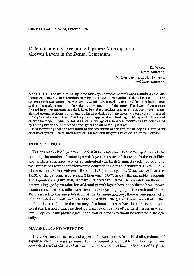
PRIMATES, 19(4): 775--784, October 1978 775
Determination of Age in the Japanese Monkey from Growth Layers in the Dental Cementum
K. WADA Kyoto University
N. OHTAISHI, and N. HACHIYA Hokkaido University
ABSTRACT. The teeth of 14 Japanese monkeys (Macaca fuscata) were examined to estab- lish an exact method of determining age by histological observation of dental cementum. The cementum showed annual growth layers, which were especially remarkable in the incisor root and in the molar cementum deposited at the junction of the roots. The layer of cementum formed in winter appears as a dark layer in stained sections and as a translucent layer in un- stained ground sections. In the incisor the first dark and light layers are formed at the age of three years, whereas in the molar they do not appear at a definite age. The layers are thick and clear in the upper medial incisor. As a result, the age of a Japanese monkey can be determined by adding two to the number of dark layers and an outer light layer.
It is interesting that the formation of the cementum of the first molar begins a few years after its eruption. The relation between this fact and the pressure of occlusion is discussed.
I N T R O D U C T I O N
Various methods of age determination in mammals have been developed recently by counting the number of annual growth layers in tissues of the teeth, in the mandible, and in other structures. Age of an individual can be determined exactly by counting the laminations found in sections of the dentin in some marine mammals (LAws, 1953), of the cementum in carnivores (RAuSCH, 1961) and ungulates (SERGEANT t~ PIMLOTT, 1959), of the ear plug in cetaceans (NIsHIWAKI, 1957), and of the mandible in rodents and lagomorphs (Ox-ITAISm, HACHIYA, & SHmATA, 1976). In primates, methods of determining age by examination of dental growth layers have not hitherto been known though a number of studies have been made regarding aging of the teeth and bones. With respect to the age estimation of the Japanese monkey, there is one report of a method based on tooth wear (KmaNO & SAHEKI, 1962), but it is obvious that in this method there is a limit to the accuracy of estimation. Therefore, the authors attempted to establish a more exact method by direct examination of the hard tissues in which annual cycles of the physiological condition of a monkey might be reflected histologi- cally.
MATERIALS A N D M E T H O D S
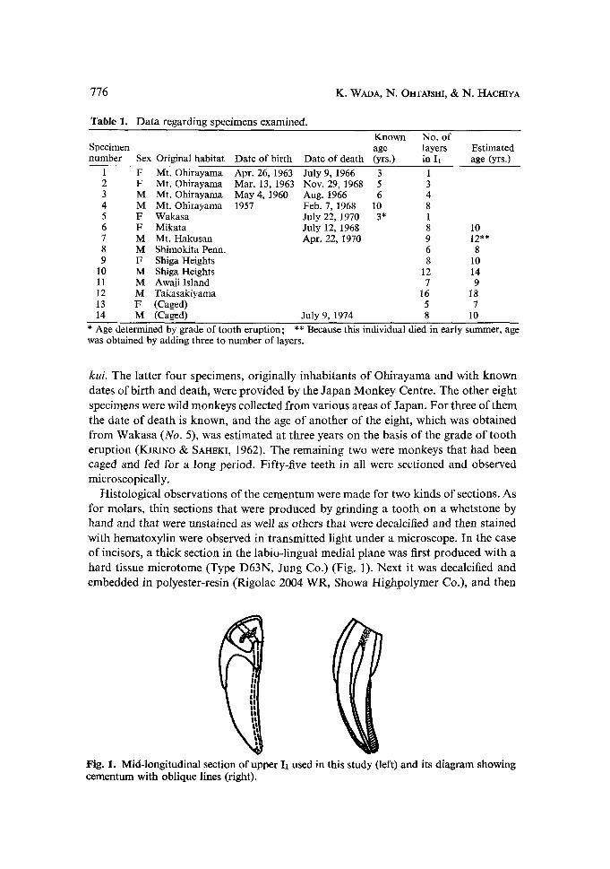
The upper medial incisors and upper and lower molars from 14 skull specimens of Japanese monkeys were examined for the present study (Table 1). These specimens comprised ten individuals of Macacafuscatafuscata and four individuals o f M. f. ya-
776
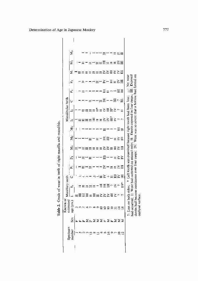
Table I. Data regarding specimens examined.
K. WADA, N, OHTAISHI, & N. HACHIYA
Known No. of Specimen age layers Estimated number Sex Original habitat Date of birth Date of death (yrs.) in 11 age (yrs.)
1 F Mt. Ohirayama Apr. 26, 1963 July 9, 1966 3 1 2 F Mt. Ohirayama Mar. 13, 1963 Nov. 29, 1968 5 3 3 M Mr. Ohirayama May 4, 1960 Aug. 1966 6 4 4 M Mt. Ohirayama 1957 Feb. 7, 1968 10 8 5 F Wakasa July 22, 1970 3* 1 6 F Mikata July 12, 1968 8 10 7 M Mt. Hakusan Apr. 22, 1970 9 12"* 8 M Shimokita Penn. 6 8 9 F Shiga Heights 8 10
10 M Shiga Heights 12 14 11 M Awaji Island 7 9 12 M Takasakiyama 16 18 13 F (Caged) 5 7 14 M (Caged) July 9, 1974 8 10
* Age determined by grade of tooth eruption; ** Because this individual died in early summer, age was obtained by adding three to number of layers.
kui. The latter four specimens, originally inhabitants o f Ohi rayama and with known dates o f birth and death, were provided by the Japan Monkey Centre. The other eight specimens were wild monkeys collected f rom various areas o f Japan. For three o f them the date o f death is known, and the age o f another o f the eight, which was obtained f rom Wakasa (No. 5), was estimated at three years on the basis o f the grade o f tooth eruption (KmlNO & SAH~KI, 1962). The remaining two were monkeys that had been caged and fed for a long period. Fifty-five teeth in all were sectioned and observed microscopically.
Histological observations o f the cementum were made for two kinds o f sections. As for molars, thin sections that were produced by grinding a too th on a whetstone by hand and that were unstained as well as others that were decalcified and then stained with hematoxylin were observed in transmitted light under a microscope. In the case o f incisors, a thick section in the labio-lingual medial plane was first produced with a hard tissue microtome (Type D63N, Jung Co.) (Fig. 1). Next it was decalcified and embedded in polyester-resin (Rigolac 2004 WR, Showa Highpolymer Co.), and then
Fig. 1. Mid-longitudinal section of upper I1 used in this study (left) and its diagram showing cementum with oblique lines (right).
Determination of Age in Japanese Monkey 777
A
e~
o
e4
, !
- : : : ~ : ~ .
m
E
,.:, ~ = : : : : ~ . - : = ~ - ~ > ~ . ~
m ~ N ~ N N ~ ~ N N
m ~
~..~o " ' ~
, .ZD
;:.~ o .~ ~ ~
~D ~..
N ~ m
o
o ~
. . ~ ~ . ,.~ " ~ 0
778 K. WADA, N. OHTAISHI, t~ N. HACHIYA
Determination of Age in Japanese Monkey 779
sectioned at a thickness of about 20 microns with the hard tissue microtome. The sec- tions were decalcified with Planck-Rychlo's solution, and stained with Delafield's hematoxylin.
RESULT A N D DISCUSSION
Apart from the microscopic study, the tooth eruption sequence and the grade of wear on the occlusal surface were examined. Twelve specimens showed fully erupted permanent dentition, and the other two specimens, Nos. 1 and 5, showed mixed denti- tion with the third molars not yet erupted.
As the first step, incisors, canines, premolars, and molars both on the maxilla and mandible in several individuals were cut into thin sections and their tissues were ex- amined to determine whether they could be used for age determination or not. As a result, it was found that the cementum of the upper median incisor and of the upper and lower first molars showed clear growth layers, and therefore these teeth were observed with special attention in all individuals.
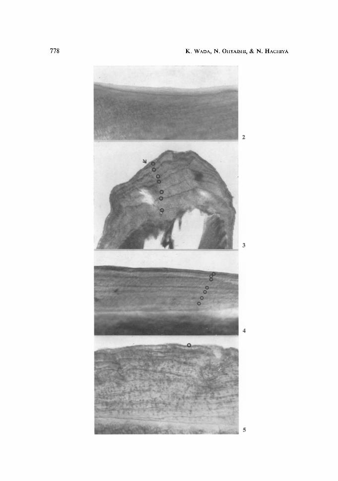
In the eight specimens for which dates of death are knowiL the cementum of the incisors, when stained with hematoxylin, showed in the outermost layer both a light layer (Figs. 2 & 3) and a dark layer (Fig. 4) in transmitted light. The dark layer must have been formed during winter, and the light layer must have been deposited on it during summer, judging from the dates of death of the individuals (Nos. 1, 4, and 6) that had the teeth shown in Figures 2, 3, and 4. In the upper medial incisors and first molars, both layers were found to be thicker and clearer, and therefore it was easier to count their number than to count any other teeth.
Since the medial incisor erupt during a period from the third winter to the fourth spring (K~RINO & SAHEKL 1962), the first light layer should have been formed during the fourth summer(Fig. 2), and the first dark layer in the fourth winter of the same age. The formation of light and dark layers was repeated alternately in the years following the eruption of each tooth. In addition, by counting the number of dark layers in the upper medial incisor of all specimens of known age, it was indicated that the first dark layers were formed in the fourth winter of age three (Table l). Similar results were ob- tained from other incisors. In the lower medial incisor and lateral incisors of specimen No. I that died in the fourth summer, only the first light layer was found. There were
VJg. 2. Upper I1 of an individual aged three years and two months (April 26, 1963-July 9, 1966). There is one light layer but no dark layer. This first light layer was formed in the sum- mer at three years of age. Specimen No. 1 : female obtained from JMC. • 200
Fig. 3. Upper I1 of an individual who died July 12. Seven dark layers are distinguished, and on outer surface a new light layer arrow formed in the summer of 1977 is seen. Specimen No. 6: female obtained from Mikata. Age was estimated at ten years. • 25
Fig. 4. Upper I1 of a 10-year old individual (1957-February 7, 1963). Dark layers are shown with small circles. This monkey died in the winter and, therefore, outermost layer is dark. Specimen No. 4: male obtained from JMC. /, 180
Fig. 5. Upper I1 of a 12-year old individual. The outer part of the cementum ends with a dark layer that is enclosed with a circle. Specimen No. 7: male collected at Mt. Hakusan. • 200
780 K. WADA, iN~. OHTAISHI, t~ N. HACHIYA
Determination of Age in Japanese Monkey 781
no dark layers. Four dark layers are seen in the upper medial incisor of specimen No. 2, aged five years and eight months and seven dark layers in specimen No. 4, aged ten years and ten months (Fig. 4). ha the upper medial incisor of specimen No. 6 that died on July 12, the eighth light layer that was formed in the last summer is seen in the outermost layer of the cementum (Fig. 3).
F rom these findings in the specimens of known age, it can generally be said that the age of a specimen of unknown age is determined by adding two to the number of dark layers and an outermost light layer of the upper medial incisor, with the exception of specimen No. 7 that died in April (Table 1). In the case of No. 7, the age is given by adding three to the number of the dark layers. As the period f rom April to May is the main birth season of the Japanese monkey, the new light layer is impossible to recog- nize clearly until June or July (Fig. 11). I f a monkey dies in April or May, the outer layer of the cementum ends with a dark layer, and for the determination of its age it is necessary to add three instead of the number of light layers. Therefore the age of indi- vidual No. 7 is estimated as 12 years.
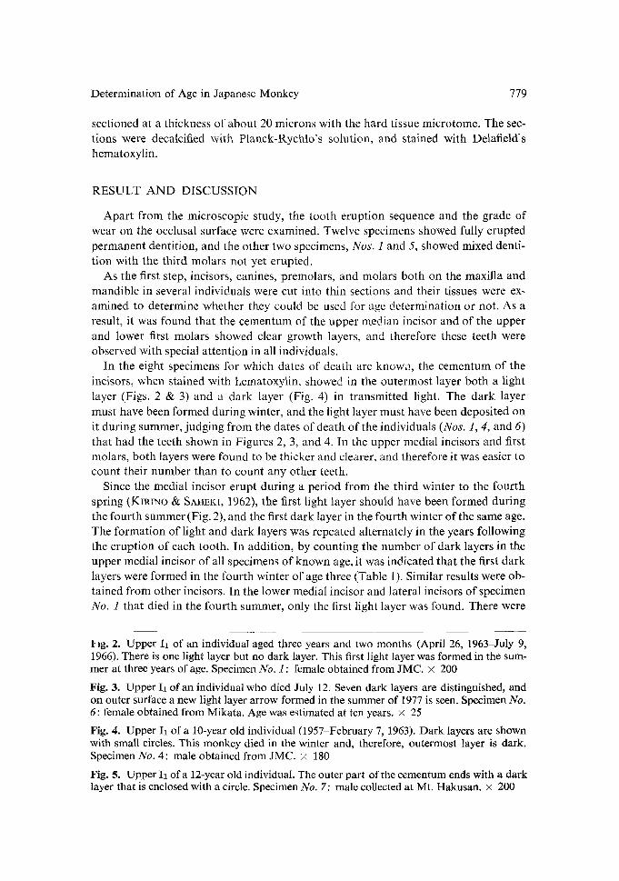
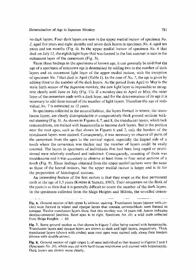
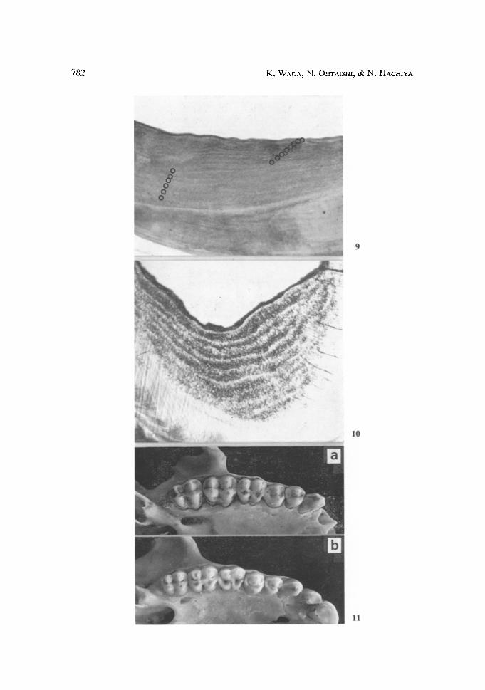
In specimens collected in the natural habitat, the layers formed in winter, the trans- lucent layers, are clearly distinguishable in comparatively thick ground sections with- out staining (Fig. 6). As shown in Figures 6, 7, and 8, the translucent layers, which lack cementoblasts, are stained with hematoxylin to become dark layers. But in the portion near the root apex, such as that shown in Figures 6 and 7, only the borders of the translucent layers were stained. Consequently, it was necessary to observe all parts of the cementum from the apex to the cervical region, especially the lingual side of a tooth where the cementum was thicker and the number of layers could be easily counted. The layers in specimens of individuals that had been long caged or provi- sioned were relatively confused and indistinct. Consequently, counting of them was troublesome and it was necessary to observe at least three or four serial sections of a tooth (Fig. 9). These findings obtained from the upper medial incisors were the same as those of the lateral incisors, but the upper medial incisor is larger and is fit for the preparation of histological sections.
An interesting feature of the first molars is that they erupt as the first permanent teeth at the age of 1.5 years (KmtNo & SAHFI<I, 1962). Their cementum, on the flank of the roots is so thin that it is generally difficult to count the number of the dark layers. In the specimens collected f rom the Shiga Heights and Mikata, the so-called cemen-
Fig. 6. Ground section of left upper I2 without staining. Translucent layers (shown with cir- cles) were formed in winter and opaque layers that contain cementoblasts were formed in summer. Twelve translucent layers show that this monkey was 14 years old. Arrow indicates dentino-cemental junction. Root apex is to right. Specimen No. 10: a wild male collected from Shiga Heights. • 60
Fig. 7. Same ground section as that shown in Figure 2 after being stained with hematoxylin. Translucent layers and opaque layers are shown as dark and light layers, respectively. Thick translucent layers (shown with circles) near root apex were stained only along their borders (shown with double arrow).
Fig. 8. Ground section of right upper I1 of same individual as that treated in Figures 2 and 3 (Specimen No. 10), which was cut with hard tissue microtome and stained with hematoxylin. Dark layers are shown more clearly.
782 K. WADA, N. OHTAISHI, & N. HACHIYA
Determination of Age in Japanese Monkey 783
turn pad is formed on the crotch of roots, where the lines of growth layers are distin- guished without staining but are branching off (Fig. 10). However, the year when the first translucent layer is formed is a few years later than the year of eruption. This fact might mean that the first molar does not exert their normal masticatory function of molars as it is located in the posterior end of the teeth for a few years after its eruption. In the second and third molars, also, the growth layers are seen in the cementum pad, but the age in which the first translucent layer was formed is not apparent. In long caged and provisioned specimens, translucent layers of the cementum pad of molars are generally obscure.
The growth layers in the cementum of the canines and the premolars were less clear than in other teeth, and, therefore, the first, second, and third molars, the canines, and the premolars are not suitable for age estimation.
Age estimation in Japanese monkeys based on tooth eruption and wear has been attempted by KmINO and SAIJEKI (1962), but the usefulness of this criterion is restrict- ed. The grade of tooth wear corresponds with advancing age up to nine years, but beyond ten years a considerable variation in the grade is found among the specimens (Table 2). For example, in the four specimens (Nos. 4, 6, 9, and 14) which were finally determined to be ten years old, the variation in the grade of wear in the mandibular first molars ranges from II to IV; that in the second molars, from II to III, and that in the third molars, from I to II (Figs. l l a & b).
In general, the individual variations in the grade of tooth wear become more and more conspicuous as the age advances beyond three to four years. The grade of wear seems to differ among individuals according to their conditions of life and food ha- bits. For example, the grade seen in the specimens collected from the Shiga Heights and Mikata, where in winter wild monkeys eat various kinds of bark and buds of trees, is higher than that in the specimens obtained from free-ranging individuals who were provisioned at Takasakiyama while alive. The method of age estimation by means of histological features in dental tissues would be far more accurate than such a macro- scopic one, but it has its own difficulties and limitation of effectiveness.
Another interesting finding obtained in the present study concerns the difference of time between incisors and molars in the formation of the dark layer. The first light layer of the cementum in the incisors is formed in summer of the year of eruption, but it would seem that the first dark layer is formed in the winter of the year of eruption. In the case of first molars, on the other hand, the first dark layer is formed a few years
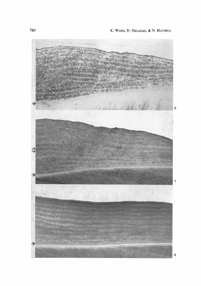
Fig. 9. Upper I1 of a monkey provisioned at 37akasakiyama. Translucent layers formed in early years are stained only on their sides. Outermost layer indicates that this individual died in autumn or early winter. Sixteen dark layers are seen and age was estimated at 18 years. Specimen No. 12: adult male. • 35
Fig. 10. Unstained ground section of so-called cement pad formed in crotch of roots of lower first molar of same individual as that treated in Figures 2 and_ 3, of which the age was estimated at 14 years. First translucent layer was formed during the winter at age six, though the first molar generally erupts at 1.5 years. After six years of age growth layers were formed constantly each year. • 45
Fig. 11. Right upper teeth of two wild females. Their ages were estimated. Amount of wear shown in a (Specimen No. 9) is more remarkable than that shown in b (No. 11).
784 K. WADA, N. OHTAISHI, & N. HACHIYA
later than the e rupt ion o f the first molars . These facts suggest tha t the pe r iod o f for-
ma t ion o f the first da rk layer is re la ted to the funct ion or the k ind o f tooth . As for p r e p a r a t i o n o f specimens, it is best to use the hard tissue mic ro tome, bu t to
use g round sections wi thout s taining is a s imple and pract ical me thod for p re l iminary es t imat ion o f age.
Acknowledgements. The Japan Monkey Centre allowed us to use many of its specimens of teeth of the Japanese monkey. Dr. O. TAKENAKA and Mr. S. GOTO, of the Primate Research Institute of Kyoto University, helped us in collecting other specimens. Dr. M. TASUMI, Depart- ment of Zoology of Kyoto University, provided careful review of the manuscript. To these institutions and people we are very grateful.
REFERENCES
KIRINO, T. & M. SAHEKI, 1962. Tooth eruption and wear with the age in Japanese monkeys of Takasakiyama. In: The Japanese Monkey o f Takasakiyama, J. ITAM, J. IKEDA, & T. TANAKA (eds.), Keiso-shobo, Tokyo, pp. 124-135, in Jap.
LAws, R. M., 1953. A new method of age determination in mammals with special reference to the elephant seal. Falkland Is. Dependencies Surv., Sei. Rept., No. 2, 11 pp.
NISHIWAKI, M., 1957. Age characteristics of ear plug of whales. Sei. Rep. Whales Res. Inst., 12: 23-32.
OHTAISHI, N., N. HACHIYA, & Y. SHIBATA, 1976. Age determination of the hare from annual layers in the mandibular bone. Aeta Theriol., 21(11): 168-171.
RAUSCH, R., 1961. Notes on the black bear, Ursus americanus PALLAS, in Alaska, with parti- cular reference to dentition and growth. Z. Saugetierku., 26(2): 77-107.
SERGEANT, D. & D. PIMLOTT, 1959. Age determination in moose from sectioned incisor teeth. J. Wildl. Mgmt., 23(3): 315-321.
- - Received February 4, 1976; Accepted October 12, 1977
Authors' Addresses: KAzuo WADA, Primate Research Institute, Kyoto University, lnuyama, Aichi 484, Japan; N. OHTAISHI and N. HACHtYA, Department of Oral Anatomy, School of Dentistry, Hokkaido University, Sapporo 060, Japan.


![Microanalysis of Root Cementum in Patients with Rapidly ......at the exposed cementum [4]. Chemical analysis of the exposed cementum has shown an increase in calcium, magnesium, and](https://img.pdfslide.net/doc/110x75/5f237b2b5d795a336e24c740/microanalysis-of-root-cementum-in-patients-with-rapidly-at-the-exposed-cementum.jpg)


![Adv in Cementum Devt[1]](https://img.pdfslide.net/doc/110x75/55cf99ce550346d0339f453c/adv-in-cementum-devt1.jpg)