Embed Size (px)
Citation preview
INFECTION AND IMMUNITY, Mar. 1977, p. 958-971 Vol. 15, No. 3Copyright © 1977 American Society for Microbiology Printed in U.S.A.
Development of Arthrospores of Trichophytonmentagrophytes
D. J. BIBEL,* D. A. CRUMRINE, K. YEE, AND R. D. KING'Department ofDermatology Research, Letterman Army Institute ofResearch,
San Francisco, California 94129
Received for publication 13 October 1976
Arthrosporogenesis of the dermatophyte Trichophyton mentagrophytes wasexamined by light and by scanning and transmission electron microscopy.Sabouraud dextrose agar plates were inoculated with microconidia and incu-bated in an atmosphere of 8% CO2. Typical germination and hyphal branchingcontinued to day 4, when hyphae began to be increasingly coated with agranular-fibrillar material. Multiple replication of nuclei and formation ofsegregating septa followed. By day 6 the thick surface mesh sometimes wasrestricted to protruding rings, probably over septa. Between days 6 and 7, afterthickening of outer and septal walls, units began to round and separate. Trian-gular gaps, which developed at the junction of septa and outer wall layers,enlarged so that spores were held together at their poles and along a tangentialring. With elongation of the spore to its barrel shape, the halves of the septumseparated and the ring pulled apart, leaving a jagged, circular flange originat-ing from the outer layer of cell wall. After the final separation of surface fibrils,the outer layer of cell wall extended toward the poles, covering the apparentlyexposed inner wall layer. Newly formed arthrospores, which measured 2.0 to 3.3by 2.9 to 3.8 /im and possessed walls of about 0.33-/im thickness, had smoothsides but somewhat rough poles.
Although dermatophytes, fungi responsiblefor ringworm and athlete's foot, have been ex-amined by both transmission electron micros-copy (TEM) (2, 3, 5, 7, 8, 10, 14, 17, 18, 20, 21)and scanning electron microscopy (SEM) (2, 6,9, 12, 16, 19), few reports have investigatedspores and their development. Only three stud-ies have briefly described arthrospores (9, 12,16), and these, using inadequate techniques,pertained to hyphal invasion of hair. Recently,a procedure (R. D. King, C. L. Dillavou, J. H.Greenberg, J. C. Jeppson, and J. S. Jaegar,Can. J. Microbiol., in press) based on the workof Chin and Knight (4) was introduced whichinitiates arthrospore development on agar. Us-ing this system, we have followed by light andelectron microscopy the growth of a dermato-phyte from microconidial germination throughhyphal branching to maturation of the arthro-spore.
MATERIALS AND METHODSMicroorganism. Microconidia of Trichophyton
mentagrophytes var. granulosum (ATCC 18748)were harvested from dermatophyte test medium (15)by the method of Reinhardt et al. (11), with the
I Present address: Department of Microbiology, Univer-sity of Texas, Health Science Center, San Antonio, TX78284.
exception of substituting distilled water for Tween40-saline washes. In brief, fungal mats were homog-enized with glass beads on a rotating shaker, fil-tered through glass wool, and centrifuged. A hemo-cytometer was used to quantitate the washed andconcentrated suspension of spores.Medium and culture conditions. Sabouraud dex-
trose agar (10 ml in 100-mm-diameter dishes) wasinoculated in duplicate with portions from 10-folddilutions of microconidia in Sabouraud dextrosebroth. One set of plates, supporting 102 to 105 thal-lus-forming units, was sealed inside anaerobic jars(Baltimore Biological Laboratory, [BBL] Cockeys-ville, Md.) in which two CO2 GasPaks (BBL) wereinserted to attain an atmosphere of 8% CO2. Thesecond set of plates was cultured in a normal atmos-phere. Incubation of all plates was at 300C. At dailyintervals up to 10 days, small agar blocks (up to 15by 15 mm) containing isolated fungal growth werecut from appropriate dishes, beginning with themore concentrated inocula.SEM. Blocks were fixed for 18 h in 2% (wt/vol)
paraformaldehyde and 2.5% (vol/vol) glutaralde-hyde in 0.1 M cacodylate buffer, pH 7.2. After beingwashed in 5% (wt/vol) sucrose in cacodylate buffer,specimens were postfixed in 1% (wt/vol) OS04 inZetterqvist salt solution (H. Zetterqvist, Ph.D. the-sis, Karolinska Institute, Stockholm, 1956). Oncedehydrated in a graded series of acetone, blockswere critical-point dried with CO2 in a Denton DCP-1 apparatus. Next, the sample was coated with suc-cessive layers of carbon, silver, and gold in a Denton
958
on May 12, 2019 by guest
http://iai.asm.org/
Dow
nloaded from
ARTHROSPORES OF T. MENTAGROPHYTES 959
DV502 vacuum evaporator with rotating stage. TheETEC Autoscan scanning electron microscope wasoperated at 10 kV.TEM. Additional blocks were first subjected for 2
h to Karnovsky's fixative (M. J. Karnovsky, J. CellBiol., 27:137A, 1965) with 1% (wt/vol) lanthanumnitrate. After postfixation for 2 h in 1% (wt/vol)OsO4 and 1% lanthanum nitrate, buffered at pH 7.3by 0.1 M cacodylic acid, specimens were dehydratedthrough a graded series of alcohol and embedded in
Spurr's low-viscosity epoxy resin (13). Sectioned on aPorter-Blum MT-2 ultramicrotome with a diamondknife, fungi were stained with lead citrate and ura-nyl acetate and viewed with a Hitachi HS8F orPhilips 201 electron microscope.
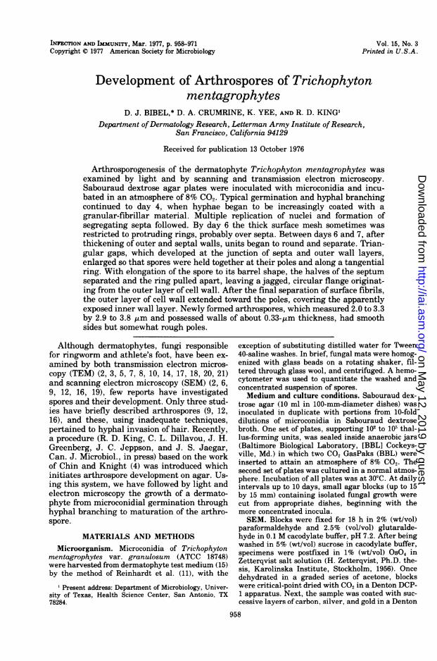
RESULTSSEM. By 24 h microconidia (Fig. 1) on all
plates had germinated (Fig. 2), sending forth
FIG. 1. Isolated microconidia of T. mentagrophytes. Note that the surface is ruffled except within the areaof the circular flange (arrows), which was once attached to the hyphal cell wall. x1 7,000. Bar, 1 ,ImM
VOL. 15, 1977
on May 12, 2019 by guest
http://iai.asm.org/
Dow
nloaded from
960 BIBEL ET AL.
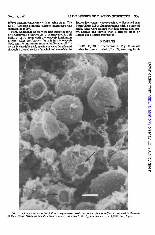
serpentine hyphae. Whereas the spherical mi-croconidia were ruffled, the germ tubes had asomewhat smooth surface (Fig. 2). Hyphae ofT. mentagrophytes incubated under normal
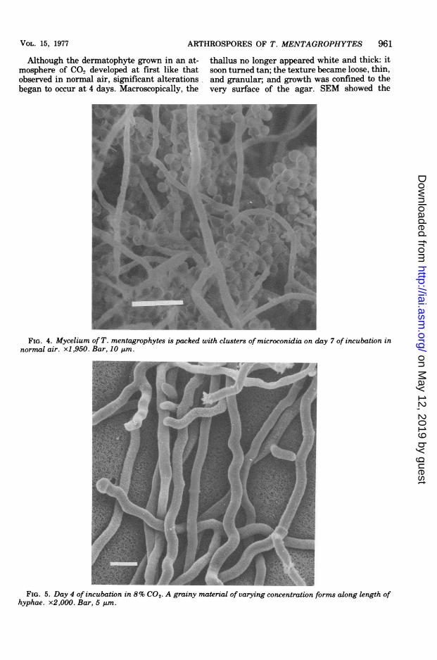
aerobic conditions rapidly extended, branched,and developed microconidia (Fig. 3). Seven-daypreparations were packed with clusters ofthesespores (Fig. 4).
_-_'_. ''-_'1' .. Wr- -. '. -
FIG. 2. Germination of microconidia 24 h after inoculation. Germ tubes are relatively smooth walled.x3,000. Bar, 5 ,um.
FIG. 3. Microconidia develop on hyphae after 3 days of incubation in normal atmosphere. x8,000. Bar, 111m.
INFECT . IMMUN .
on May 12, 2019 by guest
http://iai.asm.org/
Dow
nloaded from
ARTHROSPORES OF T. MENTAGROPHYTES 961
Although the dermatophyte grown in an at-mosphere of CO2 developed at first like thatobserved in normal air, significant alterationsbegan to occur at 4 days. Macroscopically, the
thallus no longer appeared white and thick: itsoon turned tan; the texture became loose, thin,and granular; and growth was confined to thevery surface of the agar. SEM showed the
FIG. 4. Mycelium of T. mentagrophytes is packed with clusters of microconidia on day 7 of incubation innormal air. x1,950. Bar, 10 Am.
FIG. 5. Day 4 of incubation in 8% CO2- A grainy material of varying concentration forms along length ofhyphae. x2,000. Bar, 5 kum.
VOL. 15, 1977
on May 12, 2019 by guest
http://iai.asm.org/
Dow
nloaded from
962 BIBEL ET AL.
events in detail. At 4 days only hyphae wereobserved. They were generally smooth, yet oncloser inspection (Fig. 5) a grainy material ofvarying concentration was found distributed
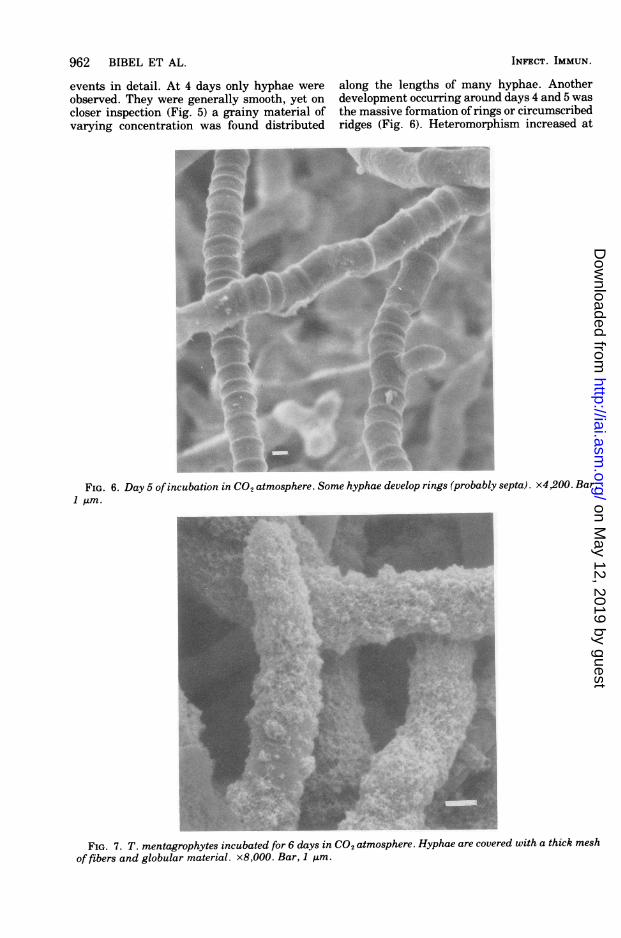
along the lengths of many hyphae. Anotherdevelopment occurring around days 4 and 5 wasthe massive formation of rings or circumscribedridges (Fig. 6). Heteromorphism increased at
FIG. 6. Day 5 ofincubation in CO2 atmosphere. Some hyphae develop rings (probably septa). x4,200. Bar,1 Jim.
4.
;'.'wc
A,
A:id
,; acen_EPs t
e
I' !ft!1;>-#31 :. svat w
jf. A
'. -8 e.
_=,rE j, _o ., e v.it's
FIG. 7. T. mentagrophytes incubated for 6 days in CO2 atmosphere. Hyphae are covered with a thick mesh
of fibers and globular material. x8,000. Bar, 1 pzm.
>-'.
IF* 71
INFECT. IMMUN.
i. j'k..VW,Al.-I.A.-,.
'.p :,
on May 12, 2019 by guest
http://iai.asm.org/
Dow
nloaded from
ARTHROSPORES OF T. MENTAGROPHYTES 963
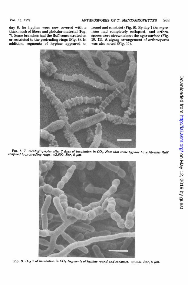
day 6, for hyphae were now covered with athick mesh of fibers and globular material (Fig.7). Some branches had the fluff concentrated onor restricted to the protruding rings (Fig. 8). Inaddition, segments of hyphae appeared to
,Vc _.
round and constrict (Fig. 9). By day 7 the myce-lium had completely collapsed, and arthro-spores were strewn about the agar surface (Fig.10, 11). A zigzag arrangement of arthrosporeswas also noted (Fig. 11).
-_ U9 1,HeFIG. 8. T. mentagrophytes after 7 days of incubation in CO2. Note that some hyphae have fibrillar fluff
confined to protruding rings. x2,500. Bar, 5 tim.
IFIG. 9. Day 7 of incubation in CO2. Segments of hyphae round and constrict. x3,300. Bar, 5 gum.
VOL. 15, 1977
on May 12, 2019 by guest
http://iai.asm.org/
Dow
nloaded from
964 BIBEL ET AL.
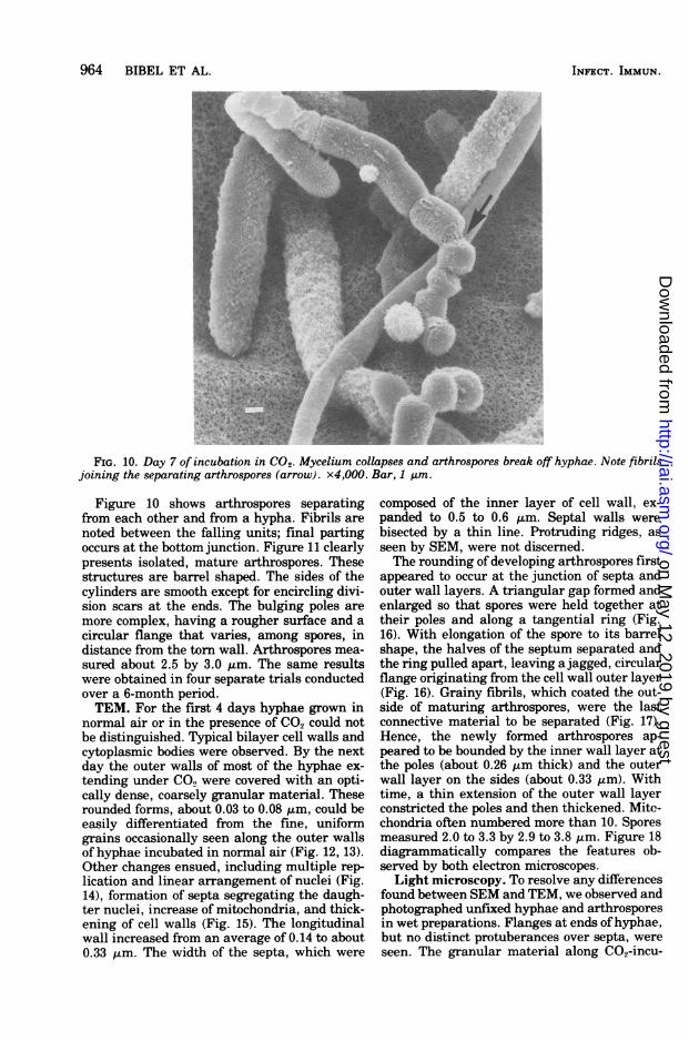
FIG. 10. Day 7 of incubation in CO2. Mycelium collapses and arthrospores break off hyphae. Note fibrilsjoining the separating arthrospores (arrow). x4,000. Bar, 1 Am.
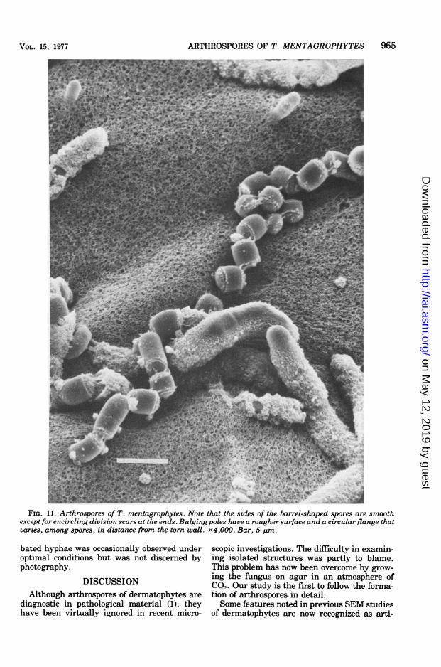
Figure 10 shows arthrospores separatingfrom each other and from a hypha. Fibrils are
noted between the falling units; final partingoccurs at the bottom junction. Figure 11 clearlypresents isolated, mature arthrospores. Thesestructures are barrel shaped. The sides of thecylinders are smooth except for encircling divi-sion scars at the ends. The bulging poles are
more complex, having a rougher surface and a
circular flange that varies, among spores, indistance from the torn wall. Arthrospores mea-sured about 2.5 by 3.0 gum. The same resultswere obtained in four separate trials conductedover a 6-month period.TEM. For the first 4 days hyphae grown in
normal air or in the presence of CO., could notbe distinguished. Typical bilayer cell walls andcytoplasmic bodies were observed. By the nextday the outer walls of most of the hyphae ex-
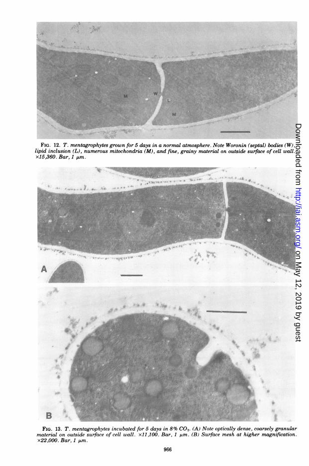
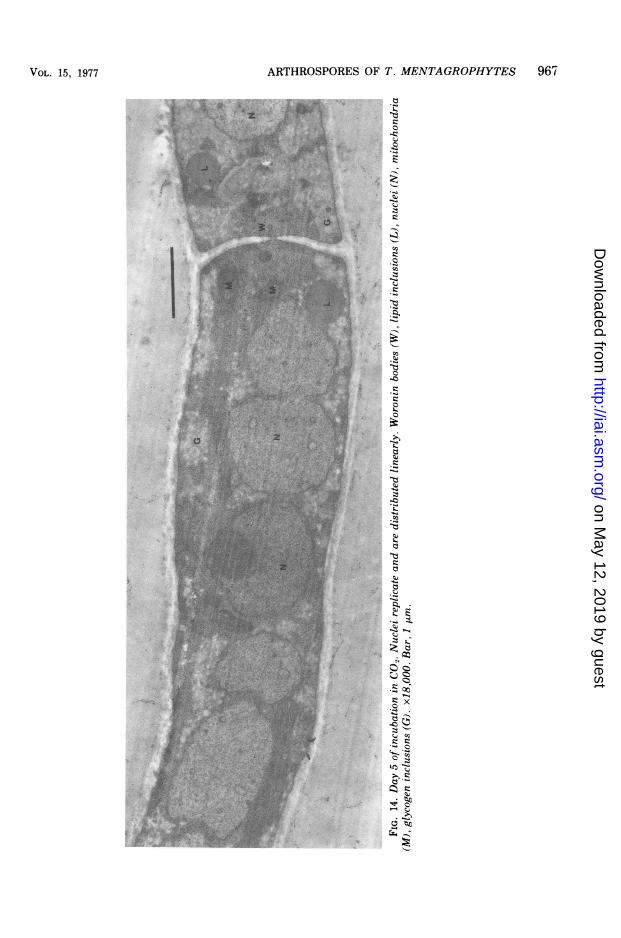
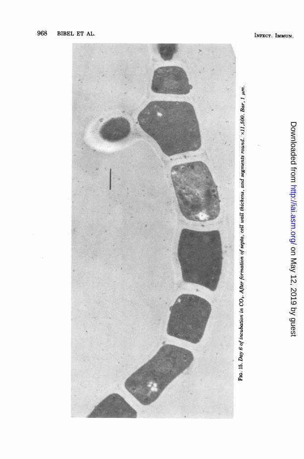
tending under CO., were covered with an opti-cally dense, coarsely granular material. Theserounded forms, about 0.03 to 0.08 ,m, could beeasily differentiated from the fine, uniformgrains occasionally seen along the outer wallsof hyphae incubated in normal air (Fig. 12, 13).Other changes ensued, including multiple rep-lication and linear arrangement of nuclei (Fig.14), formation of septa segregating the daugh-ter nuclei, increase of mitochondria, and thick-ening of cell walls (Fig. 15). The longitudinalwall increased from an average of 0.14 to about0.33 gm. The width of the septa, which were
composed of the inner layer of cell wall, ex-panded to 0.5 to 0.6 4m. Septal walls werebisected by a thin line. Protruding ridges, asseen by SEM, were not discerned.The rounding of developing arthrospores first
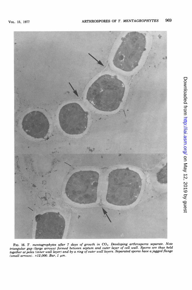
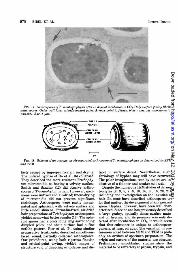
appeared to occur at the junction of septa andouter wall layers. A triangular gap formed andenlarged so that spores were held together attheir poles and along a tangential ring (Fig.16). With elongation of the spore to its barrelshape, the halves of the septum separated andthe ring pulled apart, leaving a jagged, circularflange originating from the cell wall outer layer(Fig. 16). Grainy fibrils, which coated the out-side of maturing arthrospores, were the lastconnective material to be separated (Fig. 17).Hence, the newly formed arthrospores ap-peared to be bounded by the inner wall layer atthe poles (about 0.26 ktm thick) and the outerwall layer on the sides (about 0.33 gm). Withtime, a thin extension of the outer wall layerconstricted the poles and then thickened. Mite-chondria often numbered more than 10. Sporesmeasured 2.0 to 3.3 by 2.9 to 3.8 tim. Figure 18diagrammatically compares the features ob-served by both electron microscopes.
Light microscopy. To resolve any differencesfound between SEM and TEM, we observed andphotographed unfixed hyphae and arthrosporesin wet preparations. Flanges at ends of hyphae,but no distinct protuberances over septa, wereseen. The granular material along CO2-incu-
INFECT. IMMUN.
on May 12, 2019 by guest
http://iai.asm.org/
Dow
nloaded from
ARTHROSPORES OF T. MENTAGROPHYTES 965
,.B.I*i_B'W,-
e K* w '* a h*91;i;"*~ . 1
FIG. 11. Arthrospores of T. mentagrophytes. Note that the sides of the barrel-shaped spores are smoothexcept for encircling division scars at the ends. Bulging poles have a rougher surface and a circularflange thatvaries, among spores, in distance from the torn wall. x4,000. Bar, 5 ,um.
bated hyphae was occasionally observed underoptimal conditions but was not discerned byphotography.
DISCUSSIONAlthough arthrospores of dermatophytes are
diagnostic in pathological material (1), theyhave been virtually ignored in recent micro-
scopic investigations. The difficulty in examin-ing isolated structures was partly to blame.This problem has now been overcome by grow-ing the fungus on agar in an atmosphere ofCO2. Our study is the first to follow the forma-tion of arthrospores in detail.Some features noted in previous SEM studies
of dermatophytes are now recognized as arti-
VOL. 15, 1977
on May 12, 2019 by guest
http://iai.asm.org/
Dow
nloaded from
' ' :t
Ls f -fe 4 *
M
FIG. 12. T. mentagrophytes grown for 5 days in a normal atmosphere. Note Woronin (septal) bodies (W),lipid inclusion (L), numerous mitochondria (M), and fine, grainy material on outside surface of cell wall.x15,360. Bar, 1 unm.
A
4: -; ^
-y~~>
FIG. 13. T. mentagrophytes incubated for 5 days in 8% CO2, (A) Note optically dense, coarsely granularmaterial on outside surface of cell wall. xl1,100. Bar, 1 gim. (B) Surface mesh at higher magnification.x22,000. Bar, 1 nm.
966
I
on May 12, 2019 by guest
http://iai.asm.org/
Dow
nloaded from
ARTHROSPORES OF T. MENTAGROPHYTES 967
in
zi.
C0
0co
b.0
'-.4
,..,.>i A , .- .
B' > An,', s iti'o
Tea~~ ~ ~~~~~~~~'.> ;
VOL. 15, 1977
t .- X,
x- O-w~t,
( e
on May 12, 2019 by guest
http://iai.asm.org/
Dow
nloaded from
968 BIBEL ET AL. INFECT. IMMUN.
'V
'N
C
'N'Nx
0L.U,
U,
iU,Co
0
0
Ci
2.0
0Co
on May 12, 2019 by guest
http://iai.asm.org/
Dow
nloaded from
VOL. 15, 1977 ARTHROSPORES OF T. MENTAGROPHYTES 969
4:
4~~~~~~~~~~~~~~~~~~~~~~~~~~~~~~~~~~~~~~~:
S -.
A.~~~~~~~~~~~~~4
'A~~~~~~~~4
4'6.'z< X #
I~ ~ ~ ~ ~ ~ I4
Or
FIG. 16. T. mentagrophytes after 7 days of growth in CO2. Developing arthrospores separate. Notetriangular gap (large arrows) formed between septum and outer layer of cell wall. Spores are thus heldtogether at poles (inner wall layer) and by a ring of outer wall layers. Separated spores have a jagged flange(small arrows). x12,OOO. Bar, 1 gin.
on May 12, 2019 by guest
http://iai.asm.org/
Dow
nloaded from
970 BIBEL ET AL.
A
FIG. 17. Arthrospores ofT. mentagrophytes after 10 days ofincubation in CO2. Only surface grainy fibrilsunite spores. Outer wall layer extends toward poles. Arrows point to flange. Note numerous mitochondria.x16,800. Bar, 1 tim.
1 Pm
FIG. 18. Schema ofan average, newly separated arthrospore of T. mentagrophytes as determined by SEMand TEM.
facts caused by improper fixation and drying.The unfixed hyphae of Ito et al. (6) collapsed.They described the more resistant Trichophy-ton microconidia as having a velvety surface.Smith and Sandler (12) did observe arthro-spores ofTrichophyton in hair. However, speci-mens were unfixed and air-dried; freeze-dryingof microconidia did not prevent significantshrinkage. Arthrospores were partly invagi-nated and spherical, with velvety surface andlittle embellishment. Formalin-fixed, air-driedhair preparations of Trichophyton arthrosporesyielded somewhat better results (16). The sphe-roid spores had a protruding ring surroundingdimpled poles, and their surface had a finenetlike pattern. Pier et al. (9), using similarpreparative treatments, described smooth-sur-faced, round, partially collapsed arthrospores.Our procedures, using both rigorous fixationand critical-point drying, yielded images ofstructure void of dimpling or collapse and dis-
tinct in surface detail. Nevertheless, slightshrinkage of hyphae may still have occurred.The polar invaginations seen by others are in-dicative of a thinner and weaker cell wall.
Despite the numerous TEM studies of derma-tophytes (2, 3, 5, 7, 8, 10, 14, 17, 18, 20, 21),including one investigation on the invasion ofhair (3), none have described arthrospores or,for that matter, the development ofany asexualspore. Hyphae, however, have been well char-acterized. Since no one has previously describeda large grainy, optically dense surface mate-rial on hyphae, and its presence was only de-tected after incubation in CO2, it would seemthat this substance is unique to arthrosporo-genesis, at least on agar. The variation in pro-fuseness noted between SEM and TEM is prob-ably an artifact of specimen preparation. Theorigin and nature of the material are obscure.Preliminary, unpublished studies show thematerial to be refractory to pepsin, trypsin, and
INFECT. IMMUN.
on May 12, 2019 by guest
http://iai.asm.org/
Dow
nloaded from
ARTHROSPORES OF T. MENTAGROPHYTES 971
urea, and its disappearance upon maturation ofthe spore is even more perplexing.Arthrospores have different surface features
than microconidia. The in vitro production ofthese spores holds promise for the comparativeanalysis of all spores with respect to develop-ment, morphology, antigenicity, heat toler-ance, conditions for germination, resistance tochemical agents, and so forth. Arthrosporogen-esis in other species of dermatophytes alsoneeds to be investigated.
ACKNOWLEDGMENTSWe thank C. L. Dillavou, M. V. Lancaster, and R. Aly
for their helpful criticism.
LITERATURE CITED1. Ajello, L., and A. A. Padhye. 1974. Dermatophytes and
the agents of superficial mycoses, p. 469-481. In E. H.Lennette, E. H. Spaulding, and J. P. Truant (ed.),Manual of clinical microbiology, 2nd ed. AmericanSociety for Microbiology, Washington, D. C.
2. Akin, D. E., and G. E. Michaels. 1972. Microsporumgypseum macroconidial development revealed bytransmission and scanning electron microscopy. Sa-bouraudia 10:52-55.
3. Baxter, M., and P. R. Mann. 1969. Electron microscopicstudies ofthe invasion ofhuman hair in vitro by threekeratinophilic fungi. Sabouraudia 7:33-37.
4. Chin, B., and S. G. Knight. 1957. Growth of Trichophy-ton mentagrophytes and Trichophyton rubrum in in-creased carbon dioxide tensions. J. Gen. Microbiol.16:642-646.
5. Hill, T. W. 1975. Ultrastructure of ascosporogenesis inNannizzia gypsea. J. Bacteriol. 122:743-748.
6. Ito, Y., Y. Nozawa, H. Suzuki, and T. Setoguti. 1970.Surface structure of dermatophytes as seen by thescanning electron microscope. Sabouraudia 7:270-272.
7. Ito, Y., T. Setoguti, Y. Nozawa, and S. Sakurai. 1967.An electron microscopic observation of Trichophytonviolaceum. J. Invest. Dermatol. 48:124-127.
8. Laden, E., and J. 0. Erickson. 1958. Electron micro-
scopic study ofEpidermophyton floccosum. J. Invest.Dermatol. 31:55-58.
9. Pier, A. C., K. R. Rhoades, T. L. Hayes, and J. Gal-lagher. 1972. Scanning electron microscopy of se-lected dermatophytes of veterinary importance. Am.J. Vet. Res. 33:607-613.
10. Pock-Steen, B., and T. Kobayasi. 1970. Ultrastructureof the hyphal wall and septum of Trichophyton men-tagrophytes. J. Invest. Dermatol. 55:404-409.
11. Reinhardt, J. H., A. M. Allen, D. Gunnison, and W. A.Akers. 1974. Experimental human Trichophytonmentagrophytes infections. J. Invest. Dermatol.63:419422.
12. Smith, J. M. B., and W. J. U. Sander. 1971. The surfacestructure of saprophytic and parasitic dermatophytespores. Mycopathol. Mycol. Appl. 43:153-159.
13. Spurr, A. R. 1969. A low-viscosity epoxy resin embed-ding medium for electron microscopy. J. Ultrastruct.Res. 26:3143.
14. Taplin, D., and H. Blank. 1961. Microscopic morphol-ogy of Trichophyton rubrum. J. Invest. Dermatol.37:523-528.
15. Taplin, D., N. Zaias, G. Rebell, and H. Blank. 1969.Isolation and recognition of dermatophytes on a newmedium (DTM). Arch. Dermatol. 99:203-209.
16. Tosti, A., S. Villardita, M. L. Fazzini, and R. Scalici.1970. Contribution to the knowledge ofdermatophyticinvasion of hair. An investigation with the scanningelectron microscope. J. Invest. Dermatol. 55:123-134.
17. Tsukahara, T., A. Sato, and R. Okada. 1964. Electronmicroscopic studies on the cytological structure ofTrichophyton mentagrophytes. Jpn. J. Microbiol.8:83-96.
18. Urabe, H., and T. Izu. 1969. The ultrastructure ofTrichophyton and a double cell wall in the hypha. J.Invest. Dermatol. 52:508-513.
19. Watanabe, S. 1975. Observations ofMicrosporum caniswith cryoscanning and scanning electron microscopy.Mycopathologia 57:73-76.
20. Werner, H. J., C. Catulis, H. W. Jolly, Jr., and C. L.Carpenter, Jr. 1967. Electron microscope observa-tions of the fine structure of Microsporum gypseum.J. Invest. Dermatol. 48:481-484.
21. Werner, H. J., H. W. Jolly, Jr., and B. 0. Spurlock.1966. Electron microscope observations of the finestructure ofMicrosporum canis. J. Invest. Dermatol.46:130-134.
VOL. 15, 1977
on May 12, 2019 by guest
http://iai.asm.org/
Dow
nloaded from