Embed Size (px)
Citation preview
Development of Controlled Matrix Heterogeneity on a Triphasic
Scaffold for Orthopedic Interface Tissue Engineering
JEFFREY P. SPALAZZI, M.S.,1 STEPHEN B. DOTY, Ph.D.,2 KRISTEN L. MOFFAT, M.S.,1
WILLIAM N. LEVINE, M.D.,3 and HELEN H. LU, Ph.D.1,4
ABSTRACT
Biological fixation of orthopedic soft tissue grafts to bone poses a significant clinical challenge. Theclinical success of soft tissue–based grafts for anterior cruciate ligament (ACL) reconstruction is limitedby the lack of functional graft integration with subchondral bone. Soft tissues such as the ACL connect tosubchondral bone via a complex interface whereby three distinct tissue regions (ligament, fibrocartilage,and bone) work in concert to facilitate load transfer from soft to hard tissue while minimizing stressconcentration at the interface. Although a fibrovascular tissue forms at the graft-to-bone interface fol-lowing surgery, this tissue is nonphysiologic and represents a weak link between the graft and bone. Wepropose that the re-establishment of the native multi-tissue interface is essential for biological graftfixation. In vivo observations and our in vitro monolayer co-culture results suggest that osteoblast-fibroblast interaction is important for interface regeneration. This study focuses on the design of atriphasic scaffold system mimicking the multi-tissue organization of the native ACL-to-bone interfaceand the evaluation of osteoblast-fibroblast interactions during three-dimensional co-culture on the tri-phasic scaffold. We found that the triphasic scaffold supported cell proliferation, migration and phe-notypic matrix production while maintaining distinct cellular regions and phase-specific extracellularmatrix deposition over time. This triphasic scaffold is designed to guide the eventual reestablishment ofan anatomically oriented and mechanically functional fibrocartilage interfacial region directly on bio-logical and synthetic soft tissue grafts. The results of this study demonstrate the feasibility of multi-tissueregeneration on a single scaffold, and the potential of interface tissue engineering to enable the biologicalfixation of soft tissue grafts to bone.
INTRODUCTION
THE ANTERIOR CRUCIATE LIGAMENT (ACL) is the primary
knee joint stabilizer and the most frequently injured
knee ligament, with approximately 100,000 reconstruction
procedures performed each year in the United States.1–5
Because of the poor healing potential of the ACL,6 surgical
reconstruction is necessary to restore normal knee function.
The long-term performance of ACL reconstruction grafts
depends on the structural and material properties of the graft,
the initial graft tension,7–12 the intra-articular position of the
graft,13,14 graft fixation,15,16 and optimized postoperative
rehabilitation.
Autologoushamstringtendon-basedgraftsare increasingly
used for ACL reconstruction because of the high incidence
of donor site morbidity associated with bone-patellar
1Biomaterials and Interface Tissue Engineering Laboratory, Department of Biomedical Engineering, Columbia University, New York,
New York.2Analytical Microscopy Core Laboratory, Hospital for Special Surgery, New York, New York.3Department of Orthopaedic Surgery, Columbia University, New York, New York.4College of Dental Medicine, Columbia University, New York, New York.
TISSUE ENGINEERINGVolume 12, Number 12, 2006# Mary Ann Liebert, Inc.
3497
tendon-bone grafts.17,18 Clinically, the hamstring tendon
graft is mechanically fixed within the femoral bone tunnel
with a cross-pin, while a fixation screw is used to secure the
graft within the tibial bone tunnel. While these grafts may
restore the physiologic range of motion and joint function
through mechanical fixation, biological fixation is not
achieved because disorganized scar tissue forms within the
bone tunnels. The native fibrocartilage insertion site fails to
regenerate following surgery with traditional reconstruction
techniques and mechanical fixation methods.19 Without a
functional interface, the graft-bone junction exhibits limited
mechanical stability,15,16,20 and the lack of graft integration
constitutes the primary cause of graft failure.15,16,21–23
Soft tissues such as tendons or ligaments connect to bone
through a characteristic fibrocartilage interface, with con-
trolled spatial variation in cell type and matrix composition.
Three distinct tissue regions are observed: ligament, fibro-
cartilage, and bone.24–32 The fibrocartilage region is further
divided into nonmineralized and mineralized fibrocartilage
zones. The ligament proper is composed of fibroblasts em-
bedded in a type I and type III collagen matrix. The non-
mineralized fibrocartilage matrix is composed of ovoid
chondrocytes, and type II collagen is present within the
proteoglycan-rich matrix. The mineralized fibrocartilage
consists of hypertrophic chondrocytes surrounded by a cal-
cified matrix,30 and type X collagen is detected only within
this region.28 The last region is the subchondral bone, within
which osteoblasts, osteocytes, and osteoclasts are embedded
in a mineralized type I collagen matrix. This controlled
matrix heterogeneity likely permits a gradual transition of
mechanical load between soft tissue and bone, and in turn
minimizes the formation of stress concentrations.25,33
Increased emphasis has been placed on graft fixation since
postoperative rehabilitation protocols require the immediate
ability to regain full range of motion, re-establish neuro-
muscular function, and bear weight.19,34 However, the me-
chanisms governing interface regeneration are not well
understood. While tendon-to-bone healing following ACL
reconstruction with hamstring tendon grafts does not lead to
the re-establishment of the native insertion, fibrovascular
tissue is consistently formed within the bone tunnels.19,35–46
Furthermore, Fujioka et al.47 reported that cellular re-
organization occurred at the site of surgical reattachment of
the Achilles tendon, along with the formation of non-
mineralized and mineralized fibrocartilage-like regions.
These reports collectively suggest that interactions between
cells derived from tendon (i.e., fibroblasts) and bone tissue
(i.e., osteoblasts) may play a significant role in interface
regeneration.
We propose that functional biological integration of soft
tissue–based ACL reconstruction grafts with bone may be
achieved through the regeneration of the native fibrocarti-
lage interface. Previous work from our laboratory evaluating
the interaction of osteoblasts and fibroblasts in a monolayer
co-culture model revealed that co-culture modulated cellular
phenotype, resulting in a decrease in osteoblast alkaline
phosphatase activity and the expression of type II collagen,
as well as an increase in the mineralization potential of fibro-
blasts.48 In addition, Nawata et al.49 reported that postnatal
reorganization of the ACL-to-bone insertion may involve
the transdifferentiation of fibroblasts into fibrochondrocytes.
On the basis of these observations, it is clear that interface
regeneration will require controlled cell-to-cell interactions.
Therefore, we propose that the ideal scaffold for interface
tissue engineering must support multi-tissue regeneration,
promote homotypic and heterotypic cell interactions, and
facilitate the development of distinct cellular and matrix
zones mimicking those of the native insertion.50 In addition
to supporting cell attachment and growth, each phase of the
scaffold should be designed to ensure that controlled mor-
phologic and chemical stimuli are present to promote phase-
and tissue-specific matrix elaboration. As with other tissue
engineering applications, the interface scaffold must be
biodegradable and exhibit mechanical properties compar-
able to those of the native ligament-to-bone insertion site.
This study focuses on the design and in vitro evaluation of
a multiphasic scaffold with the potential to direct the re-
generation of the multi-tissue interface between tendon
grafts and bone. Our working hypothesis is that osteoblast
and fibroblast interactions on a three-dimensional, biomi-
metic scaffold will lead to the development of distinct matrix
organization on a continuous scaffold. The first objective of
this study was to design a multiphasic scaffold mimicking
the native ACL-to-bone interface. The second objective was
to establish the feasibility of co-culturing osteoblasts and
fibroblasts on the scaffold while maintaining distinct cellular
regions. The final objective was to determine the effect of
osteoblast and fibroblast interaction on the development of
controlled matrix heterogeneity on the multiphasic scaffold.
It was anticipated that the multiphasic scaffold design and
controlled osteoblast-fibroblast interactions would result in
distinct cellular zones and interface-relevant matrix orga-
nization on a single construct, building the foundation for the
development of functional fixation devices with the poten-
tial to facilitate biological graft fixation.
MATERIALS AND METHODS
Objective I: scaffold design and characterization
Mimicking the matrix organization and the three distinct
tissue types present at the native ACL insertion, the triphasic
scaffold designed for this study consists of three phases
(Fig. 1): Phase A for soft tissue, Phase B for the development
of the fibrocartilage region, and Phase C for bone. A tri-
phasic and continuous scaffold was fabricated in four stages.
First, Phase A was formed from polyglactin 10:90 knitted
mesh sheets (Vicryl VKML, Ethicon, Somerville, NJ) by
sintering segments of the polymer mesh in cylindrical molds
at 1508C for 20 h. Phase B consisted of poly(D-L-lactide-co-
glycolide) 85:15 copolymer (PLGA, Mw& 123.6 kDa;
3498 SPALAZZI ET AL.
Alkermes, Cambridge, MA) microspheres formed by a
water/oil/wateremulsion.51 Briefly,PLGAwasfirstdissolved
in dichloromethane (10% w/v, EM Science, Gibbstown, NJ),
then poured into a 1% polyvinyl alcohol solution (Sigma,
St. Louis, MO) to form the PLGA microspheres. Phase B
was fabricated by sintering the microspheres above the poly-
mer glass transition temperature for 5 h. Phase C was com-
prised of composite microspheres consisting of a 4:1 ratio
of PLGA and 45S5 bioactive glass (BG, 20 mm; Mo-Sci
Corp., Rolla, MD), and the PLGA-BG microspheres were
sintered for 5 h to form Phase C.52 Phases A and B were
joined together and subsequently sintered onto Phase C by
heating all three phases together. The triphasic scaffolds
were sterilized with ethylene oxide and vacuum desiccated
for 24 h before characterization and in vitro evaluation.
Total scaffold diameter and thickness were measured
following fabrication. Individual phase thickness (n¼ 15)
was determined by image analysis (ImageJ, version 1.34s,
National Institutes of Health, Bethesda, MD), while phase
diameter (n¼ 5) was measured using a digital caliper. Scaf-
fold porosity and pore diameter were evaluated by mercury
porosimetry (n¼ 3; Micromeritics, Norcross, GA).
Objective II: cell localization and maintenance
of distinct cellular regions
Cells and cell culture. Primary bovine fibroblasts and
osteoblasts were obtained from, respectively, explant cul-
tures of ACL and trabecular bone tissue according to
methods described elsewhere.53,54 For osteoblast outgrowth,
trabecular bone chips were isolated from subchondral bone
using a bone rongeur. The bone chips were first rinsed tho-
roughly with phosphate-buffered saline (PBS, Sigma Che-
micals, St. Louis, MO) to remove the bone marrow, then
cultured in Dulbecco’s modified Eagle medium (DMEM)
supplemented with 10% fetal bovine serum, 1% non-
essential amino acids, and 1% penicillin/streptomycin. For
the fibroblast cultures, the ligament tissue was minced and
incubated in fully supplemented DMEM. Only cells ob-
tained from second migration were used to ensure a relatively
homogenous cell population.53 All media and supplements
were purchased from Mediatech (Herndon, VA).
Localization of osteoblast-fibroblast co-culture on the
triphasic scaffolds. To determine cell localization on the
triphasic scaffold, primary osteoblasts were seeded on Phase
C (2�105 cells/phase). The cells were permitted to attach
for 30 min before the addition of media. After 24 h, fibro-
blasts were seeded on Phase A of the triphasic scaffold
(2�105 cells/phase). Osteoblasts and fibroblasts were co-
cultured on the scaffold for 4 weeks, and the media was
supplemented with 10 mg/mL ascorbic acid and 3 mM b-
glycerophosphate beginning at day 7.
Cell attachment, migration, and growth on the triphasic
scaffolds were evaluated by fluorescence microscopy over
the 4-week period. To monitor cell distribution and mi-
gration, osteoblasts were prelabeled with CM-DiI (2 mM)
cell tracking membrane dye (Molecular Probes, Eugene,
OR) before seeding, and all cells were subsequently labeled
with calcein AM (8 mM) viability dye (Molecular Probes)
following the manufacturer’s protocols. The samples were
imaged at day 0 and 28 using fluorescence microscopy (Carl
Zeiss Inc., Thornwood, NY).
Objective III: development of controlled matrix
heterogeneity on the triphasic scaffold
Cells and cell culture. Human osteoblast-like cells and
fibroblasts were obtained from explant cultures of tissue
isolated from humeral trabecular bone and hamstring ten-
don, respectively. The human tissue samples were obtained
as surgical waste following ACL reconstruction surgery and
were exempted from institutional review board approval.
Briefly, the trabecular bone chips were rinsed thoroughly
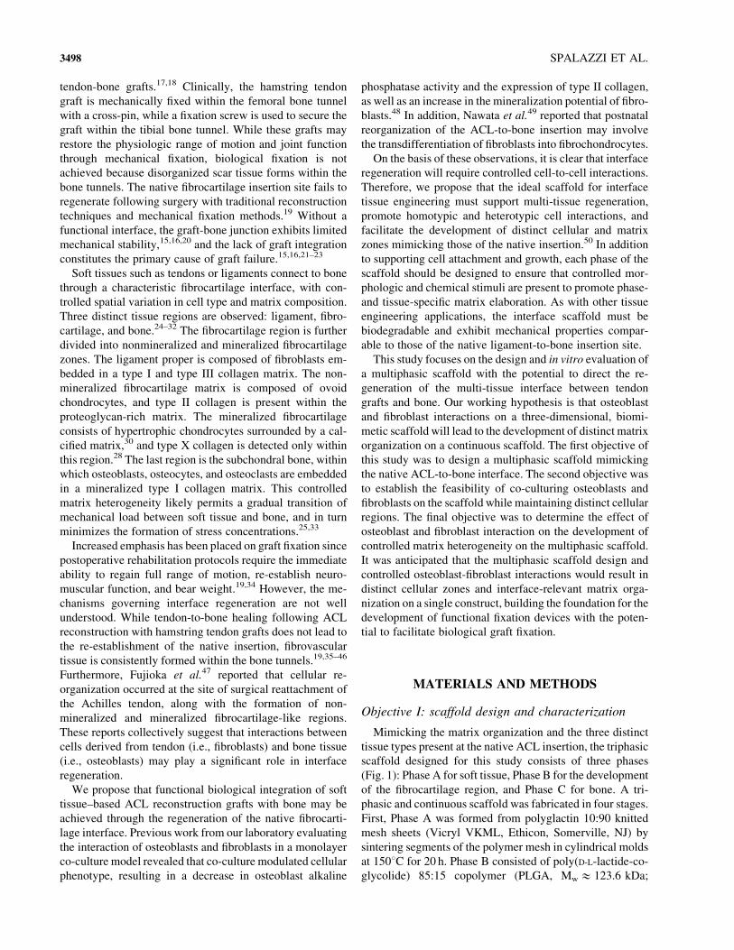
FIG. 1. Design of biomimetic triphasic scaffold. (A) Posterior view of bovine anterior cruciate ligament (ACL) with the tibial ACL
insertion into bone outlined. (B) Histologic image of neonatal bovine tibial ACL insertion showing the three main tissue types found at
the ACL-bone interface: ligament, fibrocartilage (FC), and bone (modified Goldner Masson’s trichrome stain). (C) Triphasic scaffold
mimicking the three tissue regions found at the interface.
CONTROLLED MATRIX HETEROGENEITY ON A TRIPHASIC SCAFFOLD 3499
with PBS and then cultured in fully supplemented DMEM;
cell outgrowth was monitored over time. The tendon tissue
was minced and cultured in similarly supplemented DMEM.
Only cells obtained from second and third migrations were
used in planned experiments to ensure a relatively homo-
genous cell population.53
Osteoblast-fibroblast co-culture on triphasic scaffolds.
After establishing the feasibility of osteoblast-fibroblast co-
culture on the triphasic scaffold, the effects of cell-cell
interactions on the development of controlled matrix het-
erogeneity was evaluated. Specifically, human hamstring
tendon fibroblasts (1.1�104 cells/cm2) were seeded on
Phase A. After the cells were allowed to attach for 30 min,
primary human osteoblasts (1.1�104 cells/cm2) were seeded
on Phase C of the triphasic scaffold. On the basis of por-
osimetry analysis, total surface area of Phase A was
approximately three times greater than that of Phase C.
Consequently, three times more cells were seeded on Phase
A than Phase C. After the osteoblasts were allowed to
attach for 30 min, supplemented DMEM was added and the
samples were incubated at 378C under humidified condi-
tions. Ascorbic acid (20 mg/mL) was added beginning at
day 7, and no b-glycerophosphate was added in this experi-
ment to prevent ectopic mineralization of the fibroblasts on
Phase A. Each well was precoated with agarose to limit
cell migration, and media was exchanged regularly.
Cell proliferation, gene expression, alkaline phosphatase
(ALP) activity, and extracellular matrix production were
determined as a function of scaffold phase and culturing
time. Compressive mechanical properties of the co-cultured
triphasic scaffold were measured over time, with acellular
scaffolds as controls.
Cell proliferation. Cell proliferation (n¼ 5) was de-
termined at days 1, 7, 21, and 35 by measuring total DNA per
scaffold phase using the PicoGreen double-stranded DNA
assay (Molecular Probes) following the manufacturer’s
suggested protocol.55 At designated time points, each tri-
phasic scaffold was rinsed with PBS and the three phases
were separated using a scalpel. The cells in each phase were
lysed with 0.1% Triton-X solution (Sigma), and each phase
was homogenized (Biospec, Bartlesville, OK). Fluorescence
of the samples was measured with a microplate reader
(Tecan, Research Triangle Park, NC), with excitation and
emission wavelengths of 485 and 535 nm, respectively. The
total number of cells in the sample was determined by
converting the amount of DNA per sample to cell number
using the conversion factor of 8 pg DNA/cell.56
Gene expression. We measured gene expression by using
reverse transcription polymerase chain reaction (RT-PCR)
at day 42. The three phases were separated and homogenized,
and total RNA was isolated using the Trizol extraction
method (Invitrogen, Carlsbad, CA). The isolated RNA was
reverse-transcribed into complementary DNA (cDNA)
using the SuperScript First-Strand Synthesis System (In-
vitrogen), and the cDNA product was amplified using re-
combinant Taq DNA polymerase (Invitrogen). Expression
of glyceraldehyde-3-phosphate dehydrogenase (GAPDH)
(GAPDH sense, 50-GGTGATGCTGGTGCTGAGTA-30;antisense, 50-ATCCACAGTCTTCTGGGTGG-30, 305 base
pairs) and type I collagen (type I collagen sense, 50-TGC
TGGCCAACCATGCCTCT-30; antisense, 50-TTGCACAA
TGCTCTGATC-30, 489 base pairs) were determined. All
genes were amplified for 40 cycles in a thermocycler (Ep-
pendorf Mastercycler gradient, Brinkmann, Westbury, NY).
Type I collagen band intensities were measured and nor-
malized against GAPDH (ImageJ).
Alkaline phosphatase activity. Sample ALP (n¼ 5) ac-
tivity was determined using an enzymatic assay based on
the hydrolysis of p-nitrophenyl phosphate (pNP-PO4) to
pNP.55,57 Aliquots of the sample homogenate (25 mL) were
added to 75 mL of 10 mM pNP-PO4 and were incubated at
378C for 30 min. The reaction was terminated by addition
of 100 mL of 0.1 N sodium hydroxide, and sample absor-
bance was measured at 415 nm using a microplate reader
(Tecan).
Cell attachment and phase-dependent extracellular ma-
trix deposition in co-culture. Cell attachment morphology
and growth on each phase of the triphasic scaffold (n¼ 2)
were examined immediately following seeding and at days
7, 21, and 35 using scanning electron microscopy (SEM,
3 kV; JEOL 5600LV, Tokyo, Japan, and FEI Quanta 600,
FEI Co., Hillsboro, OR). The triphasic scaffold was rinsed
in buffer and fixed with Karnovsky’s fixative58,59 for 24 h at
48C and was dehydrated with an ethanol series. The scaf-
folds were mounted on an aluminum post and were gold-
coated before analysis to reduce charging effects. Matrix
distribution on each phase of the scaffold was monitored
using SEM after 7, 21, and 35 days of culture. Elemental
composition of the elaborated matrix on each phase was
determined for non-gold coated samples using energy dis-
persive x-ray analysis (EDAX, 15 kV; FEI Quanta 600).
Scaffold mechanical properties. Compressive mechan-
ical properties of the samples (n¼ 4) were determined at
days 0, 7, 21, and 35. The samples were tested under uni-
axial compression (MTS 810, Eden Prairie, MN) following
the methods of Lu et al.52 Phase A was removed before
testing since this phase is intended for soft tissue formation,
and therefore the compressive properties of this phase are
less relevant than those of Phase B and Phase C. Moreover,
Phase A degraded rapidly over the 5-week period, and
removal of this phase standardized the testing method.
The samples were tested at a displacement rate of 1.3 mm/
min following a 10-N preload.60 A stress-strain curve was
generated and compressive modulus was determined by
calculating the slope of the elastic region of the stress-strain
curve.
3500 SPALAZZI ET AL.
Statistical analysis
Results are presented in the form of mean� standard
deviation, with n equal to the number of samples analyzed.
A two-way analysis of variance (ANOVA) was performed
to determine scaffold phase and temporal effects on total
cell number and ALP activity. Similarly, two-way ANOVA
was performed to determine effects of co-culture and time
on scaffold compressive modulus. The Tukey-Kramer post
hoc test was performed for all pairwise comparisons and
statistical significance was attained at a p value less than
.05. All statistical analyses were performed using JMP
statistical software (SAS Institute, Cary, NC).
RESULTS
Objective I: biomimetic scaffold design
and characterization
Because the native ligament-to-bone interface (Fig. 1-B)
consists of three distinct yet continuous tissue regions—
ligament, fibrocartilage, and bone—we designed a triphasic
scaffold (Fig. 1-C) with the potential to support the si-
multaneous formation of these three types of tissue. Phase A
of the triphasic scaffold is intended for fibroblast culture and
soft tissue formation, while Phase C is intended for osteo-
blasts and bone formation. The intermediate region, Phase
B, will support the co-culture of osteoblasts and fibroblasts
and will promote the development of a fibrocartilage-like
interface.
Specifically, Phase A consists of a polymer fiber mesh
intended for fibroblast attachment and matrix production,
Phase C consists of a polymer-bioactive glass composite
previously shown to be osteoconductive,52,61 and Phase B
consists of a porous polymer intermediate region where the
two cell types can interact and potentially form a fibro-
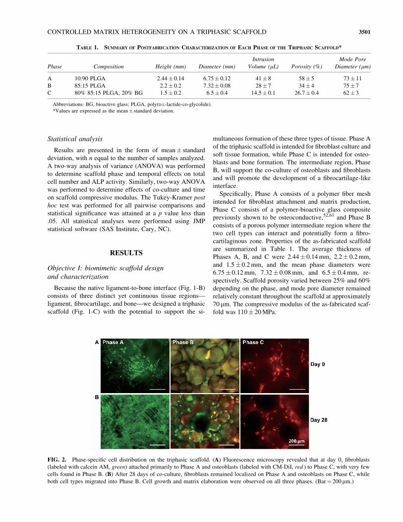
cartilaginous zone. Properties of the as-fabricated scaffold
are summarized in Table 1. The average thickness of
Phases A, B, and C were 2.44� 0.14 mm, 2.2� 0.2 mm,
and 1.5� 0.2 mm, and the mean phase diameters were
6.75� 0.12 mm, 7.32� 0.08 mm, and 6.5� 0.4 mm, re-
spectively. Scaffold porosity varied between 25% and 60%
depending on the phase, and mode pore diameter remained
relatively constant throughout the scaffold at approximately
70 mm. The compressive modulus of the as-fabricated scaf-
fold was 110� 20 MPa.
TABLE 1. SUMMARY OF POSTFABRICATION CHARACTERIZATION OF EACH PHASE OF THE TRIPHASIC SCAFFOLD*
Phase Composition Height (mm) Diameter (mm)
Intrusion
Volume (mL) Porosity (%)
Mode Pore
Diameter (mm)
A 10:90 PLGA 2.44� 0.14 6.75� 0.12 41� 8 58� 5 73� 11
B 85:15 PLGA 2.2� 0.2 7.32� 0.08 28� 7 34� 4 75� 7
C 80% 85:15 PLGA, 20% BG 1.5� 0.2 6.5� 0.4 14.5� 0.1 26.7� 0.4 62� 3
Abbreviations: BG, bioactive glass; PLGA, poly(D-L-lactide-co-glycolide).
*Values are expressed as the mean� standard deviation.
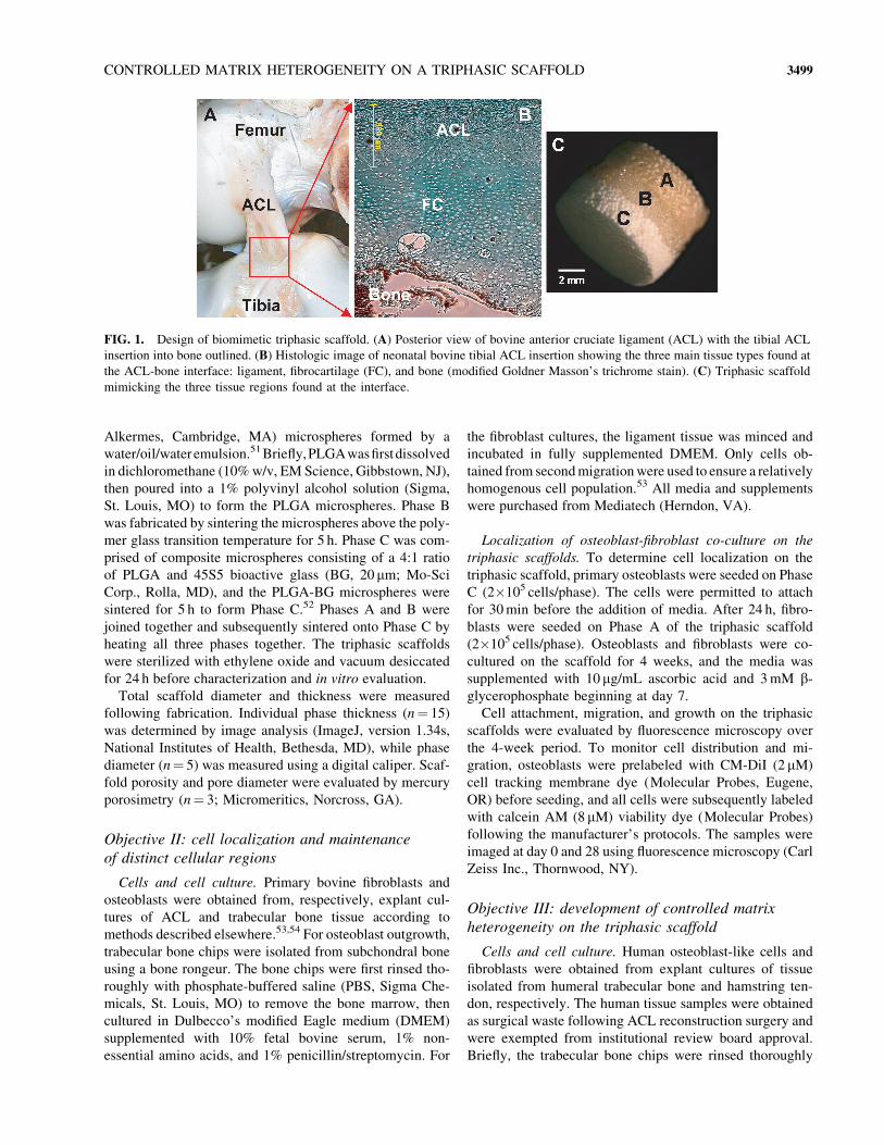
FIG. 2. Phase-specific cell distribution on the triphasic scaffold. (A) Fluorescence microscopy revealed that at day 0, fibroblasts
(labeled with calcein AM, green) attached primarily to Phase A and osteoblasts (labeled with CM-DiI, red ) to Phase C, with very few
cells found in Phase B. (B) After 28 days of co-culture, fibroblasts remained localized on Phase A and osteoblasts on Phase C, while
both cell types migrated into Phase B. Cell growth and matrix elaboration were observed on all three phases. (Bar¼ 200 mm.)
CONTROLLED MATRIX HETEROGENEITY ON A TRIPHASIC SCAFFOLD 3501
Objective II: cell localization and migration
To validate the co-culture of osteoblasts and fibroblasts
on the three-dimensional scaffold, the second objective of
this study was to monitor the migration of both cell types
into Phase B over time. As shown in Fig. 2, fluorescence
microscopy revealed that fibroblasts (labeled with calcein
AM, green) and osteoblasts (labeled with CM-DiI, red )
were indeed localized primarily on their respective phases,
with very few cells found in Phase B after initial seeding
(Fig. 2A). After 28 days of culture, fibroblasts and osteo-
blasts proliferated within their respective phases, and both
fibroblasts and osteoblasts migrated into Phase B (Fig. 2B).
With this scaffold design, osteoblasts and fibroblasts were
largely localized in their respective regions, while their
interaction was restricted to Phase B at day 28.
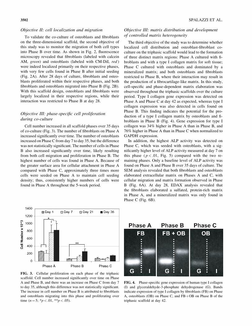
Objective III: phase-specific cell proliferation
during co-culture
Cell number increased in all scaffold phases over 35 days
of co-culture (Fig. 3). The number of fibroblasts on Phase A
increased significantly over time. The number of osteoblasts
increased on Phase C from day 7 to day 35, but the difference
was not statistically significant. The number of cells in Phase
B also increased significantly over time, likely resulting
from both cell migration and proliferation in Phase B. The
highest number of cells was found in Phase A. Because of
the greater surface area for cellular attachment in Phase A
compared with Phase C, approximately three times more
cells were seeded on Phase A to maintain cell seeding
density; thus, consistently higher numbers of cells were
found in Phase A throughout the 5-week period.
Objective III: matrix distribution and development
of controlled matrix heterogeneity
The third objective of the study was to determine whether
localized cell distribution and osteoblast-fibroblast co-
culture on the triphasic scaffold would lead to the formation
of three distinct matrix regions: Phase A cultured with fi-
broblasts and with a type I collagen matrix for soft tissue;
Phase C cultured with osteoblasts and dominated by a
mineralized matrix; and both osteoblasts and fibroblasts
restricted to Phase B, where their interaction may result in
the production of a fibrocartilage-like matrix. In this study,
cell-specific and phase-dependent matrix elaboration was
observed throughout the triphasic scaffolds over the culture
period. Type I collagen gene expression was detected on
Phase A and Phase C at day 42 as expected, whereas type I
collagen expression was also detected in cells found on
Phase B. This finding indicates the potential for the pro-
duction of a type I collagen matrix by osteoblasts and fi-
broblasts in Phase B (Fig. 4). Gene expression for type I
collagen was 34% higher in Phase A than in Phase B, and
76% higher in Phase A than in Phase C when normalized to
GAPDH expression.
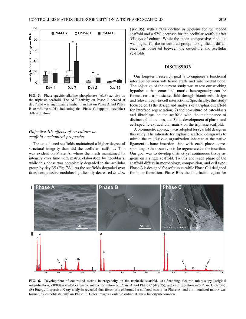
In addition, the highest ALP activity was detected on
Phase C, which was seeded with osteoblasts, with a sig-
nificantly higher level of ALP activity measured at day 7 on
this phase ( p< .01, Fig. 5) compared with the two re-
maining phases. Only a baseline level of ALP activity was
found on Phase A and Phase B over 35 days of culture. The
SEM analysis revealed that both fibroblasts and osteoblasts
elaborated extracellular matrix on Phases A and C, with
cellular migration and matrix formation observed in Phase
B (Fig. 6A). At day 28, EDAX analysis revealed that
the fibroblasts elaborated a sulfated, protein-rich matrix
in Phase A, and a mineralized matrix was only found in
Phase C (Fig. 6B).
FIG. 3. Cellular proliferation on each phase of the triphasic
scaffold. Cell number increased significantly over time on Phase
A and Phase B, and there was an increase on Phase C from day 7
to day 35, although this difference was not statistically significant.
The increase in cell number on Phase B is attributed to fibroblasts
and osteoblasts migrating into this phase and proliferating over
time (n¼ 5; *p< .01, **p< .05).
FIG. 4. Phase-specific gene expression of human type I collagen
(I) and glyceraldehyde-3-phosphate dehydrogenase (G). Bands
indicate expression of type I collagen by fibroblasts (FB) on Phase
A, osteoblasts (OB) on Phase C, and FBþOB on Phase B of the
triphasic scaffold at day 42.
3502 SPALAZZI ET AL.
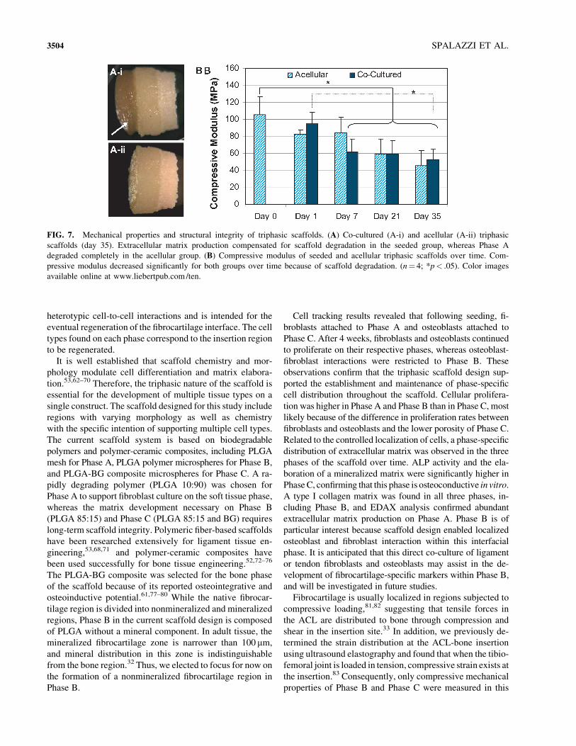
Objective III: effects of co-culture on
scaffold mechanical properties
The co-cultured scaffolds maintained a higher degree of
structural integrity than did the acellular scaffolds. This
was evident on Phase A, where the mesh maintained its
integrity over time with matrix elaboration by fibroblasts,
while this phase was completely degraded in the acellular
group by day 35 (Fig. 7A). As the scaffolds degraded over
time, compressive modulus significantly decreased in vitro
( p<.05), with a 50% decline in modulus for the seeded
scaffold and a 57% decrease for the acellular scaffold after
35 days of culture. While the mean compressive modulus
was higher for the co-cultured group, no significant differ-
ence was observed between the co-culture and acellular
scaffolds.
DISCUSSION
Our long-term research goal is to engineer a functional
interface between soft tissue grafts and subchondral bone.
The objective of the current study was to test our working
hypothesis that controlled matrix heterogeneity can be
formed on a triphasic scaffold through biomimetic design
and relevant cell-to-cell interactions. Specifically, this study
focused on 1) the design and analysis of a triphasic scaffold
for interface regeneration, 2) the co-culture of osteoblasts
and fibroblasts on the scaffold with the maintenance of
distinct cellular zones, and 3) the development of phase- and
cell-specific extracellular matrix on the triphasic scaffold.
A biomimetic approach was adopted for scaffold design in
this study. The rationale for triphasic scaffold design was to
mimic the multi-tissue organization inherent at the native
ligament-to-bone insertion site, with each phase corre-
sponding to the tissue type to be regenerated at the insertion.
Our goal was to develop distinct yet continuous tissue re-
gions on a single scaffold. To this end, each phase of the
scaffold differs in morphology, composition, and cell type.
Phase A is designed for soft tissue, while Phase C is designed
for bone formation. Phase B is the interfacial region for
FIG. 5. Phase-specific alkaline phosphatase (ALP) activity on
the triphasic scaffold. The ALP activity on Phase C peaked at
day 7 and was significantly higher than that on Phase A and Phase
B (n¼ 5; *p< .01), indicating that Phase C supports osteoblast
differentiation.
FIG. 6. Development of controlled matrix heterogeneity on the triphasic scaffold. (A) Scanning electron microscopy (original
magnification,�1000) revealed extensive matrix formation on Phase A and Phase C (day 35), and cell migration into Phase B (arrow).
(B) Energy dispersive X-ray analysis revealed that fibroblasts elaborated a sulfated matrix on Phase A, and a mineralized matrix was
formed by osteoblasts only on Phase C. Color images available online at www.liebertpub.com /ten.
CONTROLLED MATRIX HETEROGENEITY ON A TRIPHASIC SCAFFOLD 3503
heterotypic cell-to-cell interactions and is intended for the
eventual regeneration of the fibrocartilage interface. The cell
types found on each phase correspond to the insertion region
to be regenerated.
It is well established that scaffold chemistry and mor-
phology modulate cell differentiation and matrix elabora-
tion.53,62–70 Therefore, the triphasic nature of the scaffold is
essential for the development of multiple tissue types on a
single construct. The scaffold designed for this study include
regions with varying morphology as well as chemistry
with the specific intention of supporting multiple cell types.
The current scaffold system is based on biodegradable
polymers and polymer-ceramic composites, including PLGA
mesh for Phase A, PLGA polymer microspheres for Phase B,
and PLGA-BG composite microspheres for Phase C. A ra-
pidly degrading polymer (PLGA 10:90) was chosen for
Phase A to support fibroblast culture on the soft tissue phase,
whereas the matrix development necessary on Phase B
(PLGA 85:15) and Phase C (PLGA 85:15 and BG) requires
long-term scaffold integrity. Polymeric fiber-based scaffolds
have been researched extensively for ligament tissue en-
gineering,53,68,71 and polymer-ceramic composites have
been used successfully for bone tissue engineering.52,72–76
The PLGA-BG composite was selected for the bone phase
of the scaffold because of its reported osteointegrative and
osteoinductive potential.61,77–80 While the native fibrocar-
tilage region is divided into nonmineralized and mineralized
regions, Phase B in the current scaffold design is composed
of PLGA without a mineral component. In adult tissue, the
mineralized fibrocartilage zone is narrower than 100 mm,
and mineral distribution in this zone is indistinguishable
from the bone region.32 Thus, we elected to focus for now on
the formation of a nonmineralized fibrocartilage region in
Phase B.
Cell tracking results revealed that following seeding, fi-
broblasts attached to Phase A and osteoblasts attached to
Phase C. After 4 weeks, fibroblasts and osteoblasts continued
to proliferate on their respective phases, whereas osteoblast-
fibroblast interactions were restricted to Phase B. These
observations confirm that the triphasic scaffold design sup-
ported the establishment and maintenance of phase-specific
cell distribution throughout the scaffold. Cellular prolifera-
tion was higher in Phase A and Phase B than in Phase C, most
likely because of the difference in proliferation rates between
fibroblasts and osteoblasts and the lower porosity of Phase C.
Related to the controlled localization of cells, a phase-specific
distribution of extracellular matrix was observed in the three
phases of the scaffold over time. ALP activity and the ela-
boration of a mineralized matrix were significantly higher in
Phase C, confirming that this phase is osteoconductive in vitro.
A type I collagen matrix was found in all three phases, in-
cluding Phase B, and EDAX analysis confirmed abundant
extracellular matrix production on Phase A. Phase B is of
particular interest because scaffold design enabled localized
osteoblast and fibroblast interaction within this interfacial
phase. It is anticipated that this direct co-culture of ligament
or tendon fibroblasts and osteoblasts may assist in the de-
velopment of fibrocartilage-specific markers within Phase B,
and will be investigated in future studies.
Fibrocartilage is usually localized in regions subjected to
compressive loading,81,82 suggesting that tensile forces in
the ACL are distributed to bone through compression and
shear in the insertion site.33 In addition, we previously de-
termined the strain distribution at the ACL-bone insertion
using ultrasound elastography and found that when the tibio-
femoral joint is loaded in tension, compressive strain exists at
the insertion.83 Consequently, only compressive mechanical
properties of Phase B and Phase C were measured in this
FIG. 7. Mechanical properties and structural integrity of triphasic scaffolds. (A) Co-cultured (A-i) and acellular (A-ii) triphasic
scaffolds (day 35). Extracellular matrix production compensated for scaffold degradation in the seeded group, whereas Phase A
degraded completely in the acellular group. (B) Compressive modulus of seeded and acellular triphasic scaffolds over time. Com-
pressive modulus decreased significantly for both groups over time because of scaffold degradation. (n¼ 4; *p< .05). Color images
available online at www.liebertpub.com /ten.
3504 SPALAZZI ET AL.
study since these phases of the scaffold would be the sites of
fibrocartilage and bone formation, respectively, and there-
fore would be subjected to compressive loading in future
clinical application.
Stratified scaffold systems have been researched for
orthopedic tissue engineering, and in particular for osteo-
chondral applications.74,84–88 Schaefer et al.86 seeded bovine
articular chondrocytes on polyglycolic acid meshes and
periosteal cells on PLGA/polyethylene glycol foams, and
subsequently sutured these separate constructs together at 1 or
4 weeks after seeding. Integration between the two scaffolds
was observed to be superior when sutured at 1 week, sug-
gesting the importance of cellular interactions immediately
after seeding. Similarly, Gao et al.87 seeded mesenchymal
stem cell–differentiated chondrogenic cells in a hyaluronan
sponge and mesenchymal stem cell–differentiated osteo-
genic cells in a porous calcium phosphate scaffold. These
scaffolds were then joined by fibrin sealant and implanted
subcutaneously in syngeneic rats, with continuous collagen
fibers observed between the two scaffolds 6 weeks following
implantation. In addition, distinct cartilaginous and osseous
zones were also formed.
In these pioneering studies on multi-tissue integration,
scaffolds with a single cell type were joined after seeding.
This process minimized any immediate cellular interactions
necessary for eventual construct integration. In addition,
scaffold integrity is not optimal in these reported scaffolds
because the integration between the two phases is not con-
tinuous. In contrast, the triphasic scaffold design used in this
study consists of a single continuous construct with inte-
grated phases, upon which cells can interact immediately after
seeding. Moreover, osteoblast-fibroblast interaction is loca-
lized and promoted within the interface region (Phase B).
To our knowledge, this is the first reported study to focus
on developing functional scaffolds and promoting cell-to-
cell interactions for regenerating the interface between li-
gament and bone. The novel triphasic scaffold reported in
this study promoted the formation of distinct cellular and
matrix regions, and will serve as a model system for de-
veloping tissue engineering strategies aimed at regenerating
the graft-to-bone interface on soft tissue–based ACL re-
construction grafts. The results of this study demonstrate the
feasibility of multi-tissue regeneration on a single graft, with
the potential to promote biological fixation of soft tissue
grafts to bone.
This triphasic scaffold is designed to guide the eventual
reestablishment of an anatomically oriented and mechani-
cally functional fibrocartilage interfacial region directly
on both synthetic and biological soft tissue grafts. It is in-
tended to be used clinically as a graft collar for soft tissue
graft fixation. In this scenario, Phase C would be inserted
into the bone tunnel, while Phases A and B would reside
outside the bone tunnel in the joint capsule; cellular inter-
actions will promote the formation of an anatomically or-
iented and functional fibrocartilage interface. Tensile and
torsional forces would be sustained by the soft tissue graft
and Phase A. The presence of a physiologic fibrocartilage
region will enable the gradual transition of load from the
graft to bone,89 and osteointegration in the bone tunnel will
be facilitated by Phase C and the addition of osteogenic
factors.
Interface tissue engineering is an emerging field, and bio-
logical fixation of soft tissue grafts is a multifaceted problem
with many inherent challenges. Through controlled co-
culture and multiphasic scaffold design, distinct cellular and
matrix zones were developed on the triphasic scaffold. The
phase-specific cell distribution and matrix heterogeneity
were maintained for the duration of the culturing period in this
study; however, whether this zonal distribution can be sus-
tained during long-term culture and in vivo remains to be
seen. Moreover, the mechanisms governing fibrocartilage
development or regeneration are not well understood, repre-
senting one of the most significant challenges in orthopedic
interface tissue engineering. In addition, a prerequisite for
biomimetic scaffold design is a clear understanding of the
structure-function relationship at the soft tissue-to-bone in-
terface. Results from our ongoing studies in elucidating the
mechanisms of interface regeneration and characterization
of the chemical and mechanical properties of the ACL-to-
bone interface will aid in the optimization of cellular inter-
actions on this multiphasic scaffold. Future studies will focus
on strategies for in vitro and in vivo development of fi-
brocartilage-specific markers and biomimetic tissue orga-
nization on the triphasic scaffold.
In conclusion, we have reported here the design and
in vitro testing of a triphasic scaffold for soft tissue graft-
to-bone integration. The triphasic scaffold supported the
growth, migration, and phenotypic matrix production of
osteoblasts and fibroblasts. More importantly, this novel
biomimetic scaffold supported distinct zonal distributions
of cells and phase-specific extracellular matrix deposition
over time. This is the first reported study examining the
interaction of osteoblasts and fibroblasts on a multiphasic
scaffold, and our results demonstrate the feasibility of
multi-tissue regeneration on a single construct. The co-
cultured triphasic scaffold will serve as a model system for
formulating tissue engineering strategies for the regenera-
tion of a functional interface on soft tissue reconstruction
grafts.
ACKNOWLEDGMENTS
The authors would like to gratefully acknowledge Dr. X.
Edward Guo of the Bone Biomechanics Laboratory at Co-
lumbia University for his advice on mechanical testing and
use of the MTS system, and Dr. Yusuf M. Khan of the
University of Virginia and Micromeritics for assistance with
porosimetry analysis. This study was funded by National
Institutes of Health/National Institute of Arthritis and Mus-
culoskeletal and Skin Diseases (R21 AR052402-01A1)
(H.H.L.) an Early Faculty Career Award from the Wallace
CONTROLLED MATRIX HETEROGENEITY ON A TRIPHASIC SCAFFOLD 3505
H. Coulter Foundation (H.H.L.), and a National Science
Foundation Graduate Fellowship (GK-12 0338329) ( J.P.S.).
REFERENCES
1. Johnson, R.J. The anterior cruciate: a dilemma in sports
medicine. Int. J. Sports Med. 3, 71, 1982.
2. American Academy of Orthopaedic Surgeons. Arthroplasty
and Total Joint Replacement Procedures: United States 1990
to 1997. Rosemont, IL: American Academy of Orthopaedic
Surgeons, 1997.
3. Brown, C.H., Jr., and Carson, E.W. Revision anterior cruciate
ligament surgery. Clin. Sports Med. 18, 109, 1999.
4. United States Department of Health and Human Services,
Centers for Disease Control and Prevention, National Center
for Health Statistics. National Survey of Ambulatory Surgery.
Atlanta, GA: Centers for Disease Control and Prevention;
1996.
5. United States Department of Health and Human Services,
Centers for Disease Control and Prevention, National Center
for Health Statistics. National Hospital Discharge Survey.
Atlanta, GA: Centers for Disease Control and Prevention;
1996.
6. Bray, R.C., Leonard, C.A., and Salo, P.T. Vascular physiol-
ogy and long-term healing of partial ligament tears. J. Orthop.
Res. 20, 984, 2002.
7. Fleming, B., Beynnon, B., Howe, J., McLeod, W., and Pope,
M. Effect of tension and placement of a prosthetic anterior
cruciate ligament on the anteroposterior laxity of the knee.
J. Orthop. Res. 10, 177, 1992.
8. Gregor, R.J., and Abelew, T.A. Tendon force measurements
and movement control: a review. Med. Sci. Sports Exerc. 26,
1359, 1994.
9. Beynnon, B.D., Yu, J., Huston, D., Fleming, B., Johnson, R.,
Haugh, L., and Pope, M.H. A sagittal plane model of the knee
and cruciate ligaments with application of a sensitivity ana-
lysis. J. Biomech. Eng. 118, 227, 1996.
10. Beynnon, B.D., Johnson, R.J., Fleming, B.C., Peura, G.D.,
Renstrom, P.A., Nichols, C.E., and Pope, M.H. The effect of
functional knee bracing on the anterior cruciate ligament in
the weightbearing and nonweightbearing knee. Am. J. Sports
Med. 25, 353, 1997.
11. Fleming, B.C., Abate, J.A., Peura, G.D., and Beynnon, B.D.
The relationship between graft tensioning and the anterior-
posterior laxity in the anterior cruciate ligament reconstructed
goat knee. J. Orthop. Res. 19, 841, 2001.
12. Fleming, B.C., and Beynnon, B.D. In vivo measurement of
ligament/tendon strains and forces: a review. Ann. Biomed.
Eng. 32, 318, 2004.
13. Markolf, K.L., Hame, S., Hunter, D.M., Oakes, D.A., Zoric,
B., Gause, P., and Finerman, G.A. Effects of femoral tunnel
placement on knee laxity and forces in an anterior cruciate
ligament graft. J. Orthop. Res. 20, 1016, 2002.
14. Loh, J.C., Fukuda, Y., Tsuda, E., Steadman, R.J., Fu, F.H.,
and Woo, S.L. Knee stability and graft function following
anterior cruciate ligament reconstruction: comparison be-
tween 11 o’clock and 10 o’clock femoral tunnel placement.
Arthroscopy 19, 297, 2003.
15. Robertson, D.B., Daniel, D.M., and Biden, E. Soft tissue
fixation to bone. Am. J. Sports Med. 14, 398, 1986.
16. Kurosaka, M., Yoshiya, S., and Andrish, J.T. A biomecha-
nical comparison of different surgical techniques of graft
fixation in anterior cruciate ligament reconstruction. Am. J.
Sports Med. 15, 225, 1987.
17. Beynnon, B.D., Johnson, R.J., Fleming, B.C., Kannus, P.,
Kaplan, M., Samani, J., and Renstrom, P. Anterior cruciate
ligament replacement: comparison of bone-patellar tendon-
bone grafts with two-strand hamstring grafts. A prospective,
randomized study. J. Bone Joint Surg. Am. 84-A, 1503,
2002.
18. Barrett, G.R., Noojin, F.K., Hartzog, C.W., and Nash, C.R.
Reconstruction of the anterior cruciate ligament in females: a
comparison of hamstring versus patellar tendon autograft.
Arthroscopy 18, 46, 2002.
19. Rodeo, S.A., Arnoczky, S.P., Torzilli, P.A., Hidaka, C., and
Warren, R.F. Tendon-healing in a bone tunnel. A biomecha-
nical and histological study in the dog. J. Bone Joint Surg.
Am. 75, 1795, 1993.
20. Rodeo, S.A., Suzuki, K., Deng, X.H., Wozney, J., and War-
ren, R.F. Use of recombinant human bone morphogenetic
protein-2 to enhance tendon healing in a bone tunnel. Am. J.
Sports Med. 27, 476, 1999.
21. Friedman, M.J., Sherman, O.H., Fox, J.M., Del Pizzo, W.,
Snyder, S.J., and Ferkel, R.J. Autogeneic anterior cruciate
ligament (ACL) anterior reconstruction of the knee. A review.
Clin. Orthop. Relat. Res. 196, 9, 1985.
22. Jackson, D.W., Grood, E.S., Arnoczky, S.P., Butler, D.L., and
Simon, T.M. Cruciate reconstruction using freeze dried ante-
rior cruciate ligament allograft and a ligament augmentation
device (LAD). An experimental study in a goat model. Am. J.
Sports Med. 15, 528, 1987.
23. Yahia L. Ligaments and Ligamentoplasties. Berlin Heidel-
berg: Springer Verlag, 1997.
24. Cooper, R.R., and Misol, S. Tendon and ligament insertion. A
light and electron microscopic study. J. Bone Joint Surg. Am.
52, 1, 1970.
25. Benjamin, M., Evans, E.J., and Copp, L. The histology of
tendon attachments to bone in man. J. Anat. 149, 89, 1986.
26. Wei, X., and Messner, K. The postnatal development of the
insertions of the medial collateral ligament in the rat knee.
Anat. Embryol. (Berl). 193, 53, 1996.
27. Sagarriga, V.C., Kavalkovich, K., Wu, J., and Niyibizi, C.
Biochemical analysis of collagens at the ligament-bone in-
terface reveals presence of cartilage-specific collagens. Arch.
Biochem. Biophys. 328, 135, 1996.
28. Niyibizi, C., Sagarrigo, V.C., Gibson, G., and Kavalkovich,
K. Identification and immunolocalization of type X collagen
at the ligament-bone interface. Biochem. Biophys. Res. Com-
mun. 222, 584, 1996.
29. Messner, K. Postnatal development of the cruciate ligament
insertions in the rat knee. morphological evaluation and im-
munohistochemical study of collagens types I and II. Acta
Anatomica. 160, 261, 1997.
30. Petersen, W., and Tillmann, B. Structure and vascularization
of the cruciate ligaments of the human knee joint. Anat.
Embryol. (Berl). 200, 325, 1999.
31. Thomopoulos, S., Williams, G.R., Gimbel, J.A., Favata, M.,
and Soslowsky, L.J. Variations of biomechanical, structural,
3506 SPALAZZI ET AL.
and compositional properties along the tendon to bone in-
sertion site. J. Orthop. Res. 21, 413, 2003.
32. Wang, I.E., Mitroo, S., Chen, F.H., Lu, H.H., and Doty, S.B.
Age-dependent changes in matrix composition and organi-
zation at the ligament-to-bone insertion. J. Orthop. Res. 25,
1745, 2006.
33. Woo, S.L., and Buckwalter, J.A. AAOS/NIH/ORS workshop.
Injury and repair of the musculoskeletal soft tissues. Sa-
vannah, Georgia, June 18-20, 1987. J. Orthop. Res. 6, 907,
1988.
34. Brand, J. Jr., Weiler, A., Caborn, D.N., Brown, C.H. Jr., and
Johnson, D.L. Graft fixation in cruciate ligament reconstruc-
tion. Am. J. Sports Med. 28, 761, 2000.
35. Grana, W.A., Egle, D.M., Mahnken, R., and Goodhart, C.W.
An analysis of autograft fixation after anterior cruciate liga-
ment reconstruction in a rabbit model. Am. J. Sports Med. 22,
344, 1994.
36. Liu, S.H., Panossian, V., al Shaikh, R., Tomin, E., Shepherd,
E., Finerman, G.A., and Lane, J.M. Morphology and matrix
composition during early tendon to bone healing. Clin. Or-
thop. Relat. Res. 339, 253, 1997.
37. Blickenstaff, K. R., Grana, W.A., and Egle, D. Analysis of a
semitendinosus autograft in a rabbit model. Am. J. Sports
Med. 25, 554, 1997.
38. Panni, A.S., Milano, G., Lucania, L., and Fabbriciani, C.
Graft healing after anterior cruciate ligament reconstruction in
rabbits. Clin. Orthop. 343, 203, 1997.
39. Eriksson, K., Kindblom, L.G., and Wredmark, T. Semi-
tendinosus tendon graft ingrowth in tibial tunnel following
ACL reconstruction: a histological study of 2 patients with
different types of early graft failure. Acta Orthop. Scand. 71,
275, 2000.
40. Yoshiya, S., Nagano, M., Kurosaka, M., Muratsu, H., and
Mizuno, K. Graft healing in the bone tunnel in anterior
cruciate ligament reconstruction. Clin. Orthop. 376, 278, 2000.
41. Anderson, K., Seneviratne, A.M., Izawa, K., Atkinson, B.L.,
Potter, H.G., and Rodeo, S.A. Augmentation of tendon heal-
ing in an intraarticular bone tunnel with use of a bone growth
factor. Am. J. Sports Med. 29, 689, 2001.
42. Weiler, A., Hoffmann, R.F., Bail, H.J., Rehm, O., and Sud-
kamp, N.P. Tendon healing in a bone tunnel. Part II: Histo-
logic analysis after biodegradable interference fit fixation in a
model of anterior cruciate ligament reconstruction in sheep.
Arthroscopy. 18, 124, 2002.
43. Thomopoulos, S., Hattersley, G., Rosen, V., Mertens, M.,
Galatz, L., Williams, G.R., and Soslowsky, L.J. The localized
expression of extracellular matrix components in healing
tendon insertion sites: an in situ hybridization study. J. Or-
thop. Res. 20, 454, 2002.
44. Batra, G.S., Harrison, J.W., Clough, T.M., and Paul, A.S.
Failure of anterior cruciate ligament reconstruction following
calcification of the graft. Knee 9, 245, 2002.
45. Chen, C.H., Chen, W.J., Shih, C.H., Yang, C.Y., Liu, S.J., and
Lin, P.Y. Enveloping the tendon graft with periosteum to en-
hance tendon-bone healing in a bone tunnel: a biomechanical
and histologic study in rabbits. Arthroscopy 19, 290, 2003.
46. Song, E.K., Rowe, S.M., Chung, J.Y., Moon, E.S., and Lee,
K.B. Failure of osteointegration of hamstring tendon autograft
after anterior cruciate ligament reconstruction. Arthroscopy
20, 424, 2004.
47. Fujioka, H., Thakur, R., Wang, G.J., Mizuno, K., Balian, G.,
and Hurwitz, S.R. Comparison of surgically attached and non-
attached repair of the rat Achilles tendon-bone interface.
Cellular organization and type X collagen expression. Con-
nect. Tissue Res. 37, 205, 1998.
48. Wang, I.E., Jeffries, D.T., Jiang, J., Chen, F.H., and Lu, H.H.
Effects of co-culture on ligament fibroblast and osteoblast
growth and differentiation. Trans. Orthop. Res. Soc., Paper
no. 138, 2004.
49. Nawata, K., Minamizaki, T., Yamashita, Y., and Teshima, R.
Development of the attachment zones in the rat anterior
cruciate ligament: changes in the distributions of proliferating
cells and fibrillar collagens during postnatal growth. J. Or-
thop. Res. 20, 1339, 2002.
50. Mikos, A.G., Herring, S.W., Elisseef, J., Lu, H.H., Kandel, R.,
Schoen, F.J., Toner, M., Mooney, D., Atala, A., Kaplan, D.,
and Vunjak-Novakovic, G. Engineering complex tissues.
Tissue Eng. 12, 3307, 2006.
51. Borden, M., Attawia, M., Khan, Y., and Laurencin, C.T.
Tissue engineered microsphere-based matrices for bone re-
pair: design and evaluation. Biomaterials 23, 551, 2002.
52. Lu, H.H., El Amin, S.F., Scott, K.D., and Laurencin, C.T. Three-
dimensional, bioactive, biodegradable, polymer-bioactive glass
composite scaffolds with improved mechanical properties sup-
port collagen synthesis and mineralization of human osteoblast-
like cells in vitro. J. Biomed. Mater. Res. 64A, 465, 2003.
53. Lu, H.H., Cooper, J.A. Jr., Manuel, S., Freeman, J.W., Atta-
wia, M.A., Ko, F.K., and Laurencin, C.T. Anterior cruciate
ligament regeneration using braided biodegradable scaffolds:
in vitro optimization studies. Biomaterials 26, 4805, 2005.
54. Spalazzi, J.P., Dionisio, K.L., Jiang, J., and Lu, H.H. Osteo-
blast and chondrocyte interactions during coculture on scaf-
folds. IEEE Eng. Med. Biol. Mag. 22, 27, 2003.
55. Jiang, J., Nicoll, S.B., and Lu, H.H. Co-culture of osteoblasts
and chondrocytes modulates cellular differentiation in vitro.
Biochem. Biophys. Res. Commun. 338, 762, 2005.
56. Lu, H.H. 45S5 bioactive glass surface zeta potential variations
in electrolyte solutions with and without fibronectin [Ph.D.
thesis]. Department of Bioengineering, University of Penn-
sylvania, Philadelphia, Pa, 1998.
57. Teixeira, C.C., Hatori, M., Leboy, P.S., Pacifici, M., and
Shapiro, I.M. A rapid and ultrasensitive method for mea-
surement of DNA, calcium and protein content, and alkaline
phosphatase activity of chondrocyte cultures. Calcif. Tissue
Int. 56, 252, 1995.
58. Karnovsky, M.J. A formaldehyde-glutaraldehyde fixative of
high osmolarity for use in electron microscopy. J. Cell Biol.
27, 137A, 1965.
59. Langley, S.M., Chai, P.J., Miller, S.E., Mault, J.R., Jaggers,
J.J., Tsui, S.S., Lodge, A.J., Lefurgey, A., and Ungerleider,
R.M. Intermittent perfusion protects the brain during deep
hypothermic circulatory arrest. Ann. Thorac. Surg. 68, 4, 1999.
60. Standard Test Method for Compressive Properties of Rigid
Plastics, Annual Book of ASTM Standards, D 695, 1996, pp.
78–84.
61. Lu, H.H., Tang, A., Oh, S.C., Spalazzi, J.P., and Dionisio,
K. Compositional effects on the formation of a calcium
phosphate layer and the response of osteoblast-like cells on
polymer-bioactive glass composites. Biomaterials 26, 6323,
2005.
CONTROLLED MATRIX HETEROGENEITY ON A TRIPHASIC SCAFFOLD 3507
62. Brody, G.A., Eisinger, M., Arnoczky, S.P., and Warren, R.F.
In vitro fibroblast seeding of prosthetic anterior cruciate liga-
ments. A preliminary study. Am. J. Sports Med. 16, 203, 1988.
63. Dunn, M.G., Liesch, J.B., Tiku, M.L., Maxian, S.H., and
Zawadsky, J.P. The tissue engineering approach to ligament
reconstruction. Proc. Material Res. Soc. 33, 13, 1994.
64. Bellincampi, L.D., Closkey, R.F., Prasad, R., Zawadsky, J.P.,
and Dunn, M.G. Viability of fibroblast-seeded ligament analogs
after autogenous implantation. J. Orthop. Res. 16, 414, 1998.
65. Goulet, F., Germain, L., Rancourt, D., Caron, C., Normand, A.,
and Auger, F.A. Tendons and ligaments. In: Lanza, R.P.,
Langer, R., and Vacanti, J.P., eds. Principles of Tissue En-
gineering. San Diego, CA: Academic Press, 2000, pp. 639–645.
66. Altman, G.H., Horan, R.L., Lu, H.H., Moreau, J., Martin, I.,
Richmond, J.C., and Kaplan, D.L. Silk matrix for tissue en-
gineered anterior cruciate ligaments. Biomaterials 23, 4131,
2002.
67. Altman, G.H., Lu, H.H., Horan, R.L., Calabro, T., Ryder, D.,
Kaplan, D.L., Stark, P., Martin, I., Richmond, J.C., and
Vunjak-Novakovic, G. Advanced bioreactor with controlled
application of multi-dimensional strain for tissue engineering.
J. Biomed. Eng. 124, 742, 2002.
68. Cooper, J.A., Lu, H.H., Ko, F.K., Freeman, J.W., and Laur-
encin, C.T. Fiber-based tissue-engineered scaffold for ligament
replacement: design considerations and in vitro evaluation.
Biomaterials 26, 1523, 2005.
69. Kubo, K., Kakimoto, T., Kanda, C., Tsukasa, N., Uehara, M.,
Izumi, Y., Kamada, T., Kaneko, N., and Sueda, T. Bioactive
glass promoted formation of nodules in periodontal-ligament
fibroblasts in vitro. J. Biomed. Mater. Res. 27, 1175, 1993.
70. Hee, C.K., Jonikas, M.A., and Nicoll, S.B. Influence of three-
dimensional scaffold on the expression of osteogenic differ-
entiation markers by human dermal fibroblasts. Biomaterials
27, 875, 2006.
71. Dunn, M.G., Liesch, J.B., Tiku, M.L., and Zawadsky, J.P.
Development of fibroblast-seeded ligament analogs for ACL
reconstruction. J. Biomed. Mater. Res. 29, 1363, 1995.
72. Laurencin, C.T., Attawia, M.A., Elgendy, H.E., and Herbert,
K.M. Tissue engineered bone-regeneration using degradable
polymers: the formation of mineralized matrices. Bone 19,
93S, 1996.
73. Marcolongo, M., Ducheyne, P., Garino, J., and Schepers, E.
Bioactive glass fiber/polymeric composites bond to bone tis-
sue. J. Biomed. Mater. Res. 39, 161, 1998.
74. Niederauer, G.G., Slivka, M.A., Leatherbury, N.C., Korvick,
D.L., Harroff, H.H., Ehler, W.C., Dunn, C.J., and Kieswetter,
K. Evaluation of multiphase implants for repair of focal os-
teochondral defects in goats. Biomaterials 21, 2561, 2000.
75. Ma, P.X., Zhang, R., Xiao, G., and Franceschi, R. Engineer-
ing new bone tissue in vitro on highly porous poly(alpha-
hydroxyl acids)/hydroxyapatite composite scaffolds. J. Biomed.
Mater. Res. 54, 284, 2001.
76. Verrier, S., Blaker, J.J., Maquet, V., Hench, L.L., and Boc-
caccini, A.R. PDLLA/Bioglass composites for soft-tissue and
hard-tissue engineering: an in vitro cell biology assessment.
Biomaterials 25, 3013, 2004.
77. Hench, L.L. Bioceramics: from concept to clinic. J. Am. Cera.
Soc. 74, 1487, 1991.
78. Oonishi, H., Kushitani, S., Yasukawa, E., Iwaki, H., Hench,
L.L., Wilson, J., Tsuji, E., and Sugihara, T. Particulate bio-
glass compared with hydroxyapatite as a bone graft substitute.
Clin. Orthop. 334, 316, 1997.
79. Gatti, A.M., Valdre, G., and Andersson, O.H. Analysis of the
in vivo reactions of a bioactive glass in soft and hard tissue.
Biomaterials 15, 208, 1994.
80. Yao, J., Radin, S., Leboy, S., and Ducheyne, P. The effect of
bioactive glass content on synthesis and bioactivity of com-
posite poly (lactic-co-glycolic acid)/bioactive glass substrate
for tissue engineering. Biomaterials 26, 1935, 2005.
81. Benjamin, M., Ralphs, J.R. Fibrocartilage in tendons and
ligaments—an adaptation to compressive load. J. Anat. 193,
481, 1998.
82. Evanko, S.P., and Vogel, K.G. Ultrastructure and proteogly-
can composition in the developing fibrocartilaginous region
of bovine tendon. Matrix 10, 420, 1990.
83. Spalazzi, J.P., Gallina, J., Fung-Kee-Fung, S.D., Konofagou,
E.E., and Lu, H.H. Elastographic imaging of strain distribu-
tion in the anterior cruciate ligament and at the ligament-bone
insertions. J. Orthop. Res. 24, 2001, 2006.
84. Lu, H.H., Jiang, J., Tang, A., Hung, C.T., and Guo, X.E.
Development of controlled heterogeneity on a polymer-cera-
mic hydrogel scaffold for osteochondral repair. Bioceramics
17, 607, 2005.
85. Yu, H., Grynpas, M., and Kandel, R.A. Composition of car-
tilagenous tissue with mineralized and non-mineralized zones
formed in vitro. Biomaterials 18, 1425, 1997.
86. Schaefer, D., Martin, I., Shastri, P., Padera, R.F., Langer,
R., Freed, L.E., and Vunjak-Novakovic, G. In vitro genera-
tion of osteochondral composites. Biomaterials 21, 2599,
2000.
87. Gao, J., Dennis, J.E., Solchaga, L.A., Awadallah, A.S.,
Goldberg, V.M., and Caplan, A.I. Tissue-engineered fabrica-
tion of an osteochondral composite graft using rat bone
marrow-derived mesenchymal stem cells. Tissue Eng. 7, 363,
2001.
88. Hollister, S.J., Maddox, R.D., and Taboas, J.M. Optimal de-
sign and fabrication of scaffolds to mimic tissue properties
and satisfy biological constraints. Biomaterials 23, 4095,
2002.
89. Woo, S.L., Gomez, M.A., Seguchi, Y., Endo, C.M., and
Akeson, W.H. Measurement of mechanical properties of li-
gament substance from a bone-ligament-bone preparation.
J. Orthop. Res. 1, 22, 1983.
Address reprint requests to:
Helen H. Lu, Ph.D.
Department of Biomedical Engineering
Columbia University
New York, NY
E-mail: [email protected]
3508 SPALAZZI ET AL.