Embed Size (px)
Citation preview
Proc. Natl. Acad. Sci. USAVol. 87, pp. 2730-2734, April 1990Immunology
Development of B-lineage cells in the bone marrow of scid/scidmice following the introduction of functionally rearrangedimmunoglobulin transgenes
(severe combined immunodeficiency/p-transgenic scid mice/,uc-transgenic scid mice/pre-B cells/fluorescence-activated cell sorter)
MICHAL REICHMAN-FRIED, RICHARD R. HARDY, AND MELVIN J. BOSMAInstitute for Cancer Research, Fox Chase Cancer Center, 7701 Burholme Avenue, Philadelphia, PA 19111
Communicated by Michael Potter, January 8, 1990 (received for review October 16, 1989)
ABSTRACT Mice homozygous for the mutation scid (scidmice) are severely immunodeficient and generally lack detect-able numbers ofpre-B, B, and T cells. This condition is believedto result from a defect in the mechanism responsible forrearrangement of immunoglobulin and T-cell receptor genes indeveloping B and T lymphocytes. To test this hypothesis andevaluate whether scid affects only the process of gene recom-bination, we introduced functionally rearranged immunoglob-ulin genes into the scid mouse genome. As scid mice appear tocontain early lymphoid cells committed to the B lineage (pro-Bcells), we asked whether the introduction of an IgM heavy-chain gene alone (IL-transgenic scid mice) or both IgM heavy-and Kc light-chain genes (,.uc-transgenic scid mice) would allowfurther differentiation of scid pro-B cells into pre-B and B cells.We found that normal numbers of pre-B cells appeared in thebone marrow of ,u-transgenic scid mice and that both pre-B andB cells appeared in the bone marrow of ,.uc-transgenic scidmice. However, in the latter case, the number of pre-B and Bcells was 2- to 3-fold less than in the controls (juc-transgenicscid heterozygotes) and few, if any, B cells were detectable inthe peripheral lymphoid tissues. The implications of theseresults for the above hypothesis are discussed.
Mutant C.B-17/Icr scid/scid mice with severe combinedimmunodeficiency (1), here referred to as scid mice, arehomozygous for an autosomal recessive mutation (scid) onchromosome 16 (2). Although scid mice show evidence ofearly lymphoid cells committed to the B- or T-cell pathway(3), these cells generally fail to mature into detectable num-bers of pre-B, B, and T cells (1, 4). It has been hypothesized(5) that developing scid lymphocytes die prematurely as aresult of a defective lymphocyte recombinase system, whichcannot successfully recombine the variable (V), diversity(D), and joining (J) gene elements that code for immunoglob-ulin (Ig) and T-cell receptor variable regions. This hypothesiswas prompted by the observation of abnormal Ig heavy-chainand T-cell receptor gene rearrangements in transformedlymphoid cell lines recovered from scid bone marrow orthymus (5). Others have confirmed and extended thesefindings (6-8); moreover, abnormal Ig gene rearrangementsalso have been observed in long-term cultures of scid bonemarrow cells (9, 10). Further support for this hypothesis hascome from reports showing that transformed scid lympho-cytes have an abnormal VDJ recombinase activity (11, 12).Though this activity can mediate the joining of signal se-quences that flank V, D, and J coding elements and thus yieldthe reciprocal product of a standard VDJ recombinationevent, it cannot mediate with any appreciable frequency thefunctional joining of V(D)J elements. Failure to recombine
chromosome ends bearing V, D, or J coding elements pre-sumably would be lethal to developing scid lymphocytes.To test the above hypothesis, we introduced functionally
rearranged Ig transgenes [i.e., a ju heavy-chain gene alone orboth a A gene and K light-chain gene (OK)] into the scid mousegenome. In normal developing B-lineage cells, the 1L locusrearranges before the K locus and cells progress from thetL-expressing pre-B-cell stage to B cells that express both asand K chains and are surface IgM' (reviewed in ref. 13). Asneither cell type is detectable in scid bone marrow, it wasimportant to know whether the introduction of u and 1LKtransgenes into the scid mouse genome would result in astepwise appearance of pre-B cells and B cells, respectively.Such a result would be expected if (i) the effect of scid ondeveloping lymphocytes were restricted to VDJ recombina-tion and the products of the IL and / transgenes were ableto provide the necessary signals for further B-cell maturation,and (ii) the Ig transgenes were able to exclude or inhibitrearrangement ofendogenous Ig alleles (allelic exclusion) andthereby rescue developing scid lymphocytes from the pre-sumed deleterious effects of endogenous Ig gene rearrange-ments. We found that the bone marrow of ju-transgenic scidmice contained normal numbers of pre-B cells but no detect-able B cells, whereas in the bone marrow of LK-transgenicscid mice, both pre-B and B cells were detected. But in thelatter case, the number of detectable pre-B and B cells was2- to 3-fold less than in the controls (,UK-transgenic scidheterozygotes); moreover, few, if any, B cells were detect-able in the peripheral lymphoid tissues of ,uK-transgenic scidmice. We discuss the possibility that incomplete allelic ex-clusion of endogenous scid Ig alleles may be responsible forthe relatively few B cells in /LK-transgenic scid mice.
MATERIALS AND METHODSMice. The mutation scid occurred in the C.B-17/Icr (C.B-
17) inbred mouse strain, an Igh allotype congenic partnerstrain ofBALB/c AnIcr (BALB/c) (1). Mice homozygous forscid are here designated as scid mice. Mice of the M54transgenic line carry a functional tL heavy-chain transgene, asdescribed by Grosschedl et al. (14), and those of the 207-4transgenic line carry both a ,u and a K (OK) transgene, asdescribed by Storb et al. (15). The constructed mouse stockhomozygous for the SJL allele of the Ig A light-chain locus(Igl-Jb) was derived from the work of Epstein et al. (16).Western Blot Analysis. Preparation of cell lysates, electro-
phoresis, and Western blotting were performed as described(17). The blots were first sequentially overlaid with affnitny-purified goat anti-mouse IgM (15 ,ug/ml) (Fisher) and 1 I-labeled IgM-A (MOPC-104E) to reveal g chains. A secondsequential overlay of afflinity-purified goat anti-mouse Ig K (15
Abbreviations: FACS, fluorescence-activated cell sorter; V, vari-able; D, diversity; J, joining.
2730
The publication costs of this article were defrayed in part by page chargepayment. This article must therefore be hereby marked "advertisement"in accordance with 18 U.S.C. §1734 solely to indicate this fact.
Dow
nloa
ded
by g
uest
on
July
15,
202
0
Proc. Natl. Acad. Sci. USA 87 (1990) 2731
pUg/ml) (Fisher) and 125I-labeled IgG-K (MOPC 31-C) wascarried out to detect K chains. Proteins were radiolabeledwith 125I (Amersham) by using lodo-Gen (Pierce) as detailedin ref. 18.
Fluorescence-Activated Cell Sorter (FACS) Analysis. Single-cell suspensions were prepared from lymphoid tissues andexamined by multiparameter FACS analysis using a duallaser dye laser FACStarPLUs (Becton Dickinson) equippedwith filters for four-color analysis (triple immunofluores-cence plus propidium iodide staining). Cells were stained asdescribed (19) and 30,000 or 100,000 cells were examined peranalysis. Plots present cells falling within forward and large-angle light-scatter gates set to exclude nonlymphoid cells anddebris. Data are presented as 5% probability contour plots.
RESULTSConstruction of j- and #uc-Transgenic scid Mice. The
introduction of functionally rearranged g and A gene con-structs (transgenes) into the scid mouse genome was accom-plished by means of selective genetic crosses with twopreviously established transgenic mouse lines. This is de-tailed and illustrated in Fig. 1.Western Blot Analysis of ,u- and #uc-Transgenic scid Mice.
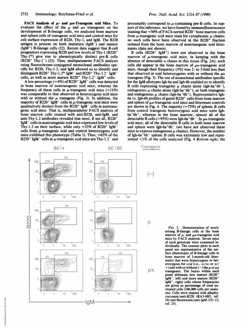
To ascertain whether IL and K chains were synthesized in g-and /LK-transgenic scid mice, lysates oftheir lymphoid tissuesalong with those of littermate controls were examined byWestern blot analysis. As shown in Fig. 2A, A& chains but not
TransgeneDonorStrains(M54 or 20Z-4)
K chains were present in the bone marrow, spleen, andthymus of tL-transgenic scid mice. No ,u chains were detectedin nontransgenic scid mice. Since the synthesis of transgene-encoded ,u chains in thymocytes is a property of Ig-transgenicmice (14, 15), the detection of a novel-size (78-kDa) 1L chainin thymus and bone marrow of A-transgenic scid mice indi-cates that the 78-kDa ,u chain corresponds to the transgeneproduct. With respect to K chains, both transgenic andendogenous K chains were present in the bone marrow ofAuK-transgenic heterozygous scid mice, whereas transgenic Kchains alone (together with u chains) were seen in the bonemarrow ofUK-transgenic scid mice (Fig. 2B). The K transgeneproduct can be distinguished from most other K light chainsdue to its slower migration (21). Relatively high levels ofendogenous K chains were present in the spleen of /LK-transgenic scid heterozygotes (Fig. 2B). In contrast, thespleen of /.LK-transgenic scid mice contained relatively lowlevels of transgenic K chains and only about 20% of such miceproduced detectable levels of serum Ig (0.2-0.5 mg/ml); themajority lacked detectable serum Ig (<0.1 mg/ml) (serolog-ical data not shown).
A q xi lG\ I\'r
_ n::..Constructed
Stock
A) Tg/+,IgI-la+llglla+ X +1+, lgb +1191- lb +
B) Tg/+, Igl- la +/lgl-lb + X +, lb +glg lb +
C) Tg/+, glg lb +1gi- lb + X +1+, Igl-la scid/lgl-la sacid
D) Tg/+, Ig- Ib +.1gi I ascid X +1+, g- la scid/lgl-1a scid
1) Tg/+, Igl-la scidigI-la sad
2) +1+, IgI-Ia scd/lgl-1 said
3) Tg/+, gI-1a sci&igl- b +
4) +/+, Igl la scid/IgIl b +
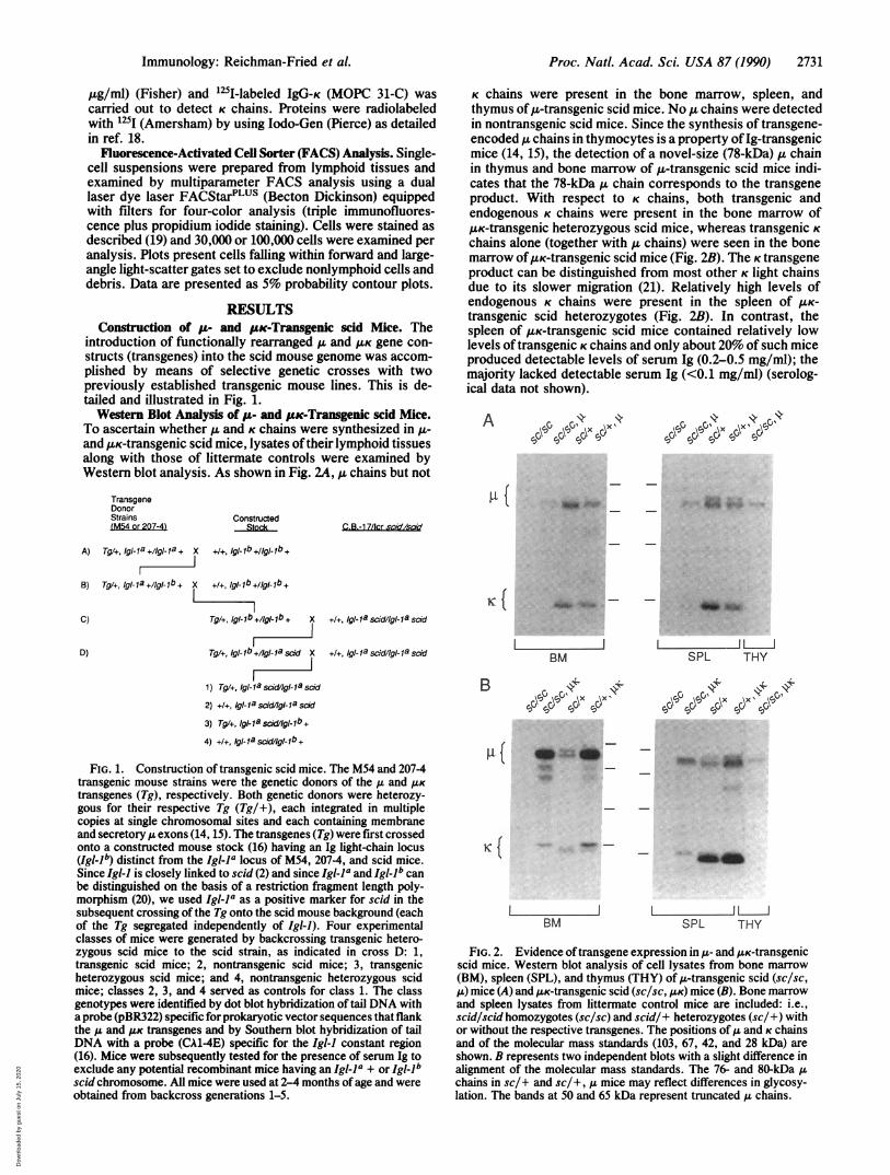
FIG. 1. Construction of transgenic scid mice. The M54 and 207-4transgenic mouse strains were the genetic donors of the A and ILKtransgenes (Tg), respectively. Both genetic donors were heterozy-gous for their respective Tg (Tg/+), each integrated in multiplecopies at single chromosomal sites and each containing membraneand secretory A exons (14, 15). The transgenes (Tg) were first crossedonto a constructed mouse stock (16) having an Ig light-chain locus(Igl-1b) distinct from the IglI-a locus of M54, 207-4, and scid mice.Since Igl-J is closely linked to scid (2) and since Igl-la and Igllb canbe distinguished on the basis of a restriction fragment length poly-morphism (20), we used Igl-la as a positive marker for scid in thesubsequent crossing of the Tg onto the scid mouse background (eachof the Tg segregated independently of Igi-l). Four experimentalclasses of mice were generated by backcrossing transgenic hetero-zygous scid mice to the scid strain, as indicated in cross D: 1,transgenic scid mice; 2, nontransgenic scid mice; 3, transgenicheterozygous scid mice; and 4, nontransgenic heterozygous scidmice; classes 2, 3, and 4 served as controls for class 1. The classgenotypes were identified by dot blot hybridization of tail DNA witha probe (pBR322) specific for prokaryotic vector sequences that flankthe ,u and AK transgenes and by Southern blot hybridization of tailDNA with a probe (CA1-4E) specific for the Igl-) constant region(16). Mice were subsequently tested for the presence of serum Ig toexclude any potential recombinant mice having an Igl-la + or Igl-lbscid chromosome. All mice were used at 2-4 months of age and wereobtained from backcross generations 1-5.
I IBM
B 044
\SPc~c ox,
I 1ISPL THY
4,0cl3Osd
4~ 4
q7CO\X~c~o11
R1( Sm4W
*_
BMBM
I 1 1iSPL THY
FIG. 2. Evidence oftransgene expression in A- and /LK-transgenicscid mice. Western blot analysis of cell lysates from bone marrow(BM), spleen (SPL), and thymus (THY) of u-transgenic scid (sc/sc,,u) mice (A) and AsK-transgenic scid (sc/sc, ,UK) mice (B). Bone marrowand spleen lysates from littermate control mice are included: i.e.,scid/scid homozygotes (sc/sc) and scid/+ heterozygotes (sc/+) withor without the respective transgenes. The positions ofA and K chainsand of the molecular mass standards (103, 67, 42, and 28 kDa) areshown. B represents two independent blots with a slight difference inalignment of the molecular mass standards. The 76- and 80-kDa Achains in sc/+ and sc/+, ,A mice may reflect differences in glycosy-lation. The bands at 50 and 65 kDa represent truncated ,u chains.
Immunology: Reichman-Fried et al.
C.B.-1 7/lcr cdsd
K{
A. Ammok
Dow
nloa
ded
by g
uest
on
July
15,
202
0
2732 Immunology: Reichman-Fried et al.
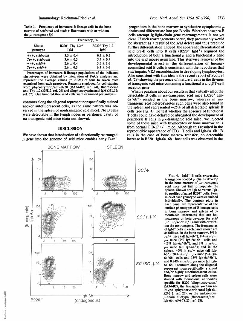
FACS Analysis of p- and FKc-Transgenic scid Mice. Toevaluate the effect of the pu and /LK transgenes on thedevelopment of B-lineage cells, we analyzed bone marrowand spleen cells of transgenic scid mice and control mice forcell surface expression of B220, Thy-i, and IgM. The B220antigen is present on both immature (IgM-) and mature(IgM+) B-lineage cells (22). Recent data suggest that B-cellprogenitors expressing B220 and low levels of Thy-1 (B220+Thy-10l) give rise to phenotypically distinct pre-B cells(B220 Thy-l-) (23). Thus, multiparameter FACS analysisusing fluorochrome-conjugated monoclonal antibodies spe-cific for B220, Thy-1.2, and IgM allowed us to identify anddistinguish B220 Thy-1.2lo IgM- and B220+ Thy-1.2- IgM-cells, as well as more mature B220' Thy-1.2- IgM' cells.A low percentage (-4%) ofB220' IgM- cells was detected
in bone marrow of nontransgenic scid mice, whereas thefrequency of these cells in pu-transgenic scid mice (fi1%)was comparable to that observed in heterozygous scid micewith or without the transgene (Fig. 3). In addition, themajority of B220 IgM- cells in pu-transgenic scid mice werequalitatively distinct from the B220 IgM- cells in nontrans-genic scid mice. That is, multiparameter FACS analysis ofbone marrow cells stained with anti-B220, anti-IgM, andanti-Thy-1.2 antibodies revealed that most, if not all, B220'IgM- cells in nontransgenic scid mice expressed low levels ofThy-1.2 on their surface, while only -35% of B220' IgM-cells from p.-transgenic scid and control heterozygous scidmice exhibited this phenotype (Table 1). Thus, ==65% of theB220 IgM- cells in tL-transgenic scid mice are Thy-1.2- and
SC/+
1 1 1 0 100,.
.1 1 10 100
.1 1
1 00
10
1 00
10
100
10
.1
10 100
SC /SC
.....................O
I...........
-..........
.1 i.10
1gM
Proc. Natl. Acad. Sci. USA 87 (1990)
presumably correspond to A.-containing pre-B cells. In sup-port ofthis inference, we have found by immunofluorescencestaining that =50% ofFACS-sorted B220' bone marrow cellsfrom A-transgenic scid mice stain for cytoplasmic pu chains;no such cells have been observed in the B220' fractionisolated from the bone marrow of nontransgenic scid litter-mates (data not shown).B cells (B220' IgM+) were not observed in the bone
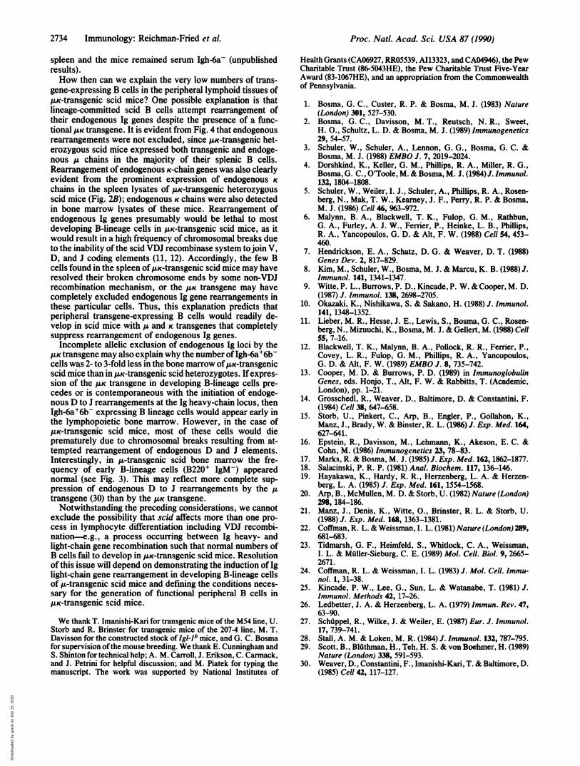
marrow of p-transgenic scid mice, in keeping with theabsence of detectable K chains in this tissue (Fig. 2A); suchcells did appear in the bone marrow of p.K-transgenic scidmice, though their frequency (3%) was 2- to 3-fold less thanthat observed in scid heterozygotes with or without the AKtransgene (Fig. 3). The use of monoclonal antibodies specificfor the IgM allotypes Igh-6a and Igh-6b enabled us to identifyB cells expressing transgenic chains alone (Igh-6a+6b-),endogenous chains alone (Igh-6a-6b+), or both transgenicand endogenous chains (Igh-6a+6b+). Representative Igh-6a vs. Igh-6b profiles of gated B220' cells from bone marrowand spleen of pK-transgenic scid mice and littermate controlsare shown in Fig. 4. The majority (=75%) of splenic B cellsfrom control transgenic heterozygous scid mice were Igh-6a+6b+, whereas in the bone marrow, almost all of thedetectable B cells (>95%) were Igh-6a+6b-. In p.K-transgenicscid mice, all of the detectable B cells in both bone marrowand spleen were Igh-6a+6b- (we have not observed thesemice to express endogenous chains). However, the numberof Igh-6a+6b- splenic B cells was extremely low and repre-sented <1% of the cells analyzed (Fig. 4 Bottom right; the
Transgene
5,I
(-)
100
...... .............
FIG. 3. Demonstration of newlyarising B-lineage cells in the bonemarrow of A- and A.K-transgenic scidmice by FACS analysis. Seven mice
!--- 13 of each genotype were examined in-100 dividually. The contour plots in each
panel are representative of the sur-face phenotypes of B-lineage cells inbone marrow of 3-month-old litter-mates that were homozygous or het-erozygous for scid (i.e., sc/sc or Sc!
+) and with or without (-) the orpAKKly transgenes. The boxes within each
panel delineate less mature (B220+IgM-, left) and more mature (B220+IgMI, right) cells whose frequenciesare given as percentage of total nu-cleated cells (100,000 cells per analy-
100 sis). Cells were stained with allophy-cocyanin/anti-B220 (RA3-6B2; ref.24) and fluorescein/anti-IgM (331.12;ref. 25).
ooI
CMm
10 ........... ... v
;ax
...........................v...;.....
1 1 1 0
1 00
1 0
.1
..
_ ......... .-......
100
1 0
.1
.1
Dow
nloa
ded
by g
uest
on
July
15,
202
0
Proc. Natl. Acad. Sci. USA 87 (1990) 2733
Table 1. Frequency of immature B-lineage cells in the bonemarrow of scid/scid and scid/+ littermates with or withoutthe /L transgene (Tg)
Frequency, %
Mouse B220+ Thy-1.210 B220' Thy-1.2-genotype IgM- IgM-
+/+, scid/scid 3.3 ± 0.4 0.3 ± 0.2Tg/+, scid/scid 3.6 ± 0.3 5.7 ± 0.9+/+, scid/+ 2.6 ± 0.4 5.5 ± 1.6Tg/+, scid/+ 2.6 ± 0.3 6.3 ± 0.6Percentages of immature B-lineage populations of the indicated
phenotypes were obtained by integration of FACS analyses andrepresent the average values (± SEM) of four to seven miceexamined from each genotype. Reagents employed for cell stainingwere phycoerythrin/anti-B220 (RA3-6B2; ref. 24), fluorescein/anti-Thy-1.2 (30H12; ref. 26) and allophycocyanin/anti-IgM (331.12;ref. 25). One hundred thousand cells were examined per analysis.
contours along the diagonal represent nonspecifically stainedand/or autofluorescent cells, as the same pattern was ob-served in the spleen of nontransgenic scid mice). No B cellswere detectable in the lymph nodes or peritoneal cavity of,uK-transgenic scid mice (data not shown).
DISCUSSIONWe have shown that introduction ofa functionally rearranged,u gene into the genome of scid mice enables early B-cell
BONE MARROW SPLEEN
progenitors in the bone marrow to synthesize cytoplasmic ,uchains and differentiate into pre-B cells. Whether these pre-Bcells attempt Ig light-chain gene rearrangements is not yetclear. If such rearrangements occur, they presumably wouldbe aberrant as a result of the scid defect and thus precludefurther differentiation. Indeed, the apparent differentiation ofscid pre-B cells into B cells (B220+ IgM+) required theintroduction of both a functional ,u and a functional K geneinto the scid mouse germ line. This stepwise removal of thedevelopmental arrest in the differentiation of lineage-committed scid B cells is consistent with the hypothesis thatscid impairs VDJ recombination in developing lymphocytes.Also consistent with this idea is the recent report of Scott etal. (29) showing the presence of mature T cells in the thymusof transgenic scid mice containing a functional a and ,f T-cellreceptor gene.What is puzzling about our results is that virtually all ofthe
detectable B cells in ILK-transgenic scid mice (B220+ Igh-6a+6bV) resided in the bone marrow, whereas in UK-
transgenic scid heterozygotes such cells were also found inthe spleen and represented =25% of all detectable splenic Bcells (see Fig. 4). To test whether the absence of functionalT cells could have delayed or abrogated the development ofperipheral B cells in IK-transgenic scid mice, we injectedsome of these mice with thymocytes or bone marrow cellsfrom normal C.B-17+/+ mice. Although this resulted in thereproducible appearance of CD3+ T cells and Igh-6a-6b+ Bcells in the case of bone marrow transfer, no detectableincrease in B220+ Igh-6a+6b- host cells was observed in the
B220 +
.1 1 10 100
100
10
1
100
10
.1
100
10
1
.1 10 100
.1 10 100
-- Igh-6b-(endogenous)
SC/+FIG. 4. IgM+ B cells expressing
transgene-encoded ,u chains developin the bone marrow of AK-transgenicscid mice but fail to populate thespleen. Shown are Igh-6a versus Igh-6b profiles of gated B220+ cells. Fourmice ofeach genotype were examinedindividually. The contour plots ineach panel are representative of thesurface phenotypes of B-lineage cellsin bone marrow and spleen of 3-
SC 1, UKmozygous or heterozygous for scid(i.e., sc/sc or sc/+) and with or with-out the K transgene. The frequenciesofIgM+ cells in each panel shown areas follows: in the bone marrow, 8% insc/+ mice (all Igh-6b+), 8% in sc/+,/K mice (7% Igh-6a+6b- cells and<1% Igh-6a+6b+), and 3% in sc/sc,
AK mice (all Igh-6a+); and in thespleen, 40o in sc/+ mice (all Igh-6b+), 20% in sc/+, AK mice (5% Igh-6a+6b- cells and 15% Igh-6a+6b+),and 0.24% in sc/sc, AK mice (all Igh-
SC /SC, JIK 6a+6b-; contours along the diagonalrepresent nonspecifically stainedand/or highly autofluorescent cells).Bone marrow and spleen cells werestained with monoclonal antibodiesspecific for B220 (allophycocyanin/RA3-6B2), the transgene ,u-chain al-lotype (phycoerythrin/anti-Igh-6a,RS-3.1; ref. 27), or the endogenous,u-chain allotype (fluorescein/anti-Igh-6b, AF6-78.25; ref. 28).
100
10
.1
100
10
.1
.1 1 10 100
_eCI CD'D
U)Cn CY
__3
.1 1 10 100
'----N
Immunology: Reichman-Fried et al.
Dow
nloa
ded
by g
uest
on
July
15,
202
0
2734 Immunology: Reichman-Fried et al.
spleen and the mice remained serum Igh-6a- (unpublishedresults).How then can we explain the very low numbers of trans-
gene-expressing B cells in the peripheral lymphoid tissues of/LK-transgenic scid mice? One possible explanation is thatlineage-committed scid B cells attempt rearrangement oftheir endogenous Ig genes despite the presence of a func-tional lK transgene. It is evident from Fig. 4 that endogenousrearrangements were not excluded, since puK-transgenic het-erozygous scid mice expressed both transgenic and endoge-nous ,u chains in the majority of their splenic B cells.Rearrangement ofendogenous K-chain genes was also clearlyevident from the prominent expression of endogenous Kchains in the spleen lysates of ,K-transgenic heterozygousscid mice (Fig. 2B); endogenous K chains were also detectedin bone marrow lysates of these mice. Rearrangement ofendogenous Ig genes presumably would be lethal to mostdeveloping B-lineage cells in UK-transgenic scid mice, as itwould result in a high frequency of chromosomal breaks dueto the inability ofthe scid VDJ recombinase system tojoin V,D, and J coding elements (11, 12). Accordingly, the few Bcells found in the spleen of JUK-transgenic scid mice may haveresolved their broken chromosome ends by some'non-VDJrecombination mechanism, or the tLK transgene may havecompletely excluded endogenous Ig gene rearrangements inthese particular cells. Thus, this explanation predicts thatperipheral transgene-expressing B cells would readily de-velop in scid mice with ,u and K transgenes that completelysuppress rearrangement of endogenous Ig genes.Incomplete allelic exclusion of endogenous Ig loci by the
/.K transgene may also explain why the number ofIgh-6a+6b-cells was 2- to 3-fold less in the bone marrow of ,uK-transgenicscid mice than in ,uK-transgenic scid heterozygotes. Ifexpres-sion of the ,. transgene in developing B-lineage cells pre-cedes or is contemporaneous with the initiation of endoge-nous D to J rearrangements at the Ig heavy-chain locus, thenIgh-6a+6b- expressing B lineage cells would appear early inthe lymphopoietic bone marrow. However, in the case of,UK-transgenic scid mice, most of these cells would dieprematurely due to chromosomal breaks resulting from at-tempted rearrangement of endogenous D and J elements.Interestingly, in A-transgenic scid bone marrow the fre-quency of early B-lineage cells (B220+ IgM-) appearednormal (see Fig. 3). This may reflect more complete sup-pression of endogenous D to J rearrangements by the ,utransgene (30) than by the MK transgene.
Notwithstanding the preceding considerations, we cannotexclude the possibility that scid affects more than one pro-cess -in lymphocyte differentiation including VDJ recombi-nation-e.g., a process occurring between Ig heavy- andlight-chain gene recombination such that normal numbers ofB cells fail to develop in AK-transgenic scid mice. Resolutionof this issue will depend on demonstrating the induction of Iglight-chain gene rearrangement in developing B-lineage cellsof u-transgenic scid mice and defining the conditions neces-sary for the generation of functional peripheral B cells in/.K-transgenic' scid mice.
We thank T. Imanishi-Kari for transgenic mice of the M54 line, U.Storb and R. Brinster for transgenic mice of the 207-4 line, M. T.Davisson for the constructed stock of Igl-1b mice, and G. C. Bosmafor supervision of the mouse breeding. We thank E. Cunningham andS. Shinton for technical help; A. M. Carroll, J. Erikson, C. Carmack,and J. Petrini for helpful discussion; and M. Piatek for typing themanuscript. The work was supported by National Institutes of
Health Grants (CA06927, RR05539, A113323, and CA04946), the PewCharitable Trust (86-5043HE), the Pew Charitable Trust Five-YearAward (83-1067HE), and an appropriation from the Commonwealthof Pennsylvania.
1. Bosma, G. C., Custer, R. P. & Bosma, M. J. (1983) Nature(London) 301, 527-530.
2. Bosma, G. C., Davisson, M. T., Reutsch, N. R., Sweet,H. O., Schultz, L. D. & Bosma, M. J. (1989) Immunogenetics29, 54-57.
3. Schuler, W., Schuler, A., Lennon, G. G., Bosma, G. C. &Bosma, M. J. (1988) EMBO J. 7, 2019-2024.
4. Dorshkind, K., Keller, G. M., Phillips, R. A., Miller, R. G.,Bosma, G. C., O'Toole, M. & Bosma, M. J. (1984)J. Immunol.132, 1804-1808.
5. Schuler, W., Weiler, I. J., Schuler, A., Phillips, R. A., Rosen-berg, N., Mak, T. W., Kearney, J. F., Perry, R. P. & Bosma,M. J. (1986) Cell 46, %3-972.
6. Malynn, B. A., Blackwell, T. K., Fulop, G. M., Rathbun,G. A., Furley, A. J. W., Ferrier, P., Heinke, L. B., Phillips,R. A., Yancopoulos, G. D. & Alt, F. W. (1988) Cell 54, 453-460.
7. Hendrickson, E. A., Schatz, D. G. & Weaver, D. T. (1988)Genes Dev. 2, 817-829.
8. Kim, M., Schuler, W., Bosma, M. J. & Marcu, K. B. (1988) J.Immunol. 141, 1341-1347.
9. Witte, P. L., Burrows, P. D., Kincade, P. W. & Cooper, M. D.(1987) J. Immunol. 138, 2698-2705.
10. Okazaki, K., Nishikawa, S. & Sakano, H. (1988) J. Immunol.141, 1348-1352.
11. Lieber, M. R., Hesse, J. E., Lewis, S., Bosma, G. C., Rosen-berg, N., Mizuuchi, K., Bosma, M. J. & Gellert, M. (1988) Cell55, 7-16.
12. Blackwell, T. K., Malynn, B. A., Pollock, R. R., Ferrier, P.,Covey, L. R., Fulop, G. M., Phillips, R. A., Yancopoulos,G. D. & Alt, F. W. (1989) EMBO J. 8, 735-742.
13. Cooper, M. D. & Burrows, P. D. (1989) in ImmunoglobulinGenes, eds. Honjo, T., Alt, F. W. & Rabbitts, T. (Academic,London), pp. 1-21.
14. Grosschedl, R., Weaver, D., Baltimore, D. & Constantini, F.(1984) Cell 38, 647-658.
15. Storb, U., Pinkert, C., Arp, B., Engler, P., Gollahon, K.,Manz, J., Brady, W. & Binster, R. L. (1986) J. Exp. Med. 164,627-641.
16. Epstein, R., Davisson, M., Lehmann, K., Akeson, E. C. &Cohn, M. (1986) Immunogenetics 23, 78-83.
17. Marks, R. & Bosma, M. J. (1985) J. Exp. Med. 162, 1862-1877.18. Salacinski, P. R. P. (1981) Anal. Biochem. 117, 136-146.19. Hayakawa, K., Hardy, R. R., Herzenberg, L. A. & Herzen-
berg, L. A. (1985) J. Exp. Med. 161, 1554-1568.20. Arp, B., McMullen, M. D. & Storb, U. (1982) Nature (London)
298, 184-186.21. Manz, J., Denis, K., Witte, O., Brinster, R. L. & Storb, U.
(1988) J. Exp. Med. 168, 1363-1381.22. Coffman, R. L. & Weissman, I. L. (1981) Nature (London) 289,
681-683.23. Tidmarsh, G. F., Heimfeld, S., Whitlock, C. A., Weissman,
I. L. & Muller-Sieburg, C. E. (1989) Mol. Cell. Biol. 9, 2665-2671.
24. Coffman, R. L. & Weissman, I. L. (1983) J. Mol. Cell. Immu-nol. 1, 31-38.
25. Kincade, P. W., Lee, G., Sun, L. & Watanabe, T. (1981) J.Immunol. Methods 42, 17-26.
26. Ledbetter, J. A. & Herzenberg, L. A. (1979) Immun. Rev. 47,63-90.
27. Schuppel, R., Wilke, J. & Weiler, E. (1987) Eur. J. Immunol.17, 739-741.
28. Stall, A. M. & Loken, M. R. (1984) J. Immunol. 132, 787-795.29. Scott, B., Bluthman, H., Teh, H. S. & von Boehmer, H. (1989)
Nature (London) 338, 591-593.30. Weaver, D., Constantini, F., Imanishi-Kari, T. & Baltimore, D.
(1985) Cell 42, 117-127.
Proc. Natl. Acad. Sci. USA 87 (1990)
Dow
nloa
ded
by g
uest
on
July
15,
202
0