Embed Size (px)
Citation preview
October 1, 2015 ◆ Volume 92, Number 7 www.aafp.org/afp American Family Physician 601
Patients will experience a wide range of skin growths and changes over their lifetime. Family physicians should be able to distinguish potentially malignant from benign skin tumors. Most lesions can be diagnosed on the basis of history and clinical examination. Lesions that are suspicious for malignancy, those with changing characteristics, symptom-atic lesions, and those that cause cosmetic problems may warrant medical therapy, a simple office procedure (e.g., excision, cryosurgery, laser ablation), or referral. Acrochordons are extremely common, small, and typically peduncu-lated benign neoplasms. Simple scissor or shave excision, electrodesiccation, or cryosurgery can be used for treatment. Sebaceous hyperplasia presents as asymptomatic, discrete, soft, pale yellow, shiny bumps on the forehead or cheeks, or near hair follicles. Except for cosmesis, they have no clinical significance. Lipomas are soft, flesh-colored nodules that are easily moveable under the overlying skin. Keratoacanthomas are rapidly growing, squamoproliferative benign tumors that resemble squamous cell carcinomas. Early simple excision is recommended. Pyogenic granuloma is a rap-idly growing nodule that bleeds easily. Treatment includes laser ablation or shave excision with electrodesiccation of the base. Dermatofibromas are an idiopathic benign proliferation of fibroblasts. No treatment is required unless there is a change in size or color, bleeding, or irritation from trauma. Epidermal inclusion cysts can be treated by simple excision with removal of the cyst and cyst wall. Seborrheic keratoses and cherry angiomas generally do not require treatment. (Am Fam Physician. 2015;92(7):601-607. Copyright © 2015 American Academy of Family Physicians.)
Diagnosing Common Benign Skin TumorsJAMES C. HIGGINS, CAPT, MC, USN, RET; MICHAEL H. MAHER, CAPT, MC, USN, RET; and MARK S. DOUGLAS, LCDR, MC, USN, Naval Hospital Jacksonville, Jacksonville, Florida
Skin problems are commonly encountered in primary care. One retrospective chart review of der-matology referrals at a university
general medicine clinic found that approxi-mately one-third of patients were referred during their initial visit to their primary care physician.1 However, family physicians can effectively treat most skin disorders.2 A review of diagnoses made by primary care physicians found they were correct 70% of the time (compared with 93% for dermatologists).3 Another multisite pro-spective cohort study found overall agree-ment in diagnoses and treatment between family physicians and dermatologists, with a concordance of 72% and 80%, respectively.2
Clinicians must take special precautions in evaluating skin tumors and screening for skin cancer. The use of dermoscopy to improve diagnosis has been addressed in a previous article in American Family Physi-cian.4 However, the preferred method of diagnosing skin cancer is physical examina-tion. This article will review some common benign skin tumors that are amenable to office procedures, as well as those that may require referral (Table 1).
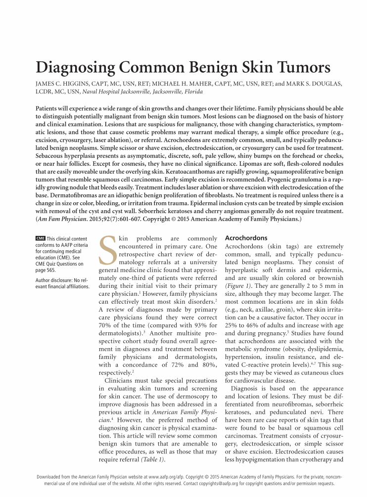
AcrochordonsAcrochordons (skin tags) are extremely common, small, and typically peduncu-lated benign neoplasms. They consist of hyperplastic soft dermis and epidermis, and are usually skin colored or brownish (Figure 1). They are generally 2 to 5 mm in size, although they may become larger. The most common locations are in skin folds (e.g., neck, axillae, groin), where skin irrita-tion can be a causative factor. They occur in 25% to 46% of adults and increase with age and during pregnancy.5 Studies have found that acrochordons are associated with the metabolic syndrome (obesity, dyslipidemia, hypertension, insulin resistance, and ele-vated C-reactive protein levels).6,7 This sug-gests they may be viewed as cutaneous clues for cardiovascular disease.
Diagnosis is based on the appearance and location of lesions. They must be dif-ferentiated from neurofibromas, seborrheic keratoses, and pedunculated nevi. There have been rare case reports of skin tags that were found to be basal or squamous cell carcinomas. Treatment consists of cryosur-gery, electrodesiccation, or simple scissor or shave excision. Electrodesiccation causes less hypopigmentation than cryotherapy and
CME This clinical content conforms to AAFP criteria for continuing medical education (CME). See CME Quiz Questions on page 565.
Author disclosure: No rel-evant financial affiliations.
Downloaded from the American Family Physician website at www.aafp.org/afp. Copyright © 2015 American Academy of Family Physicians. For the private, noncom-mercial use of one individual user of the website. All other rights reserved. Contact [email protected] for copyright questions and/or permission requests.
Benign Skin Tumors
602 American Family Physician www.aafp.org/afp Volume 92, Number 7 ◆ October 1, 2015
is the preferred treatment in nonwhite patients. An ear speculum placed over a small lesion may be helpful in directing the freeze pattern during cryosurgery.
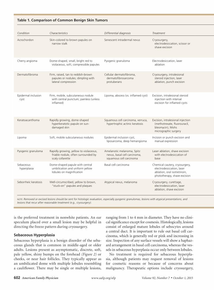
Sebaceous HyperplasiaSebaceous hyperplasia is a benign disorder of the seba-ceous glands that is common in middle-aged or older adults. Lesions present as asymptomatic, discrete, soft, pale yellow, shiny bumps on the forehead (Figure 2) or cheeks, or near hair follicles. They typically appear as an umbilicated dome with multiple lobules resembling a cauliflower. There may be single or multiple lesions,
ranging from 1 to 4 mm in diameter. They have no clini-cal significance except for cosmesis. Histologically, lesions consist of enlarged mature lobules of sebocytes around a central duct. It is important to rule out basal cell car-cinoma, which is generally red or pink and increasing in size. Inspection of any surface vessels will show a haphaz-ard arrangement in basal cell carcinoma, whereas the ves-sels in sebaceous hyperplasia occur only between lobules.
No treatment is required for sebaceous hyperpla-sia, although patients may request removal of lesions for cosmetic reasons or because of concerns about malignancy. Therapeutic options include cryosurgery,
Table 1. Comparison of Common Benign Skin Tumors
Condition Characteristics Differential diagnosis Treatment CommentsPrecautions and referral criteria
Acrochordon Skin-colored to brown papules on narrow stalk
Senescent intradermal nevus Cryosurgery, electrodesiccation, scissor or shave excision
Do not send multiple specimens in same jar
Cryosurgery should be performed with caution in persons with darker skin; refer patients with eyelid involvement
Cherry angioma Dome-shaped, small, bright red to violaceous, soft, compressible papules
Pyogenic granuloma Electrodesiccation, laser ablation
Numerous lesions (hundreds) and early onset can occur in Fabry disease
Genetic evaluation for Fabry disease in patients with multiple lesions
Dermatofibroma Firm, raised, tan to reddish-brown papules or nodules; dimpling with lateral compression
Cellular dermatofibroma, dermatofibrosarcoma protuberans
Cryosurgery, intralesional steroid injection, laser ablation, punch excision
Abrupt appearance of multiple lesions may occur in persons with human immunodeficiency virus infection or systemic lupus erythematosus
Refer patients with cellular variant and dermatofibrosarcoma protuberans (deep invasion and metastases)
Epidermal inclusion cyst
Firm, mobile, subcutaneous nodule with central punctum; painless (unless inflamed)
Lipoma, abscess (vs. inflamed cyst) Excision, intralesional steroid injection with interval excision for inflamed cysts
Presence of punctum helps differentiate cysts from lipomas; history helps differentiate between inflamed cyst and abscess (acute)
Inflamed cysts and those that have undergone previous incision and drainage can be more difficult to excise; refer patients with facial cysts
Keratoacanthoma Rapidly growing, dome-shaped hyperkeratotic papule on sun-damaged skin
Squamous cell carcinoma, verruca, hypertrophic actinic keratosis
Excision, intralesional injection (methotrexate, fluorouracil, bleomycin), Mohs micrographic surgery
Cannot be histologically differentiated from squamous cell carcinoma
Refer patients with recurrence after complete excision
Lipoma Soft, mobile subcutaneous nodules Epidermal inclusion cyst, liposarcoma, deep hemangioma
Incision or punch excision and manual expression
Ultrasonography can help differentiate lipomas from other deep neoplasms
Use caution with facial lipomas and recurrent lesions after excision
Pyogenic granuloma Rapidly growing, yellow to violaceous, friable nodule, often surrounded by scaly collarette
Amelanotic melanoma, Spitz nevus, basal cell carcinoma, squamous cell carcinoma
Laser ablation, shave excision with electrodesiccation of base
Send for histologic evaluation to rule out melanoma
Refer patients with recurrent lesions or facial lesions
Sebaceous hyperplasia
Dome-shaped papule with central umbilication and uniform yellow lobules on magnification
Basal cell carcinoma Chemical cautery, cryosurgery, electrodesiccation, laser ablation, oral isotretinoin, phototherapy, shave excision
Thin shave biopsy can rule out basal cell carcinoma
Basal cell carcinoma is generally red or pink and increases in size
Seborrheic keratosis Well-circumscribed, yellow to brown, “stuck-on” papules and plaques
Atypical nevus, melanoma Cryosurgery, curettage, electrodesiccation, laser ablation, shave excision
Consider malignancy workup for abrupt appearance of multiple lesions
Cryosurgery should be performed with caution in persons with darker skin
NOTE: Removed or excised lesions should be sent for histologic evaluation, especially pyogenic granulomas, lesions with atypical presentations, and lesions that recur after reasonable treatment (e.g., cryosurgery).
October 1, 2015 ◆ Volume 92, Number 7 www.aafp.org/afp American Family Physician 603
phototherapy, shave excision, laser ablation, electrodes-iccation with curettage, chemical cautery, or oral isotret-inoin for widespread lesions.8
LipomasLipomas are slow-growing, benign mesenchymal tumors enclosed by a thin fibrous capsule. They closely resemble normal fat and are the most common type of soft tis-sue tumor. They are usually subcutaneous but may occur in any organ because they are mesenchymal. They are generally asymptomatic but may become irritated with trauma or produce local obstructive symptoms in the
airway or gastrointestinal tract. Their prevalence is 1%.9
Lipomas must be clinically differentiated from other tumors. The primary differential diagnosis in a subcu-taneous mass is a sebaceous cyst or abscess. Lipomas are soft, flesh-colored nodules that are easily moveable under the overlying skin. Sebaceous cysts are generally identifiable by a central punctum, and abscesses can be identified by the presence of warmth, redness, and pain. Ultrasonography is increasingly used to aid in the diagnosis of lipomas. High-frequency ultrasonogra-phy (greater than 20 MHz) can provide high-resolution images of subcutaneous tumors and surrounding structures.10 The differential diagnosis of lipomas also includes liposarcomas; risk factors for malignancy are
Table 1. Comparison of Common Benign Skin Tumors
Condition Characteristics Differential diagnosis Treatment CommentsPrecautions and referral criteria
Acrochordon Skin-colored to brown papules on narrow stalk
Senescent intradermal nevus Cryosurgery, electrodesiccation, scissor or shave excision
Do not send multiple specimens in same jar
Cryosurgery should be performed with caution in persons with darker skin; refer patients with eyelid involvement
Cherry angioma Dome-shaped, small, bright red to violaceous, soft, compressible papules
Pyogenic granuloma Electrodesiccation, laser ablation
Numerous lesions (hundreds) and early onset can occur in Fabry disease
Genetic evaluation for Fabry disease in patients with multiple lesions
Dermatofibroma Firm, raised, tan to reddish-brown papules or nodules; dimpling with lateral compression
Cellular dermatofibroma, dermatofibrosarcoma protuberans
Cryosurgery, intralesional steroid injection, laser ablation, punch excision
Abrupt appearance of multiple lesions may occur in persons with human immunodeficiency virus infection or systemic lupus erythematosus
Refer patients with cellular variant and dermatofibrosarcoma protuberans (deep invasion and metastases)
Epidermal inclusion cyst
Firm, mobile, subcutaneous nodule with central punctum; painless (unless inflamed)
Lipoma, abscess (vs. inflamed cyst) Excision, intralesional steroid injection with interval excision for inflamed cysts
Presence of punctum helps differentiate cysts from lipomas; history helps differentiate between inflamed cyst and abscess (acute)
Inflamed cysts and those that have undergone previous incision and drainage can be more difficult to excise; refer patients with facial cysts
Keratoacanthoma Rapidly growing, dome-shaped hyperkeratotic papule on sun-damaged skin
Squamous cell carcinoma, verruca, hypertrophic actinic keratosis
Excision, intralesional injection (methotrexate, fluorouracil, bleomycin), Mohs micrographic surgery
Cannot be histologically differentiated from squamous cell carcinoma
Refer patients with recurrence after complete excision
Lipoma Soft, mobile subcutaneous nodules Epidermal inclusion cyst, liposarcoma, deep hemangioma
Incision or punch excision and manual expression
Ultrasonography can help differentiate lipomas from other deep neoplasms
Use caution with facial lipomas and recurrent lesions after excision
Pyogenic granuloma Rapidly growing, yellow to violaceous, friable nodule, often surrounded by scaly collarette
Amelanotic melanoma, Spitz nevus, basal cell carcinoma, squamous cell carcinoma
Laser ablation, shave excision with electrodesiccation of base
Send for histologic evaluation to rule out melanoma
Refer patients with recurrent lesions or facial lesions
Sebaceous hyperplasia
Dome-shaped papule with central umbilication and uniform yellow lobules on magnification
Basal cell carcinoma Chemical cautery, cryosurgery, electrodesiccation, laser ablation, oral isotretinoin, phototherapy, shave excision
Thin shave biopsy can rule out basal cell carcinoma
Basal cell carcinoma is generally red or pink and increases in size
Seborrheic keratosis Well-circumscribed, yellow to brown, “stuck-on” papules and plaques
Atypical nevus, melanoma Cryosurgery, curettage, electrodesiccation, laser ablation, shave excision
Consider malignancy workup for abrupt appearance of multiple lesions
Cryosurgery should be performed with caution in persons with darker skin
NOTE: Removed or excised lesions should be sent for histologic evaluation, especially pyogenic granulomas, lesions with atypical presentations, and lesions that recur after reasonable treatment (e.g., cryosurgery).
Figure 1. Acrochordons (skin tags) in a patient with meta-bolic syndrome.
Figure 2. Sebaceous hyperplasia on the forehead with the typical umbilicated, lobulated appearance without hap-hazard blood vessels.
Benign Skin Tumors
604 American Family Physician www.aafp.org/afp Volume 92, Number 7 ◆ October 1, 2015
size greater than 10 cm, older age, rapid lesion growth, location on the thigh, and invasion into deeper tissue, such as nerve or bone, leading to a firm or fixed feeling on examination. Lesions concerning for malignancy should be imaged with computed tomography or con-trast magnetic resonance imaging.11
Patients commonly present with cosmetic concerns or symptoms related to compression of surrounding tissue. A single incision or punch excision (for smaller lesions) will generally allow manual expression of the lipoma without difficulty when standard excision is not required.12
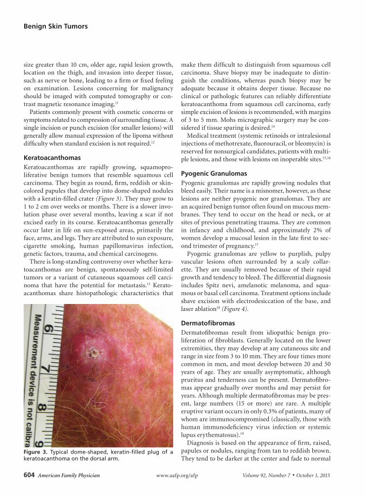
KeratoacanthomasKeratoacanthomas are rapidly growing, squamopro-liferative benign tumors that resemble squamous cell carcinoma. They begin as round, firm, reddish or skin-colored papules that develop into dome-shaped nodules with a keratin-filled crater (Figure 3). They may grow to 1 to 2 cm over weeks or months. There is a slower invo-lution phase over several months, leaving a scar if not excised early in its course. Keratoacanthomas generally occur later in life on sun-exposed areas, primarily the face, arms, and legs. They are attributed to sun exposure, cigarette smoking, human papillomavirus infection, genetic factors, trauma, and chemical carcinogens.
There is long-standing controversy over whether kera-toacanthomas are benign, spontaneously self-limited tumors or a variant of cutaneous squamous cell carci-noma that have the potential for metastasis.13 Kerato-acanthomas share histopathologic characteristics that
make them difficult to distinguish from squamous cell carcinoma. Shave biopsy may be inadequate to distin-guish the conditions, whereas punch biopsy may be adequate because it obtains deeper tissue. Because no clinical or pathologic features can reliably differentiate keratoacanthoma from squamous cell carcinoma, early simple excision of lesions is recommended, with margins of 3 to 5 mm. Mohs micrographic surgery may be con-sidered if tissue sparing is desired.14
Medical treatment (systemic retinoids or intralesional injections of methotrexate, fluorouracil, or bleomycin) is reserved for nonsurgical candidates, patients with multi-ple lesions, and those with lesions on inoperable sites.15,16
Pyogenic GranulomasPyogenic granulomas are rapidly growing nodules that bleed easily. Their name is a misnomer, however, as these lesions are neither pyogenic nor granulomas. They are an acquired benign tumor often found on mucous mem-branes. They tend to occur on the head or neck, or at sites of previous penetrating trauma. They are common in infancy and childhood, and approximately 2% of women develop a mucosal lesion in the late first to sec-ond trimester of pregnancy.17
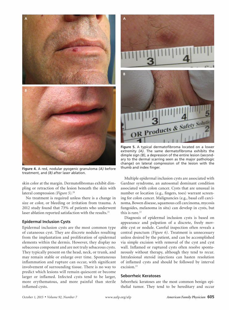
Pyogenic granulomas are yellow to purplish, pulpy vascular lesions often surrounded by a scaly collar-ette. They are usually removed because of their rapid growth and tendency to bleed. The differential diagnosis includes Spitz nevi, amelanotic melanoma, and squa-mous or basal cell carcinoma. Treatment options include shave excision with electrodesiccation of the base, and laser ablation18 (Figure 4).
DermatofibromasDermatofibromas result from idiopathic benign pro-liferation of fibroblasts. Generally located on the lower extremities, they may develop at any cutaneous site and range in size from 3 to 10 mm. They are four times more common in men, and most develop between 20 and 50 years of age. They are usually asymptomatic, although pruritus and tenderness can be present. Dermatofibro-mas appear gradually over months and may persist for years. Although multiple dermatofibromas may be pres-ent, large numbers (15 or more) are rare. A multiple eruptive variant occurs in only 0.3% of patients, many of whom are immunocompromised (classically, those with human immunodeficiency virus infection or systemic lupus erythematosus).19
Diagnosis is based on the appearance of firm, raised, papules or nodules, ranging from tan to reddish brown. They tend to be darker at the center and fade to normal
Figure 3. Typical dome-shaped, keratin-filled plug of a keratoacanthoma on the dorsal arm.
October 1, 2015 ◆ Volume 92, Number 7 www.aafp.org/afp American Family Physician 605
skin color at the margin. Dermatofibromas exhibit dim-pling or retraction of the lesion beneath the skin with lateral compression (Figure 5).20
No treatment is required unless there is a change in size or color, or bleeding or irritation from trauma. A 2012 study found that 73% of patients who underwent laser ablation reported satisfaction with the results.21
Epidermal Inclusion CystsEpidermal inclusion cysts are the most common type of cutaneous cyst. They are discrete nodules resulting from the implantation and proliferation of epidermal elements within the dermis. However, they display no sebaceous component and are not truly sebaceous cysts. They typically present on the head, neck, or trunk, and may remain stable or enlarge over time. Spontaneous inflammation and rupture can occur, with significant involvement of surrounding tissue. There is no way to predict which lesions will remain quiescent or become larger or inflamed. Infected cysts tend to be larger, more erythematous, and more painful than sterile inflamed cysts.
Multiple epidermal inclusion cysts are associated with Gardner syndrome, an autosomal dominant condition associated with colon cancer. Cysts that are unusual in number or location (e.g., fingers, toes) warrant screen-ing for colon cancer. Malignancies (e.g., basal cell carci-noma, Bowen disease, squamous cell carcinoma, mycosis fungoides, melanoma in situ) can develop in cysts, but this is rare.22
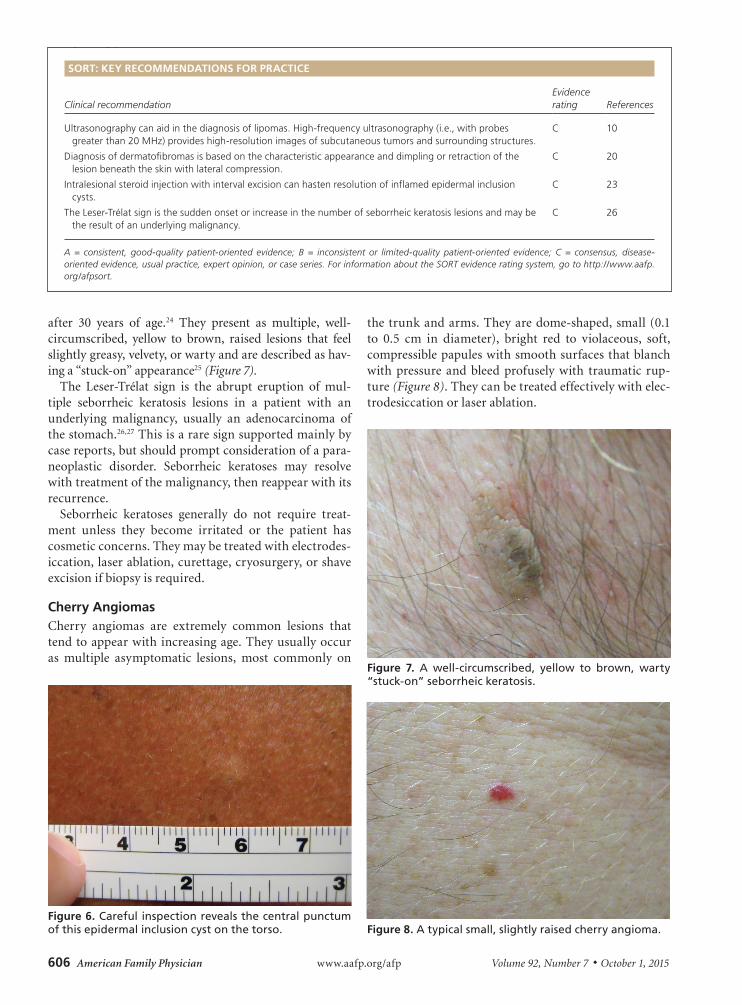
Diagnosis of epidermal inclusion cysts is based on appearance and palpation of a discrete, freely mov-able cyst or nodule. Careful inspection often reveals a central punctum (Figure 6). Treatment is unnecessary unless desired by the patient, and can be accomplished via simple excision with removal of the cyst and cyst wall. Inflamed or ruptured cysts often resolve sponta-neously without therapy, although they tend to recur. Intralesional steroid injections can hasten resolution of inflamed cysts and should be followed by interval excision.23
Seborrheic KeratosesSeborrheic keratoses are the most common benign epi-thelial tumor. They tend to be hereditary and occur
Figure 4. A red, nodular pyogenic granuloma (A) before treatment, and (B) after laser ablation.
A
B
Figure 5. A typical dermatofibroma located on a lower extremity (A). The same dermatofibroma exhibits the dimple sign (B), a depression of the entire lesion (second-ary to the dermal scarring seen as the major pathologic change) on lateral compression of the lesion with the thumb and index finger.
A
B
Benign Skin Tumors
606 American Family Physician www.aafp.org/afp Volume 92, Number 7 ◆ October 1, 2015
after 30 years of age.24 They present as multiple, well-circumscribed, yellow to brown, raised lesions that feel slightly greasy, velvety, or warty and are described as hav-ing a “stuck-on” appearance25 (Figure 7).
The Leser-Trélat sign is the abrupt eruption of mul-tiple seborrheic keratosis lesions in a patient with an underlying malignancy, usually an adenocarcinoma of the stomach.26,27 This is a rare sign supported mainly by case reports, but should prompt consideration of a para-neoplastic disorder. Seborrheic keratoses may resolve with treatment of the malignancy, then reappear with its recurrence.
Seborrheic keratoses generally do not require treat-ment unless they become irritated or the patient has cosmetic concerns. They may be treated with electrodes-iccation, laser ablation, curettage, cryosurgery, or shave excision if biopsy is required.
Cherry AngiomasCherry angiomas are extremely common lesions that tend to appear with increasing age. They usually occur as multiple asymptomatic lesions, most commonly on
the trunk and arms. They are dome-shaped, small (0.1 to 0.5 cm in diameter), bright red to violaceous, soft, compressible papules with smooth surfaces that blanch with pressure and bleed profusely with traumatic rup-ture (Figure 8). They can be treated effectively with elec-trodesiccation or laser ablation.
Figure 7. A well-circumscribed, yellow to brown, warty “stuck-on” seborrheic keratosis.
Figure 8. A typical small, slightly raised cherry angioma.
SORT: KEY RECOMMENDATIONS FOR PRACTICE
Clinical recommendationEvidence rating References
Ultrasonography can aid in the diagnosis of lipomas. High-frequency ultrasonography (i.e., with probes greater than 20 MHz) provides high-resolution images of subcutaneous tumors and surrounding structures.
C 10
Diagnosis of dermatofibromas is based on the characteristic appearance and dimpling or retraction of the lesion beneath the skin with lateral compression.
C 20
Intralesional steroid injection with interval excision can hasten resolution of inflamed epidermal inclusion cysts.
C 23
The Leser-Trélat sign is the sudden onset or increase in the number of seborrheic keratosis lesions and may be the result of an underlying malignancy.
C 26
A = consistent, good-quality patient-oriented evidence; B = inconsistent or limited-quality patient-oriented evidence; C = consensus, disease-oriented evidence, usual practice, expert opinion, or case series. For information about the SORT evidence rating system, go to http://www.aafp.org/afpsort.
Figure 6. Careful inspection reveals the central punctum of this epidermal inclusion cyst on the torso.

October 1, 2015 ◆ Volume 92, Number 7 www.aafp.org/afp American Family Physician 607
Data Sources: A series of PubMed searches were completed in Clinical Queries using the key terms acrochordon, sebaceous hyperplasia, lipoma, keratoacanthoma, pyogenic granuloma, dermatofibroma, epidermal inclusion cysts, seborrheic keratosis, and cherry angiomas. The follow-ing keywords were also searched in PubMed: benign skin lesions, benign skin tumors, skin diseases, diagnosis, and treatment. The search included reviews, meta-analyses, randomized controlled trials, and clinical trials. We also searched the National Guideline Clearinghouse, Cochrane Database of Systematic Reviews, UpToDate, and Pepid. Search date: April 30, 2014.
The opinions and assertions contained herein are the private views of the authors and are not to be construed as official or as reflecting the views of the U.S. Navy.
The Authors
JAMES C. HIGGINS, CAPT, MC, USN, RET, is a staff physician in the Family Medicine Residency Program at the Naval Hospital Jacksonville, Fla., and assistant clinical professor of family medicine at the Uniformed Services University of the Health Sciences, Bethesda, Md.
MICHAEL H. MAHER, CAPT, MC, USN, RET, is a staff physician at the Fam-ily Medicine Residency Program at the Naval Hospital Jacksonville, and an assistant clinical professor of family medicine at the Uniformed Services University of the Health Sciences.
MARK S. DOUGLAS, LCDR, MC, USN, is head of the Department of Derma-tology at Naval Hospital Jacksonville.
Address correspondence to James C. Higgins, CAPT, MC, USN, RET, Naval Hospital Jacksonville, 2080 Child St., Jacksonville, FL 32214 (e-mail: [email protected]). Reprints are not available from the authors.
REFERENCES
1. Lowell BA, Froelich CW, Federman DG, Kirsner RS. Dermatology in pri-mary care: Prevalence and patient disposition. J Am Acad Dermatol. 2001;45(2):250-255.
2. Merenstein D, Meyers D, Krist A, et al. How well do family physicians manage skin lesions? J Fam Pract. 2007;56(1):40-45.
3. Federman DG, Concato J, Kirsner RS. Comparison of dermatolgic diag-noses by primary care practioners and dermatologists. Arch Fam Med. 1999;8(2):170-172.
4. Marghoob AA, Usatine RP, Jaimes N. Dermoscopy for the family physi-cian. Am Fam Physician. 2013;88(7):441-450.
5. Banik R, Lubach D. Skin tags: localization and frequencies according to sex and age. Dermatologica. 1987;174(4):180-183.
6. Tamega AA, Aranha AM, Guiotoku MM, Miot LD, Miot HA. Associa-tion between skin tags and insulin resistance [in Portuguese]. An Bras Dermatol. 2010;85(1):25-31.
7. Sari R, Akman A, Alpsoy E, Balci MK. The metabolic profile in patients with skin tags. Clin Exp Med. 2010;10(3):193-197.
8. Yu C, Shahsavari M, Stevens G, Liskanich R, Horowitz D. Isotreti-noin as monotherapy for sebaceous hyperplasia. J Drugs Dermatol. 2010;9(6):699-701.
9. Nickloes TA. Lipomas. http://emedicine.medscape.com/article/191233-overview. Accessed July 15, 2015.
10. Schmid-Wendtner MH, Burgdorf W. Ultrasound scanning in dermatol-ogy. Arch Dermatol. 2005;141(2):217-224.
11. Kransdorf MJ, Bancroft LW, Peterson JJ, Murphey MD, Foster WC, Temple HT. Imaging of fatty tumors: distinction of lipoma and well-differentiated liposarcoma. Radiology. 2002;224(1):99-104.
12. Luba MC, Bangs SA, Mohler AM, Stulberg DL. Common benign skin tumors. Am Fam Physician. 2003;67(4):729-738.
13. Karaa A, Khachemoune A. Keratoacanthoma: a tumor in search of a classification. Int J Dermatol. 2007;46(7):671-678.
14. Shriner DL, McCoy DK, Goldberg DJ, Wagner RF Jr. Mohs micrographic surgery. J Am Acad Dermatol. 1998;39(1):79-97.
15. Annest NM, VanBeek MJ, Arpey CJ, Whitaker DC. Intralesional metho-trexate treatment for keratoacanthoma tumors: a retrospective study and review of the literature. J Am Acad Dermatol. 2007;56(6):989-993.
16. Kirby JS, Miller CJ. Intralesional chemotherapy for nonmelanoma skin cancer: a practical review. J Am Acad Dermatol. 2010;63(4):689-702.
17. Kroumpouzos G, Cohen LM. Dermatoses of pregnancy. J Am Acad Der-matol. 2001;45(1):1-19.
18. Gilmore A, Kelsberg G, Safranek S. Clinical inquiries. What’s the best treatment for pyogenic granuloma? J Fam Pract. 2010;59(1):40-42.
19. Massone C, Parodi A, Virno G, Rebora A. Multiple eruptive dermatofi-bromas in patients with systemic lupus erythematous treated with pred-nisone. Int J Dermatol. 2002;41(5):279-281.
20. Fitzpatrick TB, Gilchrest BA. Dimple sign to differentiate benign from malignant pigmented cutaneous lesions. N Engl J Med. 1977; 296(26):1518.
21. Alonso-Castro L, Boixeda P, Segura-Palacios JM, de Daniel-Rodríguez C, Jiménez-Gómez N, Ballester-Martínez A. Dermatofibromas treated with pulsed dye laser: clinical and dermoscopic outcomes. J Cosmet Laser Ther. 2012;14(2):98-101.
22. Swygert KE, Parrish CA, Cashman RE, Lin R, Cockerell CJ. Melanoma in situ involving an epidermal inclusion (infundibular) cyst. Am J Dermato-pathol. 2007;29(6):564-565.
23. Zuber TJ. Minimal excision technique for epidermoid (sebaceous) cysts. Am Fam Physician. 2002;65(7):1409-1412, 1417-1418, 1420.
24. Kyriakis KP, Alexoudi I, Askoxylaki K, Vrani F, Kosma E. Epidemiologic aspects of seborrheic keratoses. Int J Dermatol. 2012;51(2):233-234.
25. Hafner C, Vogt T. Seborrheic keratosis. J Dtsch Dermatol Ges. 2008; 6(8):664-677.
26. Schwartz RA. Sign of Leser-Trélat. J Am Acad Dermatol. 1996; 35(1):88-95.
27. Husain Z, Ho JK, Hantash BM. Sign and pseudo-sign of Leser-Trélat: case reports and a review of the literature. J Drugs Dermatol. 2013;12(5):e79-e87.
Benign Skin Tumors