Embed Size (px)
Citation preview
Diagnostic & Clinical Care Guidelinesfor Primary Immunodeficiency Diseases
Third EdiTion

This book contains general medical information which cannot be applied safely to any individual case. Medical knowledge and practice can change rapidly. Therefore, this book should not be used as a substitute for professional medical advice.
Copyrights 2008, 2009, 2015 the immune deficiency Foundation
readers may redistribute this publication to other individuals for non-commercial use, provided the text, html codes, and this notice remain intact and unaltered in any way. The immune deficiency Foundation diagnostic and Clinical Care Guidelines for Primary immunodeficiency diseases may not be resold, reprinted or redistributed for compensation of any kind without prior written permission from the immune deficiency Foundation. if you have any questions about permission, please contact: immune deficiency Foundation, 110 West road, Suite 300, Towson, Md 21204, USA or by telephone at 800-296-4433.
Diagnostic & Clinical Care Guidelinesfor Primary Immunodeficiency Diseases
Arranged by:Dr. Mehdi Adeli, MD, FAAAAI, FAPSenior Consultant, Allergy and immunologyAssistant Professor Well Cornell Medical College-QatarAllergy and immunology Awareness Program (AiAP)Pediatrics department hamad Medical Corporationdoha, Qatar


Dr. Mehdi Adeli, MDFAAAAi, FACAAi, FAPAllergy and immunology Senior ConsultantAllergy and immunology Awareness Program (AiAP)hamad Medical Corporation (hMC)doha, QatarEmail: [email protected]
Primary immunodeficiency (Pi) is considered one of the most complicated fields of medicine. Many physicians find it hard to diagnose a patient with a Pi disease and are reluctant to treat patients suffering from it.
our goal at the Allergy and immunology Awareness Program is to make the diagnosis of Pis easier and to increase the awareness by answering the most frequently asked questions by physicians, patients and their families.
it is my pleasure to present this booklet which was written by pioneers in the field of clinical immunology including those at duke's University, under whom i had the honor to be trained. This book is intended for physicians, interns and medical students and it contains a wealth of useful information in simplified manner and we hope it will guide you towards having more understanding of Pi diseases. in this book we will talk about the symptoms, methods of diagnosis and the treatments.
We are pleased to thank the immune deficiency Foundation (idF) in the United States for allowing us to use and translate this book.
For more information, visit hamad Medical Corporation web site: www.hamad.qa
if you have any questions or suggestions please contact us:
[email protected] « [email protected]
introduction
Editors Word
The immune deficiency Foundation, in partnership with
expert immunologists, developed these diagnostic and
clinical care guidelines to enhance earlier diagnosis,
improve health outcomes and increase access to
specialized healthcare and optimal treatment for patients
with primary immunodeficiency diseases.
The immune deficiency Foundation is the national patient
organization dedicated to improving the diagnosis,
treatment and quality of life of persons with primary
immunodeficiency diseases through advocacy, education
and research.
Rebecca H. Buckley, MDduke University School of Medicine
Contents
introduction ............................................................................................................................................. 4
Selected Primary immunodeficiency diseases .......................................................................... 5
Antibody Production defects ........................................................................................................... 9
Cellular or Combined defects .........................................................................................................17
Phagocytic Cell immune defects ..................................................................................................24
Mendelian Susceptibility to Mycobacterial disease ..............................................................28
Complement defects .......................................................................................................................33
Genetic Counseling: General Considerations and Practical Aspects ...............................36
Glossary .................................................................................................................................................39
4 Diagnostic & Clinical Care Guidelines forPrimary Immunodeficiency Diseases
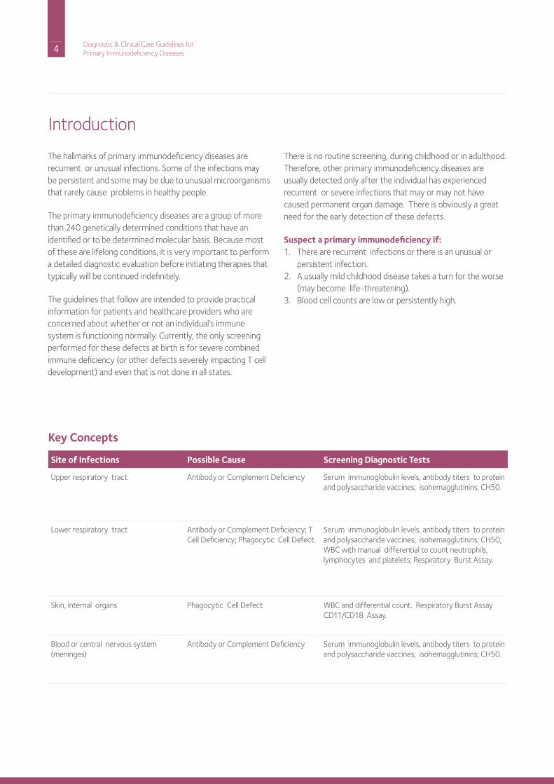
The hallmarks of primary immunodeficiency diseases are recurrent or unusual infections. Some of the infections may be persistent and some may be due to unusual microorganisms that rarely cause problems in healthy people.
The primary immunodeficiency diseases are a group of more than 240 genetically determined conditions that have an identified or to be determined molecular basis. Because most of these are lifelong conditions, it is very important to perform a detailed diagnostic evaluation before initiating therapies that typically will be continued indefinitely.
The guidelines that follow are intended to provide practical information for patients and healthcare providers who are concerned about whether or not an individual’s immune system is functioning normally. Currently, the only screening performed for these defects at birth is for severe combined immune deficiency (or other defects severely impacting T cell development) and even that is not done in all states.
introduction
There is no routine screening, during childhood or in adulthood. Therefore, other primary immunodeficiency diseases are usually detected only after the individual has experienced recurrent or severe infections that may or may not have caused permanent organ damage. There is obviously a great need for the early detection of these defects.
Suspect a primary immunodeficiency if:1. There are recurrent infections or there is an unusual or
persistent infection.2. A usually mild childhood disease takes a turn for the worse
(may become life-threatening).3. Blood cell counts are low or persistently high.
Key Concepts
Site of Infections Possible Cause Screening Diagnostic Tests
Upper respiratory tract Antibody or Complement deficiency Serum immunoglobulin levels, antibody titers to protein and polysaccharide vaccines; isohemagglutinins; Ch50.
Lower respiratory tract Antibody or Complement deficiency; T Cell deficiency; Phagocytic Cell defect.
Serum immunoglobulin levels, antibody titers to protein and polysaccharide vaccines; isohemagglutinins; Ch50; WBC with manual differential to count neutrophils, lymphocytes and platelets; respiratory Burst Assay.
Skin, internal organs Phagocytic Cell defect WBC and differential count. respiratory Burst Assay Cd11/Cd18 Assay.
Blood or central nervous system (meninges)
Antibody or Complement deficiency Serum immunoglobulin levels, antibody titers to protein and polysaccharide vaccines; isohemagglutinins; Ch50.
5 Diagnostic & Clinical Care Guidelines forPrimary Immunodeficiency Diseases
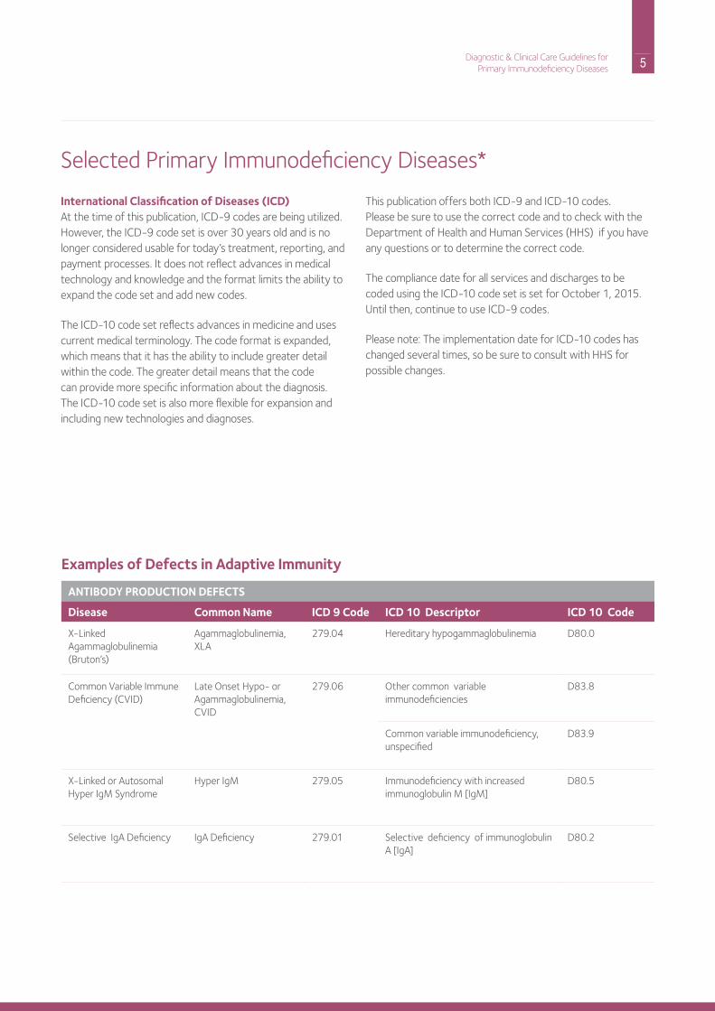
International Classification of Diseases (ICD)At the time of this publication, iCd-9 codes are being utilized. however, the iCd-9 code set is over 30 years old and is no longer considered usable for today’s treatment, reporting, and payment processes. it does not reflect advances in medical technology and knowledge and the format limits the ability to expand the code set and add new codes.
The iCd-10 code set reflects advances in medicine and uses current medical terminology. The code format is expanded, which means that it has the ability to include greater detail within the code. The greater detail means that the code can provide more specific information about the diagnosis. The iCd-10 code set is also more flexible for expansion and including new technologies and diagnoses.
Selected Primary immunodeficiency diseases*
This publication offers both iCd-9 and iCd-10 codes. Please be sure to use the correct code and to check with the department of health and human Services (hhS) if you have any questions or to determine the correct code.
The compliance date for all services and discharges to be coded using the iCd-10 code set is set for october 1, 2015. Until then, continue to use iCd-9 codes.
Please note: The implementation date for iCd-10 codes has changed several times, so be sure to consult with hhS for possible changes.
Examples of Defects in Adaptive Immunity
ANTIBODY PRODUCTION DEFECTS
Disease Common Name ICD 9 Code ICD 10 Descriptor ICD 10 Code
X-Linked Agammaglobulinemia (Bruton’s)
Agammaglobulinemia, XLA
279.04 hereditary hypogammaglobulinemia d80.0
Common Variable immune deficiency (CVid)
Late onset hypo- or Agammaglobulinemia, CVid
279.06 other common variable immunodeficiencies
d83.8
Common variable immunodeficiency, unspecified
d83.9
X-Linked or Autosomal hyper igM Syndrome
hyper igM 279.05 immunodeficiency with increased immunoglobulin M [igM]
d80.5
Selective igA deficiency igA deficiency 279.01 Selective deficiency of immunoglobulin A [igA]
d80.2
6 Diagnostic & Clinical Care Guidelines forPrimary Immunodeficiency Diseases
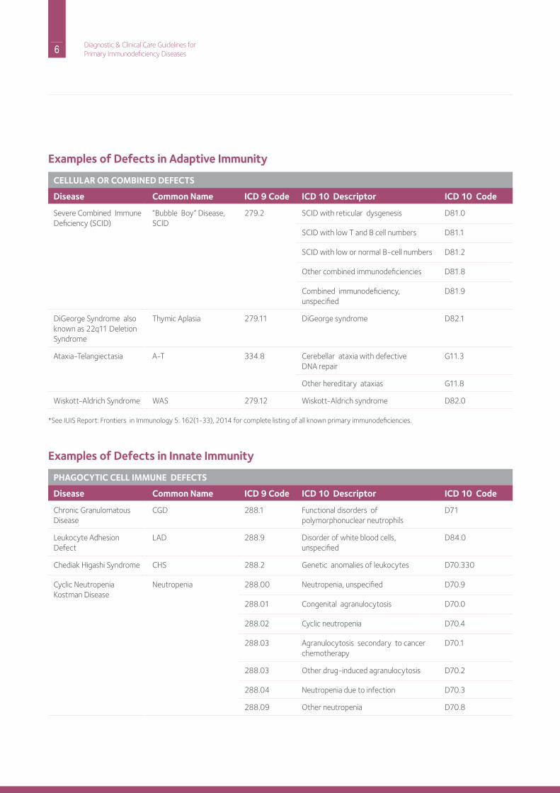
Examples of Defects in Adaptive Immunity
CELLULAR OR COMBINED DEFECTS
Disease Common Name ICD 9 Code ICD 10 Descriptor ICD 10 Code
Severe Combined immunedeficiency (SCid)
“Bubble Boy” disease, SCid
279.2 SCid with reticular dysgenesis d81.0
SCid with low T and B cell numbers d81.1
SCid with low or normal B-cell numbers d81.2
other combined immunodeficiencies d81.8
Combined immunodeficiency, unspecified
d81.9
diGeorge Syndrome also known as 22q11 deletion Syndrome
Thymic Aplasia 279.11 diGeorge syndrome d82.1
Ataxia-Telangiectasia A-T 334.8 Cerebellar ataxia with defectivednA repair
G11.3
other hereditary ataxias G11.8
Wiskott-Aldrich Syndrome WAS 279.12 Wiskott-Aldrich syndrome d82.0
*See iUiS report: Frontiers in immunology 5: 162(1-33), 2014 for complete listing of all known primary immunodeficiencies.
Examples of Defects in Innate Immunity
PHAGOCYTIC CELL IMMUNE DEFECTS
Disease Common Name ICD 9 Code ICD 10 Descriptor ICD 10 Code
Chronic Granulomatous disease
CGd 288.1 Functional disorders of polymorphonuclear neutrophils
d71
Leukocyte Adhesion defect
LAd 288.9 disorder of white blood cells, unspecified
d84.0
Chediak higashi Syndrome ChS 288.2 Genetic anomalies of leukocytes d70.330
Cyclic neutropenia Kostman disease
neutropenia 288.00 neutropenia, unspecified d70.9
288.01 Congenital agranulocytosis d70.0
288.02 Cyclic neutropenia d70.4
288.03 Agranulocytosis secondary to cancer chemotherapy
d70.1
288.03 other drug-induced agranulocytosis d70.2
288.04 neutropenia due to infection d70.3
288.09 other neutropenia d70.8
7 Diagnostic & Clinical Care Guidelines forPrimary Immunodeficiency Diseases
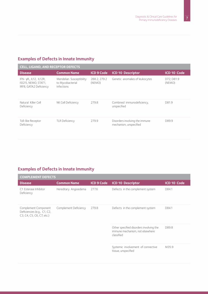
Examples of Defects in Innate Immunity
CELL, LIGAND, AND RECEPTOR DEFECTS
Disease Common Name ICD 9 Code ICD 10 Descriptor ICD 10 Code
iFn- γr,, iL12, iL12r, iSG15, nEMo, STAT1, irF8, GATA2 deficiency
Mendelian Susceptibility to Mycobacterial infections
288.2, 279.2 (nEMo)
Genetic anomalies of leukocytes d72; d81.9 (nEMo)
natural Killer Cell deficiency
nK Cell deficiency 279.8 Combined immunodeficiency, unspecified
d81.9
Toll-like receptor deficiency
TLr deficiency 279.9 disorders involving the immune mechanism, unspecified
d89.9
Examples of Defects in Innate Immunity
COMPLEMENT DEFECTS
Disease Common Name ICD 9 Code ICD 10 Descriptor ICD 10 Code
C1 Esterase inhibitordeficiency
hereditary Angioedema 277.6 defects in the complement system d84.1
Complement Componentdeficiencies (e.g., C1, C2, C3, C4, C5, C6, C7, etc.)
Complement deficiency 279.8 defects in the complement system d84.1
other specified disorders involving the immune mechanism, not elsewhere classified
d89.8
Systemic involvement of connective tissue, unspecified
M35.9

8 Diagnostic & Clinical Care Guidelines forPrimary Immunodeficiency Diseases
9 Diagnostic & Clinical Care Guidelines forPrimary Immunodeficiency Diseases
Part A: Recognition and AssessmentAn infection recurring in a single site is generally not indicative of a primary immunodeficiency disease. rather it suggests an anatomic abnormality. on the other hand, several types of infections affecting various organ systems may be indicative of an underlying immunologic deficiency.
These infections and conditions include:• recurrent sinopulmonary infections
• Pneumonia with fever
• Sinusitis documented by X-ray or computerized
tomography (C-T) scan
• otitis media (although frequent ear infections are seen in
normal children, an evaluation may still be indicated for
individuals on a case-by-case basis). Continued episodes of
otitis after placement of ear tubes should raise concern.
• Meningitis and/or sepsis (blood stream infection)• Gastrointestinal infections, chronic diarrhea or
malabsorption• Cutaneous (skin) infectionsin addition, certain types of autoimmune and allergic conditions may be associated with some types of primary immunodeficiency, including antibody deficiency disorders. Examples include autoimmune disorders, endocrine disorders, rheumatic conditions, and autoimmune hemolytic anemia, neutropenia or thrombocytopenia (low platelet count). These autoimmune disorders are seen especially in patients with igA deficiency, Common Variable immune deficiency (CVid). Allergic disorders with elevated serum igE can also be seen in igA deficiency.
Useful Physical Examination Findings• Absence or reduced size of tonsils and lymph nodes in
X-linked and autosomal recessive agammaglobulinemia and in X-linked hyper igM syndrome
• Enlarged lymph nodes and splenomegaly in CVid and autosomal recessive hyper igM syndrome
• Scarred tympanic membranes• rales and rhonchi in lungs, clubbing of the fingers
Useful Diagnostic Screening Tests• Complete blood count with differential white blood cell
count (in certain cases may require manual differential).
• These tests are of great clinical importance because they
allow the physician to know whether the lymphocyte,
neutrophil and platelet counts (and platelet size) are
normal. Many immune defects can be ruled out by these
Antibody Production defects
simple tests. in the setting of immunodeficiency disorders,
the manual differential cell count is more reliable than an
automated differential.
• Quantitative serum immunoglobulin (igG, igA, igM and igE) levels
• Quantitation of immunoglobulin levels can be performed at
any CLiA88 (Clinical Laboratory improvement Amendment
of 1988) approved laboratory. however, the assay results
should be evaluated in the context of the tested patient’s
age and clinical findings. A testing issue exists for igA levels,
which typically are reported at or above the lower limit of
test sensitivity as most commercially available assays for
igA are not sensitive enough to distinguish between very
low (<10) and absent igA levels. hypergammaglobulinemia
can be the result of hiV-1, CGd, and ALPS. results of all immunoglobulin measurements must be compared with age-adjusted normal values to evaluate their significance. igG subclass measurements are rarely helpful.
• Measurement of specific antibodies to vaccines
• These tests are of crucial importance in determining
whether there is truly an antibody deficiency disorder when
the serum immunoglobulins are not very low or even if they
are low. it is important to test for antibodies to both protein
(i.e., tetanus or diphtheria toxoids) and polysaccharide (i.e., pneumococcal polysaccharides) antigens. Patients may respond to tetanus vaccine because of the presence of memory B-cells from previous immunizations but not respond to Pneumococcal polysaccharides following Pneumovax vaccine and that still indicates a humoral immunodeficiency. isohemagglutinins (antibodies to red blood cells) are natural anti-polysaccharide antibodies primarily of the igM class; if they are missing after age 2, this also suggests an antibody deficiency disorder (unless the patient has type AB blood).
10 Diagnostic & Clinical Care Guidelines forPrimary Immunodeficiency Diseases
When these screening tests are not conclusive and the clinical suspicion of an antibody deficiency is strong, the patient should be referred to an immunologist for further evaluation before beginning immunoglobulin (ig) replacement therapy. This is particularly true for those who have been diagnosed with igG subclass deficiency or “polysaccharide antibody deficiency.” These diagnoses are often based on the results of measurements of serum igG subclass levels or tests of pneumococcal antibody titers. The results need to be interpreted in the context of the clinical history and physical exam. it is very important that all of the tests listed in this section be performed before ig replacement is started. once the patient is on ig therapy, it is difficult to perform further humoral immune testing.
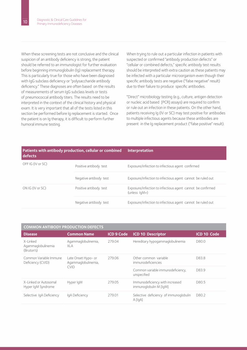
When trying to rule out a particular infection in patients with suspected or confirmed “antibody production defects” or “cellular or combined defects,” specific antibody test results should be interpreted with extra caution as these patients may be infected with a particular microorganism even though their specific antibody tests are negative (“false negative” result) due to their failure to produce specific antibodies.
“direct” microbiology testing (e.g., culture, antigen detection or nucleic acid based [PCr] assays) are required to confirm or rule out an infection in these patients. on the other hand, patients receiving ig (iV or SC) may test positive for antibodies to multiple infectious agents because these antibodies are present in the ig replacement product (“false positive” result).
Patients with antibody production, cellular or combined defects
Interpretation
oFF iG (iV or SC)Positive antibody test Exposure/infection to infectious agent confirmed
negative antibody test Exposure/infection to infectious agent cannot be ruled out
on iG (iV or SC) Positive antibody test Exposure/infection to infectious agent cannot be confirmed (unless igM+)
negative antibody test Exposure/infection to infectious agent cannot be ruled out
COMMON ANTIBODY PRODUCTION DEFECTS
Disease Common Name ICD 9 Code ICD 10 Descriptor ICD 10 Code
X-Linked Agammaglobulinemia(Bruton’s)
Agammaglobulinemia, XLA
279.04 hereditary hypogammaglobulinemia d80.0
Common Variable immunedeficiency (CVid)
Late onset hypo- or Agammaglobulinemia, CVid
279.06 other common variable immunodeficiencies
d83.8
Common variable immunodeficiency, unspecified
d83.9
X-Linked or Autosomal hyper igM Syndrome
hyper igM 279.05 immunodeficiency with increased immunoglobulin M [igM]
d80.5
Selective igA deficiency igA deficiency 279.01 Selective deficiency of immunoglobulin A [igA]
d80.2
11 Diagnostic & Clinical Care Guidelines forPrimary Immunodeficiency Diseases
Part B: Management, Expectations, Complications and Long Term MonitoringWith the exceptions of selective igA deficiency and transient hypogammaglobulinemia of infancy, patients with an identified antibody deficiency disorder are treated at regular intervals throughout life with replacement ig, either intravenously (iV) or subcutaneously (SC). ig products for replacement therapy are comprised of broad spectrum of igG antibodies purified from plasma donations from approximately 10,000 normal donors per batch. The half-life of these igG antibodies is 19-21 days or longer in immunodeficient patients and the amounts of the other classes of immunoglobulins (igA, igM) are extremely low, so they do not contribute to the patient’s blood level of these proteins. The intervals between ig doses are generally 2 to 4 weeks for the intravenous route of administration and more frequently (1 to 14 days) for the subcutaneous route, although there is now an ig product that can be administered subcutaneously every 3 to 4 weeks.
An immunologist should participate in the determination of the proper dose and interval for ig therapy in each patient. Typical total monthly doses are in the range of 400 to 600 mg/kg body weight. Trough (pre-dose) blood levels of igG should be evaluated more frequently initially and at least once a year after that to determine if there has been a change in the metabolism and resultant blood levels of igG in a specific individual. Patients on SCig replacement therapy via home care may need their serum igG checked more often to screen for compliance. ig dose adjustments are obviously necessary during childhood related to normal growth and also during pregnancy, especially during the third trimester. For patients starting at very low igG levels, the trough level should be at least at or above the lower range of normal for age-adjusted igG levels. This may vary depending on the judgment of an immunologist as to the patient’s clinical condition. For example, in one study, when igG trough levels in adult patients with agammaglobulinemia were maintained above 800 mg/dl, serious bacterial illness and enteroviral meningoencephalitis were prevented. higher trough levels (>800 mg/dl) may also have the potential to improve pulmonary outcomes. it is important to recognize that, for virtually all confirmed antibody deficiencies, lifelong ig replacement is required.
ig replacement is preventive therapy so, when a patient develops an infection, this should be treated aggressively with appropriate antibiotics. it is important to recognize that ig therapy only replaces circulating igG and does not replace immunoglobulins in the patient’s external secretions, therefore, infections involving mucosal surfaces may remain problematic. in particular, patients with antibody deficiencies
receiving ig replacement may still develop recurrent and/or chronic bacterial sinus, lung and/or gastrointestinal disease; under these circumstances the use of more prolonged or prophylactic antibiotic therapy is often indicated. in addition, in the setting of these complications, it is best to actively monitor the status of sinus disease, evaluate and monitor lung disease via spirometry and/or chest imaging (being conscious of radiation exposure issues) and evaluate chronic diarrhea or malabsorption with appropriate microbiological studies. Families should expect that effective ig replacement therapy will result in improved school and/or work attendance in their affected relatives.
Spirometry (lung breathing tests) should also be performed annually or at 6-month intervals if the disease appears to be progressing. Complete pulmonary function testing with measurement of diffusion capacity should also be done yearly in patients with CVid who have interstitial and/or granulomatous lung disease. Blood tests of liver and renal function should be checked prior to beginning ig and prophylactic antibiotic therapy and at least once a year thereafter.
in the face of any abnormal neurologic or developmental findings, a baseline lumbar puncture (spinal tap) for the examination of spinal fluid may be helpful in detecting a meningoencephalitis (brain) infection due to enterovirus, particularly in patients with X-linked (Bruton’s) agammaglobulinemia. developmental assessments of such children should also be obtained annually or at 6-month intervals if the disease appears to be progressing.
From a prognostic point of view, patients with antibody deficiencies who have B cells by flow cytometry (e.g., may have CVid) are also at risk for autoimmune disease complications.
Granulomatous lesions in the skin, liver, spleen and lungs in patients with CVid may be misdiagnosed as sarcoid. Those granulomatous complications also mean a worse overall prognosis.
Patients with CVid, X-linked (Bruton’s) agammaglobulinemia or X-linked hyper igM may present with chronic diarrhea and have malabsorption due to infection with parasites, e.g., Giardia lamblia or Cryptosporidium, or from overgrowth in the small intestines with certain types of bacteria. C. difficile causes a colon infection with diarrhea and can occur in hospitalized individuals or those on antibiotics.
12 Diagnostic & Clinical Care Guidelines forPrimary Immunodeficiency Diseases
Ig Therapy When Diagnosis Is UncertainWhen there is uncertainty of the diagnosis, and ig replacement has already been started, it is useful to reassess the need for ig treatment. This is particularly true if the patient’s serum contains igA, igM and igE, which are not present in significant amounts in ig preparations. if these classes of immunoglobulin are present in the patient’s serum, this means that the patient is producing them. however, the serum igG level and antibody titers to vaccine antigens could all be from the ig therapy. To further evaluate whether the patient can produce igG antibodies normally, the patient can be challenged with a neoantigen (e.g., a vaccine not routinely administered, such bacteriophage phi X174) for which there is no specific antibody in ig preparations. While bacteriophage immunizations are useful because it is not necessary to discontinue ig replacement when they are given, the vaccine and testing are available at only a few research institutions under an ind application. Alternately, only under the advisement of an immunologist, ig treatment can be stopped in the spring or summer when infection risks are less. After three months the patient can be reimmunized with standard killed vaccines and the antibody titers to these vaccines tested two to three weeks later. if the patient’s serum immunoglobulins and antibody titers to bacteriophage phiX174 and/or to vaccine antigens are found to be in the normal range, then ig replacement is not necessary. Skin testing for allergies is also useful; individuals who have positive allergy skin tests are producing igE antibodies to the allergens and are not likely to need ig replacement.
Monitoring Ig Therapy in Antibody Deficient PatientsFrequency of testing for trough levels• Monitor igG levels at least once a year (more often if the
patient is having infections) just before the next infusion. Be aware that gastrointestinal tract infection with the parasite Giardia lamblia or other causes of gut disease (e.g., inflammatory bowel disease) can cause loss of igG leading to unexpectedly low igG levels and this would be accompanied by a decrease is serum albumin levels. Generally, once the optimal dose of immune globulin has been established in a patient, monthly monitoring of the igG level is not indicated unless there is protein loss through the gut or urinary tract.
Long-term follow up of patients on Ig therapy• Evaluations regarding hepatitis A, B, and C by PCr
(polymerase chain reaction) screening may be indicated. Yearly PCr screening for hepatitis C is the standard of care in European Union countries.
Adverse event monitoring on Ig therapy• Every 6-12 months creatinine level and liver function tests
are useful.
Other ScreeningsCancer screening may be indicated on a periodic basis, as it is for individuals with intact immune systems. Some subgroups of those with a primary immunodeficiency disease, such as patients with CVid, particularly those with chronic lymphadenopathy may merit baseline complete pulmonary function studies, CT, Mri and/or PET scans and more intensive screening. Lymphoma evaluation is the same as for those without hypogammaglobulinemia. Useful diagnostic screening tests for malignancy include determination of uric acid, Ldh (lactic dehydrogenase) and ESr (erythrocyte sedimentation rate). Testing for hiV-infection should include a nucleic acid based PCr test.
VaccinationsPatients receiving regular infusions of ig possess passively transferred antibodies to the agents normally given in vaccines. Thus, while a patient is receiving ig, there is no need for immunizations. Some immunologists recommend influenza vaccination, but the patient is unlikely to respond to it with antibody production. however, all household contacts should receive regular immunizations with killed vaccines, particularly annual influenza immunizations. Patients with severe antibody deficiency (X-linked agammaglobulinemia, CVid) should not be given oral polio, yellow fever, live attenuated influenza, or typhoid fever vaccines, but family members and other close contacts, with the exception of oral polio vaccine, may receive other standard vaccines because transmission to an immune deficient patient is most unlikely. There are few data on the harmful effects of BCG and rotavirus vaccine, but caution is urged, since the level of T cell immunity in CVid is variable
overall, the general recommendation is to avoid live vaccines in patients with primary immunodeficiencies, but there are exceptions and patients should consult their clinical immunologist to discuss the risks and benefits. in addition there is a recent summary of vaccination recommendations in patients with primary immunodeficiency.
13 Diagnostic & Clinical Care Guidelines forPrimary Immunodeficiency Diseases
Part C: Practical Aspects of Genetic CounselingThe genetic bases of many of the common antibody production defects are currently unknown. This is especially true for most patients with Common Variable immune deficiency (CVid) and Selective igA deficiency where the underlying molecular defect has been identified in less than 10% of patients. For this reason, genetic counseling can be complicated in families affected by these disorders. The inheritance patterns and recurrence risks to family members are difficult to predict without a molecular diagnosis, but an accurate family history may be helpful in this aspect. it should be noted, however, that these disorders can also occur sporadically and the family history in those cases would be negative. Even though the inheritance pattern for some disorders may not be clearly understood, research has shown that family members of patients with CVid and Selective igA deficiency also have an increased risk of antibody deficiencies and autoimmune disorders. it is also important to note that, when the gene defect has not been identified for a specific disorder, prenatal diagnosis is not an option.
The genetic bases of other antibody production defects are known and these disorders include patients with absent B cells and agammaglobulinemia and most cases of the hyper igM syndrome. These disorders can follow either an X-linked recessive or an autosomal recessive inheritance pattern. Please see the general genetic counseling section for a more detailed explanation of inheritance. Because mutations in a number of different genes can cause these conditions, molecular testing is important to determine the specific gene involved and its mutation. This can help predict the clinical manifestations of the disorder in the affected individual. Gene identification along with an accurate family history will also help determine the pattern of inheritance in the family, risks for family members who could be affected, as well as identification of at-risk carrier females of X-linked disorders. Genetic testing for patients, carriers and prenatal diagnosis of some diseases is available through commercial and research laboratories. For a current list of these laboratories, consult your immunologist.
Part D: Frequently Asked Questions from Patients about Antibody Deficiency Disorders1. Will the patient outgrow the disease?
While it is unlikely that a patient will outgrow a primary immunodeficiency disease, identifying changes in the patient’s medical condition and their management is best performed by your immunologist.
2. Should I keep my child with primary immunodeficiency home from school to avoid infections and have my child taught at home? There are both physical and social needs for children with a primary immunodeficiency. if your child has received definitive therapy for the immunodeficiency, and your immunologist agrees that the child’s immunity has been restored by treatment, (e.g., iViG, stem cell transplant), your child can attend regular school. A child must learn how to integrate into society, attain educational skills, advance to high school and college, and seek occupational skills and placement in a job to support themselves later in life.
3. What is Ig? ig stands for immunoglobulin, a family of plasma proteins that help fight infections. Commercially available preparations of these globulins are comprised of numerous igG antibodies purified from plasma donations from approximately 10,000 donors per batch. The name iViG refers to the intravenous (in the vein) form of ig. SCiG is ig, which is given subcutaneously (SC) under the skin.
4. Is there a need for extra Ig during infections, such as pneumonia, and during surgery? during an infection, the antibodies to that infectious agent are rapidly used up, so there is a need for additional amounts of ig during an illness. ig may also provide broad protection against infections that may occur during invasive surgery. Appropriate antibiotic coverage should also be considered during surgery.
5. Can Ig be given orally and is there any place for this as a treatment? While ig has been given by mouth to some patients, trying to mimic the situation in very young animals where the infant animal receives protective antibodies in mother’s milk, there are no research trials that confirm its usefulness in people.
6. Is there protection in Ig from West Nile Virus? At the present time, this is unknown. however, there is no risk of transmitting the West nile virus by ig.
7. What is the safety of Ig? There is a remote or theoretical possibility of blood borne disease transmission. however, laboratory screening is very good and can identify infected potential donors as well as those developing infection. in addition, the manufacturing process for ig includes multiple steps that 1) remove
14 Diagnostic & Clinical Care Guidelines forPrimary Immunodeficiency Diseases
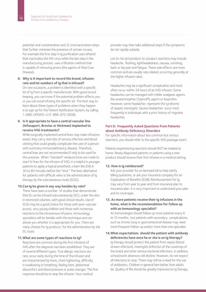
potential viral contamination and 2) viral inactivation steps that further minimize the presence of certain viruses. For example the first step in ig purification uses ethanol that inactivates the hiV virus while the last step in the manufacturing process uses a filtration method that is capable of removing prions (the agents of Mad Cow disease).
8. Why is it important to record the brand, infusion rate and lot numbers of Ig that is infused? on rare occasions, a problem is identified with a specific lot of ig from a specific manufacturer. With good record keeping, you can know if the potential problem affects you or you can avoid infusing the specific lot. The best way to learn about these types of problems when they happen is to sign up for the Patient notification System, by calling 1-888-UPdATE-U (1-888-873-2838).
9. Is it appropriate to have a central vascular line (Infusaport, Broviac or Hickman) implanted to receive IVIG treatments? While surgically implanted central lines may make infusions easier, they carry real risks of serious infection and blood clotting that could greatly complicate the care of a person with a primary immunodeficiency disease. Therefore, central lines are not recommended if only to be used for this purpose. When “standard” venipunctures are made to start iV lines for the infusion of iViG, it is helpful in younger patients to apply a topical anesthetic cream like EMLA 30 to 60 minutes before the “stick.” The best alternative for patients with difficult veins is the administration of ig therapy by the subcutaneous route (SCiG).
10. Can Ig be given in any way besides by vein? There have been a number of studies that demonstrate that iG can be infused subcutaneously (SC), under the skin in restricted volumes, with good clinical results. Use of SCiG may be a good choice for those with poor vascular access, very young children and those with numerous reactions to the intravenous infusions. immunology specialists will be familiar with this technique and can advise you whether it is appropriate for you. There are many choices for ig products for the administration by the SC route.
11. What are some types of reactions to Ig? reactions are common during the first infusions of iViG after the diagnosis has been established. They are of several different types. True allergic reactions are rare, occur early during the time of the infusion and are characterized by hives, chest tightening, difficulty in swallowing or breathing, feeling faint, abdominal discomfort and blood pressure or pulse changes. The first response should be to stop the infusion. Your medical
provider may then take additional steps if the symptoms do not rapidly subside. Lot-to-lot and product-to-product reactions may include headache, flushing, lightheadedness, nausea, vomiting, back or hip pain and fatigue. These side effects are more common and are usually rate related, occurring generally at the higher infusion rates. headaches may be a significant complication and most often occur within 24 hours of an iViG infusion. Some headaches can be managed with milder analgesic agents like acetaminophen (Tylenol®), aspirin or ibuprofen. however, some headaches represent the syndrome of aseptic meningitis. Severe headaches occur most frequently in individuals with a prior history of migraine headaches.
Part D: Frequently Asked Questions from Patients about Antibody Deficiency DisordersFor specific information about less common but serious reactions, you should refer to the specific iViG package insert.
Patients experiencing reactions should noT be treated at home. newly diagnosed patients or patients using a new product should receive their first infusion in a medical setting.
12. How is Ig reimbursed? Ask your provider for an itemized bill to help clarify billing questions, or ask your insurance company for an Explanation of Benefits (EoB). reimbursement for ig may vary from year to year and from insurance plan to insurance plan. it is very important to understand your plan and its coverages.
13. As more patients receive their Ig infusions in the home, what is the recommendation for follow up with an immunology specialist? An immunologist should follow up most patients every 6 to 12 months, but patients with secondary complications, such as chronic lung or gastrointestinal disease may need more frequent follow-up and/or more than one specialist.
14. What expectations should the patient with antibody deficiencies have once he or she is on Ig therapy? ig therapy should protect the patient from sepsis (blood stream infection), meningitis (infection of the coverings of the brain) and other serious bacterial infections. in addition, school/work absences will decline. however, do not expect all infections to stop. There may still be a need for the use of antibiotics. Children in general fare better than adults do. Quality of life should be greatly improved on ig therapy.
15 Diagnostic & Clinical Care Guidelines forPrimary Immunodeficiency Diseases
15. What is the role of antibiotics in antibody deficiency diseases? Antibiotics may be used chronically if there is evidence of chronic infection or permanent damage to the lungs (bronchiectasis) or sinuses. The antibiotics should be given in full treatment doses. Prophylactic antibiotic therapy may be useful for selected patients with antibody deficiencies.
16. What is the role of over-the-counter immune stimulants? There is no evidence that these stimulants have any helpful effects.
17. Is it OK to exercise and play sports? Yes. Physical activity and sports may help improve patients’ sense of well-being and enable them to participate in some of life’s enjoyable activities.
18. Is it OK to have pets? Yes, but be aware that animals may carry infections that possibly can be transmitted to humans.
19. Can Ig be given during pregnancy? Yes and it should be given as when not pregnant.
For additional information on the use of IVIG or subcutaneous Ig, see:orange JS, hossny EM, Weiler Cr, Ballow M, Berger M, Bonilla FA, Buckley rh et al. “Use of intravenous immunoglobulin in human disease: a review of evidence by members of the Primary immunodeficiency Committee of the American Academy of Allergy, Asthma and immunology.” J Allergy Clin immunol 117: S525-S553, 2006.
For additional information on the use of live viral and bacterial vaccines, see:Shearer WT, Fleisher, TA, Buckley rh , Ballas Z, Ballow M, Blaese M et al. “recommendations for live viral and bacterial vaccines in immunodeficient patients and their close contacts.” J Allergy Clin immunol 2014; 133(4):961-966.
16 Diagnostic & Clinical Care Guidelines forPrimary Immunodeficiency Diseases
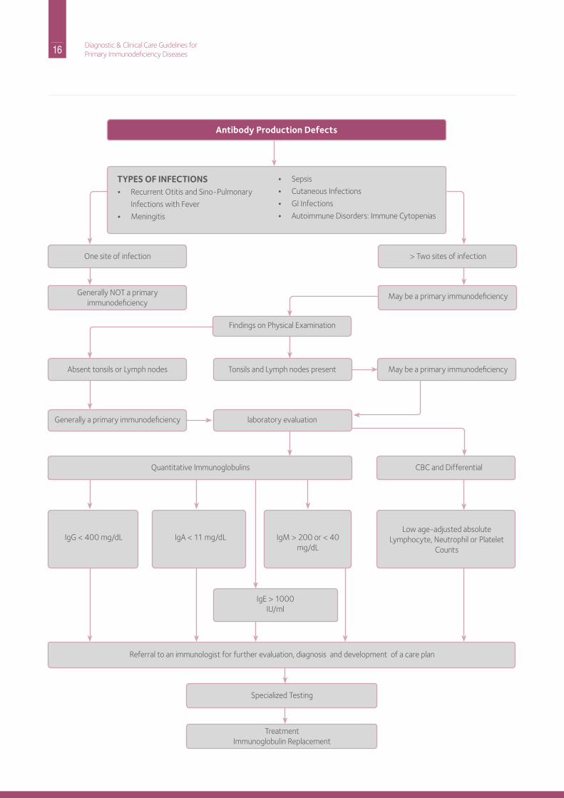
Antibody Production Defects
TYPES OF INFECTIONS• recurrent otitis and Sino-Pulmonary
infections with Fever
• Meningitis
• Sepsis
• Cutaneous infections
• Gi infections
• Autoimmune disorders: immune Cytopenias
one site of infection
Absent tonsils or Lymph nodes
Generally a primary immunodeficiency
Quantitative immunoglobulins
Generally noT a primary immunodeficiency
igG < 400 mg/dL igA < 11 mg/dL igM > 200 or < 40 mg/dL
igE > 1000iU/ml
referral to an immunologist for further evaluation, diagnosis and development of a care plan
Specialized Testing
Treatmentimmunoglobulin replacement
Low age-adjusted absolute Lymphocyte, neutrophil or Platelet
Counts
> Two sites of infection
May be a primary immunodeficiency
May be a primary immunodeficiency
CBC and differential
Findings on Physical Examination
Tonsils and Lymph nodes present
laboratory evaluation
17 Diagnostic & Clinical Care Guidelines forPrimary Immunodeficiency Diseases
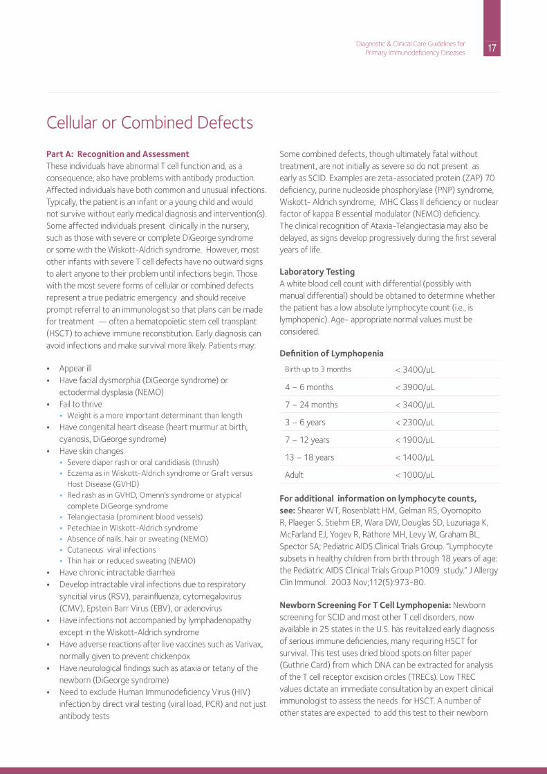
Part A: Recognition and AssessmentThese individuals have abnormal T cell function and, as a consequence, also have problems with antibody production. Affected individuals have both common and unusual infections. Typically, the patient is an infant or a young child and would not survive without early medical diagnosis and intervention(s). Some affected individuals present clinically in the nursery, such as those with severe or complete diGeorge syndrome or some with the Wiskott-Aldrich syndrome. however, most other infants with severe T cell defects have no outward signs to alert anyone to their problem until infections begin. Those with the most severe forms of cellular or combined defects represent a true pediatric emergency and should receive prompt referral to an immunologist so that plans can be made for treatment — often a hematopoietic stem cell transplant (hSCT) to achieve immune reconstitution. Early diagnosis can avoid infections and make survival more likely. Patients may:
• Appear ill• have facial dysmorphia (diGeorge syndrome) or
ectodermal dysplasia (nEMo)• Fail to thrive
• Weight is a more important determinant than length
• have congenital heart disease (heart murmur at birth, cyanosis, diGeorge syndrome)
• have skin changes• Severe diaper rash or oral candidiasis (thrush)• Eczema as in Wiskott-Aldrich syndrome or Graft versus
host disease (GVhd)• red rash as in GVhd, omenn’s syndrome or atypical
complete diGeorge syndrome• Telangiectasia (prominent blood vessels)• Petechiae in Wiskott-Aldrich syndrome • Absence of nails, hair or sweating (nEMo) • Cutaneous viral infections• Thin hair or reduced sweating (nEMo)
• have chronic intractable diarrhea• develop intractable viral infections due to respiratory
syncitial virus (rSV), parainfluenza, cytomegalovirus (CMV), Epstein Barr Virus (EBV), or adenovirus
• have infections not accompanied by lymphadenopathy except in the Wiskott-Aldrich syndrome
• have adverse reactions after live vaccines such as Varivax, normally given to prevent chickenpox
• have neurological findings such as ataxia or tetany of the newborn (diGeorge syndrome)
• need to exclude human immunodeficiency Virus (hiV) infection by direct viral testing (viral load, PCr) and not just antibody tests
Some combined defects, though ultimately fatal without treatment, are not initially as severe so do not present as early as SCid. Examples are zeta-associated protein (ZAP) 70 deficiency, purine nucleoside phosphorylase (PnP) syndrome, Wiskott- Aldrich syndrome, MhC Class ii deficiency or nuclear factor of kappa B essential modulator (nEMo) deficiency. The clinical recognition of Ataxia-Telangiectasia may also be delayed, as signs develop progressively during the first several years of life.
Laboratory TestingA white blood cell count with differential (possibly with manual differential) should be obtained to determine whether the patient has a low absolute lymphocyte count (i.e., is lymphopenic). Age- appropriate normal values must be considered.
Definition of Lymphopenia
Birth up to 3 months < 3400/µL
4 – 6 months < 3900/µL
7 – 24 months < 3400/µL
3 – 6 years < 2300/µL
7 – 12 years < 1900/µL
13 – 18 years < 1400/µL
Adult < 1000/µL
For additional information on lymphocyte counts, see: Shearer WT, rosenblatt hM, Gelman rS, oyomopito r, Plaeger S, Stiehm Er, Wara dW, douglas Sd, Luzuriaga K, McFarland EJ, Yogev r, rathore Mh, Levy W, Graham BL, Spector SA; Pediatric AidS Clinical Trials Group. “Lymphocyte subsets in healthy children from birth through 18 years of age: the Pediatric AidS Clinical Trials Group P1009 study.” J Allergy Clin immunol. 2003 nov;112(5):973-80.
Newborn Screening For T Cell Lymphopenia: newborn screening for SCid and most other T cell disorders, now available in 25 states in the U.S. has revitalized early diagnosis of serious immune deficiencies, many requiring hSCT for survival. This test uses dried blood spots on filter paper (Guthrie Card) from which dnA can be extracted for analysis of the T cell receptor excision circles (TrECs). Low TrEC values dictate an immediate consultation by an expert clinical immunologist to assess the needs for hSCT. A number of other states are expected to add this test to their newborn
Cellular or Combined defects
18 Diagnostic & Clinical Care Guidelines forPrimary Immunodeficiency Diseases
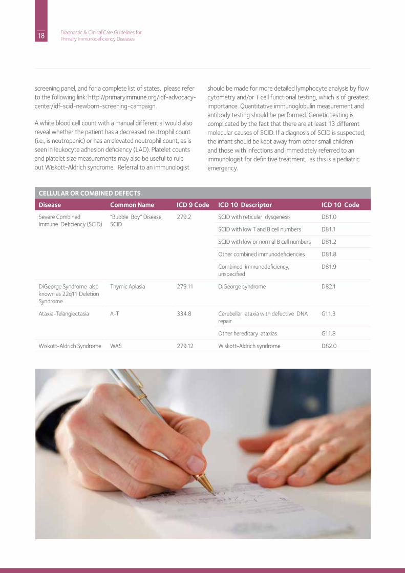
screening panel, and for a complete list of states, please refer to the following link: http://primaryimmune.org/idf-advocacy- center/idf-scid-newborn-screening-campaign.
A white blood cell count with a manual differential would also reveal whether the patient has a decreased neutrophil count (i.e., is neutropenic) or has an elevated neutrophil count, as is seen in leukocyte adhesion deficiency (LAd). Platelet counts and platelet size measurements may also be useful to rule out Wiskott-Aldrich syndrome. referral to an immunologist
should be made for more detailed lymphocyte analysis by flow cytometry and/or T cell functional testing, which is of greatest importance. Quantitative immunoglobulin measurement and antibody testing should be performed. Genetic testing is complicated by the fact that there are at least 13 different molecular causes of SCid. if a diagnosis of SCid is suspected, the infant should be kept away from other small children and those with infections and immediately referred to an immunologist for definitive treatment, as this is a pediatric emergency.
CELLULAR OR COMBINED DEFECTS
Disease Common Name ICD 9 Code ICD 10 Descriptor ICD 10 Code
Severe Combinedimmune deficiency (SCid)
“Bubble Boy” disease, SCid
279.2 SCid with reticular dysgenesis d81.0
SCid with low T and B cell numbers d81.1
SCid with low or normal B cell numbers d81.2
other combined immunodeficiencies d81.8
Combined immunodeficiency, unspecified
d81.9
diGeorge Syndrome also known as 22q11 deletion Syndrome
Thymic Aplasia 279.11 diGeorge syndrome d82.1
Ataxia-Telangiectasia A-T 334.8 Cerebellar ataxia with defective dnA repair
G11.3
other hereditary ataxias G11.8
Wiskott-Aldrich Syndrome WAS 279.12 Wiskott-Aldrich syndrome d82.0
19 Diagnostic & Clinical Care Guidelines forPrimary Immunodeficiency Diseases
Part B: Management, Expectations, Complications and Long Term Monitoringonly irradiated (5000 rAd), CMV-negative, leukocyte-depleted blood products should be used for the patient with SCid or other suspected T cell deficiencies. Before transplantation (hSCT) to restore their immune systems, infants with SCid and other serious T cell deficiencies must noT receive live viral vaccines (e.g., oral polio virus, varicella, measles, mumps, rubella, herpes zoster, rotavirus, yellow fever, smallpox, or live attenuated influenza virus/or live bacterial vaccines [BCG or typhoid fever]). For those SCid and other serious T cell deficient patients who have received hSCT but have not recovered full immune competence, or who are receiving immunosuppressive drugs (i.e., GVhd prophylaxis), these same rules of avoidance apply. For those immunodeficient patients whose immunity has been completely restored, parents and patients should be given information on the risk versus benefit of any live vaccine administration by expert clinical immunologists after appropriate laboratory assessment of immune response.
Patients with partial T cell deficiencies, such as partial diGeorge patients (birth rate 1/4000) have inadvertently received live measles, mumps, and rubella vaccines without sequelae, but other live vaccines should be avoided. Clinical judgment and determination of T cell competence as assessed by T cell responses to mitogens and antigens should both factor into deciding whether live vaccines are safe. Completely killed vaccines may be of some benefit in children with partial T cell function. Close contacts of immunodeficient patients (e.g., household contacts) should not receive live vaccines, but periodic immunization with killed vaccines is strongly encouraged. it should be noted that close contacts immunized with live attenuated influenza, have very rarely transmitted vaccine-derived influenza to immunodeficient subjects, but in an abundance of caution, avoidance is best. however, all household contacts should receive regular immunizations with killed vaccines, particularly annual influenza immunizations.
Typical serious, often overwhelming or fatal infections in SCid are due to PJP (Pneumocystis jiroveci pneumonia), candida, rSV, parainfluenza 3, CMV, EBV and/or adenovirus. if an infant is suspected of having SCid, he or she should be placed on PJP prophylaxis with trimethoprim/sulfamethoxazole.
if there is a family history of an early death due to infection, a simple screening test that would help diagnose or exclude most cases of SCid in a subsequent birth can be done by performing a white blood cell count and a manual differential count on the cord blood to look for a low lymphocyte count. if
the count is low, flow cytometry should be performed to see if T cells are present. if T cells are low or absent, T cell function should be assessed by performing mitogen stimulation studies to confirm a diagnosis of SCid. if T cells are present, they could be transplacentally transferred maternal T cells or the infant could have clonal T cells as in omenn Syndrome or other forms of “leaky SCid.” in both cases the T cell function would be low.
immune reconstitution in SCid generally requires hSCT (hematopoietic stem cell transplantation, previously known as bone marrow transplantation) early in life. Pre-transplant chemotherapy is not needed for true SCid infants because they do not have T cells. Gene therapy has been tried with notable success, but there have been serious adverse events. Patients with SCid who have received successful hSCT require at least annual follow up by an immunologist at a specialized center. There may be unanticipated complications and patients should also have the opportunity to benefit from new therapeutic developments.
Patients with combined immunodeficiencies (Cid) may have low but not absent T cell function and may additionally fail to make specific antibodies normally despite normal or elevated immune globulin levels. They also require ig replacement therapy. For example, although Wiskott-Aldrich patients may have normal serum immunoglobulin levels, they are usually treated with ig replacement because their ability to make antibodies is abnormal. in the complete form of diGeorge syndrome, there is no T cell function and thymic transplantation is the recommended treatment. The long-term outcome for partial diGeorge syndrome is generally satisfactory from an immunologic perspective. however, susceptibility to other complications such as developmental delay, seizure, severe autoimmune disease or EBV induced lymphoma remains. A-T patients (Ataxia-Telangiectasia) and patients with SCid due to Artemis and Ligase 4 gene mutations should minimize their exposure to ionizing irradiation, as they have an increased risk for chromosomal breakage and its complications.
VaccinationsThe same recommendations for live vaccines in severe antibody deficiencies apply for cellular or combined immune deficiencies (see p.9). When children with cellular immunodeficiency are immunoreconstituted, they may receive attenuated live viral vaccines (e.g., measles, mumps, rubella, and possibly chickenpox). Consultation with an immunologist is essential. This reduced vigilance for live attenuated viral vaccine also applies to children with less severe cellular immunodeficiency like partial diGeorge syndrome.
20 Diagnostic & Clinical Care Guidelines forPrimary Immunodeficiency Diseases
Part C: Practical Aspects of Genetic CounselingThe genetic basis is known for many of the cellular or combined immune defects. Several of these disorders follow X-linked inheritance; many others follow autosomal recessive inheritance. Please refer to the general genetic counseling section for an explanation of inheritance patterns. A special consideration for genetic counseling of families affected by these disorders is the fact that there are several different genes that, when mutated, result in the same clinical disorder. For example, it is currently known that mutations in one of at least thirteen genes can cause SCid. The most common form of SCid follows X-linked inheritance; all other forms of SCid follow autosomal recessive inheritance. it is therefore crucial that genetic testing be done to determine the specific gene involved in these disorders to provide accurate estimates of risks for family members being affected. however, genetic testing should noT dELAY initiation of appropriate treatment of the affected patient. Genetic testing for many of the cellular or combined immunodeficiency disorders is available in commercial and specialized laboratories in the U.S. and abroad. For more specific information refer to the general section on genetic counseling.
diGeorge syndrome is a primary immunodeficiency that may follow autosomal dominant inheritance; however most cases are sporadic. it is caused by deletion of a portion of a region on chromosome 22 in more than half of the cases, by mutations in a gene on chromosome 10 in another 10% and is of unknown cause in the other cases; it affects both males and females.
While many of these cases occur as new mutations in the genes on chromosome 22, it is also important to do molecular testing of the parents of a child with this condition because there can be clinical variability and an affected parent may have previously gone undiagnosed. Whether or not the chromosome defect is inherited or due to a new mutation has significant impact on recurrence risk for a family. Genetic testing for chromosome 22 deletions is widely available in laboratories across the United States.
21 Diagnostic & Clinical Care Guidelines forPrimary Immunodeficiency Diseases
Part D: Frequently Asked Questions1. What happens if my child is exposed to chickenpox?
You need to let your physician know immediately so he or she can receive VariZiG (Varicella iG), an investigational hyperimmune globulin, within 48 hours of exposure. ig replacement therapy in the usual doses can also provide antibodies against the chickenpox virus. The incubation period of varicella is 11 to 21 days. if your child already has a vesicular rash, which looks like small blisters, he or she will need to be treated with acyclovir, an antiviral agent. intravenous (iV) acyclovir is superior to oral.
2. Should I keep my child with primary immunodeficiency home from school to avoid infections and have my child taught at home? There are both physical and social needs for children with a primary immunodeficiency. if your child has received definitive therapy for the immunodeficiency, and your immunologist agrees that the child’s immunity has been restored by treatment, (e.g., iViG, stem cell transplant), your child can attend regular school. A child must learn how to integrate into society, attain educational skills, advance to high school and college, and seek occupational skills and placement in a job to support themselves later in life.
3. What kind of vaccines can my child receive? All of the killed vaccines are safe, but he or she should not receive any live vaccines such as rotovirus (rotateq® or rotarix®), oral polio, measles, mumps, rubella (MMr®), varicella (Varivax®), or intranasal influenza vaccine (FLuMist®) or BCG. Antibodies in ig may give protection against some or all of these viruses. in general, immunodeficient patients who are receiving ig replacement should not be given vaccines, although some immunologists give killed influenza immunizations. if the patient is immune deficient enough to need ig replacement, he or she probably would not be able to respond with antibody production. it is uncertain whether there would be a T cell response that could be helpful. however, the antibodies in the ig replacement therapy would neutralize most live vaccines and they would be ineffective.
4. What is the difference between public and private cord blood banks? Cord blood is rich in the stem cells that can restore immunity in SCid and other serious T cell deficiencies.Cord blood is being stored in banks that are either public or private. Storage of cord blood in public cord banks is free, whereas, storage in private (for profit) cord blood banks has a large fee for service. other differences for private cord blood banks are underutilization and the fact
that they are less regulated for quality control. Almost all stem cell transplantation associations throughout the world prefer public cord blood banks. There may be some need for private cord blood banks for families with known stem T cell immunodeficiencies where the cord blood from normal offspring could rescue another child in the family who inherited the disease. Thus, the general public, parents, pediatricians and obstetricians need to become better educated on the issue of cord blood being stored in either public or private banks for use in transplantation. For additional information on public vs private cord blood banks see the American Academy of Pediatrics Section on hematology/oncology; American Academy of Pediatrics Section on Allergy/immunology, Lubin Bh, Shearer WT. “Cord blood banking for potential future transplantation.” Pediatrics 2007 Jan;119 (1):165-70. (A revised policy statement regarding cord blood banking for transplantation is being prepared by the American Academy of Pediatrics and should be available soon).
22 Diagnostic & Clinical Care Guidelines forPrimary Immunodeficiency Diseases
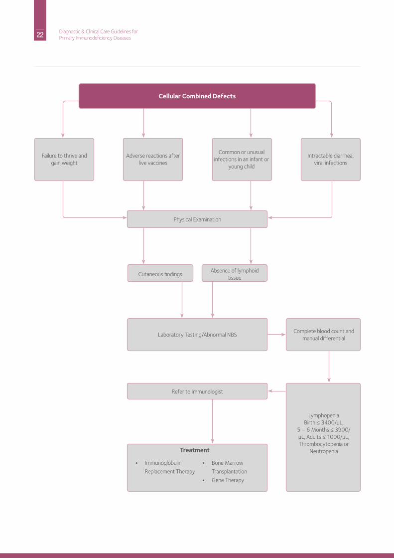
Cellular Combined Defects
Failure to thrive and gain weight
Adverse reactions after live vaccines
Common or unusual infections in an infant or
young child
intractable diarrhea, viral infections
Cutaneous findingsAbsence of lymphoid
tissue
Laboratory Testing/Abnormal nBS
refer to immunologist
• immunoglobulin
replacement Therapy
• Bone Marrow
Transplantation
• Gene Therapy
Complete blood count and manual differential
LymphopeniaBirth ≤ 3400/µL,
5 – 6 Months ≤ 3900/µL, Adults ≤ 1000/µL, Thrombocytopenia or
neutropenia
Physical Examination
Treatment

23 Diagnostic & Clinical Care Guidelines forPrimary Immunodeficiency Diseases
24 Diagnostic & Clinical Care Guidelines forPrimary Immunodeficiency Diseases
Part A: Recognition and AssessmentSigns of defects in the phagocytic cells are manifest in many organ systems. The onset of symptoms is usually in infancy or early childhood.
• Skin – abscesses (boils) (seen in Chronic Granulomatous disease [CGd] and the hyper igE syndrome) and/or cellulitis (inflammation of the skin) (seen in Leukocyte Adhesion deficiency [LAd]).
• Lymph nodes – may be swollen and contain pus in patients with CGd.
• in LAd there may be delayed shedding of the umbilical cord or infection of the cord base (omphalitis) and cellulitis but no abscesses or pus.
• Osteomyelitis – an infection of bone seen frequently in patients with CGd.
• Hepatic Abscess – liver abscesses may also be seen in CGd.
• Lungs – Aspergillus (mold) lung disease is common in patients with CGd. Abscesses and other infections may occur in these patients due to pathogens that do not result in abscesses in normal hosts. infections due to nontuberculous mycobacteria and/or salmonella may also occur.
• Gastrointestinal tract outlet and/or urinary tract obstruction resulting in abdominal or back pain is often seen in CGd, as is constipation.
• Mouth (gingivitis) – gum inflammation, mouth ulcers.• Unexplained fever – without identifiable cause.• Malaise and fatigue• Albinism – may be seen in Chediak-higashi syndrome.
Laboratory Testsdefects in phagocytic cells can be due to an insufficient number of such cells, an inability of the cells to get to an infected area, or to an inability to kill ingested bacteria or fungi normally.
• A complete blood count and differential count are needed to determine whether phagocytic cells (neutrophils) are present in normal number. in the case of cyclic neutropenia, the test (absolute neutrophil count or AnC) has to be repeated sequentially (e.g., 2 times per week for 1 month).
• A test for Cd11/Cd18 expression on white cells is needed to exclude LAd.
Phagocytic Cell immune defects
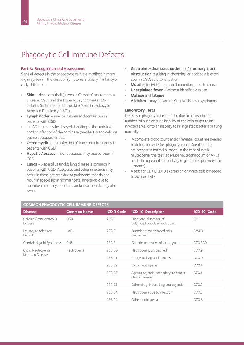
COMMON PHAGOCYTIC CELL IMMUNE DEFECTS
Disease Common Name ICD 9 Code ICD 10 Descriptor ICD 10 Code
Chronic Granulomatous disease
CGd 288.1 Functional disorders of polymorphonuclear neutrophils
d71
Leukocyte Adhesion defect
LAd 288.9 disorder of white blood cells, unspecified
d84.0
Chediak higashi Syndrome ChS 288.2 Genetic anomalies of leukocytes d70.330
Cyclic neutropenia Kostman disease
neutropenia 288.00 neutropenia, unspecified d70.9
288.01 Congenital agranulocytosis d70.0
288.02 Cyclic neutropenia d70.4
288.03 Agranulocytosis secondary to cancer chemotherapy
d70.1
288.03 other drug-induced agranulocytosis d70.2
288.04 neutropenia due to infection d70.3
288.09 other neutropenia d70.8
25 Diagnostic & Clinical Care Guidelines forPrimary Immunodeficiency Diseases
Part A: Recognition and Assessment• A respiratory Burst Assay (dhr flow cytometry assay,
the replacement for the nBT assay) should be performed to determine if phagocytic cells can produce the oxygen radicals needed to kill bacteria and fungi; neutrophils from patients with CGd do not produce these oxygen radicals. As is the case in CGd, patients with the hyper igE syndrome also present with abscesses (boils), although they have a normal number of neutrophils and a normal respiratory Burst Assay result. Thus, a serum igE level should be measured in patients with recurrent abscesses to make certain that the hyper igE syndrome is not the underlying cause.
Part B: Management, Expectations, Complications and Long Term Monitoringin general, neutropenia (reduced numbers of phagocytes) is most commonly secondary to a drug or an infection, and the patient should be referred to a hematologist or other specialist for management. in individuals with a primary immunodeficiency disease affecting phagocytic cells, prophylactic antibiotics are appropriate. These antibiotics include trimethoprim/sulfamethoxizole or if the patient is allergic to it, cephalexin. Prophylactic antifungal agents are also often administered in patients with CGd.
Cd40 Ligand (Cd40L or X-linked hyper igM) deficient patients are often profoundly neutropenic. individuals with severe neutropenia (Kostmann’s Syndrome is one form) may be responsive to granulocyte colony stimulating factor (G-CSF), as may be the neutropenia associated with Cd40 Ligand deficiency. it is important to obtain bacterial and fungal cultures when these patients are sick in order to correctly direct antibiotic treatment.
With appropriate antibiotic therapy, individuals with CGd should live into their 40’s or older. however, there are differences in infection susceptibility in terms of the X-linked and autosomal recessive types, with somewhat more frequent infections in the X-linked type. Meticulous medical care from an expert in immunology will increase the patient’s chances of longer survival. Typically, hemogloblin, hematocrit, ESr (erythrocyte sedimentation rate) and/or CrP (C-reactive protein) and chest imaging should be performed regularly in CGd. if there is any fever, malaise or change in health status, the patient requires immediate medical evaluation. Patients with CGd have normal T cell and B cell function.
interferon gamma has been used to prevent infections in CGd. There is no change in in vitro tests of phagocytic cell function with this treatment, although some clinical benefit (e.g., reduced number of serious infections) has been reported. over the past 14 years, hSCT has been successful in more than 150 patients with CGd, with a high survival rate.
VaccinationsExcept for Bacille Calmette Guerin (BCG) and live Salmonella vaccine, phagocytic cell deficient patients have no contraindication and should receive all age-recommended vaccines.
Home SchoolingPatients with CGd or other phagocytic cell disorders should attend school and may visit public places, such as malls.
Part C: Practical Aspects of Genetic CounselingThe genetic basis is known for most of the phagocytic cell immune defects. however, as with SCid, mutations in several different genes can be responsible for a clinically similar condition. For example, mutations in any of five genes are known to cause Chronic Granulomatous disease. The most common form of this disorder is X-linked and the other forms follow autosomal recessive inheritance. A detailed family history may be helpful in determining the type of CGd. however, genetic testing is crucial in determining the specific gene involved.
With this information, the clinical prognosis can be assessed and patterns of inheritance determined in the family. Genetic testing for the phagocytic cell immune defects is available in commercial and specialized laboratories. Please see the general section on genetic counseling for more information about testing.

26 Diagnostic & Clinical Care Guidelines forPrimary Immunodeficiency Diseases
Part D: Frequently Asked Questions1. What activities and places should my child avoid?
There is no general recommendation for avoiding infections. however, swimming in lakes or ponds should be avoided. Exposure to aspergillus and mold occurs with gardening and digging or handling or being around mulch. These and any other activities that will expose the child with a phagocytic cell primary immunodeficiency to potentially harmful bacteria or fungi should be avoided.
2. Can my child receive all types of vaccines? Yes, except for Bacille Calmette Guerin (BCG) and live Salmonella vaccine because patients with CGd or other phagocytic cell disorders typically have normal T and B cell function.
27 Diagnostic & Clinical Care Guidelines forPrimary Immunodeficiency Diseases
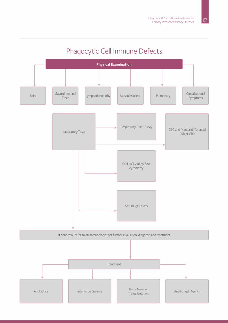
Physical Examination
Skin Lymphadenopathy
Treatment
Pulmonary Gastrointestinal
TractMusculoskeletal
ConstitutionalSymptoms
Laboratory TestsCBC and Manual differential
ESr or CrP
respiratory Burst Assay
Cd11/Cd/18 by flow cytometry
Serum igE Levels
Antibiotics interferon GammaBone Marrow
TransplantationAnti Fungal Agents
if abnormal, refer to an immunologist for further evaluation, diagnosis and treatment
Phagocytic Cell immune defects
28 Diagnostic & Clinical Care Guidelines forPrimary Immunodeficiency Diseases
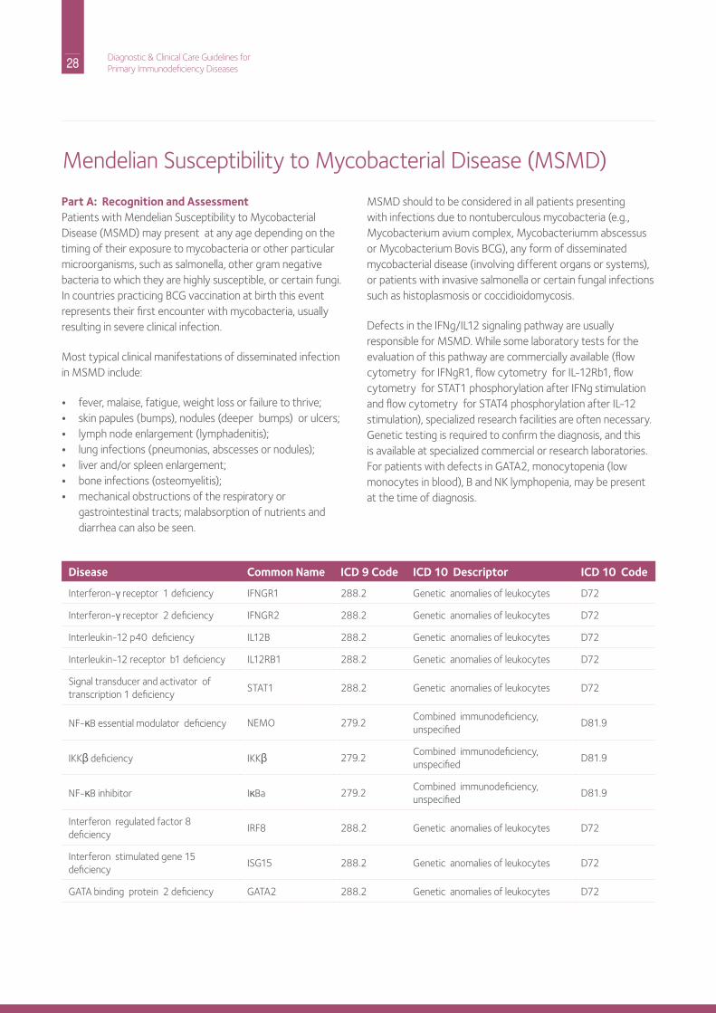
Part A: Recognition and AssessmentPatients with Mendelian Susceptibility to Mycobacterial disease (MSMd) may present at any age depending on the timing of their exposure to mycobacteria or other particular microorganisms, such as salmonella, other gram negative bacteria to which they are highly susceptible, or certain fungi. in countries practicing BCG vaccination at birth this event represents their first encounter with mycobacteria, usually resulting in severe clinical infection.
Most typical clinical manifestations of disseminated infection in MSMd include:
• fever, malaise, fatigue, weight loss or failure to thrive;• skin papules (bumps), nodules (deeper bumps) or ulcers;• lymph node enlargement (lymphadenitis);• lung infections (pneumonias, abscesses or nodules);• liver and/or spleen enlargement;• bone infections (osteomyelitis);• mechanical obstructions of the respiratory or
gastrointestinal tracts; malabsorption of nutrients and diarrhea can also be seen.
MSMd should to be considered in all patients presenting with infections due to nontuberculous mycobacteria (e.g., Mycobacterium avium complex, Mycobacteriumm abscessus or Mycobacterium Bovis BCG), any form of disseminated mycobacterial disease (involving different organs or systems), or patients with invasive salmonella or certain fungal infections such as histoplasmosis or coccidioidomycosis.
defects in the iFng/iL12 signaling pathway are usually responsible for MSMd. While some laboratory tests for the evaluation of this pathway are commercially available (flow cytometry for iFngr1, flow cytometry for iL-12rb1, flow cytometry for STAT1 phosphorylation after iFng stimulation and flow cytometry for STAT4 phosphorylation after iL-12 stimulation), specialized research facilities are often necessary. Genetic testing is required to confirm the diagnosis, and this is available at specialized commercial or research laboratories. For patients with defects in GATA2, monocytopenia (low monocytes in blood), B and nK lymphopenia, may be present at the time of diagnosis.
Mendelian Susceptibility to Mycobacterial disease (MSMd)
Disease Common Name ICD 9 Code ICD 10 Descriptor ICD 10 Code
interferon-γ receptor 1 deficiency iFnGr1 288.2 Genetic anomalies of leukocytes d72
interferon-γ receptor 2 deficiency iFnGr2 288.2 Genetic anomalies of leukocytes d72
interleukin-12 p40 deficiency iL12B 288.2 Genetic anomalies of leukocytes d72
interleukin-12 receptor b1 deficiency iL12rB1 288.2 Genetic anomalies of leukocytes d72
Signal transducer and activator of transcription 1 deficiency
STAT1 288.2 Genetic anomalies of leukocytes d72
nF-кB essential modulator deficiency nEMo 279.2Combined immunodeficiency, unspecified
d81.9
iKKβ deficiency iKKβ 279.2Combined immunodeficiency, unspecified
d81.9
nF-кB inhibitor iкBa 279.2Combined immunodeficiency, unspecified
d81.9
interferon regulated factor 8 deficiency
irF8 288.2 Genetic anomalies of leukocytes d72
interferon stimulated gene 15 deficiency
iSG15 288.2 Genetic anomalies of leukocytes d72
GATA binding protein 2 deficiency GATA2 288.2 Genetic anomalies of leukocytes d72
29 Diagnostic & Clinical Care Guidelines forPrimary Immunodeficiency Diseases
Part B: Management, Expectations, Complications and Long Term MonitoringTreatment of mycobacterial diseases in MSMD patients usually involves:• A multidrug approach (when treated with a single agent,
mycobacteria tend to become resistant to that antibiotic);• Long term treatment (some mycobacteria grow very
slowly, needing months of treatment before the infection is controlled or eradicated);
• Parenteral (intravenous or intramuscular) treatment (some of the best drugs to treat mycobacteria are only available as intravenous medications, and when gastrointestinal absorption is compromised, use of a parenteral route is necessary);
• Use of biologicals (such as recombinant iFng or iL-12) as adjuvant therapeutic tools;
• hSCT should be considered on a case-by-case basis.
regular monitoring for therapeutic response is important in treating these infections. depending the site of the infection, biopsies, sputum or bronchoalveolar lavage may help determine if mycobacteria are present and alive. imaging studies (ultrasound, CT scan, Mri), and laboratory markers of inflammation (leukocytosis, ESr, CrP) are also useful in following these patients.
VaccinationsExcept for Bacille Calmette Guerin (BCG) and live Salmonella vaccine, MSMd patients have no contraindication and should receive all age-recommended vaccines.
Home Schooling and Visiting Public PlacesPatients with MSMd should attend school and may visit public places.
Part C: Practical Aspects of Genetic CounselingGenetic defects associated with MSMd may be inherited as X-linked (e.g., nEMo defects), autosomal recessive (Ar) (e.g., iFnγr1, iFnγr2, iL-12p40, iKKβ, irF8, iSG15, STAT1 and iL-12rβ1) or autosomal dominant (Ad) traits (e.g., iFnγr1, iFnγr2, STAT1, irF8, ikBa and GATA2). A particular gene may be associated with either recessive or dominant inheritance patterns, depending on the type of mutation affecting it. A detailed family history is very helpful to determine the inheritance pattern. importantly, not every individual carrying mutations associated with MSMd will manifest the disease, so genetic testing is critical for accurate genetic counseling.
Part D: Frequently Asked Questions About Mendelian Susceptibility to Mycobacterial Disease1. Which activities and places should my child avoid?
Bacille Calmette-Guerin (BCG) vaccination should be avoided in all children with MSMd or in families with suspected MSMd. Encourage the use of gloves when handling fish tanks (certain mycobacteria can grow on them). do not eat raw eggs or foods prepared with raw eggs (risk of salmonella infections). Avoid reptiles (turtles, snakes and lizards) as pets, as they can carry salmonella to which MSMd patients are highly susceptible. Besides these precautions, there are no particular activities that patients with MSMd should avoid: they can (and should) go to school with other kids, practice sports, and when possible have otherwise normal lives.
2. Can my child receive all types of vaccines? Except for Bacille Calmette-Guerin (BCG) and live salmonella vaccines, patients with MSMd can (and should) get all the other recommended vaccines.

30 Diagnostic & Clinical Care Guidelines forPrimary Immunodeficiency Diseases
31 Diagnostic & Clinical Care Guidelines forPrimary Immunodeficiency Diseases
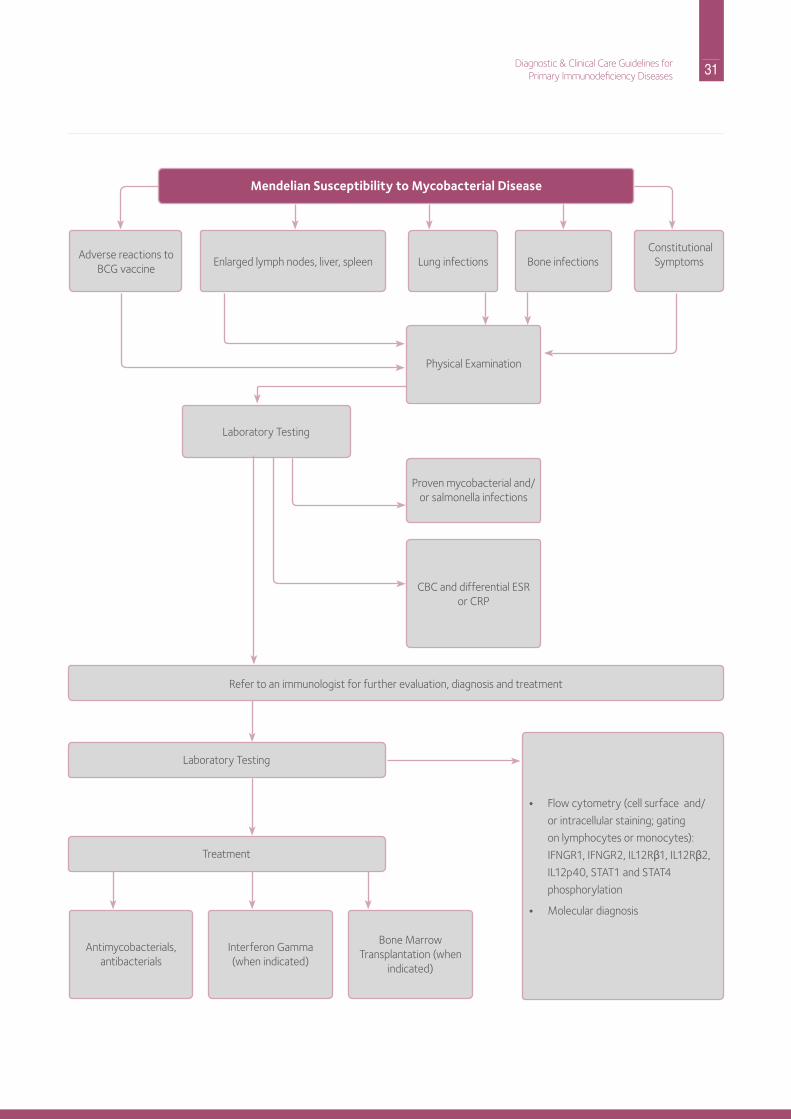
Mendelian Susceptibility to Mycobacterial Disease
Physical Examination
Proven mycobacterial and/or salmonella infections
CBC and differential ESr or CrP
Antimycobacterials, antibacterials
interferon Gamma(when indicated)
Bone Marrow Transplantation (when
indicated)
• Flow cytometry (cell surface and/
or intracellular staining; gating
on lymphocytes or monocytes):
iFnGr1, iFnGr2, iL12rβ1, iL12rβ2,
iL12p40, STAT1 and STAT4
phosphorylation
• Molecular diagnosis
Laboratory Testing
Treatment
refer to an immunologist for further evaluation, diagnosis and treatment
Adverse reactions to BCG vaccine
Lung infections Bone infections Constitutional
SymptomsEnlarged lymph nodes, liver, spleen
Laboratory Testing

32 Diagnostic & Clinical Care Guidelines forPrimary Immunodeficiency Diseases
33 Diagnostic & Clinical Care Guidelines forPrimary Immunodeficiency Diseases
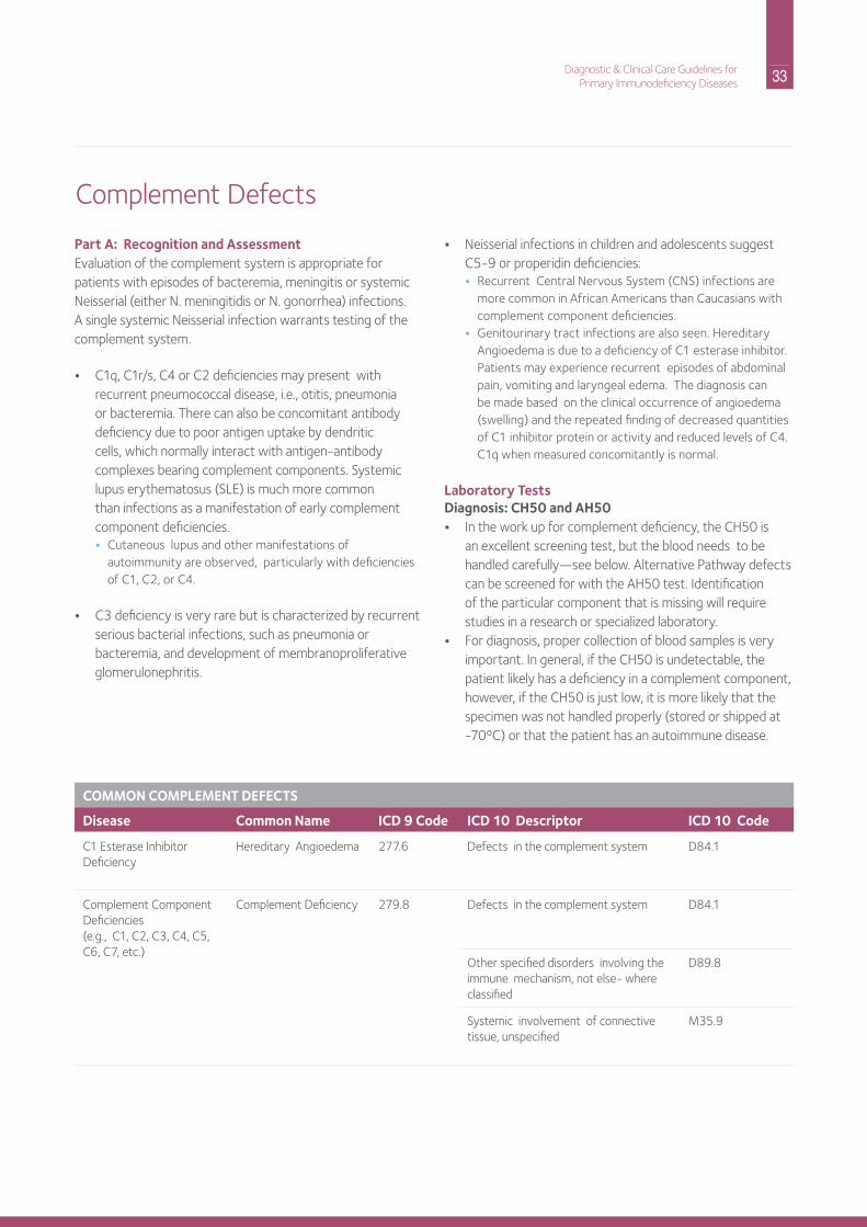
Part A: Recognition and AssessmentEvaluation of the complement system is appropriate for patients with episodes of bacteremia, meningitis or systemic neisserial (either n. meningitidis or n. gonorrhea) infections. A single systemic neisserial infection warrants testing of the complement system.
• C1q, C1r/s, C4 or C2 deficiencies may present with recurrent pneumococcal disease, i.e., otitis, pneumonia or bacteremia. There can also be concomitant antibody deficiency due to poor antigen uptake by dendritic cells, which normally interact with antigen-antibody complexes bearing complement components. Systemic lupus erythematosus (SLE) is much more common than infections as a manifestation of early complement component deficiencies.• Cutaneous lupus and other manifestations of
autoimmunity are observed, particularly with deficiencies of C1, C2, or C4.
• C3 deficiency is very rare but is characterized by recurrent serious bacterial infections, such as pneumonia or bacteremia, and development of membranoproliferative glomerulonephritis.
• neisserial infections in children and adolescents suggest C5-9 or properidin deficiencies:• recurrent Central nervous System (CnS) infections are
more common in African Americans than Caucasians with complement component deficiencies.
• Genitourinary tract infections are also seen. hereditary Angioedema is due to a deficiency of C1 esterase inhibitor. Patients may experience recurrent episodes of abdominal pain, vomiting and laryngeal edema. The diagnosis can be made based on the clinical occurrence of angioedema (swelling) and the repeated finding of decreased quantities of C1 inhibitor protein or activity and reduced levels of C4. C1q when measured concomitantly is normal.
Laboratory TestsDiagnosis: CH50 and AH50• in the work up for complement deficiency, the Ch50 is
an excellent screening test, but the blood needs to be handled carefully—see below. Alternative Pathway defects can be screened for with the Ah50 test. identification of the particular component that is missing will require studies in a research or specialized laboratory.
• For diagnosis, proper collection of blood samples is very important. in general, if the Ch50 is undetectable, the patient likely has a deficiency in a complement component, however, if the Ch50 is just low, it is more likely that the specimen was not handled properly (stored or shipped at -70ºC) or that the patient has an autoimmune disease.
Complement defects
COMMON COMPLEMENT DEFECTS
Disease Common Name ICD 9 Code ICD 10 Descriptor ICD 10 Code
C1 Esterase inhibitor deficiency
hereditary Angioedema 277.6 defects in the complement system d84.1
Complement Componentdeficiencies(e.g., C1, C2, C3, C4, C5, C6, C7, etc.)
Complement deficiency 279.8 defects in the complement system d84.1
other specified disorders involving the immune mechanism, not else- where classified
d89.8
Systemic involvement of connective tissue, unspecified
M35.9
34 Diagnostic & Clinical Care Guidelines forPrimary Immunodeficiency Diseases
Part B: Management, Expectations, Complications and Long Term MonitoringProphylactic antibiotics may be appropriate for deficiencies of any of the components of the complement cascade. Meningococcal vaccine and/or antibiotic prophylaxis may be helpful for any person diagnosed with a C5 through C9 deficiency.
All patients with complement deficiencies should receive immunization with Prevnar® or Pneumovax or both for pneumococcal infection prevention. Early recognition of fevers and prompt evaluation (including blood cultures) is very important.
Complications of complement deficiency include autoimmune disease, especially systemic lupus erythematosus, lupus-like syndromes, glomerulonephritis, and infections.
Mannose binding lectin (MBL), a protein of the innate immune system, is involved in opsonization and phagocytosis of micro- organisms. MBL recognizes oligosaccharides and can activate the classical C-pathway. however, MBL deficiency is relatively common and while it may modify other diseases, like cystic fibrosis, it does not appear to be a major immunodeficiency by itself.
Part C: Practical Aspects of Genetic CounselingThe genetic basis is known for all of the complement defects but testing is only available in specialized laboratories. All forms of inheritance have been reported for the complement defects, and it is very important to determine the specific complement factor involved as well as its molecular basis.
The genetic basis of hereditary angioedema is known and
follows autosomal dominant inheritance. Please see the general genetics section for a more complete description of autosomal dominant inheritance. Family history is important in the evaluation of angioedema. Genetic testing is available for C1 Esterase inhibitor deficiency and can be important in distinguishing the hereditary form of the disorder from the acquired forms.
Part D: Frequently Asked Questions1. Can my child receive all types of vaccines?
Yes, individuals with complement defects usually have normal T and B cell function.
2. Are there infection risks if my child with a complement defect attends public school or goes to public places? no, in general the problems with infections are due to those that your child would come in contact with in a variety of physical settings. however, if there is a known outbreak of meningitis in your community, it may be prudent to keep your child at home out of the school environment and administer antibiotics recommended by the public health authorities.
3. What should I do if my child develops fever and appears sick? in this case, the child should be taken to your doctor’s office or the closest Er for evaluation. inform the healthcare personnel that your child has a Complement deficiency and needs a blood culture (or a spinal fluid evaluation if somnolent), followed by starting treatment with intravenous antibiotics that cover neisseria bacteria. it is recommended that you carry a letter from your (immunology) physician explaining the situation and the need for such an approach.
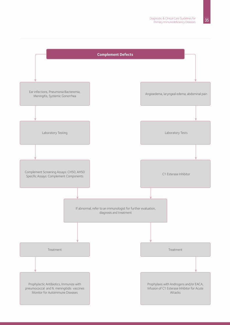
35 Diagnostic & Clinical Care Guidelines forPrimary Immunodeficiency Diseases
Complement Defects
Ear infections, Pneumonia Bacteremia, Meningitis, Systemic Gonorrhea
Angioedema, laryngeal edema, abdominal pain
Laboratory Testing Laboratory Tests
Complement Screening Assays: Ch50, Ah50Specific Assays: Complement Components
C1 Esterase inhibitor
if abnormal, refer to an immunologist for further evaluation, diagnosis and treatment
Treatment Treatment
Prophylactic Antibiotics, immunize with pneumococcal and n. meningitidis vaccines
Monitor for Autoimmune diseases
Prophylaxis with Androgens and/or EACA, infusion of C1 Esterase inhibitor for Acute
Attacks
36 Diagnostic & Clinical Care Guidelines forPrimary Immunodeficiency Diseases
due to the genetic nature of the primary immunodeficiency diseases, genetic counseling for the individual affected by these diseases, as well as family members, is a very important part of comprehensive care. Genetic counseling is typically provided once the individual has been diagnosed with a specific primary immunodeficiency disease. While most immunologists are knowledgeable about the genetic aspects of primary immunodeficiency disease, it is sometimes helpful to refer the individual or family member to a genetic counselor who has expertise in providing complex information in an unbiased and easily understood manner. issues addressed in genetic counseling for a primary immunodeficiency disease should include:
• discussion of the clinical diagnosis and the gene(s) responsible for the disorder, if known.
• determination of available molecular genetic testing for confirmation of the diagnosis, if applicable, depending on the disorder in question.
• An accurate intake of the patient’s family history, preferably in the form of a pedigree.
• determination and discussion of inheritance pattern and recurrence risk for future children.
• identification of family members who may be at risk of being affected with the disorder or at risk of being carriers.
• Brief discussion of the availability of prenatal diagnosis options for the particular primary immunodeficiency. This can be discussed again in more detail in future genetic counseling sessions when appropriate.
• discussion of whether the family should save the cord blood of future infants born to them. if the infant is affected, this would not be helpful. if the infant is normal, the cord blood could be used as a source of stem cells for affected hLA- identical members of the family who might need a transplant for immune reconstitution.
Patterns of InheritanceThe pattern of inheritance is very important when evaluating the patient with a primary immunodeficiency disease. Many of the primary immunodeficiency diseases follow X-linked recessive inheritance. This means that the genes responsible for these disorders are located on the X chromosome and these conditions affect only males. Affected males have either inherited the gene defect from their mothers who were carriers of the gene defect or the gene defect occurred as a new mutation for the first time in the egg that formed the affected male.
About one third of all X-linked defects are identified as new mutations. in these cases, the mothers are not carriers or are the first affected (carrier) member of the family due to a spontaneous (“de novo”) mutation affecting the father’s sperm, or, rarely, the mother’s egg; the family history is otherwise negative. Female carriers of the gene defect do not typically show clinical symptoms. however, half of all boys born to female carriers are affected with the disorder and half of the daughters may be carriers like their mothers. Therefore, in families with X-linked disorders, it is important to determine whether the gene defect is inherited or is a new mutation, because this will greatly affect the recurrence risk to other family members. All biologic daughters of an affected male will be carriers, while the boys will be genetically normal.
The other most common pattern of inheritance for primary immunodeficiency diseases is autosomal recessive. disorders following this pattern are caused by gene defects on any of the 22 pairs of numbered chromosomes (not the X or Y) and therefore, affect both males and females. in this type of inheritance, the condition is only expressed when both parents are carriers of the gene defect and both have passed the defective gene on to their child. These couples have a one in four (25%) chance of having an affected child, and a two in four (50%) chance of having a child that is a carrier with each pregnancy.
Some primary immunodeficiency diseases follow autosomal dominant inheritance. These disorders are caused by gene defects on any of the numbered chromosomes and affect both males and females. Unlike autosomal recessive inheritance, only one copy of the gene defect needs to be present for the condition to be expressed in an individual. The defective copy overrides the individual’s normal copy of the gene. individuals affected with these disorders have a 50% chance of passing the gene defect on to each of their children, regardless of gender. it is important to note that some of these disorders occur as new mutations in the affected individual and a family history may be negative.
A number of factors complicate genetic counseling for primary immunodeficiency diseases. it is important that the process of genetic counseling for primary immunodeficiency diseases be done by either an immunologist or genetic counselor knowledgeable about the specific intricacies of immune disorders. Some of the complicating factors include:
Genetic Counseling: General Considerations & Practical Aspects
37 Diagnostic & Clinical Care Guidelines forPrimary Immunodeficiency Diseases
• Primary immunodeficiency diseases are a group of more than 240 different disorders and there are at least that many genes involved in these disorders.
• Some primary immunodeficiency diseases have the same or similar clinical presentation, but different genetic causes. This impacts the accurate determination of inheritance pattern and recurrence risk to other family members.
• The genetic basis is still not known for many of the primary immunodeficiency diseases, including some of the most common disorders, making it difficult to accurately determine inheritance patterns and risk to other family members. This also makes it difficult to offer prenatal diagnostic options.
• Very few commercial laboratories perform molecular diagnostic procedures for primary immunodeficiency diseases and, for those that do, costs may be $2,500-$3,000 or more per family. Genetic testing is also possible in some research laboratories. These considerations may influence accessibility to testing as well as timely receipt of test results. For a current list of laboratories performing genetic testing of primary immunodeficiency diseases, consult your immunologist.
Genetic counseling also involves a psychosocial component. This is true when providing genetic counseling to families affected by the primary immunodeficiency disorders. The emotional aspect of having a genetic disease in the family can be a heavy burden and this can be explored in a genetic counseling session. in addition, the discussion of prenatal diagnostic options can be a sensitive topic, since interruption of a pregnancy may be a consideration. Prenatal diagnosis for primary immunodeficiency diseases may be an option if the genetic defect is known in the family.
This could be done through a chorionic villus sampling performed in the first trimester of pregnancy or through amniocentesis performed in the second trimester. Each of these procedures has a risk of miscarriage of the pregnancy associated with it, so these risks need to be discussed thoroughly by the genetic counselor. Prenatal diagnosis for a primary immunodeficiency disease may be considered when a couple wants to better prepare for the birth of an infant with the disease in question. For example, knowing that a fetus is affected can give a couple time to start looking for a match for a bone marrow donor if this is the therapy for the disease. Knowing that a fetus is unaffected can offer great relief to the couple for the rest of the pregnancy.
Prenatal diagnosis may also be considered when a couple would choose to terminate a pregnancy of an affected fetus. Again, these considerations are thoroughly discussed in a genetic counseling session.
Finally, the discussion of gene therapy may be addressed in the genetic counseling of a family affected by a primary immunodeficiency disease. The anticipation is that gene therapy will become available for several diseases over the coming years. however, even if the treatment is perfected and found to be safe, it cannot be done unless the abnormal gene is known in the family.
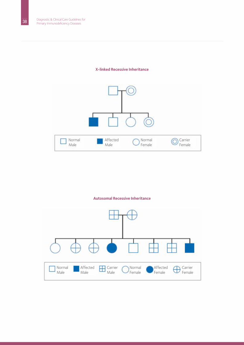
38 Diagnostic & Clinical Care Guidelines forPrimary Immunodeficiency Diseases
X-linked Recessive Inheritance
Autosomal Recessive Inheritance
normalMale
normalMale
AffectedMale
AffectedMale
normalFemale
normalFemale
AffectedFemale
CarrierFemale
CarrierFemale
CarrierMale
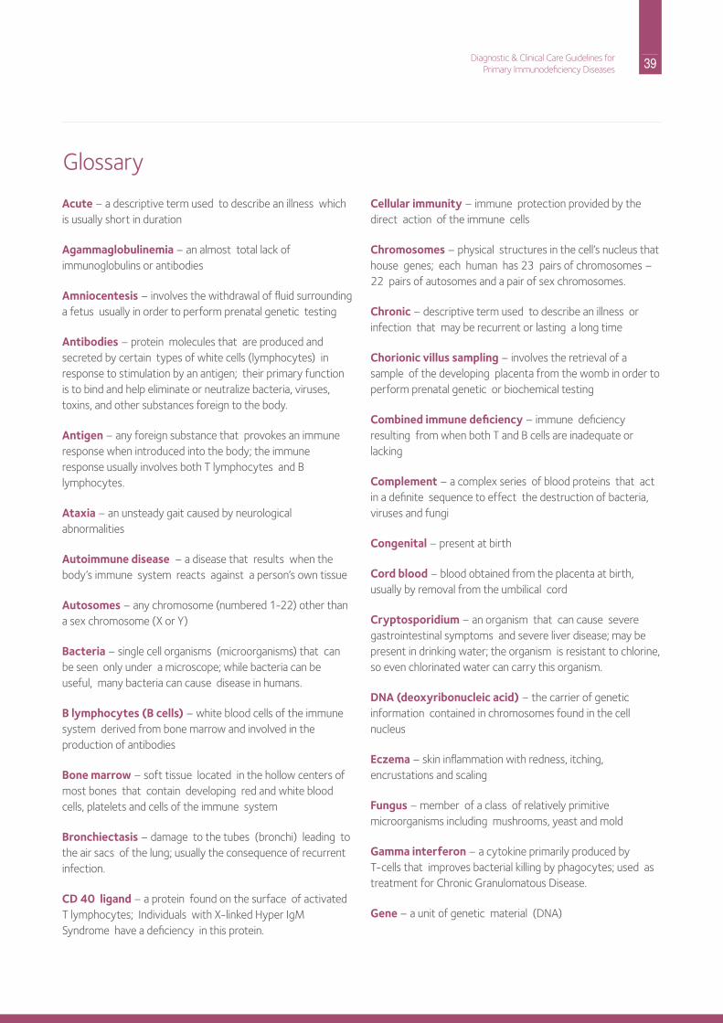
39 Diagnostic & Clinical Care Guidelines forPrimary Immunodeficiency Diseases
Acute – a descriptive term used to describe an illness which is usually short in duration
Agammaglobulinemia – an almost total lack of immunoglobulins or antibodies
Amniocentesis – involves the withdrawal of fluid surrounding a fetus usually in order to perform prenatal genetic testing
Antibodies – protein molecules that are produced and secreted by certain types of white cells (lymphocytes) in response to stimulation by an antigen; their primary function is to bind and help eliminate or neutralize bacteria, viruses, toxins, and other substances foreign to the body.
Antigen – any foreign substance that provokes an immune response when introduced into the body; the immune response usually involves both T lymphocytes and B lymphocytes.
Ataxia – an unsteady gait caused by neurological abnormalities
Autoimmune disease – a disease that results when the body’s immune system reacts against a person’s own tissue
Autosomes – any chromosome (numbered 1-22) other than a sex chromosome (X or Y)
Bacteria – single cell organisms (microorganisms) that can be seen only under a microscope; while bacteria can be useful, many bacteria can cause disease in humans.
B lymphocytes (B cells) – white blood cells of the immune system derived from bone marrow and involved in the production of antibodies
Bone marrow – soft tissue located in the hollow centers of most bones that contain developing red and white blood cells, platelets and cells of the immune system
Bronchiectasis – damage to the tubes (bronchi) leading to the air sacs of the lung; usually the consequence of recurrent infection.
CD 40 ligand – a protein found on the surface of activated T lymphocytes; individuals with X-linked hyper igM Syndrome have a deficiency in this protein.
Cellular immunity – immune protection provided by the direct action of the immune cells
Chromosomes – physical structures in the cell’s nucleus that house genes; each human has 23 pairs of chromosomes – 22 pairs of autosomes and a pair of sex chromosomes.
Chronic – descriptive term used to describe an illness or infection that may be recurrent or lasting a long time
Chorionic villus sampling – involves the retrieval of a sample of the developing placenta from the womb in order to perform prenatal genetic or biochemical testing
Combined immune deficiency – immune deficiency resulting from when both T and B cells are inadequate or lacking
Complement – a complex series of blood proteins that act in a definite sequence to effect the destruction of bacteria, viruses and fungi
Congenital – present at birth
Cord blood – blood obtained from the placenta at birth, usually by removal from the umbilical cord
Cryptosporidium – an organism that can cause severe gastrointestinal symptoms and severe liver disease; may be present in drinking water; the organism is resistant to chlorine, so even chlorinated water can carry this organism.
DNA (deoxyribonucleic acid) – the carrier of genetic information contained in chromosomes found in the cell nucleus
Eczema – skin inflammation with redness, itching, encrustations and scaling
Fungus – member of a class of relatively primitive microorganisms including mushrooms, yeast and mold
Gamma interferon – a cytokine primarily produced by T-cells that improves bacterial killing by phagocytes; used as treatment for Chronic Granulomatous disease.
Gene – a unit of genetic material (dnA)
Glossary
40 Diagnostic & Clinical Care Guidelines forPrimary Immunodeficiency Diseases
Gene (or genetic) testing – testing performed to determine if an individual possesses a specific gene or genetic trait; may include studies of whether the gene is altered (mutated) in any way.
Gene therapy – treatment of genetic diseases by providing the correct or normal form of the abnormal gene causing the disease
Graft-versus-host disease – a life-threatening reaction in which transplanted or transfused immunocompetent T cells attack the tissue of the recipient
Graft rejection – the immunologic response of the recipient to the transplanted tissue resulting in rejection of the transplant
Granulocyte – a white cell of the immune system characterized by the ability to ingest (phagocytize) foreign material; a granulocyte is identified by the presence of many granules when seen under a microscope; neutrophils, eosinophils, and basophils are examples of granulocytes.
Haplotype – a series of gene clusters on one human chromosome 6 that encodes a set of histocompatibility (hLA) antigens
Helper lymphocytes (Helper T cells) – a subset of T cells that help B cells and other immune cells to function optimally
Humoral immunity – immune protection provided by soluble factors, such as antibodies, which circulate in the body’s fluid
Hypogammaglobulinemia – lower than normal levels of immunoglobulins and antibodies in the blood; low levels need to be assessed as to whether the level requires replacement immunoglobulin therapy; this assessment is best made by a qualified experienced immunologist.
IgA – an immunoglobulin found in blood, tears, saliva, and fluids bathing the mucous membranes of respiratory and intestinal tracts
IgD – an immunoglobulin whose function is poorly understood at this time
IgE – an immunoglobulin found in trace amounts in the blood; antibodies of this type are responsible for allergic reactions.
IgG – the most abundant and common of the immunoglobulins; igG functions mainly against bacteria and some viruses; it is the only immunoglobulin that can cross the placenta.
IgM – an immunoglobulin found in the blood; igM functions in much the same way as igG but is formed earlier in the immune response; it is also very efficient in activating complement.
Immune response – the activity or response of the immune system against foreign substances
Immunodeficiency – a state of either a congenital (present at birth) or an acquired abnormality of the immune system that prevents adequate immune responsiveness
Immunoglobulins (Ig) – the antibody proteins; there are five major classes: igG, igA, igM, igd, and igE.
Intravenous immunoglobulin (IVIG) – immunoglobulin therapy injected directly into the vein
Killer lymphocytes – T lymphocytes that directly kill microorganisms or cells that are infected with microorganisms, also called “cytotoxic T lymphocytes”
Leukemia – type of cancer of the immune system
Live vaccines – live viruses are used in the vaccine; live vaccines (particularly oral polio and the chickenpox vaccine) when injected into immunodeficient individuals, can transmit the diseases they were designed to prevent.
Less virulent agents – organisms that do not generally infect humans; they may cause disease in individuals with weak immune systems, these are called “opportunistic infections.”
Leukocyte (white blood cell) – a group of small colorless blood cells that play a major role in the body’s immune system; there are five basic white blood cells: monocytes, lymphocytes, neutrophils, eosinophils and basophils.
Lymph – fluid made up of various components of the immune system; it flows throughout tissues of the body via the lymph nodes and lymphatic vessels.
Lymph nodes – small bean-sized organs of the immune system, distributed widely throughout the body; each lymph node contains a variety of specialized compartments that house B cells, T cells and macrophages; lymph nodes unite in one location the several factors needed to produce an immune response.
41 Diagnostic & Clinical Care Guidelines forPrimary Immunodeficiency Diseases
Lymphocytes – small white cells, normally present in the blood and in lymphoid tissue, that bear the major responsibility for carrying out the functions of the immune system; there are three major forms of lymphocytes, B cells, T cells and natural killer (nK) cells, each of which have distinct but related functions in generating an immune response.
Lymphoma – type of cancer of the immune system
Macrophages – phagocytic cells found in the tissue, able to destroy invading bacteria or foreign material
Metabolism – a general term which summarizes the chemical changes within a cell resulting in either the building up or breaking down of living material
Microorganisms – minute living organisms, usually one-cell organisms, which include bacteria, protozoa and fungi
Molecules – the smallest unit of matter of an element or compound
Monocyte – phagocytic cell found in the blood that acts as a scavenger, capable of destroying invading bacteria or other foreign material; these cells develop into macrophages in tissues.
Mucosal surfaces – surfaces that come in close contact with the environment, such as mouth, nose, gastrointestinal tract, eyes, etc. igA antibodies protect these surfaces from infection.
Neutropenia – a lower than normal amount of neutrophils in the blood
Neutrophils – a type of granulocyte, found in the blood and tissues that can ingest microorganisms
Opportunistic infection – an infection caused by a usually benign or less virulent agent, but resulting when those organisms become established under certain conditions, such as in immunodeficient individuals
Organism – an individual living thing
Osteomyelitis – infection of the bone
Parasite – a plant or animal that lives, grows and feeds on or within another living organism
Persistent infections – infections marked by the continuance of an infectious episode despite appropriate medical interventions
Petechiae – pinhead sized red spots resulting from bleeding into the skin, usually caused by low platelet numbers (thrombocytopenia)
Phagocyte – a general class of white blood cells that ingest microbes and other cells and foreign particles; monocytes, macrophages and neutrophils are types of phagocytes.
Plasma cells – antibody-producing cells descended from B cells
Platelets – smallest of the blood cells; their primary function is associated with the process of blood clotting.
Polysaccharides – complex sugars
Primary immunodeficiency – immunodeficiency that is intrinsic to the cells and tissues of the immune system, not due to another illness, medication or outside agent damaging the immune system
Prophylactic – medical therapy initiated to prevent or guard against disease or infection
Protein – a class of chemicals found in the body made up of chains of amino acids (building blocks); immunoglobulins (antibodies) are proteins.
Recurrent infections – infections, such as otitis, sinusitis, pneumonia, deep-seated abscess, osteomyelitis, bacteremia or meningitis, that occur repeatedly
Secondary immunodeficiency – immunodeficiency due to another illness or agent, such as human immunodeficiency virus (hiV), cancer or chemotherapy
Sepsis – an infection of the blood
Spleen – an organ in the abdominal cavity; it is directly connected to the blood stream and like lymph nodes contains B cells, T cells, and macrophages.
Stem cells – cells from which all blood cells and immune cells are derived; bone marrow is rich in stem cells. The stem cells referred to in this glossary do not include embryonic stem cells, which are involved in the generation of all cells and tissues.
42 Diagnostic & Clinical Care Guidelines forPrimary Immunodeficiency Diseases
Subcutaneous infusion (SC) – administration of a drug or biologic (e.g., immunoglobulin) slowly, directly under the skin often using a small pump
Telangiectasia – dilation of small blood vessels
Thrombocytopenia – low platelet count
Thrush – a fungal disease on mucous membranes caused by Candida albicans infections
Thymus gland – a lymphoid organ located behind the upper portion of the sternum (breastbone) where T cells develop; this organ increases in size from infancy to adolescence and then begins to shrink.
T lymphocytes (or T cells) – lymphocytes that are processed in the thymus; they have a central role in carrying out the immune response.
Unusual infectious agents – these are normally non-pathogenic agents or those not generally found in humans which can cause serious disease in immunocompromised patients
Vaccine – a substance that contains components from an infectious organism which stimulates an immune response in order to protect against subsequent infection by that organism
Virus – a submicroscopic microbe causing infectious disease which can only reproduce in lining cells
White blood cells – see leukocyte

Notes
43 Diagnostic & Clinical Care Guidelines forPrimary Immunodeficiency Diseases