Embed Size (px)
Citation preview
1
5 ORIGINAL ARTICLE DIAGNOSTIC COMPARISON OF ULTRASONOGRAPHY WITH MRCP IN CHOLANGIO PANCREATIC LESIONS 1. DR (MAJ) DEEPAK KUMAR RAJPUT Associate Professor Radiology dept, AMC MET Medical College& LG Hospital Ahmedabad Gujarat. 2. DR DRUSTY MAJMUDAR *Assistant Professor Radiology dept, AMC MET Medical College& LG Hospital Ahmedabad Gujarat. * Correspondence author DR DRUSTY MAJMUDAR ABSTRACT OBJECTIVE :- To evaluate the diagnostic accuracy between USG & MRCP in the patients suspected of biliary and pancreatic Pathology. MATERIAL & METHODS : Sixty patients of all age groups and both sexes, attending the hospital, suspected of biliary and pancreatic pathology, were examined first by USG and followed by MRCP. The findings were co-related with ERCP and biopsy reports. RESULTS :- Out of 60 patients, 41 patients had biliary pathology and 19 patients had pancreatic pathology. Out of this, MRCP was 98 % accurate in diagnosis when results were compared in all cases. USG didn’t help in case of CBD stricture, in evaluating Pancreatic duct in case of Chronic pancreatitis and in lower end of CBD pathology. CONCLUSION :- USG is the cheap and easily available modality so, it is the primary investigative modality for suspected patients of biliary and pancreatic pathology, but MRCP has high diagnostic value. KEY WORDS :- Ultrasonography, MRCP (MRI).
2
INTRODUCTION:- MRCP (magnetic resonance cholangiopancreaticography) is emerging as an exciting tool for the non- invasive evaluation of the pancreatic and biliary duct system [I, II, III]. Non invasive imaging modalities such as Ultrasonography and CT Scan are often the primary imaging modalities in the evaluation of the biliary tree and pancreatic ducts [IV, V]. So, two noninvasive, non-radiation modalities for the evaluation of pancreatic and biliary duct system are USG & MRCP. AIM :- Comparison of diagnostic accuracy between USG and MRCP in the patients of suspected biliary and pancreatic pathology in L.G Hospital, Ahmedabad, Gujarat from July 2015 to Dec 2016. MATERIALS AND METHODS:- Sixty patients of all age groups and both sexes of suspected biliary and pancreatic pathology were subjected to real time Ultrasonographic examination of biliary tree (IHBR, CHD, CBD, CYSTIC DUCT), GB and pancreas including Main Pancreatic Duct (GE Logiq P5, 4 to 11 MHz probes used for study), subsequently followed by MRCP (SIEMENS, 1.5 T machine was used for study). Results were compared with either ERCP or biopsy findings with histopathological reports and post-operative findings. RESULTS:- Comparison of diagnostic accuracy between USG & MRCP in
3
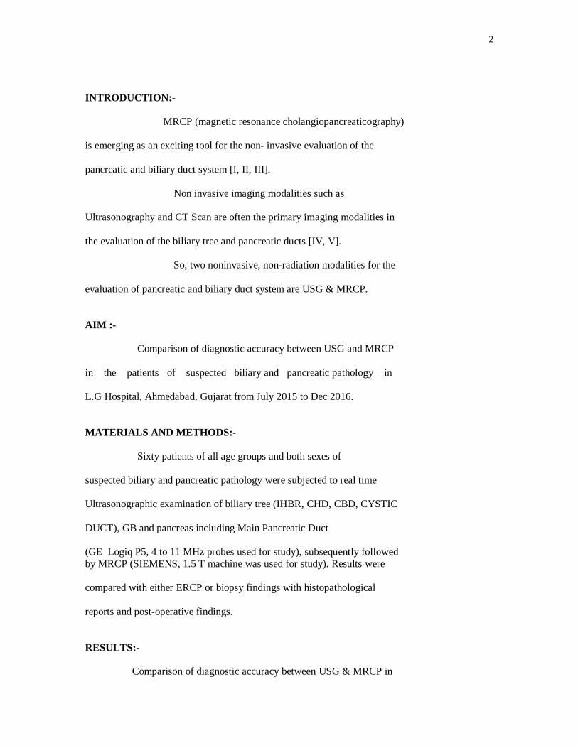
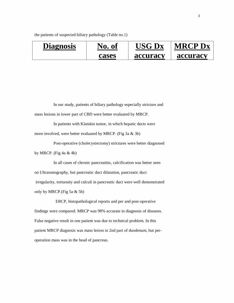
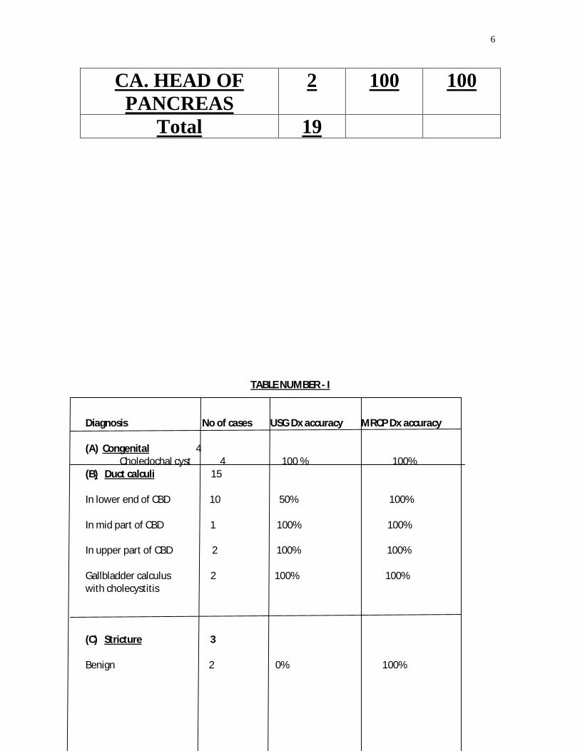
the patients of suspected biliary pathology (Table no.1)
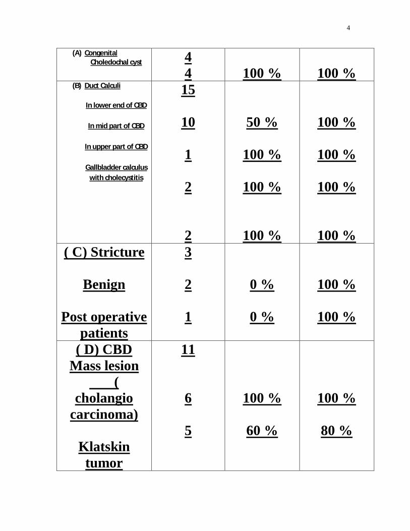
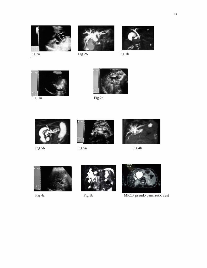
In our study, patients of biliary pathology especially stricture and mass lesions in lower part of CBD were better evaluated by MRCP. In patients with Klatskin tumor, in which hepatic ducts were more involved, were better evaluated by MRCP. (Fig 3a & 3b) Post-operative (cholecystectomy) strictures were better diagnosed by MRCP. (Fig 4a & 4b) In all cases of chronic pancreatitis, calcification was better seen on Ultrasonography, but pancreatic duct dilatation, pancreatic duct irregularity, tortuosity and calculi in pancreatic duct were well demonstrated only by MRCP.(Fig 5a & 5b) ERCP, histopathological reports and per and post-operative findings were compared. MRCP was 98% accurate in diagnosis of diseases. False negative result in one patient was due to technical problem. In this patient MRCP diagnosis was mass lesion in 2nd part of duodenum, but per- operation mass was in the head of pancreas.
Diagnosis No. of cases
USG Dx accuracy
MRCP Dx accuracy
4
(A) Congenital Choledochal cyst 4
4
100 %
100 % (B) Duct Calculi
In lower end of CBD
In mid part of CBD
In upper part of CBD
Gallbladder calculus
with cholecystitis
15
10
1
2
2
50 %
100 %
100 %
100 %
100 %
100 %
100 %
100 % ( C) Stricture
Benign
Post operative
patients
3
2
1
0 %
0 %
100 %
100 %
( D) CBD Mass lesion
( cholangio
carcinoma)
Klatskin tumor
11
6
5
100 %
60 %
100 %
80 %
5
TABLE II TABLE II
PANCREATIC LESION
NO. OF
CASES
USG accurac
y
MRCP accurac
y
ACUTE PANCREATITIS
2 50 100
CHRONIC PANCREATITIS
5 60 80
ACUTE ON CHRONIC
PANCREATITIS
3 66 100
PSEUDOPANCREATIC CYST
5 100 100
HYDATID CYST 2 50 100
Lower CBD
mass (E)
Periampullary mass
7 85.6 % 71.4 %
(F) Gallbladder
mass
1 100 % 100 %
Total 41
6
CA. HEAD OF PANCREAS
2 100 100
Total 19
TABLE NUMBER - I Diagnosis No of cases USG Dx accuracy MRCP Dx accuracy (A) Congenital 4
Choledochal cyst 4 100 % 100% (B) Duct calculi 15 In lower end of CBD 10 50% 100% In mid part of CBD 1 100% 100% In upper part of CBD 2 100% 100% Gallbladder calculus 2 100% 100% with cholecystitis (C) Stricture 3 Benign 2 0% 100%
7
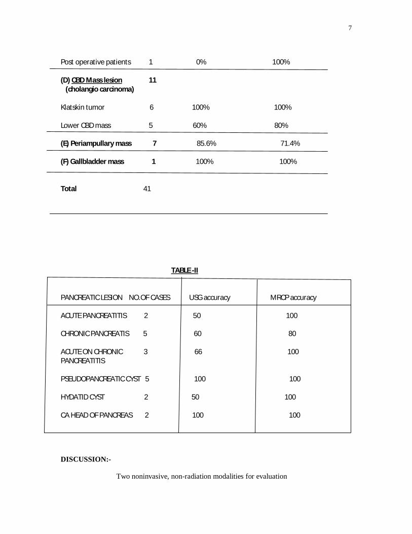
Post operative patients 1 0% 100% (D) CBD Mass lesion 11 (cholangio carcinoma) Klatskin tumor 6 100% 100% Lower CBD mass 5 60% 80% (E) Periampullary mass 7 85.6% 71.4% (F) Gallbladder mass 1 100% 100% Total 41 TABLE -II PANCREATIC LESION NO.OF CASES USG accuracy MRCP accuracy ACUTE PANCREATITIS 2 50 100 CHRONIC PANCREATIS 5 60 80 ACUTE ON CHRONIC 3 66 100 PANCREATITIS PSEUDOPANCREATIC CYST 5 100 100 HYDATID CYST 2 50 100 CA HEAD OF PANCREAS 2 100 100 DISCUSSION:- Two noninvasive, non-radiation modalities for evaluation
8
of biliary & pancreatic pathologies are USG & MRCP. Magnetic resonance cholangiopancreaticography (MRCP) is a radiologic technique that produces images of the pancreaticobiliary tree
that are similar in appearance to those obtained by invasive radiographic methods, such as endoscopic retrograde cholangiopancreaticography (ERCP) The basic principle underlying MRCP is that body fluids, such as bile and pancreatic secretions, have high signal intensity on heavily T2-weighted magnetic resonance sequences (i.e., they appear bright), whereas background tissues generate little signal (i.e., they appear dark) [VI]. Since its introduction by wallner et al in 1991[VII], MRCP has undergone tremendous technical changes essentially in the search for an optional imaging sequence. In 1991 - wallner BK et al introduced MRCP which was used as a breath hold two dimensional, T-2 gradient echo sequence using steady state Free Precession (SSFP) [ VII]. Marimoto improved image quality by introducing - 3D SSFP sequences [III]. Modified FSE sequences were introduced recently. These are the RARE (Rapid Acquisition with Rapid Enhancement Sequence) and HASTE (Half Fourier Acquisition Single Shot Turbo Spin Echo Sequences). So, now HASTE & RARE sequences are ideal cholangiographic sequences for MRCP and a combination of HASTE & RARE takes only 10 minutes imaging time [VIII]. Currently, diagnostic accuracy of MRCP is considered to be equivalent to ERCP for a broad spectrum of benign and malignant pancreatic & biliary diseases [IX]. Ultrasonography has limitation especially in the
9
evaluation of distal CBD where bowel gas, debris/ fluid in the duodenum and obesity can degrade the image quality [IV, V]. Other imaging modalities are invasive, hence MRCP is an excellent modality for evaluation of biliary and pancreatic diseases. Meta-analysis including 60 patients study shows that MRCP is 97% sensitive & 98% specific for defining the biliary tract obstruction [X]. The overall sensitivity, specificity and accuracy of MRCP in the detection of bile duct lesions were 97%, 98% and 97%, respectively [XI]. Specificity for detecting chronic pancreatitis was 99% [XII]. Results of studies show clearly that USG is not able to diagnose cases of stricture, mass lesion and calculi in lower end of CBD. CONCLUSION:- In the patients of suspected biliary and pancreatic pathology, USG is the primary imaging modality of choice, but it has very less diagnostic accuracy in evaluation of benign, malignant stricture of lower end of CBD, calculi in lower end of CBD, congenital anatomical variants, post-operative biliary tree anatomy and pathology. So, MRCP based on heavily T-2 weighted images (HASTE & RARE sequences) produces remarkable increased contrast between stagnant fluid (bile) and background (abdominal fat, hepatic, pancreatic parenchyma) has almost 100% diagnostic accuracy. So, all patients having biliary and pancreatic pathology, not clearly diagnosed by USG must be evaluated by MRCP for diagnostic accuracy.
10
REFERENCES:-
I. Barish M A, Yuces E H, Soto J A et al. 1995 M R Cholangiography: Efficiency of three-dimentional turbo spin echo technique : Am J Roentgenol 165 : 295-300.
II. Soto JA, Barish MA, Yucel EK, et al. Pancreatic duct: MR cholangiopancreatography with three-dimensional fast spin-echo technique. Radiology 1995; 196:459-464.
III. Morimoto K, Shimoi M, Shirakawa T et al. (1992) Biliary
obstruction evaluation with 3 dimensional MRCP. Radiology 183:578-580.
IV. Baron RL: Common bile duct stones. Reassesment of criteria for CT diagnosis. Radiology 162:419-424,1987.
V. Baron RL: Computed tomography of the biliary tree. Radiologic Clin North America 29(6) 1235-1250,1991.
VI. Matthew A Barish, E. Kent Yucel and Joseph T. ferrucci, Magnetic Resonance Cholangiopancreatography. The New England J Of Medicine July 22 1999;341:258-264.
VII. Wallner BK, Schumacher K.A. Weidenmaier W. Fariedrich jm (1991) Dilated biliary tract: evaluation with MR Cholangiography with a T2 weighted contrast-enhanced fast sequence. Radiology 181:805-808.
11
VIII. Hiroyuki Irie, MD., Hiroshi Honda, MD., et al. Optimal MR
Cholangiopancreatographic Sequence and Its Clinical Application. Radiology 1998; 206 : 379-38.
IX. Soto JA, Barish MA, Yucel EK, Siegenberg D, Ferrucci JT,
Chuttani R. Magnetic resonance cholangiography: comparison to
endoscopic retrograde cholangiopancreatography. Gastroenterology
1996;110:589-597.
X. Joseph Romagnuolo, Marc Bardou, Elham Rahme, et al. Magnetic
Resonance Cholangiopancreatography: A Meta-Analysis of Test
Performance in Suspected Biliary Disease. Ann Intern Med
2003;139:547-557.
XI. Varghese JC, Farrell MA, Courtney G, et al. A prospective
comparison of magnetic resonance cholangiopancreatography with
endoscopic retrograde cholangiopancreatography in the evaluation of
patients with suspected biliary tract disease. Clin Radiol 1999
Aug; 54(8):513-20.
XII. Alcaraz MJ, De la Morena EJ, Polo A, et al. A comparative study of
magnetic resonance cholangiography and direct cholangiography.
Rev Esp Enferm Dig. 2000 Jul;92(7):427-38.
LEGENDS: FIGURE 1a :- USG - CBD is dilated upto lower end, calculi could not be
12
not seen. FIGURE 1b :- MRCP - CBD is dilated upto lower end, calculi seen at distal end. FIGURE 2a :- USG - Markedly dilated CBD upto lower end, cause could not be evaluated. FIGURE 2b :- MRCP - Markedly dilated CBD upto lower end, malignant stricture at lower end by mass in lower end of CBD. FIGURE 3a :- USG - Markedly dilated both hepatic ducts and IHBR, Common Hepatic Duct not seen-suggestive of Klatskin tumor. FIGURE 3b :- MRCP - Right Hepatic duct is more dilated than left hepatic duct suggestive of right hepatic duct is more involved in Klatskin tumor than left hepatic duct. FIGURE 4a :- USG - Dilated Common Hepatic duct, Gall Bladder not Seen, history of cholecystectomy before two month, CBD not seen. FIGURE 4b :- MRCP - Post –operative (cholecystectomy) stricture at lower end of Common Hepatic Duct. FIGURE 5a :- USG - Pancreatic calcification is better seen in chronic pancreatitis. FIGURE 5b :- MRCP - Main pancreatic duct and its branches are irregular, tortuous and dilated.
13
Fig 3a Fig 2b Fig 1b Fi g
Fig. 1a Fig 2a
Fig 5b Fig 5a Fig 4b
Fig 4a Fig 3b MRCP pseudo pancreatic cyst