Embed Size (px)
Citation preview
jourtal of Herpetology. Vol. 35, No. 1, pp. 92-103. 2001Copyright 2001 Society for the Study of Amphibians and Reptiles
Diagnostic Histological Findings in Yosemite Toads (Bufo canorus)from a Die-off in the 1970s
D. EARL GREEN' 2 AND CYNTHIA KAGARISE SHERMAN 3
'National Institutes of Health, Office of Research Support, ieterinary Resources Program, 28 Library Drive, Bethesda,
Marvland 20892-5230 USA
342 Sparrow Crest, Ithlaca, Ni9ew York 14850 USA; E-mnail: [email protected]
ABSTRACT.-TWelVe adult and 25 larval Yosemite toad (Bufo canorus) specimens from the eastern Sierra
Nevada of California were examined histologically for evidence of infectious, toxicological, and degenerative
diseases. The preserved toads were selected from 21 that had been salvaged or collected during a die-off
in 1976-1979 that immediately preceded a population decline. Causes of death of four toads were determined
histologically; clinical signs and field observations suggested causes of death of three more. Four toads died
of infectious diseases, including chytridiomycosis of the skin (N = 1), bacillary septicemia (N = 2), and
combined chytridiomycosis and bacterial septicemia (N = 1). Infections by a funguslike organism (Der-
mosporidiu1ni penneri), renal myxozoa (Leptotheca ohimacheri), larval Rhabdias, various gastrointestinal nem-
atodes. urinary bladder flukes, and lung flukes were detected in five specimens. No evidence of degenerative
diseases, virus infections, or intoxications was found. The variety of lethal diseases and our inability to
determine the causes of death of five specimens suggests that one or more histologically undetectable
diseases or intoxications may have also contributed to the deaths and population decline.
Amphibian populations have declinedthroughout the world (Heyer et al., 1988; Brad-ford. 1991; Carey, 1993; Pounds and Crump,1994; Stebbins and Cohen, 1995; Laurance et al.,1996). Many declines can be traced to anthro-pogenic factors, but some have occurred in rel-atively protected habitats with minimal humanimpacts (e.g., national parks or remote moun-tain regions: Crump et al., 1992; Drost and Fell-ers, 1996; Lips, 1998, 1999). These declines havebeen of two general types: (1) abrupt die-offs inwhich many sick and dead animals are found,.followAed by population crashes 3-24 months lat-er (Kagarise Sherman and Morton, 1993; Laur-ance et al., 1996; Lips, 1998, 19991; and (2) de-clines without documented die-offs or detectedfield casualties (Fellers and Drost, 1993; Poundsand Crump, 1994; Drost and Fellers, 1996; Fisherand Shaffer, 1996).
The significance of field casualties is twofold.First. the observation of dead and dying toads(or other small vertebrates) suggests that actualmortality numbers are much higher because theremoval of carcasses by predators and scaven-gers has been overwhelmed by the number ofdeaths (Wobeser and Wobeser, 1992). Second,the sudden occurrence of casualties has epide-miologic features characteristic of an introducedinfectious disease or intoxication.
In Califomia, population declines have been
2 Present Address: National W7ildlife Health Center,U.S. Geological Survey, U.S. Department of Interior,6006 Schroeder Road, Madison, Wisconsin 53711-6223USA; E-mail: [email protected]
documented or suggested for Ambystoma califor-niense, Bufo boreas, Bufo canorus, Bufio microscaphuscalifornicus, Rana aurora, Rana boylii, Rana casca-dae, Rana nmuscosa, Scaphiopus hammondii, Scaphio-pus intermontanus, and Taricha spp. (Bradford,1991; Fellers and Drost, 1993; Kagarise Shermanand Morton, 1993; Stebbins and Cohen, 1995;Drost and Fellers, 1996; Fisher and Shaffer,1996). A documented die-off in one B. canoruspopulation near Yosemite National Park preced-ed widespread population declines (KagariseSherman and Morton, 1993; Drost and Fellers,1996); this die-off coincided with observed massmortality, rapid population declines, and localextinctions in the mountain yellow-legged frog(R. mnuscosa) in nearby Kings Canyon NationalPark (Bradford, 1991).
Recent studies involving mass casualties ofentire amphibian assemblages in Costa Rica,Panama, and Australia (Berger et al., 1998; Lips,1998, 1999) as well as more localized and spe-cies-specific mass mortality of eggs, larvae, neo-tenes, and postmetamorphic adults (W'Vorthylakeand Hovingh, 1989; Bradford, 1991; Bl'austein etal., 1994a; Jancovich et al., 1997) have implicatedbacteria, fungi, and viruses as etiologies. Themass mortalitv among Yosemite toads at TiogaPass was similar in its epidemiology and symp-tomatology to amphibian casualties reported inCentral America and Australia. To determinewvhether infectious disease was a factor in theSierra Nevada die-offs, histologic examinationswere performed on adult toads preserved in1976-1979, normal-appearing tadpoles collected
1 1 1 .1 ... ... I ' l l ... .
CAUSES OF DEATH IN YOSEMITE TOADS
in 1977 and 1978, and skeletal and skin remainspreserved in 1990.
MATERIALS AND METHODS
Study Species.-Bufo canorus is a montane spe-cies that historically inhabited an area of 240 kmx 60 km in the central Sierra Nevada (Karis-trom, 1962). The species is diurnal (Mullally,1953; Mullally and Cunningham, 1956) and sex-ually dichromatic (males are olive-green, fe-males are mottled black and gravish-white). In-dividuals are active above ground for onlyabout four months each year between May andSeptember, and hibernate beneath deep snowfor the remainder of the year (Karlstrom, '1962;Kagarise Sherman, 1980; Morton, 1981; KagariseSherman and Morton, 1993).
From 1971 to 1982, a population of B. canowrsadjacent to Yosemite National Park at TiogaPass, California, was censused yearly (Mortonand Sokolski, 1978; Kagarise Sherman, 1980;Mlorton, 1981, 1982; Kagarise Sherman and Mor-ton, 1993). Beginning in the late spring of 1976,field casualties were observed for three years,and by 1982, the population had declined by90% (Kagarise Sherman and Morton, 1993). Themajority of observed field casualties (19 of 36)had no gross abnormalities or apparent causesof death; six toads died while crossing a largesnowfield near the pools, and two females werefound dead after being amplexed by severalmales. However, nine (25%) casualties had evi-dence of infectious disease, characterized as red-dened skin of the ventrum and toes (N = 8), orventral skin nodules (N - 1). An additional 21toads were fotmd alive but with similarlv red-dened ventral skin and toes (Kagarise Shermanand Morton, 1993).
Collection of Specimens.-Twenty-one dead ordying adult B. canorus were collected at two lo-cations: at or near breeding sites in Tioga PassMeadow (TP1M) at the southern end of TiogaLake, Mono Countv (119'E, 380N, elevation 3030m) in 1976-1979 (N 1 19); and at a breedingarea at the northwest end of Saddlebag Lake,Mono Countv (11.90E, 38°N, 3072 m, 6.8 km fromTioga Pass) in 1977 (N = 2). In addition, theremains of 22 B. canorus (skins, partial skeletons,and ovaries and oviducts of females) were sal-vaged in 1990 from Mildred Lake, Mono County(119'E, 37'N, elevation 2975 m, 53.1 km fromTioga Pass). Twenty-five Yosemite toad tad-poles, alive and normal-appearing, were col-lected in 1977 and 1978 from the main breedingarea in TPM where sick and dead adults werefound. All collection sites were east of the crestof the Sierra Nevada.
The specimens from Tioga Pass Meadow andSaddlebag Lake were fixed in neutral buffered10% formalin and deposited in the Museum of
Zoology, University of Michigan at Ann Arbor,where they were stored in ethanol in the her-petological collection. Partial carcasses fromMildred Lake and the tadpoles were fixed andstored in isopropyl alcohol and 10% formalin,respectively, and kept in the personal collectionof CKS.
From 1971 to 1975, and from 1979 to 1982,toads were captured by searching throughoutTPM and around Saddlebag Lake. In addition,during 1976-1978, toads entering the mainbreeding area at TPM were caught by a driftfence with pitfall traps. Adult toads were per-manently marked by clipping a unique combi-nation of toes (Kagarise Sherman, 1980; Kagar-ise Sherman and Morton, 1993).
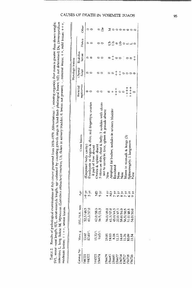
Pathology and Histology.-Adult specimenswere selected to span the range of collectiondates, sites, and causes of death as assessed atthe time of collection. Toad carcasses were ex-amined, weighed, and portions of organs andtissues were removed. Skin from the pelvicpatch and one whole hind-limb digit (usuallydigit IV, but in three toads another digit becauseof ante mortem toe clipping) were examinedfrom 12 toads collected from 1976-1979 (11 atTPM, one from Saddlebag Lake). Digits weredecalcified in saturated EDTA solution for 24-48 h. The livers, spleens, mesonephroi, lungs,urinary bladders, stomachs, and cloacae of eighttoads were examined histologically. At leastfour pieces of skin were selected for histologicalevaluation from each of 14 carcasses from Mil-dred Lake; wherever possible, yellowish ventralskin was included. Tadpoles' oral discs (tooth-rows and jaw sheaths) were examined under adissecting microscope. Tadpoles were Gosner'sstages 27-36, with one exception (stage 43-44).Tissues and whole tadpoles were processed rou-tinely through ethanol and xyiene, and embed-ded in paraffin. Six micron-thick sections werestained with hematoxylin and eosin (H&E) andGiemsa stains.
RESULTS
Three toads that had been accidentally killedin May 1.976 were chosen to serve as pre-die-offspecimens (Table 1). Gross and histological ex-aminations of these specimens revealed no sig-nificant infectious diseases (Table 2).
Ages of 10 toads, determined by countinggrowth rings in phalangeal bones, ranged fromfour to nine years (Table 2). The minimum agesof six toads., calculated from first capture datesand likely age at sexual maturity, ranged fromfive to eight years (Table 1). In five toads, agewas determined by both methods. In four cases,the toad's age was estimated within 1-2 yr bythe two methods. One toad was nine years by
93
9D. E. GREEN AND C. KAGARISE SHERMAN
TABLE 1. Field data for Bufo canorus preserved from 1976-1979. Abbreviations: TPM, Tioga Pass Meadow
breeding areas; IPM*, nonbreeding areas; SBNW, breeding areas at northwest end of Saddlebag Lake; M, male;
F, female; t, sex determined by external characteristics only; Minimum age calculated from first capture date
in breeding area, assuming that males were 3 years old and femnales 4 years old when first captured in breeding
area (Kagarise Sherman, 1980); ND, not determined; Conditior. status when captured or collected for preser-
vation: P. pre-die-off specimen.
Catalog Collection Toe clip Minimumno. date date Location Sex age Condition Presumed cause of death
144321 16May76 12June71 TPM Mt 8 yr Dead Probably trampled, P
144322 21May76 10June74 TPM* F 6 yr Dead Probably trampled, P
144323 27May76 none TPM F ND Alive Died after chalk injection, P
156674 5June77 4june77 TPM1 Ft ND Dead Amplexed by 6 males
156678 15June77 none TPM M ND Dead Unknown
156685 16June77 24June76 SBNW F 5 vr Dead Unknown
156681 24June77 12June77 TPIM4 M ND Dead Dead in shallow burrow at fence
156677 4July77 19June74 TPM M 6 yr Alive Accidental hvperthermia in plastic bag
156714 6July78 27May76 TPM F 6 yr Dead On snow bank after breeding
156717 14July78 29June78 TPM F ND Dead Swollen red toes
156718 14July78 30June78 TPM F ND Sick Swollen red toes
156716 8June79 25May76 TPM M 6 vr Dead Unknown, possibly trampled
growth rings and was known to be at least six
years old based on its first capture date.Beginning in June 1976, Yosemite toads at Tio-
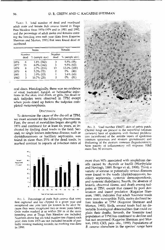
ga Pass Meadow began to show signs of illness(CKS, pers. obs.). One male was bloated andweak when captured and was found dead fivedays later; two emaciated individuals werefound in late summer, and several other maleswere found dead but with no extemal abnor-malities. During the next three years, more deadand moribund toads were found throughout thepopulation at TPM (Table 3). The number andpercentage of dead and moribund males variedfrom 1976 to 1982. Female deaths peaked in1978, and only one dead female was found after1978. One dead and one dying B. canorus werealso found during a survey of the SaddlebagLakle (northwest) population on 16 June 1977.
Between 34.9% and 68.4% of the adult malesthat had been toe clipped at or near the main
breeding area in TPM were recaptured in thesame area one year after being marked (Fig. 1).These one-year recapture rates were significant-ly lower for males marked from 1976-1978 com-pared to those marked from 1971-1974 and in1979 and 1981 (unpaired t-Test, t = 3.23, df =
7, P = 0.015, percentages transformed usingsquare-root of arcsine values). Males marked in1978 had the lowest one-year recapture rates(Fig. 1). In addition, only 13.3% (2/15) of themales toe clipped in 1978 that were recapturedone year later were known to be alive two yearsafter marking. Comparable recapture percent-ages two years after marking for males toeclipped in the other study years were: 1971,
91.7% (33/36); 1972, 94.4% (17/18); 1973, 68.4%(80/117); 1974, 87.9% (80/91); 1976, 74.2% (89/
120); 1977, 46.3% (25154); and 1979, 50.0% (2/4).
Serious infectious diseases were detected inhistological examinations of four of 12 toads
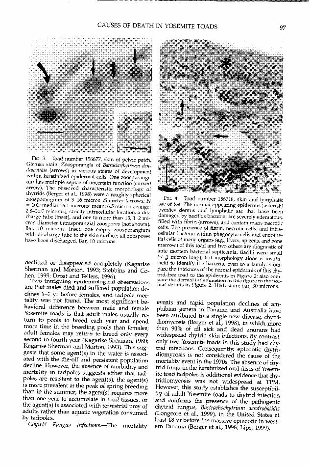
(Table 2). These diseases were identified as epi-dermal chytridiomycosis (N = 1; Figs. 2-3), ba-
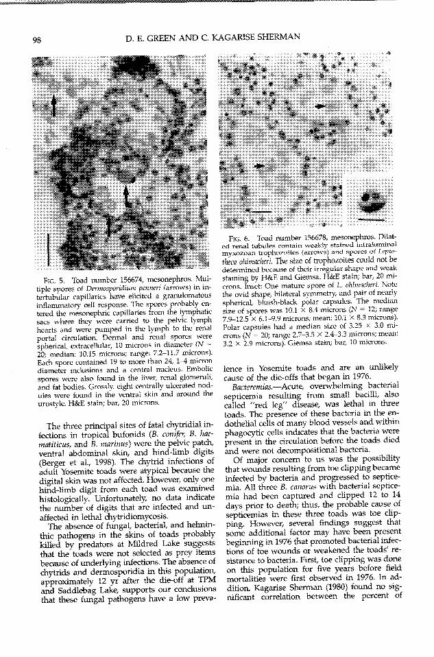
ciliary bacterial septicemia (N = 2; Fig. 4), anda combination of both diseases (N = 2). Infec-tious diseases of uncertain significance were de-tected in an additional five specimens (Table 2):a systemic fungal infection by Dermosporidiumsp. (N = 1; Fig. 5), mvxozoan infection (Lepto-theca ohlmacheri) of the mnesonephros (N = 1; Fig.6), and systemic larval Rhabdias sp. infections (N= 3; Fig. 7). A variety of helminths were foundin the gastrointestinal tracts, lungs, and urinarybladders of five toads (Table 2). The trematodesin the urinary bladders (N = 3) were consistentwith Gorgodera sp., Gorgoderina sp., or Megalod-iscus sp. (Flynn, 1973) and were considered in-nocuous. The trematodes in the lungs (N = 2)caused very mild inflammatory reaction andprobably were Haemiatoloechus sp.
The skins of 14 toads that probably werekilled by ravens (Cormus corax, Kagarise Sher-man and Morton, 1993) at Mildred Lake in 1990were free of chytridial and dermosporidial fun-gi. Autolysis (decomposition) hindered exami-nations of about half of the skins; postmortembacterial and water mold invasion of the skinwas evident in seven of the toads.
Because of autolysis of internal organs, his-tologic examinations of the 25 tadpoles werelimited to skin, oral discs, and gills. Grossly,there was no evidence of deformed, malformed,or supemumerary limbs, nor depigmentation orulceration of the keratinized segments of the
11 ''I ......
94
X~~~~~~~~~~~~~~~~~: C:, CD|
< a F I r ~ ~~~~~~ + +C
V si QJ % C5,% + +~+ z z £ v.,J: o o o o o 4 + + a
8 = R s - ° ° °° ° ++ o o+ +c
., O CJ I V. V.~~~
. E , { , .I v' £ 8 r~~~~~~~~~~~~~~(i
_O C < W ~~~~~~~~~~~~~~~~~~~~~O' i
O <U r M
0, _ j v e >oE 0
It bG
°e~~~~~~~~" Lr Lf i ) Lo LI Ln
5~~~~~~~~, c- 06 "ca-, o Eh O :~ L, On OU mVL C,L:
+ < < v.0 < 5O<'%< ^ 5 bb i > ~ ~~~~~~ m L1 WI mx l 0~ 't O_
grl v, .° Q ] O > <' $ 95 4 <? 95 '>5 £ '5 <
(,, 5 E j , t ~~~~~~~~~~~Lr) L^ Lr) L^ L^. LI'.^ .
I
I
CAtJSES OF DEATFI IN YOSEMI"TE TOADS 95
o
D. E. GREEN AND C. KAGARISE SHERMAN
TABLE 3. Total number of dead and moribund
adult male and female Bufo canorus found in TiogaPass Meadow from 1976-1979 and in 1981 and 1982,
and the percentage of adult males and females enter-ing the breeding area each year (data from KagariseSherman and Morton, 1993) that were found dead or
moribund.
Males Females
Number Numberdead % (sample size) dead % (sample size)
1976 6 1.8%6 (342) 3 5.5% (55)1977 13 8.0% (162) 3 9.5% (63)1978 8 3.7% (216) 13 13.0% (100)1979 4 5.3% (75) 0 0% (46)1981 1 3.9% (33) 1 1.6% (63)1982 3 10.7% (28) 0 0% (81)
oral discs. Histologically, there was no evidenceof viral, bacterial, fungal, or helminthic infec-tions in the skin, oral discs, or gills. No dead orsick tadpoles were observed in TPM exceptwhen pools dried up before the tadpoles corn-pleted metamorphosis.
DISCUSSION
To determine the cause of the die-off at TPM,we must account for the following observations.First, the onset of mortalities began abnrptly in1976 and continued for at least six years, as in-dicated by finding dead toads in the field. Sec-ond, no single known infectious disease, such aschytridiomycosis or bacillary septicernia, wasfound in more than 25% of the dead toads, inmarked contrast to reports of infection rates of
70- ; 171
Per-ee 60 * 7
Rcaptured 7
Yr Late., sO 7
340
20
10
71 72 73 74 75 76 77 78 79 80 8t
Yea. of Rrt: Captute and MSrkng
FIG. 1. Percentage of male Bufo canorus that werefirst captured and toe clipped in a given year andrecaptured one year later (or known to be alive be-cause thev were recaptured two or more years later).Only males marked at or within 200 m of the mainbreeding area at Tioga Pass Meadow are included.Numbers above bar are total number toe clipped eachyear; data from 1975 are not included because of par-tiallv missing marking records; no marking was donein 1980.
FIG. 2. Toad number 156677, skn of pelvic patch.Chytrid fungi are present in the superficial (stratumcorneum) layer of epidermis with marked prolifera-tion (acanthosis) of the middle layers of epidermis(stratum spinosum and stratum granulosum) andthickening of the stratum corneum (hyperkeratosis).Note paucitv of inflammatory cell response. h&Estain: bar, 50 microns.
more than 90% associated with amphibian die-offs caused by chytrids or bacilli (Worthylakeand Hovingh, 1989; Berger et al., 1998). Third, avariety of serious or potentially serious diseaseswere found in the toads (chytridiomycosis, ba-cillary septicemia, systemic dermosporidiosis,and systemic rhabdiasis). Fourth, the absence oflesions, observed illness, and death among tad-poles at TPM, except that caused by pool des-iccation and insect predation (Kagarise Sher-man, 1980), indicates that adult Yosemite toadswere more susceptible. Fifth, males declined be-fore females at TPM (Kagarise Sherman andMorton, 1993). Sixth, several toads had no de-tectable histological abnormalities that could ex-plain their deaths. Seventh, the Yosemite toadpopulation at TPM has continued to decline andhas not recovered (Kagarise Sherman and Mor-ton, 1993; CKS, pers. obs., 1999). Populations ofB. canorus elsewhere in the species' range have
...
96
CAUSES OF DEATH IN YOSEMITE TOADS 97
..000000''' X "....... ........ . ..f
FIG. 3. Toad number 156677, skin of pelic patch,Giemsa stain. Zoosporangia of Batraclwchigrium den-drob7atidis (arrows) in various stages of developmentwithin keratinized epidermal cells. One zoosporangi-urn has multiple septae of uncertain function (curvedarrow). The observed characteristic morphology ofchytrids (Berger et al., 1998) were a roughly sphericalzoosporangium of 3-16 micron diameter (arrows; N= 100; median: 6.1 microns; mean: 6.5 microns; range:2.3-16.Q rnicrons), strictly intracellular location, a dis-charge tube (inset), and one to more than 15, 1-2 mi-cron diameter intrasporangial zoospores (not showzn).Bar, 1 0 mricrons. Inset: one empty zoosporangiumwith discharge tube to the skin surface; all zoosporeshave been discharged. Bar, 10 microns.
declined or disappeared completely (KagariseSherman and Morton, 1993; Stebbins and Co-hen, 1995; Drost and Fellers, 1996).
TwTo intriguing epidemriological observationsare that males died and suffered population de-clines 1-2 yr before females, and tadpole mor-tality was not found. The most significant be-havioral dlifference betw7een male and femnaleYosemite toads is that adult males usually re-turn to pools to breed each year and spendmore time in the breeding pools than females;adult females may return to breed only everysecond to fourth year (Kagarise Sherman, 1980;Kagarise Sherman and Morton, 1993). This sug-gests that some agent(s) in the wvater is associ-ated with the die-off and persistent populationdecline. However, the absence of morbidity antdmortality in tadpoles suggests either that tad-poles are resistant to the agent(s), the agent(s)is more prevalent at the peak of spring breedingthan in the summner, the agent(s) requires morethan one year to accumulate in toad tissues, orthe agent(s) is associated with terrestrial prey ofadults rather than aqusatic vegetation consumedby tadpoles.
Chytrid Fungues Infections.-The mortality
FIG;. 4. Toad number 156718, skin and lymphaticsac of toe. The normal-appearing epidermis (asterisk)overlies dermis and lymphatic sac that have beendamaged bv bacillus bacteria, are severely edematous,filled with fibrin (arrows), and contain manym necroticcells. The presence of fibrin, necrotic cells, and intra-cellular bacteria within phagocytic cells and endothe-lial cells of many organs (e.g., livers, spleens, and bonemarrow) of this toad and two others are diagnostic ofante mortem bacterial septicemia. Bacilli were small(< 4 micron long), but morphology alone is insuffi-dent to identify the bacteria, even to a familv. Com-pare the thickness of the normal epidermis of this chy-trid-free toad to the epidermis in Figure 2; also com-pare the dermal inflammation in this figure to the nor-mal dermis in Figure 2. H&E stain; bar, 30 microns.
events and rapid population declines of am-phibian genera in Panama and Australia havebeen attributed to a single new disease, chytri-diomycosis (Berger et al., 1998), in which morethan 90% of all sick and dead anurans hadwidespread chytrid skin infections. By contrast,only two Yosemite toads in this study had chy-trid infections. Consequently, epizootic chytri-diomvcosis is not considered the cause of themortality event in the 1970s. The absence of chy-trid fungi in the keratinized oral discs of Yosem-ite toad tadpoles is additional evidence that chy-tridiomycosis was not widespread at TPM.However, this study establishes the susceptibil-ity of adult Yosemite toads to chytrid infectionand confirms the presence of the pathogenicchytrid fungus, Bactrachochytrium dendrobatidis(Longcore et al., 1999), in the United States atleast 18 yr before the massive epizootic in west-ern Panama (Berger et al., 1998; Lips, 1999).
D. E. GREEN AND C. KAGARISE SHERMAN
FiG. 5. Toad number 156674, mesonephros. Mul-tiple spores of Dermosporidiurn penneri (arrows) in in-
tertubular capillaries have elicited a granulomatous
inflammatory cell response. The spores probably en-
tered the mesonephric capillaries from the lymphaticsacs where they were carried to the pelvic lymph
hearts and were pumped in the lymph to the renal
portal circulation, Dermal and renal spores were
spherical, extracellular, 10 microns in diameter (N --
20; median: 10.15 microns; range: 7.2-11.7 microns).Each spore contained 19 to more than 24, 1-4 microndiameter inclusions and a central nucleus. Embolic
spores were also found in the liver, renal glomeruli,and fat bodies. Grossly. eight centrallv ulcerated nod-ules were found in the ventral skin and around the
urostyle. H&E stain: bar, 20 microns.
The three principal sites of fatal chytridial in-fections in tropical bufonids (B. conifer, B. hae-matiticus, and B. marinus) were the pelvic patch,ventral abdominal skin, and hind-limb digits(Berger et al., 1998). The chytrid infections ofadult Yosemite toads were atypical because thedigital skin was not affected. However, only onehind-limb digit from each toad was examinedhistologically. Unfortunately, no data indicatethe number of digits that are infected and un-affected in lethal chytridiomycosis.
The absence of fungal, bacterial, and helmin-thic pathogens in the skins of toads probablykilled by predators at Mildred Lake suggeststhat the toads were not selected as prey itemsbecause of underlying infections. The absence ofchytrids and dermosporidia in this population,approximately 12 vr after the die-off at TPMand Saddlebag Lake, supports our conclusionsthat these fungal pathogens have a low preva-
FiG. 6. Toad number 156678, mesonephros. Dilat-ed renal tubules contain weakly stained intraluminalmyxozoan trophozoites (arrows) and spores of Lepto-
theca ohlmacheri. The size of trophozoites could not bedetermined because of their irregular shape and weakstaining by H&E and Giemsa. H&E stain; bar, 20 mi-
crons. Inset: One mnature spore of L. ohimacheri. Notethe ovid shape, bilateral symmetry, and pair of nearly
spherical, bluish-black polar capsules. The mediansize of spores was 10.1 X 8.4 microns (N = 12; range7.9-12.5 x 6.1-9.9 microns; mean: 10.1 X 8.3 microns).
Polar capsules had a median size of 3.25 X 3.0 mi-crons (N = 20; range 2.7-3.5 X 2.4-3.3 microns; mean:3.2 X 2.9 microns). Giemsa stain; bar, 10 microns.
lence in Yosemite toads and are an unlikely
cause of the die-offs that began in 1976.
Bacteremias.-Acute, overwhelming bacterial
septicemia resulting from small bacilli, also
called "red leg" disease, was lethal in three
toads. The presence of these bacteria in the en-
dothelial cells of many blood vessels and withinphagocytic cells indicates that the bacteria werepresent in the circulation before the toads diedand were not decompositional bacteria.
Of major concern to us was the possibilitythat wounds resulting from toe clipping becameinfected by bacteria and progressed to septice-mia. All three B. canorus with bacterial septice-mia had been captured and clipped 12 to 14days prior to death; thus, the probable cause ofsepticemias in these three toads was toe clip-ping. However, several findings suggest thatsome additional factor may have been presentbeginning in 1976 that promoted bacterial infec-tions of toe wounds or weakened the toads' re-sistance to bacteria. First, toe clipping was doneon this population for five years before fieldmortalities were first observed in 1976. In ad-dition, Kagarise Sherman (1980) found no sig-nificant correlation between the percent of
-......................
98
CAUSES OF DEATH IN YOSEMITE TOADS
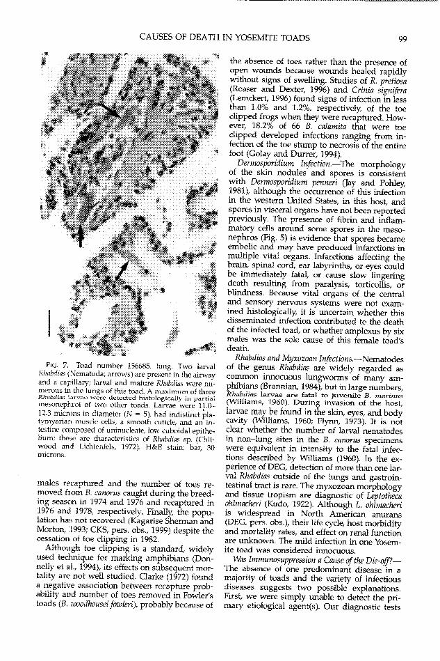
FiG. 7. Toad number 156685., lung. Two larvalRhabdias (Nemnatoda; arrows) are present in the airwayand a capillary; larval and mature RhabdiaLs were nu-merous in tlhe lungs of this toad. A maximum of threeRhlabdias larvae were detected histologically in partialmesonephroi of two other toads. Larvae were 11.0-12.3 microns in diameter (N = 5), had indistinct pla-tymyariari muscie cells, a smooth cuticle, and an in-testine composed of uninucleate, low cuboidal epithe-lium; these are characteristics of Rhabdias sp. (Chit-wood and Lichtenfels, 1972). H&E stain; bar, 30microns.
males recaptured and the number of toes re-moved from B. canorus caught during the breed-ing seascon in 1974 and 1976 and recaptured in1976 and 1978, respectively. Finally, the popu-lation has not recovered (Kagarise Sherman andMorton, [L993; CKS, pers. obs., 1999) despite thecessation of toe clipping in 1982.
Although toe clipping is a standard, widelyused tedimique for marking amphibians (Don-nelly et al., 1994, its effects on subsequent mor-tality are not wAell studied. Clarke (1972) founda negative association between recapture prob-ability and number of toes removed in Fowler'stoads (B. zvoodhouseifoawleri), probably because of
the absence of toes rather than the presence ofopen wounds because wounds healed rapidlvwithout signs of swelling. Studies of R. pretiosa(Reaser and Dexter, 1996) and Crinia signifera(Lemckert, 1996) found signs of infection in lessthan 1.0% and 1.2%, respectively, of the toeclipped frogs when thev were recaptured. How-ever, 18.2% of 66 B. calamita that were toeclipped developed infections ranging from in-fection of the toe stump to necrosis of the entirefoot (Golay and Durrer, 1994).
Dermosporidium Infection.-The morphologyof the skin nodules and spores is consistentwith Dermosporidium pen-neri (Jay and Pohley,1981), although the occurrence of this infectionin the western United States, in this host, andspores in visceral organs have not been reportedpreviously. The presence of fibrin and inflam-matory cells around some spores in the meso-nephros (Fig. 5) is evidence that spores becameembolic and may have produced infarctions inmultiple vital organs. Infarctions affecting thebrain, spinal cord, ear labyrinths, or eyes couldbe immediately fataL or cause slow lingeringdeath resulting from paralysis, torticollis, orblindness. Because vital organs of the centraland sensory nervous svstems were not exam-ined histologically, it is uncertain whether thisdisseminated infection contributed to the deathof the infected toad, or whether amplexus by sixmales was the sole cause of this female toad'sdeath.
Rhabdias and Myxozoan Infections.-Nematodesof the genus Rhabdias are widely regarded ascommon innocuous lungworms of many am-phibians (Brannian, 1984), but in large numbers,Rhabdias larvae are fatal to juvenile B. marinus(Williams, 1960). During invasion of the host,larvae may be found in the skin, eyes, and bodycavity (Williams, 1960; Flynn, 1973). It is notclear whether the number of larval nematodesin non-lung sites in the B. canorus specimenswere equivalent in intensity to the fatal infec-tions described by Williams (1960). In the ex-perience of DEG, detection of more than one lar-val Rhabdias outside of the lungs and gastroin-testinal tract is rare. The myxozoan morphologyand tissue tropism are diagnostic of Leptothecaohlmacheri (Kudo, 1922). Although L. ohirnacheriis widespread in North American anurans(DEG, pers. obs.), their life cycle, host morbidityand mortality rates, and effect on renal functionare unknown. The mild infection in one Yosem-ite toad was considered innocuous.
Was Immunosuppression a Cause of the Die-off?-The absence of one predominant disease in amajority of toads and the variety of infectiousdiseases suggests two possible explanations.First, we were simply unable to detect the pri-mary etiological agent(s). Our diagnostic tests
99z
D. E. GREEN AND C. KAGARISE SHERMAN
on the formalin-fixed toads were limited togross and histological examinations. To preservethe museum specimens intact, we did not ex-arnine histologically the special sensory andcentral nervous systems, hearts, and some tis-sues of the immune system (thymus, lympho-myeloid organs, etc.) and would not have de-tected any infectious or toxic agents that hadtheir principal effects on these systems. Al-though histological examinations can detect in-fections caused by iridoviruses and most bac-teria, fungi, protozoa, and helminths, oftenthere are no observable abnormalities with somevirus infections and most intoxications (Has-chek and Rousseaux, 1998).
Second, the variety of infections in these Yo-semite toads suggests that their immune sys-tems were suppressed. Virus infections and in-toxications by pesticides and other contami-nants may be acutely lethal, impair the immunesVstem, or cause other nonlethal injuries (e.g.,malformations, disruption of hormones, impair-
ment of other organs; Carey and Bryant, 1995;Carey et al., 1999). Two studies have docu-mented immune impairment and lethal second-ary bacterial infections following virus infec-tions in frogs (Cunningham et al., 1996) andpesticide exposure in toads (Taylor et al., 1999).Cunningham et al. (1996) found that iridovirus-infected adult R. temporaria were prone to sec-ondary bacterial infections, and Taylor et al.(1999) found that adult B. vxodhousei had muchhigher mortality rates with combined exposureto a pesticide (malathion) and a Gram-negativebacteria (Aeromonas hydrophila) than when ex-posed to only one of the two agents. Such im-mune suppression is not selective for bacterialinfections but allows opportunistic infections bya variety of bacterial, fungal, protozoan, andhelminthic organisms (Griffin, 1997).
Evidence of virus infection was not found inthese Yosemite toads. Most viruses isolatedfrom amphibians have been ranaviruses in thefamily, Iridoviridae (Tweedell and Granoff,1968; Wolf et al., 1968; Smith et al., 1986; Speareet al., 1991; Speare and Smith, 1992; Bennati etal., 1994; Cullen et al., 1995; Cunningham et al.,1996; Jancovich et al., 1997; Mao et al., 1997,1999). Adult and larval B. catorus lacked the his-tological abnormalities associated with acuteamphibian ranavirus infections. However, theabnormalities associated with chronic ranavirusinfections have not been described, and infor-mation on amphibian virus diversity and pa-thologv is inadequate. Thus, the Yosemite toadsmight have had an unrecognized virus infectionthat could have been lethal to some individualsand caused immune suppression in others.
A variety of chemicals are known to produceimmunosuppression in vertebrates, including
halogenated hydrocarbons, (e.g., dioxin, hexach-lorobenzene), polycyclic (nonhalogenated) hy-drocarbons, insecticides, organotins, heavy met-als, and oxidizing air pollutants (Haschek andRosseaux, 1998; Taylor et al., 1999; Voccia et al.,1999). Many agrichemicals and pollutants areknown to be lethal to amphibians (Kaplan andOverpeck, 1964; Kaplan et al., 1964, 1967; Mat-sui and Hayashi, 1992; Berrill et al., 1993;Schneeweiss and Schneeweiss, 1997; Marco etal., 1999). Direct application of pesticides (J. VanW'Vagtendonk, pers. com.), toxic chemical spills,industrial effluents, and agricultural run-offshave not been reported at TPM. However. at-mospheric drift of pesticides from the heavilvagriculturalized Central Valley is well docu-mented in the Sierra Nevada (Cory et al., 1970;Zabik and Seiber, 1993; Aston and Seiber, 1997;McConnell et al., 1998), and concentrations ofsome pesticides in mountain surface waters aresufficient to be lethal to arnphipods but not am-phibians (LeNoir et al., 1999). Bioaccumulationof pesticides into tissues of Sierra Nevada am-phibians has been reported (Cory et al., 1970;Datta et al., 1998). Howvever, the synergistic ef-fects of pollutants and concentrations necessaryto cause immunosuppression or other sublethaleffects are unknown. Histological changes as-sociated with intoxications by most xenochemi-cals, even at acutelv lethal doses, are slight andnionspecific (Haschek and Rousseaux, 1998).Toad specimens that predate the use of agri-chemicals in the Central Vallev were not avail-able for comparison, and our histological ex-aminations did not detect changes associatedwith chronic intoxication(s). Nevertheless, weconclude that the seven epidemiological andpathological features of this die-off of Yosemitetoads are consistent with immunosuppressionby a virus infection or chronic intoxication bvone or more chemicals.
Other Possible Causes of Amphibian Die-offs.-Several hypothesized causes of amphibian die-offs and population declines were ruled out asfactors in the die-off at TPM. Acute lethal intox-ications, as occur in a direct application of pes-ticides or accidental chemical spill (Kirk, 1988;Matsui and. Hayashi, 1992), were unlikely forfour reasons: (1) there are no records of pesti-cide applications at Tioga Pass (e.g., for the con-trol of lodgepole needle miners, Coleotechnitesmilleri; Struble, 1972); (2) the deaths spannedseveral years rather than being limited to a fewdays; (3) tadpoles were not affected; and (4) noother taxa suffered concurrent mortalities. Thehabitat at TPM is protected and has not beendegraded by farming, ranching, or logging (Ka-garise Sherman and Morton, 1993), and therehas been no introduction of potential predatorsor competitors such as bullfrogs (R. catesbeiana)
100
CAUSES OF DEATH IN YOSEMITE TOADS
at TPM (CKS, pers. obs.). Although nonnativetrout have been stocked in lakes throughout theSierra N.evada, Yosemite toads breed in shallow,ephemeral pools that are not inhabited by fish.
Physical and environmental stressors, such ashandling, toe clipping, unusual temperatures orweather, and ultraviolet radiation, mav causedeaths and immune suppression in amphibians(Carey, 1993; Carey et al., 1999). At TPM, Yosem-ite toads were captured, handled, and toeclipped for five years before casualties occurred(Kagarise Sherman, 1980; Morton, 1981., 1982;Kagarise Sherman and Morton, 1993). Toadpopulations declined at Saddlebag Lake, Mil-dred Lake, and elsewhere in the eastern SierraNevada (Kagarise Sherman and Morton, 1993;Drost and Fellers, 1996) where human visitorswere infrequent, and handling and toe clippingwere rare. Weather fluctuations (Pounds andCrump, 1994), or atmospheric warming (Poundset al., 1999), may be causally linked to some am-phibian declines. Although temperature andweather at TPM in 1.977 and 1978 may have tem-porarily stressed adult B. canorus (KagariseSherman., 1980; Kagarise Sherman and Morton,1993), such variations, which probablv have oc-curred repeatedly for many millenria, cannotexplain the persistence of the poptllation declinefor two decades. Ultraviolet radiation cannot beruled out completely as a factor in the die-offsand persistent population declines; however itshould be noted that casualties associated withrising levels of WV-B radiation have been re-ported only in some amphibian eggs, embryos,and larvae (Blaustein et al., 1994a; 1994b; Kie-secker and1 Blaustein, 1995; Blaustein et al., 1998;Hatch and Burton, 1998; Cummins et al., 1999)but not in adult amphibians.
This study demonstrates the potential valueof museum specimens for investigating amphib-ian mortality events and population declines.We encourage salvaging and preservation offield casualties for museum specimens orprompt diagnostic examination. We also rec-ommend the collection of apparentlv healthv in-dividuals from declining amphibian popula-tions and casualty sites for thorough diagnosticexaminations and placement in museums. Notonly can museum collections be used to assesschanges in biodiversity (Shaffer et al., 1998), butpreserved specimens may yield critical infor-mation to help understand the causes of historicamphibian population declines.
Acknawledgments.-This research was sup-ported by grants to CKS from the AmericanMuseum of Natural History Theodore RooseveltMemorial Fund, Horace H. Rackham GraduateSchool and Museum of Zoology Hinsdale andWalker Scholarships at the University of Michi-
gan, Sigma Xi Grants-in-Aid of Research, andthe American Philosophical Society PenroseFund and by fellowships from the Horace H.Rackham Graduate School and the AmericanAssociation of Universitv Women. For their co-operation and support in the field, we thank E.P. Pister of the California Department of Fishand Game, and the Inyo National Forest. Wethank R. Nussbaum for allowing us to examinespecimens from the University of Michigan Mu-seum of Zoology collection and P. W Shermanfor his comments on the manuscript.
LITERATURE CITED
ASTON, L. S., AND J. N. SEIBER. 1997. Fate of sum-mertime airborne organophosphate pesticide resi-dues in the Sierra Nevada Mountains. J. Environ.Qual. 26:1483-1492.
BENNATi, R., M. BONETTI, A. LAVAZZA, AND D. GEL.-MEmTI. 1994. Skin lesions associated with herpesvirus-like particle in frogs (Rana daimatina). Vet.Rec. 135:625-626.
BERGER. L., R. SPEARE, P. DASZAK, D. E. GREEN, A. A.CUNNINGHAM, C. L. GOGGIN, R. SLOCOMBE, M. A.RAGAN, A. D. HIYATT, K. R. MCDONALD, H. B.HINES, K. R. LIPS, G. MARANTELI.I, AND H. PARKES.1998. Chytridiomycosis causes amphibian mortal-ity associated with population declines in the rainforests of Australia and Centra' America. Proc.Natl. Acad. Sci. USA 95:9031-9036,
BERRILL, M., S. BERTRAM, A. WILSON, S. LoUis, D.BRIGHAM, AND C. STROMBERG. 1993. Lethal andsublethal impacts of pyrethroid insecticides onamphibian embryos and tadpoles. Environ. Toxi-col. Chem. 12:525-539.
BLAUSTEIN, A. R., D. G. HOKIT, R. K. O'HARA, AND R.A. HOLT. 1994a. Pathogenic fungus contributes toamphibian losses in the Pacific Northwest. Biol.Conserv. 67:251-254.
BLAUSTEIN, A. R., P. D. HOFFMAN, D G. HOKrr, J. M.KIESECKER, S. C. WALLS, AND J. B. HAYS. 1994b.UV repair and resistance to solar UV-B in amphib-ian eggs; a link to population declines? Proc. Natl.Acad. Sc. USA 91:1791-1795.
BLAUSTEIN, A. R., J. M. KIESECKER, D. P. CHIVERS, D.G. HOKIT, A. MARCO, L. K. BELDEN, AND A.HATCH. 1998. Effects of ultraviolet radiation onamphibians: field experiments. Am. Zool. 38:799-812.
BRADFORD, D, F. 1991. Mass mortality and extinctionin a high-elevation population of Rana rnuscosa. J.HIerpetol. 25:174-177.
BRANNIAN, R. E. 1984. Lungworms. In G. L. Hoff, EL. Frye, and E. R. Jacobson (eds.), Diseases of Am-phibians and Reptiles, pp. 213-217. Plenum Press,New York.
CAREY, C. 1993. Hypothesis concerning the causes ofthe disappearance of boreal toads from the moun-tains of Colorado. Conserv. Biol. 7:355-362.
CAREY, C., AND C. J. BRYANT. 1995. Possible interre-lations among environmental toxicants, amphibiandevelopment, and decline of amphibian Popula-tions. Environ. Health Perspect. 103:13-17.
CAREY, C., N. COHEN, AND L. ROL.LINS-SMITI . 1999.
101
D. E. GREEN AND C. KAGARISE SHERMAN
Amphibian declines: an immunological perspec-tive, Dev. Comp. immunol. 23:459-472.
CHITWOOD, M., AND J. R. LICE-ITENFELS. 1972. Identi-fication of parasitic metazoa in tissue sections. Exp.Parasitol. 32:407-519.
CLARKE, R. D. 1972. The effect of toe clipping on sur-vival in Fowler's toad (Bufo woodhousei fawleri).Copeia 1972:182-185.
CoRY, L., P. FIELD, AND W. SERAT. 1970. Distribution
patterns of DDT residues in the Sierra NevadaMountains. Pestic. Mionit. J. 3:204-211.
CRUMP, M. L., R R. FIENSLEY, AND K. L. CLARK. 1992.
Apparent decline of the golden toad: undergroundor extinct? Copeia 1992:413-420.
CULLEN, B. R., L. OWENS, AND R. J. WFIITTINGTON.
1995. Experimental infection of Australian an-urans (Limnodynastes terraereginae and Litoria lato-palmata) with Bohle iridovirus. Dis. Aquat. Org. 23:83-92.
CuMMiNs. C. P., P. D. GREENSLADE, AND A. R. Mc-
LEOD. 1999. A test of the effect of supplementalUV-B radiation on the common frog, Rana tempor-
aria L., during embrvonic development. GlobalChange Biol. 5:471-479.
CUNNINGHAM, A. A., T. E. S. LANGTON, P. M. BEN-
NETT, J. F LEWIN, S. E. N. DRURY, R. E. GOUGII,
AND S. K. MAcGREGOR. 1996. Pathological and
microbiological findings from incidents of unusualmortality of the common frog (Rana temporaria). R.Soc. Lond. Philos. Trans. Ser. B Biol. Sd. 351:1539-1557.
DATTA, S., L. HANSEN, L. MCCONNELL, J. BAKER, J. LE-
NOIR, AND J. N. SEIBER. 1998. Pesticides and PCB
contaminants in fish and tadpoles from the Kaw-eah River Basin, California. Bull. Environ. Contain.Toxicol. 60:829-836.
DONNELLY, M. A., C. GUYER, J. E. JUTERBOCK, AND R.
A. ALFORD. 1994. Techniques for marking am-phibians. In W. R. Heyer, M. A. Donnelly, R. WMcDiarmid, L. C. Hayek, and M. S. Foster (eds.),Measuring and Monitoring Biological Diversity.Standard Methods for Amphibians, pp. 277-284.Smithsonian Institution Press, Washington, DC.
DROST, C. A., AND G. M. FELLERS. 1996. Collapse of
a regional frog fauna in the Yosemite area of theCalifornia Sierra Nevada, USA. Conserv. Biol. 10:414-425.
FELLERS, G. M., AND C. A. DROST. 1993. Disappear-
ance of the cascades frog Rana cascadae at thesouthern end of its range, California, USA. Biol.Conserv. 65:177-181.
FISHER, R. N., AND H. B. SHAFFER. 1996. The decline
of amphibians in California's Great Central Valley.Conserv. Biol. 10:1387-1397.
FLYNN, R. J. 1973. Parasites of Laboratory Animals.Iowa State Univ. Press, Ames.
GOLAY, N., AND H. DURRER. 1994. Inflammation due
to toe-clipping in natteriack toads (Bufo calamnita).Amphib.-Reptilia 15:81-83.
GRIFFIN, D. E. 1997. Virus-induced immune suppres-sion. In N. Nathanson (ed.), Viral Pathogenesis, pp.207-233. Lippincott-Raven Publishers, Philadel-phia, Pennsylvania.
HASCIIEK, W M., AND C. G. ROUSSEAUX. 1998. Fun-
damentals of Toxicologic Pathology. AcademicPress, San Diego, California.
HATcli, A. C., AND G. A. BURTON. 1998. Effects ofphotoinduced toxicity of fluoranthene on amphib-ian embryos and larvae. Environ. Toxicoi. Chem.17:1777-1785.
HEYER, W. R., A. S. RAND, C. A. CONCALVEZ DA CRUZ,AND 0. L. PEIXOTO. 1988. Decimations, extinctions,
and colonizations of frog populations in south-eastern Brazil and their evolutionary implications.Biotropica 20:230-235.
JANCOVICH, J. K., E. WM DAVIDSON, J. F M4ORADo, B. L.JACOBS, AND J. PCOL.LINS. 1997. Isolation of a le-thal virus from the endangered tiger salamanderAmbystoma tigrinum stebbinsi. Dis. Aquat. Org. 31:161-167.
JAY, 1. M., AND W. J. POHI.EY. 1981. Dermosporitdiun
penneri sp. n. from the skin of the American toad,Bufo amrericanus (Amphibia: Bufonidae). J. Parasitol.67:108-110.
KAGARISE SHERMAN, C. 1980. A comparison of the
natural history and mating system of two anurans:Yosemite toads (Bufo canorus) and black toads (Bufoexsul). Unpubl. Ph.D. diss., Univ. of Michigan, AnnArbor.
KAGARISE SHERMAN, C., AND M. L. MORTON. 1993.
Population declines of Yosemite toads in the east-ern Sierra Nevada of California. J. i-lerpetol. 27:186-198.
KAPLAN, H. M., ANI) J. G. OVERPECK. 1964. ToxiCity
of halogenated hydrocarbon insecticides for thefrog, Rana pipiens, Herpetologica 20:163-169.
KAPLAN, H. M., N. YEE, AND S. S. GLACZENSKI. 1964.
Toxicity of fluoride for frogs. Lab. Anim. Care 14:185-188.
KAPLAN, H. M., T. J. ARNHOLT, AND J. E. PAYNE. 1967.
Toxicity of lead nitrate solutions for frogs (Ranapipiens). Lab. Anim. Care 17:240-246.
KARLSTROM, E. L. 1962. The toad genus Bufo in the
Sierra Nevada of California. Univ. Calif. (Berkeley)Publ. Zool. 62:1-104.
KIESECKER, J. M., AND A. R. BLAUSTEIN. 1995. Syner-
gism between UV-B radiation and a pathogenmagnifies amphibian embryo mortalitv in nature.Proc. Natl. Acad. Sci. USA 92:111049-11052.
KIRK, J. J. 1988. Western spotted frog (Rana pretiosa)mortality following forest spraying of DDT. Her-petol. Rev. 19:51-53.
KUDO, R. 1922. On the morphology and life historyof a myxosporidian, Leptotheca o01nacheri, parasitic
in Rana clamitans and R. pipiens. Parasitol. 14:221-242.
LAURANCE, W E, K. R. MCDONALD, AND R. SPEARE.
1996. Epidemic disease and the catastrophic de-cline of Australian rain forest frogs. Conserv Biol.10:406-413.
LEMCKERT, F 1996. Effects of toe-clipping on the sur-vival and behaviour of the Australian frog Criniasignifera. Amphib.-Reptilia 17:287-290.
LENOIR, J. S., L. L. MCCONNELL., G. M. FELLERS, T. M.
CAlIILL, AND J. N. SEIBER. 1999. Summertime
transport of current-use pesticides from Califor-niSas Central Valley to the Sierra Nevada Mountainrange, USA. Environ. Toxicoi. Chem. 18:2715-2722.
LIPS, K. R. 1998. Decline of a tropical montane am-phibian fauna. Conserv. Biol. 12:106-117.
- 1999. Mass mortality and population declines
102
CAUSES OF DEATH IN YOSEMITE TOADS
of anurans at an upland site in western Panama.Conserv. Biol. 13:117-125.
LONGCORE, J. E., A. P. PESSIER, AND D. K. NICFIOLS.1]999. Batrachochyfrium dendrobatidis gen. et sp. nov.,a chytrid pathogenic to amphibians. Mycol. 91:219-227.
MAO, J. H., R. P. HEDRICK, AND V. G. CHINCHAR. 1997.Molecular characterization, sequence analysis, andtaxonomic position of newlv isolated fish iridovi-ruses. Virol. 229:212-220.
MAO, J. H., D. E. GREEN, G. FELLERS, AND V. G. CHIN-CHAR. 1999. Molecular characterization of irido-viruses isolated fromrr svmpatric amphibians andfish. Virus Res. 63:45-52.
MARCO. A., C. QUILCHiANO, AND A. R. BLAUSTEIN.
1999. Sensitivity to nitrate and nitrite in pond-breeding amphibians from the Pacific Northwest,USA. Environ. Toxicol. Chem. 18:2836-2839.
MATsuI, M.; AND T. HAYASHI. 1992. Genetic unifor-mity ini the Japanese giant salamander, Andrias ja-ponicu':. Copeia 1992:232-235.
MCCONNI.LL, L. L., J. S. LENOIR, S. DATTA, AND J. N.SEIBER, 1998. Wet deposition of current-use pes-ticides in the Sierra Nevada Mountain Range, Cal-ifornia. USA. Environ. Toxicol. Chem. 17:1908-1916.
MORTON, M. L. 1981. Seasonal changes in total bodylipid and liver weight in the Yosemite toad. Copeia1981:234-238.
. 1982. Natural history of the Yosemite toad.Nat. Geog. Soc. R. Rep. 14:499-503.
MORTON, M. L., AND K. N. SOKOLSKI. 1978. Svmpatryin Bufo boreas and Bufo canorus and additional ev-idence of natural hybridization. Bull. S. Calif. Acad.Sci. 77:52-55.
MULLALLY. D. P. 1953. Observations on the ecologyof the toad Bufo canorus. Copeia 1953:182-183.
MULLALLY, D. P., AND J. D. C£1NNINGHAM. 1956. As-pects of the thermal ecology of the Yosemite toad.Herpetologica 12:57-67.
POUNDS, j. A., AND M. L. CRUMP. 1994. Amnphibiandeclines and climate disturbance: the case of thegolden toad and harlequin frog. Conserv. Biol. 8:72-85.
POUNDS, J. A., M. P. L. FOGDEN, AND j. H. CAMPBELL.1999. Biological response to climate change on atropical mountain. Nature 398:611-615.
REASER, J. K., AND R. E. DEXTER. 1996. Rana pretiosa(spotted frog). Toe clipping effects. Herpetol. Rev.27:195-196.
SCHNEEWE]S, N., AND U. SCHNEEWEIS. 1997. Amphi-
bienveriuste infolge mineralischer Dungung aufAgraflachen. Salamandra 33:1-8.
SHIAFFER, H. B., R. N. FISHER, AND C. DAVIDSON. 1998.The role of natural history collections in docu-menting species declines. TR Ecol. Evol. 13:27-30.
SMITH, A. W, M. P. ANDERSON, D. E. SKILLING, J. E.BARLOUGH, AND P. K. ENSLEY. 1986. First isolationof calicivirus from reptiles and amphibians. Am. J.Vet. Res. 47:1718-1721.
SPEARE, R., AND J. R. SM-IH. 1992. An iridovirus-likeagent isolated from the ornate burrowing frogLimnodynastes ornatus in nortAhern Australia. Dis.Aquat. Org. 14:51-57.
SPEARE, R., W J. FREEILAND, AND S. J. BOLTON. 1991.A possible iridovirus in erythrocytes of Bufo nmar-inus in Costa Rica. J. WildI. Dis. 27:457-462.
STEBBINS, R. C., AND N. W. COHIEN. 1995. A NaturalHistory of Amphibians. Princeton Univ. Press,Princeton, New Jersey.
STRUBIE, G. R. 1972. Biology, ecology, and control ofthe lodgepole needle miner. U.S. Dep. Agric. Tech.Bull. 1458:1-38.
TAYLOR, S. K., E. S. WILLIAMS, AND K. W MILLS. 1999.Effects of malathion on disease susceptibility inWoodhouse's toads. J. Wildl. Dis. 35:536-541.
TwEEDELL, K., AND A. GRANOFF. 1968. Viruses andrenal carcinoma of Rana pipiens. V. Effect of frogvirus 3 on developing frog embryos and larvae. J.Nat. Cancer Inst. 40:407-410.
VOCCIA, I., B. BLAKLEY, P. BROUSSEAU, AND M. FOUR-NIER. 1999. Immunotoxicity of pesticides: a review.Toxicol. Indust. Health 15:120-132.
WILLIAMS, R. W 1960. Observations on the life historyof Rhabdias sphaerocephala Goodey 1924 from Bufomarinus L., in the Bermuda Islands. J. He7minth. 34:93-98.
WOBESER, G., AND A. G. WOBESER. 1992. Carcass dis-appearance and estimation of mortality in a sim-ulated die-off of small birds. J. Wildl. Dis. 28:548-554.
WOLF, K., G. L. BULLOCK, C. E. DUNBAR, AND M. C.QUIMBY. 1968. Tadpole edema virus: a viscero-tropic pathogen for anuran amphibians. J. Infect.Dis. 118:253-262.
WORTHYLAKF, K. M., AND P. HOVINGH. 1989. Massmortality of salamanders (Ambystoma tigrinumi bybacteria (Acinetobacter) in an oligotrophic seepagemountain lake. Great Basin Nat. 49:364-372.
ZABIK, J. M., AND J. N. SEIBER. 1993. Atmospherictransport of organophosphate pesticides from Cal-ifornia's Central Valley to the Sierra Nevada Moun-tains. J. Environ. Qual. 22:80-89.
Accepted: 4 October 2000.
103
COPYRIGHT INFORMATION
TITLE: Diagnostic histological findings in Yosemite toads (Bufocanorus) froma die-off in the 1970s
SOURCE: Journal of Herpetology 35 no1 Mr 2001WN: 0106005199013
The magazine publisher is the copyright holder of this article and itis reproduced with permission. Further reproduction of this article inviolation of the copyright is prohibited.
Copyright 1982-2001 The H.W. Wilson Company. All rights reserved.


![LOCOMOTION IN TOADS (BUFO WOODHOUSII FOWLER]) BY …LOCOMOTION IN TOADS (BUFO WOODHOUSII FOWLER]) BY BRUCE D. ANDERSON1, MARTIN E. FEDER1* AND ROBERT J. FULL2 1Department of Organismal](https://img.pdfslide.net/doc/110x75/5e68af72d372f7064435ec23/locomotion-in-toads-bufo-woodhousii-fowler-by-locomotion-in-toads-bufo-woodhousii.jpg)