Embed Size (px)
Citation preview
Research ArticleDifferential Diagnosis of Entamoeba spp inClinical Stool Samples Using SYBR Green Real-TimePolymerase Chain Reaction
Thiago dos Santos Gomes1 Mariana Coimbra Garcia2 Flavia de Souza Cunha1
Heloisa Werneck de Macedo3 Joseacute Mauro Peralta2 and Regina Helena Saramago Peralta34
1 Departamento de Doencas Infecciosas e Parasitarias Faculdade de Medicina Universidade Federal do Rio de Janeiro21941-902 Rio de Janeiro RJ Brazil
2 Instituto de Microbiologia Prof Paulo de Goes Universidade Federal do Rio de Janeiro 21941-590 Rio de Janeiro RJ Brazil3 Faculdade de Medicina Universidade Federal Fluminense 24030-210 Niteroi RJ Brazil4Departamento de Patologia Hospital Universitario Antonio Pedro Rua Marques do Parana 303 Predio Principal4∘ andar Centro 24030-210 Niteroi RJ Brazil
Correspondence should be addressed to Regina Helena Saramago Peralta rhperaltaglobocom
Received 5 November 2013 Accepted 29 December 2013 Published 12 February 2014
Academic Editors J P Ackers and H Hooshyar
Copyright copy 2014 Thiago dos Santos Gomes et al This is an open access article distributed under the Creative CommonsAttribution License which permits unrestricted use distribution and reproduction in any medium provided the original work isproperly cited
Amoebiasis a disease caused by Entamoeba histolytica is usually diagnosed by microscopic examination which does notdifferentiate the morphologically identical species of the E histolyticaE dispar complex Furthermore morphologically similarspecies such as Entamoeba hartmanni contribute to misidentification Therefore there is a need for more sensitive and specificmethods This study standardized a multiplex real-time PCR system for E histolytica and E dispar and a single real-time PCR forE hartmanniThemultiplex protocol detected up to 00143 pg of E histolyticaDNA and 05156 pg of E disparDNA and the averagemelting temperature (119879
119898
) was 73∘C and 70∘C respectively For E hartmanni the 119879119898
was 73∘C and the amplification was successfuldown to 003 fg of plasmidDNANegative controls and other intestinal parasites presented no amplification Among the 48 samplestested E dispar DNA was detected in 37 none exhibited E histolytica DNA and 11 were negative in the multiplex protocol In 4 ofthese 11 samples however E hartmanni DNA was amplified SYBR Green is demonstrated to be an interesting option and thesecombined PCR reactions can improve laboratory diagnosis of amoebiasis in developing countries
1 Introduction
The genus Entamoeba contains many species six of which arefound in the human intestinal tract Entamoeba histolyticaEntamoeba dispar Entamoeba moshkovskii Entamoeba coliEntamoeba hartmanni and Entamoeba polecki Of thesespecies only E histolytica is associated with pathologicalinjuries the others are considered to be nonpathogenic spe-cies [1] E histolytica is responsible for approximately 50million cases of amoebiasis worldwide each year resulting inup to 100000 deaths [2 3] Because of these characteristicsE histolytica is globally considered to be a leading par-asitic cause of human mortality [1] However historical
epidemiological data concerning E histolytica infections canlead to overestimates because they were obtained before theformal separation of this parasite into two morphologicallyidentical species the potentially pathogenic E histolytica andthe nonpathogenic E dispar [4] More importantly it is alsoestimated that 90 of diagnosed infections are caused by Edispar [5]
Diagnosis of E histolytica has historically relied onmicroscopic examination of protozoan morphology Opticalmicroscopy is a very useful tool for the diagnosis of amoe-biasis because it is simple and cheap to execute Howeverconcentration techniques and stained smears are requiredto increase its low sensitivity [1 6] It is also influenced by
Hindawi Publishing Corporatione Scientific World JournalVolume 2014 Article ID 645084 8 pageshttpdxdoiorg1011552014645084
2 The Scientific World Journal
the intermittent release of cysts requiring the examinationof multiple samples from different days [7] Moreover thismethod cannot differentiate between morphologically iden-tical species such as E dispar and E moshkovskii and theproficiency of the examiner can also be crucial for preciseidentification as other Entamoeba species can present struc-tures very similar to the features of E histolytica [2 8 9]
Entamoeba hartmanni is one of these species with similarmorphological forms because its cysts share characteristicswith those of the E histolyticaE disparE moshkovskii com-plexThemajor difference between these species is the size ofthe cysts which in E hartmanni are generally smaller than inthose from the complex [4 10] For this differentiation to beconducted bymicroscopy an appropriatemicroscope capableof performing morphometric analysis is also required Giventhat this organism can still be difficult to identify dependingon the circumstances and the condition of the sample Con-tractile trophozoites or cysts of E histolytica can be erro-neously identified as E hartmanni and larger structures ofE hartmanni can also be misdiagnosed as the pathogenicspecies Alternative molecular methods could be helpful toprovide accurate and reliable identification of Entamoebaspecies Polymerase Chain Reaction (PCR) including real-time PCR has provided the means to identify E histolyticaE dispar and E moshkovskii in a variety of samples [11ndash17]However little attention has been devoted to the possibilityof identifying E hartmanni whose structures confuse exam-iners at different proficiency levels [8] Previously data det-ermined by microscopic examination have demonstrated aprevalence of E hartmanni of 133 inHIV patients and 25in children in Brazilian groups [18 19] Identification of Enta-moeba species other than the complex using molecular tech-niques has been conducted in only a single study in Brazilin which the evaluation of Brazilian clinical samples previ-ously identified as positive for the complex by microscopicexamination demonstrated the presence of E hartmanniDNA in more than one sample while E moshkovskii DNAwas not detected among the samples analyzed [20] Theseresults showed the presence of E hartmanni in Brazil butyielded no data about E moshkovskii
Real-time PCR is a very sensitive methodology thatcould become an important tool to achieve these goals andSYBR Green (N1015840N1015840-dimethyl-N-[4-[(E)-(3-methyl-13-ben-zothiazol-2-ylidene)methyl]-1-phenylquinolin-1-ium-2-yl]-N-propylpropane-13-diamine) technology with its probe-free assays is a less expensive choice for establishing moreefficient protocols in developing countries [12] Because ofthese possibilities the objectives of this study were to stand-ardize SYBR Green real-time PCR protocols for the identifi-cation of E histolytica E dispar and E hartmanni and toevaluate their efficiency on clinical stool samples
2 Material and Methods
21 DNA Samples The positive controls used in the SYBRGreen multiplex real-time PCR were genomic DNA fromthe standard strain of E histolytica HM1-IMSS and E disparP2 (a strain isolated from patient samples in Piauı Brazil)The DNA was measured in a Qubit fluorometer (Invitrogen
Carlsbad CA USA) and 2-fold dilutions in water wereprepared for testing using the SYBR Green real-time PCRprotocol ForE hartmanni however a cloning procedure wasnecessary to obtain a positive control because a protozoanculture of E hartmanni was not available For this processpositive DNA samples characterized in a previous study wereused [20] The specific E hartmanni sequence was amplifiedwith primers EhartF and EhartR2 this fragment was thencloned using the pCR 21-TOPO vector as described in theprotocol with the pCR 21-TOPO TA Cloning Kit (Invit-rogen) The extraction and purification of plasmid DNAcontaining the cloned fragment were performed using theIllustra PlasmidPrep Mini Spin Kit (GE Healthcare UK Lim-ited Buckinghamshire UK) according to the manufacturerrsquosinstructions This cloned plasmid DNA was measured ina Qubit fluorometer (Invitrogen) and 10-fold dilutions inwater were prepared for testing with the SYBR Green real-time PCR protocol In both protocols DNA solutions fromother intestinal parasites (two of each) were tested for a speci-ficity assessment E moshkovskii Cryptosporidium hominisGiardia lamblia and Schistosoma mansoni
22 Clinical Samples Stool samples from 2263 patientswho were treated at Hospital Universitario Antonio Pedro(HUAP) Niteroi Rio de Janeiro Brazil betweenAugust 2008and June 2009 were submitted to a microscopic examinationin order to detect the presence of parasitesTheprocedurewasconducted at the HUAPrsquos parasitology laboratory Moreovera group of 292 stool samples from individual residents ofSumidouro Rio de Janeiro Brazil a rural area was also sub-jected to the same examination Those samples identified aspositive for the E histolyticaE dispar complex were selectedto evaluate this SYBR Green real-time PCR system Addi-tional stool samples from 50 individuals with negativedirect parasitological examinations were included as negativecontrols This study was reviewed and approved by theHuman InvestigationCommittee of theUniversidade FederalFluminense with protocol number 02007
23 Microscopic Examination Themicroscopic examinationwas conducted byHUAPrsquos parasitology laboratory staff Priorto morphologic analysis this service concentrated the stoolsusing a spontaneous sedimentation method [21] Then thesediments were analyzed in wet mounts stained with Lugolrsquosiodine solution For themicroscopic examination the labora-tory staff used a Nikon Eclipse E20 optical microscope(Nikon) After the analysis the sediments were stored atminus20∘C for DNA extraction
24 DNA Extraction DNA templates were extracted usingthe Fast PrepDNAKit and the FastPrep FP120Disrupter (MPBiomedicals Solon OH USA) Further Purification was per-formed using the QIAquick PCR purification Kit (QIAGENInc Valencia CA USA) and the purified DNA was storedat minus20∘C Both methods were performed according to themanufacturerrsquos instructions
25 Conventional PCR Amplification Amultiplex PCR reac-tion was performed according to a protocol previously
The Scientific World Journal 3
described by Santos et al [22] with some modificationsThe specific primers for E histolytica and E dispar werepreviously defined by Nunez et al [23] E histolytica(EHP1mdash51015840CGATTTTCCCAGTAGAAATTA31015840 and EHP2mdash51015840CAAAATGGTCGTCTAGGC31015840) and E dispar (EDP1mdash51015840ATGGTGAGGTTGTAGCAGAGA31015840 and EDP2mdash51015840CGA-TATTGACCTAGTACT31015840) These primer sets were used toamplify specific sequences of 132 bp for E histolytica and96 bp for E dispar Each reaction was performed in a volumeof 50120583L using PCR Supermix (Invitrogen) 20 pmol of eachprimer 01 bovine serum albumin (BSA Sigma Chem CoUSA) and 2 120583L of the DNA sample The PCR reaction wascarried out using a Veriti 96 Well Thermal Cycler (AppliedBiosystems CA USA) and the amplification parameterswere as follows 3 minutes at 94∘C 30 cycles of 30 seconds at94∘C 30 seconds at 55∘C and 30 seconds at 72∘C and finally7 minutes at 72∘CThe amplified product was visualized withethidium bromide staining after electrophoresis on a 20agarose gel
26 Multiplex SYBR Green Real-Time PCR StandardizationThis PCR reaction was standardized for the simultaneousdetection and identification of E histolytica and E disparThespecific primers used for the detection of E histolytica andE dispar were the same oligonucleotide sequences employedfor conventional multiplex PCR Initially a single format wasdesigned in order to amplify E histolytica and E dispar sep-arately This format allowed us to know the specific meltingtemperature (119879
119898) of the sequence amplified from each species
and the feasibility of standardizing a multiplex reaction withthese primersThe simplex PCR cycling parameters consistedof 2 minutes at 50∘C 2 minutes at 95∘C and 35 cycles of15 seconds at 95∘C and 33 seconds at 55∘C using PlatinumSYBR Green qPCR SuperMix-UDG (Invitrogen) and thesame PCR reagents and conditions A dissociation stage wasthen performed the parameters consisted of 15 seconds at95∘C 1 minute at 60∘C 15 seconds at 95∘C and 15 secondsat 60∘C Finally the multiplex format was standardized ina final volume of 25 120583L Four different amounts of primerswere tested 125 25 375 and 5 pmol Two amounts ofROX Reference Dye (Invitrogen) were also tested in thestandardization process 025 pmol and 125 pmol All reac-tions were carried out in an ABI 7500 System thermocycler(Applied Biosystems) and the PCR cycling parameters werethe same as described for the single format with an additionalcomparison between two annealing temperatures 55∘C and60∘C The amplified products were visualized with ethidiumbromide staining after electrophoresis on a 20 agarose gelfor confirmation
27 Standardization of SYBR Green Real-Time PCR for Iden-tification of E hartmanni DNA sequence alignment wasperformed using CLC Sequence Viewer software (CLC bio)The sequences aligned were rRNA genes of the highly homol-ogous species E histolytica E dispar E moshkovskii andE hartmanni Specific regions in the E hartmanni sequence(GenBank access AF149907) were identified A forwardprimer EhartF (51015840-CGTTCAAGACATGAGTGTGA-31015840) and
two reverse primers (EhartR1mdash51015840-CCGTAGATCTCCTAT-TCACTTT-31015840) EhartR2 (51015840-ACAACACATTCATGTCGC-A-31015840) were designed
To verify the identity of the target sequence of theprimers a conventional PCR was performed in a volume of50 120583L using PCR Supermix (Invitrogen) 20 pmol of eachprimer 01 bovine serum albumin (BSA Sigma Chem Co)and 2 120583L of DNA sample The PCR reaction was carried outusing a Veriti 96-Well Thermal Cycler (Applied Biosystems)and the amplification parameters were as follows 3 minutesat 94∘C 40 cycles of 30 seconds at 94∘C 30 seconds at55∘C and 30 seconds at 72∘C and finally 7 minutes at 72∘CThe amplified product was visualized by ethidium bromidestaining after electrophoresis on a 20 agarose gel andsent to the sequencing platform of the Genomic Unit atthe Laboratory of Cell Physiology Darcy Fontoura (FederalUniversity of Rio de Janeiro Brazil) After confirming theidentity of the amplified product on the GenBank databasethe standardization process of the real-time PCR beganThe PCR reaction using primers EhartF and EhartR2 wasperformed in a reaction volume of 25 120583L using PlatinumSYBR Green qPCR SuperMix-UDG (Invitrogen) Analysesconcerning the amounts of primers and ROX Reference Dyewere conducted just as described in the previous sectionfor the multiplex protocol Also the reactions were carriedout in the same thermocycler The PCR cycling parametersconsisted of 2minutes at 50∘C 2minutes at 95∘Cand 35 cyclesof 15 seconds at 95∘C and 33 seconds at 60∘C A dissociationstage was then performed the parameters consisted of 15seconds at 95∘C 1 minute at 60∘C 15 seconds at 95∘C and 15seconds at 60∘CThe amplified products were visualized withethidium bromide staining after electrophoresis on a 20agarose gel for confirmation
28 Stool Sample Evaluation For a preliminary evaluationof the real-time PCR protocols during the standardizationprocess a selection of Entamoeba positive samples was runThus only samples exhibiting structures of the E histolyt-icaE dispar complex under microscopic examination wereanalyzed by multiplex conventional PCR and multiplex real-time PCR for the accurate identification of E histolytica andE dispar Those samples with negative results obtained byboth methods were submitted for further investigation withreal-time PCR for the identification of E hartmanni
3 Results
31 Multiplex SYBR Green Real-Time PCR The preliminarystep represented by standardizing a single format of thisreaction was essential to achieve the final protocol forthe multiplex SYBR Green real-time PCR This reactiondetermined that the mean melting temperatures (119879
119898) of the
amplified sequences of E histolytica and E disparwere 732∘C(728 to 735∘C) and 702∘C (691∘C to 709∘C) respectivelyThese data demonstrated the possibility of standardizinga multiplex protocol to detect and identify these speciesusing SYBRGreen technologyThusmultiplex real-timePCRusing those primers was performed and DNA amplifica-tion was exhibited as expected The melting curve analysis
4 The Scientific World Journal
10000
1000
15 16 17 18 19 20 21 22 23 24 25 26 27 28 29 30 31 32 33 34 35
Cycle number
120575 Rn versus cycle
120575Rn
0010
0100
65 70 73 75
∘C)(65 70 75
∘C)(65 70 75
Temperature (∘C)
(c)
(b)
(a)
(A) (B) (C)
96bp132bp
10000
1000
15 16 17 18 19 20 21 22 23 24 25 26 27 28 29 30 31 32 33 34 35
Cycle number
120575 Rn versus cycle
120575Rn
0010
0100
65 70 73 75
∘C)(65 70 75
∘C)(65 70 75
Temperature (∘C)
(b)
(a)
(A) (B) (C)
96bp132bp
b
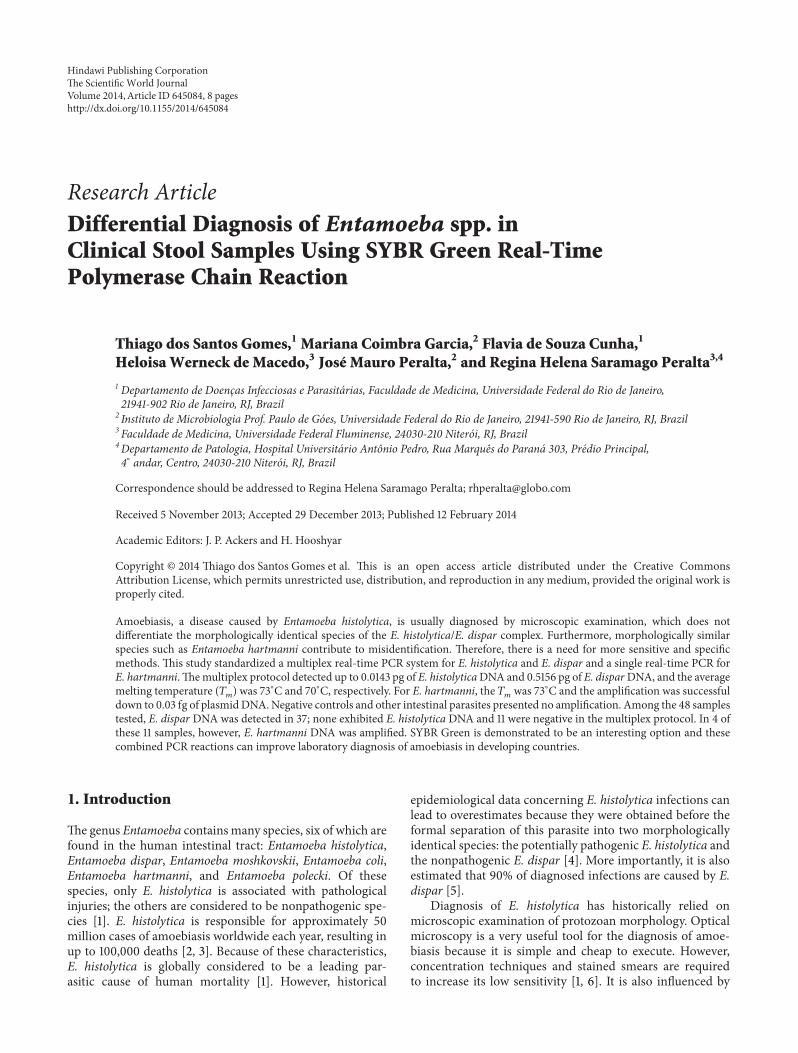
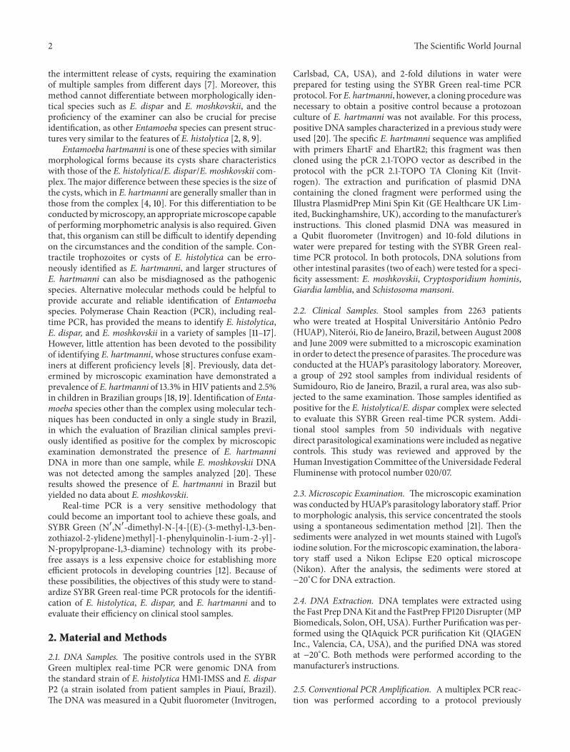
Figure 1 (a) Melting curve graph obtained through the SYBR Green real-time PCR for E histolytica (A) and E dispar (B) and the multiplexSYBR Green real-time PCR (C) (b) The softwarersquos graph demonstrating the DNA amplification of E histolytica and E dispar mixed DNAsin different amounts at the multiplex SYBR Green real-time PCR in descending order 1835 and 1980 pg (E histolytica and E dispar resp)915 and 990 pg 459 and 495 pg 229 and 247 pg 115 and 124 pg 8 57 and 62 pg 29 and 31 pg 14 and 16 pg 07 and 08 pg 035 and039 pg (c) Agarose gel frommultiplex real-time PCR products Lane 1 100 bp ladder standard lane 2 no sample lanes 3 to 12 PCR productsin descending order of concentration
demonstrated similar 119879119898
values for each species and theconcomitant presence of genetic material from both speciesdid not alter these conditions (Figure 1(a))
Under the conditions for real-time PCR the optimalconcentration of primers was found to be 25 pmol Otherconcentrations decreased the sensitivity of the assay orincreased primer dimer formation A concentration of ROXReference Dye equivalent to 125 pmol gave the most repro-ducible and stable resultsThe best annealing temperature foramplification of both fragments was 55∘C
In the specificity assessment no amplification wasdetected either in the presence of other intestinal parasiteDNAs or in the negative control group Specific DNAamplification was observed at all concentrations tested forboth species separately For E histolytica amplification wastested successfully from 734 pg (119862
119905 212) to 00143 pg (119862
119905
302) ForE dispar amplificationwas tested successfully from264 pg (119862
119905 234) to 05156 pg (119862
119905 330) In order to model
a mixed infection genetic material from both species wastested together A concomitant specific DNA amplification
The Scientific World Journal 5
1000e + 001
1000e + 000
15 16 17 18 19 20 21 22 23 24 25 26 27 28 29 30 31 32 33 34 35
Cycle number
120575 Rn versus cycle
1000e minus 002
1000e minus 003
1000e minus 004
1000e minus 001
(a)
65 70 75
Temperature (∘C)
(b)
Standard curve31097
30000
28000
26000
24000
23001minus4523 minus4000 minus3000 minus2000 minus1000 0000 0477
Log
Ct
C0
(c)
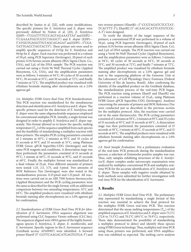
Figure 2 DNA amplification and the melting curve analysis using different DNA concentrations on SYBR Green real-time PCR for Ehartmanni DNA concentrations 30 pg 03 pg 003 pg 0003 pg 00003 pg and 000003 pg
Table 1 Results of multiplex PCR obtained with different DNAconcentration
DNA (pg)119862119905
valueE histolytica E dispar1835 1980 231915 990 239459 495 254229 247 265115 124 27357 62 28129 31 29314 16 30007 08 311036 039 315
was observed at all concentrations tested and the DNA ofboth species was amplified at levels as low as 03584 pg forE histolytica and 03867 pg for E dispar (Figures 1(b) and1(c) Table 1) As expected decreasing theDNAconcentrationalso increased the 119862
119905value revealing an inverse relationship
between these values In the latter situation the specific 119879119898
value must be checked in the melting temperature graph toevaluate the presence of two peaks (Figure 1(a))
32 SYBR Green Real-Time PCR for Identification of E hart-manni The alignment revealed highly specific regions in theE hartmanni sequence that allowed the design of a forward
primer (EhartF1) and a reverse primer (EhartR1) The frag-ment defined by these primers was 184 bp However Gen-Bank database analysis showed a small region of homol-ogy with Cryptosporidium hominis In an attempt to avoidhomology with other intestinal parasites a second reverseprimer EhartR2 was designed to exclude this homologousregion The new set of primers amplifies a sequence of 90 bpIn this real-time PCR protocol the melting curve analysisrevealed a mean melting temperature (119879
119898) of 73∘C for the
amplified product (Figure 2) Agarose gel electrophoresisanalysis confirmed the amplification of the specific 90 bpfragment In the conditions for real-time PCR the optimalconcentration of primers was found to be 375 pmol Otherconcentrations either decreased the sensitivity of the assay orincreased primer dimer formation For the ROX ReferenceDye 025 pmol was determined to be the concentration thatgave the most reproducible and stable results
In the specificity assessment no amplification wasdetected either in the presence of other intestinal parasiteDNAs or in the negative control group Specific DNAamplification was observed down to a concentration of003 fg As noted above the decrease in DNA concentrationcorresponded to an increase in the119862
119905valueThe119879
119898value was
between 728 and 736∘C (Figure 2 Table 2)
33 Stool Sample Evaluation For this study all sampleswere analyzed through microscopic examination Of the292 samples collected at Sumidouro 24 specimens exhibitedmorphological structures characteristic of theE histolyticaEdispar complex for a prevalence of 82 In the HUAPrsquos
6 The Scientific World Journal
Ib 420Ena 21
Ec 6250Bh 6870
Hookworm620
Ss 210
Polyparasitism 88
(a)
0
20
40
60
80
Ed EhEharm
Unknown
71
0
29
77
0 83 147
Multiplex PCRrt-multiplex PCR
()
(b)
Figure 3 Results of the 48 samples with Entamoeba histolyticaEntamoeba dispar complex (a) Frequency of polyparasitism and each detectedparasite onmicroscopic examination (b) Frequency ofE histolytica and E dispar in conventional andmultiplex real-time PCR and frequencyof E hartmanni by real-time PCR in negative samples from multiplex real-time PCR
Table 2 119862119905
and 119879119898
values obtained with different DNA concentra-tion from E hartmanni
DNA (pg) 119862119905
value 119879119898
(∘C)30 230 72803 250 732003 260 7320003 260 73600003 270 732000003 311 732
laboratory these structures were detected in 24 specimensfrom the 2263 samples collected with a prevalence of 11These 48 samples were then selected and submitted to real-time multiplex PCR The samples were also submitted tothe corresponding multiplex conventional PCR format forcomparison Of these samples 88 had polyparasitism withone or more parasites mainly with Blastocystis hominis andEntamoeba coli (Figure 3(a)) The conventional multiplexPCR revealed amplification of the specific sequence of Edispar in 34 samples while real-time multiplex PCR was ableto detect E dispar in these 34 samples and in three othersamples (77) Amplification of the specific sequence of Ehistolytica however was not observed in the samples testedby either methods (Figure 3(b)) The 11 samples negativefor E histolytica and E dispar by both methods were thensubmitted to the SYBR Green real-time PCR protocol for Ehartmanni identification to investigate whether the morpho-logical structure observed wasmisidentified In four samplesthe specific sequence of E hartmanni was amplified whilethe remaining 7 samples remained unidentified (Figure 3(b))To investigate the presence of inhibitory substances thesesamples were contaminated with 117 fg of E hartmanniDNAand subsequently submitted to E hartmanni real-time PCRDNA amplification was successful after this step indicatingthat inhibitory substances did not influence the negativeresults
4 Discussion
Several authors have described the crucial importance ofaccurate differentiation of E histolytica from E dispar Emoshkovskii and other morphologically similar species ofEntamoeba for proper clinical management of patients andfor assessment of their actual prevalence in different geo-graphical regions [13] To date several microscopy-based epi-demiological surveys to study the prevalence of E histolyticaand E dispar have been performed in different parts of Brazilbut these studies were carried out without using molecularmethods to accurately identify the species In Brazil thestandard clinical approach is to treat all asymptomatic indi-viduals who present with cysts in their feces with an anti-protozoal agent This approach to treatment results in theindiscriminate use of antiamoebic drugs and has led toincreased minimum inhibitory concentrations of these ther-apeutic agents against E histolytica with the potential forresistant strains to appear [24 25]
In this study we described the development of a real-time PCR system for the differential diagnosis of three speciesof Entamoeba that share identical or similar morphology asboth cysts and trophozoites The standardization of thesePCR reactions is an important step in developing an efficientsystem for the identification of Entamoeba species Mostmolecular diagnostic tools detect only E histolytica anddo not detect other morphologically identical or similarspecies giving rise to several questions A previous study hasalready demonstrated a situation in which samples that werepositive by microscopic examination for the E histolyticaEdispar complex were identified as E hartmanni after analysisby two different molecular methods [20] To avoid thispotential confusion and provide complete epidemiologic dataon parasitic infections it is important to develop a reliablesystem for the identification and differentiation of pathogenicand nonpathogenic parasites For this approach real-timePCR methodology is an interesting possibility because ofits high sensitivity and specificity as well as its speed inprocessing samples while SYBRGreen technology represents
The Scientific World Journal 7
a less expensive option for developing real-time PCR proto-cols in developing countries Additionally the SYBR Greenapproach is especially attractive for those who already havethe conventional PCR assay running and want to convert tothe real-time format [12]
The presented assays successfully amplified positive con-trols from E histolytica E dispar and E hartmanni demon-strating no amplification of DNA from other intestinalparasites Furthermore the amplification was successfullytested on amounts of DNA ranging between 734 pg and00143 pg for E histolytica and between 264 pg and 05156 pgfor E dispar These results demonstrated a high sensitivityOther studies have presented detection limits at higher DNAconcentrations such as 02 pg for E histolytica and 20 pgfor E dispar [16 26] For E hartmanni amplification wasobserved even with a lower DNA concentration Howeverthe lack of E hartmanni parasites must be considered inanalyzing the E hartmanni detection protocol a limitationthat was circumvented by subjecting the specific fragmentamplified to a cloning procedure Certainly the high numberof copies of the target sequence in the cloned plasmidsallowed the amplification of specific E hartmanni DNA withlower quantities of DNA
Among the 48 samples that were diagnosed as contain-ing the complex by microscopic examination 37 sampleswere identified as E dispar However there was no DNAamplification of E histolytica among these samples neitherwith conventional PCR nor with real-time PCRThese resultsdemonstrate the importance of accurate identification ofEntamoeba species by alternative and sensitive molecularmethods considering the relevance of these results to ensur-ing adequate treatment Two samples with E dispar also pre-sented Strongyloides stercoralis andhookworms parasites thatneed specific treatment Because E histolytica was notdetected by 2 different and sensitive protocols there is a highchance no antiamoebic treatment was actually necessaryThese sample results can be compared to the natural distribu-tion of the parasite in Brazil which has remained unknownsince E histolytica was separated into two species It ispossible that the pathogenic species has a low prevalence inRio de Janeiro in southeast Brazil while E dispar exists ata higher frequency in this area This hypothesis corroboratesother studies conducted over the past few years in differentparts of Brazil which have suggested thatE histolytica ismorecommon in the north and extreme northeast of Brazil and israre in other regions [7 27] If this is true accurate diagnosisbecomes even more important to clinical conduct becauseother Entamoeba species can lead to misidentification
The presence of other species identified as part of thecomplex is a problem that has been previously reported inthe literature [22] For this reason this study also proposesthe standardization of real-time PCR protocols for the iden-tification of Entamoeba species other than the complex Ofthe samples analyzed 11 samples remained unidentified afterthe multiplex real-time PCR protocol and were submittedto screening for E hartmanni DNA this speciesrsquo DNA wassuccessfully amplified in four samples Seven further sam-ples remained negative after this analysis However furthermicroscopic examination of these samples revealed another
possible explanation for this negative result the presence ofEntamoeba coli cysts in the majority of these samples It ispossible that the presence of immature cysts in those samplesresulted in a false positive diagnosis of the E histolyticaEdispar complex on microscopic examination
Our results highlight the importance of accurate identifi-cation of Entamoeba species In the diagnosis of amoebiasisa simple detection at the level of the E histolyticaE disparcomplex has been considered sufficient cause for clinicaltreatment It is understandable for hospitals operating withlimited funds to proceed in this manner However referencecenters such as university hospitals or public health centersshould use techniques such as PCR or antigen detectiondescribed in the literature for complete identification at thespecies level of bacteria viruses fungi and parasites [2228] Accurate identification is extremely important for exactdiagnosis and for providing correct epidemiological data
Accurate diagnosis of amoebiasis is crucial in order forphysicians to prescribe proper treatment These laboratoryresults present important information that will help cliniciansdecide whether to apply an antiamoebic treatment or searchfor a different etiology for the presented symptoms Onlythe specific detection of E histolytica confirms a diagnosis ofamoebiasis while the identification of other nonpathogenicamoebas can lead clinical investigators to search for differentpathologies with similar symptoms that would not be consid-ered without this information
5 Conclusion
The specificity and sensitivity demonstrated by the SYBRGreen real-time PCR protocols indicate an interestingapproach to the identification and differentiation of theseEntamoeba species The application of those protocols indifferent regions can be a helpful tool to providemore reliableparasitological diagnosis and more detailed epidemiologicaldata on the prevalence of Entamoeba species around theworldThe combination of both real-time PCR protocolsmayallow the development of a simple and useful Entamoebaidentification system to improve the laboratory diagnosis ofamoebiasis in developing countries
Conflict of Interests
The authors declare that there is no conflict of interestsregarding the publication of this paper
References
[1] R Fotedar D Stark N Beebe D Marriott J Ellis and JHarkness ldquoLaboratory diagnostic techniques for Entamoebaspeciesrdquo Clinical Microbiology Reviews vol 20 no 3 pp 511ndash532 2007
[2] World Health Organization ldquoAmoebiasisrdquoWeekly Epidemiolog-ical Record vol 72 pp 97ndash99 1997
[3] M Tanyuksel and W A Petri Jr ldquoLaboratory diagnosis ofamebiasisrdquoClinical Microbiology Reviews vol 16 no 4 pp 713ndash729 2003
8 The Scientific World Journal
[4] I K M Ali C G Clark and W A Petri Jr ldquoMolecularepidemiology of amebiasisrdquo Infection Genetics and Evolutionvol 8 no 5 pp 698ndash707 2008
[5] N Delialioglu G Aslan C Ozturk H Ozturhan S Sen and GEmekdas ldquoDetection of Entamoeba histolytica antigen in stoolsamples in Mersin Turkeyrdquo Journal of Parasitology vol 94 no2 pp 530ndash532 2008
[6] S A Tengku and M Norhayati ldquoPublic health and clinicalimportance of amoebiasis in Malaysia a reviewrdquo TropicalBiomedicine vol 28 no 2 pp 194ndash222 2011
[7] F L N Santos M S Goncalves and N M Soares ldquoValidationand utilization of PCR for differential diagnosis and prevalencedetermination of Entamoeba histolyticaEntamoeba dispar inSalvador City Brazilrdquo Brazilian Journal of Infectious Diseasesvol 15 no 2 pp 119ndash125 2011
[8] L S Garcia ldquoIntestinal protozoa amebaerdquo in DiagnosticMedical Parasitology L S Garcia Ed pp 6ndash25 ASM PressWashington DC USA 5th edition 2007
[9] B G Sard R T Navarro and J G E Sanchis ldquoAmebas intesti-nales no patogenas una vision clinicoanalıticardquo EnfermedadesInfecciosas y Microbiologıa Clınica vol 29 supplement 3 pp20ndash28 2011
[10] R B Burrows ldquoMorphological differentiation of Entamoebahartmanni and E polecki from E Histolyticardquo The AmericanJournal of Tropical Medicine and Hygiene vol 8 no 5 pp 583ndash589 1959
[11] J Blessmann H Buss P A T Nu et al ldquoReal-time PCR fordetection and differentiation of Entamoeba histolytica and Enta-moeba dispar in fecal samplesrdquo Journal of Clinical Microbiologyvol 40 no 12 pp 4413ndash4417 2002
[12] Y Qvarnstrom C James M Xayavong et al ldquoComparison ofreal-time PCR protocols for differential laboratory diagnosis ofamebiasisrdquo Journal of Clinical Microbiology vol 43 no 11 pp5491ndash5497 2005
[13] R Fotedar D Stark N Beebe D Marriott J Ellis and JHarkness ldquoPCR detection of Entamoeba histolytica Entamoebadispar and Entamoeba moshkovskii in stool samples fromSydney Australiardquo Journal of Clinical Microbiology vol 45 no3 pp 1035ndash1037 2007
[14] K Khairnar and S C Parija ldquoA novel nested multiplex poly-merase chain reaction (PCR) assay for differential detection ofEntamoeba histolytica E moshkovskii and E dispar DNA instool samplesrdquo BMCMicrobiology vol 7 article 47 2007
[15] D L Beck N Dogan V Maro N E Sam J Shao and ER Houpt ldquoHigh prevalence of Entamoeba moshkovskii in aTanzanianHIV populationrdquoActa Tropica vol 107 no 1 pp 48ndash49 2008
[16] Z Hamzah S Petmitr M Mungthin S Leelayoova and PChavalitshewinkoon-Petmitr ldquoDevelopment of multiplex real-time polymerase chain reaction for detection of Entamoebahistolytica Entamoeba dispar and Entamoeba moshkovskii inclinical specimensrdquo The American Journal of Tropical Medicineand Hygiene vol 83 no 4 pp 909ndash913 2010
[17] R Haque M Kabir Z Noor et al ldquoDiagnosis of amebic liverabscess and amebic colitis by detection of Entamoeba histolyticaDNA in blood urine and saliva by a real-time PCR assayrdquoJournal of Clinical Microbiology vol 48 no 8 pp 2798ndash28012010
[18] H Moura O Fernandes J P B Viola S P Silva R H Passosand D B Lima ldquoEnteric parasites and HIV infection occur-rence in AIDS patients in Rio de Janeiro Brazilrdquo Memorias doInstituto Oswaldo Cruz vol 84 no 4 pp 527ndash533 1989
[19] E R Machado D S Santos and J M Costa-Cruz ldquoEnteropar-asites and commensals among children in four peripheraldistricts of Uberlandia state of Minas Geraisrdquo Revista daSociedade Brasileira de Medicina Tropical vol 41 no 6 pp 581ndash585 2008
[20] H L C Santos R Bandea L A F Martins et al ldquoDifferentialidentification of Entamoeba spp based on the analysis of 18SrRNArdquo Parasitology Research vol 106 no 4 pp 883ndash888 2010
[21] A V Lutz ldquoShistosoma mansoni e schistosomose segundoobservacoes feitas no Brasilrdquo Memorias do Instituto OswaldoCruz vol 11 pp 121ndash125 1919
[22] H L C Santos R H S Peralta H W de Macedo M GM Barreto and J M Peralta ldquoComparison of multiplex-PCRand antigen detection for differential diagnosis of Entamoebahistolyticardquo Brazilian Journal of Infectious Diseases vol 11 no 3pp 365ndash370 2007
[23] Y O Nunez M A Fernandez D Torres-Nunez et al ldquoMul-tiplex polymerase chain reaction amplification and differentia-tion of Entamoeba histolytica and Entamoeba dispar DNA fromstool samplesrdquo The American Journal of Tropical Medicine andHygiene vol 64 no 5 pp 293ndash297 2001
[24] A Martınez-Palomo andMMartınez-Baez ldquoSelective primaryhealth care strategies for control of disease in the developingworld X Amebiasisrdquo Reviews of Infectious Diseases vol 5 no6 pp 1093ndash1102 1983
[25] D Bansal R Sehgal Y Chawla R C Mahajan and N MallaldquoIn vitro activity of antiamoebic drugs against clinical isolates ofEntamoeba histolytica and Entamoeba disparrdquoAnnals of ClinicalMicrobiology and Antimicrobials vol 3 article 27 2004
[26] Z Hamzah S Petmitr M Mungthin S Leelayoova andP Chavalitshewinkoon-Petmitr ldquoDifferential detection ofEntamoeba histolytica Entamoeba dispar and Entamoebamoshkovskii by a single-round PCR assayrdquo Journal of ClinicalMicrobiology vol 44 no 9 pp 3196ndash3200 2006
[27] A Dourado A Maciel and I D S Aca ldquoOccurrence ofEntamoeba histolyticaEntamoeba dispar in ambulatory patientsof Recife PErdquo Revista da Sociedade Brasileira de MedicinaTropical vol 39 no 4 pp 388ndash389 2006
[28] S J Furrows A H Moody and P L Chiodini ldquoComparison ofPCR and antigen detectionmethods for diagnosis ofEntamoebahistolytica infectionrdquo Journal of Clinical Pathology vol 57 no 12pp 1264ndash1266 2004
2 The Scientific World Journal
the intermittent release of cysts requiring the examinationof multiple samples from different days [7] Moreover thismethod cannot differentiate between morphologically iden-tical species such as E dispar and E moshkovskii and theproficiency of the examiner can also be crucial for preciseidentification as other Entamoeba species can present struc-tures very similar to the features of E histolytica [2 8 9]
Entamoeba hartmanni is one of these species with similarmorphological forms because its cysts share characteristicswith those of the E histolyticaE disparE moshkovskii com-plexThemajor difference between these species is the size ofthe cysts which in E hartmanni are generally smaller than inthose from the complex [4 10] For this differentiation to beconducted bymicroscopy an appropriatemicroscope capableof performing morphometric analysis is also required Giventhat this organism can still be difficult to identify dependingon the circumstances and the condition of the sample Con-tractile trophozoites or cysts of E histolytica can be erro-neously identified as E hartmanni and larger structures ofE hartmanni can also be misdiagnosed as the pathogenicspecies Alternative molecular methods could be helpful toprovide accurate and reliable identification of Entamoebaspecies Polymerase Chain Reaction (PCR) including real-time PCR has provided the means to identify E histolyticaE dispar and E moshkovskii in a variety of samples [11ndash17]However little attention has been devoted to the possibilityof identifying E hartmanni whose structures confuse exam-iners at different proficiency levels [8] Previously data det-ermined by microscopic examination have demonstrated aprevalence of E hartmanni of 133 inHIV patients and 25in children in Brazilian groups [18 19] Identification of Enta-moeba species other than the complex using molecular tech-niques has been conducted in only a single study in Brazilin which the evaluation of Brazilian clinical samples previ-ously identified as positive for the complex by microscopicexamination demonstrated the presence of E hartmanniDNA in more than one sample while E moshkovskii DNAwas not detected among the samples analyzed [20] Theseresults showed the presence of E hartmanni in Brazil butyielded no data about E moshkovskii
Real-time PCR is a very sensitive methodology thatcould become an important tool to achieve these goals andSYBR Green (N1015840N1015840-dimethyl-N-[4-[(E)-(3-methyl-13-ben-zothiazol-2-ylidene)methyl]-1-phenylquinolin-1-ium-2-yl]-N-propylpropane-13-diamine) technology with its probe-free assays is a less expensive choice for establishing moreefficient protocols in developing countries [12] Because ofthese possibilities the objectives of this study were to stand-ardize SYBR Green real-time PCR protocols for the identifi-cation of E histolytica E dispar and E hartmanni and toevaluate their efficiency on clinical stool samples
2 Material and Methods
21 DNA Samples The positive controls used in the SYBRGreen multiplex real-time PCR were genomic DNA fromthe standard strain of E histolytica HM1-IMSS and E disparP2 (a strain isolated from patient samples in Piauı Brazil)The DNA was measured in a Qubit fluorometer (Invitrogen
Carlsbad CA USA) and 2-fold dilutions in water wereprepared for testing using the SYBR Green real-time PCRprotocol ForE hartmanni however a cloning procedure wasnecessary to obtain a positive control because a protozoanculture of E hartmanni was not available For this processpositive DNA samples characterized in a previous study wereused [20] The specific E hartmanni sequence was amplifiedwith primers EhartF and EhartR2 this fragment was thencloned using the pCR 21-TOPO vector as described in theprotocol with the pCR 21-TOPO TA Cloning Kit (Invit-rogen) The extraction and purification of plasmid DNAcontaining the cloned fragment were performed using theIllustra PlasmidPrep Mini Spin Kit (GE Healthcare UK Lim-ited Buckinghamshire UK) according to the manufacturerrsquosinstructions This cloned plasmid DNA was measured ina Qubit fluorometer (Invitrogen) and 10-fold dilutions inwater were prepared for testing with the SYBR Green real-time PCR protocol In both protocols DNA solutions fromother intestinal parasites (two of each) were tested for a speci-ficity assessment E moshkovskii Cryptosporidium hominisGiardia lamblia and Schistosoma mansoni
22 Clinical Samples Stool samples from 2263 patientswho were treated at Hospital Universitario Antonio Pedro(HUAP) Niteroi Rio de Janeiro Brazil betweenAugust 2008and June 2009 were submitted to a microscopic examinationin order to detect the presence of parasitesTheprocedurewasconducted at the HUAPrsquos parasitology laboratory Moreovera group of 292 stool samples from individual residents ofSumidouro Rio de Janeiro Brazil a rural area was also sub-jected to the same examination Those samples identified aspositive for the E histolyticaE dispar complex were selectedto evaluate this SYBR Green real-time PCR system Addi-tional stool samples from 50 individuals with negativedirect parasitological examinations were included as negativecontrols This study was reviewed and approved by theHuman InvestigationCommittee of theUniversidade FederalFluminense with protocol number 02007
23 Microscopic Examination Themicroscopic examinationwas conducted byHUAPrsquos parasitology laboratory staff Priorto morphologic analysis this service concentrated the stoolsusing a spontaneous sedimentation method [21] Then thesediments were analyzed in wet mounts stained with Lugolrsquosiodine solution For themicroscopic examination the labora-tory staff used a Nikon Eclipse E20 optical microscope(Nikon) After the analysis the sediments were stored atminus20∘C for DNA extraction
24 DNA Extraction DNA templates were extracted usingthe Fast PrepDNAKit and the FastPrep FP120Disrupter (MPBiomedicals Solon OH USA) Further Purification was per-formed using the QIAquick PCR purification Kit (QIAGENInc Valencia CA USA) and the purified DNA was storedat minus20∘C Both methods were performed according to themanufacturerrsquos instructions
25 Conventional PCR Amplification Amultiplex PCR reac-tion was performed according to a protocol previously
The Scientific World Journal 3
described by Santos et al [22] with some modificationsThe specific primers for E histolytica and E dispar werepreviously defined by Nunez et al [23] E histolytica(EHP1mdash51015840CGATTTTCCCAGTAGAAATTA31015840 and EHP2mdash51015840CAAAATGGTCGTCTAGGC31015840) and E dispar (EDP1mdash51015840ATGGTGAGGTTGTAGCAGAGA31015840 and EDP2mdash51015840CGA-TATTGACCTAGTACT31015840) These primer sets were used toamplify specific sequences of 132 bp for E histolytica and96 bp for E dispar Each reaction was performed in a volumeof 50120583L using PCR Supermix (Invitrogen) 20 pmol of eachprimer 01 bovine serum albumin (BSA Sigma Chem CoUSA) and 2 120583L of the DNA sample The PCR reaction wascarried out using a Veriti 96 Well Thermal Cycler (AppliedBiosystems CA USA) and the amplification parameterswere as follows 3 minutes at 94∘C 30 cycles of 30 seconds at94∘C 30 seconds at 55∘C and 30 seconds at 72∘C and finally7 minutes at 72∘CThe amplified product was visualized withethidium bromide staining after electrophoresis on a 20agarose gel
26 Multiplex SYBR Green Real-Time PCR StandardizationThis PCR reaction was standardized for the simultaneousdetection and identification of E histolytica and E disparThespecific primers used for the detection of E histolytica andE dispar were the same oligonucleotide sequences employedfor conventional multiplex PCR Initially a single format wasdesigned in order to amplify E histolytica and E dispar sep-arately This format allowed us to know the specific meltingtemperature (119879
119898) of the sequence amplified from each species
and the feasibility of standardizing a multiplex reaction withthese primersThe simplex PCR cycling parameters consistedof 2 minutes at 50∘C 2 minutes at 95∘C and 35 cycles of15 seconds at 95∘C and 33 seconds at 55∘C using PlatinumSYBR Green qPCR SuperMix-UDG (Invitrogen) and thesame PCR reagents and conditions A dissociation stage wasthen performed the parameters consisted of 15 seconds at95∘C 1 minute at 60∘C 15 seconds at 95∘C and 15 secondsat 60∘C Finally the multiplex format was standardized ina final volume of 25 120583L Four different amounts of primerswere tested 125 25 375 and 5 pmol Two amounts ofROX Reference Dye (Invitrogen) were also tested in thestandardization process 025 pmol and 125 pmol All reac-tions were carried out in an ABI 7500 System thermocycler(Applied Biosystems) and the PCR cycling parameters werethe same as described for the single format with an additionalcomparison between two annealing temperatures 55∘C and60∘C The amplified products were visualized with ethidiumbromide staining after electrophoresis on a 20 agarose gelfor confirmation
27 Standardization of SYBR Green Real-Time PCR for Iden-tification of E hartmanni DNA sequence alignment wasperformed using CLC Sequence Viewer software (CLC bio)The sequences aligned were rRNA genes of the highly homol-ogous species E histolytica E dispar E moshkovskii andE hartmanni Specific regions in the E hartmanni sequence(GenBank access AF149907) were identified A forwardprimer EhartF (51015840-CGTTCAAGACATGAGTGTGA-31015840) and
two reverse primers (EhartR1mdash51015840-CCGTAGATCTCCTAT-TCACTTT-31015840) EhartR2 (51015840-ACAACACATTCATGTCGC-A-31015840) were designed
To verify the identity of the target sequence of theprimers a conventional PCR was performed in a volume of50 120583L using PCR Supermix (Invitrogen) 20 pmol of eachprimer 01 bovine serum albumin (BSA Sigma Chem Co)and 2 120583L of DNA sample The PCR reaction was carried outusing a Veriti 96-Well Thermal Cycler (Applied Biosystems)and the amplification parameters were as follows 3 minutesat 94∘C 40 cycles of 30 seconds at 94∘C 30 seconds at55∘C and 30 seconds at 72∘C and finally 7 minutes at 72∘CThe amplified product was visualized by ethidium bromidestaining after electrophoresis on a 20 agarose gel andsent to the sequencing platform of the Genomic Unit atthe Laboratory of Cell Physiology Darcy Fontoura (FederalUniversity of Rio de Janeiro Brazil) After confirming theidentity of the amplified product on the GenBank databasethe standardization process of the real-time PCR beganThe PCR reaction using primers EhartF and EhartR2 wasperformed in a reaction volume of 25 120583L using PlatinumSYBR Green qPCR SuperMix-UDG (Invitrogen) Analysesconcerning the amounts of primers and ROX Reference Dyewere conducted just as described in the previous sectionfor the multiplex protocol Also the reactions were carriedout in the same thermocycler The PCR cycling parametersconsisted of 2minutes at 50∘C 2minutes at 95∘Cand 35 cyclesof 15 seconds at 95∘C and 33 seconds at 60∘C A dissociationstage was then performed the parameters consisted of 15seconds at 95∘C 1 minute at 60∘C 15 seconds at 95∘C and 15seconds at 60∘CThe amplified products were visualized withethidium bromide staining after electrophoresis on a 20agarose gel for confirmation
28 Stool Sample Evaluation For a preliminary evaluationof the real-time PCR protocols during the standardizationprocess a selection of Entamoeba positive samples was runThus only samples exhibiting structures of the E histolyt-icaE dispar complex under microscopic examination wereanalyzed by multiplex conventional PCR and multiplex real-time PCR for the accurate identification of E histolytica andE dispar Those samples with negative results obtained byboth methods were submitted for further investigation withreal-time PCR for the identification of E hartmanni
3 Results
31 Multiplex SYBR Green Real-Time PCR The preliminarystep represented by standardizing a single format of thisreaction was essential to achieve the final protocol forthe multiplex SYBR Green real-time PCR This reactiondetermined that the mean melting temperatures (119879
119898) of the
amplified sequences of E histolytica and E disparwere 732∘C(728 to 735∘C) and 702∘C (691∘C to 709∘C) respectivelyThese data demonstrated the possibility of standardizinga multiplex protocol to detect and identify these speciesusing SYBRGreen technologyThusmultiplex real-timePCRusing those primers was performed and DNA amplifica-tion was exhibited as expected The melting curve analysis
4 The Scientific World Journal
10000
1000
15 16 17 18 19 20 21 22 23 24 25 26 27 28 29 30 31 32 33 34 35
Cycle number
120575 Rn versus cycle
120575Rn
0010
0100
65 70 73 75
∘C)(65 70 75
∘C)(65 70 75
Temperature (∘C)
(c)
(b)
(a)
(A) (B) (C)
96bp132bp
10000
1000
15 16 17 18 19 20 21 22 23 24 25 26 27 28 29 30 31 32 33 34 35
Cycle number
120575 Rn versus cycle
120575Rn
0010
0100
65 70 73 75
∘C)(65 70 75
∘C)(65 70 75
Temperature (∘C)
(b)
(a)
(A) (B) (C)
96bp132bp
b
Figure 1 (a) Melting curve graph obtained through the SYBR Green real-time PCR for E histolytica (A) and E dispar (B) and the multiplexSYBR Green real-time PCR (C) (b) The softwarersquos graph demonstrating the DNA amplification of E histolytica and E dispar mixed DNAsin different amounts at the multiplex SYBR Green real-time PCR in descending order 1835 and 1980 pg (E histolytica and E dispar resp)915 and 990 pg 459 and 495 pg 229 and 247 pg 115 and 124 pg 8 57 and 62 pg 29 and 31 pg 14 and 16 pg 07 and 08 pg 035 and039 pg (c) Agarose gel frommultiplex real-time PCR products Lane 1 100 bp ladder standard lane 2 no sample lanes 3 to 12 PCR productsin descending order of concentration
demonstrated similar 119879119898
values for each species and theconcomitant presence of genetic material from both speciesdid not alter these conditions (Figure 1(a))
Under the conditions for real-time PCR the optimalconcentration of primers was found to be 25 pmol Otherconcentrations decreased the sensitivity of the assay orincreased primer dimer formation A concentration of ROXReference Dye equivalent to 125 pmol gave the most repro-ducible and stable resultsThe best annealing temperature foramplification of both fragments was 55∘C
In the specificity assessment no amplification wasdetected either in the presence of other intestinal parasiteDNAs or in the negative control group Specific DNAamplification was observed at all concentrations tested forboth species separately For E histolytica amplification wastested successfully from 734 pg (119862
119905 212) to 00143 pg (119862
119905
302) ForE dispar amplificationwas tested successfully from264 pg (119862
119905 234) to 05156 pg (119862
119905 330) In order to model
a mixed infection genetic material from both species wastested together A concomitant specific DNA amplification
The Scientific World Journal 5
1000e + 001
1000e + 000
15 16 17 18 19 20 21 22 23 24 25 26 27 28 29 30 31 32 33 34 35
Cycle number
120575 Rn versus cycle
1000e minus 002
1000e minus 003
1000e minus 004
1000e minus 001
(a)
65 70 75
Temperature (∘C)
(b)
Standard curve31097
30000
28000
26000
24000
23001minus4523 minus4000 minus3000 minus2000 minus1000 0000 0477
Log
Ct
C0
(c)
Figure 2 DNA amplification and the melting curve analysis using different DNA concentrations on SYBR Green real-time PCR for Ehartmanni DNA concentrations 30 pg 03 pg 003 pg 0003 pg 00003 pg and 000003 pg
Table 1 Results of multiplex PCR obtained with different DNAconcentration
DNA (pg)119862119905
valueE histolytica E dispar1835 1980 231915 990 239459 495 254229 247 265115 124 27357 62 28129 31 29314 16 30007 08 311036 039 315
was observed at all concentrations tested and the DNA ofboth species was amplified at levels as low as 03584 pg forE histolytica and 03867 pg for E dispar (Figures 1(b) and1(c) Table 1) As expected decreasing theDNAconcentrationalso increased the 119862
119905value revealing an inverse relationship
between these values In the latter situation the specific 119879119898
value must be checked in the melting temperature graph toevaluate the presence of two peaks (Figure 1(a))
32 SYBR Green Real-Time PCR for Identification of E hart-manni The alignment revealed highly specific regions in theE hartmanni sequence that allowed the design of a forward
primer (EhartF1) and a reverse primer (EhartR1) The frag-ment defined by these primers was 184 bp However Gen-Bank database analysis showed a small region of homol-ogy with Cryptosporidium hominis In an attempt to avoidhomology with other intestinal parasites a second reverseprimer EhartR2 was designed to exclude this homologousregion The new set of primers amplifies a sequence of 90 bpIn this real-time PCR protocol the melting curve analysisrevealed a mean melting temperature (119879
119898) of 73∘C for the
amplified product (Figure 2) Agarose gel electrophoresisanalysis confirmed the amplification of the specific 90 bpfragment In the conditions for real-time PCR the optimalconcentration of primers was found to be 375 pmol Otherconcentrations either decreased the sensitivity of the assay orincreased primer dimer formation For the ROX ReferenceDye 025 pmol was determined to be the concentration thatgave the most reproducible and stable results
In the specificity assessment no amplification wasdetected either in the presence of other intestinal parasiteDNAs or in the negative control group Specific DNAamplification was observed down to a concentration of003 fg As noted above the decrease in DNA concentrationcorresponded to an increase in the119862
119905valueThe119879
119898value was
between 728 and 736∘C (Figure 2 Table 2)
33 Stool Sample Evaluation For this study all sampleswere analyzed through microscopic examination Of the292 samples collected at Sumidouro 24 specimens exhibitedmorphological structures characteristic of theE histolyticaEdispar complex for a prevalence of 82 In the HUAPrsquos
6 The Scientific World Journal
Ib 420Ena 21
Ec 6250Bh 6870
Hookworm620
Ss 210
Polyparasitism 88
(a)
0
20
40
60
80
Ed EhEharm
Unknown
71
0
29
77
0 83 147
Multiplex PCRrt-multiplex PCR
()
(b)
Figure 3 Results of the 48 samples with Entamoeba histolyticaEntamoeba dispar complex (a) Frequency of polyparasitism and each detectedparasite onmicroscopic examination (b) Frequency ofE histolytica and E dispar in conventional andmultiplex real-time PCR and frequencyof E hartmanni by real-time PCR in negative samples from multiplex real-time PCR
Table 2 119862119905
and 119879119898
values obtained with different DNA concentra-tion from E hartmanni
DNA (pg) 119862119905
value 119879119898
(∘C)30 230 72803 250 732003 260 7320003 260 73600003 270 732000003 311 732
laboratory these structures were detected in 24 specimensfrom the 2263 samples collected with a prevalence of 11These 48 samples were then selected and submitted to real-time multiplex PCR The samples were also submitted tothe corresponding multiplex conventional PCR format forcomparison Of these samples 88 had polyparasitism withone or more parasites mainly with Blastocystis hominis andEntamoeba coli (Figure 3(a)) The conventional multiplexPCR revealed amplification of the specific sequence of Edispar in 34 samples while real-time multiplex PCR was ableto detect E dispar in these 34 samples and in three othersamples (77) Amplification of the specific sequence of Ehistolytica however was not observed in the samples testedby either methods (Figure 3(b)) The 11 samples negativefor E histolytica and E dispar by both methods were thensubmitted to the SYBR Green real-time PCR protocol for Ehartmanni identification to investigate whether the morpho-logical structure observed wasmisidentified In four samplesthe specific sequence of E hartmanni was amplified whilethe remaining 7 samples remained unidentified (Figure 3(b))To investigate the presence of inhibitory substances thesesamples were contaminated with 117 fg of E hartmanniDNAand subsequently submitted to E hartmanni real-time PCRDNA amplification was successful after this step indicatingthat inhibitory substances did not influence the negativeresults
4 Discussion
Several authors have described the crucial importance ofaccurate differentiation of E histolytica from E dispar Emoshkovskii and other morphologically similar species ofEntamoeba for proper clinical management of patients andfor assessment of their actual prevalence in different geo-graphical regions [13] To date several microscopy-based epi-demiological surveys to study the prevalence of E histolyticaand E dispar have been performed in different parts of Brazilbut these studies were carried out without using molecularmethods to accurately identify the species In Brazil thestandard clinical approach is to treat all asymptomatic indi-viduals who present with cysts in their feces with an anti-protozoal agent This approach to treatment results in theindiscriminate use of antiamoebic drugs and has led toincreased minimum inhibitory concentrations of these ther-apeutic agents against E histolytica with the potential forresistant strains to appear [24 25]
In this study we described the development of a real-time PCR system for the differential diagnosis of three speciesof Entamoeba that share identical or similar morphology asboth cysts and trophozoites The standardization of thesePCR reactions is an important step in developing an efficientsystem for the identification of Entamoeba species Mostmolecular diagnostic tools detect only E histolytica anddo not detect other morphologically identical or similarspecies giving rise to several questions A previous study hasalready demonstrated a situation in which samples that werepositive by microscopic examination for the E histolyticaEdispar complex were identified as E hartmanni after analysisby two different molecular methods [20] To avoid thispotential confusion and provide complete epidemiologic dataon parasitic infections it is important to develop a reliablesystem for the identification and differentiation of pathogenicand nonpathogenic parasites For this approach real-timePCR methodology is an interesting possibility because ofits high sensitivity and specificity as well as its speed inprocessing samples while SYBRGreen technology represents
The Scientific World Journal 7
a less expensive option for developing real-time PCR proto-cols in developing countries Additionally the SYBR Greenapproach is especially attractive for those who already havethe conventional PCR assay running and want to convert tothe real-time format [12]
The presented assays successfully amplified positive con-trols from E histolytica E dispar and E hartmanni demon-strating no amplification of DNA from other intestinalparasites Furthermore the amplification was successfullytested on amounts of DNA ranging between 734 pg and00143 pg for E histolytica and between 264 pg and 05156 pgfor E dispar These results demonstrated a high sensitivityOther studies have presented detection limits at higher DNAconcentrations such as 02 pg for E histolytica and 20 pgfor E dispar [16 26] For E hartmanni amplification wasobserved even with a lower DNA concentration Howeverthe lack of E hartmanni parasites must be considered inanalyzing the E hartmanni detection protocol a limitationthat was circumvented by subjecting the specific fragmentamplified to a cloning procedure Certainly the high numberof copies of the target sequence in the cloned plasmidsallowed the amplification of specific E hartmanni DNA withlower quantities of DNA
Among the 48 samples that were diagnosed as contain-ing the complex by microscopic examination 37 sampleswere identified as E dispar However there was no DNAamplification of E histolytica among these samples neitherwith conventional PCR nor with real-time PCRThese resultsdemonstrate the importance of accurate identification ofEntamoeba species by alternative and sensitive molecularmethods considering the relevance of these results to ensur-ing adequate treatment Two samples with E dispar also pre-sented Strongyloides stercoralis andhookworms parasites thatneed specific treatment Because E histolytica was notdetected by 2 different and sensitive protocols there is a highchance no antiamoebic treatment was actually necessaryThese sample results can be compared to the natural distribu-tion of the parasite in Brazil which has remained unknownsince E histolytica was separated into two species It ispossible that the pathogenic species has a low prevalence inRio de Janeiro in southeast Brazil while E dispar exists ata higher frequency in this area This hypothesis corroboratesother studies conducted over the past few years in differentparts of Brazil which have suggested thatE histolytica ismorecommon in the north and extreme northeast of Brazil and israre in other regions [7 27] If this is true accurate diagnosisbecomes even more important to clinical conduct becauseother Entamoeba species can lead to misidentification
The presence of other species identified as part of thecomplex is a problem that has been previously reported inthe literature [22] For this reason this study also proposesthe standardization of real-time PCR protocols for the iden-tification of Entamoeba species other than the complex Ofthe samples analyzed 11 samples remained unidentified afterthe multiplex real-time PCR protocol and were submittedto screening for E hartmanni DNA this speciesrsquo DNA wassuccessfully amplified in four samples Seven further sam-ples remained negative after this analysis However furthermicroscopic examination of these samples revealed another
possible explanation for this negative result the presence ofEntamoeba coli cysts in the majority of these samples It ispossible that the presence of immature cysts in those samplesresulted in a false positive diagnosis of the E histolyticaEdispar complex on microscopic examination
Our results highlight the importance of accurate identifi-cation of Entamoeba species In the diagnosis of amoebiasisa simple detection at the level of the E histolyticaE disparcomplex has been considered sufficient cause for clinicaltreatment It is understandable for hospitals operating withlimited funds to proceed in this manner However referencecenters such as university hospitals or public health centersshould use techniques such as PCR or antigen detectiondescribed in the literature for complete identification at thespecies level of bacteria viruses fungi and parasites [2228] Accurate identification is extremely important for exactdiagnosis and for providing correct epidemiological data
Accurate diagnosis of amoebiasis is crucial in order forphysicians to prescribe proper treatment These laboratoryresults present important information that will help cliniciansdecide whether to apply an antiamoebic treatment or searchfor a different etiology for the presented symptoms Onlythe specific detection of E histolytica confirms a diagnosis ofamoebiasis while the identification of other nonpathogenicamoebas can lead clinical investigators to search for differentpathologies with similar symptoms that would not be consid-ered without this information
5 Conclusion
The specificity and sensitivity demonstrated by the SYBRGreen real-time PCR protocols indicate an interestingapproach to the identification and differentiation of theseEntamoeba species The application of those protocols indifferent regions can be a helpful tool to providemore reliableparasitological diagnosis and more detailed epidemiologicaldata on the prevalence of Entamoeba species around theworldThe combination of both real-time PCR protocolsmayallow the development of a simple and useful Entamoebaidentification system to improve the laboratory diagnosis ofamoebiasis in developing countries
Conflict of Interests
The authors declare that there is no conflict of interestsregarding the publication of this paper
References
[1] R Fotedar D Stark N Beebe D Marriott J Ellis and JHarkness ldquoLaboratory diagnostic techniques for Entamoebaspeciesrdquo Clinical Microbiology Reviews vol 20 no 3 pp 511ndash532 2007
[2] World Health Organization ldquoAmoebiasisrdquoWeekly Epidemiolog-ical Record vol 72 pp 97ndash99 1997
[3] M Tanyuksel and W A Petri Jr ldquoLaboratory diagnosis ofamebiasisrdquoClinical Microbiology Reviews vol 16 no 4 pp 713ndash729 2003
8 The Scientific World Journal
[4] I K M Ali C G Clark and W A Petri Jr ldquoMolecularepidemiology of amebiasisrdquo Infection Genetics and Evolutionvol 8 no 5 pp 698ndash707 2008
[5] N Delialioglu G Aslan C Ozturk H Ozturhan S Sen and GEmekdas ldquoDetection of Entamoeba histolytica antigen in stoolsamples in Mersin Turkeyrdquo Journal of Parasitology vol 94 no2 pp 530ndash532 2008
[6] S A Tengku and M Norhayati ldquoPublic health and clinicalimportance of amoebiasis in Malaysia a reviewrdquo TropicalBiomedicine vol 28 no 2 pp 194ndash222 2011
[7] F L N Santos M S Goncalves and N M Soares ldquoValidationand utilization of PCR for differential diagnosis and prevalencedetermination of Entamoeba histolyticaEntamoeba dispar inSalvador City Brazilrdquo Brazilian Journal of Infectious Diseasesvol 15 no 2 pp 119ndash125 2011
[8] L S Garcia ldquoIntestinal protozoa amebaerdquo in DiagnosticMedical Parasitology L S Garcia Ed pp 6ndash25 ASM PressWashington DC USA 5th edition 2007
[9] B G Sard R T Navarro and J G E Sanchis ldquoAmebas intesti-nales no patogenas una vision clinicoanalıticardquo EnfermedadesInfecciosas y Microbiologıa Clınica vol 29 supplement 3 pp20ndash28 2011
[10] R B Burrows ldquoMorphological differentiation of Entamoebahartmanni and E polecki from E Histolyticardquo The AmericanJournal of Tropical Medicine and Hygiene vol 8 no 5 pp 583ndash589 1959
[11] J Blessmann H Buss P A T Nu et al ldquoReal-time PCR fordetection and differentiation of Entamoeba histolytica and Enta-moeba dispar in fecal samplesrdquo Journal of Clinical Microbiologyvol 40 no 12 pp 4413ndash4417 2002
[12] Y Qvarnstrom C James M Xayavong et al ldquoComparison ofreal-time PCR protocols for differential laboratory diagnosis ofamebiasisrdquo Journal of Clinical Microbiology vol 43 no 11 pp5491ndash5497 2005
[13] R Fotedar D Stark N Beebe D Marriott J Ellis and JHarkness ldquoPCR detection of Entamoeba histolytica Entamoebadispar and Entamoeba moshkovskii in stool samples fromSydney Australiardquo Journal of Clinical Microbiology vol 45 no3 pp 1035ndash1037 2007
[14] K Khairnar and S C Parija ldquoA novel nested multiplex poly-merase chain reaction (PCR) assay for differential detection ofEntamoeba histolytica E moshkovskii and E dispar DNA instool samplesrdquo BMCMicrobiology vol 7 article 47 2007
[15] D L Beck N Dogan V Maro N E Sam J Shao and ER Houpt ldquoHigh prevalence of Entamoeba moshkovskii in aTanzanianHIV populationrdquoActa Tropica vol 107 no 1 pp 48ndash49 2008
[16] Z Hamzah S Petmitr M Mungthin S Leelayoova and PChavalitshewinkoon-Petmitr ldquoDevelopment of multiplex real-time polymerase chain reaction for detection of Entamoebahistolytica Entamoeba dispar and Entamoeba moshkovskii inclinical specimensrdquo The American Journal of Tropical Medicineand Hygiene vol 83 no 4 pp 909ndash913 2010
[17] R Haque M Kabir Z Noor et al ldquoDiagnosis of amebic liverabscess and amebic colitis by detection of Entamoeba histolyticaDNA in blood urine and saliva by a real-time PCR assayrdquoJournal of Clinical Microbiology vol 48 no 8 pp 2798ndash28012010
[18] H Moura O Fernandes J P B Viola S P Silva R H Passosand D B Lima ldquoEnteric parasites and HIV infection occur-rence in AIDS patients in Rio de Janeiro Brazilrdquo Memorias doInstituto Oswaldo Cruz vol 84 no 4 pp 527ndash533 1989
[19] E R Machado D S Santos and J M Costa-Cruz ldquoEnteropar-asites and commensals among children in four peripheraldistricts of Uberlandia state of Minas Geraisrdquo Revista daSociedade Brasileira de Medicina Tropical vol 41 no 6 pp 581ndash585 2008
[20] H L C Santos R Bandea L A F Martins et al ldquoDifferentialidentification of Entamoeba spp based on the analysis of 18SrRNArdquo Parasitology Research vol 106 no 4 pp 883ndash888 2010
[21] A V Lutz ldquoShistosoma mansoni e schistosomose segundoobservacoes feitas no Brasilrdquo Memorias do Instituto OswaldoCruz vol 11 pp 121ndash125 1919
[22] H L C Santos R H S Peralta H W de Macedo M GM Barreto and J M Peralta ldquoComparison of multiplex-PCRand antigen detection for differential diagnosis of Entamoebahistolyticardquo Brazilian Journal of Infectious Diseases vol 11 no 3pp 365ndash370 2007
[23] Y O Nunez M A Fernandez D Torres-Nunez et al ldquoMul-tiplex polymerase chain reaction amplification and differentia-tion of Entamoeba histolytica and Entamoeba dispar DNA fromstool samplesrdquo The American Journal of Tropical Medicine andHygiene vol 64 no 5 pp 293ndash297 2001
[24] A Martınez-Palomo andMMartınez-Baez ldquoSelective primaryhealth care strategies for control of disease in the developingworld X Amebiasisrdquo Reviews of Infectious Diseases vol 5 no6 pp 1093ndash1102 1983
[25] D Bansal R Sehgal Y Chawla R C Mahajan and N MallaldquoIn vitro activity of antiamoebic drugs against clinical isolates ofEntamoeba histolytica and Entamoeba disparrdquoAnnals of ClinicalMicrobiology and Antimicrobials vol 3 article 27 2004
[26] Z Hamzah S Petmitr M Mungthin S Leelayoova andP Chavalitshewinkoon-Petmitr ldquoDifferential detection ofEntamoeba histolytica Entamoeba dispar and Entamoebamoshkovskii by a single-round PCR assayrdquo Journal of ClinicalMicrobiology vol 44 no 9 pp 3196ndash3200 2006
[27] A Dourado A Maciel and I D S Aca ldquoOccurrence ofEntamoeba histolyticaEntamoeba dispar in ambulatory patientsof Recife PErdquo Revista da Sociedade Brasileira de MedicinaTropical vol 39 no 4 pp 388ndash389 2006
[28] S J Furrows A H Moody and P L Chiodini ldquoComparison ofPCR and antigen detectionmethods for diagnosis ofEntamoebahistolytica infectionrdquo Journal of Clinical Pathology vol 57 no 12pp 1264ndash1266 2004
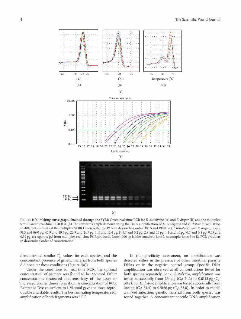
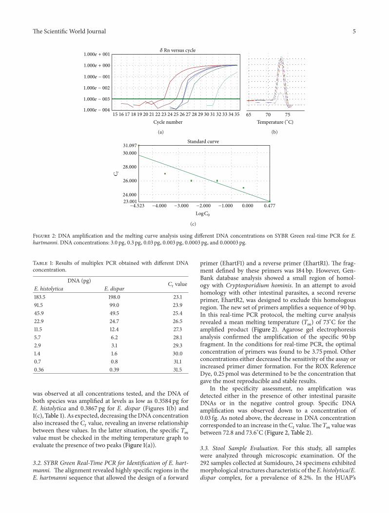
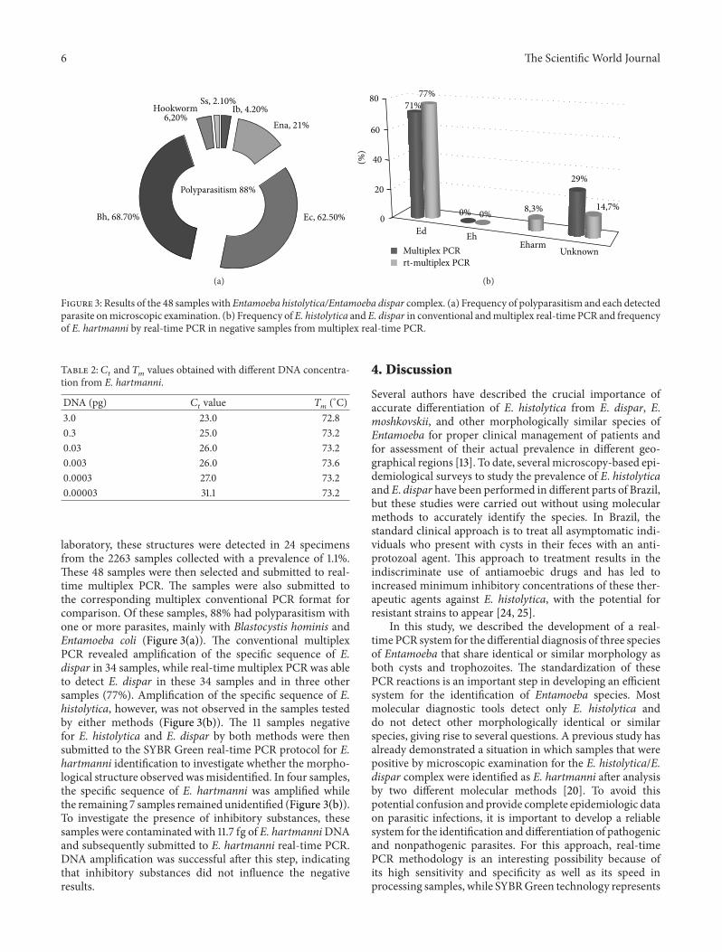
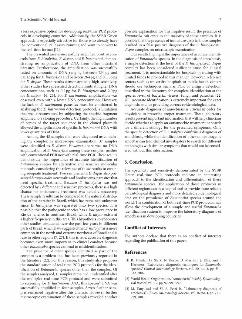


![multiplexing stool MD short.ppt [Compatibiliteitsmodus] · 8 0394-0527 Giardia Entamoeba coli Giardia 9 0394-0526 Giardia Entamoeba coli Giardia 10 1032-1221 Giardia negative Giardia](https://img.pdfslide.net/doc/110x75/5e4182dad3a23a3d0b082f2c/multiplexing-stool-md-shortppt-compatibiliteitsmodus-8-0394-0527-giardia-entamoeba.jpg)