Embed Size (px)
Citation preview
1
Digestive needs
Physiologic Integrity and TherapeuticNursing Interventions for the Patients with
Digestive NeedsNsg 4037
2006
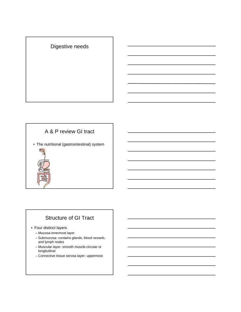
A & P review GI tract
• The nutritional (gastrointestinal) system
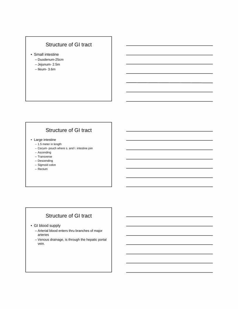
Structure of GI Tract
• Four distinct layers– Mucosa-innermost layer– Submucosa- contains glands, blood vessels,
and lymph nodes– Muscular layer- smooth muscle-circular or
longitudinal– Connective tissue serosa layer- uppermost
2
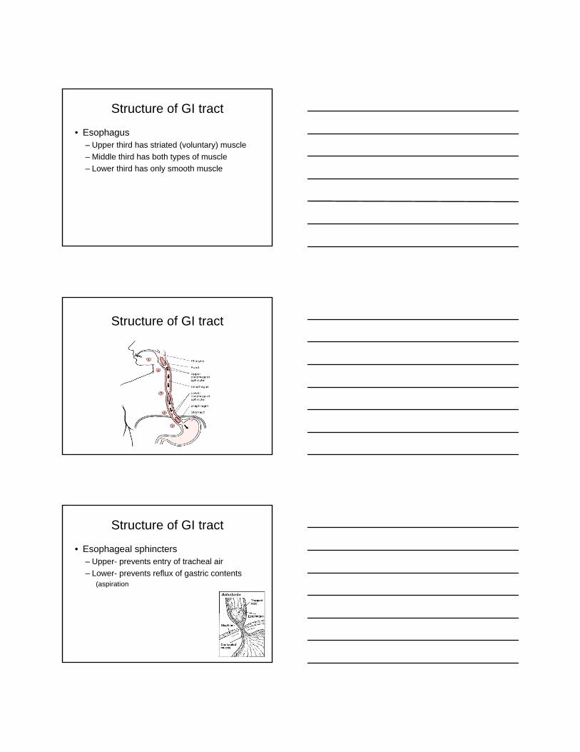
Structure of GI tract
• Esophagus– Upper third has striated (voluntary) muscle– Middle third has both types of muscle– Lower third has only smooth muscle
Structure of GI tract
Structure of GI tract
• Esophageal sphincters– Upper- prevents entry of tracheal air– Lower- prevents reflux of gastric contents
(aspiration
3
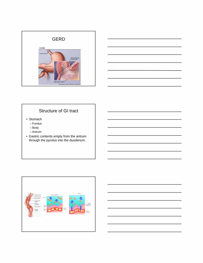
GERD
Structure of GI tract
• Stomach– Fundus– Body– Antrum
• Gastric contents empty from the antrumthrough the pyrolus into the duodenum.
4
Structure of GI tract
• Small intestine– Duodenum-25cm– Jejunum- 2.5m– Ileum- 3.6m
Structure of GI tract
• Large intestine– 1.5 meter in length– Cecum- pouch where s. and l. intestine join– Ascending– Transverse– Descending– Sigmoid colon– Rectum
Structure of GI tract
• GI blood supply– Arterial blood enters thru branches of major
arteries– Venous drainage, is through the hepatic portal
vein.
5
Structure of GI tract
• Neural regulation– Enteric nervous system-regulates motility and
secretion along the entire GI tract– Sympathetic- inhibit activity in enteric
plexuses• Constrict GI system blood vessels,• Decrease glandular secretions
Structure of GI tract
• Parasympathetic- vagus nerve is primarynerve supply to the GI tract– Stimulate motor activity– Stimulate secretory activity– Stimulate endocrine secretions
Function of GI tract
• Motility• Secretion• Digestion• Absorption
6
Function of GI tract
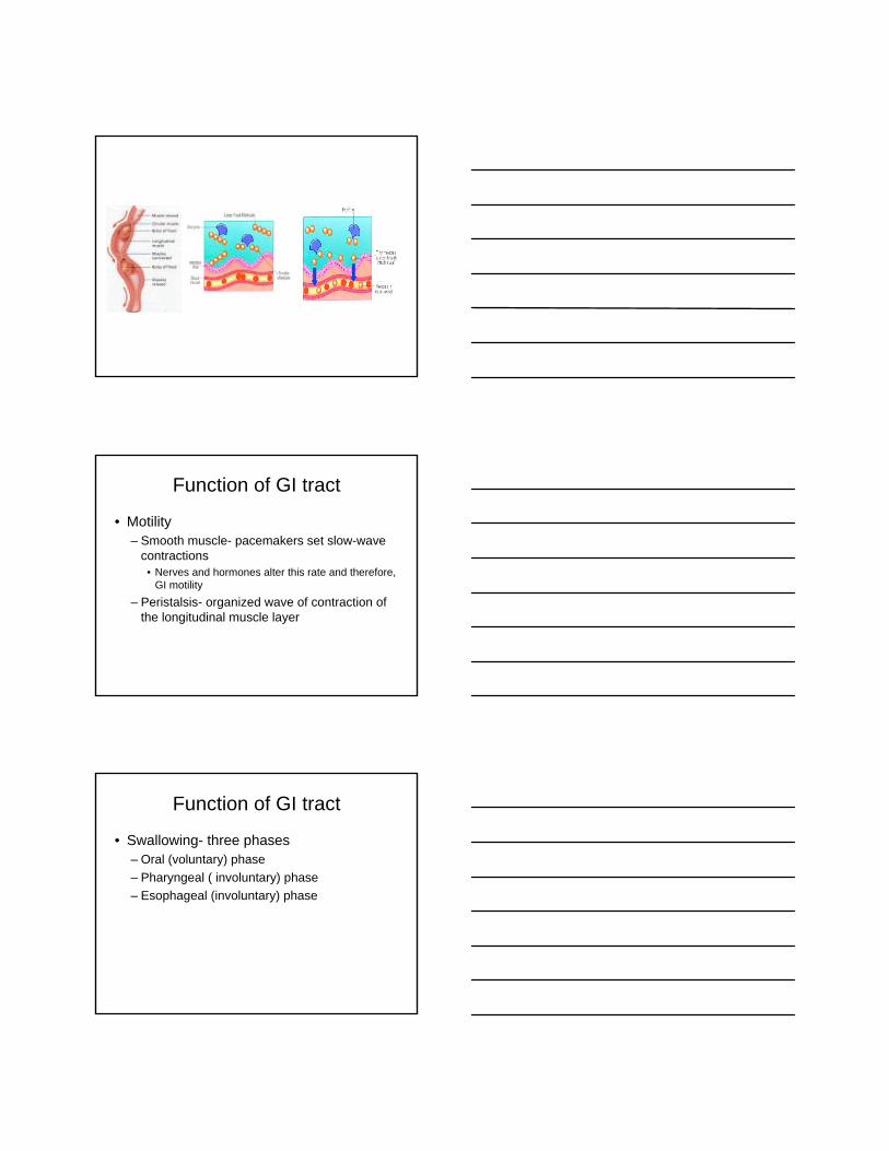
• Motility– Smooth muscle- pacemakers set slow-wave
contractions• Nerves and hormones alter this rate and therefore,
GI motility– Peristalsis- organized wave of contraction of
the longitudinal muscle layer
Function of GI tract
• Swallowing- three phases– Oral (voluntary) phase– Pharyngeal ( involuntary) phase– Esophageal (involuntary) phase
7
Function of GI tract
• Stomach– Occur after feeding at a rate of three per
minute– Antrum has strong contractions that fragment
food into smaller particles and mix the foodwith gastric secretions to initiate digestion.
Function of GI tract
• Gastroduodenal junction– Pylorus and terminal end of antrum contract
simultaneously– The rate of gastric emptying must match the
duodenal buffering ability or acid may damageduodenal mucosa
Function of GI tract
• Small intestine– Peristalsis moves chyme aborally 10cm per
contraction.– Chyme takes 2-4 hours to move 6 meters
8
Function of GI tract
• Small intestine– Peristalsis moves chyme aborally 10cm per
contraction.– Chyme takes 2-4 hours to move 6 meters
Function of GI tract
• Large intestine– Takes 1-3 days to complete its passage
through the LI– Sodium and water absorbed– Bacteria consume more nutrients and release
some vitamins (K)
Function of GI tract
• Vomiting9 things occur during the vomiting process.
9
Functions of GI tract
• Secretions– Aid in absorption by helping digest foods to
absorbable componentsTypes
lubricantsionsabsorption facilitatorsbile
Functions of GI tract
• Digestion and absorption– Carbohydrates- D & A occurs primarily in duodenum
and jejunum.– Protein- stomach and small intestine for digestion,
abs. in the duodenum and jejunum– Lipids- duodenum lipids emulsified by bile acids and
digested to form micelles with bile acids. The micellesare absorbed at the intestinal brush border.
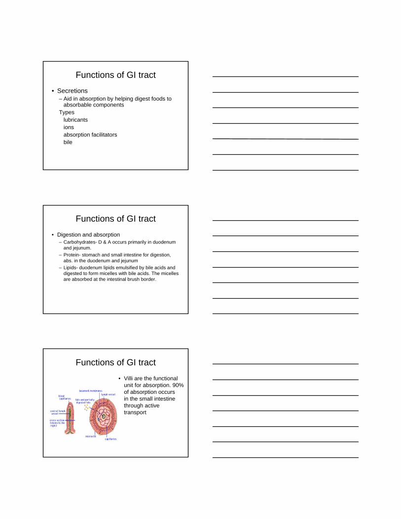
Functions of GI tract
• Villi are the functionalunit for absorption. 90%of absorption occursin the small intestinethrough activetransport
10
Function of GI tract
• Water and electrolytes- 99% of ingestedwater is absorbed.– Greatest in the jejunum and least in the colon.
GI assessment
• Screening of nutritional health– Ask about dietary intake– 24 hour recall– Ask for a 1-3 day food diary
GI assessment
• Malnutrition– Primary- occurs when adequate nutrition is
not delivered to the upper GI tract over anextended period of time.
– Secondary- occurs when the upper GI tractfails to absorb, metabolize or use nutrients
11
GI assessment
• Chronic pancreatitis• Short-bowel syndrome• Pressure ulcers• Cancer• AIDS• Prior gastric surgery
History
• Biographical data-– Age– Gender– Religious affiliation– Marital status
History
• Chief complaint– Nausea, vomiting– Indigestion– Abdominal pain– Diarrhea– Weight changes, appetite
12
History
• Symptom analysis– When does N or V occur? How long does it
last? Relation to food intake– Indigestion- “burping” or burning, relation to
food intake, type of foods– Abdominal pain- rapid or gradual in
onset,intensity, radiate?, duration, worsen orimprove with movement,
History• Abdominal pain
– Bowel obstruction- intermittent, colicky pain• Bowel sounds may progress from high pitch to absent
– Peritoneal inflammation- steady, aching pain directlyover area of inflammation. Pain increases with motion
– Vascular catastrophe (AAA or infarction)- may bepreceded by 2-3 days of mild-mod. pain followed bysevere abdominal pain and manifestations of shock.Back and flank pain are common with aorticaneurysms.
History
• Diarrhea- how many stools? How much?Stools liquid or solid? What color?painwith defecation?
• Appetite and weight change- describeappetite, and change, change in taste,smell, activity level, mood states? Weightgain, loss intentional.

13
Past health history
• Major illnesses and hospitalizations• Medications• Allergies
Family health history• Genetics and family environment play a role in
the development of some GI disorders– Ulcerative colitis– Crohn’s disease– Alcoholism– Liver disease– Cancer– Peptic ulcer disease– Irritable bowel disease
Psychosocial history
• Occupation• Diet
14
Review of systems
• Assess condition of mouth• Dental habits• Dentures• Swallowing
Physical examination
• Height and weight• Body mass index• Frame size• Circumferential measurements
Physical examination
• Mouth– Inspection and palpation– Symmetry, color, hydration, lesions, nodules.– Teeth for malocclusion or missing teeth– Check for loose teeth, masses, swelling,
tenderness
15
Physical examination
• Abdomen– Ask client to void first– Supine position– Small pillow under knees
Physical examination
• Inspection– Skin, contour, hair distribution– Notice scars, striae, petechia, rashes– At eye level, look for peristalsis movements or
pulsations
Physical examination
• Auscultation– Beginning in RLQ– Clockwise to each quadrant– Frequency and character– Vascular sounds
16
Palpation
• Non-tender areas first• Light palpation• Masses and areas of tenderness• Guarding, rigidity• Deep palpation
Diagnostic tests
• Laboratory tests• Radiography• Ultrasonography• Endoscopy• Exfoliative cytologic analysis• Gastric analysis
Management of clients withmalnutrition
• Protein-energy malnutrition– Need is not supplied through dietary intake
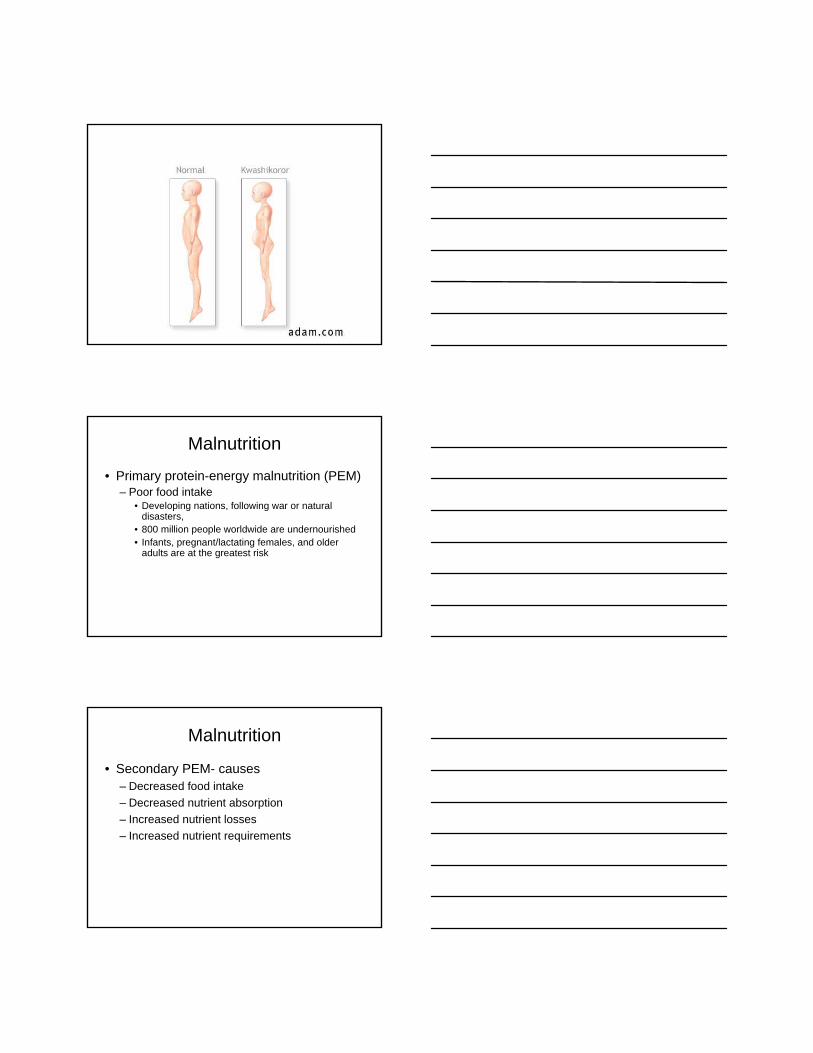
• Syndromes– Kwashiorkor-chronic protein deficiency– Marasmus- prolonged caloric deficiency
17
Malnutrition
• Primary protein-energy malnutrition (PEM)– Poor food intake
• Developing nations, following war or naturaldisasters,
• 800 million people worldwide are undernourished• Infants, pregnant/lactating females, and older
adults are at the greatest risk
Malnutrition
• Secondary PEM- causes– Decreased food intake– Decreased nutrient absorption– Increased nutrient losses– Increased nutrient requirements

18
Malnutrition
• Etiology and risk factors– Socioeconomic– Physiologic changes– Medical therapies
Malnutrition
• Pathophysiology– Total supply of nutrients is less than the
body’s requirement– Unintentional weight loss of 5% in 1 month or
10% in 6 months
Malnutrition
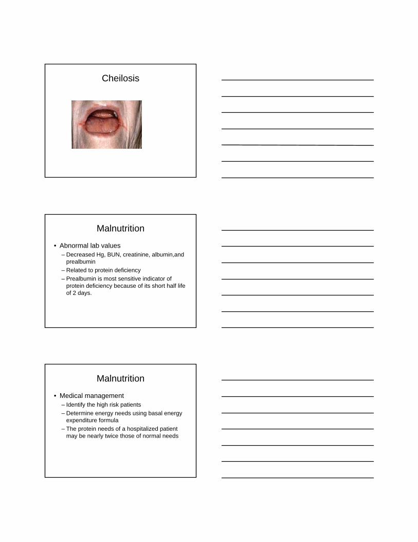
• Clinical manifestations– Hair loss or dull, dry hair– Dry, bruised skin– Brittle nails– Periodontal disease, bleeding gums– Anemia– Cheilosis
19
Cheilosis
Malnutrition
• Abnormal lab values– Decreased Hg, BUN, creatinine, albumin,and
prealbumin– Related to protein deficiency– Prealbumin is most sensitive indicator of
protein deficiency because of its short half lifeof 2 days.
Malnutrition
• Medical management– Identify the high risk patients– Determine energy needs using basal energy
expenditure formula– The protein needs of a hospitalized patient
may be nearly twice those of normal needs
20
Malnutrition
• Determine route of feeding– Supplements to boost calories– Enteral feedings used when unable to take
oral fluids– Parenteral feeding when GI tract is non
functional
Malnutrition
• Nursing management– Continued monitoring of clients nutritional
status– ND- Feeding self-care deficit related to
impaired motor function, impaired cognitivefunction, sensory-perceptual alterations, ordecreased appetite
Malnutrition• Interventions
– Improve nutritional intake• Dietician consult, improve menu items
– Increase appetite• Pleasant environment, adequate pain control,• Regular exercise, oral care,
– Increased social interaction– Minimize sensory-perceptual deficits– Minimize the impact of neuromuscular deficits– Minimize the impact of cognitive impairments– Minimize fatigue

21
Malnutrition
• Impaired swallowing– Team approach- physicians, PT,OT, Speech,
nurses
Malnutrition
• Assess swallowing– Assess LOC– Assess gag reflex– Have the client produce an audible cough– Have the client produce a voluntary swallow
Malnutrition• Swallowing techniques
– Calm quiet environment– Assist in placing food bolus in unaffected side of
mouth and toward pharynx– Tilt chin down to decrease risk of aspiration– Massage throat on affected side– Watch the thyroid cartilage for swalowing– Inspect the mouth before more food– Allow sufficient time between mouthfuls

22
Malnutrition
• Enteral nutrition– Tube feeding– Contraindicated in complete intestinal
obstruction, severe ileus, severe diarrhea,malabsorption syndrome
– If the gut works, use it
Malnutrition
• Enteral access– NG- into the stomach– Gastrostomy– Jejunal tube (J-tube)-jejunum
Malnutrition
• Short term- NG tubes• Small bore enteral feeding tubes are made
of silicone or polyurethane (softer)• Long-term- G-tubes, J-tubes are placed
surgically, endoscopically.• PEG tubes- most common long term
23
malnutrition
• Nursing management of enteral nutrition– Monitor for aspiration– Prevent contamination of formula and delivery
systems– Assess tube location– Administer feedings– Prevent aspiration– Maintain enteral access
Malnutrition
• Parenteral nutrition– Clinical indications
• Short bowel syndrome• Severe prolonged radiation enteritis• High output fistulae• Motility disorders
– Ileus, persistent vomiting
Malnutrition
• Parenteral feedings– Consist of carbohydrates- 50-70%– Fat emulsions- 10-30%– Amino acids– Fluids, electrolytes, vitamins and trace
elements
24
Malnutrition
• Vascular access– Central venous line– Must be positioned in a high flow vein– Superior or inferior vena cava
Malnutrition• Interventions
– Administer PN• Check solution for exp. date, correct ingredients, appearance
of solution• Must use a pump
– Monitor blood glucose levels• Every 6 hours initially, then q 24 hours.
– Observe for allergy to PN components– Maintain vascular access– Prevent infection– Provide dressing changes
Malnutrition
• Evaluate interventions– Assess tolerance, fluid status and GI status– Monitor VS, lab tests, and function of
nutritional access device– Therapies are routinely used in home
settings.
25
Eating disorders
• Obesity– Characterized by an excess accumulation of
fat– Causes are complex and pervasive– Defined by BMI greater than 30kg/m2
Eating disorders- obesity
• Environmental• Genetic tendency• Socioeconomic factors• Ethnic disparities
Eating disorders- obesity
• Clinical manifestations– Type 2 diabetes– Cardiovascular disease– Hypertension– Stroke, sleep apnea– Cancers-breast, colon and prostate
26
Eating disorders-obesity
• Medical management– Skin and wound problems are common– Diet, exercise, occasionally medication– Diet that provides 500-1000 calories less than
expenditure is ideal for 1-2 pound per weekweight loss.
– Lifestyle modifications
Eating disorders-obesity
• Surgical management– Gastric restrictive– Restrictive plus malabsorptive
Eating disorders- anorexia andbulimia nervosa
• Risk factors– Young women– Women 10 times greater than men– More prevalent in Western cultures– Low self-esteem
27
Eating disorders-anorexia andbulimia
• Similar to starvation– Body uses fat stores– Shifts in fluid and electrolyte balance– Can be life threatening– Alterations in the metabolism of insulin,
thyroid hormones and catecholamines
Eating disorders-anorexia andbulimia
• Clinical manifestations– Clients may limit themselves to 200-500
calories/day– Dry skin, pallor, bradycardia, hypotension– Intolerance to cold– Constipation– Amenorrhea
Eating disorders-anorexia andbulimia
• Bulimia– Eating and vomiting usually in late afternoon– Abuse of laxatives and diuretics– Depression symptoms– Erosion of tooth enamel
28
Eating disorders-anorexia andbulimia
• Medical management– Psychological as well as nutritional
components– Enteral and parenteral therapy may be
needed in serious cases
Management of Clients withIngestive Disorders
• Problems occur in the oral cavity oresophagus
• Dental disorders
Dental disorders
• Preserve client’s own teeth as long as possible• Natural teeth are more functional in masticating
food than dentures• Effective mastication helps promote efficient
digestion• Efficient digestion results in healthy GI function
and general health
29
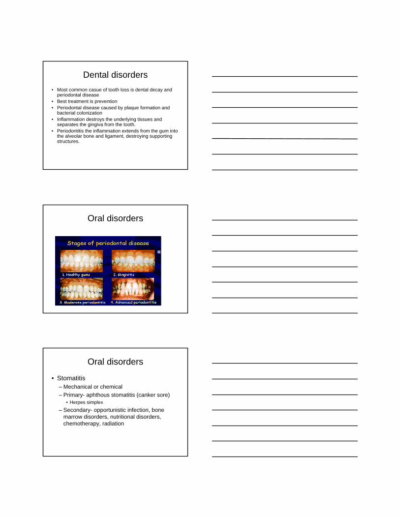
Dental disorders• Most common casue of tooth loss is dental decay and
periodontal disease• Best treatment is prevention• Periodontal disease caused by plaque formation and
bacterial colonization• Inflammation destroys the underlying tissues and
separates the gingiva from the tooth.• Periodontitis the inflammation extends from the gum into
the alveolar bone and ligament, destroying supportingstructures.
Oral disorders
Oral disorders
• Stomatitis– Mechanical or chemical– Primary- aphthous stomatitis (canker sore)
• Herpes simplex– Secondary- opportunistic infection, bone
marrow disorders, nutritional disorders,chemotherapy, radiation
30
Oral disorders
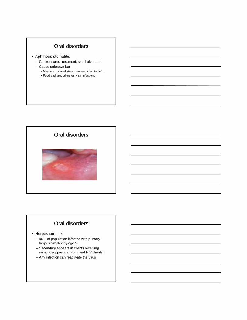
• Aphthous stomatitis– Canker sores- recurrent, small ulcerated.– Cause unknown but-
• Maybe emotional stress, trauma, vitamin def.,• Food and drug allergies, viral infections
Oral disorders
Oral disorders
• Herpes simplex– 90% of population infected with primary
herpes simplex by age 5– Secondary appears in clients receiving
immunosuppresive drugs and HIV clients– Any infection can reactivate the virus
31
Oral disorders
• Vincent’s angina (trench mouth)– Acute bacterial infection of the gingivae
caused by the resident flora of the mouth– Caused by poor oral hygiene, increased age,
DM, lack of sleep, viral infections– Not contagious
Oral disorders
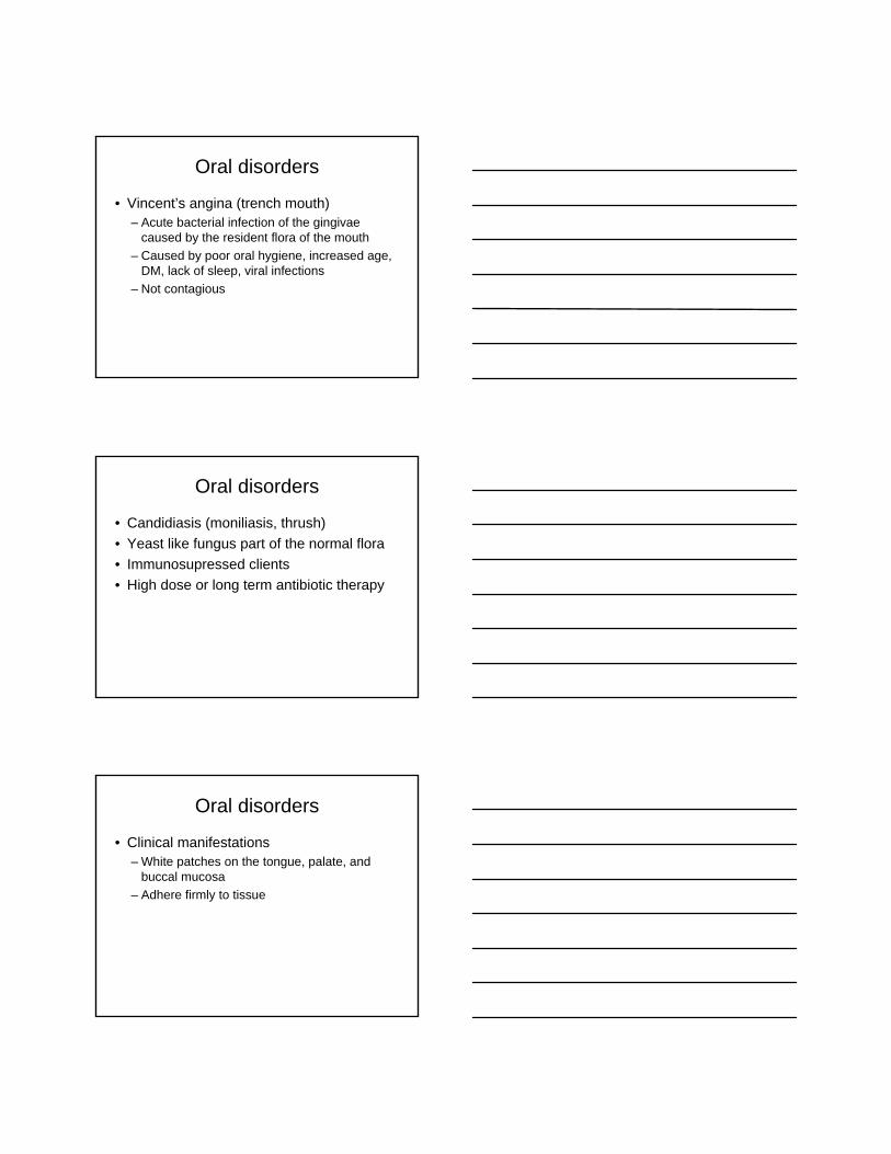
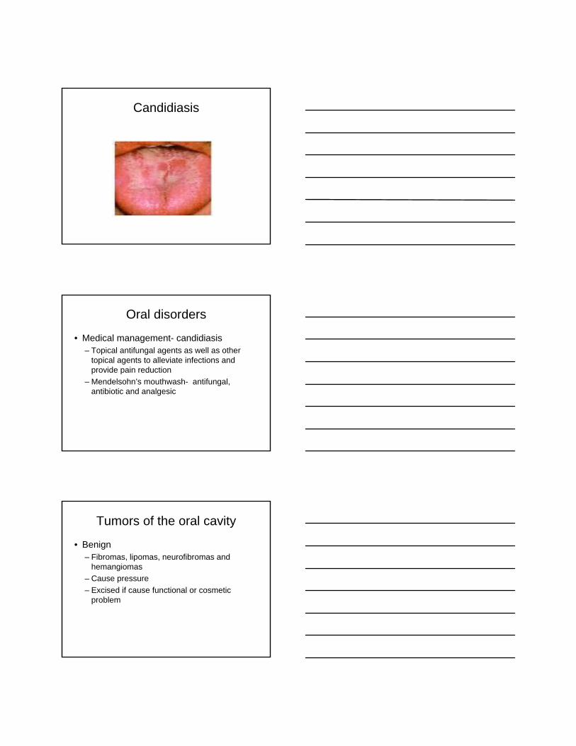
• Candidiasis (moniliasis, thrush)• Yeast like fungus part of the normal flora• Immunosupressed clients• High dose or long term antibiotic therapy
Oral disorders
• Clinical manifestations– White patches on the tongue, palate, and
buccal mucosa– Adhere firmly to tissue
32
Candidiasis
Oral disorders
• Medical management- candidiasis– Topical antifungal agents as well as other
topical agents to alleviate infections andprovide pain reduction
– Mendelsohn’s mouthwash- antifungal,antibiotic and analgesic
Tumors of the oral cavity
• Benign– Fibromas, lipomas, neurofibromas and
hemangiomas– Cause pressure– Excised if cause functional or cosmetic
problem
33
Tumors of the oral cavity
• Premalignant• Leukoplakia
– Yellow-white or gray-white lesion– Any region of mouth.– Leathery surface– Clearly defined borders– Men twice as frequently as women
Tumors of the oral cavity
• Erythroplakia– Red, velvety appearing patch that commonly
indicates early squamous cell carcinoma– Most frequently 50-60 years of age– Men and women the same
Tumors of the oral cavity
• Malignant tumors– Most commonly seen at 40-50 years old– Men more than women– Associated with long alcohol consumption and
tobacco use
34
Tumors of the oral cavity
• Basal cell carcinoma– Second most common cancer of the oral
cavity– Occurs on the lips– Characteristic pearly border– Due to excessive exposure to sunlight
Tumors of oral cavity
• Squamous cell carcinoma– Leading type of oral cancer– Older than 45 years of age– Lower lip and tongue– Sore or lesion that does not heal
Tumors of the oral cavity
• Medical management– Inhibit tumor growth
• Survival depends on site and staging
• Radiation therapy• Chemotherapy
35
Tumors of the oral cavity
• Nursing management– Interventions
• Avoid oral irritants• Promote comfort• Promote nutrition• Relieve mouth dryness
Tumors of the oral cavity
• Surgical management– Local excision of small tumors– Extensive surgery for invasive tumors
• Radical and modified radical neck dissection• Requires extensive preparation of the client for
surgery
Tumors of the oral cavity
• Nursing management– Make sure client understands implications of
surgery– Assess rehabilitative needs– Speech therapy, coping with disfigurement,
depression
36
Tumors of the oral cavity
• Interventions– Maintain airway
• Semi to high Fowler’s position– Provide wound care– Monitor for bleeding– Administer supplemental nutrition– Discuss eating modifications
Tumors of the oral cavity• Interventions- impaired verbal communications
– Promote alternate forms of communication• Paper/pencil, magic slate, laptop• Nurses’ manner should communicate acceptance,
compassion, and caring.– Relieve anxiety
• Check on them frequently• Respond promptly• Label intercom re: client’s inability to speak
Tumors of the oral cavity
• Client should heal within 6 weeks to 3months.
• Need tremendous emotional support• Self-feed via tube until healing occurs• Home health referral for respiratory
support, suctioning nutritional support andwound and trach care
37
Disorders of the salivary gland
• Parotitis- inflammation of the parotidglands– Caused by inactivity of the glands due to
certain drugs (diuretics), prolonged NGintubation, and lack of oral intake.
– Calculi- stones form in salivary glands– Tumors- most are benign in salivary glands
Disorders of the esophagus
• Dysphagia– Mechanical obstruction– Cardiovascular abnormalities– Neurologic diseases– Other causes
Disorders of the esophagus
• Regurgitation– Ejection of small amounts of chyme or gastric
juice from the mouth without nausea– Immediately after swallowing results from
structural or motor abnormalities in the LES
38
Disorders of the esophagus
• Acute pain-(odynophagia) pain withswallowing– Can be sharp, stabbing, crushing, knife-like– May be constant or only with swallowing
(esophageal spasm)– Reflux disease– Radiation– Viral infection
Disorders of the esophagus
• Heartburn or pyrosis– Substernal, midline burning tends to radiate,
generally in waves, upward to the neck,resulting from abnormalities of the LES
– Occurs in obesity, postural changes, duringpregnancy, or alcohol intake.
Disorders of esophagus
• Achalasia– Progressively increasing dysphagia– Unknown cause, no risk factors, occurs in 20s
and 30s– Impaired mobility of the lower 2/3 of
esophagus.– LES fails to relax normally with swallowing
39
Disorders of esophagus
• Clinical manifestations– Dysphagia– Substernal pain– Regurgitation of undigested food– URI, emotional disturbance, pregnancy,
obesity exacerbate problem
Disorders of esophagus
• Medical management– Relieve manifestations
• PEG tube, or surgical procedures
– Administer medications• Relax the sphincter (anticholinergic drugs)• Antacids, H2 receptor antagonists, and proton pump
inhibitors
– Modify diet– Alternate positions
Disorders of esophagus
• Nursing management– Consult with client about dietary habits– Possibility of PEG tube placement– Pain eased thru medications
40
Disorders of the esophagus
• Surgical management– Dilation of LES– PEG placement
Disorders of esophagus
• GERD– Results from backward flow of gastric
contents into the esophagus– Causes reflux esophagitis– Associated with hiatal hernia
Disorders of the esophagus
• GERD– Cause
• An alteration in the innervation of thegastroesophageal sphincter
• Displacement of the angle of the GE junction• Incompetent LES
41
Disorders of the esophagus
• Risk factors– Obesity– Weight gain– Pregnancy– Chewing tobacco, smoking– High fat foods– Theophylline– Caffeine, chocolate
Disorders of esophagus
• Discomfort usually begins after meals• Increased intraabdominal pressure• Lying or supine positions
Disorders of esophagus
• Medical management– NSAIDs, anticholinergic drugs, calcium
channel blockers, theophylline should beavoided
– Lifestyle and diet changes– Endoluminal gastroplication
42
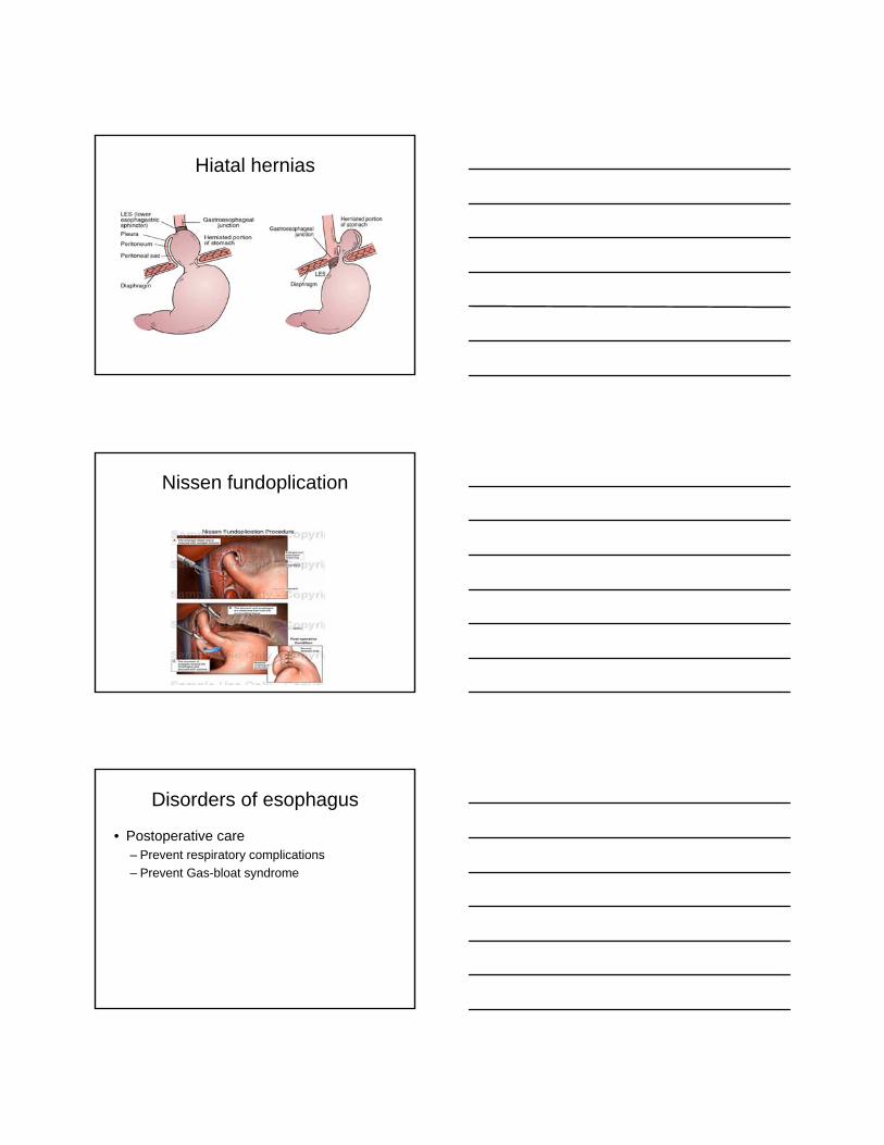
Disorders of the esophagus
• Hiatal hernia– Cardiac sphincter becomes enlarged, allowing
a part of the stomach to pass into the thoraciccavity.
• Sliding (type I)• Rolling (type II)
Disorders of the esophagus
• Causes and risk factors– Related to muscle weakness in esophageal
hiatus, loosens the esophageal support– Aging, trauma, surgery– Anything that increases intraabdominally
pressure such as lifting, coughing, pregnancy,obesity
Disorders of the esophagus
• Clinical manifestation– Heartburn 30-60 minutes after a meal– Reflux– Fullness after eating, difficulty breathing– Worse when lying down
43
Hiatal hernias
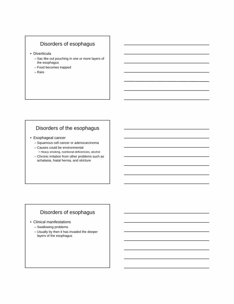
Nissen fundoplication
Disorders of esophagus
• Postoperative care– Prevent respiratory complications– Prevent Gas-bloat syndrome
44
Disorders of esophagus
• Diverticula– Sac like out pouching in one or more layers of
the esophagus– Food becomes trapped– Rare
Disorders of the esophagus
• Esophageal cancer– Squamous cell cancer or adenocarcinoma– Causes could be environmental
• Heavy smoking, nutritional deficiencies, alcohol– Chronic irritation from other problems such as
achalasia, hiatal hernia, and stricture
Disorders of esophagus
• Clinical manifestations– Swallowing problems– Usually by then it has invaded the deeper
layers of the esophagus
45
Disorders of the esophagus
• Medical management– Inhibit tumor growth– Radiation therapy– Chemotherapy– Photodynamic therapy– Maintain nutrition
Disorders of the esophagus
• Nursing management– Assess nutritional status, dysphagia– Encourage soft food– Assess odynophagia, regurgitation, chronic
cough, increased secretions and hoarseness
Disorders of the esophagus
• Interventions– Monitor nutritional status, weight changes, I &
O– Teach diet changes– Assess skin around feeding tube– Provide emotional support– Poor prognosis frequently
46
Disorders of the esophagus
• Vascular disorders– Esophageal varices
• Trauma– Chemical burns– Foreign bodies– External forces
Management of clients withdigestive disorders
• Clinical manifestation– Pain– Anorexia– Nausea and vomiting– Bleeding– Diarrhea– Belching flatulence– Indigestion
Gastrointestinal intubation
• Used for– Decompression– Lavage– Gastric analysis– Tube feedings
47
Gastrointestinal intubation
• Types of tubes– Short tubes
• levin tubes• Salem sump tubes
Gastrointestinal intubation
• Medium tubes– Variety of nasoduodenal tubes– Extend from nose to the duodenum and are
for short-term feeding.– Weighted tip-less likely to cause aspiration
Gastrointestinal intubation
• Long tubes– Extend into small bowel, sometimes the entire
length– Not used much– Types- Miller-Abbott Tube– Cantor tube– Harris tube
48
Gastrointestinal intubation
• Other tubes– G-tubes or J- tubes are for long term enteral
feedings.– PEG tubes or PEJ tubes also
Gastrointestinal intubation
• Insertion of tubes– High Fowler’s position– Measure the distance on the tube– Lubricate and gently insert– Have the client swallow– Verify placement
Gastrointestinalintubation
• Suctioning– Ensure that gastric mucosa is not traumatized– Intermittent suction is used
49
Gastrointestinal intubation
• Nursing management– Comfort– Clean and lubricate nares– Tape the tube to prevent irritation of nares– Frequent oral hygiene– Chew gum or ice chips– Request order for anesthetic mouthrinse or
lozenges
Gastritis
• Acute gastritis– Inflammation of gastric mucosa– Risk factors- seen with nausea and vomiting,
bleeding, malaise, anorexia– Aspirin and NSAIDs, digitalis, chemo, steroids,
acute alcoholism and food poisoning.
Gastritis
• Health promotion behaviors– Limit use of NSAIDs, alcohol, caffeine– Avoid nicotine products, smoking
• Health maintenance behaviors– Use of enteric coated aspirin, COX-2
inhibitors, proton pump inhibitors to blockgastric acid production
50
Gastritis
• Mucosal lining of the stomach acts as abarrier to protect it from the gastric acid
• When barrier is penetrated, gastritisoccurs
Gastritis
• Clinical manifestations– Epigastric discomfort– Abdominal tenderness– Reflux– Nausea and vomiting– Hematemesis
Gastritis
• Medical management– Removal of cause and treat symptoms– Withhold foods and fluid until N & V subside
51
Gastritis
• Chronic gastritis– Superficial gastritis
– Atrophic gastritis
– Hypertrophic gastritis
Gastritis
• Risk factors– Peptic ulcer disease (PUD)– Infection with Helicobacter pylori bacteria– Gastric surgery– Others similar to acute gastritis– Age
Gastritis
• Mucosal lining becomes thickened anderythmatous and then thin and atrophic.
• Loss of function of parietal cells• Decreased acid secretion leads to inability
to absorb vitamin B12.• Also a risk factor for gastric cancer
52
Gastritis
• Clinical manifestations– Vague– Anorexia– Feeling of fullness– Nausea– Intolerance to spicy foods– Epigastric pain
Gastritis
• Complications– Bleeding– Pernicious anemia– Gastric cancer
Gastritis
• Nursing management– Reduce pain
• Teach about foods that worsen, avoid smokingalcohol
• Gaviscon is best antacid for gastritis• H2 receptors and PPI enhance mucosal defenses
and reduce pain
53
Gastritis
• Nursing management (cont)– Promote self care
• Instruct to keep appointments with provider• Especially if H. pylori present, closely r/t gastric
cancer
Peptic ulcer disease
• Break in the continuity of mucosa– Occurs in 10% of population
PUD
• Duodenal ulcers– Characterized by high gastric secretions– Rapid emptying of the stomach
54
PUD
• Gastric ulcers– Heal within few weeks– Form within an inch of the pylorus– Incompetent pylorus may decrease mucus production
allowing gastric juices to injure mucosa– Incompetent pylorus may allow bile acids to reflux into
the stomach and break the barrier
PUD
• Stress and drug induced ulcers– Usually occur after an medical crisis– Severe trauma or major illness– Severe burns– Head injury– Ingestion of drug– Shock– sepsis
PUD
• Causes and risk factors– 90% attributed to H. pylori– PUD results when the aggressive factors of
PUD exceed the defensive barrier.– Smoking, chewing, alcohol, stress, steroids,
ASA, NSAIDs,– Zollinger-Ellison, Crohn’s dz, hepatic and
biliary disease may play a role also
55
PUD
• Pathophysiology of gastric ulcers– Protection factors- tight, nonpermeated
junctions between epithelial cells and thealkaline layer of the mucus that coats thesurface of the gastric epithelium
– This barrier may be interrupted by the chronicpresence of the injurious substances such asASA, NSAIDs, steroids
PUD
• Pathogenesis of duodenal ulcers– Activity of the vagus nerve is increase– Stimulates the pyloric cells to release gastrin,
which stimulates the release of HCl acid
PUD
• Another factor is emotional stress,– Thalamic stimulation of vagal nerves results in
increase in gastric secretion, blood supply,and gastric motility
– Stress reactions upset the aggressive-defensive balance.
56
PUD
• Zollinger –Ellison syndrome– Abnormal secretion of gastrin by rare islet cell tumor
in the pancreas– Hypergastinemia and diarrhea secondary to fat
malabsorption– Hyperplasia of the gastric mucosa due to the trophic
effects of gastrin– Treatment aimed at suppression of acid secretion
PUD
• Clinical manifestations– Acute pain
• Aching, burning, cramp-like pain• Definite relation to eating
– Gastric ulcers- food causes pain, vomiting relieves it– Duodenal ulcers- pain on empty stomach, relieved by
food or antacids.
• Location – 2-10 cm between the xiphoid and theumbilicus
PUD
• Clinical manifestations– Nausea and vomiting
• Vomiting- gastric ulcer, esp. in the pylorum orantrum of stomach
• Results from gastric stasis or pyloric obstruction• Usually vomits undigested food
57
PUD
• Clinical manifestations– Bleeding may be massive or occult
PUD
• Medical management– Provide stomach rest– Neutralize HCl acid– Eradicate H. pylori– Dietary management– Stress reduction
PUD• Prevent and treat complications
– Hemorrhage- assess bleeding, tarry stools.– Prevent shock with IV fluids, NPO, NG tube to assess
bleeding and also to administer room temperaturesaline.
– Replace fluids– Administer vasopressin- arterial admin.– Inject artery with emboli- via angiography– Maintain rest
58
PUD
• Maintain high gastric pH• Stop bleeding surgically• Perform multipolar electrocoagulation or
heater probe therapy
PUD
• Perforation– Surgical emergency– Gastroduodenal contents escape through the
stomach wall into the peritoneal cavity.– Assess pain- sudden sharp severe pain in the
midepigastrium.– Replace fluids- immediate replacement of fluids,
electrolytes, and blood as well as antibiotics– Correct perforation surgically
PUD
• Obstruction– Scarring causes pyloric obstruction– Pain at night– Vomiting– Surgery required
59
PUD
• Nursing management– Monitor for development of complications– Assess for pain and document occurrence
and location– Promote rest and relaxation– Provide teaching– Provide support
PUD
• Surgical management– See p.756-758– Different options
PUD
• Nursing management of surgical patient– Postop interventions
• Maintain NG tube• Monitor for complication• Promote comfort
60
Gastric cancer
• Risk factors• Men more than women• Chronic atrophic gastritis• Pernicious anemia• Smoking• Metal crafts workers, miners, bakers,
dusty, smoky environments
Gastric cancer
• Arises from the mucosal lining• Prognosis best for polypoid lesions• Worse for ulcerating cancers• Poorest for infiltrating forms
Gastric cancer
• Clinical manifestations– Seldom detected in early stages– Palpable mass, ascites or bone pain may be
first manifestation– Weight loss, vague indigestion, anorexia,
61
Gastric cancer
• Nursing management– Control pain– Management of nutritional therapy– Explanation of disease and all treatment
options
A and P review
• Large intestine– Cecum– Ascending– Transverse– Descending
Assessment of elimination
• Similar assessment questions as with theupper digestive systems
• Travel history of client is particularlyimportant in assessing for eliminationdisorders
• E. coli is most common cause of diarrhea
62
Physical examination
• Abdomen• Anus• Rectum
Anus and rectum
• Most nurses do a visual inspection• Rectal anatomy is important in assessing
for digital impaction
Diagnostic tests
• Similar to ingestive diagnostic tests• Laboratory tests
– CEA- High CEA levels characteristic ofmalignancies of breast, colorectal cancer
• Often called tumor markers when used to monitoreffectiveness of treatment
63
Diagnostic tests
• Fecal analysis– Color, consistency, odor,– Stool specimen required for diagnosis of
infectious diseases, GI bleeding and other GIdisorders
– Fecal occult blood• Screening for colorectal cancer
Diagnostic tests
• Stool examination for ova and parasites• Stool cultures• Fecal lipids
Diagnostic tests
• Endoscopy– Protosigmoidoscopy
• Lining of the sigmoid colon, the rectum and theanal canal using a proctoscope and asigmoidoscope.
– Colonoscopy• Visual exam of the entire lining of the colon with a
flexible fiberoptic scope. Screen clients at high riskof colon cancer.
64
Management of clients withintestinal disorders
• Bleeding – blood in stool, color is affectedby the digestive processes on the bloodand the rapidity with which the chymepasses thru the bowel.
• Pain- acute or chronic, caused bymechanical, inflammatory or ischemicchanges.
Intestinal disorders
• Nausea and vomiting– Distention of the duodenum– Changes in the integrity of intestinal wall– Changes in the motility– Vomitus that contains fecal matter usually
indicates a distal obstruction in S.I.
Intestinal disorders
• Distention– Caused by excessive gas in the intestines– Flatus is another clinical manifestation
65
Intestinal disorders
• Diarrhea– Increase in frequency, volume, and fluid
content of stool– Common causes- infections, malabsorption
syndromes, medications, allergies, andsystemic diseases
Intestinal disorders
• Constipation– Infrequent or difficult passage of stool– Passage of hard stool
• Abnormalities of fecal content– Presence of fats or other abnormal
constituents normally absorbed from the stool
Inflammatory disorders
• Viral and bacterial infections– Gastroenteritis
• Inflammation of stomach and intestinal tract• Diarrhea, abd. pain, and cramping• Contaminated food and water• C.difficile bacterial dysentery
66
Inflammatory disorders
• Pathophysiology– Disruption of intestinal flora by
• Harmful bacteria and viruses that cause tissuedamage and inflammation
• Depressed by antibiotic therapy, adminsteredeither orally or parenterally.
Inflammatory disorders
• Parasitic infections– Protozoa
• Replicate in the intestinal tract of the host andexcreted in the feces.
• Water borne disease usually but more food bornerecently
• Giardiasis- spoiled food or fecal contaminatedsurfaces or contaminated recreational water
Inflammatory disorders
• Parasitic infections– Helminths- parasitic worms
• Contracted through the skin or from ingestingcontaminated food or water
– Schistosomiasis- parasitic flatworm-
67
Inflammatory disorders
• Nursing management– Rest the bowel– Decrease diarrhea– Restore fluid and electrolytes– Assess diarrhea stools– Assess bowel sounds– Prevent spread of disease
Inflammatory disorders
• Appendicitis– Caused by fecalith that occludes the lumen of
the appendix– Kinking of the appendix– Swelling of the bowel wall– Fibrous conditions of the bowel wall
Inflammatory disorders
• Manifestations– Acute abdominal pain that comes in waves– Guarding– Drawing up the legs to relieve tension– Vomiting– Low grade fever
68
Inflammatory disorders
• Peritonitis• Inflammation of the peritoneal membrane
– Peritoneal membrane is a semipermeable twolayered sac filled with 1500 ml of fluid
– Primary or secondary peritonitis
Inflammatory disorders
• Clinical manifestations– Pain may be localized or generalized– Pain that causes rigidity of the abdomen– Nausea and vomiting– Absence of bowel sounds– Shallow respirations
Inflammatory disorders
• Inflammatory bowel disease– Crohn’s disease– Ulcerative colitis
69
Inflammatory disorders
• Clinical manifestations– Both similar– Abdominal pain, diarrhea, fluid imbalances, weight
loss– Can be very thin, wasted appearance, abdomen is flat
or concave with visible peristalsis– Tenderness on palpation– Rectal bleeding with ulcerative colitis
Inflammatory disease
• Medical interventions are aimed atcontrolling symptoms such as diarrheaand pain
• TPN is required if client does not respondto medical intervention
Inflammatory disorders
• Surgical management– Ulcerative colitis- undergo colectomy with
permanent ileostomy– Crohn’s disease- surgery due to
complications
70
Inflammatory disorders
• Ostomies– Pouch should fit close around stoma– Assess skin for irritation each change– Reduce odor– Discuss medications– Emphasize fluid intake– Explain dietary recommendations– Prevent kidney stones
Neoplastic disorders
• Benign tumors of the bowel– Polyps
• Can become cancerous and can cause obstruction
Neoplastic disorders
• Cancer of the small bowel– Surgery is only option for cure
• Colorectal cancer– Most common GI cancer– Incidence declining with increased screening– Most tumors found in distal portion
71
Neoplastic disorders
• Colorectal cancers– Risk factors
• High fat diets, few fruits and vegetables• Hereditary links• Increased age• History of breast, ovarian, endometrial cancers and
ulcerative colitis
Neoplastic disorders
• Colorectal cancer– 95% develop from polyps– Spread by direct invasion of surrounding
tissue– Lymphatic and circulatory channels– Seeding of cells into the peritoneal cavity
Neoplastic disorders
• Manifestations– Rectal bleeding, change in bowel habits,– Abdominal pain, weight loss, anemia and
anorexia– Tumors in large intestine rarely have early
signs– 1/3 of tumors in distal colon and rectum can
be palpated with digital rectal exam
72
Neoplastic disorders
• Prognosis– Depends on health of client– How early the disease is diagnosed– How effective the treatment is– Overall 51% survive 10 years
Neoplastic disorders
• Medical management– Decrease tumor growth– Radiation therapy– Chemotherapy
Neoplastic disorders
• Colostomies– Single barrel-permanent– Double barrel- temporary– Loop –temporary– Abdominal-perineal resection
73
Neoplastic disorders
• Nursing management– Assess for peristalsis– Advance diet as tolerated– Reduce pain– Monitor stoma drainage– Prevent thrombophlebitis– Emotional support r/t disturbed body image
Neoplastic disorders
• Nursing management– Colostomies
• Teach ostomy care, encourage self-care• Teach stoma irrigation
Herniations
• Abnormal protrusion of bowel through aweakness of abdominal musculature
• Reducible• Irreducible• Incarcerated• Strangulated
74
Diverticular disease
• Diverticulum- out pouching or herniation ofintestinal mucosa through the muscularcoat of the large intestine
• Diverticulosis- presence of non inflameddiverticulum
• Diverticulitis- inflammation of diverticulum
Meckel’s diverticulum
• Out pouching of the bowel, is a vestige ofembryonic development found on thececum near the ileum
• May have gastric mucosa or pancreatictissue
• May ulcerate and bleed or perforate
Intestinal obstruction
• Partial or complete impairment of theforward flow of intestinal contents
• Mostly in small bowel, especially ileum• Nausea, vomiting, dehydration, pain• High mortality if not treated in 24 hours
75
Intestinal obstruction
• Mechanical factors– Hernia– Volvulus– Intussusception– Cancers
Intestinal obstruction
• Neurogenic factors– Paralytic ileus
• Occurs after abdominal surgery• Trauma• Hypokalemia• Vascular insufficiency
Intestinal obstruction
• Vascular factors– Occlusion of mesenteric artery
• Mesenteric infarction– Partial occlusion
• Abdominal angina
76
Intestinal obstruction
• Manifestations– Vomiting– Loss of fluid and electrolytes– Abdominal pain in waves– Distention– High pitched bowel sounds- tinkling sound
Intestinal obstruction
• Management– Decompress the bowel
• Bowel rest• Intestinal tube to relieve pressure• Maintain fluid balance• Note the amount and color of fluid from tube
Irritable bowel syndrome
• Functional disorder of motility• There is no organic disease or abnormality• Diets high in fat, lactose, caffeine and
alcohol• High stress
77
IBS
• Health promotion strategies– High fiber diet, low-fat, avoid problem food– Reduce stress, avoid smoking and alcohol– Regular exercise and sleep
Celiac disease
• Causes severe malabsorption• marked atrophy in the villi in the small
intestine• Induced by ingestion of gluten-containing
foods• Gluten is found in rye, oats, barley and
wheat.
Anorectal area disorders
• Hemorrhoids- perianal varicose veins– Enlarged mass at the anus– Bleeding– Itching and pain at rectal area
78
Anorectal area disorders
• Anal fissure- ulceration or tear of the liningof the anal canal
• Anal fistula- a sinus tract that developsbetween the anal canal to the skin outsidethe anus or from an abscess to either theanal canal or the perianal area.
Neural regulation
• Sympathetics inhibit activity in enteric plexuses,constrict GI system blood vessels, and decreaseglandular secretions
• Parasympathetics (vagus) innervate allstructures from the salivary glands to thetransverse colon. Stimulate motor activity,secretory activity, and endocrine secretions.