Embed Size (px)
Citation preview
Digestive system II.
Morphology
© David Kachlík 30.9.2015
Digestive tube
• Oesophagus
• Stomach
• Small intestine
• Large intestine
• Rectum
• Anus
© David Kachlík 30.9.2015
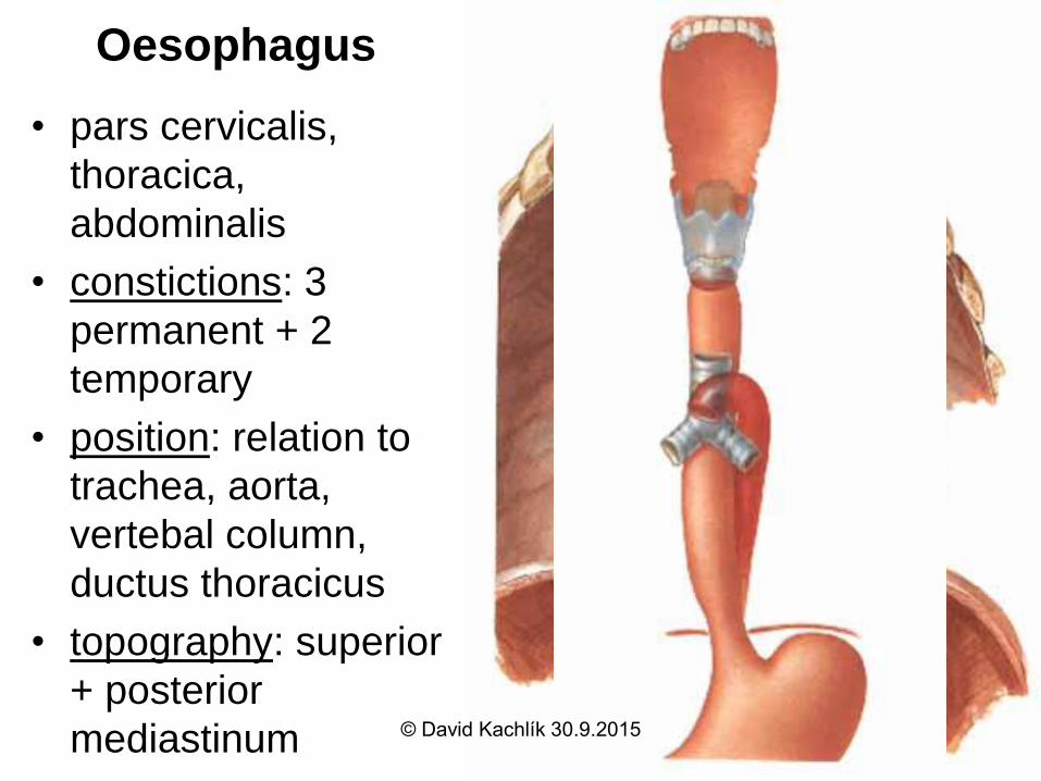
Oesophagus
• pars cervicalis,
thoracica,
abdominalis
• constictions: 3
permanent + 2
temporary
• position: relation to
trachea, aorta,
vertebal column,
ductus thoracicus
• topography: superior
+ posterior
mediastinum © David Kachlík 30.9.2015
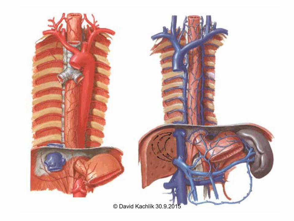
Oesophagus – blood supply
• Arteries:
– a. thyroidea inf.
– arcus aortae + aorta thoracica and their branches
– a. gastrica sin.
• Veins: vv. oesophageales
– vv. thyroideae inf.
– v. azygos + hemiazygos + hemiazygos acc.
– vv. gastricae breves
portocaval anastomosis – varices
© David Kachlík 30.9.2015
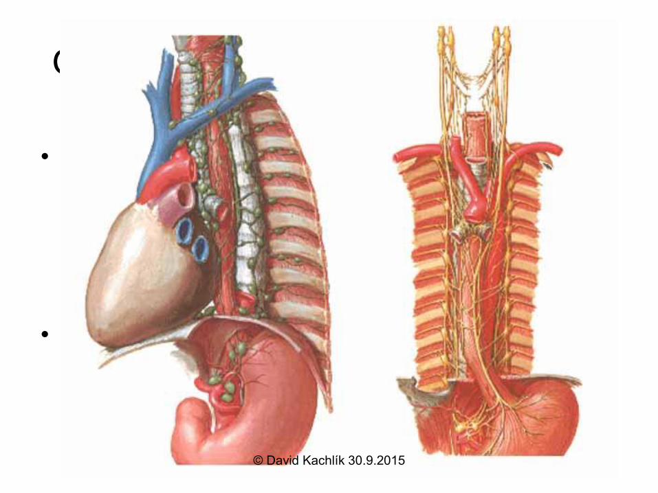
Oesophagus – Lymph and Nerves
• Lymph: nodes or directly into ductus thoracicus
– n.l. cervicales prof.
– n.l. juxtaoesophageales + paravertebrales (=
n.l. mediastinales post.)
– n.l. gastrici sin.
• Nerves: n.X – plexus oesophageus – left
anteriorly (rotation 90 degrees !) –
parasympathetic
– truncus symphaticus – sympathetic + sensory
© David Kachlík 30.9.2015
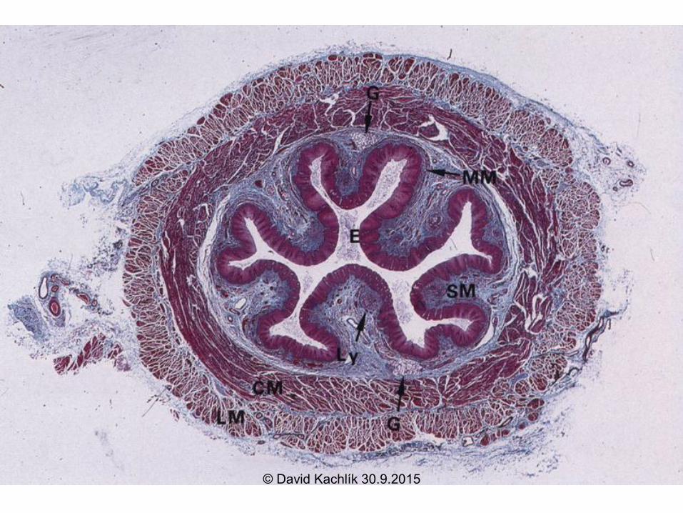
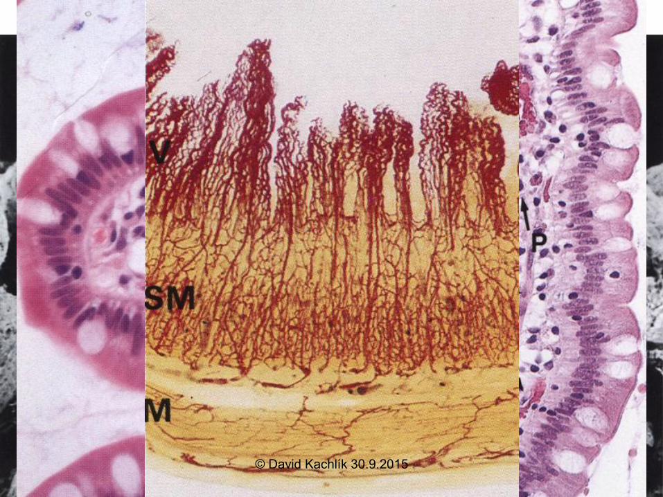
Oesophageus – wall structure
– stratified squamous nonkeratinising
epithelium
– lamina propria mucosae – distally
oesophageal cardial glands
– in submucosis mucinous glands - gl.
oesophageae
– proximally skeletal musculature
– adventitia
• serosa on the short abdominal part only
© David Kachlík 30.9.2015
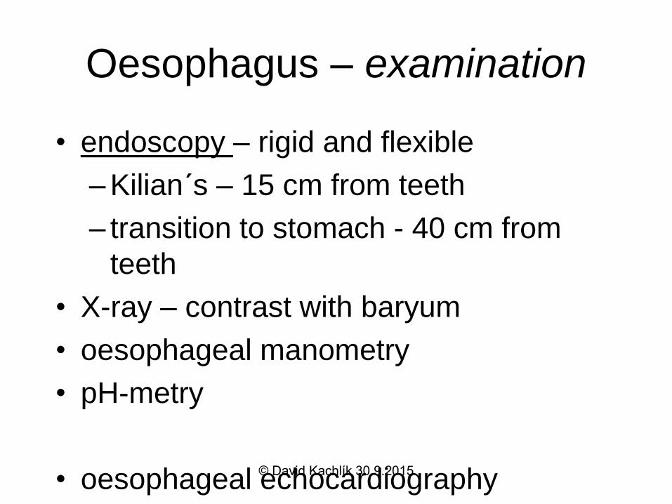
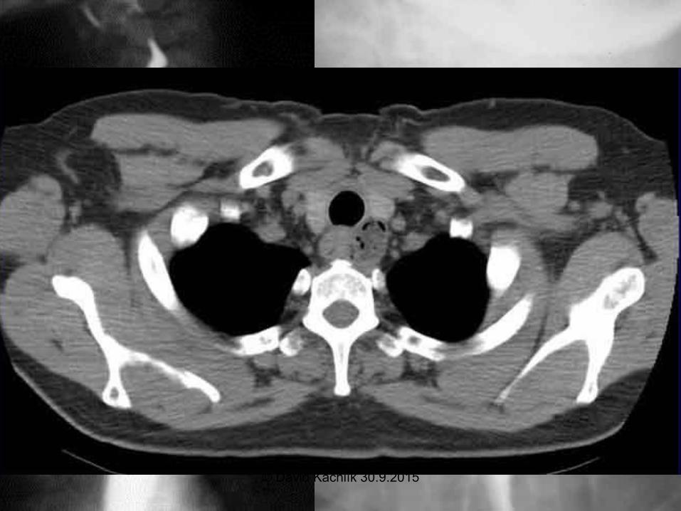
Oesophagus – examination
• endoscopy – rigid and flexible
– Kilian´s – 15 cm from teeth
– transition to stomach - 40 cm from
teeth
• X-ray – contrast with baryum
• oesophageal manometry
• pH-metry
• oesophageal echocardiography© David Kachlík 30.9.2015
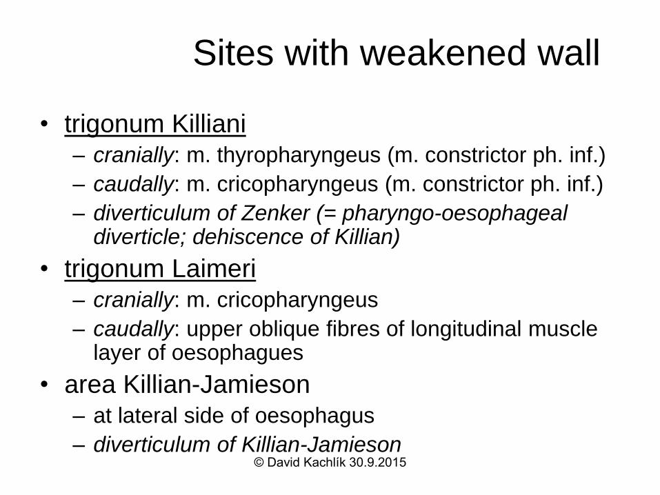
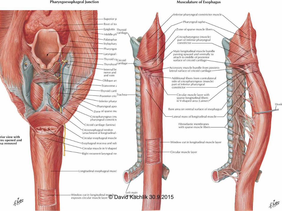
Sites with weakened wall
• trigonum Killiani– cranially: m. thyropharyngeus (m. constrictor ph. inf.)
– caudally: m. cricopharyngeus (m. constrictor ph. inf.)
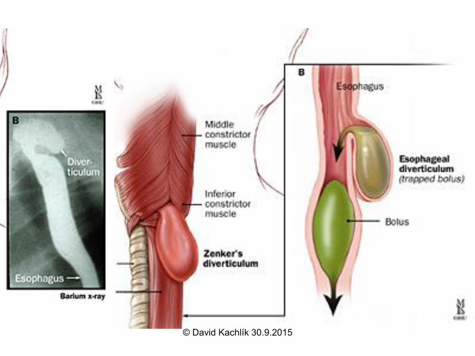
– diverticulum of Zenker (= pharyngo-oesophageal diverticle; dehiscence of Killian)
• trigonum Laimeri– cranially: m. cricopharyngeus
– caudally: upper oblique fibres of longitudinal muscle layer of oesophagues
• area Killian-Jamieson– at lateral side of oesophagus
– diverticulum of Killian-Jamieson© David Kachlík 30.9.2015
© David Kachlík 30.9.2015
© David Kachlík 30.9.2015
© David Kachlík 30.9.2015
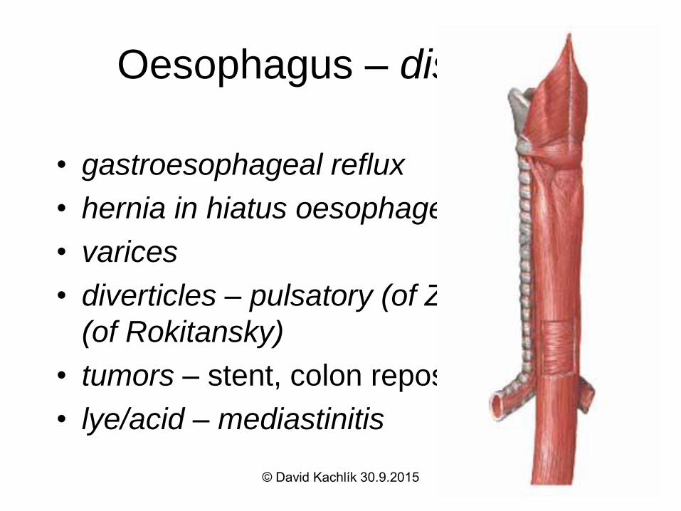
Oesophagus – diseases
• gastroesophageal reflux
• hernia in hiatus oesophageus
• varices
• diverticles – pulsatory (of Zenker), traction
(of Rokitansky)
• tumors – stent, colon reposition
• lye/acid – mediastinitis
© David Kachlík 30.9.2015
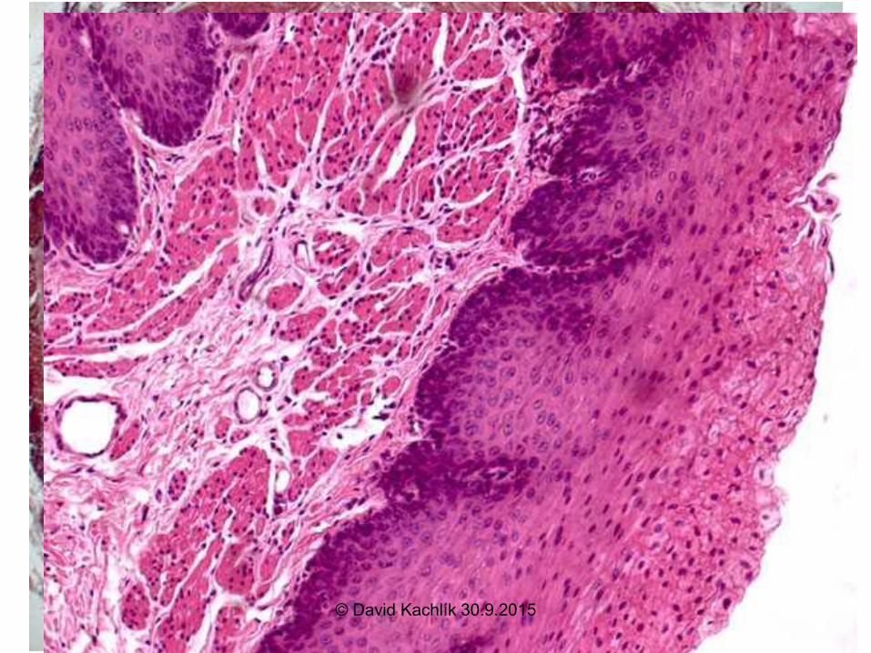
Oesophageus - HE
© David Kachlík 30.9.2015
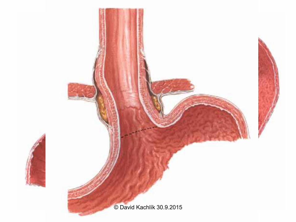
Stomach = gaster (ventriculus, stomachus)
• paries anterior + posterior
• curvatura major + minor
• cardia, fundus /fornix/, corpus
/canalis/, pars pylorica (antrum,
canalis, pylorus)
• incisura angularis
• ostium cardiacum + pyloricum© David Kachlík 30.9.2015
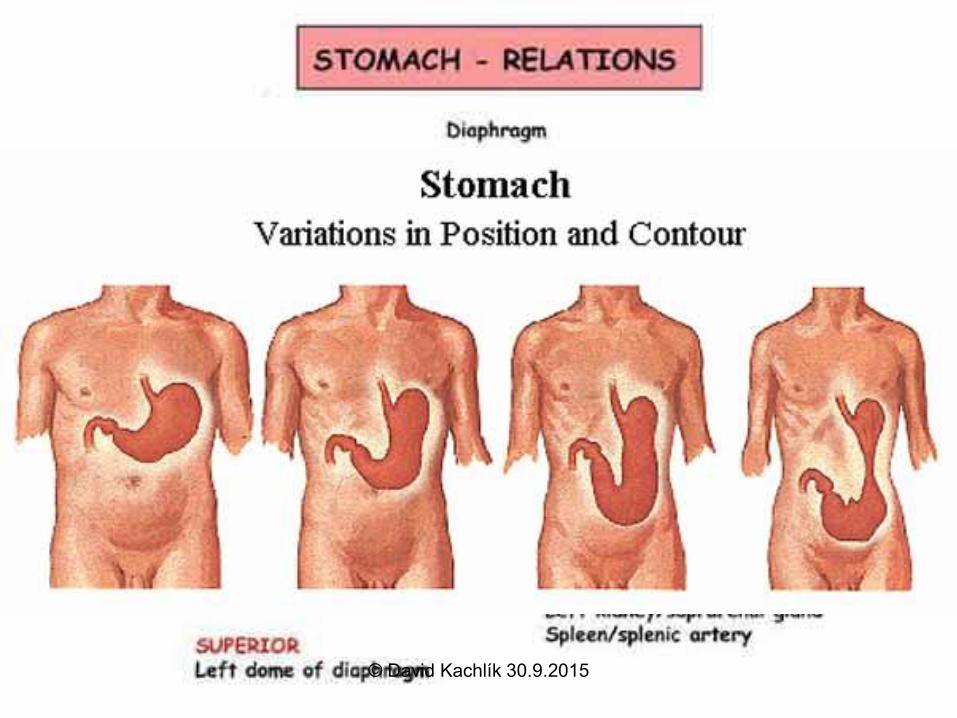
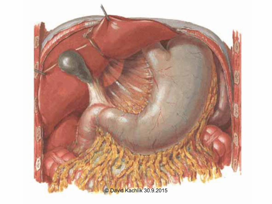
Stomach
• shape: hook, spindle, bull horn
• position: Th11-L3
• projection: Labbé´s
• topography: organ impressions
• relation to peritoneum: intraperitoneally
• suspended with: omentum majus +
minus, bursa omentalis (= lesser sac)
© David Kachlík 30.9.2015
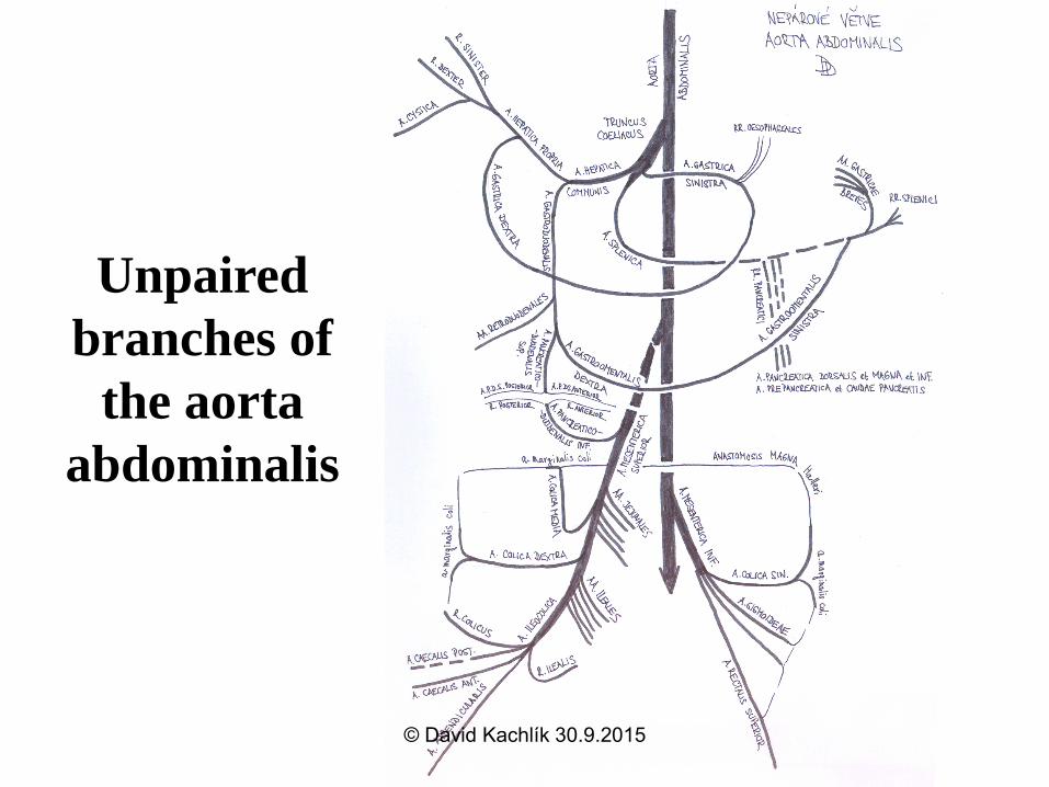
Unpaired
branches of
the aorta
abdominalis
© David Kachlík 30.9.2015
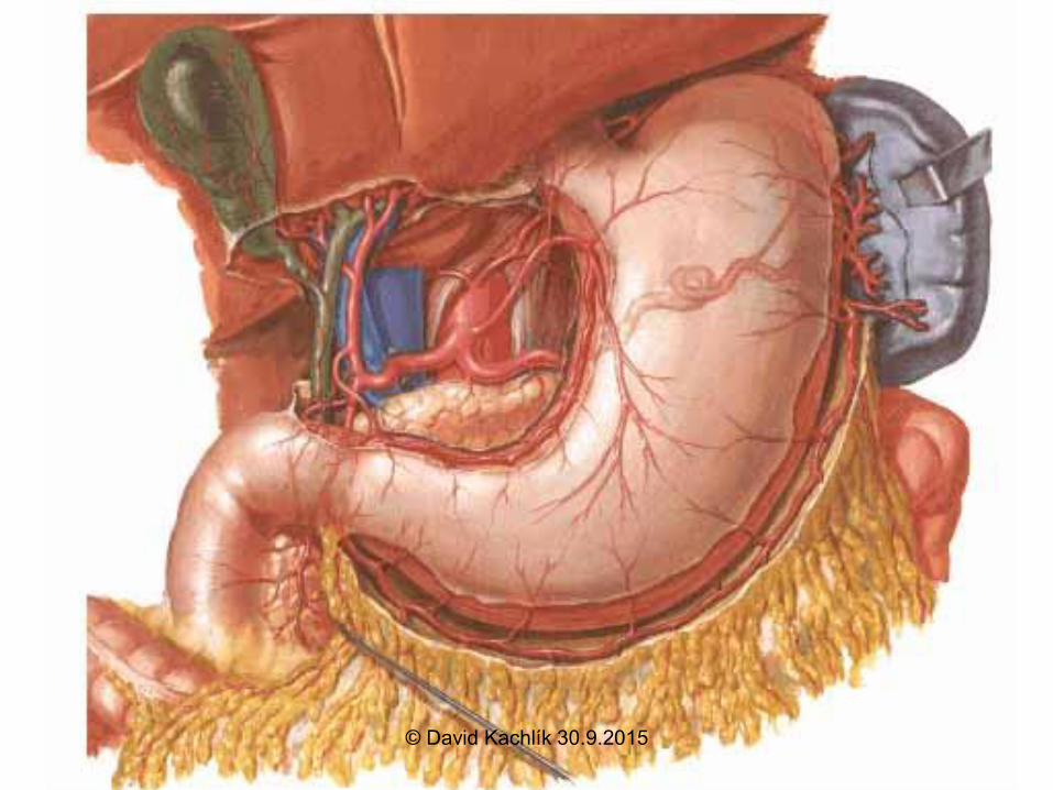
Stomach – arterial supply
truncus coelicacus
• aa. gastrica sin.
• a. hepatica communis
– a. hepatica propria a. gastrica dx.
– a. gastroduodenalis a. gastroomentalis dx.
• a. splenica a. gastroomentalis sin., aa.
gastricae breves (fundus), a. gastrica posterior
(variability)
© David Kachlík 30.9.2015
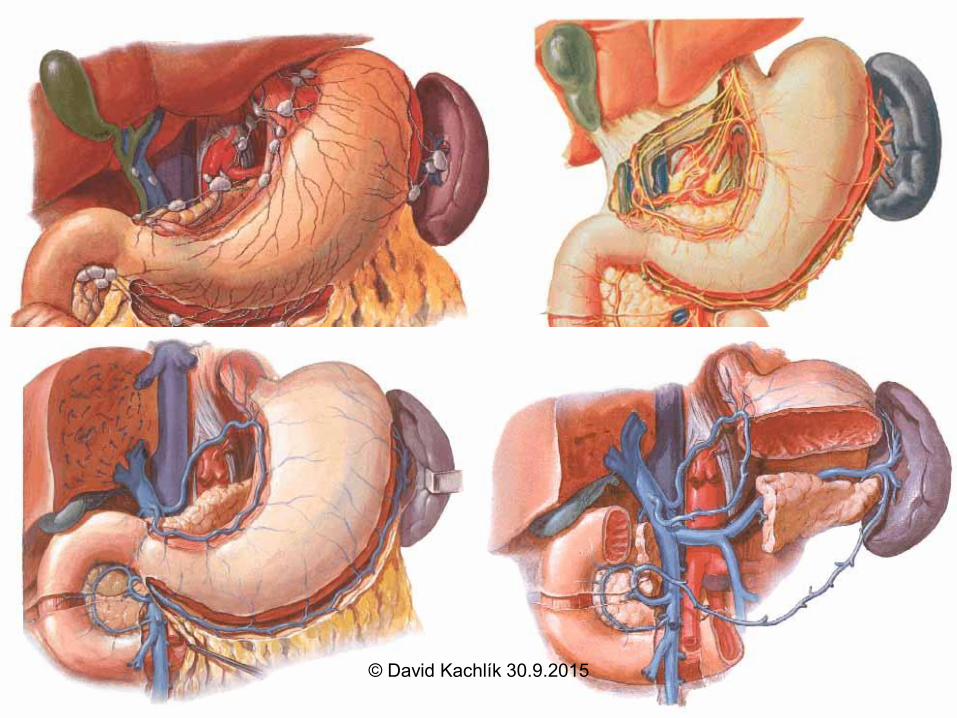
Stomach – other supply
• Veins: correlate to arteries + v. prepylorica v.
portae
portocaval anastomosis between v. gastica sin.
and vv. oesophageales varices
• Lymph: n.l. gastrici, gastroomentales, pylorici,
splenici, pancratici n.l. coeliaci
• Nerves: parasympathetic – n. X
sympathetic – nn. splanchnici major+minor
ggl. coeliacum + mesentericum sup.© David Kachlík 30.9.2015
Stomach – wall structure
• tunica mucosa
– plicae gastricae (sulcus salivatorius
Waldeyeri) areae gastricae foveolae
gastricae
– simple columnar epithelium – mucous production
– lamina propria mucosae
• gastrical glands, lymphatic follicles
• tunica muscularis– inner - fibrae obliquae
– medial - circular - m. sphincter pylori
– outer - longitudinal© David Kachlík 30.9.2015
Tunica mucosa of gaster
• gastric pits• deeper in pyloric part
• glands enter these pits
– gl. cardiacae - deep
– gl. gastricae propriae
– gl. pyloricae - shallow
© David Kachlík 30.9.2015
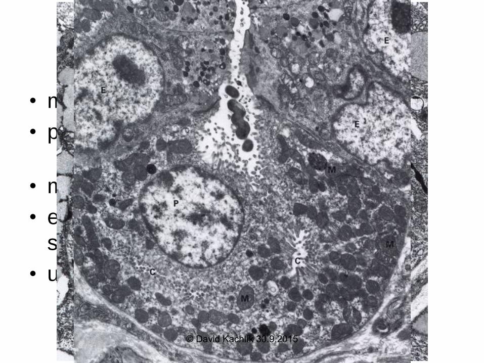
Gastric cells
• main cells - pepsinogen, lipase
• parietal cells - HCl, intrinsic factor• abundant intracellular channels
• mucinous cells – mucus
• enteroendocrine cells (DNES) - gastrin,
somatostatin
• undiferentiated cells - mitotically active
© David Kachlík 30.9.2015
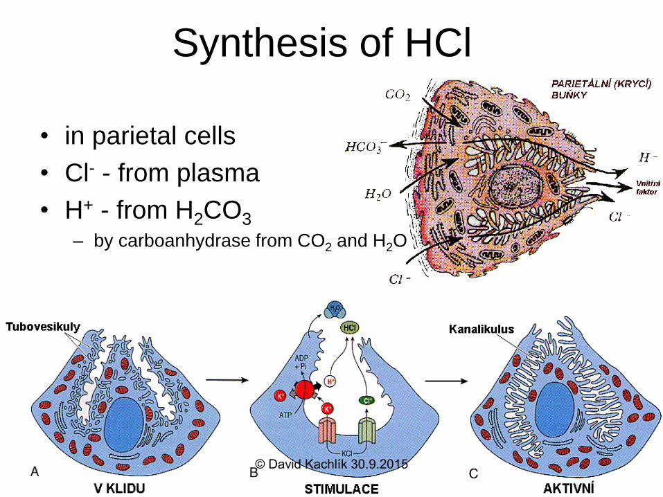
Synthesis of HCl
• in parietal cells
• Cl- - from plasma
• H+ - from H2CO3
– by carboanhydrase from CO2 and H2O
© David Kachlík 30.9.2015
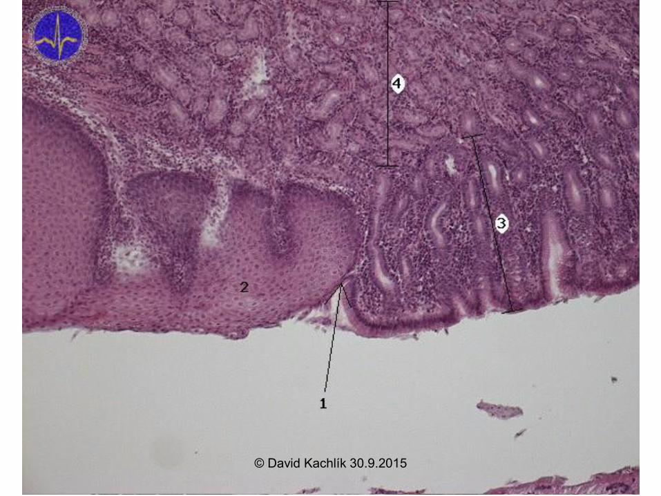
Cardia gastri - HE
© David Kachlík 30.9.2015
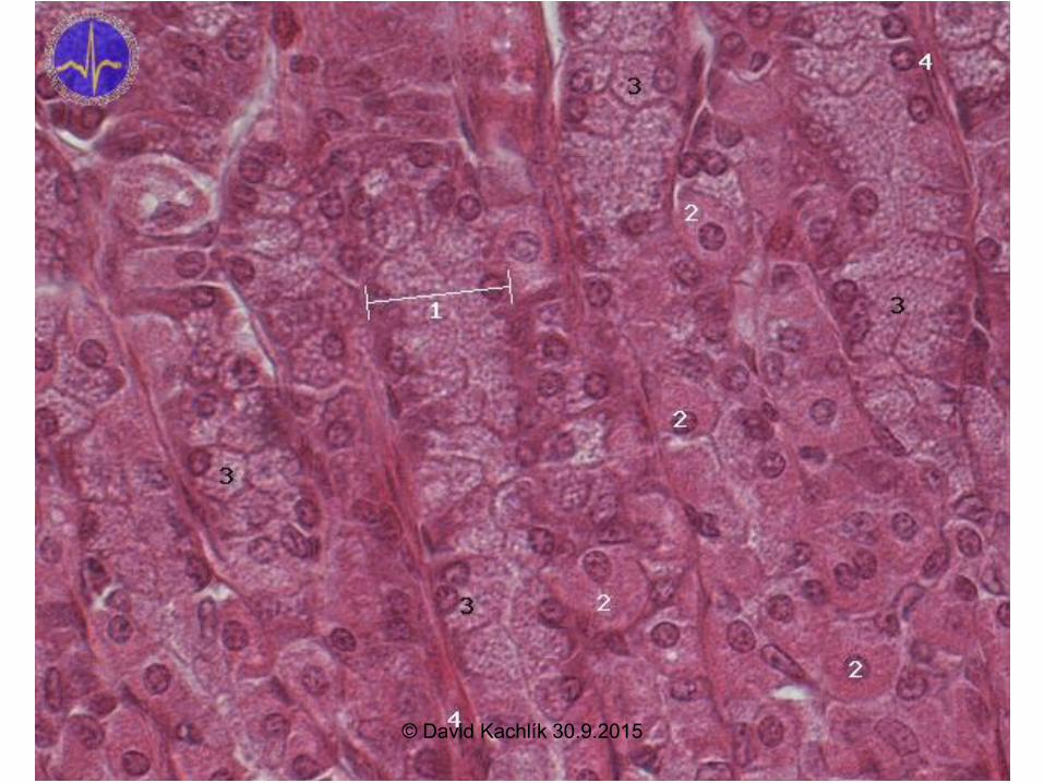
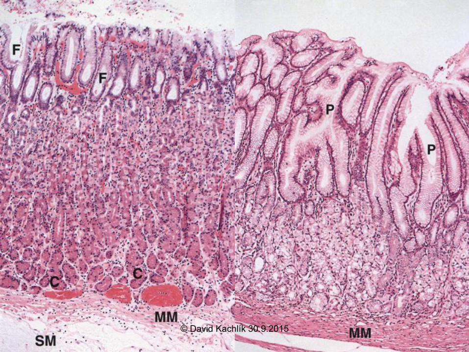
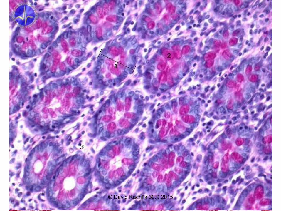
Fundus gastri - HE
© David Kachlík 30.9.2015
Stomach – examination + diseases
• peptic ulcer
• gastritis A,B (Helicobacter pylori)
• tumors
• pylorostenosis
gastroscopy
X-ray – contrast with baryum
gastrostomy© David Kachlík 30.9.2015
Small intestine = Intestinum tenue
• duodenum
• jejunum
• ileum
mesenterium - radix mesenterii
© David Kachlík 30.9.2015
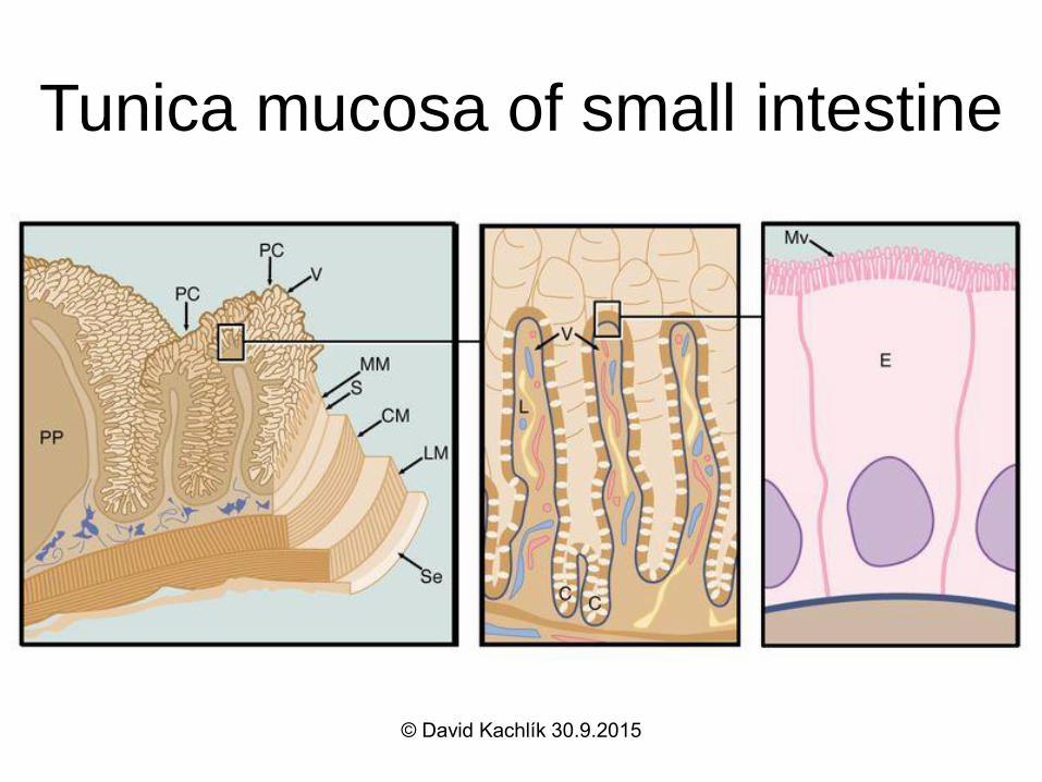
Tunica mucosa of small intestine
• plicae circulares (Kerkringi) villi
intestinales microvilli
• glandulae intestinales = Lieberkühn´s
crypts
• simple columnar epithelium
• lamina propria mucosae
– vessels, smooth muscles, noduli lymphoidei
© David Kachlík 30.9.2015
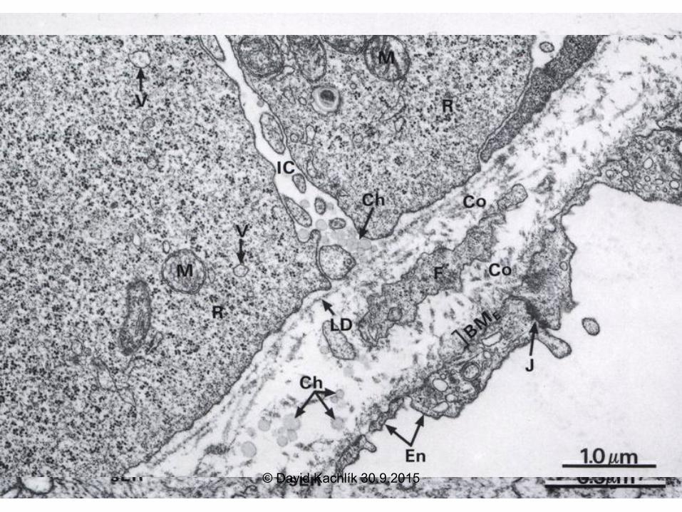
Cells of small intestine mucosa
• enterocytes - resorption– microvilli, interdigitations, lipid droplets
• goblet cells – mucus production
• Paneth cells – lysozym production
• endocrine cells (DNES) - 12 types– cholecystokinin, sekretin
• M-cells - over noduli lymph. aggregati (Peyer‘s plates)
• undiferentiated cells
© David Kachlík 30.9.2015
Villi intestinales
• digit- to leaf-formed elements
• about 10times surface enlargement
• surface – enterocytes, goblet cells
• smooth muscle „skeleton“
• capillary network
• lymphoid (=lacteal) inhte centre
© David Kachlík 30.9.2015
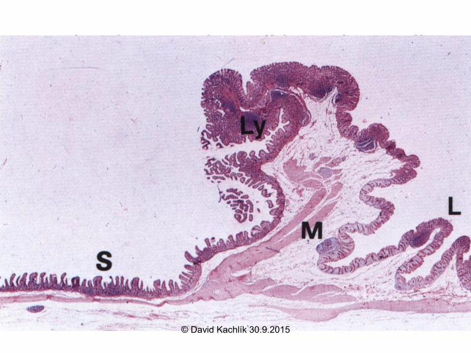
Other layers of small intestine wall
• tela submucosa
– duodenum – glandulae duodenales Brunneri
• tuboalveolar mucinous
• alcalic secretion
– ileum – noduli lymphoidei aggregati („agmina Peyeri“) = Peyer´s plates
• accumulation of lymphoid tissue
• other layers correnspond to the standard form of the tube
© David Kachlík 30.9.2015
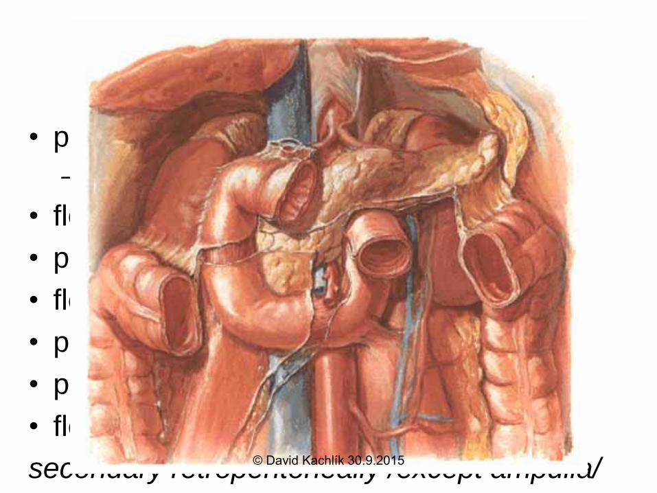
Duodenum
• pars superior
– ampulla=bulbus
• flexura duodeni sup.
• pars descendens
• flexura duodeni inf.
• pars horizontalis/inferior
• pars ascendens
• flexura duodenojejunalis
secondary retroperitoneally /except ampulla/© David Kachlík 30.9.2015
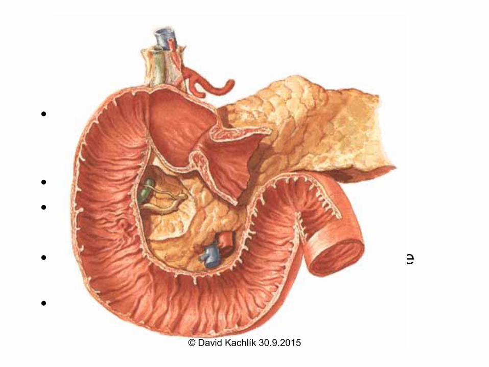
Duodenum
• structure: plica longitudinalis – papilla d. major Vateri
– papilla d. minor Santorini
• fixation: lig. + m. suspensorius Treitzi d.
• position: duodenal window L2
• plicae ciculares Kerckringi – highest of the intestine
• glandulae duodenales Brunneri– submucosal
© David Kachlík 30.9.2015
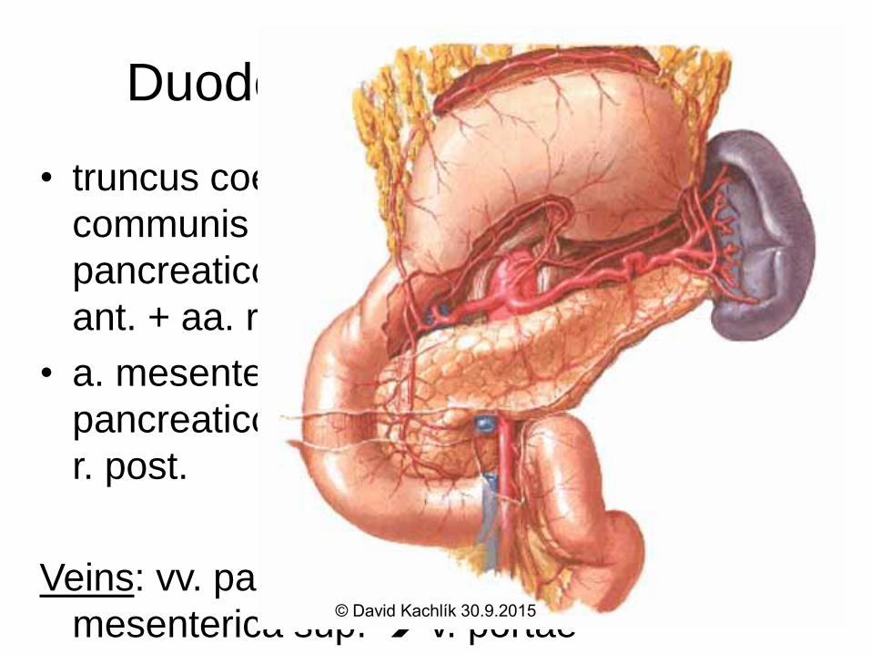
Duodenum – blood supply
• truncus coelicacus a. hepatica
communis a. gastroduodenalis a.
pancreaticoduodenalis sup. post. + sup.
ant. + aa. retroduodenales
• a. mesenterica sup. a.
pancreaticoduodenalis inf. ramus ant. +
r. post.
Veins: vv. pancreaticoduodenales v.
mesenterica sup. v. portae© David Kachlík 30.9.2015
Duodenum – Lymph and Nerves
Lymph: n.l. pylorici
• n.l. hepatici n.l. coeliaci n.l.
preaortici
• down directly to n.l. preaortici
Nerves: parasympathetic – n. X
sympathetic – nn. splanchnici major+minor
ggl. coeliacum + mesentericum sup.© David Kachlík 30.9.2015
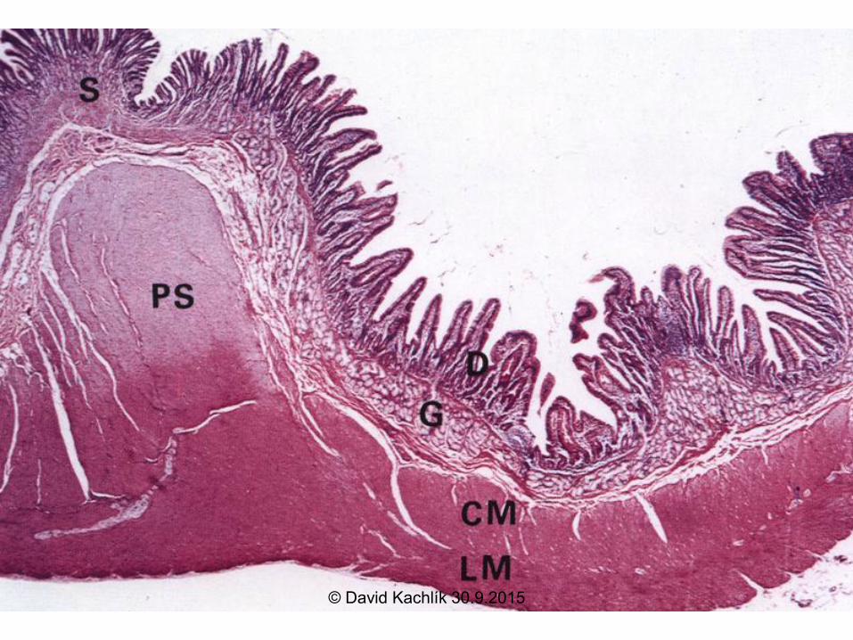
Transition: Pylorus - Duodenum HE
© David Kachlík 30.9.2015
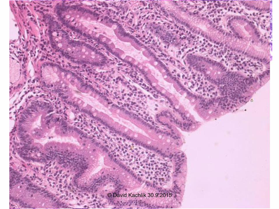
Duodenum - HE
© David Kachlík 30.9.2015
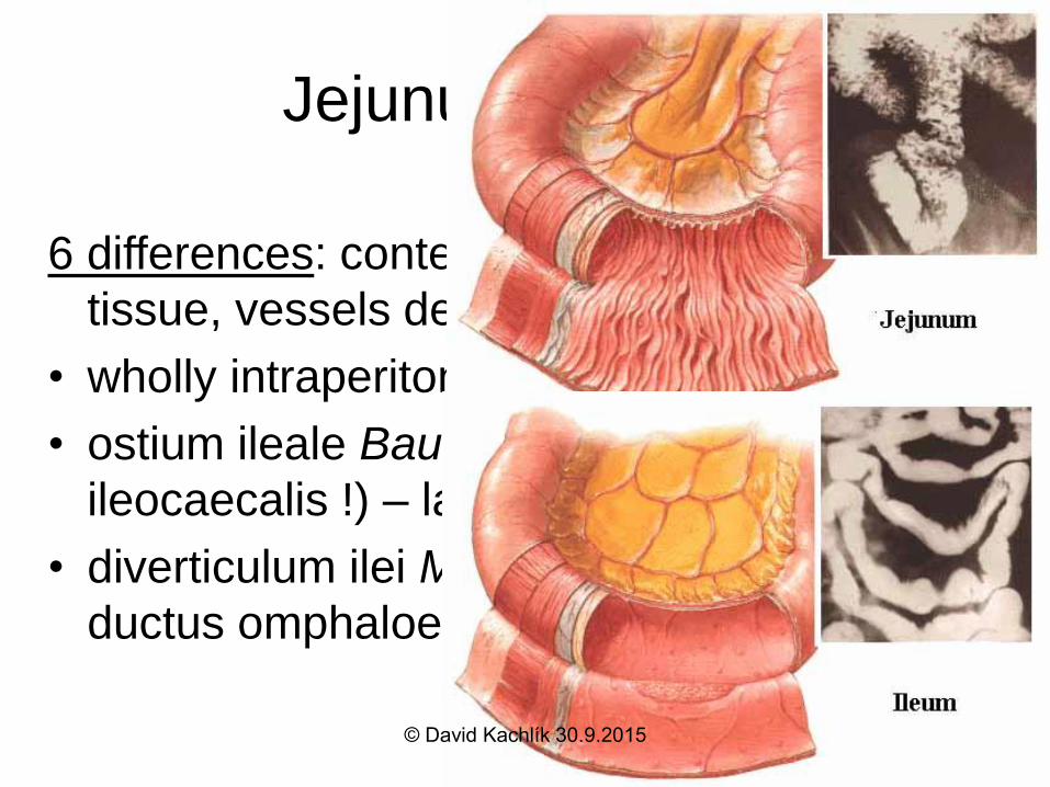
Jejunum et ileum
6 differences: content, width, folds, lymphoid
tissue, vessels density and arrangement
• wholly intraperitoneally, radix mesenterii
• ostium ileale Bauhini s. Tulpi (former valva
ileocaecalis !) – labrum sup. + inf.
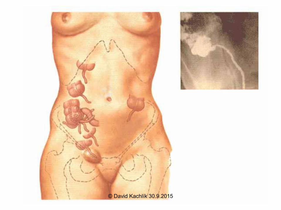
• diverticulum ilei Meckeli (2%) – remnant of
ductus omphaloentericus
© David Kachlík 30.9.2015
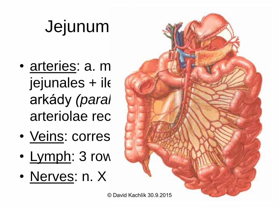
Jejunum + ileum - supply
• arteries: a. mesenterica sup. aa.
jejunales + ileales + aa. ileocolica
arkády (parallel Dwigth´s artery
arteriolae rectae
• Veins: correspon to arteries
• Lymph: 3 rows of n.l.mesenterici sup.
• Nerves: n. X + sympathetic© David Kachlík 30.9.2015

Small intestine - HE
© David Kachlík 30.9.2015
Small intestine - diseases
• duodenal ulcer
• inflammation – morbus Crohn, colitis
ulcerosa
• tumors – very rare – carcinoid
• coeliakia
© David Kachlík 30.9.2015
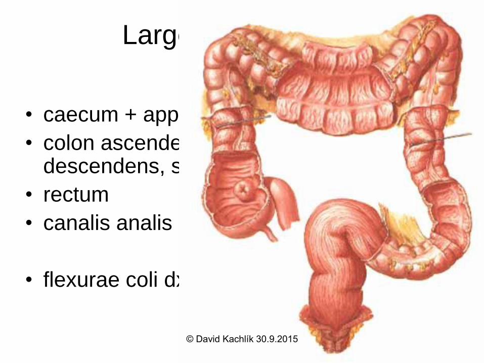
Large intestine = Intestinum
crassum
• caecum + appendix vermiformis
• colon ascendens, transversum, descendens, sigmoideum
• rectum
• canalis analis
• flexurae coli dx.+ sin.
© David Kachlík 30.9.2015
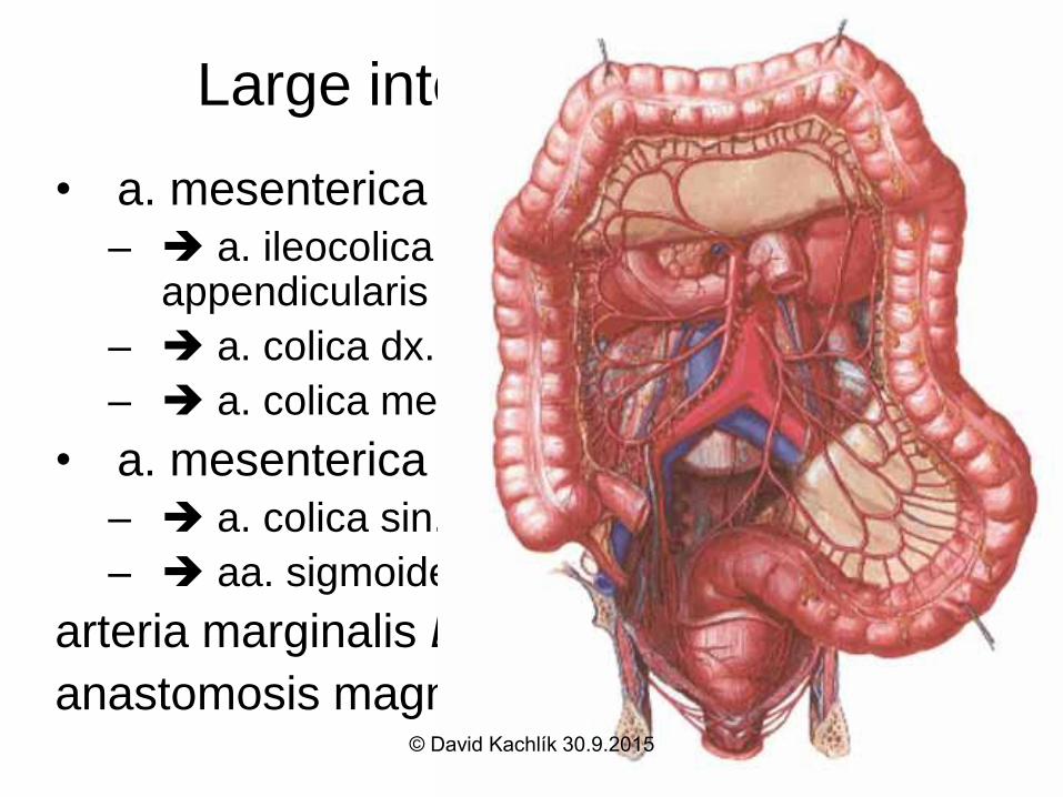
Large intestine - arteries
• a. mesenterica sup.
– a. ileocolica a. ceacalis ant. + post., a. appendicularis
– a. colica dx. (colon ascendens)
– a. colica media (colon transversum)
• a. mesenterica inf.
– a. colica sin. (pro colon descendens)
– aa. sigmoideae (3-4)
arteria marginalis Drummondi
anastomosis magna Halleri = arcus Riolani© David Kachlík 30.9.2015

Large intestine – other supply
• Veins: correspond to arteries v. portae
• Lymph: 3 rows of n.l. colici n.l.
preaortici
• Nerves:
– parasympathetic: n. X down to flexura coli
sin. (= Cannon-Böhme´s ), then sacral
parasympathetic (S2-4)
– sympathetic: z ggl. coeliacum, mesentericum
sup. + inf.© David Kachlík 30.9.2015
Mucosa of large intestine
• plicae semilunares
• no villi
• deeper Lieberkühn´s crypt
– enterocytes – less of microvilli
– goblet cells are numerous
– Paneth cells are missing
– endocrine cells (DNES) are present
© David Kachlík 30.9.2015
Other layers of large intestine
wall
• tunica muscularis externa
– inner circular - haustrations
– outer longitudinal – reduced to taenie coli
• mesenterica
• omentalis
• libera
• besides appendix and rectum !
© David Kachlík 30.9.2015
Large intestine - tunica serosa
Fixation and relation to peritoneum
• intraperitoneal: appendix, colon transversum +
sigmoideum - having their meso-
• mesoperitoneal: colon ascendens + descendens
– half-grown with the posterior wall
• rectum – partially intraperitoneal + subperitoneal
• caecum – variability – see picture
• appendices omentales • adipose tissue
• unclear function© David Kachlík 30.9.2015
Large intestine - HE
© David Kachlík 30.9.2015
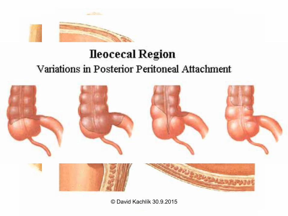
Caecum
• papilla et ostium ileale Bauhini s. Tulpi(frenulum, labrum)
• intraperioneally, often no mesocaecum
appendix vermiformis: 2-30 cm, mesoappendix, ostium, lig. appendiculoovaricum Cladoi
• 6 positions: positio pelvina, retro-, pre-, sub-, latero- et ileocaecalis /most frequent positio retrocaecalis/
• projection: McBurney´s (on Monro´s line) and Lanz´s point (on linea interspinosa)© David Kachlík 30.9.2015
Appendix vermiformis caeci
• Paneth cells are present
• lamina propria mucosae
– fulfilled with lymphatic tissue = noduli lymphoidei
aggregati („tonsilla abdominalis“)
• longitudinal musculature forms no taenia
– circular one strongly reduced
• Amyand´s hernia – appendix in sac of
inguinal hernia
– Claudius Amyand, 1735 – physician to George II.
© David Kachlík 30.9.2015

Appendix vermiformis - HE
© David Kachlík 30.9.2015
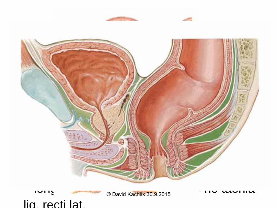
Rectum
• description: ampulla, flexura sacralis et laterales (superodextra lat., intermedisinistra lat., inferodextra lat.),
• structure: plicae transversae
– plica dx. Kohlrauschi
– plicae sin. sup. + inf. Houstoni s. Nelatoni
• longitudinal musculature forms no taenia
lig. recti lat.© David Kachlík 30.9.2015
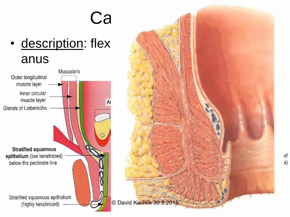
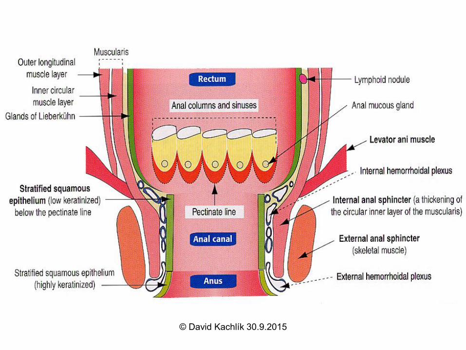
Canalis analis
• description: flexura anorectalis = perinealis,
anus
• structure: columnae, valvulae, sinus, lineae
anocutanea et pectinata, pecten, zona
transitionalis
• muscles: m. sphincter ani ext. et int.
• peritoneum: on upper ¼ only = partially
intraperitoneal + subperitoneal organ
• psition: septum
rectovesicale♂/rectovaginale♀, fossa
ischioanalis© David Kachlík 30.9.2015
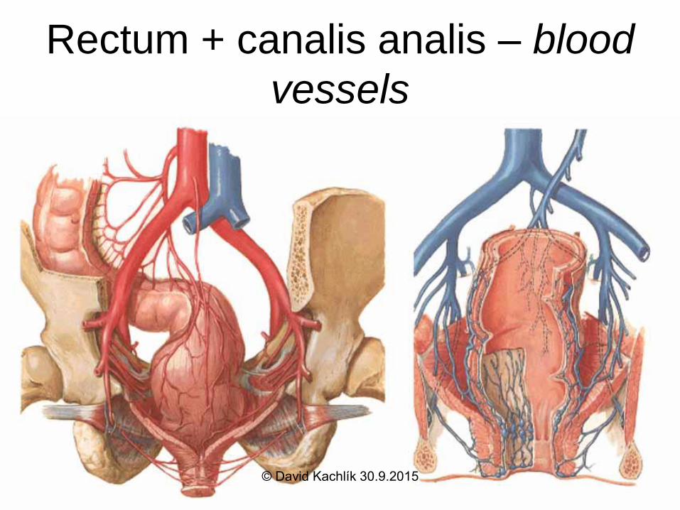
Rectum + canalis analis – blood
vessels• a. mesenterica sup. a. rectalis sup.
• a. iliaca int. a. rectalis media
• a. iliaca int. a.pudenda int. a. rectalis
inf.
Veins: plexus venosus rectalis
correspond to arteries possible
portocaval anastomosis
© David Kachlík 30.9.2015
Rectum + canalis analis –
Lymph and Nerves• n.l. mesenterici inf. n.l. preaortici
• n.l. iliaci int.
• n.l. sacrales
• n.l. inguinales superficales
Nerves: - sacral parasympathetic
– sympathetic by plexus hypogastricus sup. +
inf., n. pudendus
© David Kachlík 30.9.2015
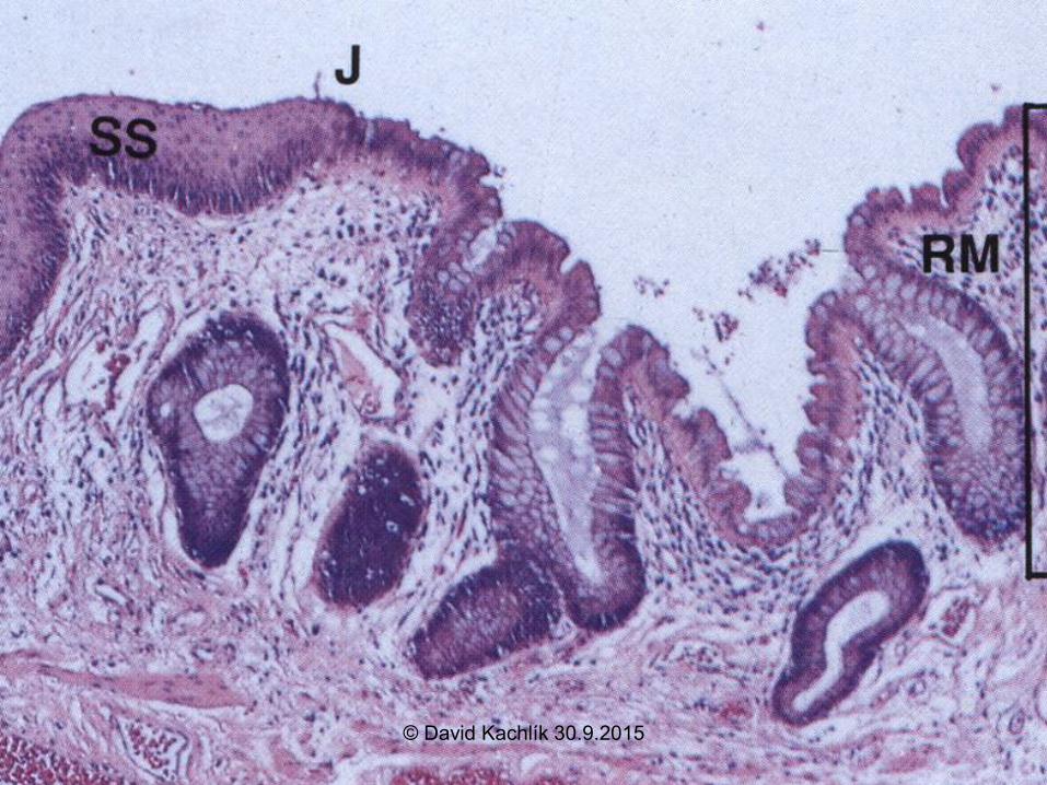
Canalis analis - structure
• zona haemorrhoidalis - m. sphincter ani
int.
– columnae anales (6-10)– change of epithelium – stratified squamous
nonkeratinising
– Anal venous plexus – „cavernous
body“ – maintains continention inner
haemorrhoids
• zona cutanea – keratinising epithelium– glandulae circumanales (sweat and apocrine)
© David Kachlík 30.9.2015
© David Kachlík 30.9.2015
Large intestine - examination
• Hemocult – hidden bleeding examination
• endoscopy – rectoscopy (rigid), coloscopy
(flexible)
• X-ray- native, contrast - passage,
irigography
• CT
© David Kachlík 30.9.2015
Large intestine - diseases
• polyps
• tumors – most frequent !!!
• diverticulosis diverticulitis (Graser´s diverticle)
• inflammation – colitis ulcerosa, morbus Crohn
• appendicitis – „the sun may not rise or fall“
– most frequent sudden abdominal accident
• internal / external haemorrhoids
• colostomy – temporary / permanent
© David Kachlík 30.9.2015


![DOG INTESTINUM TENUE · 19. Flexura diaphragmatica [=flexura diaphragmatica dorsalis] 20. Colon dorsale dextrum - (3 teniae) tenia libera mediale, laterale, tenia mesocolica 21. Ampulla](https://img.pdfslide.net/doc/110x75/5e204c809b010200564bc8f6/dog-intestinum-tenue-19-flexura-diaphragmatica-flexura-diaphragmatica-dorsalis.jpg)