Embed Size (px)
Citation preview
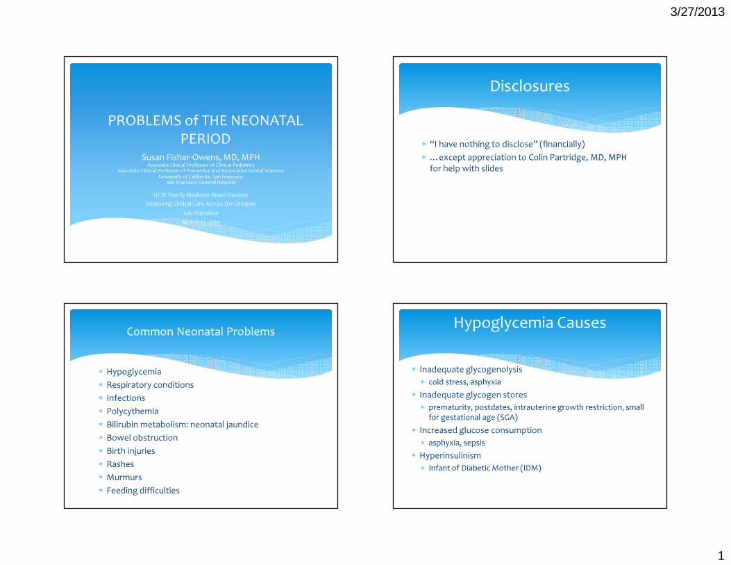
3/27/2013
1
PROBLEMS of THE NEONATAL PERIOD
Susan Fisher-Owens, MD, MPHAssociate Clinical Professor of Clinical Pediatrics
Associate Clinical Professor of Preventive and Restorative Dental SciencesUniversity of California, San Francisco
San Francisco General Hospital
UCSF Family Medicine Board Review: Improving Clinical Care Across the Lifespan
San FranciscoMarch 27, 2013
∗ “I have nothing to disclose” (financially)∗ …except appreciation to Colin Partridge, MD, MPH
for help with slides
Disclosures
∗ Hypoglycemia∗ Respiratory conditions∗ Infections∗ Polycythemia∗ Bilirubin metabolism: neonatal jaundice∗ Bowel obstruction∗ Birth injuries∗ Rashes∗ Murmurs∗ Feeding difficulties
Common Neonatal Problems
∗ Inadequate glycogenolysis ∗ cold stress, asphyxia
∗ Inadequate glycogen stores ∗ prematurity, postdates, intrauterine growth restriction, small
for gestational age (SGA)∗ Increased glucose consumption
∗ asphyxia, sepsis∗ Hyperinsulinism
∗ Infant of Diabetic Mother (IDM)
Hypoglycemia Causes
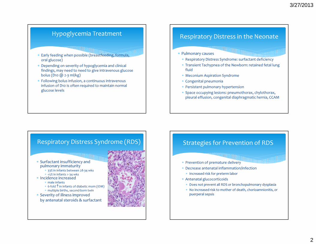
3/27/2013
2
∗ Early feeding when possible (breastfeeding, formula, oral glucose)
∗ Depending on severity of hypoglycemia and clinical findings, may need to need to give intravenous glucose bolus (D10 @ 2-3 ml/kg)
∗ Following bolus infusion, a continuous intravenous infusion of D10 is often required to maintain normal glucose levels
Hypoglycemia Treatment
∗ Pulmonary causes∗ Respiratory Distress Syndrome: surfactant deficiency∗ Transient Tachypnea of the Newborn: retained fetal lung
fluid∗ Meconium Aspiration Syndrome∗ Congenital pneumonia∗ Persistent pulmonary hypertension∗ Space occupying lesions: pneumothorax, chylothorax,
pleural effusion, congenital diaphragmatic hernia, CCAM
Respiratory Distress in the Neonate
∗ Surfactant insufficiency and pulmonary immaturity ∗ 33% in infants between 28-34 wks∗ <5% in infants > 34 wks
∗ Incidence increased∗ male infants∗ 6-fold ↑ in infants of diabetic mom (IDM)∗ multiple births, second-born twin
∗ Severity of illness improvedby antenatal steroids & surfactant
Respiratory Distress Syndrome (RDS)
∗ Prevention of premature delivery∗ Decrease antenatal inflammation/infection
∗ Increased risk for preterm labor∗ Antenatal glucocorticoids
∗ Does not prevent all RDS or bronchopulmonary dysplasia∗ No increased risk to mother of death, chorioamnionitis, or
puerperal sepsis
Strategies for Prevention of RDS
3/27/2013
3
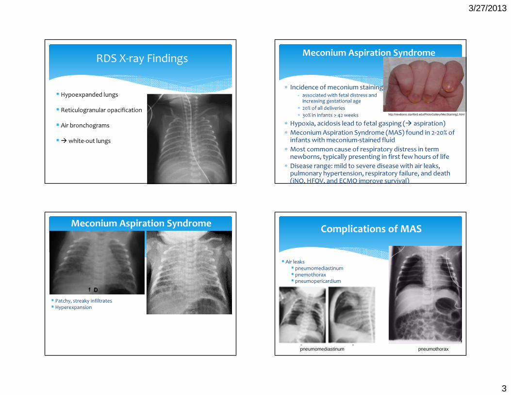
RDS X-ray Findings
� Hypoexpanded lungs� Reticulogranular opacification� Air bronchograms�� white-out lungs
∗ Incidence of meconium staining∗ associated with fetal distress and
increasing gestational age ∗ 20% of all deliveries ∗ 30% in infants > 42 weeks
∗ Hypoxia, acidosis lead to fetal gasping (� aspiration) ∗ Meconium Aspiration Syndrome (MAS) found in 2-20% of
infants with meconium-stained fluid ∗ Most common cause of respiratory distress in term
newborns, typically presenting in first few hours of life∗ Disease range: mild to severe disease with air leaks,
pulmonary hypertension, respiratory failure, and death (iNO, HFOV, and ECMO improve survival)
Meconium Aspiration Syndrome
http://newborns.stanford.edu/PhotoGallery/MecStaining1.html
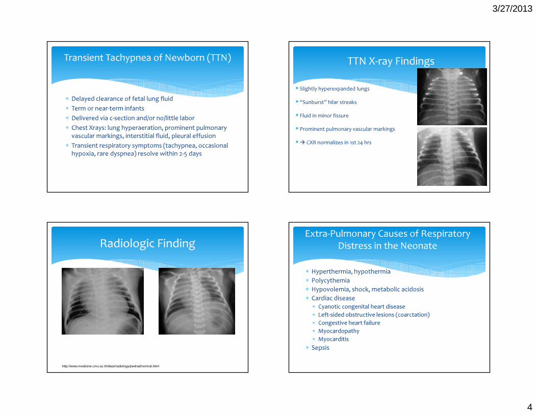
Meconium Aspiration Syndrome
� Patchy, streaky infiltrates� Hyperexpansion
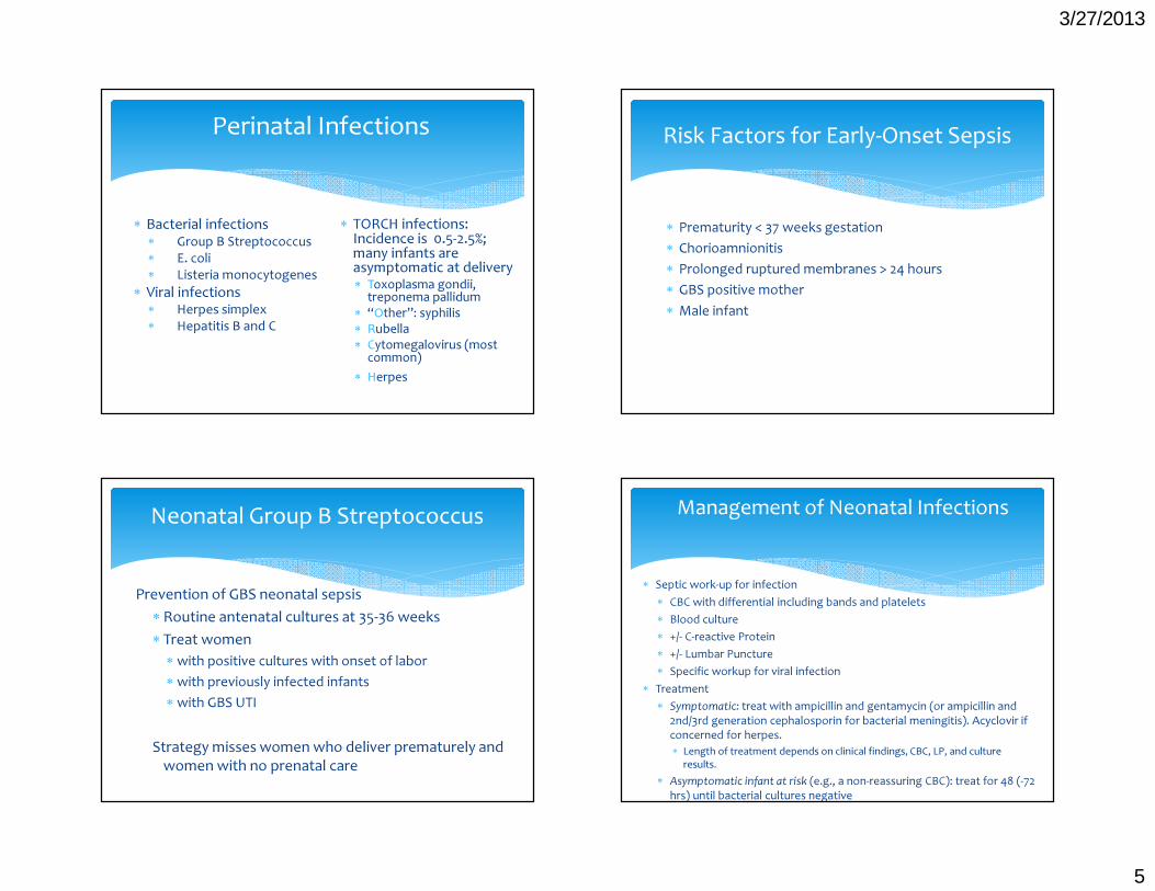
Complications of MAS
pneumothorax
� Air leaks� pneumomediastinum� pnemothorax� pneumopericardium
pneumomediastinum
3/27/2013
4
∗ Delayed clearance of fetal lung fluid∗ Term or near-term infants∗ Delivered via c-section and/or no/little labor∗ Chest Xrays: lung hyperaeration, prominent pulmonary
vascular markings, interstitial fluid, pleural effusion∗ Transient respiratory symptoms (tachypnea, occasional
hypoxia, rare dyspnea) resolve within 2-5 days
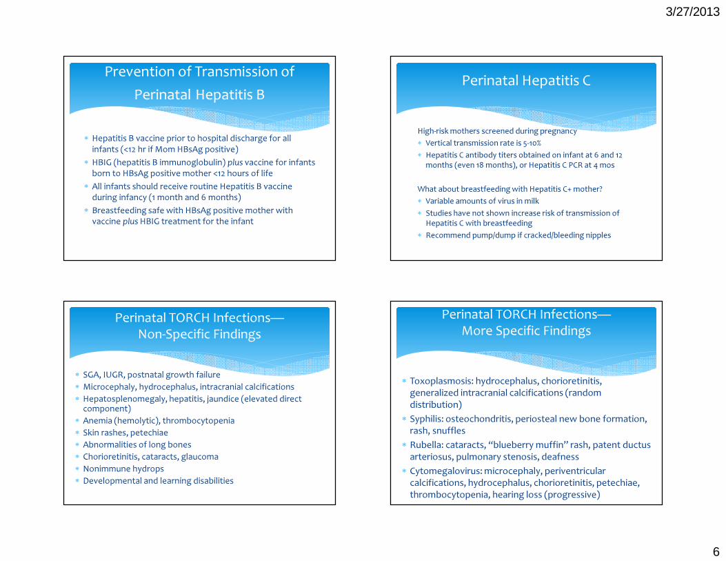
Transient Tachypnea of Newborn (TTN) TTN X-ray Findings� Slightly hyperexpanded lungs� “Sunburst” hilar streaks� Fluid in minor fissure� Prominent pulmonary vascular markings �� CXR normalizes in 1st 24 hrs
Radiologic Finding
http://www.medicine.cmu.ac.th/dept/radiology/pedrad/normal.html
∗ Hyperthermia, hypothermia∗ Polycythemia∗ Hypovolemia, shock, metabolic acidosis∗ Cardiac disease
∗ Cyanotic congenital heart disease∗ Left-sided obstructive lesions (coarctation)∗ Congestive heart failure∗ Myocardopathy∗ Myocarditis
∗ Sepsis
Extra-Pulmonary Causes of Respiratory Distress in the Neonate
3/27/2013
5
∗ Bacterial infections∗ Group B Streptococcus ∗ E. coli ∗ Listeria monocytogenes
∗ Viral infections∗ Herpes simplex ∗ Hepatitis B and C
Perinatal Infections
∗ TORCH infections: Incidence is 0.5-2.5%; many infants are asymptomatic at delivery∗ Toxoplasma gondii, treponema pallidum∗ “Other”: syphilis ∗ Rubella∗ Cytomegalovirus (most common)∗ Herpes
∗ Prematurity < 37 weeks gestation∗ Chorioamnionitis∗ Prolonged ruptured membranes > 24 hours∗ GBS positive mother∗ Male infant
Risk Factors for Early-Onset Sepsis
Prevention of GBS neonatal sepsis∗ Routine antenatal cultures at 35-36 weeks∗ Treat women∗ with positive cultures with onset of labor∗ with previously infected infants ∗ with GBS UTI
Strategy misses women who deliver prematurely and women with no prenatal care
Neonatal Group B Streptococcus
∗ Septic work-up for infection∗ CBC with differential including bands and platelets ∗ Blood culture∗ +/- C-reactive Protein ∗ +/- Lumbar Puncture ∗ Specific workup for viral infection
∗ Treatment∗ Symptomatic: treat with ampicillin and gentamycin (or ampicillin and
2nd/3rd generation cephalosporin for bacterial meningitis). Acyclovir if concerned for herpes.∗ Length of treatment depends on clinical findings, CBC, LP, and culture
results.∗ Asymptomatic infant at risk (e.g., a non-reassuring CBC): treat for 48 (-72
hrs) until bacterial cultures negative
Management of Neonatal Infections
3/27/2013
6
∗ Hepatitis B vaccine prior to hospital discharge for all infants (<12 hr if Mom HBsAg positive)
∗ HBIG (hepatitis B immunoglobulin) plus vaccine for infants born to HBsAg positive mother <12 hours of life
∗ All infants should receive routine Hepatitis B vaccine during infancy (1 month and 6 months)
∗ Breastfeeding safe with HBsAg positive mother with vaccine plus HBIG treatment for the infant
Prevention of Transmission of Perinatal Hepatitis B
High-risk mothers screened during pregnancy∗ Vertical transmission rate is 5-10% ∗ Hepatitis C antibody titers obtained on infant at 6 and 12
months (even 18 months), or Hepatitis C PCR at 4 mos
What about breastfeeding with Hepatitis C+ mother?∗ Variable amounts of virus in milk∗ Studies have not shown increase risk of transmission of
Hepatitis C with breastfeeding∗ Recommend pump/dump if cracked/bleeding nipples
Perinatal Hepatitis C
∗ SGA, IUGR, postnatal growth failure∗ Microcephaly, hydrocephalus, intracranial calcifications∗ Hepatosplenomegaly, hepatitis, jaundice (elevated direct
component)∗ Anemia (hemolytic), thrombocytopenia∗ Skin rashes, petechiae∗ Abnormalities of long bones ∗ Chorioretinitis, cataracts, glaucoma∗ Nonimmune hydrops∗ Developmental and learning disabilities
Perinatal TORCH Infections—Non-Specific Findings
∗ Toxoplasmosis: hydrocephalus, chorioretinitis, generalized intracranial calcifications (random distribution)
∗ Syphilis: osteochondritis, periosteal new bone formation, rash, snuffles
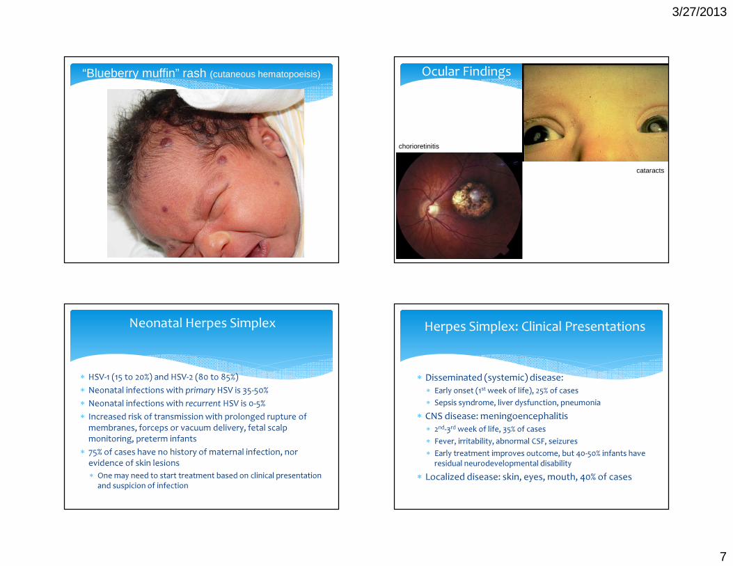
∗ Rubella: cataracts, “blueberry muffin” rash, patent ductus arteriosus, pulmonary stenosis, deafness
∗ Cytomegalovirus: microcephaly, periventricular calcifications, hydrocephalus, chorioretinitis, petechiae, thrombocytopenia, hearing loss (progressive)
Perinatal TORCH Infections—More Specific Findings
3/27/2013
7
“Blueberry muffin” rash (cutaneous hematopoeisis) Ocular Findings
chorioretinitis
cataracts
∗ HSV-1 (15 to 20%) and HSV-2 (80 to 85%) ∗ Neonatal infections with primary HSV is 35-50%∗ Neonatal infections with recurrent HSV is 0-5%∗ Increased risk of transmission with prolonged rupture of
membranes, forceps or vacuum delivery, fetal scalp monitoring, preterm infants
∗ 75% of cases have no history of maternal infection, nor evidence of skin lesions∗ One may need to start treatment based on clinical presentation
and suspicion of infection
Neonatal Herpes Simplex
∗ Disseminated (systemic) disease: ∗ Early onset (1st week of life), 25% of cases∗ Sepsis syndrome, liver dysfunction, pneumonia
∗ CNS disease: meningoencephalitis∗ 2nd-3rd week of life, 35% of cases∗ Fever, irritability, abnormal CSF, seizures∗ Early treatment improves outcome, but 40-50% infants have
residual neurodevelopmental disability∗ Localized disease: skin, eyes, mouth, 40% of cases
Herpes Simplex: Clinical Presentations
3/27/2013
8
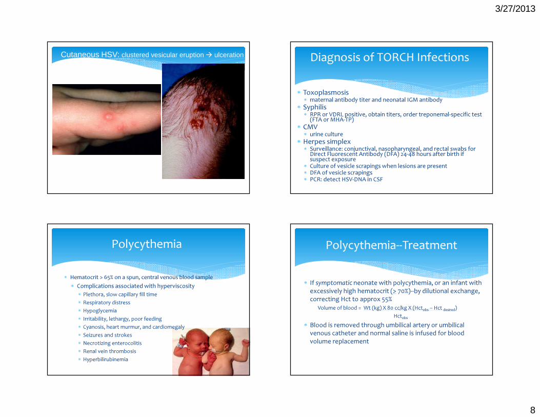
Cutaneous HSV: clustered vesicular eruption � ulceration
∗ Toxoplasmosis∗ maternal antibody titer and neonatal IGM antibody
∗ Syphilis∗ RPR or VDRL positive, obtain titers, order treponemal-specific test (FTA or MHA-TP)
∗ CMV∗ urine culture
∗ Herpes simplex∗ Surveillance: conjunctival, nasopharyngeal, and rectal swabs for Direct Fluorescent Antibody (DFA) 24-48 hours after birth if suspect exposure∗ Culture of vesicle scrapings when lesions are present∗ DFA of vesicle scrapings ∗ PCR: detect HSV-DNA in CSF
Diagnosis of TORCH Infections
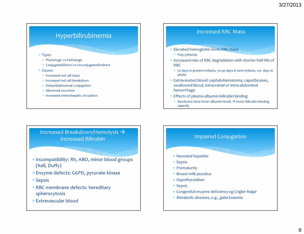
∗ Hematocrit > 65% on a spun, central venous blood sample∗ Complications associated with hyperviscosity
∗ Plethora, slow capillary fill time∗ Respiratory distress∗ Hypoglycemia∗ Irritability, lethargy, poor feeding∗ Cyanosis, heart murmur, and cardiomegaly∗ Seizures and strokes∗ Necrotizing enterocolitis∗ Renal vein thrombosis∗ Hyperbilirubinemia
Polycythemia
∗ If symptomatic neonate with polycythemia, or an infant with excessively high hematocrit (> 70%)--by dilutional exchange, correcting Hct to approx 55%
Volume of blood = Wt (kg) X 80 cc/kg X (Hctobs – Hct desired)Hctobs
∗ Blood is removed through umbilical artery or umbilical venous catheter and normal saline is infused for blood volume replacement
Polycythemia--Treatment
3/27/2013
9
∗ Types∗ Physiologic vs Pathologic∗ Conjugated/Direct vs Unconjugated/Indirect
∗ Causes∗ Increased red cell mass∗ Increased red cell breakdown∗ Delayed/abnormal conjugation∗ Abnormal excretion∗ Increased enterohepatic circulation
Hyperbilirubinemia∗ Elevated hemoglobin level, RBC mass
∗ Polycythemia∗ Increased rate of RBC degradation with shorter half-life of
RBC ∗ 70 days in preterm infants, 70-90 days in term infants, 120 days in
adults∗ Extravasated blood: cephalohematoma, caput/bruises,
swallowed blood, intracranial or intra-abdominal hemorrhage
∗ Effects of plasma albumin-bilirubin binding∗ Newborns have lower albumin levels � lower bilirubin-binding
capacity
Increased RBC Mass
∗ Incompatibility: Rh, ABO, minor blood groups (Kell, Duffy)
∗ Enzyme defects: G6PD, pyruvate kinase∗ Sepsis∗ RBC membrane defects: hereditary
spherocytosis∗ Extravascular blood
Increased Breakdown/Hemolysis �Increased Bilirubin
∗ Neonatal hepatitis ∗ Sepsis∗ Prematurity∗ Breast milk jaundice∗ Hypothyroidism∗ Sepsis∗ Congenital enzyme deficiency eg Crigler-Najjar∗ Metabolic diseases, e.g., galactosemia
Impaired Conjugation
3/27/2013
10
∗ Obstruction to biliary flow: biliary atresia, choledocal cyst, cystic fibrosis, stones ∗ Dark urine (urine + for bilirubin), light colored
stools, persistent jaundice (> 3weeks)∗ Hepatic cell injury : syphilis, TORCH infections∗ Hepatic dysfunction: E. coli (UTI)∗ Toxic effects: hyperalimentation cholestasis∗ Metabolic errors: galactosemia∗ Chronic “overload”: erythroblastosis fetalis, G6PD,
spherocytosis
Conjugated (Direct) Hyperbilirubinemia: Impaired Excretion
∗ Conjugated bilirubin is unconjugated and reabsorbed ∗ Enterohepatic circulation and reabsorption is
enhanced by:∗ Gut sterility (urobilinogen and stercobilinogen)∗ Bowel dysmotility (preterm infants, effects of magnesium or
morphine)∗ Ileus∗ Obstruction: atresia, pyloric stenosis, meconium plugs, cystic
fibrosis∗ Delayed feeding (“breast-feeding jaundice”)
Enterohepatic Circulation
∗ Hemolysis∗ Onset of jaundice in 1st 24 hours∗ Rapid rate of rise of bili (>0.5mg/dL per hour)∗ Hepatosplenomegaly, pallor∗ Family history (G6PD, spherocytosis)∗ “Set-up” with incompatibility, Coombs (+DAT), elevated
reticulocytes, abnormal hemolytic smear
∗ Sepsis or inborn error∗ Emesis, lethargy, poor feeding∗ Hepatosplenomegaly, tachypnea, temperature instability
Causes Suggested by Clinical Findings
∗ Increased susceptibility to neurotoxicity seen with asphyxia, sepsis, acidosis, prematurity, and hemolysis∗ Consider treatment at lower levels of unconjugated bilirubin
in these cases∗ When to worry
∗ Visible jaundice in the first 24 hours of life∗ Serum bilirubin rising rapidly > 5 mg/dl/24 hrs∗ Prolonged hyperbilirubinemia > 1 week term infant and > 2
weeks in the preterm∗ Direct bilirubin > 2mg/dl
Management of Indirect Hyperbilirubinemia
3/27/2013
11
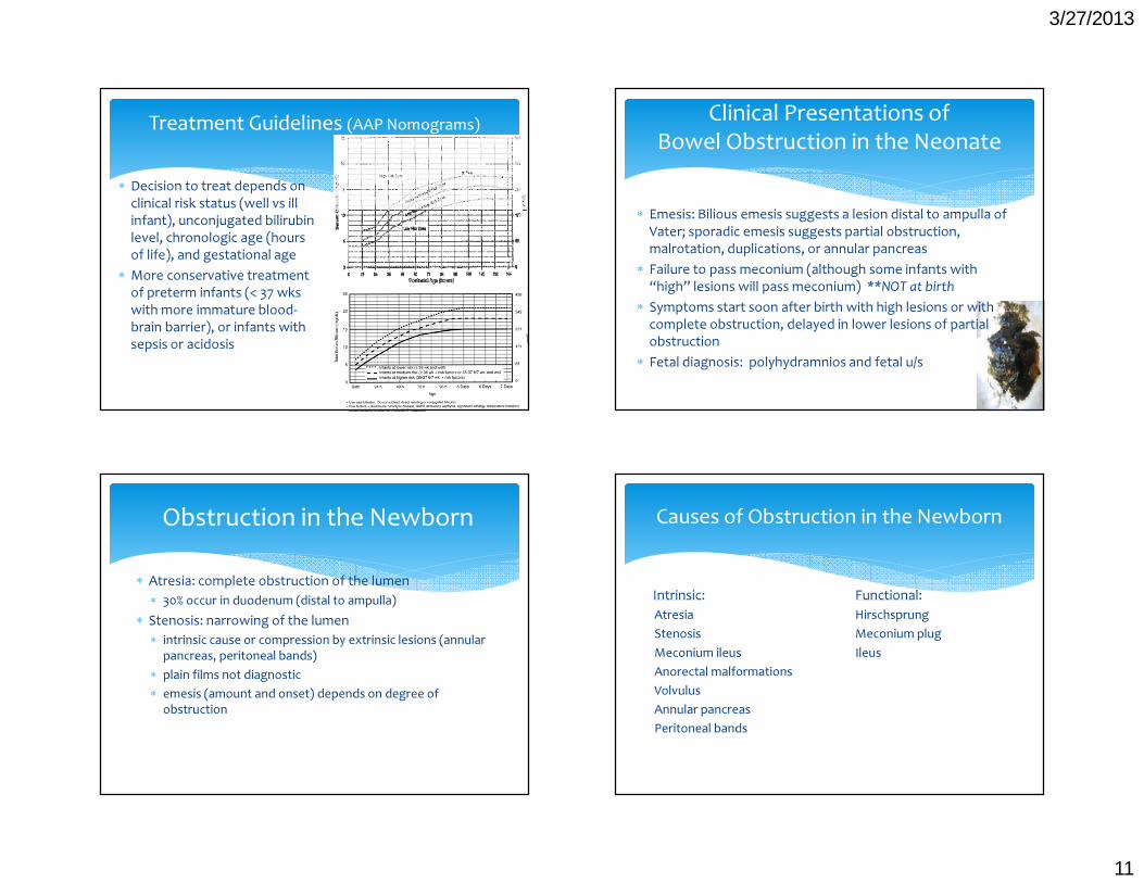
∗ Decision to treat depends on clinical risk status (well vs ill infant), unconjugated bilirubin level, chronologic age (hours of life), and gestational age
∗ More conservative treatment of preterm infants (< 37 wks with more immature blood-brain barrier), or infants with sepsis or acidosis
Treatment Guidelines (AAP Nomograms)
∗ Emesis: Bilious emesis suggests a lesion distal to ampulla of Vater; sporadic emesis suggests partial obstruction, malrotation, duplications, or annular pancreas
∗ Failure to pass meconium (although some infants with “high” lesions will pass meconium) **NOT at birth
∗ Symptoms start soon after birth with high lesions or with complete obstruction, delayed in lower lesions of partial obstruction
∗ Fetal diagnosis: polyhydramnios and fetal u/s
Clinical Presentations ofBowel Obstruction in the Neonate
∗ Atresia: complete obstruction of the lumen∗ 30% occur in duodenum (distal to ampulla)
∗ Stenosis: narrowing of the lumen∗ intrinsic cause or compression by extrinsic lesions (annular
pancreas, peritoneal bands)∗ plain films not diagnostic∗ emesis (amount and onset) depends on degree of
obstruction
Obstruction in the Newborn
Intrinsic: Functional: Atresia HirschsprungStenosis Meconium plugMeconium ileus IleusAnorectal malformationsVolvulusAnnular pancreasPeritoneal bands
Causes of Obstruction in the Newborn
3/27/2013
12
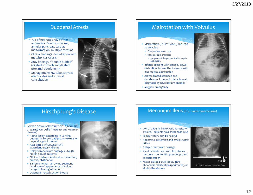
∗ 70% of neonates have other anomalies: Down syndrome, annular pancreas, cardiac malformation, multiple atresias
∗ Clinical findings: dehydration with metabolic alkalosis
∗ Xray findings: “double-bubble” (dilated stomach and dilated proximal duodenum)
∗ Management: NG tube, correct electrolytes and surgical consultation
Duodenal Atresia
∗ Malrotation (8th-10th week) can lead to volvulus ∗ Complete obstruction ∗ Vascular compromise:
∗ gangrene of the gut, peritonitis, sepsis, and shock.
∗ Infants present with emesis, bowel distention. Intermittent emesis with incomplete obstruction
∗ Xrays: dilated stomach and duodenum, little air in distal bowel, diagnosis by UGI (barium enema)
∗ Surgical emergency
Malrotation with Volvulus
∗ Lower bowel obstruction: agenesis of ganglion cells (Auerbach and Meissner plexuses)∗ Rectal lesion extending in varying degree; in 80-90% patients no extension beyond sigmoid colon∗ Associated w/ Downs (15%), Waardenburg syndrome∗ Delayed meconium passage (>24-48 hrs) in 90% of patients∗ Clinical findings: Abdominal distention, emesis, obstipation∗ Barium enema: narrowing segment, “corkscrew” appearance of colon, delayed clearing of barium ∗ Diagnosis: rectal suction biopsy
Hirschprung’s Disease
∗ 90% of patients have cystic fibrosis, 10-15% of CF patients have meconium ileus
∗ Family history may be helpful∗ Abdominal distention and emesis within
48 hrs∗ Delayed meconium passage∗ 1/3 of patients have volvulus, atresia,
meconium peritonitis, pseudocyst, and present earlier
∗ Xrays: dilated bowel loops, intra-abdominal calcification (peritonitis), no air-fluid levels seen
Meconium Ileus (inspissated meconium)
3/27/2013
13
∗ Etiology: colonic dysmotility ∗ Hirschsprung’s disease in 50% of these
patients∗ Clinical findings:
∗ Delayed meconium passage: (24-48 hrs)∗ Abdominal distention, emesis∗ Barium enema diagnostic and
therapeutic
Meconium Plug Syndrome∗ Cephalhematoma∗ Caput succedaneum∗ Subgaleal hematoma∗ Erb’s palsy∗ Klumpke’s palsy∗ Clavicular fracture∗ Phrenic nerve injury with diaphragmatic paralysis
Birth Injuries
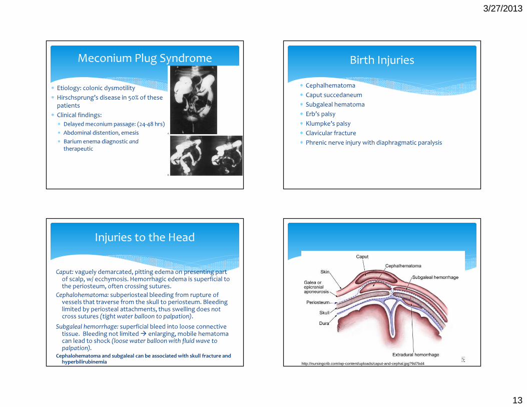
Caput: vaguely demarcated, pitting edema on presenting part of scalp, w/ ecchymosis. Hemorrhagic edema is superficial to the periosteum, often crossing sutures.
Cephalohematoma: subperiosteal bleeding from rupture of vessels that traverse from the skull to periosteum. Bleeding limited by periosteal attachments, thus swelling does notcross sutures (tight water balloon to palpation).
Subgaleal hemorrhage: superficial bleed into loose connective tissue. Bleeding not limited � enlarging, mobile hematoma can lead to shock (loose water balloon with fluid wave to palpation).
Cephalohematoma and subgaleal can be associated with skull fracture and hyperbilirubinemia
Injuries to the Head
http://nursingcrib.com/wp-content/uploads/caput-and-cephal.jpg?9d7bd4
3/27/2013
14
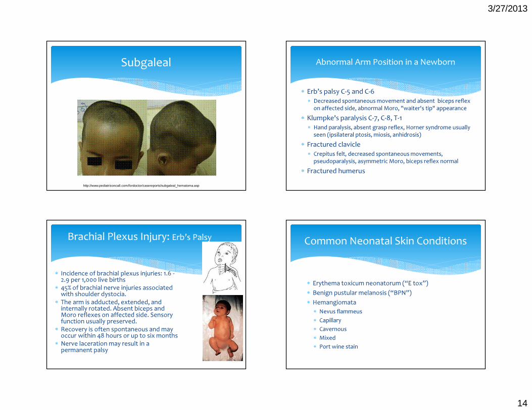
Subgaleal
http://www.pediatriconcall.com/fordoctor/casereports/subgaleal_hematoma.asp
∗ Erb’s palsy C-5 and C-6∗ Decreased spontaneous movement and absent biceps reflex
on affected side, abnormal Moro, "waiter's tip" appearance ∗ Klumpke's paralysis C-7, C-8, T-1
∗ Hand paralysis, absent grasp reflex, Horner syndrome usually seen (ipsilateral ptosis, miosis, anhidrosis)
∗ Fractured clavicle ∗ Crepitus felt, decreased spontaneous movements,
pseudoparalysis, asymmetric Moro, biceps reflex normal∗ Fractured humerus
Abnormal Arm Position in a Newborn
∗ Incidence of brachial plexus injuries: 1.6 -2.9 per 1,000 live births∗ 45% of brachial nerve injuries associated with shoulder dystocia.∗ The arm is adducted, extended, and internally rotated. Absent biceps and Moro reflexes on affected side. Sensory function usually preserved. ∗ Recovery is often spontaneous and may occur within 48 hours or up to six months∗ Nerve laceration may result in a permanent palsy
Brachial Plexus Injury: Erb’s Palsy
∗ Erythema toxicum neonatorum (“E tox”)∗ Benign pustular melanosis (“BPN”)∗ Hemangiomata
∗ Nevus flammeus∗ Capillary∗ Cavernous∗ Mixed∗ Port wine stain
Common Neonatal Skin Conditions
3/27/2013
15
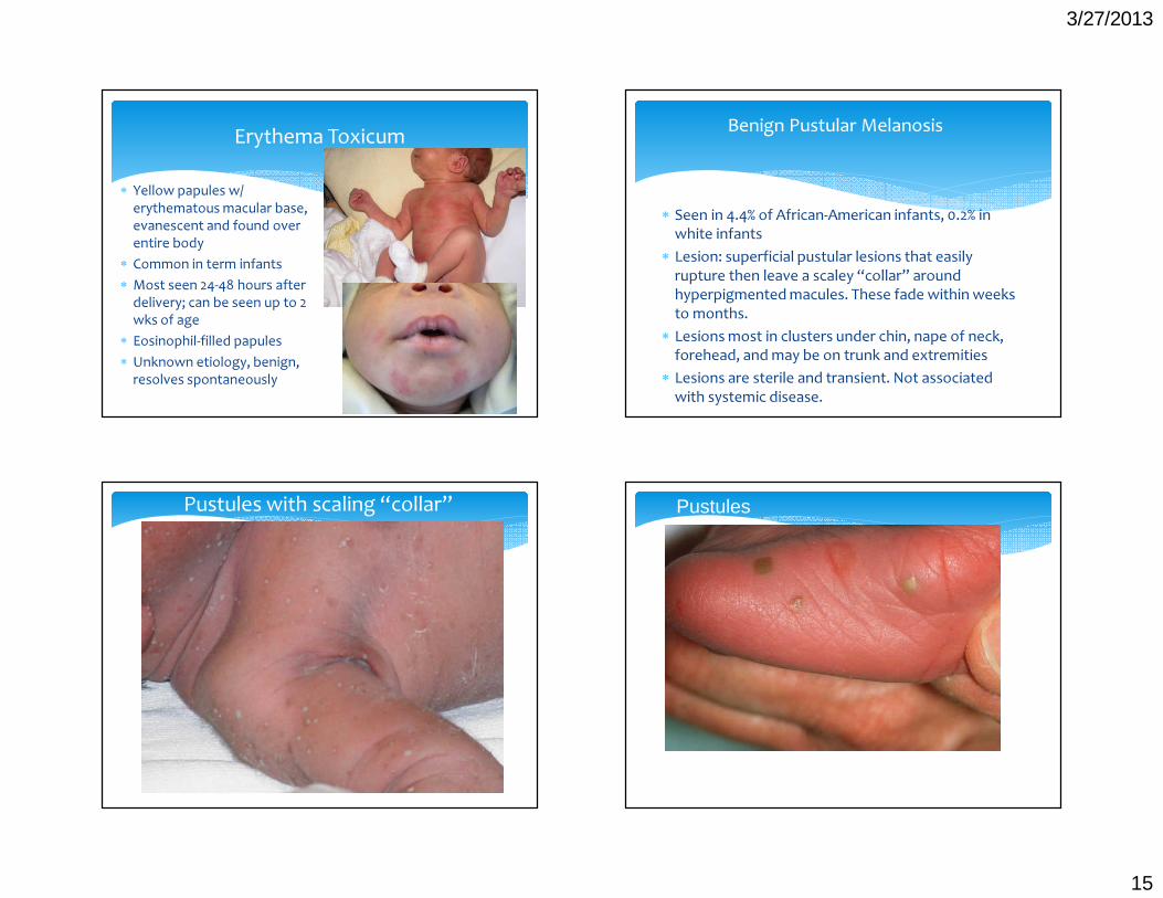
∗ Yellow papules w/ erythematous macular base, evanescent and found over entire body
∗ Common in term infants∗ Most seen 24-48 hours after
delivery; can be seen up to 2 wks of age
∗ Eosinophil-filled papules∗ Unknown etiology, benign,
resolves spontaneously
Erythema Toxicum
∗ Seen in 4.4% of African-American infants, 0.2% in white infants
∗ Lesion: superficial pustular lesions that easily rupture then leave a scaley “collar” around hyperpigmented macules. These fade within weeks to months.
∗ Lesions most in clusters under chin, nape of neck, forehead, and may be on trunk and extremities
∗ Lesions are sterile and transient. Not associated with systemic disease.
Benign Pustular Melanosis
Pustules with scaling “collar” Pustules
3/27/2013
16
Post-inflammatory hyperpigmentation
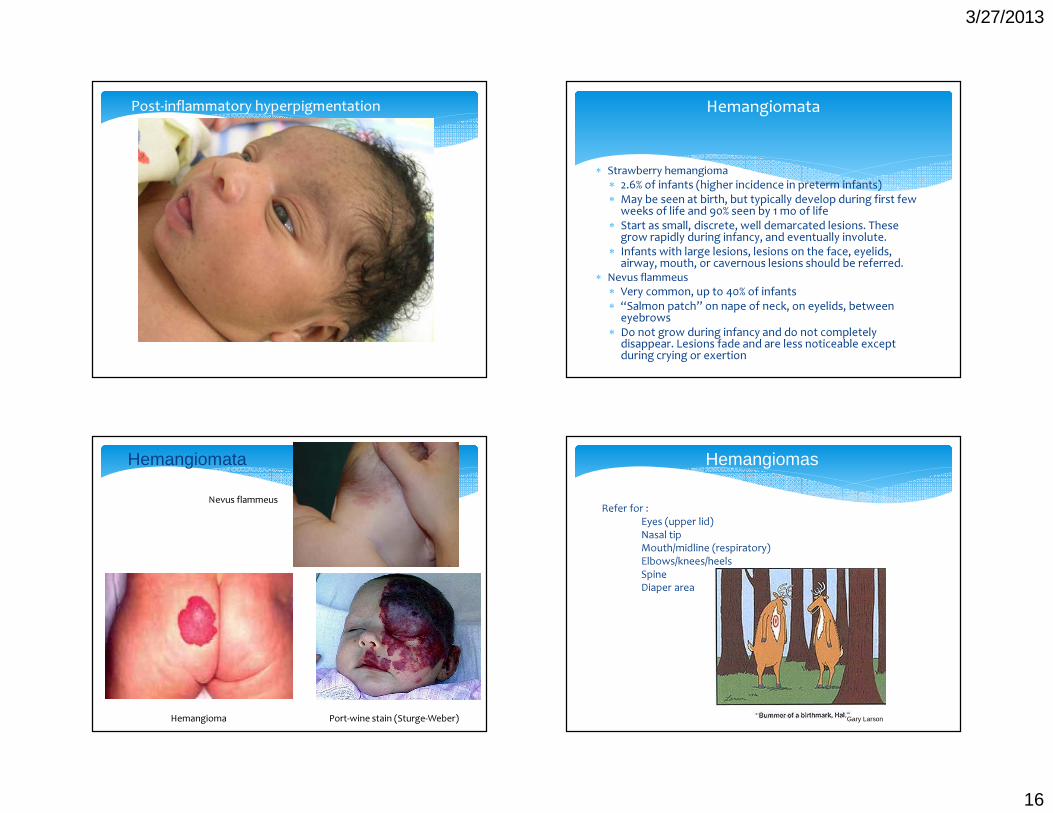
∗ Strawberry hemangioma∗ 2.6% of infants (higher incidence in preterm infants)∗ May be seen at birth, but typically develop during first few weeks of life and 90% seen by 1 mo of life∗ Start as small, discrete, well demarcated lesions. These grow rapidly during infancy, and eventually involute. ∗ Infants with large lesions, lesions on the face, eyelids, airway, mouth, or cavernous lesions should be referred.
∗ Nevus flammeus∗ Very common, up to 40% of infants∗ “Salmon patch” on nape of neck, on eyelids, between eyebrows∗ Do not grow during infancy and do not completely disappear. Lesions fade and are less noticeable except during crying or exertion
Hemangiomata
Hemangioma Port-wine stain (Sturge-Weber)
Hemangiomata
Nevus flammeus
Hemangiomas
Refer for :Eyes (upper lid)Nasal tipMouth/midline (respiratory)Elbows/knees/heelsSpineDiaper area
Gary Larson
3/27/2013
17
∗ What you hear when∗ Day of birth/1st DOL—outflow stenoses∗ After 1st day—coarctation∗ 1st week—left-to-right shunts (PDA, VSD, etc)∗ “Tachycardia”/bradycardia of newborn (DOL ~3)
∗ How? Training ear to VSD vs patent ductus∗ Stanford’s newborn nursery site:
http://newborns.stanford.edu/PhotoGallery/Heart.html
Cardiac/Murmurs
∗ What else to see?∗ Congestive heart failure—sweating, poor feeding,
failure to grow, HSM∗ What else to do? Standard to check post-ductal
saturation after 24 HOL∗ Post-ductal <95%, or gradient >3%
Cardiac/Murmurs, cont’d
Breastfeeding
∗ Benefits∗ Challenges
∗ Who is your patient?∗ Resources
∗ Lactation∗ Public health nurses∗ Local groups/stores/insurance
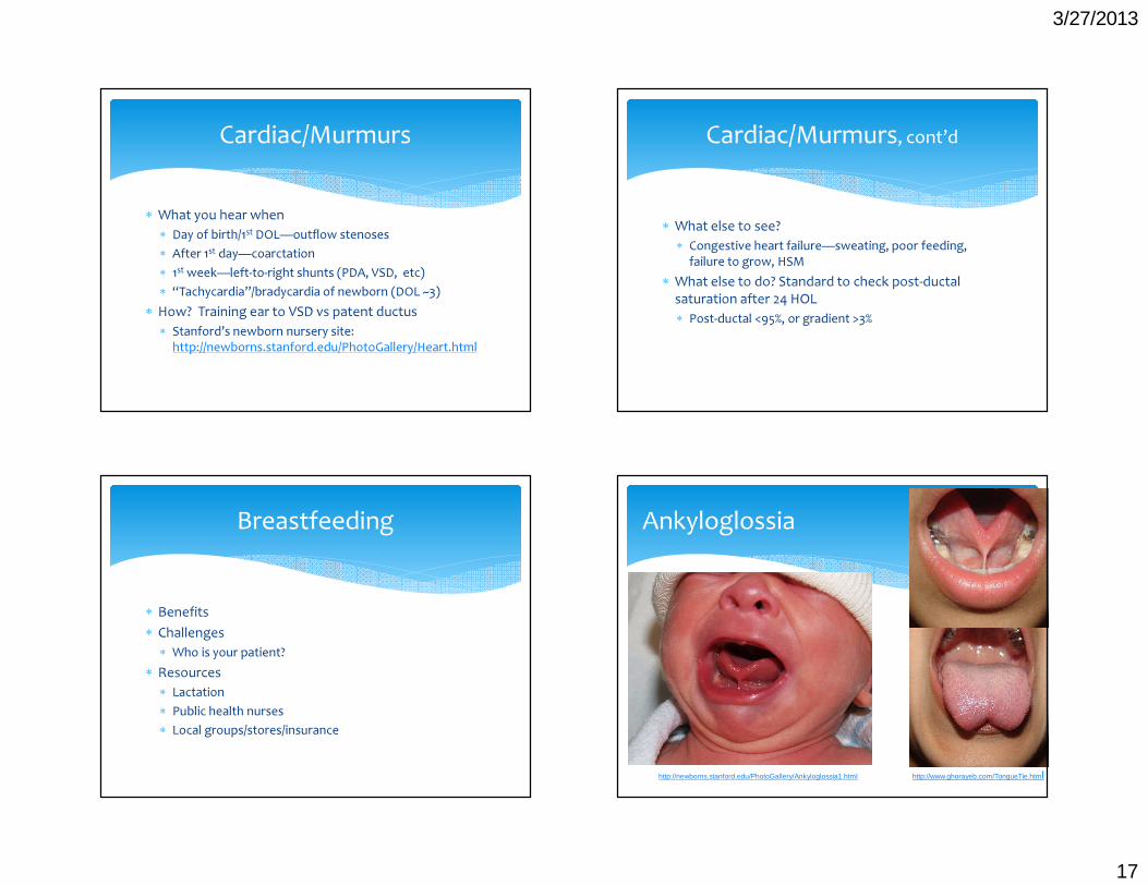
Ankyloglossia
http://newborns.stanford.edu/PhotoGallery/Ankyloglossia1.html http://www.ghorayeb.com/TongueTie.html
3/27/2013
18
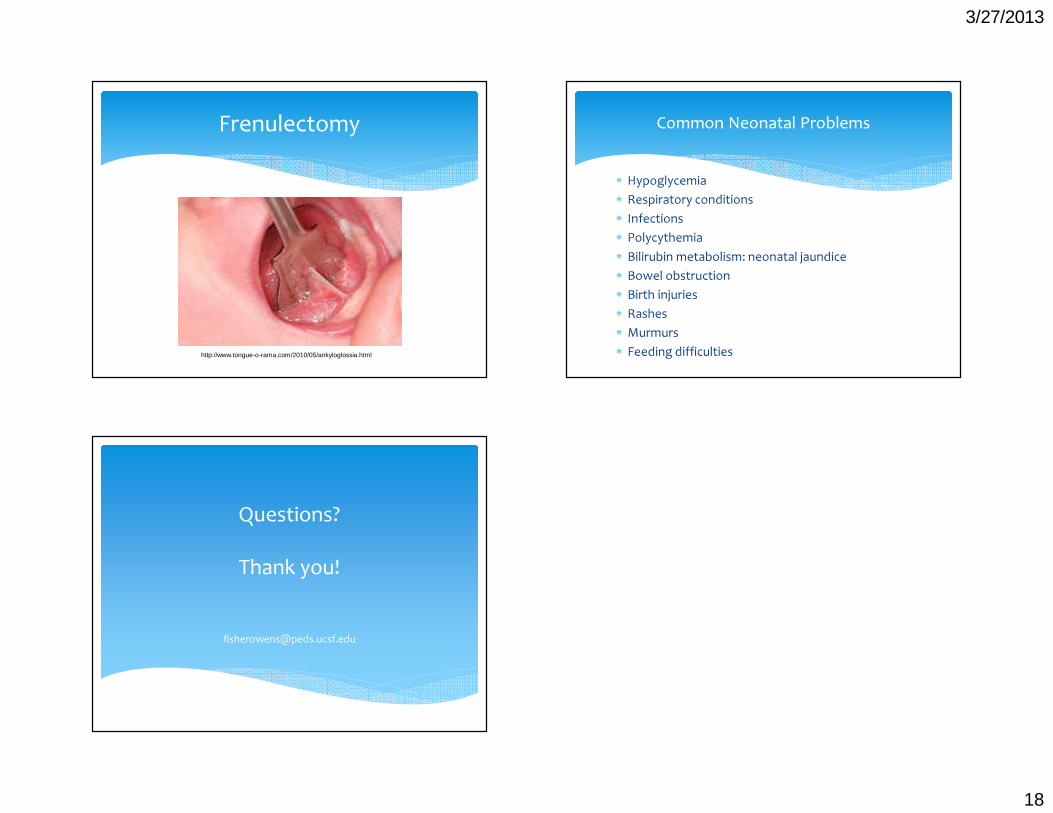
Frenulectomy
http://www.tongue-o-rama.com/2010/05/ankyloglossia.html
∗ Hypoglycemia∗ Respiratory conditions∗ Infections∗ Polycythemia∗ Bilirubin metabolism: neonatal jaundice∗ Bowel obstruction∗ Birth injuries∗ Rashes∗ Murmurs∗ Feeding difficulties
Common Neonatal Problems
Questions?Thank you!


![Indikationen für die Abrechnung der Pauschalen für ... · 8 Bösartige Neubildungen der Verdauungsorgane2_15 C24.1 Bösartige Neubildung: Ampulla hepatopancreatica [Ampulla Vateri]](https://img.pdfslide.net/doc/110x75/5e04fd523baf0e25b840bc29/indikationen-fr-die-abrechnung-der-pauschalen-fr-8-bsartige-neubildungen.jpg)