Embed Size (px)
Citation preview
Microenvironment of osteoarthritic cartilage and subchondral bone influences chondrogenic differentiation, extracellular matrix production and composition of bone
marrow-derived stem cells and articular chondrocytes
Dissertation to obtain the doctoral degree (Dr. rer. nat.) in natural science from the Faculty of Biology of the University Regensburg
By
Michaela Leyh (geb. Priller)
from Karlskron, Germany
-2014-
2
This work was carried out between September 2008 and May 2014 at the Department of Experimental Orthopedics of the University Hospital of Regensburg, Centre for Medical Biotechnology, Germany.
Submitted December 2014
Under the supervision of Prof. Dr. Rainer Deutzmann
and Prof. Dr. Susanne Grässel
Request for examination submitted on: November 2014
Date of examination: 9 April 2015
Examination board: Chairman: Prof. Dr. Arne Dittmer
First reviewer: Prof. Dr. Rainer Deutzmann
Second reviewer: Prof. Dr. Susanne Grässel
External reviewer: Prof. Dr. Reinhard Sterner
<Table of content
3
Table of content Table of content --------------------------------------------------------------------------------------------------------------- 3
Acknowledgments ------------------------------------------------------------------------------------------------------------ 6
Abbreviations ------------------------------------------------------------------------------------------------------------------ 7
Abstract ----------------------------------------------------------------------------------------------------------------------- 10
Zusammenfassung auf Deutsch ----------------------------------------------------------------------------------------- 12
1 State of the Art / General introduction ------------------------------------------------------------------- 14
1.1 Epidemiology of Osteoarthritis (OA) in human joints ------------------------------------------------- 14
1.2 Biology of articular cartilage -------------------------------------------------------------------------------- 14
1.2.1 Organization of articular cartilage ------------------------------------------------------------------------- 14
1.2.2 Differences between normal and OA cartilage --------------------------------------------------------- 15
1.2.3 Biomechanical properties of cartilage -------------------------------------------------------------------- 18
1.3 Biology of subchondral bone -------------------------------------------------------------------------------- 19
1.3.1 Organization of subchondral bone ------------------------------------------------------------------------ 19
1.3.2 Differences of normal and OA subchondral bone ------------------------------------------------------ 21
1.4 Biology of bone marrow derived mesenchymal stem cells (BMSC) ------------------------------- 22
1.4.1 BMSC and their regenerative potential ------------------------------------------------------------------- 22
1.4.2 Chondrogenic differentiation of BMSC ------------------------------------------------------------------- 24
1.4.3 BMSC as trophic mediators ---------------------------------------------------------------------------------- 26
1.5 Cartilage repair and tissue engineering ------------------------------------------------------------------ 27
1.5.1 Cartilage repair studies --------------------------------------------------------------------------------------- 27
1.5.2 Biomaterials and fibrin in tissue engineering ----------------------------------------------------------- 28
1.5.3 Coculture models in tissue engineering ------------------------------------------------------------------ 29
1.6 Animals in cartilage trauma repair models -------------------------------------------------------------- 33
1.6.1 Animal models in cartilage repair -------------------------------------------------------------------------- 33
1.6.2 Ovine cartilage repair models ------------------------------------------------------------------------------- 34
2 Aim of the thesis ----------------------------------------------------------------------------------------------- 36
3 Material and methods ---------------------------------------------------------------------------------------- 38
3.1 Culture and isolation of articular cartilage, subchondral bone and chondrocytes------------- 38
3.2 Culture and isolation of mesenchymal stem cells (BMSC) and adipose-derived stem cells
(ASC) --------------------------------------------------------------------------------------------------------------- 40
3.3 Flow cytometric characterization of BMSC and differentiation into different lineages ------ 41
3.4 Cocultivation of fibrin gel embedded BMSC/chondrocytes during chondrogenic
differentiation -------------------------------------------------------------------------------------------------- 43
3.5 Stimulation of fibrin gel embedded BMSC and/or chondrocytes ---------------------------------- 44
<Table of content
4
3.6 Cell vitality and proliferation in fibrin gel cocultures -------------------------------------------------- 45
3.7 Isolation and quantification of messenger RNA -------------------------------------------------------- 46
3.8 Histology and immunofluorescence of fibrin gel embedded cultures ---------------------------- 48
3.9 Biochemical analysis of fibrin gel cell lysates ----------------------------------------------------------- 48
3.9.1 DMMB Assay ---------------------------------------------------------------------------------------------------- 48
3.9.2 Collagen I and II ELISA ----------------------------------------------------------------------------------------- 49
3.9.3 Collagen III Dot-Blot ------------------------------------------------------------------------------------------- 49
3.10 Immunoblotting of Sox9 and phospho Sox9 ------------------------------------------------------------ 49
3.11 Analysis of supernatants (DMMB und hydroxyproline-assay, PTHrP, Fibronectin, bFGF,
IL-1β, IL-6 and IL-8 ELISA) ------------------------------------------------------------------------------------ 51
3.11.1 Hydroxyproline -assay ---------------------------------------------------------------------------------------- 51
3.11.2 PTHrP, fibronectin, bFGF, IL-1ß, IL-6 and IL-8 ELISA --------------------------------------------------- 51
3.11.3 DMMB Assay ---------------------------------------------------------------------------------------------------- 52
3.12 Screening for differentially expressed factors in coculture supernatant with LC-MS --------- 52
3.13 Biomechanical testing----------------------------------------------------------------------------------------- 53
3.14 Statistical analysis ---------------------------------------------------------------------------------------------- 54
4 Results ------------------------------------------------------------------------------------------------------------ 55
4.1 Part I: Microenvironment of cartilage influences chondrogenic differentiation and ECM
production of BMSC and chondrocytes------------------------------------------------------------------- 55
4.1.1 Bone marrow derived BMSC can be differentiated into different lineages --------------------- 55
4.1.2 Cells embedded in fibrin gel co- or tricultured with OA-cartilage remain vital and
proliferative for up to 28 days ------------------------------------------------------------------------------ 56
4.1.3 mRNA expression of differentiation/dedifferentiation markers is altered in cartilage
cocultures -------------------------------------------------------------------------------------------------------- 57
4.1.4 Microenvironment of cartilage alters immunofluorescent staining pattern of collagens --- 58
4.1.5 Collagen I, II and III and GAG contents in fibrin gel cell lysates of mono-, co- and
tricultures -------------------------------------------------------------------------------------------------------- 60
4.1.6 Sox9 is not influenced by OA-cartilage ------------------------------------------------------------------- 62
4.1.7 Comparison of soluble ECM fragments, PTHrP, bFGF and cytokines in supernatants of
mono-, co- and tricultures ----------------------------------------------------------------------------------- 63
4.1.8 LC-MS analysis revealed differentially expressed factors in BMSC cartilage coculture
supernatants ---------------------------------------------------------------------------------------------------- 66
4.1.9 Microenvironment of OA-cartilage alters biomechanical properties of new extracellular
matrix ------------------------------------------------------------------------------------------------------------- 67
4.2 Part II: Microenvironment of subchondral bone influences chondrogenic differentiation
and ECM production of BMSC and chondrocytes ----------------------------------------------------- 69
<Table of content
5
4.2.1 Cells embedded in fibrin gel and co- or tricultured with subchondral bone remain vital
for up to 28 days ----------------------------------------------------------------------------------------------- 69
4.2.2 mRNA expression of differentiation/dedifferentiation markers is altered in subchondral
bone co- and tricultures -------------------------------------------------------------------------------------- 69
4.2.3 Microenvironment of subchondral bone alters immunofluorescent staining pattern of
collagens --------------------------------------------------------------------------------------------------------- 72
4.2.4 Fibrin gel cell lysates reveal different GAG, collagen I, II and III content in mono-, co- and
tricultures -------------------------------------------------------------------------------------------------------- 74
4.2.5 Sox9 in BMSC is not influenced by subchondral bone ------------------------------------------------ 75
4.2.6 Comparison of soluble ECM molecule fragments, PTHrP and cytokines in supernatants of
mono-, co- and tricultures ----------------------------------------------------------------------------------- 76
4.2.7 Microenvironment of OA-subchondral bone alters biomechanical properties of newly
regenerated cartilage ----------------------------------------------------------------------------------------- 80
4.3 Part III: Effect of IL-1β, IL-6 or IL-8 stimulation on chondrogenic differentiation and ECM
production of BMSC and chondrocytes------------------------------------------------------------------- 82
4.3.1 mRNA expression of (de)-differentiation markers is altered in cytokine stimulated fibrin
gels ---------------------------------------------------------------------------------------------------------------- 82
4.3.2 Stimulation with cytokines revealed different GAG, collagen I, II and III content in fibrin
gel lysates -------------------------------------------------------------------------------------------------------- 86
4.4 Part IV: Influence of normal ovine cartilage on mRNA expression of ovine BMSC, mixed
and chondrocyte cultures ------------------------------------------------------------------------------------ 87
5 Discussion -------------------------------------------------------------------------------------------------------- 89
5.1 General discussion --------------------------------------------------------------------------------------------- 89
5.2 Part I: Microenvironment of cartilage coculture influences BMSC differentiation and
ECM production ------------------------------------------------------------------------------------------------ 93
5.3 Part II: Microenvironment of subchondral bone coculture influences BMSC differentiation
and ECM production ---------------------------------------------------------------------------------------- 103
5.4 Part III: Stimulation with IL-1β, IL-6 and IL-8 during chondrogenesis --------------------------- 110
5.5 Part IV: Influence of coculture with normal ovine cartilage on BMSC differentiation ------ 114
6 Summary and conclusion ---------------------------------------------------------------------------------- 115
References ------------------------------------------------------------------------------------------------------------------ 117
Publications, Awards, Posters and Presentations ----------------------------------------------------------------- 129
Acknowledgments
6
Acknowledgments
I want to thank Prof. Dr. Susanne Grässel, Department of Orthopedics, University Hospital
Regensburg, for being my mentor and greatly contributing to the development of my thesis. The
versatile administrative and academic skills that I have acquired during the last few years are due to
her excellent support. The gained skills and the achieved qualification are a good basis for a
successful career. I cannot thank her enough.
Special thanks to Prof. Dr. Rainer Deutzmann, Department of Biology and Preclinical Medicine,
University of Regensburg for allowing me to work on this thesis under his patronage. He was
supporting the success of my thesis by LC-MS analysis together with Dr. Astrid Bruckmann, who I also
want to thank for her abundant help and guidelines.
I am also deeply grateful that Prof. Dr. Joachim Grifka, Director of the Department of Orthopedics,
University Hospital Regensburg, provided the basic conditions for the accomplishment of this work.
An exceptional thank to all of my amiable colleagues, especially to Lilly Weger for her excellent
technical assistance, for being a great friend and a mental support. She really made my days brighter.
I also want to convey my gratitude to Prof. Dr. Jürgen Strutz and to Prof. Dr. Holger Gassner,
Department of Otorhinolaryngology, University Hospital Regensburg, who made it possible that I
could finish my doctoral thesis in parallel to my duties in their lab. I am indebted to the remarkable
indulgence and support of Dr. Frank Haubner and Petra Eberl.
Additionally, I want to thank Jens Schaumburger and Hans-Robert Springorum for being an abundant
source of bone marrow and explanted knee joints, Johann Haas (Landgasthof Haas, Karlskron) and
Albert Obermayr (Metzgerei Obermayr, Deimhausen) for provision of ovine femur from
slaughterhouse waste and Drs. Uta Delling and Henriette Juelke for isolation of ovine BMSC.
Grateful thanks to my collaboration partners Dr. Lutz Dürselen and Andreas Seitz, University of Ulm,
Institute of Orthopedic Research and Biomechanics, ZMFU, who supported me with generation,
analyses and interpretation of the biomechanical properties.
I also want to thank R. Holmdahl and K. Rubin from the Developmental Studies Hybridoma Bank,
University of Iowa, who developed the CIIC1 antibody under the auspices of the NICHD.
Finally, I want to thank my family and friends for being a great pool of hope, for their encouragement
to never give up and to brighten my mood. Especially I have to thank my parents for their financial
support that enabled me to finalize my thesis.
This study was supported by funds from the DFG (GR 1301/8-1) and from the German Society for
Orthopedics and Orthopedic Surgery (DGOOC).
Abbreviations
7
Abbreviations
3D Three-dimensional
ACAN Aggrecan gene
ALP Alkaline phosphatase
APS Ammonium peroxodisulfate solution
ASC Adipose derived stem cells
BCA Bicinchoninic acid
bFGF basic fibroblast growth factor
BMSC Bone marrow derived stem cells
BMP Bone morphogenetic protein
BSA Bovine serum albumin
cDNA Complementary DNA
Ch Chondrocytes
Col Collagen
COMP Cartilage oligomeric matrix protein
Ct Threshold cycle
Da Dalton
DAPI 4´,6´-diamidino-2-phenylindole, dihydrochloride
DMEM Dulbecco´s modified eagle´s medium
DMMB Dimethylmethylene blue
DMSO dimethylsulfoxide
DNA Deoxyribonucleic acid
dNTP mixture of deoxyribonucleotides dATP, dCTP, dGTP, dTTP
E. coli Escherichia coli
ECM Extracellular matrix
EDTA Ethylene-diamine-tetra acetic acid
Abbreviations
8
ELISA enzyme linked immunosorbent assay
et al. et alii (from Latin: and others)
EtOH Ethanol
E∞ equilibrium modulus
F monocultures (without subchondral bone explants)
FACS Fluorescence activated cell sorting
FB co- and tricultures with subchondral bone explants
FC co- and tricultures with articular cartilage explants
FCS Fetal calf serum
g gravitation
GAG Glycosaminoglycan
HA aggregate modulus
IBMX 3-isobutyl-1-methylxanthine
IGF Insulin-like growth factor
Ihh Indian hedgehog homolog
IL Interleukin
k hydraulic permeability
kb kilo bases
LC-MS Liquid chromatography–mass spectrometry
LDH Lactate dehydrogenase
MALDI-TOF Matrix Assisted Laser Desorption/Ionization- Time Of Flight
Mixed mixed cultures of BMSC and chondrocytes
MMP Matrix metalloprotease
mRNA messenger RNA
MW molecular weight
n number of independent samples
N Newton
Abbreviations
9
NB normal bone
OA osteoarthritis
oBMSC ovine bone marrow derived stem cells
OD optical density
PAGE Polyacrylamide gel electrophoresis
PBS Phosphate buffered saline
PCR Polymerase chain reaction
PCNA Proliferating cell nuclear antigen
PDGF Platelet-derived growth factor
PFA Paraformaldehyde
PTHrP Parathyroid hormone related peptide
qPCR Quantitative PCR
RNA Ribonucleic acid
RT-PCR Reverse transcriptase PCR
SD Standard deviation
SDS Sodium dodecyl sulphate
Sox Sex related homeobox containing transcription factors
TEMED Tetramethylethylenediamine
TGF-β Transforming growth factor beta
TNF-α Tumor necrosis factor-α
Tris Tris(hydroxymethyl)-aminomethan
U units
Abstract
10
Abstract
Objective
Osteoarthritis (OA) is characterized by an imbalance in cartilage and subchondral bone homeostasis,
which could be potentially treated or improved by cell-based therapies. In the present study, a
reproducible in vitro coculture model was established to evaluate the influence of normal and OA-
cartilage or subchondral bone explants on chondrogenic differentiation of human bone marrow
derived stem cells (BMSC), human adipose derived stem cells (ASC) and phenotype of OA-
chondrocytes. Signals from the articular cartilage or the underlying subchondral bone were
hypothesized to induce phenotypic shifts and an altered re- and differentiation potential in
cocultured BMSC, ASC and differentiated chondrocytes from OA patients. To provide a chondrogenic
environment, cells were embedded in fibrin gel and kept in chondrogenic medium for up to 28 days.
Methods
A reproducible coculture model of ASC, BMSC, mixed cultures (BMSC and chondrocytes in equal
ratio) and chondrocytes embedded in fibrin gel seeded on articular cartilage or subchondral bone
explants (= co- and tricultures) compared with monocultured cells in fibrin gel without explants was
established. Human OA-tissues, OA-chondrocytes and BMSC were derived from patients subjected to
arthroplasty. Normal (healthy) human tissues were received from knees of rare trauma affected
donors (treated for sports accidents). ASC were isolated from subcutaneous fat tissue obtained from
patients undergoing elective body contouring procedures. Ovine cartilage, chondrocytes and BMSC
were obtained from normal (healthy) pasture sheep. Gene expression analysis, biochemical assays
(ELISA, Dot-blot, DMMB, Hydroxyproline), immunofluorescence staining and biomechanical tests
were used to characterize the properties of newly generated extracellular matrix (ECM) from
chondrocytes and chondrogenically differentiated ASC and BMSC.
Results
In general, all cell regimens cocultured with OA-explants exhibited reduced gene expression patterns
of collagens I, II, III and X in comparison with monocultures. Significant lower levels of collagen I and
II protein (BMSC) and significant lower collagen I and III protein (mixed cultures) were detected in
cartilage co- or tricultures, while co- and triculture with subchondral bone inhibited collagens in
general. In contrast, no changes in glycosaminoglycan (GAG) synthesis were observed for cartilage
co- and tricultures, while reduced GAG production was observed in subchondral bone co- and
triculture lysates. Repetition of key experiments with normal ASC confirmed inhibitory effects for OA-
subchondral bone. Co- and triculture with normal cartilage or subchondral bone explants showed no
Abstract
11
or only reduced inhibitory effects on chondrogenic differentiation or collagen gene expression. In
addition, biomechanical properties of the OA-cartilage or subchondral bone co- and tricultured cell-
fibrin gels were affected. All co- and triculture regimens tended to exhibit lower Young´s modulus
and aggregate modulus compared with monocultures. In contrast, hydraulic permeability seemed to
be higher in co- and tricultures. Supernatants of cartilage and subchondral bone co- and tricultures
contained significant higher IL-1β, IL-6 and IL-8 levels, as well as significant more soluble GAGs
compared with controls. In general, stimulation of monocultures with IL-1β induced matrix
metalloproteinase (MMP)2, and MMP3 and reduced collagens I, II and X gene expression and led to a
downregulation of aggrecan gene expression. Stimulation with IL-6 reduced aggrecan, MMP3 and
MMP13 gene expression and mainly reduced collagen I, II and III gene expression of BMSC and/or
chondrocytes. In contrast, IL-8 stimulation of mixed and chondrocytes monocultures had only little
effects, while IL-8 stimulated BMSC showed reduced collagen I, II and III gene expression.
Conclusions
Taken together our results suggest an inhibitory effect of factors from the microenvironment of OA-
cartilage and subchondral bone on production of collagens. This indicates a distinct modulatory
influence, which affects the composition of the de novo produced ECM from cocultured cells and
leads to impaired mechanical strength and biochemical properties of the newly formed matrix.
Experiments with ASC, normal cartilage and subchondral bone explants either might hint to disease
status induced effects (OA vs. trauma) or to an effect caused by different mean age of cell and tissue
donors. Soluble signal factors, i.e. pro-inflammatory cytokines (including IL-1β and IL-6), released
from OA-cartilage, OA-subchondral bone, chondrocytes and osteoblasts, might partly mediate these
effects on newly formed extracellular matrix properties. Thus, the microenvironment of neighbored
OA-cartilage seems to provide both: promoting and inhibiting signals for BMSC differentiation and
suggests that the balance of these factors determines the destiny of BMSC. This knowledge can be
used to develop new strategies for cell based cartilage regeneration.
Zusammenfassung auf Deutsch
12
Zusammenfassung auf Deutsch
Die Mikroumgebung von arthrotischem Knorpel und subchondralem Knochengewebe
beeinflusst die chondrogene Differenzierung sowie die ECM Produktion und
Zusammensetzung von BMSC und artikulären Chondrozyten
Charakteristisch für Osteoarthrose (OA) ist ein Ungleichgewicht der Knorpel- und subchondralen
Knochen Homöostase. Die Behandlung und der Verlauf dieser Erkrankung könnte durch zellbasierte
Therapien deutlich verbessert werden. In dieser Studie wurde ein reproduzierbares in vitro
Kokulturmodell entwickelt, um den Einfluss von OA-Knorpel und -Knochen Explantaten auf die
chondrogene Differenzierung von adipogenen Stammzellen (ASC), von humanen mesenchymalen
Stammzellen aus dem Knochenmark (BMSC) und den Phänotyp von OA-Chondrozyten zu
untersuchen. Vermutlich verursachen Signalfaktoren aus dem artikulären Knorpel und der
darunterliegenden subchondralen Knochenschicht einen Shift im Phänotyp oder eine Veränderung
im (Re-) Differenzierungspotential von ASC, BMSC und differenzierten Chondrozyten. Um eine
chondrogene Mikroumgebung zu schaffen, wurden die Zellen in Fibringel eingebettet und bis zu 28
Tage in chondrogenem Medium kultiviert.
In einem Kokulturmodell wurden in Fibringel eingebettete ASC, BMSC, Chondrozyten oder ein
Gemisch aus BMSC und Chondrozyten (zu gleichen Teilen) entweder zusammen mit
osteoarthrotischem Knorpel oder subchondralem Knochen kultiviert (= Ko- und Trikultur) und mit
den entsprechenden Zellen in Monokultur ohne Explantat verglichen. Alle humanen OA-
Gewebestücke wurden aus Patienten entnommen, die einer Arthroplastik unterzogen wurden.
Gesundes humanes Gewebe wurde aus Knien von Unfallpatienten gewonnen, die wegen einer
Sportverletzung behandelt wurden. ASC wurden aus Unterhautfettgewebe isoliert, das von Patienten
einer kosmetischen Fettabsaugung stammt. Genexpressionsanalysen, biochemische Assays (ELISA,
Dot-blot, DMMB, Hydroxyprolin), Immunfluoreszenzfärbungen und biomechanische Tests wurden
verwendet, um die Eigenschaften der neu gebildeten Matrix von Chondrozyten und chondrogen
differenzierten BMSC zu bestimmen.
Im Allgemeinen zeigten alle kokultivierten Zellbedingungen verminderte Genexpression von Kollagen
I, II, III und X verglichen mit Monokulturen. Auf Proteinebene konnte signifikant weniger Kollagen I
und II (BMSC) und signifikant weniger Kollagen I und III (gemischte Kulturen) in den Knorpel-Ko- und
Trikulturen nachgewiesen werden. Die Ko- und Trikultur mit subchondralem Knochen zeigte eine
generelle Verminderung der Expression aller Kollagene. Im Gegensatz dazu konnte keine Änderung in
Zusammenfassung auf Deutsch
13
der Glykosaminoglykan (GAG) Synthese der Knorpel Ko- und Trikultur nachgewiesen werden,
während in der Ko- und Trikultur mit subchondralem Knochen temporär eine Reduktion der GAG
Synthese beobachtet werden konnte. Die Wiederholung von Schlüsselexperimenten mit gesunden
ASC konnte die inhibitorischen Effekte induziert durch Ko- und Trikultur mit OA-subchondralem
Knochen bestätigen. Ko- und Trikultur mit normalem Knorpel oder subchondralem Knochen zeigte
keine oder nur verminderte inhibierende Effekte auf die chondrogene Differenzierung oder Gen
Expression von Kollagen. Die biomechanischen Eigenschaften der kokultivierten Fibringele waren
ebenfalls beeinträchtigt. Alle Ko- und Trikulturbedingungen zeigten tendenziell ein geringeres
Elastizitätsmodul und Aggregationsmodul im Vergleich zur Monokultur. Im Gegensatz dazu schien die
hydraulische Permeabilität in den Ko-und Trikulturen höher zu sein. Die Überstände von Knorpel-
und Knochen Ko- und Trikulturen enthielten im Vergleich mit den Kontrollen signifikant mehr IL-1β,
IL-6 und IL-8, und auch mehr lösliche GAGs, die größtenteils von den Explantaten stammten. Die
Stimulation der Monokulturen mit IL-1β induzierte im Allgemeinen MMP2, MMP3 und MMP13 und
reduzierte die Genexpression der Kollagene I, II und X sowie von Aggrekan. Die Stimulation mit IL-6
verminderte die Aggrekan, MMP3 und MMP13 Genexpression und größtenteils auch der Kollagene I,
II und III in BMSC und/oder Chondrozyten. Im Gegensatz dazu hatte die Stimulation mit IL-8 nur
geringe Effekte auf gemischte und Chondrozyten Monokulturen, während in den IL-8 stimulierten
BMSC eine Reduktion der Kollagene I, II und III beobachtet werden konnte.
Zusammenfassend deuten die Ergebnisse dieser Studie auf einen inhibitorischen Effekt von Faktoren
aus der Mikroumgebung von OA-Knorpel oder -subchondralem Knochen auf die Kollagenproduktion
hin. Diese Ergebnisse lassen auf einen modulierenden Einfluss schließen, der zu verminderten
mechanischen und biochemischen Eigenschaften der neu gebildeten extrazellulären Matrix führt. Die
Experimente mit ASC sowie mit normalen Knorpel und subchondralen Knochen Explantaten könnten
entweder auf krankheitsbedingte Effekte (OA vs. Trauma) hinweisen oder auf einen Effekt aufgrund
des unterschiedlichen Durchschnittsalters der Zell- und Gewebe Spender. Vermutlich regulieren
lösliche Signalfaktoren wie z.B. proinflammatorische Zytokine, abgegeben von OA-Knorpel,
OA-subchondralem Knochen, Chondrozyten oder Osteoblasten, zumindest teilweise diese negativen
Effekte auf die chondrogene BMSC Differenzierung und die Qualität der neu gebildeten ECM.
Dennoch birgt die direkte Mikroumgebung von OA-Knorpel beides: fördernde und inhibierende
Signale der BMSC Differenzierung. Dies lässt darauf schließen, dass die Balance dieser Faktoren das
Schicksal der BMSC bestimmt. Die in dieser Studie neu gewonnenen Erkenntnisse können in Zukunft
dazu beitragen, neue Strategien für die zellbasierte Knorpelregeneration zu entwickeln und vor allem
die Qualität des neu gebildeten Knorpels entscheidend zu verbessern.
State of the Art / General introduction
14
1 State of the Art / General introduction
1.1 Epidemiology of Osteoarthritis (OA) in human jo ints
The human knee joint is one of the most important sources of pain in the western population, in
people over 55 years old and nearly everybody over 65 years is affected by osteoarthritis (OA)
(Hunter and Felson 2006). Possible causes for OA development are increasing age, mechanical stress,
a high body mass index and a genetic component (Breedveld 2004). OA is a chronic joint disease and
develops usually undetected many years before it is diagnosed. Notably, a correlation with pain is
not mandatory and OA becomes painful in about 50% of patients (Pereira, Peleteiro et al. 2011). Still
it is the leading cause of disability, which besides limitations in motility and quality of life is the high
economic burden in form of direct and indirect health-related costs (Felson 2004; Hiligsmann, Cooper
et al. 2013). Thus, ineffective treatments focusing only on symptoms - mainly pain reduction – should
better include a change of lifestyle like weight reduction, adequate footwear and continuing
exercises (Roddy and Doherty 2006). Anyhow, after diagnosis and radiographic confirmation for
patients with severe end stage OA, a total replacement of the joint and implantation of a prosthesis
is inevitable. Drawback in this regard is that the prosthesis has a limited lifespan and patients risk a
revision surgery (Wiegant, van Roermund et al. 2013). Therefore, alternatives for successful OA-
treatment are urgently required to regenerate cartilage tissue and avoid implantation of an artificial
prosthesis.
1.2 Biology of articular cartilage
1.2.1 Organization of articular cartilage
Normal articular cartilage is accurately organized and consists of different zones (Fig. 1); the
outermost region is called lamina splendens, named after the light breaking reflections of parallel
fibers (Hollander, Dickinson et al. 2010). The upper zone of cartilage is rich in thin collagen fibrils
parallel to the articular surface, which protects the cartilage from shear forces. Characteristic is a
high collagen content, with an increased collagen I content and lowest amount of aggrecan
compared with deeper zones. Cells in the upper cartilage region are relatively flat, small and
separated. Additionally, regulatory proteins like TGF-β1 and -3 as well as BMP1-6 (Anderson, Hodges
et al. 2000; Yamane, Cheng et al. 2007) and MMPs (-1, -3, -8 and -13) are expressed but are only
involved in matrix turnover, not in matrix degradation (Tetlow, Adlam et al. 2001; Tchetina 2011)
State of the Art / General introduction
15
In the middle zone, resting round shaped chondrocytes are residing in chondrons, surrounded by
thick, low organized fibers and an ECM consisting of high collagen II and aggrecan amounts (Pfander,
Swoboda et al. 2001).
The deep zone of articular cartilage has the highest aggrecan and lowest water content and the
collagen fibrils with the largest diameter, which are oriented perpendicular to the joint surface
(Maroudas, Bayliss et al. 1980). Chondrocytes in the deep zone are enlarged and arranged in columns
right angled to the cartilage surface (Pearle, Warren et al. 2005).
The deep zone merges into the hypertrophic zone, which is separated by the so-called tidemark
region from calcified cartilage that links the cartilage layer with the subchondral bone. The calcified
cartilage zone is rich in hydroxylapatit crystals and hypertrophy markers like collagen X, ALP and
MMP13 are expressed (Aigner, Hemmel et al. 2001; Pearle, Warren et al. 2005).
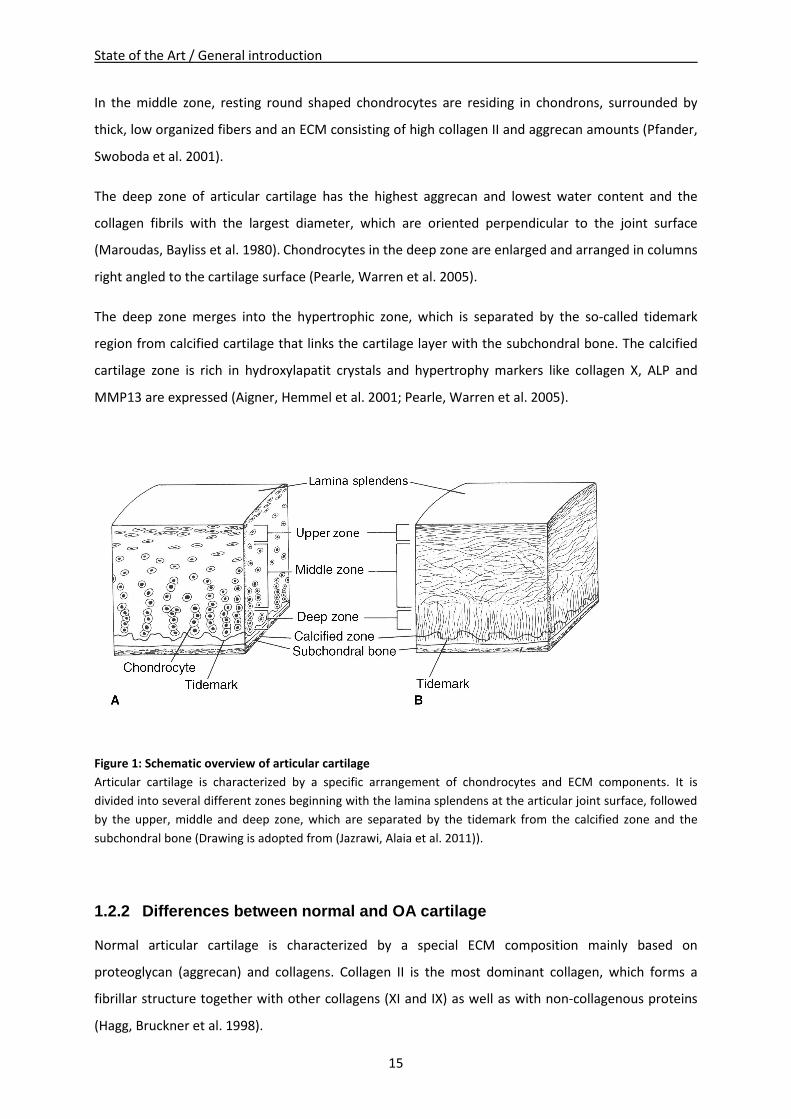
Figure 1: Schematic overview of articular cartilage
Articular cartilage is characterized by a specific arrangement of chondrocytes and ECM components. It is
divided into several different zones beginning with the lamina splendens at the articular joint surface, followed
by the upper, middle and deep zone, which are separated by the tidemark from the calcified zone and the
subchondral bone (Drawing is adopted from (Jazrawi, Alaia et al. 2011)).
1.2.2 Differences between normal and OA cartilage
Normal articular cartilage is characterized by a special ECM composition mainly based on
proteoglycan (aggrecan) and collagens. Collagen II is the most dominant collagen, which forms a
fibrillar structure together with other collagens (XI and IX) as well as with non-collagenous proteins
(Hagg, Bruckner et al. 1998).
State of the Art / General introduction
16
Mature normal articular cartilage has a low cell density as with the end of growth, articular
chondrocytes are hardly proliferating and exhibit only a low metabolism. Nevertheless, articular
cartilage shows a carefully balanced activity of matrix turnover with respect to cartilage ECM
degradation and synthesis (Poole 1997; Buckwalter and Mankin 1998; Tchetina 2011). In general,
intact hyaline cartilage shows only low gene expression of collagens I, II, VI, IX, X and XI but a high
turnover of aggrecan (Hardingham, Fosang et al. 1994; Aigner and McKenna 2002; Poole 2003).
Importantly, hyaline cartilage has no innervations and no blood flow and thus is characterized by a
low supply with nutrients and oxygen. Additionally neither pain nor overloading can be perceived
(Benedek 2006). For exactly that reason, a high impact load is unfavorable, because it is able to
provoke cartilage damage and bears the risk for OA development (Newberry, Mackenzie et al. 1998).
The impact level and its tenure are important, because the time period of maximal compression is
more fatal if it occurs abruptly than gradually despite of the same amplitude. During this kind of
strain, rupture of the collagen meshwork could happen if a critical impact is reached (Newberry,
Garcia et al. 1998; Torzilli, Grigiene et al. 1999). The rupture of cartilage ECM - typically for traumatic
sports injuries - represents a severe structural damage of the joint and induces the beginning of
structural cartilage degradation because of constant loading alterations. Additionally it causes post-
traumatic cell death, mediates acute joint inflammation and initiates repair mechanisms like an
increase of pro-inflammatory cytokines, basic fibroblast growth factor (bFGF), MMPs, aggrecanases,
and release of matrix fragments (Buckwalter, Anderson et al. 2013).
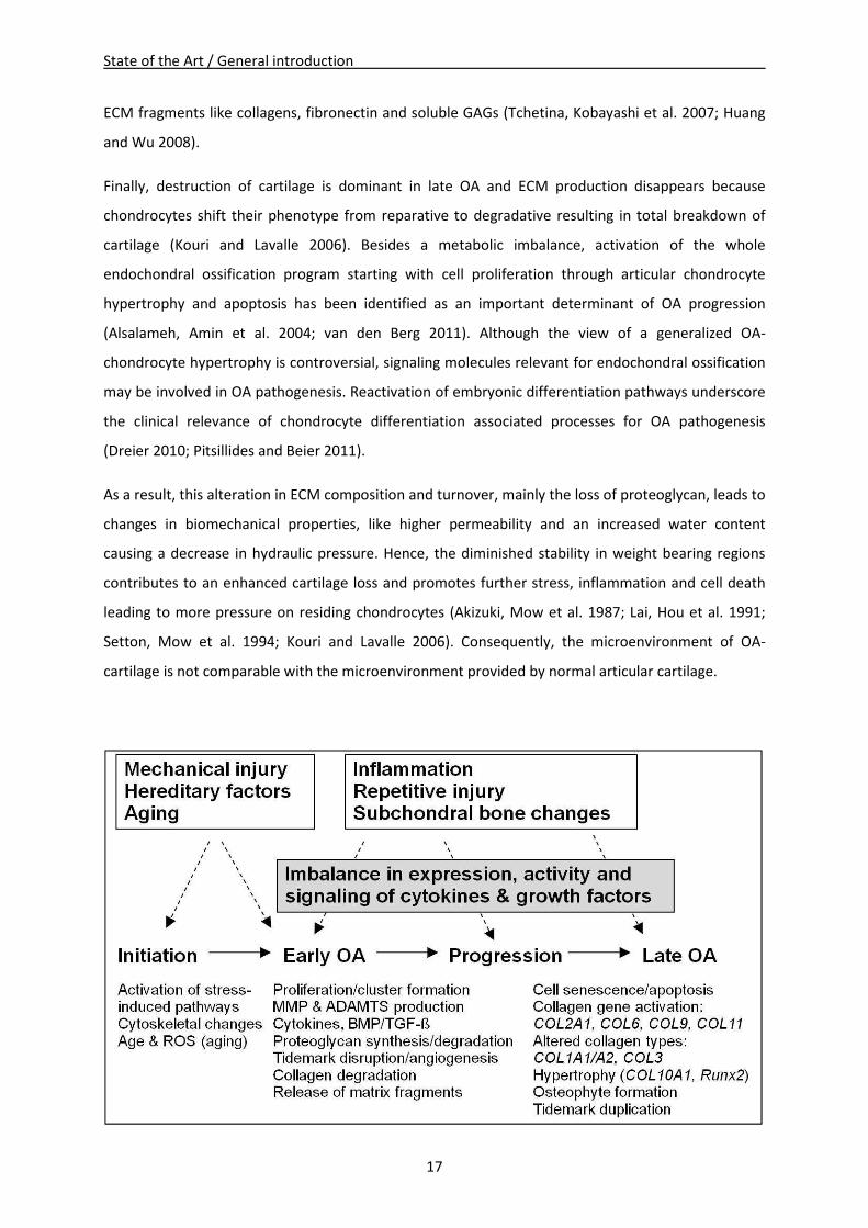
Figure 2 shows an overview of initiation and progression of OA including responsible factors and
changes in gene expression and phenotype of chondrocytes and cartilage. More detailed, in early OA,
cells residing in arthritic cartilage regions become active and restart metabolism. Especially TGF-β
and collagen II production is enhanced most probably as an attempt to repair destroyed cartilage
(Kouri and Lavalle 2006). Additionally, collagenases, aggrecanases, cytokines, collagen X, Indian
hedgehog homolog (Ihh) and caspase 3 are upregulated in early stage cartilage lesions. During
progression of OA, the balance of cartilage turnover is disturbed and MMPs – especially MMP13 –
aggrecanases, collagen X and alkaline phosphatase (ALP) are further upregulated. In combination
with downregulation of Sox9, TGF-β, TNF-α and aggrecan expression, a dramatic loss of aggrecan
occurs in the upper zone. Additionally, the natural zonal organization of cartilage breaks down and
results in large areas of destroyed inflamed cartilage tissue (Tchetina, Squires et al. 2005; Appleton,
McErlain et al. 2007; Tchetina 2011). These regions represent a unique OA-microenvironment with
release of high amounts of pro-inflammatory cytokines like IL-1β, IL-6, IL-8, IL-10 or TNF (Moldovan,
Pelletier et al. 2000; Lu, Evans et al. 2011), and growth factors like FGF, PTHrP or TGF-β as well as
State of the Art / General introduction
17
ECM fragments like collagens, fibronectin and soluble GAGs (Tchetina, Kobayashi et al. 2007; Huang
and Wu 2008).
Finally, destruction of cartilage is dominant in late OA and ECM production disappears because
chondrocytes shift their phenotype from reparative to degradative resulting in total breakdown of
cartilage (Kouri and Lavalle 2006). Besides a metabolic imbalance, activation of the whole
endochondral ossification program starting with cell proliferation through articular chondrocyte
hypertrophy and apoptosis has been identified as an important determinant of OA progression
(Alsalameh, Amin et al. 2004; van den Berg 2011). Although the view of a generalized OA-
chondrocyte hypertrophy is controversial, signaling molecules relevant for endochondral ossification
may be involved in OA pathogenesis. Reactivation of embryonic differentiation pathways underscore
the clinical relevance of chondrocyte differentiation associated processes for OA pathogenesis
(Dreier 2010; Pitsillides and Beier 2011).
As a result, this alteration in ECM composition and turnover, mainly the loss of proteoglycan, leads to
changes in biomechanical properties, like higher permeability and an increased water content
causing a decrease in hydraulic pressure. Hence, the diminished stability in weight bearing regions
contributes to an enhanced cartilage loss and promotes further stress, inflammation and cell death
leading to more pressure on residing chondrocytes (Akizuki, Mow et al. 1987; Lai, Hou et al. 1991;
Setton, Mow et al. 1994; Kouri and Lavalle 2006). Consequently, the microenvironment of OA-
cartilage is not comparable with the microenvironment provided by normal articular cartilage.
State of the Art / General introduction
18
Figure 2: Scheme of initiation and progression of OA
Schematic overview of events initiating early OA and leading to progression of late OA. Possible contributing
factors are in the boxes above and cellular and morphological changes are specified below (scheme is adopted
from (Goldring and Goldring 2007)).
1.2.3 Biomechanical properties of cartilage
With every step we take our knee joint bears a multiple of our body weight and the articular cartilage
has the important duty to distribute the load equally and frictionless over the whole joint. In return,
normal articular cartilage has a unique composition of natural biphasic hyaline ECM which is mainly
based on water (70-85%), proteoglycans (5-7%), foremost aggrecan, and collagens (10-20%) mainly
collagen II (Mow, Holmes et al. 1984; Pearle, Warren et al. 2005). In the porous and permeable solid
matrix, aggrecan is responsible for properties like elasticity and compressibility (Chambers, Cox et al.
2001; Lu and Mow 2008), while collagen II is in charge of shear and tensile properties of cartilage
which enables a high viscoelastic and mechanical stability (Pearle, Warren et al. 2005). At last, water
contributes 90% to load transmission on account of low permeability of cartilage. Pressurization of
water provides an equal weight distribution and protects the solid phase of the matrix from load
burden (Maroudas 1976; Mankin 1982; Soltz and Ateshian 1998).
In case of cartilage, diseases like OA, a rupture of tendons, damage of meniscus or lesions in the
cartilage due to age, the normal/optimal distribution of weight is disturbed and the mechanical
stimulation of additional local pressure on cartilage and chondrocytes can lead to alterations in cell-
gene expression or to further cartilage wear out (Kouri, Aguilera et al. 2000; Abramson, Attur et al.
2006). Moreover, Salter et al. even suggest that OA-cartilage has a different sensing of the
mechanical environment compared with normal cartilage leading to an inappropriate response to
mechanical stimulation resulting in progression of disease by changes in the ECM and MMP gene
expression (Salter, Hughes et al. 1992; Millward-Sadler, Wright et al. 2000; Salter, Millward-Sadler et
al. 2002).
Key roles in biomechanical properties have, among others, the correct deposition of proteoglycans
and collagens. Collagen fibrils which form the fibrillar matrix and proteoglycans which form the extra-
fibrillar matrix determine the level of hydration and stability of the ECM and due to that the
mechanical properties of articular cartilage. Therefore, GAG and collagen contents of ECM-tissue are
useful for prediction of mechanical tissue properties (Steinert, Ghivizzani et al. 2007; Yan, Zhou et al.
2009).
In general, analysis of biomechanical properties covers attributes of stiffness, elasticity,
compressibility and permeability (Busby, Grant et al. 2013). These attributes are dependent on load,
State of the Art / General introduction
19
time and type of compression (Gannon, Nagel et al. 2012). Unconfined compression is a simple
laboratory test method where no radial space limitations occur and specimens were compressed
uniaxial (Hatami-Marbini and Etebu 2013). Confined compression is performed in a confining
chamber, which simulates the in situ situation. That means the top and bottom of a cylindrical
specimen is limited in space and only e.g. buffer solution is able to disappear via an effluent.
Therefore, the fibrin gel is loaded hydrostatically to the desired confining pressure on all sides. The
resulting measured parameters are used for calculation of common specimen properties (Mow and
Guo 2002; Busby, Grant et al. 2013).
Biomechanical properties of a tissue are defined by the following parameters: Young´s modulus,
aggregate modulus and hydraulic permeability. More detailed, Young´s modulus provides
information about the stiffness and is performed under unconfined compression. Aggregate modulus
provides information about the compressibility and hydraulic permeability about the flow rate of
water (both applied under confined compression) (Mow and Guo 2002; Busby, Grant et al. 2013).
1.3 Biology of subchondral bone
1.3.1 Organization of subchondral bone
The so-called subchondral bone of the human knee joint is located beneath the layer of articular
cartilage (Sanchez, Deberg et al. 2005). In contrast to articular cartilage, which has only one resident
cell type (chondrocytes), subchondral bone consists of different cell types: osteoblasts, osteocytes,
osteoclasts, and due to the bone marrow also of HSC and BMSC. The main cell-type, osteoblasts, are
bone forming cells with an active metabolism secreting ECM components like collagen I which
accounts for about 90% of the non mineralized bone matrix. The remaining 10% of non-mineralized
bone matrix consists of glycoproteins and proteoglycans. Osteoblasts are also responsible for
mineralization of the matrix, by secretion of proteins that are essential for mineralization like alkaline
phosphatase (ALP), osteocalcin and osteopontin. When osteoblasts are trapped in the ECM they have
produced by themselves, they become osteocytes (Grabowski 2009). Recent discoveries show that
osteocytes are multifunctional cells, with important regulatory functions in bone and mineral
homeostasis (Dallas, Prideaux et al. 2013; Li, Song et al. 2014). They are perceptive to
mechanotransduction and turn mechanical stress into endocrine signals, which regulate bone
remodeling of osteoclasts and osteoblasts (Bonewald 2002; Dallas, Prideaux et al. 2013). Another
important cell type in bone are osteoclasts, which arise through fusion of hematopoietic cells of the
monocyte/macrophage lineage and thus are characterized by numerous cell nuclei. Osteoclasts are
in charge of bone resorption and remodeling and can be found next to remodeling bone surfaces
State of the Art / General introduction
20
were they secrete acid and proteolytic enzymes forming a resorption lacuna. They are known to be
attracted by chondrocytes, which release MMPs and generate fragments of ECM molecules that
allure osteoclasts and promote vascularization (Grabowski 2009).
Since the adjective ‘‘subchondral’’ is vague, in the present study the subchondral bone is defined as a
layer of bony lamella beneath the calcified cartilage, which are separated by the cement line (Fig. 3).
Though the structure of the subchondral bone is very inconsistent, it always consists of a thin woven
layer of the subchondral bone plate and the subarticular spongiosa (trabecular bone), which expands
into the marrow cavity, a supporting three-dimensional structure with impact absorbing properties
(van der Harst, Brama et al. 2004). Notably, the structure and assortment of the mineral content and
the organic bone matrix assigns properties of subchondral bone like absorption capacity or
distribution and transfer of load (Meunier and Boivin 1997; Day, Ding et al. 2001; Donnelly, Chen et
al. 2010).
Subchondral bone primarily is based on collagen I fibrils which form parallel sheets continuing to the
lamellae of the bone trabeculae (Inoue 1981), which are arranged in right angles to the joint surface
and are perpendicular crossed by smaller trabeculae (Meachim and Allibone 1984). In a section
tangential to the articular surface, little spaces built of joined plates look like a honeycomb. Further
from the surface, these spaces enlarge and progressively extend right angular to the articular surface
to form the subarticular spongiosa (Singh 1978). A direct connection of the uncalcified cartilage with
the spongiosa cavities is provided by little spaces that form an osteochondral connection via vascular
channels (Green, Martin et al. 1970; Inoue 1981; Milz and Putz 1994).
In contrast to the tidemark, which is crossed by collagen fibrils, no collagen fibrils cross the cement
line. Importantly, sympathetic (Grassel 2014) and sensory (Ashraf and Walsh 2008) nerve fibers can
grow along with subchondral blood vessels through canals in the subchondral bone plate into the
calcified cartilage, causing nutrients and factors to reach chondrocytes in the calcified zone. Calcified
cartilage nutrition depends on diffusion, however, a high density of channel formation in
mechanically stressed zones suggests a particularly good blood supply as a response to long-term
stress (Berry, Thaeler-Oberdoerster et al. 1986; Madry, van Dijk et al. 2010).
Variations in natural organization concern the shape of the cement line, the thickness, density and
composition of the subchondral bone plate as well as number and type of perforations going into the
calcified cartilage (Madry, van Dijk et al. 2010). Additionally, variations in the trabecular structure
and in mechanical properties are reported in weight bearing vs. non-weight bearing areas (Bullough,
Yawitz et al. 1985; Armstrong, Read et al. 1995).
State of the Art / General introduction
21
In summary, the subchondral bone has two main functions: it contributes to shock absorption during
mechanical stress and it maintains the articular cartilage with nutrients.
1.3.2 Differences of normal and OA subchondral bone
Research has focused on chondrocytes and cartilage as mediators of OA but there is strong evidence
that also other cells and tissues of the joint like synovium or subchondral bone are influenced in OA
pathogenesis (Sanchez, Deberg et al. 2005). Because of a close connection to articular cartilage via
channels in the cement line, subchondral bone is not just an essential impact absorber, it additionally
influences articular cartilage metabolism.
In early OA, the connecting interface between cartilage and subchondral bone is characterized by
high rates of bone remodeling and turnover, mainly in the damaged regions. Proliferation of bone
cells leads to thickening of the subchondral plate and diminishes the mineral density of subchondral
bone, which influences its load bearing properties (Dore, Quinn et al. 2009). In this case, bone
deformation under load is raised and a decrease in the elastic modulus can be observed. Under
impact, these reduced biomechanical properties (Day, Ding et al. 2001) can lead to cracks or local
defects in the subchondral bone plate, where a continuous pressure of water from the cartilage into
the subarticular spongiosa causes bone resorption (Aspenberg and Van der Vis 1998).
Altered load distribution is followed by changes in anatomy demonstrating that the subchondral
bone plate responds sensitively to modifications in the local environment. Since the subchondral
bone plate provides mechanical and metabolic functions and is a dynamic location of remodeling,
mechanical distress in the interphase of cartilage and bone results in enhanced OA progression
(Kouri, Aguilera et al. 2000; Goldring and Goldring 2007), namely an increased remodeling and
turnover of subchondral bone, thinning trabecular structures, subchondral bone cysts at the borders
of the joint, bone marrow lesions and sclerosis of subchondral plate (Burr 2004; Burr 2004; Felson
and Neogi 2004; Karsdal, Leeming et al. 2008). Additionally, calcification of the tidemark region leads
to a decrease in cartilage thickness and increase of subchondral plate thickness (Fig. 3) (Lane, Villacin
et al. 1977; Hill, Gale et al. 2001; Burr 2004).
Importantly, an OA rat model of the osteochondral junction and tissue samples from OA patients
show presence of sensory nerve fibers and expression of nerve growth factor (NGF) in the region of
vascular channels. Therefore, osteochondral angiogenesis and sensory fibers probably can cause
symptomatic pain (Suri, Gill et al. 2007; Walsh, Bonnet et al. 2007; Ashraf and Walsh 2008).
State of the Art / General introduction
22
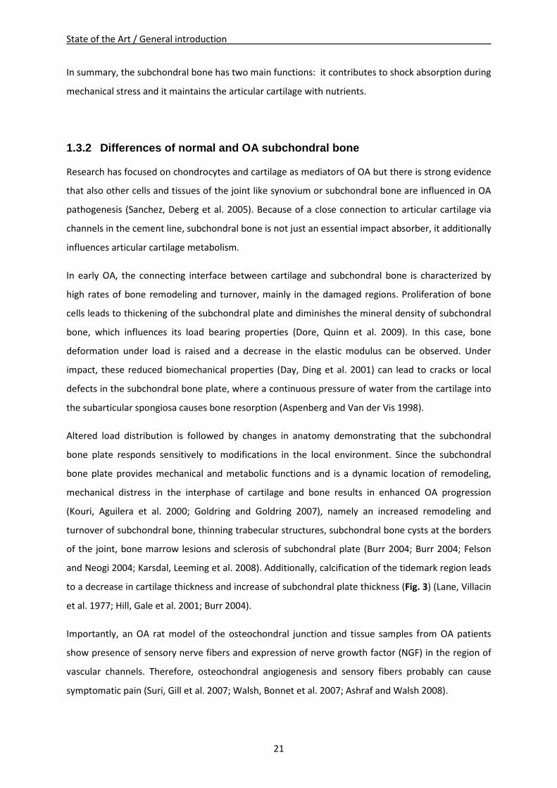
Taken together, each of both anatomically connected tissues - articular cartilage and subchondral
bone - is affected by changes in the mechanical properties of the other one (Aspenberg and Van der
Vis 1998; Van der Vis, Aspenberg et al. 1998). Therefore, OA has to be seen completely as a disease
of the whole joint and not separately of single tissues as it involves both: the articular cartilage and
the subchondral bone as shown in figure 3.
Figure 3: Anatomy of normal and osteoarthritic articular cartilage and subchondral bone
Normal human articular cartilage (left side) is separated by the tidemark from a thin zone of calcified cartilage.
Followed by subchondral bone, wich is connected via numerous canals with the calcified cartilage. In contrast,
osteoarthritic human articular cartilage (right side) shows clear signs of surface fibrillation and cartilage ECM
degradation, in detail, increase of cartilage calcification, duplication of the tidemark and enhanced vascular
invasion from subchondral bone (Drawing is adopted from (Goldring 2012)).
1.4 Biology of bone marrow derived mesenchymal stem cells (BMSC)
1.4.1 BMSC and their regenerative potential
There are in general two types of stem cells: embryonic stem cells and adult stem cells. The latter are
a rich source for a variety of repair cells, which are able to help out in case of tissue injuries and
replace damaged cells (Sylvester and Longaker 2004). They can be found in almost every tissue and
can differentiate into a broad range of specialized cells. Stem cells isolated from respective tissues
are predestined to differentiate into cells hosted along these lineages. In the bone marrow, even two
different types of stem cells coexist, in particular hematopoietic stem cells (HSC) and mesenchymal
State of the Art / General introduction
23
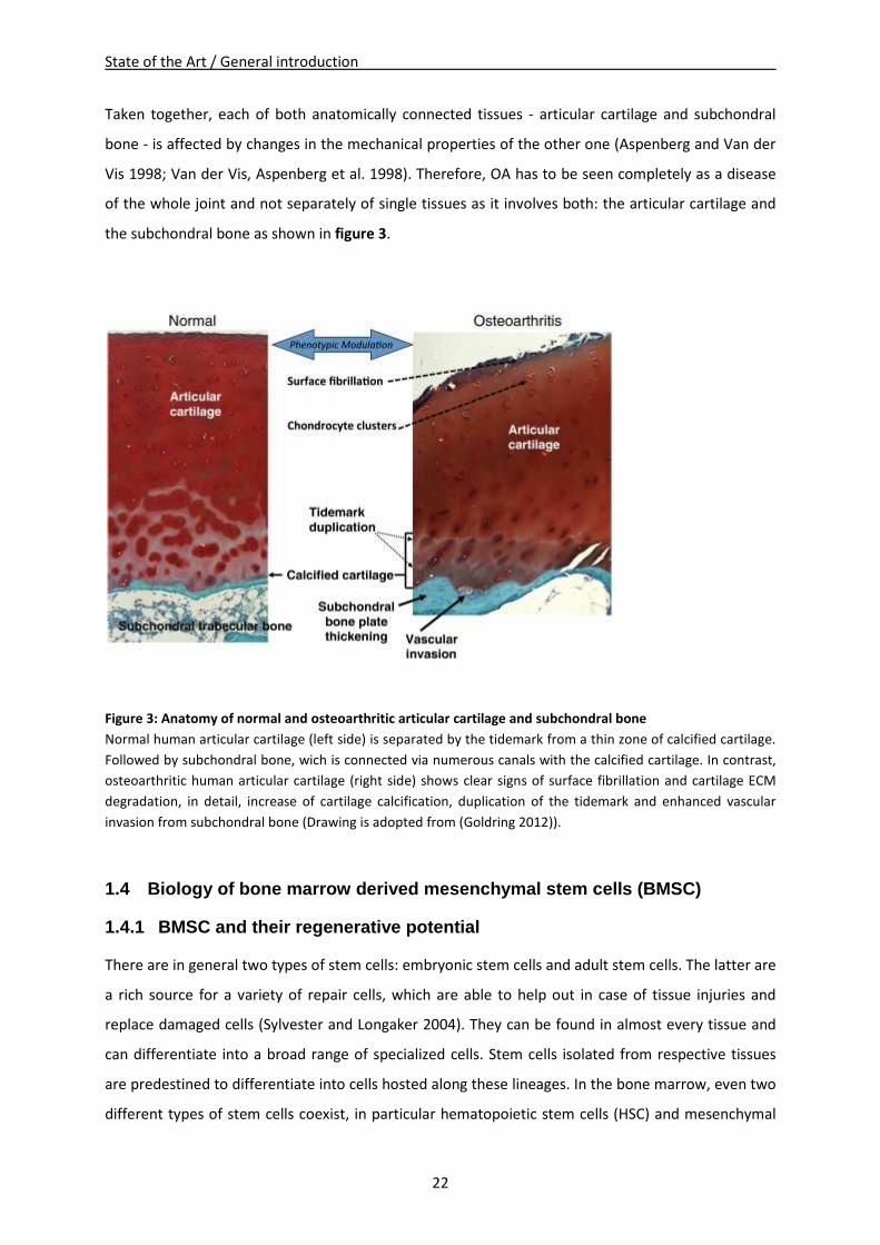
stem cells (MSC) (Sylvester and Longaker 2004). These adult bone marrow derived MSC (BMSC) are
capable for differentiation in vitro after an appropriate stimulation into several different lineages and
become amongst others adipocytes, osteoblasts or chondrocytes (Pittenger, Mackay et al. 1999; Kolf,
Cho et al. 2007) (Fig. 4).
Figure 4: Regenerative potential of MSC
MSC have the potential to differentiate into a broad range of specialized cells and form new tissues like bone
(osteocytes), cartilage (chondrocytes), muscle (myocytes), marrow stroma (marrow stromal cells),
tendon/ligament (tenocytes/ligament cells), fat (adipocytes) and connective tissues (connective tissue cells).
Growth factors and cytokines control differentiation into the different lineages. Therefore, therapeutically
approaches in regenerative medicine, which use adult BMSC are promising as they facilitate tissue repair and
regeneration of damaged tissue. (Drawing is adopted from (Caplan and Dennis 2006)).
The differentiation potential of BMSC, implanted into focal cartilage defects and their ability to
produce a proper ECM, which integrates closely with the adjacent normal tissue is highly dependent
on response of the BMSC to the tissue microenvironment. The microenvironment is provided by cell-
cell interactions with surface proteins of neighbor cells via integrins or CD44 and by cell-matrix
interactions with insoluble matrix molecules like collagens, glycoproteins and proteoglycans (Lutolf
and Hubbell 2005; Steinert, Ghivizzani et al. 2007). Additionally soluble molecules released from the
State of the Art / General introduction
24
neighboring tissue like growth factors (IGF I, TGF-β, FGF) (Denker, Nicoll et al. 1995), cytokines (IL-1,
TNFα) (van den Berg, Joosten et al. 1999) and chemokines (IL-8) are important for proper
differentiation (Garner, Stoker et al. 2011). Therefore, an important focus for successful
differentiation lies on the interaction of BMSC and their microenvironment. A better understanding
of these cues will facilitate regenerative therapies using adult BMSC, which have a promising future
in musculoskeletal tissue repair.
1.4.2 Chondrogenic differentiation of BMSC
Regulation of chondrogenic differentiation of BMSC and production of a typical hyaline cartilage
involves a variety of molecular mechanisms: stem cell intrinsic factors, paracrine factors derived from
neighboring cells like undifferentiated BMSC, osteoblasts or chondrocytes and microenvironmental
components independent from cells like availability of nutrients and oxygen, adhesion molecules,
ECM epitopes or a 3D surrounding (Coimbra, Jimenez et al. 2004; Goldring, Tsuchimochi et al. 2006;
Birmingham, Niebur et al. 2012). Additionally, chondrogenic differentiation of BMSC and deposition
of new ECM can be induced in vitro by stimulation with growth factors like BMPs or TGF-β
superfamily members in a controlled culture condition with accurately defined culture medium.
Stimulated BMSC start upregulation of chondrocyte specific genes like fibronectin, N-cadherin,
COMP, Collagen IX and during progressed differentiation also of typical articular proteins like
collagen II, aggrecan, hyaluronan and chondroadherin (Majumdar, Wang et al. 2001; Sekiya, Larson
et al. 2005; Park, Yang et al. 2009).
Molecular mechanisms during the tightly and complex regulated chondrogenic differentiation are
not yet fully understood. BMSC pass through several maturation stages in which characteristic
factors are activated. After initiation of chondrogenesis, BMSC start cell proliferation and
differentiate to chondrocytes under the influence of growth and differentiation factors, like bFGF or
bone morphogenetic protein (BMP). In early chondrogenesis, stem cells differentiate to resting
chondrocytes with the largest increase in expression of genes coding for cartilage ECM
macromolecules like proteoglycans, cartilage oligomeric matrix protein (COMP) and collagen II, IX
and XI. In case of hyaline cartilage, this type of chondrocyte is stably maintained in the joint
(Mastrogiacomo, Cancedda et al. 2001; Goldring, Tsuchimochi et al. 2006).
Further, interactions of transcription factors, such as Sox9 and Runx2 determine whether
chondrocytes remain articular or undergo terminal differentiation before ossification
(Mastrogiacomo, Cancedda et al. 2001; Goldring, Tsuchimochi et al. 2006). In embryonic
development, chondrocyte differentiation processes during endochondral ossification resulting in
State of the Art / General introduction
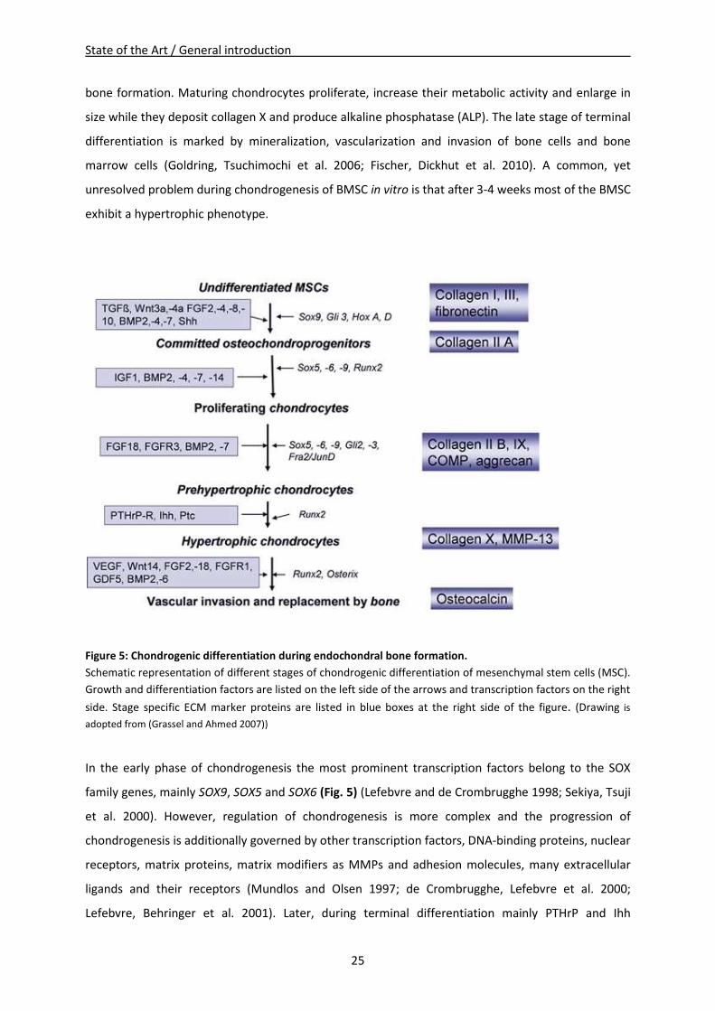
25
bone formation. Maturing chondrocytes proliferate, increase their metabolic activity and enlarge in
size while they deposit collagen X and produce alkaline phosphatase (ALP). The late stage of terminal
differentiation is marked by mineralization, vascularization and invasion of bone cells and bone
marrow cells (Goldring, Tsuchimochi et al. 2006; Fischer, Dickhut et al. 2010). A common, yet
unresolved problem during chondrogenesis of BMSC in vitro is that after 3-4 weeks most of the BMSC
exhibit a hypertrophic phenotype.
Figure 5: Chondrogenic differentiation during endochondral bone formation.
Schematic representation of different stages of chondrogenic differentiation of mesenchymal stem cells (MSC).
Growth and differentiation factors are listed on the left side of the arrows and transcription factors on the right
side. Stage specific ECM marker proteins are listed in blue boxes at the right side of the figure. (Drawing is
adopted from (Grassel and Ahmed 2007))
In the early phase of chondrogenesis the most prominent transcription factors belong to the SOX
family genes, mainly SOX9, SOX5 and SOX6 (Fig. 5) (Lefebvre and de Crombrugghe 1998; Sekiya, Tsuji
et al. 2000). However, regulation of chondrogenesis is more complex and the progression of
chondrogenesis is additionally governed by other transcription factors, DNA-binding proteins, nuclear
receptors, matrix proteins, matrix modifiers as MMPs and adhesion molecules, many extracellular
ligands and their receptors (Mundlos and Olsen 1997; de Crombrugghe, Lefebvre et al. 2000;
Lefebvre, Behringer et al. 2001). Later, during terminal differentiation mainly PTHrP and Ihh
State of the Art / General introduction
26
stimulate upregulation of collagen X, ALP and MMP13 in chondrocytes, which synthesize mineralized
matrix followed by vascular invasion and cell death at the end (Fig. 5) (Barry, Boynton et al. 2001;
Goldring, Tsuchimochi et al. 2006).
A better understanding of the mechanisms of chondrogenic differentiation and hypertrophy is
fundamental for further stem cell based therapies, because for a successful cartilage repair a stable
articular phenotype and prevention of terminal differentiation is essential. To obtain a high quality
functional regenerative tissue with superior mechanical load capacity, proper differentiated BMSC
which produce a highly organized functional ECM are required, as ECM components are strongly
correlated to mechanical strength of engineered cartilage-like tissue (Mauck, Yuan et al. 2006).
1.4.3 BMSC as trophic mediators
Moreover, BMSC are trophic mediators, which secrete a variety of cytokines and growth factors that
reflect their functional status in their specific microenvironment (Prockop 1997; Caplan and Dennis
2006; Kolf, Cho et al. 2007). These factors are able to feed back to the cell itself and govern the
functional status and physiology by inducing intracellular signaling or by instructing another cell in
the neighborhood to secrete functionally active factors (Caplan and Dennis 2006). A profound clinical
use of BMSC may be due to their trophic effects, when BMSC at the site of an injury produce
paracrine and autocrine acting factors that are known to suppress the local immune system,
stimulate angiogenesis, reduce scar formation and apoptosis and enhance mitosis and differentiation
of residing stem cells into regenerative tissue (Caplan and Dennis 2006). Additionally, the
therapeutical potential of BMSC is provided by paracrine secretion of cytokines and chemokines like
VEGF, FGF-2 or IL-6 via stimulation of survival pathways, induction of differentiation and stemness or
regulation of anti-inflammatory effects (Burdon, Paul et al. 2011).
A recent study by Pricola et al. affirmed that IL-6 is probably a “stemness” factor, which contributes
to the MSC undifferentiated status and that IL-6 is necessary for proliferation of MSC, protects from
apoptosis and inhibits adipogenic and chondrogenic differentiation in an ERK1/2 dependent pathway
(Pricola, Kuhn et al. 2009). BMSC used for therapy of chronically inflammatory diseases as OA have
two main functions: Firstly, they can either differentiate into chondrocytes or they can mediate
mitosis and differentiation of residing neighbor cells and secondly, BMSC can suppress apoptosis and
inflammatory processes by release of trophic factors.
State of the Art / General introduction
27
1.5 Cartilage repair and tissue engineering
1.5.1 Cartilage repair studies
Most tissues containing stem cells have an elaborated self-repair ambition, with exception of adult
articular cartilage. Damaged articular cartilage reveals only minimal or low self-repair capacity,
especially in defects of critical size and therefore it urgently requires potent tissue engineering-based
therapeutic methods (Caplan, Elyaderani et al. 1997; Cheng, Hardingham et al. 2014).
Lars Peterson and Mats Brittberg were clinical pioneers, who performed the first cell based approach
for treatment of full-thickness cartilage or osteochondral lesions via autologous chondrocyte
implantation (ACI) in 1994 (Brittberg, Lindahl et al. 1994). Until now, there appeared to be many
approaches with promising clinical results for cell based cartilage defect treatment of symptomatic
osteochondral defects. Examples are autologous mosaicplasty (Reverte-Vinaixa, Joshi et al. 2013),
matrix-assisted autologous chondrocyte transplantation (MACT) or microfracturing with or without
biomaterial support. For autologous mosaicplasty, ACI and MACT, cartilage is harvested from a
healthy non-weight bearing region of the joint and grafts or ex vivo expanded cells were re-
implanted into the defect. Drawbacks of these methods are limited cell and graft availability as well
as risk of donor morbidity (damage at the donor site) (Brittberg, Peterson et al. 2003; Bedi, Feeley et
al. 2010). During microfracturing, little cavities were created in the subchondral bone leading to
bleeding into the defect. Natural occurring stem cells and the developing fibrin-clot built the
fundament of defect repair. Anyhow, recent studies showed a risk for promotion of fibrocartilage
development by using this method (Harris, Siston et al. 2010; Holtzman, Theologis et al. 2010).
With increasing cell demand for regeneration of large damaged cartilage areas, chondrocytes need to
be expanded in vitro, but chondrocytes de-differentiate and their chondrogenic capacities to produce
stable cartilage ECM disappear (Mandelbaum, Browne et al. 2007). Hence, re-differentiation capacity
of chondrocytes is desirable and studies showed that a 3D environment like it is provided by scaffolds
or hydro gels is able to reactivate a chondrogenic phenotype after expansion (Endres, Neumann et al.
2012). An alternative autologous cell source to replace chondrocytes are chondrogenic differentiated
BMSC, which have a high chondrogenic differentiation potential triggered by growth factors and
cytokines (Caplan and Dennis 2006). Notably, regulatory obstacles in clinical use of BMSC-based
treatments are impeding progress of tissue engineering strategies for cartilage repair (Grassel and
Lorenz 2014).
Nevertheless, BMSC might be superior to expanded chondrocytes or cartilage grafts, as they are well
expandable in vitro, retain their differentiation capacity over several passages and are capable of
cartilage like matrix production in a proper environment. As a result, therapeutically approaches in
State of the Art / General introduction
28
regenerative medicine which use adult BMSC, would be promising as they might facilitate tissue
repair and regeneration of damaged cartilage.
1.5.2 Biomaterials and fibrin in tissue engineering
Interaction between BMSC and ECM components provides an instructive microenvironment and
contributes to differentiation of BMSC into chondrocytes. This suggests a beneficial effect by using
BMSC in conjunction with synthetic or natural scaffolds, which provide a 3D environment (Lutolf and
Hubbell 2005; Lee, Yu et al. 2008). Additionally, a conductive effect of a 3D culture is given for
chondrocytes, which maintain their articular character instead of de-differentiating in monolayer
(Schagemann, Mrosek et al. 2006; Francioli, Candrian et al. 2010).
Several studies proved the suitability of different 3D culture models for cartilage repair, like collagen
or gelatin scaffolds (George, Kuboki et al. 2006; Brochhausen, Sanchez et al. 2013) and hydrogels
including photopolymerized hyaluronic acid, agarose (Williams, Kim et al. 2003), Matrigel, PuraMatrix
(Dickhut, Gottwald et al. 2008) or alginate (Ho, Cool et al. 2010).
A commonly used hydrogel scaffold is based on fibrin (fibrin gel or fibrin glue) that encapsulates
BMSC or chondrocytes and mimics the structure and function of a natural ECM by maintaining a
round morphology of cells and thus facilitating deposition of new ECM components (Dickhut,
Gottwald et al. 2008; Ho, Cool et al. 2010). Fibrin is a natural non-cytotoxic, biocompatible and
biodegradable polymer that can be smoothly polymerized and casted from its basic constituents and
has been in clinical use for several years. A big advantage of fibrin gel is a close contact of the
implanted BMSC/chondrocytes to the defect tissue borders, as it can directly integrate. Additionally,
various in vivo and in vitro studies demonstrate the potential of fibrin gels in the field of tissue
engineering together with human or animal cells. The benefit of combining cells with fibrin is well
established for cartilage regeneration, since numerous studies managed to successfully repair
defects in animal models. For example, Fussenegger et al. accomplished to reconstruct cartilage in a
rabbit-model using stabilized autologous fibrin-chondrocyte constructs (Fussenegger, Meinhart et al.
2003), Sims et al. produced a well formed cartilaginous matrix using bovine chondrocytes suspended
in fibrin glue in vivo (Sims, Butler et al. 1998) and Mesa et al. demonstrated that ovine articular
chondrocytes from 8 year old sheep can be rejuvenated in vivo when encapsulated in fibrin gel
(Mesa, Zaporojan et al. 2006). Moreover, fibrin constructs containing rabbit BMSC which were
provided with constant levels of growth factors during culture time enable the development of
articular cartilage in vitro and in vivo (Park, Yang et al. 2009). Even a human pilot study using fibrin-
ACI reports good clinical improvement 2 years after surgery with a favorable clinical outcome and
State of the Art / General introduction
29
regenerated tissue shows evidence for similar characteristics to normal cartilage (Kim, Choi et al.
2010).
Depending on the experimental setup, some studies show that fibrin is degraded too fast
accompanied with extensive shrinkage or replacement by fibrous tissue (Homminga, Buma et al.
1993; van Susante, Buma et al. 1999), while other groups were successful with cartilage
reconstruction (Ting, Sims et al. 1998). Degradation of fibrin can be slowed by addition of high
concentrations of antifibrinolytic substances like aprotinin and tranexamic acid or by elevating the
fibrinogen/thrombin ratio causing alterations in the crosslinking during polymerization (Fussenegger,
Meinhart et al. 2003).
Taken together, a 3D environment, as is provided by biodegradable porous scaffolds or hydro gels
like fibrin gel, positively contributes to chondrogenic differentiation capacity and ECM production of
BMSC and is beneficial for cartilage neogenesis (Williams, Kim et al. 2003; Erickson, Huang et al.
2009).
1.5.3 Coculture models in tissue engineering
1.5.3.1 Microenvironment of cartilage and chondrocy tes
For long-term repair and regeneration of focal cartilage defects, chondrocytes or BMSC are
implanted at the site of injury, however, not much attention has been paid to microenvironmental
effects of neighboring cartilage or subchondral bone. This is specifically evident in diseases affecting
diarthrodial joints as osteoarthritis (OA). The differentiation potential of BMSC, implanted into focal
cartilage defects and their ability to produce a proper and stable ECM, which integrates closely with
the adjacent normal tissue is highly dependent on response of the BMSC to the local tissue
microenvironment provided by cell-cell and cell-matrix interactions and factors released from the
neighboring tissue. Soluble and insoluble factors are working together to regulate cell commitment
and tissue morphogenesis in native tissue (Lutolf and Hubbell 2005; Steinert, Ghivizzani et al. 2007;
Leyh, Seitz et al. 2014 a).
Moreover, it has been demonstrated that BMSC are able to differentiate into a specific cell type,
depending on the environment they are actually residing (Djouad, Delorme et al. 2007). Crosstalk
between BMSC and cartilage ECM components could be a strongly determining factor for the
differentiation of BMSC into chondrocytes. These interactions provide an instructive
microenvironment suggesting a beneficial effect on differentiation of BMSC into chondrocytes (Lutolf
and Hubbell 2005; Lee, Yu et al. 2008). In addition, normal articular rat cartilage microenvironment
State of the Art / General introduction
30
enhances chondrogenic differentiation capacity of rat BMSC and leads to a higher collagen and
glycosaminoglycan content in the ECM while at the same time it prevents hypertrophic
differentiation (Ahmed, Dreier et al. 2007).
However, the effect of diseased cartilage and OA-chondrocytes on chondrogenic differentiation of
BMSC is poorly understood. OA-chondrocytes secrete factors, such as pro-inflammatory cytokines
and chemokines that are believed to have a negative effect on locally residing stem cells and inhibit
cartilage repair in vivo. OA related cartilage lesions and fissures release high amounts of ECM
degradation products like fragments of collagen, fibronectin or GAGs. So far, they have not been
target for BMSC-based therapies, as this would imply to implant cells into the neighborhood of
diseased tissue, where they are confronted with an altered microenvironment of the neighboring
cartilage and subchondral bone tissue (Aung, Gupta et al. 2011).
Articular cartilage is unique because residing chondrocytes express a stable chondrogenic phenotype
whereas in the growth plate or during endochondral ossification chondrocytes underwent terminal
differentiation and hypertrophic cells are replaced by bone due to ossification of cartilage (Goldring,
Tsuchimochi et al. 2006).
Recent studies demonstrate the ability of paracrine factors released by articular cartilage tissue or
articular chondrocytes to induce chondrogenesis of BMSC and to suppress terminal differentiation
and matrix calcification of growth plate chondrocytes in vitro and might stabilize articular
differentiation of BMSC. Candidate factors are for example FGF-2, TGF-β or PTHrP (Bohme,
Winterhalter et al. 1995; Jikko, Kato et al. 1999). Studies analyzing the effect of chondrocyte
coculture on spontaneous chondrogenic differentiation of BMSC also show positive effects on
collagen II gene and protein expression even without exogenous TGF-β supplementation, suggesting
secreted factors that induce chondrogenic differentiation in BMSC (Vats, Bielby et al. 2006; Ahmed,
Dreier et al. 2007; Lettry, Hosoya et al. 2010). However, the identity of these factors needs to be
determined yet in order to develop protocols, which allow stabilization of the chondrogenic
phenotype of BMSC and to reduce their tendency to undergo hypertrophy. Until now, numerous
scientists have set up many different coculture studies using chondrocytes and BMSC to enlighten
the accurate mechanism for chondrogenic induction and to find the best protocol for generation of a
stable articular chondrocyte phenotype.
Interestingly, the ratio of cocultured BMSC and articular chondrocytes regulate whether
differentiation proceeds towards a cartilaginous or osseous phenotype. For example, culturing
articular chondrocytes with BMSC in a 2:1 ratio induces both phenotypes simultaneous in a 3D-
alginate hydrogel construct indicating that chondrocytes provide the necessary factor(s) or cell
State of the Art / General introduction
31
contact in both processes (Mo, Guo et al. 2009). Moreover, Bian et. al found that mixed cell
populations of MSC and chondrocytes (4:1) reveal higher total collagen content than constructs of
only one population, while collagen X deposition was significantly lower in the mixed populations
than in MSC alone. Notably, culture of separate cell populations together in the same well did not
improve biomechanical or biochemical properties, implying that close proximity is necessary (Bian,
Zhai et al. 2011). This is in line with observations of Kang et al. where coculture of BMSC and
chondrocytes in same ratios leads to newly engineered cartilage with the densest elastic fibers and
highest Young's modulus as well as to cartilage matrix related gene expression profiles (Kang, Liu et
al. 2012). Finally, Fischer et al. found that even conditioned medium from chondrocytes leads to
significantly reduced expression ratios and deposition of collagen X relative to collagen II. This means
direct coculture and soluble factors from chondrocytes can improve chondrogenesis and are able to
prevent hypertrophy of BMSC (Fischer, Dickhut et al. 2010).
Taken together, there are many aspects, which manipulate the influence of chondrocytes on BMSC
differentiation. The mixing ratio of both cell types, the coculture time and even the kind of coculture
– direct, indirect or via conditioned medium – act together creating an unique condition for BMSC
fate.
1.5.3.2 Microenvironment of subchondral bone and bo ne cells
Bone is a central organ of the body: it supports structural stability, carries mechanical load, provides
hostage for hematopoiesis, is a depot for minerals and regulates tissue development, remodeling
and repair. Several cell types with different functions, namely osteoblasts, osteocytes, osteoclasts,
and due to the bone marrow HSC and BMSC, are residing in the subchondral bone. Several studies
including these bone cells suggest that they are not only responsible for bone formation and
turnover but also have regulatory paracrine functions. Therefore, an influence on metabolism,
differentiation and ECM production of neighboring cells like chondrocytes or BMSC is likely. The most
prominent bone cell type used for cartilage or chondrocyte coculture studies are osteoblasts.
For example, Westacott et al. shows that subchondral osteoblasts are able to modulate the
metabolism of chondrocytes and can induce phenotypic shifts in OA-chondrocytes (Westacott, Webb
et al. 1997). In vitro cocultures of OA-chondrocytes with OA-osteoblasts reveal increased IL-6 and IL-8
levels, induced inhibition of aggrecan production and a concomitant significant increase in MMP13
synthesis in chondrocytes. Cocultured osteoblasts also decrease COL2A1, SOX9 and PTHrP/PTH-
receptor gene expression in chondrocytes, which was considered as a phenotypic shift towards the
hypertrophic stage (Jiang, Nicoll et al. 2005; Sanchez, Deberg et al. 2005; Sanchez, Deberg et al. 2005;
State of the Art / General introduction
32
Sanchez, Deberg et al. 2008). Further, Lin et al. show that articular chondrocytes cocultured with
osteoblasts reveal a phenotypic shift to hypertrophic chondrocytes and that this effect is enhanced
by using mechanically stressed ostoblasts. These results indicate that osteoblasts can cause
alterations in cartilage metabolism and provide a possible explanation for the onset and progression
of OA (Lin, Tanaka et al. 2010). Bianco et al. even suggest that hypertrophic chondrocytes are able to
become osteoblast-like cells, but solely if they reside close to the osteogenic tissue border (Bianco,
Cancedda et al. 1998). In contrast, a study from Jiang et al. revealed negative effects of chondrocyte
derived factors on osteoblast mineralization in coculture, leading to reduced biomechanical
properties of newly produced ECM (Jiang, Nicoll et al. 2005). These studies suggest that there are
regulatory influneces of osteoblasts concerning chondrocyte metabolism and phenotype. Maybe also
a cooperation of signals from both cell types are necessary in a variety of normal processes and both
are inevitable for regeneration of cartilage and mineralized tissue (Nakaoka, Hsiong et al. 2006).
Indeed, soluble factors are exchanged from subchondral bone and articular cartialge via diffusion
through channels or vascular invasion (Burr and Radin 2003), what could explain the influence of
subchondral bone factors on distant articular cartilage (Amin, Huntley et al. 2009; Pan, Zhou et al.
2009; Findlay 2010), and suggests that residing cells of both tissues are able to influence each other
(Guevremont, Martel-Pelletier et al. 2003; Sanchez, Deberg et al. 2005). Presence of such factors is
postulated by several groups, which report an altered phenotype of OA-osteoblasts compared with
normal osteoblasts, including enhanced secretion of ALP, IL-6 and TGF-β1 (Mansell, Tarlton et al.
1997; Massicotte, Lajeunesse et al. 2002; Neilson, White et al. 2004).
Anyhow, though there are several coculture studies with osteoblasts concerning their influence on
chondrocytes and their matrix generation, studies with osteoblasts and BMSC under chondrogenic
differentiation are marginal. Moreover, the role of osteocytes or osteoclasts in chondrogenesis of
BMSC or their influence on chondrocytes is mostly still unknown. However, osteoclasts are reported
to secrete TNF and other para- and outocrine cytokines, such as IL-6 and IL-1 contributing to
inflammatory processes of the bone (Boyce, Yao et al. 2009). Therefore, also osteoclasts might
contribute to communication of subchondral bone with chondrocytes or BMSC via secreted factors.
Further, not only bone cells but also subchondral bone matrix itself is likely to provide regulatory
features. Especially in case of OA, the increased turnover of subchondral bone leads to an elevated
release of matrix bound factors into the microenvironment (Canalis, McCarthy et al. 1988). For
example, demineralized bone matrix is known to contain factors like BMP-1 to BMP-7, TGF-β1 and
TGF-β2, acidic fibroblast growth factor (FGF), basic FGF and insulin-like growth factor- I (IGF-I), which
possibly mediate induction of chondrogenesis in MSC in vitro (Syftestad and Caplan 1984; Denker,
Nicoll et al. 1995). There is also evidence of diffusion permitting the exchange of soluble substances,
State of the Art / General introduction
33
Thus, the microenvironment of OA-subchondral bone has likely an influence on BMSC differentiation
and matrix generation capacities, as implanted stem cells may respond to specific ECM components
and present regulatory factors. To analyze the differentiation of BMSC in this special surrounding, a
coculture system that promotes diffusion of soluble and paracrine factors is needed (Grassel and
Ahmed 2007; Hwang, Varghese et al. 2008; Bian, Zhai et al. 2011).
Taken together, bone cells should not only be seen as structural mediators but also as trophic
mediators creating a special microenvironment for chondrocytes. In vitro coculture of osteoblasts
and chondrocytes even suggests reciprocal instructive interactions of both cell types. Thus indicating
the essential role of subchondral bone for maintaining articular cartilage and joint homeostasis, the
role of normal and OA-subchondral bone for molecular regulatory mechanisms and signaling
pathways leading to BMSC differentiation are still unidentified.
1.6 Animals in cartilage trauma repair models
1.6.1 Animal models in cartilage repair
Experiments with normal cartilage are very important for a more detailed knowledge of the influence
of unaffected articular cartilage tissue on BMSC differentiation and the differences that occur in OA-
cartilage. Unfortunately, normal human cartilage is hard to obtain for ethical reasons. In this case,
mammalian animal models provide good requisites to investigate cartilage trauma because many
aspects like the genomic, cellular and immunologic structure are close to the human system (Schulze-
Tanzil, Muller et al. 2009). Therefore, animal tissues and cells are an alternative, not only for in vivo,
but also for in vitro studies. However, some aspects have to be considered before adopting results
obtained from animal studies to the human system. Humans and animals have a different
evolutionary background, sometimes resulting in differences in genetic and proteomic regulation
(Greek and Menache 2013). For example, studies concerning responsiveness on BMPs to induce
chondrogenesis are contradictory in different species partly because of variations in distinctive
receptors (Osyczka, Diefenderfer et al. 2004; Hennig, Lorenz et al. 2007). However, in most cases
mammalian animal models correspond to physiological processes and mechanisms in humans.
Especially in the field of cartilage tissue engineering differences in anatomy of the joint like size, load
and weight distribution as well as features like cartilage thickness are essential. Additionally,
distribution of load is important because commonly used animals are quadruped and not biped like
humans (McLure, Fisher et al. 2012). Further, Stockwell et al. show that human articular cartilage is
thicker compared with most animals, were it increases proportional to body mass of mouse, rat, cat,
State of the Art / General introduction
34
rabbit, dog, sheep, man, and cow (Stockwell 1971). Therefore, in vivo studies with large animal
models like sheep or swine are probably more suitable for comparison with human than studies with
small animal models like rat or rabbit. Interestingly the cellular density decreases with increasing
body mass, but biochemical composition of cartilage is surprisingly constant. Therefore, small
animals with a high cell density have a benefit in natural cartilage repair in vivo (Malda, de Grauw et
al. 2013).
Additionally, Taylor et al. show species-specific differences in re-differentiation potential of passaged
chondrocytes for bovine chondrocytes compared with human chondrocytes (Taylor, Ahmed et al.
2010). Subsequently, choice of cell species is as important as age of the donor and culture conditions
for further interpretation of animal coculture results (Otsuki, Grogan et al. 2010). Some studies even
successfully use xenogeneic human BMSC for treatment of an animal recipient. Lack of immune
reaction to the xenogeneic regenerated cartilage is possibly due to immunosuppressive activities of
BMSC (Li, Chiang et al. 2009).
Taken together, in vivo cartilage defect repair is successful in a variety of animal studies like goat,
sheep or swine using embedded BMSC, suggesting a future use for minimally invasive implantation
and engineering of novel cartilage like tissue for humans (Williams, Kim et al. 2003; Li, Chiang et al.
2009; Zscharnack, Hepp et al. 2010).
1.6.2 Ovine cartilage repair models
Ovine cartilage models are a very potent and often used tool in biomedical research for studying
normal articular cartilage tissues in large animals. Ovine BMSC (oBMSC) share similar properties with
human BMSC like capability for growth factor induced proliferation or differentiation into multiple
stromal lineages and they express stem cell associated surface markers (McCarty, Gronthos et al.
2009).
Usually, studies use ovine cartilage or animal models to study the treatment of osteochondral
defects in vivo or in vitro with re-implantation of autologous BMSC into the defect (McLure, Fisher et
al. 2012). For example, Mrugala et al. established an ovine patella defect model and embedded
BMSC into a chitosan matrix resulting in a cartilage like tissue successfully filling the defect after 9
weeks (Mrugala, Bony et al. 2008). In another study, ex vivo chondrogenic pre-differentiated
BMSC/collagen I gel constructs are implanted into an ovine osteochondral defect model. MACT using
pre-differentiated MSC shows potential for repair of chronic osteochondral defects including
collagen II deposition and chondrogenic characteristic of cells (Zscharnack, Hepp et al. 2010).
State of the Art / General introduction
35
Several in vitro and in vivo studies prove usage of ovine cells embedded in hydrogel - especially fibrin
for successful cartilage repair. For example re-differentiation potential of de-differentiated expanded
ovine chondrocytes in PGA-fibrin scaffolds show formation of cartilaginous matrix accompanied by
induction of collagen II and aggrecan (Endres, Neumann et al. 2012). Munirah et al. reveal that
implantation of autologous chondrocyte-fibrin constructs results in hyaline-like cartilage
regeneration in vivo (Munirah, Samsudin et al. 2007). Finally, Schagemann et al. conclude that a 3D
hydrogel culture keeps chondrocytes in a differentiated status and offers a chondrogenic
environment for in vitro cartilage regeneration (Schagemann, Mrosek et al. 2006).
Taken together, normal ovine tissue and chondrocytes or BMSC embedded in a 3D fibrin gel seem to
be a suitable alternative to normal human tissue. To work out the differences between factors
released from normal or diseased human cartilage it is crucial to use a suitable cartilage defect
model. An ovine model appears to be a good choice and allows realistic conclusions that might be
crucial for further progress in cartilage tissue engineering.
Aim of the thesis
36
2 Aim of the thesis
OA is a painful joint degenerative disease, which is characterized by degradation of articular
cartilage. As an alternative treatment for implantation of prosthesis, the use of tissue engineering for
cartilage regeneration systems is rising. Scientists are keen on finding a cell-based therapy, which
succeeds in production of high quality cartilage like tissue and finding a source that provides a
sufficient number of hyaline ECM building cells with a stable articular phenotype including
prevention of donor morbidity. To date mainly two cell types are suitable for such a regeneration
approach: adult stem cells with chondrogenic differentiation potential and articular chondrocytes. So
far, an optimal therapy method was not found though several studies established suitable treatment
options (see 1.1.2). Clinical and experimental trials with a 3D surrounding provided by a biomaterial
in combination with cells gave best results. However, drawbacks like inadequate quality of
regeneration tissue, unstable articular phenotype resulting in hypertrophy of BMSC or simply an
insufficient number of donor chondrocytes prevented the standard usage of tissue engineering for
cartilage defect treatment. Further, also regulatory obstacles in clinical use of BMSC-based
treatments are impeding progress of tissue engineering strategies for cartilage repair.
In the present study, the aim was to find answers, why treatment of articular cartilage defects –
especially OA defects – with cells embedded in a degradable biomaterial fails or does not develop the
supposed hyaline cartilage like tissue.
Therefore, focus of this study is set on the following goals:
• Establishment of a reproducible in vitro paracrine coculture model, which allows to
investigate the influence of soluble factors from normal and OA-tissue explants on matrix
forming capacity of adult ASC, BMSC and articular chondrocytes as well as of mixed cultures
(BMSC and chondrocytes in equal ratio).
• Gain of a better understanding of mechanisms for BMSC differentiation and generation of a
stable articular phenotype and a better integration and quality of regenerated tissue.
• Examination of the effects induced by articular cartilage or subchondral bone explants on
ASC and BMSC differentiation, phenotype of articular chondrocytes and the biochemical and
biomechanical properties of their newly formed ECM.
• Analysis of the effects induced by a mixed culture of BMSC and chondrocytes (in equal ratio)
in respective explant cocultures on BMSC differentiation, phenotype of articular
chondrocytes and the biochemical and biomechanical properties of their newly formed
ECM.
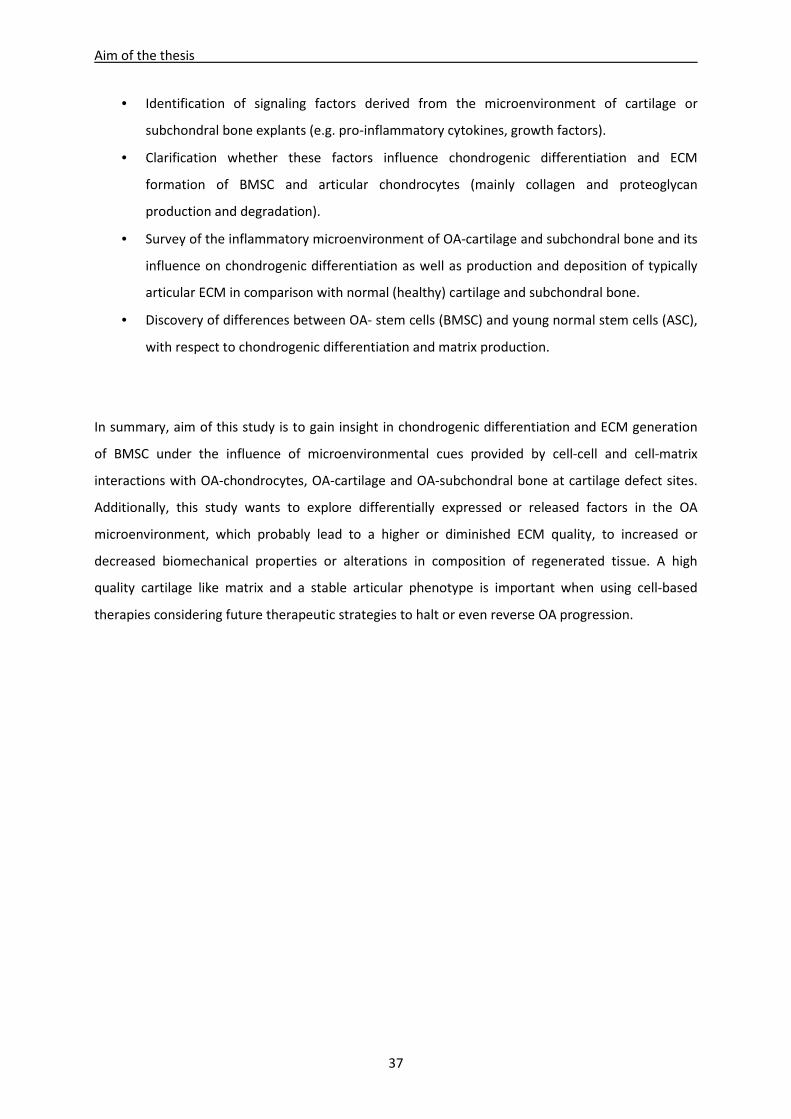
Aim of the thesis
37
• Identification of signaling factors derived from the microenvironment of cartilage or
subchondral bone explants (e.g. pro-inflammatory cytokines, growth factors).
• Clarification whether these factors influence chondrogenic differentiation and ECM
formation of BMSC and articular chondrocytes (mainly collagen and proteoglycan
production and degradation).
• Survey of the inflammatory microenvironment of OA-cartilage and subchondral bone and its
influence on chondrogenic differentiation as well as production and deposition of typically
articular ECM in comparison with normal (healthy) cartilage and subchondral bone.
• Discovery of differences between OA- stem cells (BMSC) and young normal stem cells (ASC),
with respect to chondrogenic differentiation and matrix production.
In summary, aim of this study is to gain insight in chondrogenic differentiation and ECM generation
of BMSC under the influence of microenvironmental cues provided by cell-cell and cell-matrix
interactions with OA-chondrocytes, OA-cartilage and OA-subchondral bone at cartilage defect sites.
Additionally, this study wants to explore differentially expressed or released factors in the OA
microenvironment, which probably lead to a higher or diminished ECM quality, to increased or
decreased biomechanical properties or alterations in composition of regenerated tissue. A high
quality cartilage like matrix and a stable articular phenotype is important when using cell-based
therapies considering future therapeutic strategies to halt or even reverse OA progression.
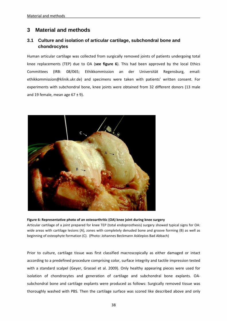
Material and methods
38
3 Material and methods
3.1 Culture and isolation of articular cartilage, s ubchondral bone and chondrocytes
Human articular cartilage was collected from surgically removed joints of patients undergoing total
knee replacements (TEP) due to OA (see figure 6). This had been approved by the local Ethics
Committees (IRB: 08/065; Ethikkommission an der Universität Regensburg, email:
[email protected]) and specimens were taken with patients’ written consent. For
experiments with subchondral bone, knee joints were obtained from 32 different donors (13 male
and 19 female, mean age 67 ± 9).
Figure 6: Representative photo of an osteoarthritic (OA) knee joint during knee surgery
Articular cartilage of a joint prepared for knee TEP (total endoprosthesis) surgery showed typical signs for OA:
wide areas with cartilage lesions (A), zones with completely denuded bone and groove forming (B) as well as
beginning of osteophyte formation (C). (Photo: Johannes Beckmann Asklepios Bad Abbach)
Prior to culture, cartilage tissue was first classified macroscopically as either damaged or intact
according to a predefined procedure comprising color, surface integrity and tactile impression tested
with a standard scalpel (Geyer, Grassel et al. 2009). Only healthy appearing pieces were used for
isolation of chondrocytes and generation of cartilage and subchondral bone explants. OA-
subchondral bone and cartilage explants were produced as follows: Surgically removed tissue was
thoroughly washed with PBS. Then the cartilage surface was scored like described above and only
Material and methods
39
justly evaluated pieces were accepted for usage. Cartilage was cut off for isolation of chondrocytes
and preparation of cartilage explants. The completely denuded bone was used for generation of
explants of 8mm in diameter and 4mm height. Cartilage explants were punched out (8 mm2 x 2 mm;
Biopsy punch, Stiefel GmbH, Munich, Germany) from intact cartilage slices including the superficial
zone (Fig.7). Chondrocytes were isolated from cartilage slices upon overnight digestion with
collagenase II (PAA, Piscataway/ New Jersey, USA) at 37°C. Isolated cells were resuspended in
Dulbecco’s modified Eagle´s medium (DMEM, Gibco Invitrogen, UK) containing 10% fetal calf serum
(FCS, Sigma-Aldrich, St. Louis/ Missouri, USA) and 1% penicillin/streptomycin (P/S, PAA, Piscataway/
New Jersey, USA). Chondrocytes were kept in monolayer in an initial density of 17.000 cells per cm2
cultured in a humidified 37°C / 5% CO2 incubator for 7 to 14 days and were used when confluent
(passage 1) (Dreier, Grassel et al. 2004).
Figure 7: Preparation of cartilage explants from OA-knees
Knee joints were obtained from different donors and taken with patients’ written consent (A). Cartilage tissue
was classified macroscopically according to a predefined procedure comprising color, surface integrity and
tactile impression tested with a standard scalpel (B). Intact cartilage slices including the superficial zone were
cut off and denuded subchondral bone was prepared (C) either for cell isolation or for coculture setups.
Cartilage explants were punched out from cartilage slices including the superficial zone (D-F).
Material and methods
40
Normal human cartilage and subchondral bone tissue (2 male and 2 female, mean age 35 ± 11) was
received from knee joints of rare trauma affected patients treated for sports accidents. The tissue
used in this study was not injured directly but was collected during the surgical treatment from
another non-affected region of the same joint and explants were prepared like described above.
Normal ovine articular cartilage was prepared from surgically removed joints of sheep euthanized for
unrelated experiments obtained from Dr. med. vet. Uta Delling (University of Leipzig, Germany,
Faculty of Veterinary Medicine, Large Animal Clinic for Surgery) or from slaughterhouse waste. Ovine
chondrocytes (oCh) and ovine cartilage explants were prepared according to the protocol of human
chondrocyte isolation.
3.2 Culture and isolation of mesenchymal stem cells (BMSC) and adipose-derived stem cells (ASC)
Human BMSC used for this study have either been purchased from Lonza (Basel, Switzerland) or
isolated from bone marrow aspirates obtained from patients (14 male and 16 female, mean age
61 ± 8) undergoing a hip replacement surgery due to OA. This had been approved by the local Ethics
Committee (IRB 08/065; Ethikkommission an der Universität Regensburg, email:
[email protected]) and specimens were taken with patients’ written consent. The bone
marrow was centrifuged and cells were fractionated on a density-gradient (Biocoll Separating
Solution, Biochrom, Berlin, Germany). The low-density cell-fraction concentrated in the interphase
(“buffy coat”) was washed, seeded in cell culture flasks supplied with MesenchymStem Medium
(PAA, Piscataway/ New Jersey, USA) supplemented with Mycozap (Lonza, Basel, Switzerland) and
non-adherent cells were removed after 5 to 7 days. Adherent cells were cultured until they reached
approximately 80% confluence. After splitting, BMSC were seeded at a density of 4 x 10⁴ cells per cm2
and kept in culture for up to three passages before they were used for experiments.
oBMSC were obtained from Dr. med. vet. Uta Delling (University of Leipzig, Germany, Faculty of
Veterinary Medicine, Large Animal Clinic for Surgery). Briefly, oBMSC were harvested from ovine
bone marrow aspirated from iliac crest. Cells were isolated and cultured as described above followed
by cryo-preservation at passage 0. After thawing, cells were seeded at a density of 4 x 10⁴ cells/cm2
in cell culture flasks, supplied with low glucose DMEM (PAA, Piscataway/ New Jersey, USA)
supplemented with 1% P/S (PAA, Piscataway/ New Jersey, USA), 10% FCS (Sigma-Aldrich, St. Louis/
Missouri, USA) and ascorbat 12.5 mg/mL (PAA, Piscataway/ New Jersey, USA) and kept in culture for
up to three passages before they were used for experiments.
Material and methods
41
Adipose-derived stem cells (ASC, isolated as described previously by Gehmert et. al.) (Gehmert,
Hidayat et al. 2011) were a friendly gift from the Department of Otorhinolaryngology, Division of
Facial Plastic Surgery, University of Regensburg. Five different patients were used for our coculture
setup (female, age < 50). Written consent from the local Ethics Committee to harvest ASC from
patients was obtained from the Institutional Review Board (IRB) of the University of Regensburg (IRB:
08/117; Ethikkommission an der Universität Regensburg, email: [email protected])
Briefly, the ASC were isolated from subcutaneous fat tissue, which was obtained from patients
undergoing elective body-contouring procedures. Fat tissue was washed, minced and digested in
serum-free MEM (1 mL/1 g tissue) with LiberaseBlendzyme 3 (2U/1g tissue; Roche Diagnostics, Basel,
Switzerland) at 37°C for 45 minutes. The lysate was filtered (100 and 40 μm filters; Fisher Scientific,
Schwerte, Germany) and centrifuged at 450g for 10 minutes. The cell pellet was washed twice with
Hanks’ balanced salt solution (Cellgro, Manassas, VA, USA), cells were seeded in culture vials (Greiner
Bio-one, Frickenhausen, Germany) and daily washed to remove unwanted red blood cells or non
adherent cells. After reaching a confluence of 80%, ASC were seeded at a density of 3000 cells/cm2,
maintained in ASC medium (αMEM containing 20% FBS, 2 mM L-glutamine and 1%
penicillin/streptomycin, Sigma, St. Louis, MO, USA) and used for experiments at passage 5.
ASC were characterized in accordance with guidelines of the Declaration of Helsinki for biomedical
research from the Applied Stem Cell Research Center of the University of Regensburg.
3.3 Flow cytometric characterization of BMSC and di fferentiation into different lineages
Plastic adherent BMSC at passage 3 were trypsinized, centrifuged and the cell pellet was
resuspended into blocking buffer (PBS with 0.1% sodium azide, 5% FCS and 10%FcR Blocking
Reagent) at a concentration of 106 cells/mL. After blocking for 15 minutes at 4°C the cell suspension
was stained with a conjugated (40% CD19-PE, 10% CD105-FITC), or a unconjugated (2% CD34, 2%
CD44) primary antibody or with corresponding isotype controls (40% IgG1-FITC, 40% IgG1-PE, 20%
IgG2a and 20% IgG1) in 50 µl blocking buffer (Table 1) for 15 minutes on ice in the dark. In case of
unconjugated primary antibodies (CD34, CD44, IgG2a and IgG1) the two times washing step with
FACS buffer (PBS containing 0.1% sodium azide and 5% BSA, Sigma-Aldrich, St. Louis/ Missouri, USA)
was followed by incubation with an immunofluorescent secondary antibody Alexa 568 or Alexa 488
1:400 in blocking buffer for 30 minutes on ice. After washing, the cells were resuspended in 250 µl
FACS buffer and analysed by FACScanto® (Becton Dickinson, USA) linked with the CellQuest 3.1 data
Material and methods
42
acquisition software (Becton Dickinson, Franklin Lakes, New Jersey, USA). Data analysis was
performed with FlowJo 7.6.5 software (Tree Star Inc., Ashland, Oregon, USA).
To demonstrate pluripotency of BMSC, 2 x 10⁵ BMSC were differentiated in a 6-well in adipogenic or
osteogenic medium for 21 days. Adipogenic medium consisted of DMEM F-12 (Gibco Invitrogen, UK)
supplemented with 10% FCS (Sigma-Aldrich, St. Louis/ Missouri, USA), 1%P/S (PAA, Piscataway/ New
Jersey, USA), 0.1% dexamethasone (Sigma-Aldrich, St. Louis/ Missouri, USA), 0.1% 3-Isobutyl-1-
methylxanthin (IBMX, Sigma-Aldrich, St. Louis/ Missouri, USA) and 0.001% insulin (Sigma-Aldrich, St.
Louis/ Missouri, USA). Osteogenic medium consisted of DMEM high glucose (Gibco Invitrogen, UK)
supplemented with 10% FCS (Sigma-Aldrich, St. Louis/ Missouri, USA), 1% P/S (PAA, Piscataway/ New
Jersey, USA), 0.1% Na-β-glycerophosphate (Sigma-Aldrich, St. Louis/ Missouri, USA), 50 µg/mL
ascorbic acid (Sigma-Aldrich, St. Louis/ Missouri, USA), and 60 ng/mL dexamethasone (Sigma-Aldrich,
St. Louis/ Missouri, USA). Chondrogenic medium consisted of DMEM high glucose (Gibco, Invitrogen,
UK) supplemented with 1% P/S (PAA, Piscataway/ New Jersey, USA), 0.1% ITS+ premix (6.25 μg/mL
insulin, 6.25 ng/mL selenium acid, 6.25μg/mL transferrin, 1.25 mg/mL BSA and
5.35 μg/mL, linoleic acid, BD Biosciences, USA.), 50 µg/mL ascorbic acid (Sigma-Aldrich, St. Louis/
Missouri, USA), 110 µg/mL pyruvate (PAA, Piscataway/ New Jersey, USA), 40 µg/mL proline
(AppliChem, Darmstadt, Germany), 0.6 µg/mL dexamethasone (Sigma-Aldrich, St. Louis/ Missouri,
USA) and 10 ng/ml transforming growth factor β-3 (TGFβ-3, R&D Systems, Minneapolis/ Minnesota,
USA).
Evaluation of differentiation capacity of isolated BMSC was performed microscopically after Oil Red
staining (adipogenic differentiation) and alizarin staining (osteogenic differentiation). Differentiated
cells were washed with PBS, fixed for 10 minutes with PFA 4% and stained for 5 minutes with oil-red
(Sigma-Aldrich, St. Louis/ Missouri, USA) in 60% isopropanol (Roth, Arlesheim, Germany) or they
were fixed for 10 minutes with methanol (Roth, Arlesheim, Germany) and stained for 2 minutes with
alizarin staining solution (0.1% alizarin and 2.5% ammonium, Sigma-Aldrich, St. Louis/ Missouri, USA,
in H2O), respectively. Alizarin stained plates were dried and stored, oil-red stained plates were
microscoped and photographed with an Olympus BX 61 imaging system and cellP software (Olympus,
Hamburg, Germany) immediately after the staining.
Material and methods
43
Table 1: Antibodies used for FACS analysis
Isolated plastic adherent BMSC in passage 3 were analyzed by flow cytometry using specific antibodies against
the negative markers CD19 and CD34 and the positive markers CD44 and CD 105. Specificity of epitope staining
was proofed using isotype controls and an FcR Blocking reagent.
Antibody Company Ref. number
CD44 Acris Antibodies, Herford, Germany BM2431PT
CD34 Acris Antibodies, Herford, Germany DM3515P
CD19-PE BD Pharmingen, San Diego, USA 555413
CD105-FITC BD Pharmingen, San Diego, USA 561443
Alexa Fluor 488 F(ab´) Invitrogen life technologies, Paisley, UK A-11017
Alexa Fluor 568 F(ab´) Invitrogen life technologies, Paisley, UK A-21124
FITC mouse IgG1k Isotype Control BD Pharmingen, San Diego, USA 555748
PE mouse IgG1k Isotype Control BD Pharmingen, San Diego, USA 555749
Mouse IgG1 Isotype Control MACS Miltenyi Biotec, Bergisch Gladbach, Germany 140-001-521
IgG2a Isotype Control Immunotech SAS, Marseille, France A12689
FcR Blocking Reagent human MACS Miltenyi Biotec, Bergisch Gladbach, Germany 130-059-901
3.4 Cocultivation of fibrin gel embedded BMSC/chond rocytes during chondrogenic differentiation
A suspension of 10 µl fibrinogen (100 mg/mL, Sigma-Aldrich, St. Louis/ Missouri, USA) and 1 x 10⁶
BMSC, 1 x 10⁶ ASC, 2 x 10⁶ chondrocytes or a mixture of 5 x 10⁵ BMSC and 5 x 10⁵ chondrocytes was
homogenously mixed with 18 µl thrombin (5 U/mL, Baxter, Munich, Germany). Because of the
smaller size of chondrocytes, their cell number had to be adjusted to obtain a comparable volume of
fibrin gel and to avoid effects due to different medium diffusion capacity resulting in an altered
maintenance with nutrients. In case of ovine chondrocytes or ovine BMSC, 100 µg/mL Aprotinin, a
protease inhibitor (3000 KIE/mL, Tissuecol immune kit, Baxter, Munich, Germany) was added to the
fibrinogen solution. The cell-fibrinogen suspension was either applied on the surface of the
superficial zone of articular cartilage explants, on subchondral bone explants (cocultures) or as a
droplet on the bottom of a 24-well plate (monocultures). Accordingly, the mixed cell-fibrinogen
suspension was applied on respective explants (tricultures) or as a droplet at the 24-well plate
bottom (mixed monocultures) (Fig. 8). Notably cartilage, chondrocyte and BMSC donors were not
used autologous. Therefore, in the mixed tricultures up to three different patients were merged (one
patient for chondrocytes, one for the BMSC and a third patient for the explant). Full polymerization
of the suspension was reached after 45 minutes at 37°C and resulted in a stable and clear hydrogel
with a pore size of approximately 50 µm. Cell-free cartilage and subchondral bone explants were
included to our setups. Mono-, co- and tricultures as well as cell-free explants were kept in
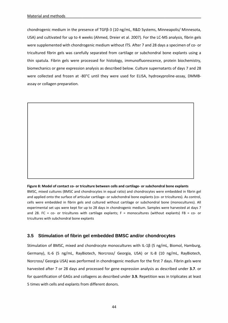
Material and methods
chondrogenic medium in the presence of TGF
USA) and cultivated for up to 4 weeks
were supplemented with chondrogenic m
tricultured fibrin gels was carefully separated from cartilage or subchondral bone explants using a
thin spatula. Fibrin gels were proce
biomechanics or gene expression analysis as described below.
were collected and frozen at -80°C until they were used for ELISA, hydroxyproline
assay or collagen preparation.
Figure 8: Model of contact co- or tri
BMSC, mixed cultures (BMSC and chondrocytes
and applied onto the surface of articular cartilage
cells were embedded in fibrin gels and cultured without cartilage
experimental set ups were kept for up to 28 days
and 28. FC = co- or tricultures with cartilage explants; F = monocultures (without explants)
tricultures with subchondral bone explants
3.5 Stimulation of fibrin gel
Stimulation of BMSC, mixed and chondrocyte
Germany), IL-6 (5 ng/mL, RayBiotech
Norcross/ Georgia USA) was performed in chondrogenic medium for the first 7 days
harvested after 7 or 28 days and
for quantification of GAGs and collagens as described under
5 times with cells and explants from different donors.
44
in the presence of TGFβ-3 (10 ng/mL, R&D Systems, Minneapolis/ Minnesota,
and cultivated for up to 4 weeks (Ahmed, Dreier et al. 2007). For the LC-MS
chondrogenic medium without ITS. After 7 and 28 days
tricultured fibrin gels was carefully separated from cartilage or subchondral bone explants using a
were processed for histology, immunofluorescence, protein biochemistry,
biomechanics or gene expression analysis as described below. Culture supernatants of day
80°C until they were used for ELISA, hydroxyproline
or triculture between cells and cartilage- or subchondral bone
and chondrocytes in equal ratio) and chondrocytes were embedded in
o the surface of articular cartilage- or subchondral bone explants (co- or tri
s and cultured without cartilage or subchondral bone
experimental set ups were kept for up to 28 days in chondrogenic medium. Samples were harvested at days 7
cultures with cartilage explants; F = monocultures (without explants)
cultures with subchondral bone explants
fibrin gel embedded BMSC and/or cho ndrocytes
of BMSC, mixed and chondrocyte monocultures with IL-1β (5 ng/mL
, RayBiotech, Norcross/ Georgia, USA) or IL-8 (10
) was performed in chondrogenic medium for the first 7 days
harvested after 7 or 28 days and processed for gene expression analysis as described under
and collagens as described under 3.9. Repetition was in triplicates at least
5 times with cells and explants from different donors.
, R&D Systems, Minneapolis/ Minnesota,
MS analysis, fibrin gels
After 7 and 28 days a specimen of co- or
tricultured fibrin gels was carefully separated from cartilage or subchondral bone explants using a
ssed for histology, immunofluorescence, protein biochemistry,
upernatants of days 7 and 28
80°C until they were used for ELISA, hydroxyproline-assay, DMMB-
or subchondral bone explants
) and chondrocytes were embedded in fibrin gel
or tricultures). As control,
or subchondral bone (monocultures). All
in chondrogenic medium. Samples were harvested at days 7
cultures with cartilage explants; F = monocultures (without explants) FB = co- or
ndrocytes
mL, Biomol, Hamburg,
ng/mL, RayBiotech,
) was performed in chondrogenic medium for the first 7 days. Fibrin gels were
processed for gene expression analysis as described under 3.7. or
Repetition was in triplicates at least
Material and methods
45
3.6 Cell vitality and proliferation in fibrin gel c ocultures
In order to evaluate vitality of cells in our different culture set ups, lactate dehydrogenase (LDH)
concentration in supernatants of BMSC, mixed cultures and chondrocytes was determined in
monocultures and co- or tricultures with OA-cartilage or OA-subchondral bone kept in chondrogenic
medium. Content of LDH was analyzed at days 7, 14, 21 and 28 with an LDH-based cytotoxicity
detection Kit (Roche, Penzberg, Germany) and compared with respective assay controls (high control
= all cells in fibrin gels were lysed; low control = spontaneous cell death of an equivalent cell amount
in monolayer) according to the manufacturer´s instructions. LDH concentration released from dead
cells into supernatant was determined on photometrical basis at absorption of 490 nm (Tecan
GENios with Magellan 6.5, Crailsheim, Germany). Due to high inter-experimental variability,
presumably caused by personal living conditions and physical activity, medical treatment or general
health status of tissue donors, the raw data was calculated as percent of control per individual
experiment. Repetition was in triplicates at least 3 times with cells and explants from different
donors.
For proliferation tests of monocultures and OA-cartilage cocultures, the fibrin gels were processed as
described under 3.8. Sections were fixed in 4% PFA (Sigma-Aldrich, St. Louis/ Missouri, USA) for 10
minutes and endogen peroxidase was blocked by treatment with 3% H2O2 containing 10% EtOH for 5
minutes. This was followed by an epitope demasking by boiling sections with 10mM sodium citrate
containing 0.05% Tween for 2 minutes at 650 watt and for another 5 minutes at 350 watt in the
microwave. Sections were blocked with 2% goat serum (Dako, Glostrup, Denmark) diluted in PBS for
1 hour and labeled over night at 4°C with an antibody against PCNA (DAKO, Glostrup, Denmark,
mouse anti PCNA clone PC10-M0879). For detection, all sections were incubated with a biotinylated
secondary antibody (Dako, Glostrup, Denmark) for 1 hour at 37°C followed by an incubation with
streptavidin peroxidase in PBS for 1 hour at 37°C. Staining was performed with the liquid substrate
system (DAKO Glostrup, Denmark) for 15 minutes. Sections were counterstained with Mayer´s
hemalaun solution (Roth, Arlesheim, Germany) for 5 minutes followed by successive dehydration
with upraising alcohol concentrations and were mounted with 1-2 drops of Depex (Serva
Electrophoresis, Heidelberg, Germany). Stained sections were photographed with Olympus BX 61
imaging system and cellP software, the number of total nuclei and number of PCNA stained nuclei
was counted and the proliferation ratio was calculated.
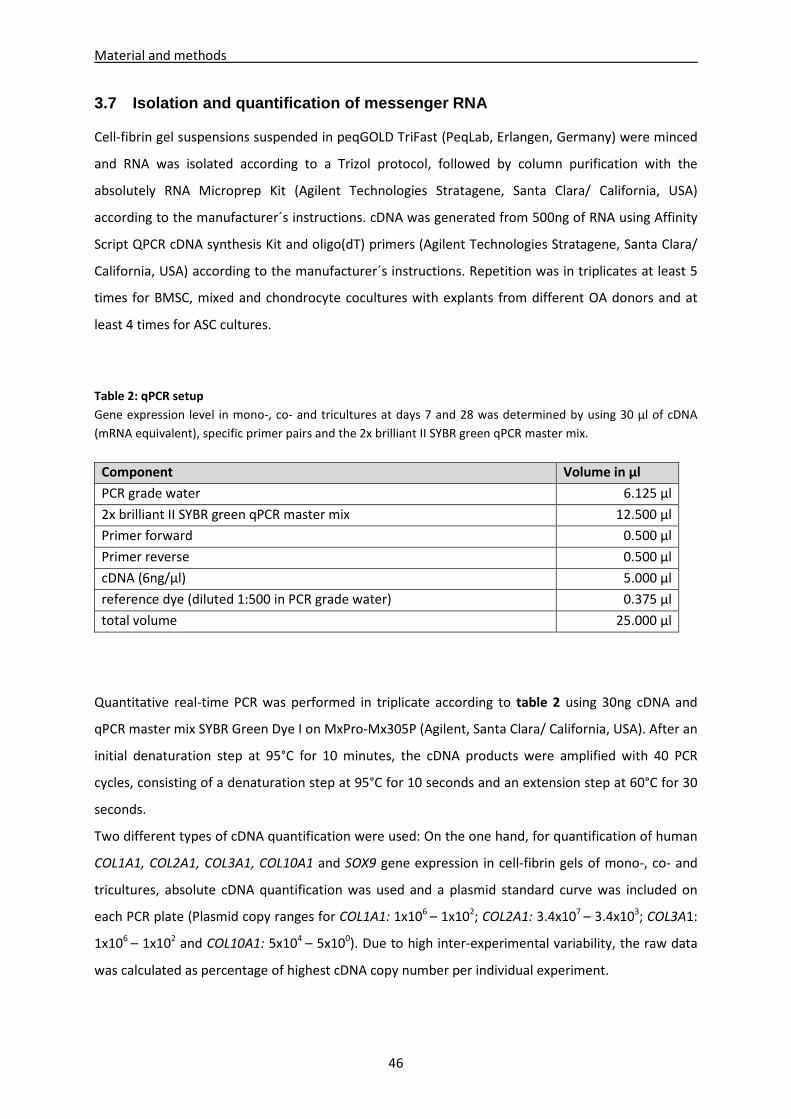
Material and methods
46
3.7 Isolation and quantification of messenger RNA
Cell-fibrin gel suspensions suspended in peqGOLD TriFast (PeqLab, Erlangen, Germany) were minced
and RNA was isolated according to a Trizol protocol, followed by column purification with the
absolutely RNA Microprep Kit (Agilent Technologies Stratagene, Santa Clara/ California, USA)
according to the manufacturer´s instructions. cDNA was generated from 500ng of RNA using Affinity
Script QPCR cDNA synthesis Kit and oligo(dT) primers (Agilent Technologies Stratagene, Santa Clara/
California, USA) according to the manufacturer´s instructions. Repetition was in triplicates at least 5
times for BMSC, mixed and chondrocyte cocultures with explants from different OA donors and at
least 4 times for ASC cultures.
Table 2: qPCR setup
Gene expression level in mono-, co- and tricultures at days 7 and 28 was determined by using 30 µl of cDNA
(mRNA equivalent), specific primer pairs and the 2x brilliant II SYBR green qPCR master mix.
Component Volume in µl
PCR grade water 6.125 µl
2x brilliant II SYBR green qPCR master mix 12.500 µl
Primer forward 0.500 µl
Primer reverse 0.500 µl
cDNA (6ng/µl) 5.000 µl
reference dye (diluted 1:500 in PCR grade water) 0.375 µl
total volume 25.000 µl
Quantitative real-time PCR was performed in triplicate according to table 2 using 30ng cDNA and
qPCR master mix SYBR Green Dye I on MxPro-Mx305P (Agilent, Santa Clara/ California, USA). After an
initial denaturation step at 95°C for 10 minutes, the cDNA products were amplified with 40 PCR
cycles, consisting of a denaturation step at 95°C for 10 seconds and an extension step at 60°C for 30
seconds.
Two different types of cDNA quantification were used: On the one hand, for quantification of human
COL1A1, COL2A1, COL3A1, COL10A1 and SOX9 gene expression in cell-fibrin gels of mono-, co- and
tricultures, absolute cDNA quantification was used and a plasmid standard curve was included on
each PCR plate (Plasmid copy ranges for COL1A1: 1x10⁶ – 1x10²; COL2A1: 3.4x10⁷ – 3.4x10³; COL3A1:
1x10⁶ – 1x10² and COL10A1: 5x10⁴ – 5x100). Due to high inter-experimental variability, the raw data
was calculated as percentage of highest cDNA copy number per individual experiment.
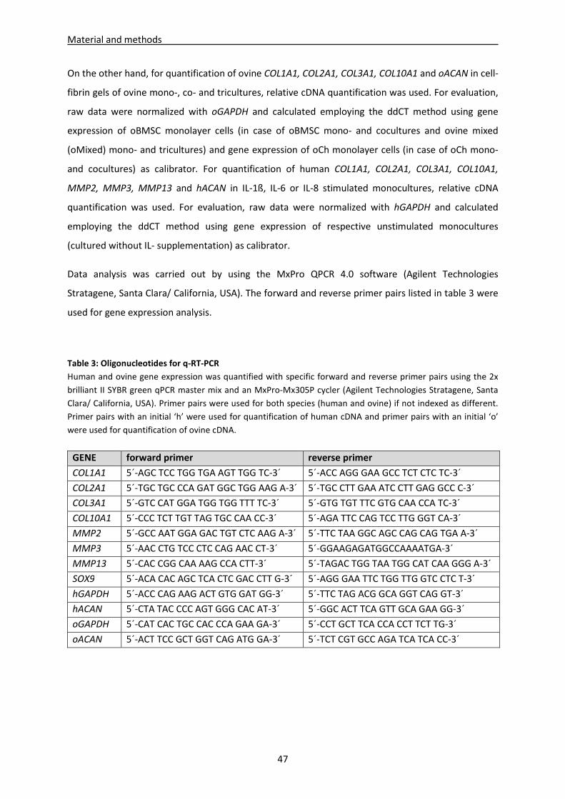
Material and methods
47
On the other hand, for quantification of ovine COL1A1, COL2A1, COL3A1, COL10A1 and oACAN in cell-
fibrin gels of ovine mono-, co- and tricultures, relative cDNA quantification was used. For evaluation,
raw data were normalized with oGAPDH and calculated employing the ddCT method using gene
expression of oBMSC monolayer cells (in case of oBMSC mono- and cocultures and ovine mixed
(oMixed) mono- and tricultures) and gene expression of oCh monolayer cells (in case of oCh mono-
and cocultures) as calibrator. For quantification of human COL1A1, COL2A1, COL3A1, COL10A1,
MMP2, MMP3, MMP13 and hACAN in IL-1ß, IL-6 or IL-8 stimulated monocultures, relative cDNA
quantification was used. For evaluation, raw data were normalized with hGAPDH and calculated
employing the ddCT method using gene expression of respective unstimulated monocultures
(cultured without IL- supplementation) as calibrator.
Data analysis was carried out by using the MxPro QPCR 4.0 software (Agilent Technologies
Stratagene, Santa Clara/ California, USA). The forward and reverse primer pairs listed in table 3 were
used for gene expression analysis.
Table 3: Oligonucleotides for q-RT-PCR
Human and ovine gene expression was quantified with specific forward and reverse primer pairs using the 2x
brilliant II SYBR green qPCR master mix and an MxPro-Mx305P cycler (Agilent Technologies Stratagene, Santa
Clara/ California, USA). Primer pairs were used for both species (human and ovine) if not indexed as different.
Primer pairs with an initial ‘h’ were used for quantification of human cDNA and primer pairs with an initial ‘o’
were used for quantification of ovine cDNA.
GENE forward primer reverse primer
COL1A1 5´-AGC TCC TGG TGA AGT TGG TC-3´ 5´-ACC AGG GAA GCC TCT CTC TC-3´
COL2A1 5´-TGC TGC CCA GAT GGC TGG AAG A-3´ 5´-TGC CTT GAA ATC CTT GAG GCC C-3´
COL3A1 5´-GTC CAT GGA TGG TGG TTT TC-3´ 5´-GTG TGT TTC GTG CAA CCA TC-3´
COL10A1 5´-CCC TCT TGT TAG TGC CAA CC-3´ 5´-AGA TTC CAG TCC TTG GGT CA-3´
MMP2 5´-GCC AAT GGA GAC TGT CTC AAG A-3´ 5´-TTC TAA GGC AGC CAG CAG TGA A-3´
MMP3 5´-AAC CTG TCC CTC CAG AAC CT-3´ 5´-GGAAGAGATGGCCAAAATGA-3´
MMP13 5´-CAC CGG CAA AAG CCA CTT-3´ 5´-TAGAC TGG TAA TGG CAT CAA GGG A-3´
SOX9 5´-ACA CAC AGC TCA CTC GAC CTT G-3´ 5´-AGG GAA TTC TGG TTG GTC CTC T-3´
hGAPDH 5´-ACC CAG AAG ACT GTG GAT GG-3´ 5´-TTC TAG ACG GCA GGT CAG GT-3´
hACAN 5´-CTA TAC CCC AGT GGG CAC AT-3´ 5´-GGC ACT TCA GTT GCA GAA GG-3´
oGAPDH 5´-CAT CAC TGC CAC CCA GAA GA-3´ 5´-CCT GCT TCA CCA CCT TCT TG-3´
oACAN 5´-ACT TCC GCT GGT CAG ATG GA-3´ 5´-TCT CGT GCC AGA TCA TCA CC-3´
Material and methods
48
3.8 Histology and immunofluorescence of fibrin gel embedded cultures
ECM composition was microscopically analyzed with respect to collagens I, II, III and X and GAG
expression at the end of coculture time. Therefore, fibrin gels were rinsed with phosphate buffered
saline (PBS, PAA, Piscataway/ New Jersey, USA), embedded in TissueTec (Sakura Finetek, Alphen aan
den Rijn, the Netherlands), frozen in liquid nitrogen and cross-sectioned (10 µm thick) with a cryo-
microtom (Mikrom® HM 500 OM, Walldorf, Germany). For histological evaluation, sections were
fixed in 4% paraformaldehyde (PFA, Sigma-Aldrich, St. Louis/ Missouri, USA) for 10 minutes at room
temperature (RT) and stained with 1% Alcian blue 8GX (Sigma-Aldrich, St. Louis/ Missouri, USA) for
GAGs and counterstained with nuclear fast red aluminum sulfate solution (Roth, Arlesheim,
Germany). For immunofluorescence analysis, sections were fixed in 4% PFA for 10 minutes at room
temperature (RT), treated for 15 minutes at 37°C with pepsin (3 mg/mL in 0.01 M HCl, Sigma-Aldrich,
St. Louis/ Missouri, USA) or in case of collagen X with hyaluronidase (0,2% in acetate buffer pH5,
Sigma-Aldrich, St. Louis/ Missouri, USA) and were blocked with 3% BSA (Sigma-Aldrich, St. Louis/
Missouri, USA) diluted in PBS for one hour. Sections were labeled over night at 4°C with an antibody
against collagen I (C-2456, Sigma-Aldrich, St. Louis/ Missouri, USA), collagen II (CIIC1, DSHB, Iowa
City/ Iowa, USA), collagen III (MAB3392, Merck Millipore, Billerica/ Massachusetts, USA) or collagen X
(2031501005, Quartett, Berlin, Germany). For fluorescence detection all sections were incubated
with an Alexa 488 secondary antibody (Invitrogen, Paisley, UK) for 1 hour at 37°C. Finally, samples
were mounted in Vectashield mounting medium with DAPI (1 µg/mL, Vector, Burlingame, Canada)
and were analyzed with an Olympus BX 61 imaging system and cellP software (Olympus, Hamburg,
Germany). Repetition was in triplicate at least 5 times with cells and explants from different donors
and two observers chose representative slides.
3.9 Biochemical analysis of fibrin gel cell lysates
3.9.1 DMMB Assay
Fibrin gels were removed from explants, homogenized and digested with pepsin (1 mg/mL in 0.5 M
acetic acid containing 0.4 M NaCl, Sigma-Aldrich, St. Louis/ Missouri, USA) for 48 hours at 4°C and
further digested with elastase (1 mg/mL in TBS pH 8, Serva Electrophoresis, Heidelberg, Germany) for
24 hours at 4°C. Samples were stored at -20°C until they were analyzed for GAG and collagen
contents. At least seven culture setups (different donors) were analyzed in triplicate.
GAG concentration was measured spectrophotometrically using 25 µl of the digested cell lysates or
25 µl undiluted cell supernatant supplemented with dimethylmethylene blue (DMMB, AppliChem,
Darmstadt, Germany) which forms a complex with GAG (Farndale, Buttle et al. 1986). Quantification
Material and methods
49
was performed in µg per 1 x 10⁶ cells with a chondroitin sulfate standard at 525 nm (Tecan GENios
with Magellan 6.5, Crailsheim, Germany).
3.9.2 Collagen I and II ELISA
Collagen I and II contents of day 28 cell lysates digested as described above were measured with
specific sandwich ELISAs, which recognize the native conformation of collagen I and II chains
(Chondrex, Redmond/ Washington, USA) according to the manufacturer´s instructions.
3.9.3 Collagen III Dot-Blot
Human collagen I, II and III protein in digested cell lysates was quantified with a dot blot assay using
an aliquot of 1 µl from 500 µl of cell lysate and including a standard curve (range: 100 ng/µl, 50 ng/µl,
25 ng/µl, 12.5 ng/µl, 6.25 ng/µl and 3.125 ng/µl) of recombinant collagen I or collagen II (Chondrex,
Redmond/ Washington, USA) or (range: 100 ng/µl, 50 ng/µl, 25 ng/µl, 12.5 ng/µl, 6.25 ng/µl and
3.125 ng/µl) of recombinant collagen III (Abcam, Cambridge, UK). Quantification was performed in µg
per 1 x 10⁶ cells densitometrically using a Chemi-Smart 500 (PeqLab, Erlangen, Germany) for
chemiluminescence detection and the CS4 for Windows software for calculation of luminescence
intensities. Repetition was in triplicates at least 5 times with cells and explants from different donors.
3.10 Immunoblotting of Sox9 and phospho Sox9
For immunoblotting of Sox9 or phospho Sox9 (pSox9) total cell proteins were isolated from mono-,
co- or tricultures with peqGOLD TriFast (PeqLab, Erlangen, Germany) and total protein content was
determined by a bicinchoninic acid (BCA) assay with the Pierce BCA Protein Assay Kit (Thermo
Scientific, Waltham, Massachusetts, USA) according to the manufacturer´s instructions. For Western
blotting of pSox9 25 µg of total cell lysate were denaturated with 6x SDS-Buffer containing
β-mercaptoethanol for 5 minutes at 95 °C and separated by denaturing sodium dodecyl sulfate
polyacrylamide (SDS) gel electrophoresis using 100 V (Table 4). Proteins were blotted to Protran®
nitrocellulose transfer membrane for 1 to 2 hours at 120 ampere (Whatman® GE Helthcare,
Buckinghamshire, UK) using a tank-blot apparatus (Bio-Rad, Hercules, California, USA). After blocking
with 5% non-fat milk for 1 hour the membrane was probed with a specific antibody against pSox9
and β-Actin (ab59252 and ab8227, both abcam, Cambridge, UK) over night at 4°C. After incubation
with a peroxidase-coupled goat-anti-mouse secondary antibody (Santa Cruz Biotechnology, Dallas,
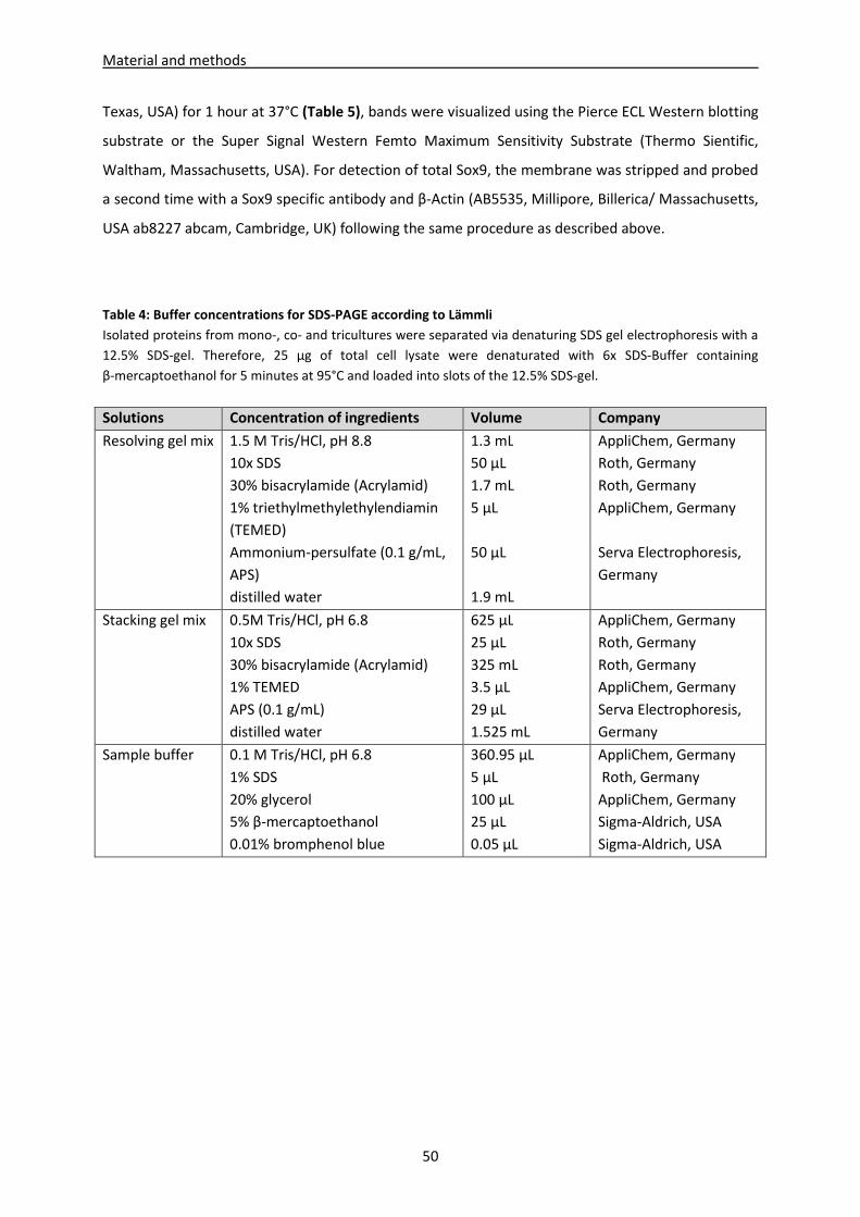
Material and methods
50
Texas, USA) for 1 hour at 37°C (Table 5), bands were visualized using the Pierce ECL Western blotting
substrate or the Super Signal Western Femto Maximum Sensitivity Substrate (Thermo Sientific,
Waltham, Massachusetts, USA). For detection of total Sox9, the membrane was stripped and probed
a second time with a Sox9 specific antibody and β-Actin (AB5535, Millipore, Billerica/ Massachusetts,
USA ab8227 abcam, Cambridge, UK) following the same procedure as described above.
Table 4: Buffer concentrations for SDS-PAGE according to Lämmli
Isolated proteins from mono-, co- and tricultures were separated via denaturing SDS gel electrophoresis with a
12.5% SDS-gel. Therefore, 25 µg of total cell lysate were denaturated with 6x SDS-Buffer containing
β-mercaptoethanol for 5 minutes at 95°C and loaded into slots of the 12.5% SDS-gel.
Solutions Concentration of ingredients Volume Company
Resolving gel mix 1.5 M Tris/HCl, pH 8.8
10x SDS
30% bisacrylamide (Acrylamid)
1% triethylmethylethylendiamin
(TEMED)
Ammonium-persulfate (0.1 g/mL,
APS)
distilled water
1.3 mL
50 µL
1.7 mL
5 µL
50 µL
1.9 mL
AppliChem, Germany
Roth, Germany
Roth, Germany
AppliChem, Germany
Serva Electrophoresis,
Germany
Stacking gel mix 0.5M Tris/HCl, pH 6.8
10x SDS
30% bisacrylamide (Acrylamid)
1% TEMED
APS (0.1 g/mL)
distilled water
625 µL
25 µL
325 mL
3.5 µL
29 µL
1.525 mL
AppliChem, Germany
Roth, Germany
Roth, Germany
AppliChem, Germany
Serva Electrophoresis,
Germany
Sample buffer 0.1 M Tris/HCl, pH 6.8
1% SDS
20% glycerol
5% β-mercaptoethanol
0.01% bromphenol blue
360.95 µL
5 µL
100 µL
25 µL
0.05 µL
AppliChem, Germany
Roth, Germany
AppliChem, Germany
Sigma-Aldrich, USA
Sigma-Aldrich, USA
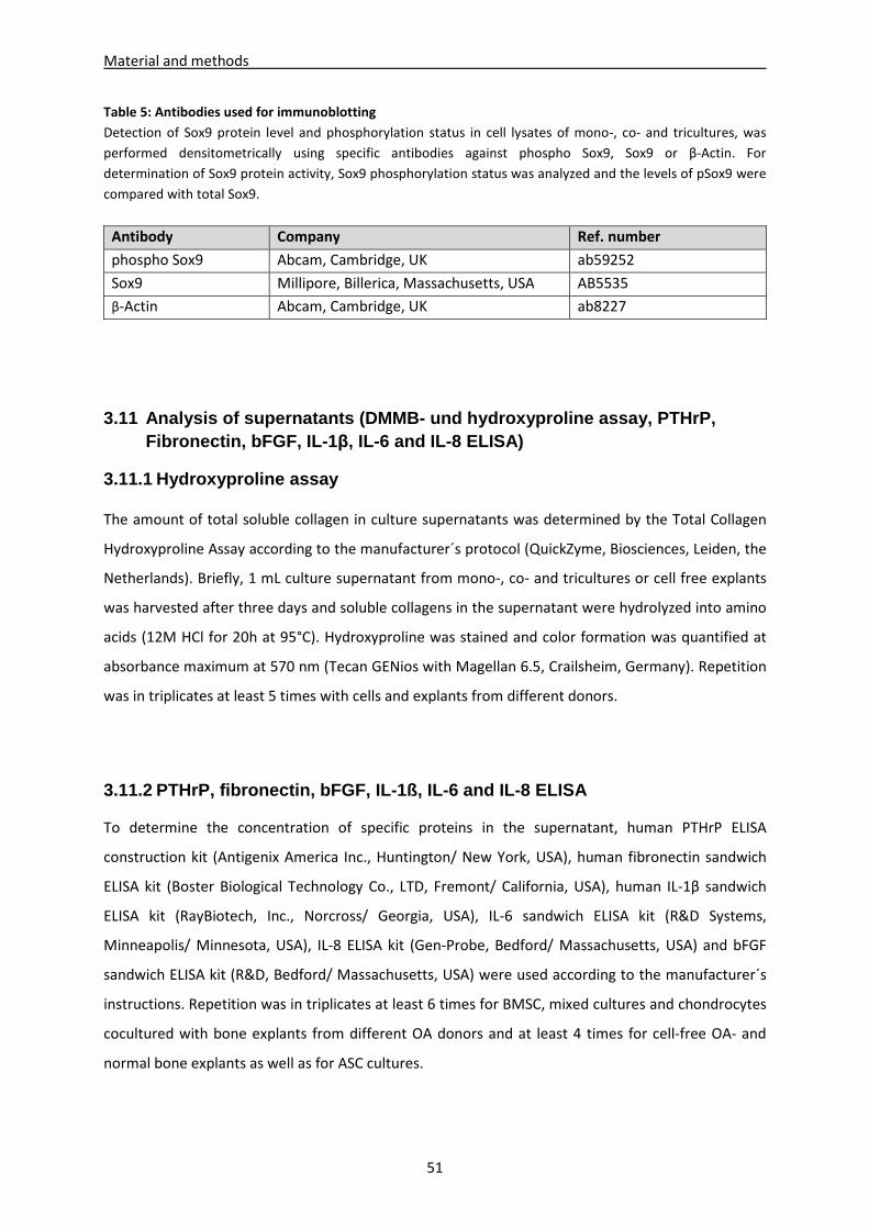
Material and methods
51
Table 5: Antibodies used for immunoblotting
Detection of Sox9 protein level and phosphorylation status in cell lysates of mono-, co- and tricultures, was
performed densitometrically using specific antibodies against phospho Sox9, Sox9 or β-Actin. For
determination of Sox9 protein activity, Sox9 phosphorylation status was analyzed and the levels of pSox9 were
compared with total Sox9.
Antibody Company Ref. number
phospho Sox9 Abcam, Cambridge, UK ab59252
Sox9 Millipore, Billerica, Massachusetts, USA AB5535
β-Actin Abcam, Cambridge, UK ab8227
3.11 Analysis of supernatants (DMMB- und hydroxypro line assay, PTHrP, Fibronectin, bFGF, IL-1 β, IL-6 and IL-8 ELISA)
3.11.1 Hydroxyproline assay
The amount of total soluble collagen in culture supernatants was determined by the Total Collagen
Hydroxyproline Assay according to the manufacturer´s protocol (QuickZyme, Biosciences, Leiden, the
Netherlands). Briefly, 1 mL culture supernatant from mono-, co- and tricultures or cell free explants
was harvested after three days and soluble collagens in the supernatant were hydrolyzed into amino
acids (12M HCl for 20h at 95°C). Hydroxyproline was stained and color formation was quantified at
absorbance maximum at 570 nm (Tecan GENios with Magellan 6.5, Crailsheim, Germany). Repetition
was in triplicates at least 5 times with cells and explants from different donors.
3.11.2 PTHrP, fibronectin, bFGF, IL-1ß, IL-6 and IL -8 ELISA
To determine the concentration of specific proteins in the supernatant, human PTHrP ELISA
construction kit (Antigenix America Inc., Huntington/ New York, USA), human fibronectin sandwich
ELISA kit (Boster Biological Technology Co., LTD, Fremont/ California, USA), human IL-1β sandwich
ELISA kit (RayBiotech, Inc., Norcross/ Georgia, USA), IL-6 sandwich ELISA kit (R&D Systems,
Minneapolis/ Minnesota, USA), IL-8 ELISA kit (Gen-Probe, Bedford/ Massachusetts, USA) and bFGF
sandwich ELISA kit (R&D, Bedford/ Massachusetts, USA) were used according to the manufacturer´s
instructions. Repetition was in triplicates at least 6 times for BMSC, mixed cultures and chondrocytes
cocultured with bone explants from different OA donors and at least 4 times for cell-free OA- and
normal bone explants as well as for ASC cultures.
Material and methods
52
3.11.3 DMMB assay
GAG concentration was measured spectrophotometrically using 25 µl of the digested cell lysates or
undiluted cell supernatant supplemented with dimethylmethylene blue (DMMB, AppliChem,
Darmstadt, Germany) which forms a complex with GAG. Quantification was performed in µg per
1 x 10⁶ cells with a chondroitin sulfate standard at an absorption maximum at 525 nm (Tecan GENios
with Magellan 6.5, Crailsheim, Germany).
3.12 Screening for differentially expressed factors in coculture supernatant with LC-MS
In order to identify differentially expressed proteins in mono- and coculture with OA-cartilage, a SDS-
PAGE/LC-MS approach was performed in cooperation with Prof. Dr. R. Deutzmann (Department of
Biochemistry I, University of Regensburg) as described previously (Hamperl, Brown et al. 2014).
Supernatants of BMSC mono- and cocultures with OA-cartilage explants and of cartilage explants
with cell-free fibrin gel were precipitated and separated by SDS-PAGE. The gel was stained with
Coomassie Blue and the pattern of protein bands between lines with proteins from different
coculture setups was compared. For analysis, differentially expressed bands were identified and
either single bands or whole lines were cut out. Lanes of the different culture setups were divided in
three different areas (high > 70kd, medium 70-35kd and low < 35kd). Proteins were isolated,
digested with trypsin and identified by MALDI-TOF mass spectrometry using standard protocols.
Briefly, LC-MS analyses were operated in positive ion reflector mode on an Applied Biosystems 4700
or 4800 Proteomics Analyzer MALDI-TOF/TOF mass spectrometer and evaluated by searching the
NCBI protein sequence database with the Mascot search engine (Matrix Science) implemented in the
GPS Explorer software (Applied Biosystems, Darmstadt, Germany). Intensity of the laser was adjusted
due to sample concentration and laser condition. This approach was performed in collaboration with
the AG from Prof. Deutzmann (Inst. Biochemistry, Microbiology and Genetics; University of
Regensburg).
Material and methods
53
3.13 Biomechanical testing
Fibrin gel constructs were removed from respective explants and mono- co- and tricultures were
adapted to pieces of same size by punching out constructs with an outer diameter of 2.6 mm (Biopsy
punch, Stiefel GmbH, Munich, Germany). All biomechanical tests were carried out in a standard
material testing machine (Z010; Zwick GmbH, Ulm, Germany) using a 40N load cell (Fig. 9). The initial
sample height (h0) was measured under a preload of 0.1N using a laser displacement transducer
(optoNCDT 2200-20, Micro-Epsilon GmbH & Co. KG, 0.3 μm resolution, ±0.03% accuracy).
An unconfined compression test was performed by placing the samples in a cell culture dish filled
with 0.9% NaCl and loading it by a flat ended cylinder at a strain rate of 100% h0/minutes until 50%
strain was reached. In order to measure the stiffness of the new generated ECM in our constructs,
the Young´s modulus was graphically determined from the related stress-strain diagrams. Therefore,
two typical regions were evaluated: the progressive region at 0-10% strain and the linear region at
40-50% strain.
After adequate relaxation time of 24 hours an additional relaxation test was performed under
confined compression conditions. The samples were placed in a confining chamber of 2.6 mm
diameter filled with 0.9% NaCl and loaded by a flat ended porous ceramic cylinder (Al2O3) allowing
for fluid flow. After application of 50% strain at a loading rate of 100% h0/minutes the strain was held
constant for 10 minutes until the equilibrium state was reached.
Based on this data hydraulic permeability (k) was calculated referred to a given diffusion equation
(Mow, Kuei et al. 1980; Frank and Hochmuth 1987) using Formula 1 and the aggregate modulus (HA)
at equilibrium state with 50% strain was assessed using Formula 2 considered that Δl/h0 is the
applied strain, H the modulus, σ the stress and�� the stress at equilibrium state with 50% strain.
Repetition was for 4 times with cells and explants from different donors.
Formula 1 �� =�� + 2� ∗ �� ∗ �(� �� �
�∗�∗�∗�)
Formula 2 �� = ���� %
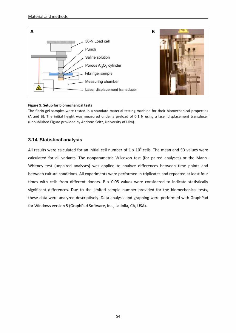
Material and methods
54
A B
Figure 9: Setup for biomechanical tests
The fibrin gel samples were tested in a standard material testing machine for their biomechanical properties
(A and B). The initial height was measured under a preload of 0.1 N using a laser displacement transducer
(unpublished Figure provided by Andreas Seitz, University of Ulm).
3.14 Statistical analysis
All results were calculated for an initial cell number of 1 x 10⁶ cells. The mean and SD values were
calculated for all variants. The nonparametric Wilcoxon test (for paired analyses) or the Mann-
Whitney test (unpaired analyses) was applied to analyze differences between time points and
between culture conditions. All experiments were performed in triplicates and repeated at least four
times with cells from different donors. P < 0.05 values were considered to indicate statistically
significant differences. Due to the limited sample number provided for the biomechanical tests,
these data were analyzed descriptively. Data analysis and graphing were performed with GraphPad
for Windows version 5 (GraphPad Software, Inc., La Jolla, CA, USA).
Results
4 Results
4.1 Part I: Microenvironment of cartilage influencesdifferentiation and ECM production of BMSC and chon drocytes
4.1.1 Bone marrow derived
To prove stemness of isolated plastic adherent b
selection of surface markers was
characteristic surface markers and were
(CD44pos and CD105 pos) and negative for the expression o
(Fig. 10A). A further criteria for human BMSC is that they are
and adipogenic lineages additionally to
the tested BMSC specimens were
oil-red stained oil drops in differentiated cells and
by alizarin staining of mineralized
Figure 10: FACS analysis of BMSC surface markers and staining of adipogenic and osteogenic differentiation
(A) Isolated plastic adherent BMSC in passage 3 were analy
against BMSC surface markers CD19
was proofed using an isotype control (black line). (B) BMSC were stained with oil red for oil dr
during adipogenic differentiation (upper row) and with alizarin red for matrix mineralization during osteogenic
differentiation (lower row).
55
Microenvironment of cartilage influences chondrogenic differentiation and ECM production of BMSC and chon drocytes
Bone marrow derived BMSC can be differentiated into different lineages
s of isolated plastic adherent bone marrow cells (see 3.2.)
selection of surface markers was analyzed by FACS. BMSC showed a typical expression pattern of
characteristic surface markers and were shown to be positive for the expression of CD44 and CD105
nd negative for the expression of CD19 and CD34 (CD19
criteria for human BMSC is that they are capable to differentiate
dditionally to chondrogenic lineage (Dominici, Le Blanc et al. 2006)
re shown to reveal adipogenic differentiation, proofed by
ps in differentiated cells and osteogenic differentiation, which
mineralized bone matrix (Fig. 10B).
FACS analysis of BMSC surface markers and staining of adipogenic and osteogenic differentiation
(A) Isolated plastic adherent BMSC in passage 3 were analyzed by flow cytometry using specific antibodies
markers CD19neg
, CD34neg
, CD44pos
and CD105pos
(red line). Specificity of epitope staining
was proofed using an isotype control (black line). (B) BMSC were stained with oil red for oil dr
during adipogenic differentiation (upper row) and with alizarin red for matrix mineralization during osteogenic
chondrogenic differentiation and ECM production of BMSC and chon drocytes
can be differentiated into different lineages
(see 3.2.) the expression of a
BMSC showed a typical expression pattern of
shown to be positive for the expression of CD44 and CD105
(CD19neg and CD34neg)
to differentiate into osteogenic
(Dominici, Le Blanc et al. 2006). All of
adipogenic differentiation, proofed by numerous
osteogenic differentiation, which was confirmed
FACS analysis of BMSC surface markers and staining of adipogenic and osteogenic differentiation
zed by flow cytometry using specific antibodies
(red line). Specificity of epitope staining
was proofed using an isotype control (black line). (B) BMSC were stained with oil red for oil drops incorporated
during adipogenic differentiation (upper row) and with alizarin red for matrix mineralization during osteogenic
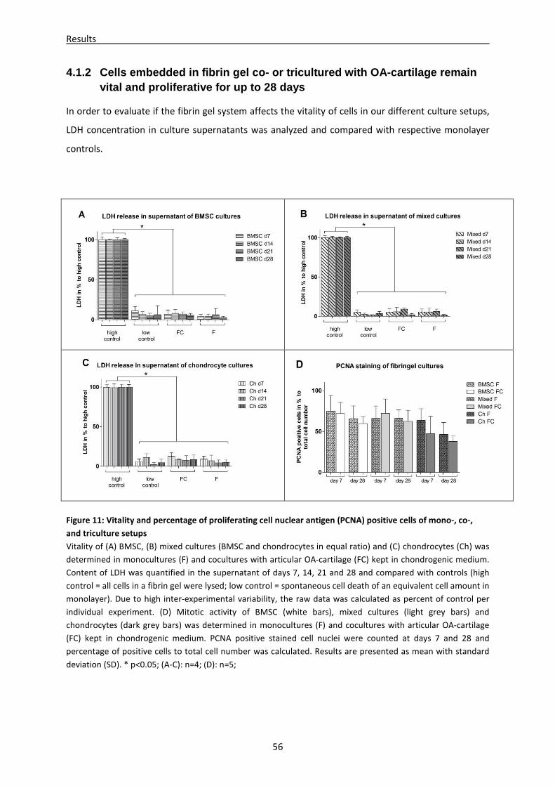
Results
56
4.1.2 Cells embedded in fibrin gel co- or tricultur ed with OA-cartilage remain vital and proliferative for up to 28 days
In order to evaluate if the fibrin gel system affects the vitality of cells in our different culture setups,
LDH concentration in culture supernatants was analyzed and compared with respective monolayer
controls.
Figure 11: Vitality and percentage of proliferating cell nuclear antigen (PCNA) positive cells of mono-, co-,
and triculture setups
Vitality of (A) BMSC, (B) mixed cultures (BMSC and chondrocytes in equal ratio) and (C) chondrocytes (Ch) was
determined in monocultures (F) and cocultures with articular OA-cartilage (FC) kept in chondrogenic medium.
Content of LDH was quantified in the supernatant of days 7, 14, 21 and 28 and compared with controls (high
control = all cells in a fibrin gel were lysed; low control = spontaneous cell death of an equivalent cell amount in
monolayer). Due to high inter-experimental variability, the raw data was calculated as percent of control per
individual experiment. (D) Mitotic activity of BMSC (white bars), mixed cultures (light grey bars) and
chondrocytes (dark grey bars) was determined in monocultures (F) and cocultures with articular OA-cartilage
(FC) kept in chondrogenic medium. PCNA positive stained cell nuclei were counted at days 7 and 28 and
percentage of positive cells to total cell number was calculated. Results are presented as mean with standard
deviation (SD). * p<0.05; (A-C): n=4; (D): n=5;
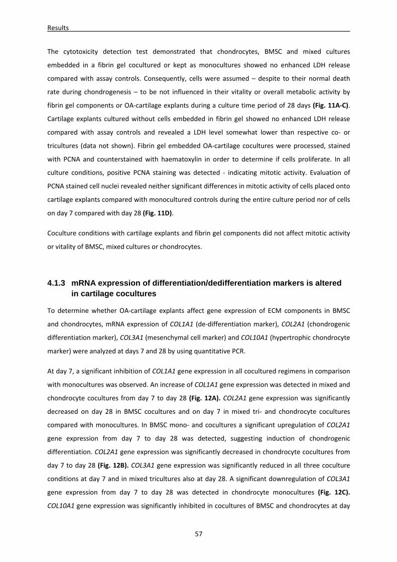
Results
57
The cytotoxicity detection test demonstrated that chondrocytes, BMSC and mixed cultures
embedded in a fibrin gel cocultured or kept as monocultures showed no enhanced LDH release
compared with assay controls. Consequently, cells were assumed – despite to their normal death
rate during chondrogenesis – to be not influenced in their vitality or overall metabolic activity by
fibrin gel components or OA-cartilage explants during a culture time period of 28 days (Fig. 11A-C).
Cartilage explants cultured without cells embedded in fibrin gel showed no enhanced LDH release
compared with assay controls and revealed a LDH level somewhat lower than respective co- or
tricultures (data not shown). Fibrin gel embedded OA-cartilage cocultures were processed, stained
with PCNA and counterstained with haematoxylin in order to determine if cells proliferate. In all
culture conditions, positive PCNA staining was detected - indicating mitotic activity. Evaluation of
PCNA stained cell nuclei revealed neither significant differences in mitotic activity of cells placed onto
cartilage explants compared with monocultured controls during the entire culture period nor of cells
on day 7 compared with day 28 (Fig. 11D).
Coculture conditions with cartilage explants and fibrin gel components did not affect mitotic activity
or vitality of BMSC, mixed cultures or chondrocytes.
4.1.3 mRNA expression of differentiation/dedifferen tiation markers is altered in cartilage cocultures
To determine whether OA-cartilage explants affect gene expression of ECM components in BMSC
and chondrocytes, mRNA expression of COL1A1 (de-differentiation marker), COL2A1 (chondrogenic
differentiation marker), COL3A1 (mesenchymal cell marker) and COL10A1 (hypertrophic chondrocyte
marker) were analyzed at days 7 and 28 by using quantitative PCR.
At day 7, a significant inhibition of COL1A1 gene expression in all cocultured regimens in comparison
with monocultures was observed. An increase of COL1A1 gene expression was detected in mixed and
chondrocyte cocultures from day 7 to day 28 (Fig. 12A). COL2A1 gene expression was significantly
decreased on day 28 in BMSC cocultures and on day 7 in mixed tri- and chondrocyte cocultures
compared with monocultures. In BMSC mono- and cocultures a significant upregulation of COL2A1
gene expression from day 7 to day 28 was detected, suggesting induction of chondrogenic
differentiation. COL2A1 gene expression was significantly decreased in chondrocyte cocultures from
day 7 to day 28 (Fig. 12B). COL3A1 gene expression was significantly reduced in all three coculture
conditions at day 7 and in mixed tricultures also at day 28. A significant downregulation of COL3A1
gene expression from day 7 to day 28 was detected in chondrocyte monocultures (Fig. 12C).
COL10A1 gene expression was significantly inhibited in cocultures of BMSC and chondrocytes at day
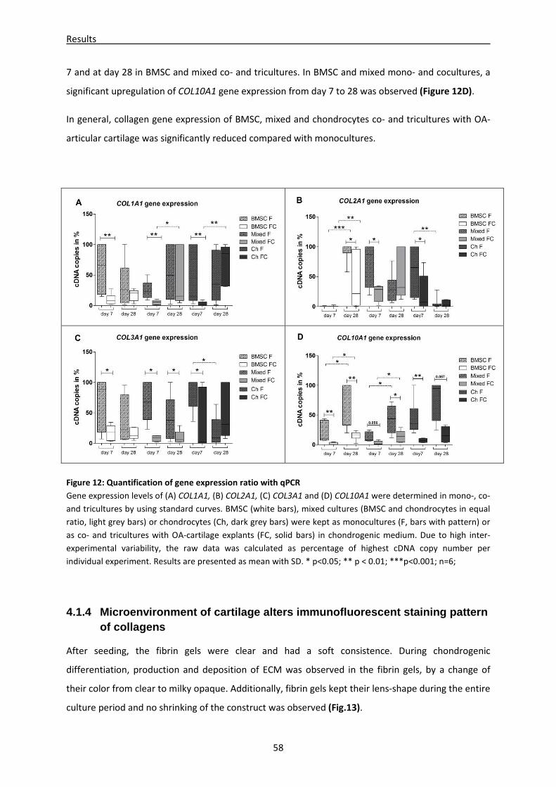
Results
58
7 and at day 28 in BMSC and mixed co- and tricultures. In BMSC and mixed mono- and cocultures, a
significant upregulation of COL10A1 gene expression from day 7 to 28 was observed (Figure 12D).
In general, collagen gene expression of BMSC, mixed and chondrocytes co- and tricultures with OA-
articular cartilage was significantly reduced compared with monocultures.
Figure 12: Quantification of gene expression ratio with qPCR
Gene expression levels of (A) COL1A1, (B) COL2A1, (C) COL3A1 and (D) COL10A1 were determined in mono-, co-
and tricultures by using standard curves. BMSC (white bars), mixed cultures (BMSC and chondrocytes in equal
ratio, light grey bars) or chondrocytes (Ch, dark grey bars) were kept as monocultures (F, bars with pattern) or
as co- and tricultures with OA-cartilage explants (FC, solid bars) in chondrogenic medium. Due to high inter-
experimental variability, the raw data was calculated as percentage of highest cDNA copy number per
individual experiment. Results are presented as mean with SD. * p<0.05; ** p < 0.01; ***p<0.001; n=6;
4.1.4 Microenvironment of cartilage alters immunofl uorescent staining pattern of collagens
After seeding, the fibrin gels were clear and had a soft consistence. During chondrogenic
differentiation, production and deposition of ECM was observed in the fibrin gels, by a change of
their color from clear to milky opaque. Additionally, fibrin gels kept their lens-shape during the entire
culture period and no shrinking of the construct was observed (Fig.13).
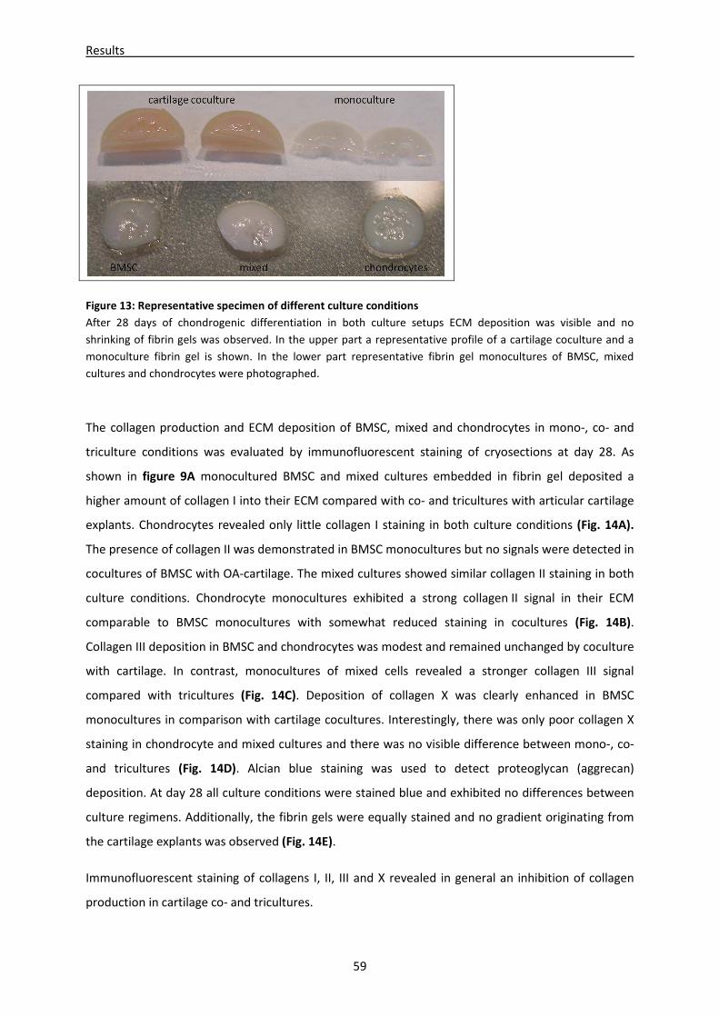
Results
59
Figure 13: Representative specimen of different culture conditions
After 28 days of chondrogenic differentiation in both culture setups ECM deposition was visible and no
shrinking of fibrin gels was observed. In the upper part a representative profile of a cartilage coculture and a
monoculture fibrin gel is shown. In the lower part representative fibrin gel monocultures of BMSC, mixed
cultures and chondrocytes were photographed.
The collagen production and ECM deposition of BMSC, mixed and chondrocytes in mono-, co- and
triculture conditions was evaluated by immunofluorescent staining of cryosections at day 28. As
shown in figure 9A monocultured BMSC and mixed cultures embedded in fibrin gel deposited a
higher amount of collagen I into their ECM compared with co- and tricultures with articular cartilage
explants. Chondrocytes revealed only little collagen I staining in both culture conditions (Fig. 14A).
The presence of collagen II was demonstrated in BMSC monocultures but no signals were detected in
cocultures of BMSC with OA-cartilage. The mixed cultures showed similar collagen II staining in both
culture conditions. Chondrocyte monocultures exhibited a strong collagen II signal in their ECM
comparable to BMSC monocultures with somewhat reduced staining in cocultures (Fig. 14B).
Collagen III deposition in BMSC and chondrocytes was modest and remained unchanged by coculture
with cartilage. In contrast, monocultures of mixed cells revealed a stronger collagen III signal
compared with tricultures (Fig. 14C). Deposition of collagen X was clearly enhanced in BMSC
monocultures in comparison with cartilage cocultures. Interestingly, there was only poor collagen X
staining in chondrocyte and mixed cultures and there was no visible difference between mono-, co-
and tricultures (Fig. 14D). Alcian blue staining was used to detect proteoglycan (aggrecan)
deposition. At day 28 all culture conditions were stained blue and exhibited no differences between
culture regimens. Additionally, the fibrin gels were equally stained and no gradient originating from
the cartilage explants was observed (Fig. 14E).
Immunofluorescent staining of collagens I, II, III and X revealed in general an inhibition of collagen
production in cartilage co- and tricultures.
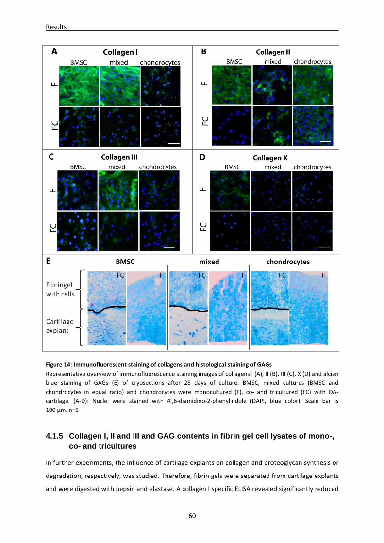
Results
60
E BMSC mixed chondrocytes
Figure 14: Immunofluorescent staining of collagens and histological staining of GAGs
Representative overview of immunofluorescence staining images of collagens I (A), II (B), III (C), X (D) and alcian
blue staining of GAGs (E) of cryosections after 28 days of culture. BMSC, mixed cultures (BMSC and
chondrocytes in equal ratio) and chondrocytes were monocultured (F), co- and tricultured (FC) with OA-
cartilage. (A-D): Nuclei were stained with 4’,6-diamidino-2-phenylindole (DAPI, blue color). Scale bar is
100 µm. n=5
4.1.5 Collagen I, II and III and GAG contents in fi brin gel cell lysates of mono-, co- and tricultures
In further experiments, the influence of cartilage explants on collagen and proteoglycan synthesis or
degradation, respectively, was studied. Therefore, fibrin gels were separated from cartilage explants
and were digested with pepsin and elastase. A collagen I specific ELISA revealed significantly reduced
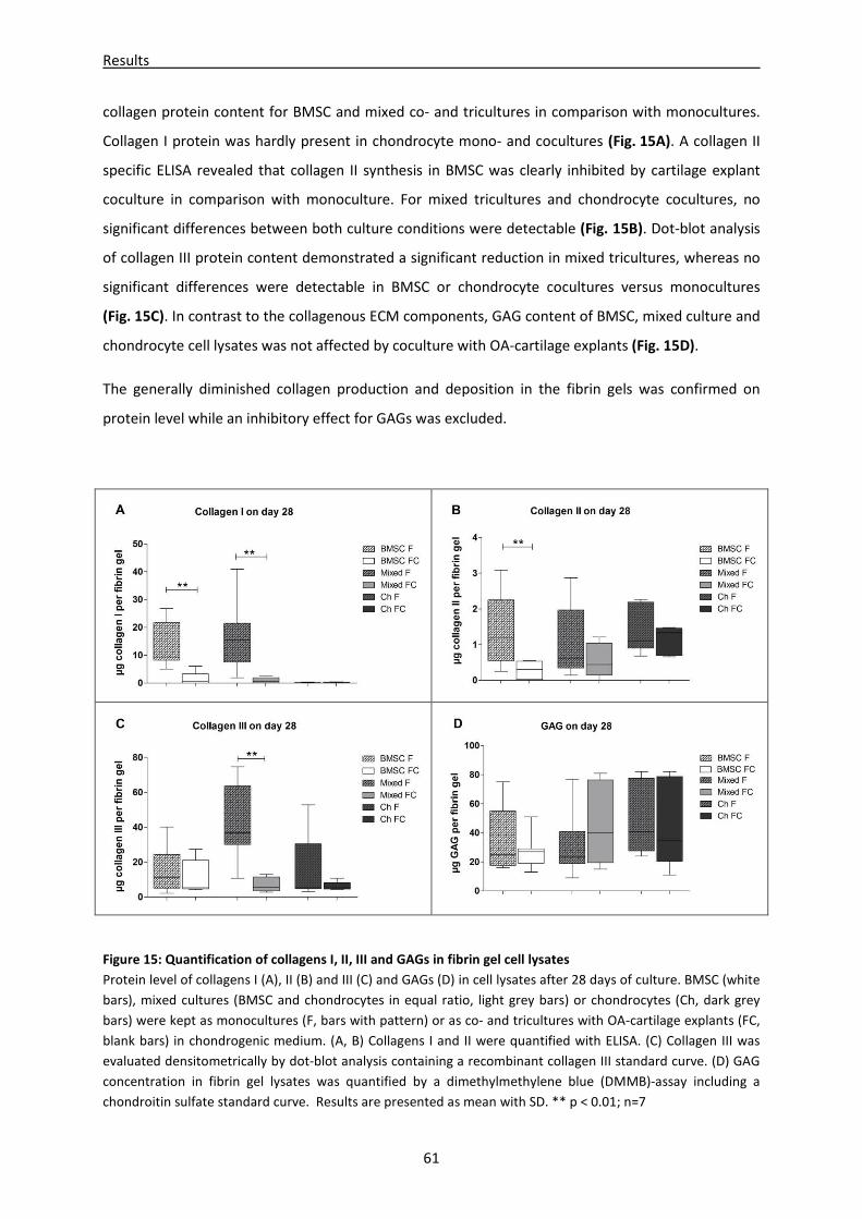
Results
61
collagen protein content for BMSC and mixed co- and tricultures in comparison with monocultures.
Collagen I protein was hardly present in chondrocyte mono- and cocultures (Fig. 15A). A collagen II
specific ELISA revealed that collagen II synthesis in BMSC was clearly inhibited by cartilage explant
coculture in comparison with monoculture. For mixed tricultures and chondrocyte cocultures, no
significant differences between both culture conditions were detectable (Fig. 15B). Dot-blot analysis
of collagen III protein content demonstrated a significant reduction in mixed tricultures, whereas no
significant differences were detectable in BMSC or chondrocyte cocultures versus monocultures
(Fig. 15C). In contrast to the collagenous ECM components, GAG content of BMSC, mixed culture and
chondrocyte cell lysates was not affected by coculture with OA-cartilage explants (Fig. 15D).
The generally diminished collagen production and deposition in the fibrin gels was confirmed on
protein level while an inhibitory effect for GAGs was excluded.
Figure 15: Quantification of collagens I, II, III and GAGs in fibrin gel cell lysates
Protein level of collagens I (A), II (B) and III (C) and GAGs (D) in cell lysates after 28 days of culture. BMSC (white
bars), mixed cultures (BMSC and chondrocytes in equal ratio, light grey bars) or chondrocytes (Ch, dark grey
bars) were kept as monocultures (F, bars with pattern) or as co- and tricultures with OA-cartilage explants (FC,
blank bars) in chondrogenic medium. (A, B) Collagens I and II were quantified with ELISA. (C) Collagen III was
evaluated densitometrically by dot-blot analysis containing a recombinant collagen III standard curve. (D) GAG
concentration in fibrin gel lysates was quantified by a dimethylmethylene blue (DMMB)-assay including a
chondroitin sulfate standard curve. Results are presented as mean with SD. ** p < 0.01; n=7
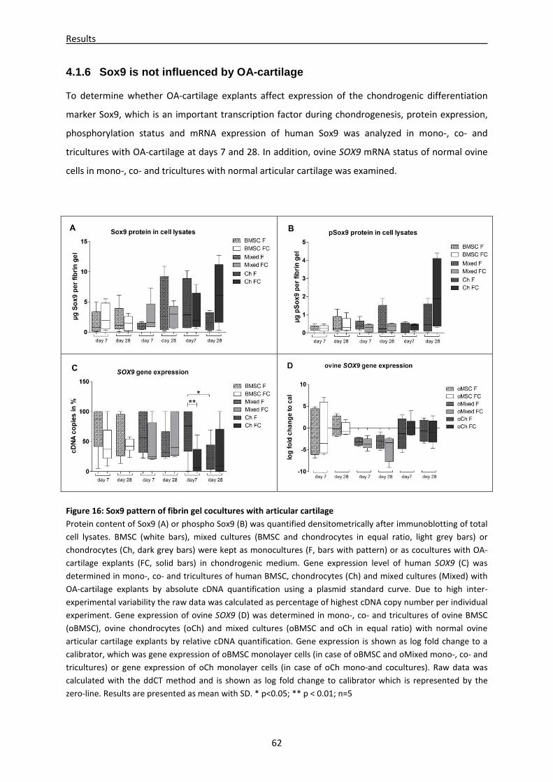
Results
62
4.1.6 Sox9 is not influenced by OA-cartilage
To determine whether OA-cartilage explants affect expression of the chondrogenic differentiation
marker Sox9, which is an important transcription factor during chondrogenesis, protein expression,
phosphorylation status and mRNA expression of human Sox9 was analyzed in mono-, co- and
tricultures with OA-cartilage at days 7 and 28. In addition, ovine SOX9 mRNA status of normal ovine
cells in mono-, co- and tricultures with normal articular cartilage was examined.
Figure 16: Sox9 pattern of fibrin gel cocultures with articular cartilage
Protein content of Sox9 (A) or phospho Sox9 (B) was quantified densitometrically after immunoblotting of total
cell lysates. BMSC (white bars), mixed cultures (BMSC and chondrocytes in equal ratio, light grey bars) or
chondrocytes (Ch, dark grey bars) were kept as monocultures (F, bars with pattern) or as cocultures with OA-
cartilage explants (FC, solid bars) in chondrogenic medium. Gene expression level of human SOX9 (C) was
determined in mono-, co- and tricultures of human BMSC, chondrocytes (Ch) and mixed cultures (Mixed) with
OA-cartilage explants by absolute cDNA quantification using a plasmid standard curve. Due to high inter-
experimental variability the raw data was calculated as percentage of highest cDNA copy number per individual
experiment. Gene expression of ovine SOX9 (D) was determined in mono-, co- and tricultures of ovine BMSC
(oBMSC), ovine chondrocytes (oCh) and mixed cultures (oBMSC and oCh in equal ratio) with normal ovine
articular cartilage explants by relative cDNA quantification. Gene expression is shown as log fold change to a
calibrator, which was gene expression of oBMSC monolayer cells (in case of oBMSC and oMixed mono-, co- and
tricultures) or gene expression of oCh monolayer cells (in case of oCh mono-and cocultures). Raw data was
calculated with the ddCT method and is shown as log fold change to calibrator which is represented by the
zero-line. Results are presented as mean with SD. * p<0.05; ** p < 0.01; n=5
Results
63
No significant differences in total Sox9 protein or phospho Sox9 protein expression in all coculture
regimens compared with monocultures were observed (Fig. 16A and B). Moreover, no differences in
SOX9 gene expression of BMSC and mixed culture conditions was found, but a significant
upregulation of SOX9 gene expression in chondrocyte monocultures in comparison with cartilage
cocultures on day 7. SOX9 gene expression in chondrocyte monocultures was significantly
downregulated from day 7 to day 28 (Fig. 16C). SOX9 gene expression of normal oBMSC, oMixed and
oCh mono-, co- or tricultures with normal ovine cartilage revealed no differences between culture
conditions (Fig. 16D).
In general, neither coculture with OA-cartilage nor with normal ovine cartilage had an effect on Sox9
gene or protein expression or phosphorylation status.
4.1.7 Comparison of soluble ECM fragments, PTHrP, b FGF and cytokines in supernatants of mono-, co- and tricultures
To quantify pro-inflammatory cytokines released from cell-free cartilage explants into culture
supernatants, ELISAs for IL-1β, IL-6 and IL-8 were performed. IL-1β concentration in supernatants of
normal and OA-cartilage explants cultured without cells showed no differences. Additionally, no
temporary differences were detectable for cell-free normal cartilage explants, whereas cell-free OA-
cartilage explants showed a significant increase of IL-1ß level from day 7 (1.1pg/mL +/- 0.7) to day 28
(3.0pg +/- 0.7) (Fig. 17A). IL-6 was only detected at day 7 in supernatant of cell-free normal cartilage
(103pg +/-292), whereas it was not detectable at day 28 or in cell-free OA-cartilage explants at days 7
and 28 (Fig. 17B). Concentration of IL-8 in supernatant of normal cell-free cartilage explants was
significantly higher at day 7 (4347pg/mL +/-1604) compared with day 28 (527pg +/-667). Similarly,
concentration of IL-8 in supernatants of cell-free OA-cartilage explants was significantly higher at
day 7 (2905pg/mL +/-1953) and was not detectable at day 28. Additionally, IL-8 concentration in
supernatant of normal cartilage explants was significantly higher than in OA-explants at day 28
(Fig. 17C). In cell-free normal and OA-cartilage explant supernatants soluble GAG concentration
(43µg/mL +/- 23) was comparable to soluble GAG measured in coculture supernatants (Fig. 17D).
ELISAs were performed to quantify the representative pro-inflammatory cytokines IL-1β, IL-6 and
IL-8, which are released into culture supernatants of mono-, co- and tricultures. Results revealed that
BMSC (day 28), mixed cultures (days 7 and 28) and chondrocytes (day 28) cultured with cartilage
explants secrete significant higher IL-1β levels at day 28 than monocultures. Monocultured
chondrocytes significantly reduced release of IL-1β from day 7 to day 28 (Fig. 18A). Furthermore,
cocultured BMSC, mixed cultures and chondrocytes had significantly elevated IL-6 and IL-8 levels at
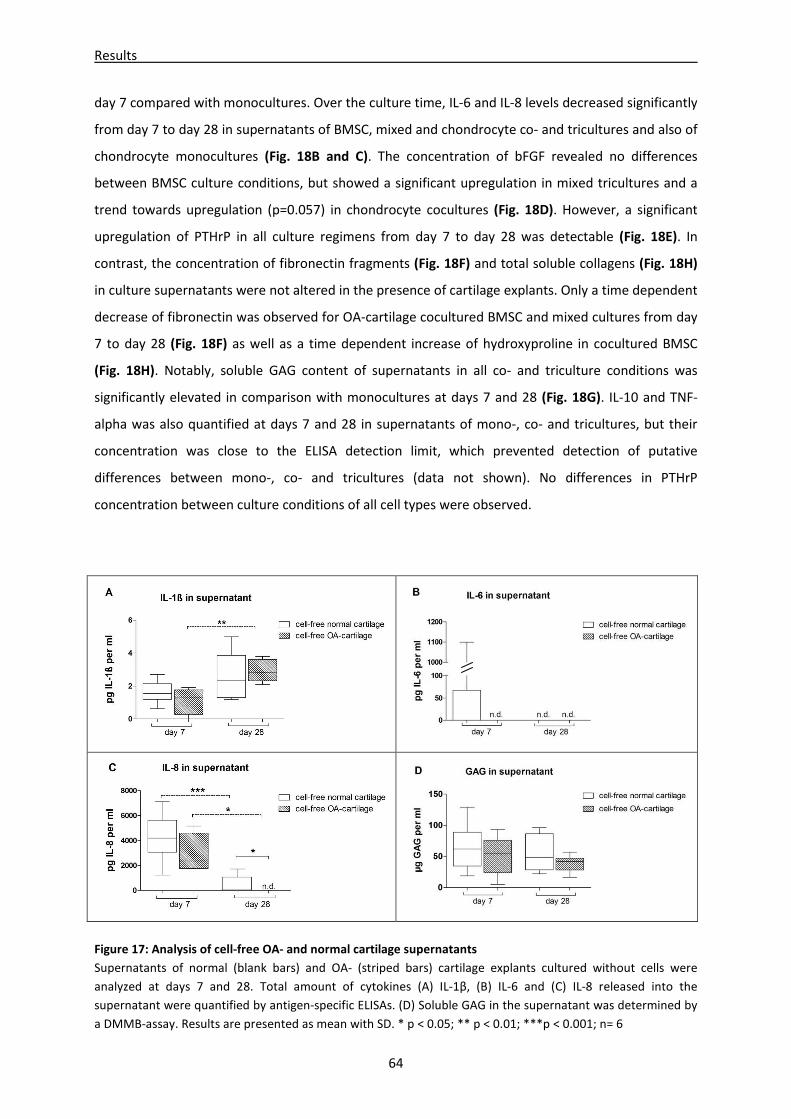
Results
64
day 7 compared with monocultures. Over the culture time, IL-6 and IL-8 levels decreased significantly
from day 7 to day 28 in supernatants of BMSC, mixed and chondrocyte co- and tricultures and also of
chondrocyte monocultures (Fig. 18B and C). The concentration of bFGF revealed no differences
between BMSC culture conditions, but showed a significant upregulation in mixed tricultures and a
trend towards upregulation (p=0.057) in chondrocyte cocultures (Fig. 18D). However, a significant
upregulation of PTHrP in all culture regimens from day 7 to day 28 was detectable (Fig. 18E). In
contrast, the concentration of fibronectin fragments (Fig. 18F) and total soluble collagens (Fig. 18H)
in culture supernatants were not altered in the presence of cartilage explants. Only a time dependent
decrease of fibronectin was observed for OA-cartilage cocultured BMSC and mixed cultures from day
7 to day 28 (Fig. 18F) as well as a time dependent increase of hydroxyproline in cocultured BMSC
(Fig. 18H). Notably, soluble GAG content of supernatants in all co- and triculture conditions was
significantly elevated in comparison with monocultures at days 7 and 28 (Fig. 18G). IL-10 and TNF-
alpha was also quantified at days 7 and 28 in supernatants of mono-, co- and tricultures, but their
concentration was close to the ELISA detection limit, which prevented detection of putative
differences between mono-, co- and tricultures (data not shown). No differences in PTHrP
concentration between culture conditions of all cell types were observed.
Figure 17: Analysis of cell-free OA- and normal cartilage supernatants
Supernatants of normal (blank bars) and OA- (striped bars) cartilage explants cultured without cells were
analyzed at days 7 and 28. Total amount of cytokines (A) IL-1β, (B) IL-6 and (C) IL-8 released into the
supernatant were quantified by antigen-specific ELISAs. (D) Soluble GAG in the supernatant was determined by
a DMMB-assay. Results are presented as mean with SD. * p < 0.05; ** p < 0.01; ***p < 0.001; n= 6
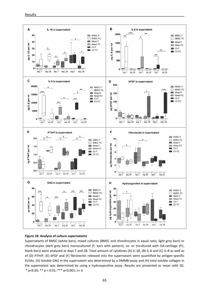
Results
65
Figure 18: Analysis of culture supernatants
Supernatants of BMSC (white bars), mixed cultures (BMSC and chondrocytes in equal ratio, light grey bars) or
chondrocytes (dark grey bars) monocultured (F, bars with pattern), co- or tricultured with OA-cartilage (FC,
blank bars) were analyzed at days 7 and 28. Total amount of cytokines (A) IL-1β, (B) IL-6 and (C) IL-8 as well as
of (D) PTHrP, (E) bFGF and (F) fibronectin released into the supernatant were quantified by antigen-specific
ELISAs. (G) Soluble GAG in the supernatant was determined by a DMMB-assay and (H) total soluble collagen in
the supernatant was determined by using a hydroxyproline assay. Results are presented as mean with SD.
* p<0.05; ** p < 0.01; *** p<0.001; n= 6
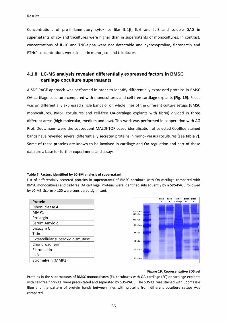
Results
66
Concentrations of pro-inflammatory cytokines like IL-1β, IL-6 and IL-8 and soluble GAG in
supernatants of co- and tricultures were higher than in supernatants of monocultures. In contrast,
concentrations of IL-10 and TNF-alpha were not detectable and hydroxyproline, fibronectin and
PTHrP concentrations were similar in mono-, co- and tricultures.
4.1.8 LC-MS analysis revealed differentially expres sed factors in BMSC cartilage coculture supernatants
A SDS-PAGE approach was performed in order to identify differentially expressed proteins in BMSC
OA-cartilage coculture compared with monocultures and cell-free cartilage explants (Fig. 19). Focus
was on differentially expressed single bands or on whole lines of the different culture setups (BMSC
monocultures, BMSC cocultures and cell-free OA-cartilage explants with fibrin) divided in three
different areas (high molecular, medium and low). This work was performed in cooperation with AG
Prof. Deutzmann were the subsequent MALDI-TOF based identification of selected CooBlue stained
bands have revealed several differentially secreted proteins in mono- versus cocultures (see table 7).
Some of these proteins are known to be involved in cartilage and OA regulation and part of these
data are a base for further experiments and assays.
Table 7: Factors identified by LC-SM analysis of supernatant
List of differentially secreted proteins in supernatants of BMSC coculture with OA-cartilage compared with
BMSC monocultures and cell-free OA cartilage. Proteins were identified subsequently by a SDS-PAGE followed
by LC-MS. Scores > 100 were considered significant.
Protein
Ribonuclease 4
MMP1
Prolargin
Serum Amyloid
Lysozym C
Titin
Extracellular superoxid dismutase
Chondroadherin
Fibronectin
IL-8
Stromelysin (MMP3)
Figure 19: Representative SDS gel
Proteins in the supernatants of BMSC monocultures (F), cocultures with OA-cartilage (FC) or cartilage explants
with cell-free fibrin gel were precipitated and separated by SDS-PAGE. The SDS gel was stained with Coomassie
Blue and the pattern of protein bands between lines with proteins from different coculture setups was
compared.
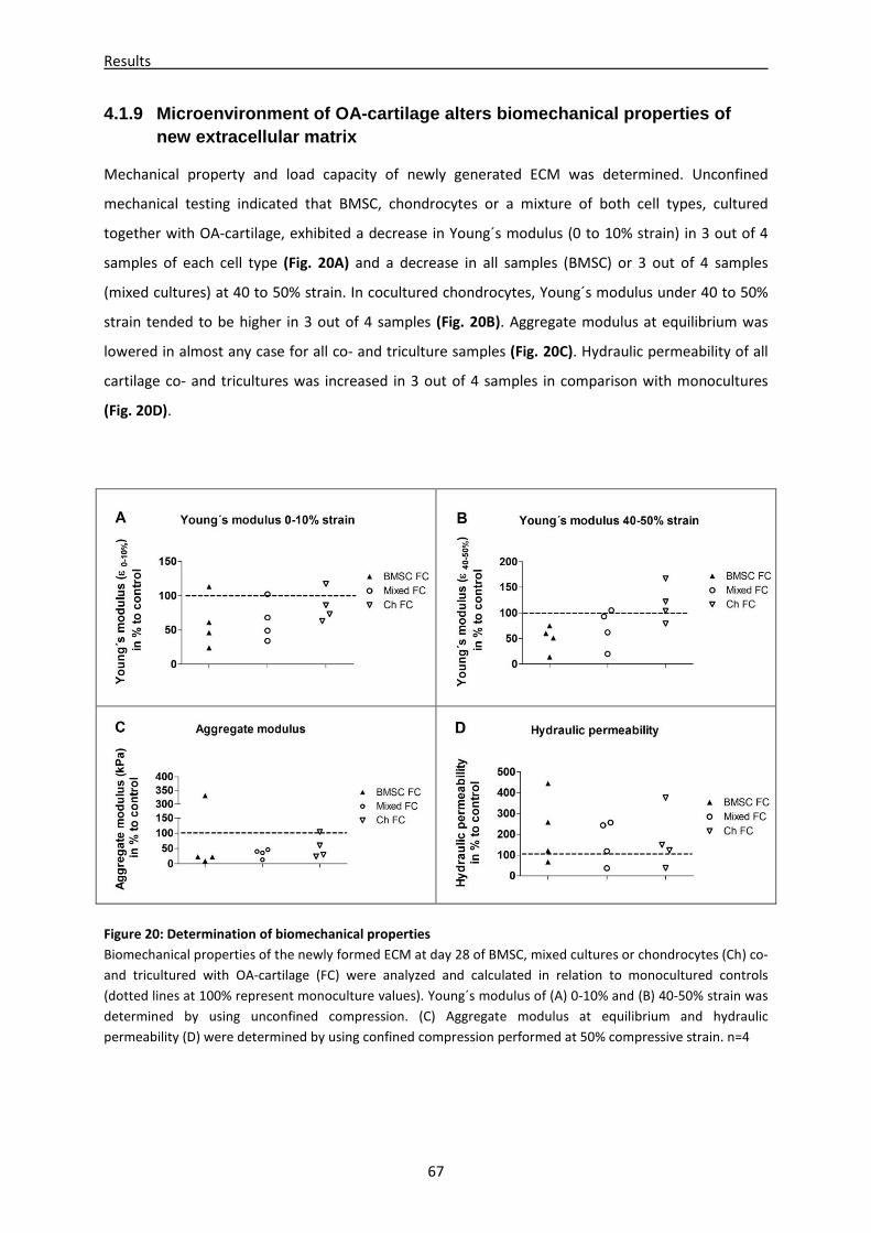
Results
67
4.1.9 Microenvironment of OA-cartilage alters biome chanical properties of new extracellular matrix
Mechanical property and load capacity of newly generated ECM was determined. Unconfined
mechanical testing indicated that BMSC, chondrocytes or a mixture of both cell types, cultured
together with OA-cartilage, exhibited a decrease in Young´s modulus (0 to 10% strain) in 3 out of 4
samples of each cell type (Fig. 20A) and a decrease in all samples (BMSC) or 3 out of 4 samples
(mixed cultures) at 40 to 50% strain. In cocultured chondrocytes, Young´s modulus under 40 to 50%
strain tended to be higher in 3 out of 4 samples (Fig. 20B). Aggregate modulus at equilibrium was
lowered in almost any case for all co- and triculture samples (Fig. 20C). Hydraulic permeability of all
cartilage co- and tricultures was increased in 3 out of 4 samples in comparison with monocultures
(Fig. 20D).
Figure 20: Determination of biomechanical properties
Biomechanical properties of the newly formed ECM at day 28 of BMSC, mixed cultures or chondrocytes (Ch) co-
and tricultured with OA-cartilage (FC) were analyzed and calculated in relation to monocultured controls
(dotted lines at 100% represent monoculture values). Young´s modulus of (A) 0-10% and (B) 40-50% strain was
determined by using unconfined compression. (C) Aggregate modulus at equilibrium and hydraulic
permeability (D) were determined by using confined compression performed at 50% compressive strain. n=4

Results
68
Overall, biomechanical tests showed that newly formed ECM in BMSC and mixed co- or tricultures
had lower Young´s and aggregate modulus, and increased hydraulic permeability compared with
monocultures. Mechanical parameters in chondrocyte cocultures were lower in case of Young´s
modulus (0-10%) and aggregate modulus and higher in case of Young´s modulus (40-50%) compared
with monocultures.
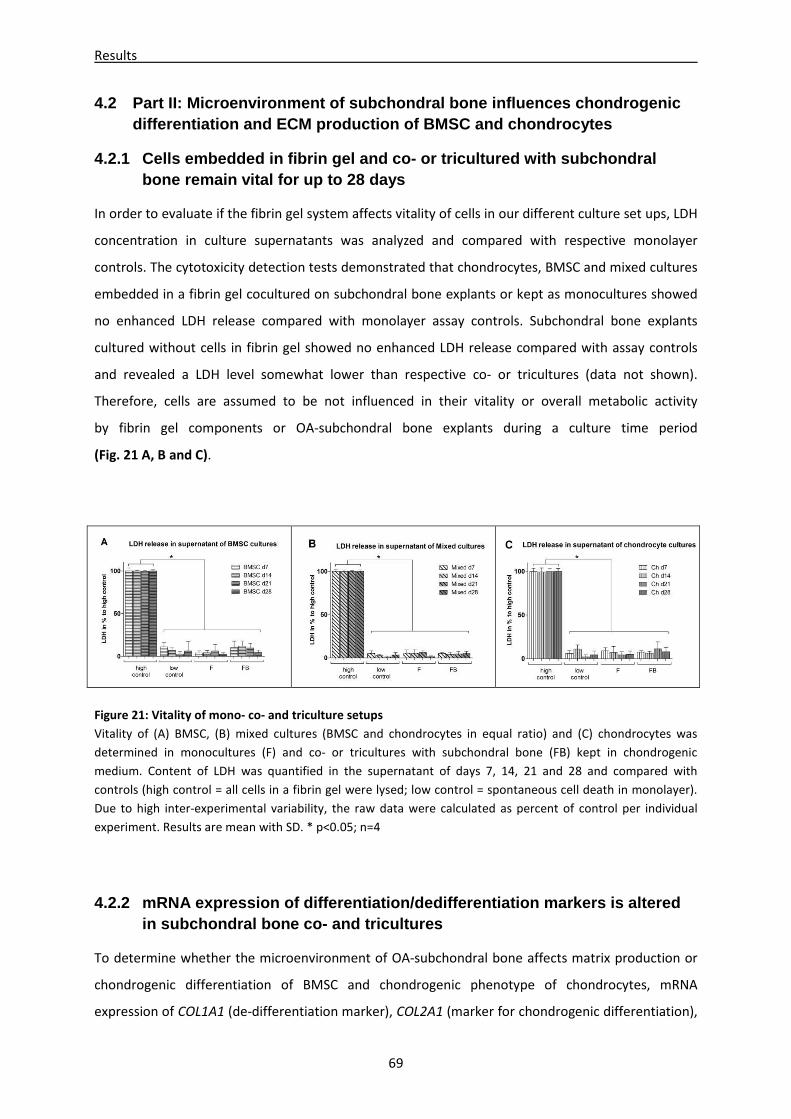
Results
69
4.2 Part II: Microenvironment of subchondral bone i nfluences chondrogenic differentiation and ECM production of BMSC and chon drocytes
4.2.1 Cells embedded in fibrin gel and co- or tricu ltured with subchondral bone remain vital for up to 28 days
In order to evaluate if the fibrin gel system affects vitality of cells in our different culture set ups, LDH
concentration in culture supernatants was analyzed and compared with respective monolayer
controls. The cytotoxicity detection tests demonstrated that chondrocytes, BMSC and mixed cultures
embedded in a fibrin gel cocultured on subchondral bone explants or kept as monocultures showed
no enhanced LDH release compared with monolayer assay controls. Subchondral bone explants
cultured without cells in fibrin gel showed no enhanced LDH release compared with assay controls
and revealed a LDH level somewhat lower than respective co- or tricultures (data not shown).
Therefore, cells are assumed to be not influenced in their vitality or overall metabolic activity
by fibrin gel components or OA-subchondral bone explants during a culture time period
(Fig. 21 A, B and C).
Figure 21: Vitality of mono- co- and triculture setups
Vitality of (A) BMSC, (B) mixed cultures (BMSC and chondrocytes in equal ratio) and (C) chondrocytes was
determined in monocultures (F) and co- or tricultures with subchondral bone (FB) kept in chondrogenic
medium. Content of LDH was quantified in the supernatant of days 7, 14, 21 and 28 and compared with
controls (high control = all cells in a fibrin gel were lysed; low control = spontaneous cell death in monolayer).
Due to high inter-experimental variability, the raw data were calculated as percent of control per individual
experiment. Results are mean with SD. * p<0.05; n=4
4.2.2 mRNA expression of differentiation/dedifferen tiation markers is altered in subchondral bone co- and tricultures
To determine whether the microenvironment of OA-subchondral bone affects matrix production or
chondrogenic differentiation of BMSC and chondrogenic phenotype of chondrocytes, mRNA
expression of COL1A1 (de-differentiation marker), COL2A1 (marker for chondrogenic differentiation),
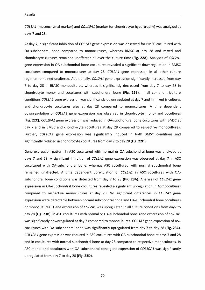
Results
70
COL3A1 (mesenchymal marker) and COL10A1 (marker for chondrocyte hypertrophy) was analyzed at
days 7 and 28.
At day 7, a significant inhibition of COL1A1 gene expression was observed for BMSC cocultured with
OA-subchondral bone compared to monocultures, whereas BMSC at day 28 and mixed and
chondrocyte cultures remained unaffected all over the culture time (Fig. 22A). Analyses of COL2A1
gene expression in OA-subchondral bone cocultures revealed a significant downregulation in BMSC
cocultures compared to monocultures at day 28. COL2A1 gene expression in all other culture
regimen remained unaltered. Additionally, COL2A1 gene expression significantly increased from day
7 to day 28 in BMSC monocultures, whereas it significantly decreased from day 7 to day 28 in
chondrocyte mono- and cocultures with subchondral bone (Fig. 22B). In all co- and triculture
conditions COL3A1 gene expression was significantly downregulated at day 7 and in mixed tricultures
and chondrocyte cocultures also at day 28 compared to monocultures. A time dependent
downregulation of COL3A1 gene expression was observed in chondrocyte mono- and cocultures
(Fig. 22C). COL10A1 gene expression was reduced in OA-subchondral bone cocultures with BMSC at
day 7 and in BMSC and chondrocyte cocultures at day 28 compared to respective monocultures.
Further, COL10A1 gene expression was significantly induced in both BMSC conditions and
significantly reduced in chondrocyte cocultures from day 7 to day 28 (Fig. 22D).
Gene expression pattern in ASC cocultured with normal or OA-subchondral bone was analyzed at
days 7 and 28. A significant inhibition of COL1A1 gene expression was observed at day 7 in ASC
cocultured with OA-subchondral bone, whereas ASC cocultured with normal subchondral bone
remained unaffected. A time dependent upregulation of COL1A1 in ASC cocultures with OA-
subchondral bone conditions was detected from day 7 to 28 (Fig. 23A). Analyses of COL2A1 gene
expression in OA-subchondral bone cocultures revealed a significant upregulation in ASC cocultures
compared to respective monocultures at day 28. No significant differences in COL2A1 gene
expression were detectable between normal subchondral bone and OA-subchondral bone cocultures
or monocultures. Gene expression of COL2A1 was upregulated in all culture conditions from day7 to
day 28 (Fig. 23B). In ASC cocultures with normal or OA-subchondral bone gene expression of COL3A1
was significantly downregulated at day 7 compared to monocultures. COL3A1 gene expression of ASC
cocultures with OA-subchondral bone was significantly upregulated from day 7 to day 28 (Fig. 23C).
COL10A1 gene expression was reduced in ASC cocultures with OA-subchondral bone at days 7 and 28
and in cocultures with normal subchondral bone at day 28 compared to respective monocultures. In
ASC mono- and cocultures with OA-subchondral bone gene expression of COL10A1 was significantly
upregulated from day 7 to day 28 (Fig. 23D).
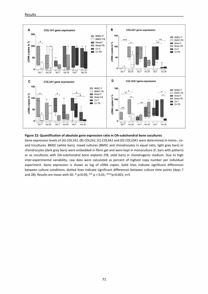
Results
71
Figure 22: Quantification of absolute gene expression ratio in OA-subchondral bone cocultures
Gene expression levels of (A) COL1A1, (B) COL2A1, (C) COL3A1 and (D) COL10A1 were determined in mono-, co-
and tricultures. BMSC (white bars), mixed cultures (BMSC and chondrocytes in equal ratio, light grey bars) or
chondrocytes (dark grey bars) were embedded in fibrin gel and were kept in monoculture (F, bars with pattern)
or as cocultures with OA-subchondral bone explants (FB, solid bars) in chondrogenic medium. Due to high
inter-experimental variability, raw data were calculated as percent of highest copy number per individual
experiment. Gene expression is shown as log of cDNA copies. Solid lines indicate significant differences
between culture conditions, dotted lines indicate significant differences between culture time points (days 7
and 28). Results are mean with SD. * p<0.05; ** p < 0.01; ***p<0.001; n=5
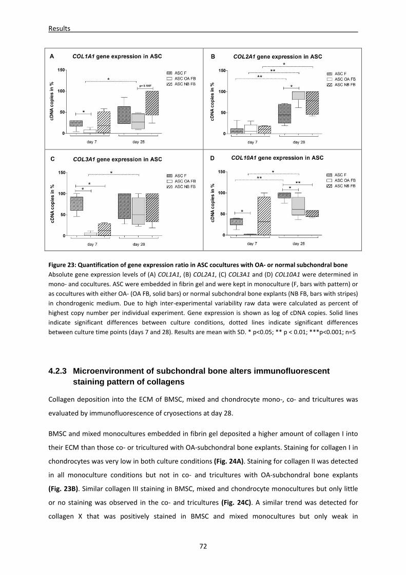
Results
72
Figure 23: Quantification of gene expression ratio in ASC cocultures with OA- or normal subchondral bone
Absolute gene expression levels of (A) COL1A1, (B) COL2A1, (C) COL3A1 and (D) COL10A1 were determined in
mono- and cocultures. ASC were embedded in fibrin gel and were kept in monoculture (F, bars with pattern) or
as cocultures with either OA- (OA FB, solid bars) or normal subchondral bone explants (NB FB, bars with stripes)
in chondrogenic medium. Due to high inter-experimental variability raw data were calculated as percent of
highest copy number per individual experiment. Gene expression is shown as log of cDNA copies. Solid lines
indicate significant differences between culture conditions, dotted lines indicate significant differences
between culture time points (days 7 and 28). Results are mean with SD. * p<0.05; ** p < 0.01; ***p<0.001; n=5
4.2.3 Microenvironment of subchondral bone alters i mmunofluorescent staining pattern of collagens
Collagen deposition into the ECM of BMSC, mixed and chondrocyte mono-, co- and tricultures was
evaluated by immunofluorescence of cryosections at day 28.
BMSC and mixed monocultures embedded in fibrin gel deposited a higher amount of collagen I into
their ECM than those co- or tricultured with OA-subchondral bone explants. Staining for collagen I in
chondrocytes was very low in both culture conditions (Fig. 24A). Staining for collagen II was detected
in all monoculture conditions but not in co- and tricultures with OA-subchondral bone explants
(Fig. 23B). Similar collagen III staining in BMSC, mixed and chondrocyte monocultures but only little
or no staining was observed in the co- and tricultures (Fig. 24C). A similar trend was detected for
collagen X that was positively stained in BMSC and mixed monocultures but only weak in
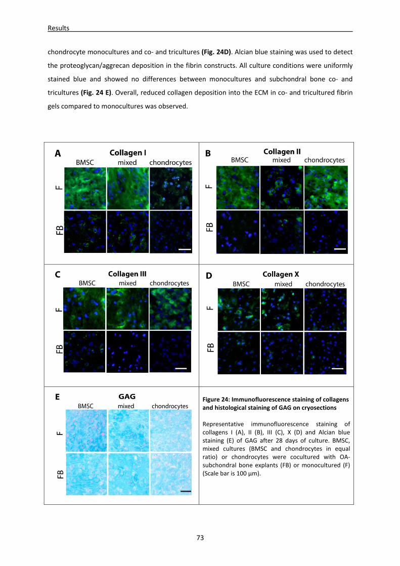
Results
73
chondrocyte monocultures and co- and tricultures (Fig. 24D). Alcian blue staining was used to detect
the proteoglycan/aggrecan deposition in the fibrin constructs. All culture conditions were uniformly
stained blue and showed no differences between monocultures and subchondral bone co- and
tricultures (Fig. 24 E). Overall, reduced collagen deposition into the ECM in co- and tricultured fibrin
gels compared to monocultures was observed.
Figure 24: Immunofluorescence staining of collagens
and histological staining of GAG on cryosections
Representative immunofluorescence staining of
collagens I (A), II (B), III (C), X (D) and Alcian blue
staining (E) of GAG after 28 days of culture. BMSC,
mixed cultures (BMSC and chondrocytes in equal
ratio) or chondrocytes were cocultured with OA-
subchondral bone explants (FB) or monocultured (F)
(Scale bar is 100 µm).
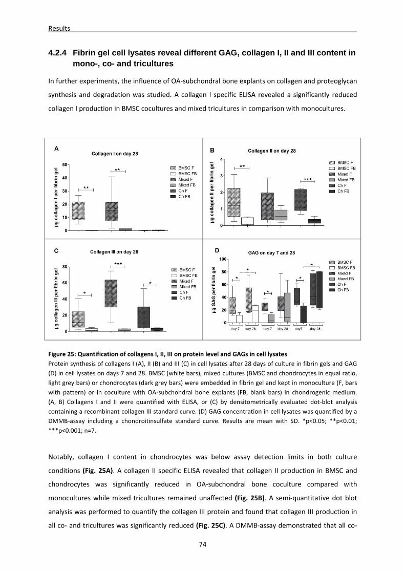
Results
74
4.2.4 Fibrin gel cell lysates reveal different GAG, collagen I, II and III content in mono-, co- and tricultures
In further experiments, the influence of OA-subchondral bone explants on collagen and proteoglycan
synthesis and degradation was studied. A collagen I specific ELISA revealed a significantly reduced
collagen I production in BMSC cocultures and mixed tricultures in comparison with monocultures.
Figure 25: Quantification of collagens I, II, III on protein level and GAGs in cell lysates
Protein synthesis of collagens I (A), II (B) and III (C) in cell lysates after 28 days of culture in fibrin gels and GAG
(D) in cell lysates on days 7 and 28. BMSC (white bars), mixed cultures (BMSC and chondrocytes in equal ratio,
light grey bars) or chondrocytes (dark grey bars) were embedded in fibrin gel and kept in monoculture (F, bars
with pattern) or in coculture with OA-subchondral bone explants (FB, blank bars) in chondrogenic medium.
(A, B) Collagens I and II were quantified with ELISA, or (C) by densitometrically evaluated dot-blot analysis
containing a recombinant collagen III standard curve. (D) GAG concentration in cell lysates was quantified by a
DMMB-assay including a chondroitinsulfate standard curve. Results are mean with SD. *p<0.05; **p<0.01;
***p<0.001; n=7.
Notably, collagen I content in chondrocytes was below assay detection limits in both culture
conditions (Fig. 25A). A collagen II specific ELISA revealed that collagen II production in BMSC and
chondrocytes was significantly reduced in OA-subchondral bone coculture compared with
monocultures while mixed tricultures remained unaffected (Fig. 25B). A semi-quantitative dot blot
analysis was performed to quantify the collagen III protein and found that collagen III production in
all co- and tricultures was significantly reduced (Fig. 25C). A DMMB-assay demonstrated that all co-
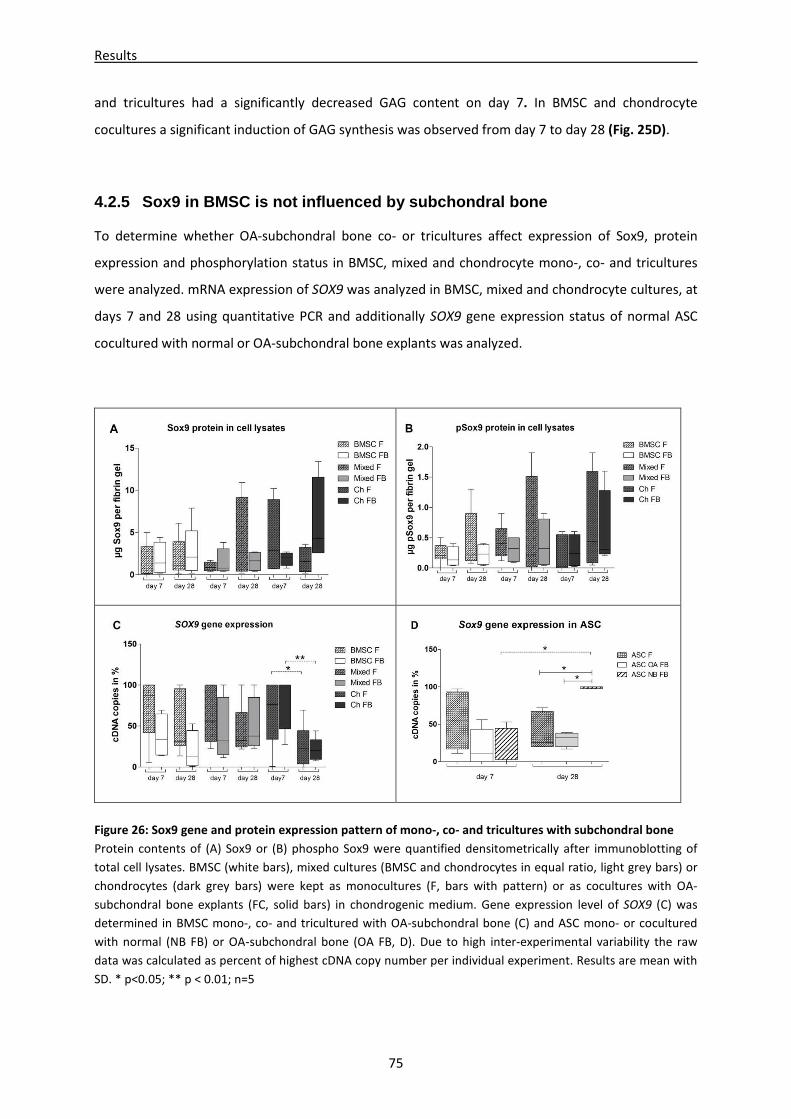
Results
75
and tricultures had a significantly decreased GAG content on day 7. In BMSC and chondrocyte
cocultures a significant induction of GAG synthesis was observed from day 7 to day 28 (Fig. 25D).
4.2.5 Sox9 in BMSC is not influenced by subchondral bone
To determine whether OA-subchondral bone co- or tricultures affect expression of Sox9, protein
expression and phosphorylation status in BMSC, mixed and chondrocyte mono-, co- and tricultures
were analyzed. mRNA expression of SOX9 was analyzed in BMSC, mixed and chondrocyte cultures, at
days 7 and 28 using quantitative PCR and additionally SOX9 gene expression status of normal ASC
cocultured with normal or OA-subchondral bone explants was analyzed.
Figure 26: Sox9 gene and protein expression pattern of mono-, co- and tricultures with subchondral bone
Protein contents of (A) Sox9 or (B) phospho Sox9 were quantified densitometrically after immunoblotting of
total cell lysates. BMSC (white bars), mixed cultures (BMSC and chondrocytes in equal ratio, light grey bars) or
chondrocytes (dark grey bars) were kept as monocultures (F, bars with pattern) or as cocultures with OA-
subchondral bone explants (FC, solid bars) in chondrogenic medium. Gene expression level of SOX9 (C) was
determined in BMSC mono-, co- and tricultured with OA-subchondral bone (C) and ASC mono- or cocultured
with normal (NB FB) or OA-subchondral bone (OA FB, D). Due to high inter-experimental variability the raw
data was calculated as percent of highest cDNA copy number per individual experiment. Results are mean with
SD. * p<0.05; ** p < 0.01; n=5
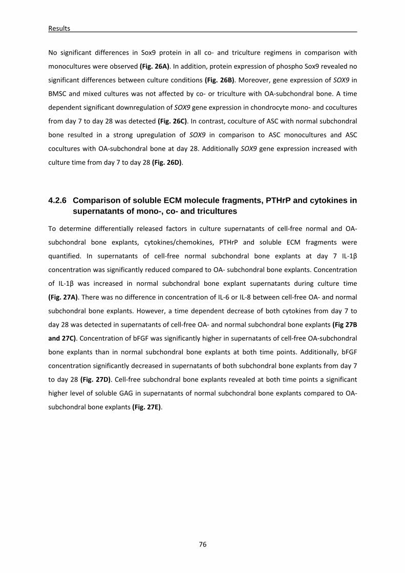
Results
76
No significant differences in Sox9 protein in all co- and triculture regimens in comparison with
monocultures were observed (Fig. 26A). In addition, protein expression of phospho Sox9 revealed no
significant differences between culture conditions (Fig. 26B). Moreover, gene expression of SOX9 in
BMSC and mixed cultures was not affected by co- or triculture with OA-subchondral bone. A time
dependent significant downregulation of SOX9 gene expression in chondrocyte mono- and cocultures
from day 7 to day 28 was detected (Fig. 26C). In contrast, coculture of ASC with normal subchondral
bone resulted in a strong upregulation of SOX9 in comparison to ASC monocultures and ASC
cocultures with OA-subchondral bone at day 28. Additionally SOX9 gene expression increased with
culture time from day 7 to day 28 (Fig. 26D).
4.2.6 Comparison of soluble ECM molecule fragments, PTHrP and cytokines in supernatants of mono-, co- and tricultures
To determine differentially released factors in culture supernatants of cell-free normal and OA-
subchondral bone explants, cytokines/chemokines, PTHrP and soluble ECM fragments were
quantified. In supernatants of cell-free normal subchondral bone explants at day 7 IL-1β
concentration was significantly reduced compared to OA- subchondral bone explants. Concentration
of IL-1β was increased in normal subchondral bone explant supernatants during culture time
(Fig. 27A). There was no difference in concentration of IL-6 or IL-8 between cell-free OA- and normal
subchondral bone explants. However, a time dependent decrease of both cytokines from day 7 to
day 28 was detected in supernatants of cell-free OA- and normal subchondral bone explants (Fig 27B
and 27C). Concentration of bFGF was significantly higher in supernatants of cell-free OA-subchondral
bone explants than in normal subchondral bone explants at both time points. Additionally, bFGF
concentration significantly decreased in supernatants of both subchondral bone explants from day 7
to day 28 (Fig. 27D). Cell-free subchondral bone explants revealed at both time points a significant
higher level of soluble GAG in supernatants of normal subchondral bone explants compared to OA-
subchondral bone explants (Fig. 27E).
Results
77
Figure 27: Analysis of cell-free OA- and normal
subchondral bone supernatants
Supernatants of normal (blank bars) and OA- (striped
bars) subchondral bone explants cultured without
cells were analyzed at days 7 and 28. Total amount
of cytokines (A) IL-1β, (B) IL-6 and (C) IL-8 released
into the supernatant were quantified by antigen-
specific ELISAs. (D) Soluble GAG in the supernatant
was determined by a DMMB-assay. Results are
presented as mean with SD. * p < 0.05; ** p < 0.01;
***p < 0.001; n= 6
In supernatants of ASC, BMSC, mixed and chondrocyte co- or tricultures with OA-subchondral bone
explants, significant more IL-1β was detected at day 28 compared to monocultures whereas
coculture of ASC with normal subchondral bone did not affect IL-1β release. In addition, mixed
tricultures and chondrocyte cocultures with OA-subchondral bone released more IL-1β into the
culture supernatant than ASC normal subchondral bone cocultures (Fig. 28A). All coculture regimen
(except for ASC OA-subchondral bone coculture) released more IL-6 into the culture supernatants at
day 7 than respective monocultures. In addition, a significantly higher IL-6 level was detected in
normal subchondral bone ASC cocultures compared to all other OA-subchondral bone co- and
triculture regimen at day 7 (Fig. 28B). IL-8 concentrations in supernatant of normal subchondral bone
cocultures of ASC and OA-subchondral bone cocultures of BMSC and chondrocytes at day 7 were
significantly elevated compared with respective monocultures. IL-8 levels of mixed cultures remained
unchanged. Moreover, IL-8 level in ASC cocultures with OA-subchondral bone was significantly lower
Results
78
than in ASC cocultured with normal subchondral bone (Fig. 28C). ASC, BMSC and chondrocyte co-
and tricultures released significantly more bFGF into supernatants in comparison with respective
monocultures while bFGF levels of mixed cultures remained unaffected by culture condition. In
supernatants of ASC cocultures with normal subchondral bone a significant higher level of bFGF was
detected than in supernatants of BMSC cocultures with OA-subchondral bone (Fig. 28D).
Furthermore, PTHrP concentration was not altered in BMSC, mixed and chondrocyte co- or
tricultures with OA-subchondral bone in comparison with respective monocultures at days 7 and 28.
However, a significant upregulation of PTHrP was observed in all samples from day 7 to day 28
(Fig. 28E). Analysis of supernatants for soluble fibronectin content did not reveal significant
differences (Fig. 28F). A significantly higher soluble GAG level was observed in supernatants of all co-
and tricultures at day 7 and of BMSC cocultures at day 28 (Fig. 28G). However, a hydroxyproline
assay revealed no significant differences in total soluble collagen content between all culture
conditions (Fig. 28H). As IL-10 and TNF-α were both at assay detection limits, no differences were
detected between co- and tricultures compared to respective monocultures (data not shown).
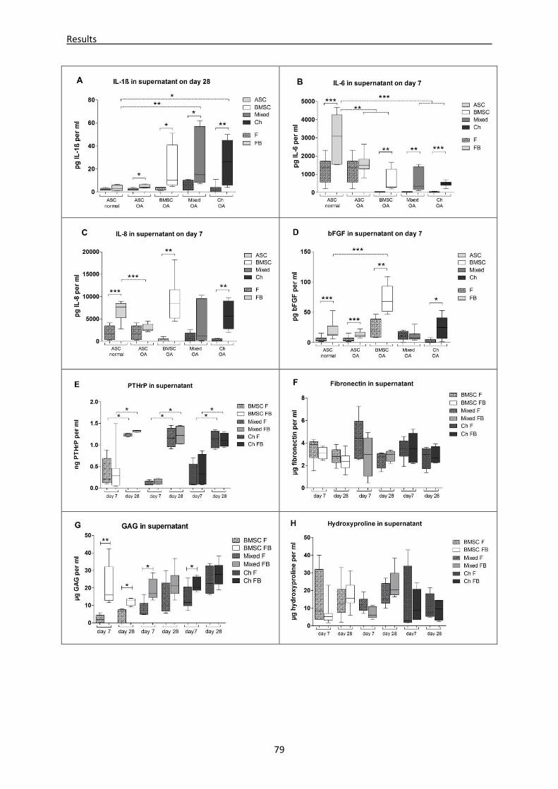
Results
79
Results
80
Figure 28: Quantification of cytokines, bFGF, PTHrP and GAGs in culture supernatants
Analysis of supernatants at days 7 and 28 of ASC (light grey bars), BMSC (white bars), mixed cultures (BMSC and
chondrocytes in equal ratio, medium grey bars) or chondrocytes (dark grey bars) monocultured (F, bars with
pattern) or cocultured with OA-subchondral bone explants (FB, blank bars). Total amount of cytokines (A) IL-1β,
(B) IL-6, (C) IL-8, (D) PTHrP, bFGF (E) and fibronectin (G) released into the supernatant were quantified by
antigen-specific ELISAs. Analysis of soluble GAG was carried out using a DMMB-assay containing a chondroitin
sulfate standard curve for quantification (F). Total soluble collagen was quantified using a hydroxyproline assay
(H). Solid lines indicate significant differences between culture conditions, (A-D): dotted lines indicate
significant differences between normal and OA- cartilage co- and tricultures, (E): dotted lines indicate
significant differences between culture time points (days 7 and 28). Results are mean with SD. *p<0.05,
**p<0.01, ***p<0.001; n= 7
Cell-free normal or OA-subchondral bone explants released IL-1β, IL-6, IL-8, bFGF and GAG into the
supernatant indicating an additive effect of bone tissue and co- or tricultured cells. Additionally,
differences in supernatants of normal bone cultures compared to OA-subchondral bone cultures
were detected for IL-1β, bFGF and soluble GAG concentrations. Overall, an increased level of IL-1β,
IL-6, IL-8, bFGF and GAG was observed in supernatants of co- and tricultures compared to respective
monocultures.
4.2.7 Microenvironment of OA-subchondral bone alter s biomechanical properties of newly regenerated cartilage
The quality and load capacity of newly generated cartilage-like tissue was determined by measuring
its biomechanical properties. Unconfined mechanical testing indicated that OA-subchondral bone co-
and tricultures with BMSC, chondrocytes or a mixture of both cell types (in equal ratio) embedded in
fibrin gel exhibited a decrease in Young´s modulus 0-10% strain in 4 of 4 samples (BMSC) and 3 of 4
samples (mixed and chondrocytes, Fig. 29A). Young´s modulus at 40-50% strain showed a decrease in
BMSC and mixed OA-subchondral bone co- and tricultures (4 of 4) while Young´s modulus of
chondrocytes (3 of 4) was increased (Fig. 29B). Aggregate modulus at equilibrium was reduced in all
OA-subchondral bone co- and tricultures compared to monocultures (Fig. 29C). Further, 3 of 4 BMSC
OA-subchondral bone coculture samples had a more than threefold higher hydraulic permeability
compared with monocultures, while no clear differences in hydraulic permeability were detected for
mixed and chondrocyte co- and tricultures compared with monocultures (Fig. 29D).
Taken together, our results suggest that co- and triculture of BMSC, mixed culture or chondrocytes
with OA-subchondral bone explants leads to changes in biomechanical properties of newly
regenerated cartilage like tissues.
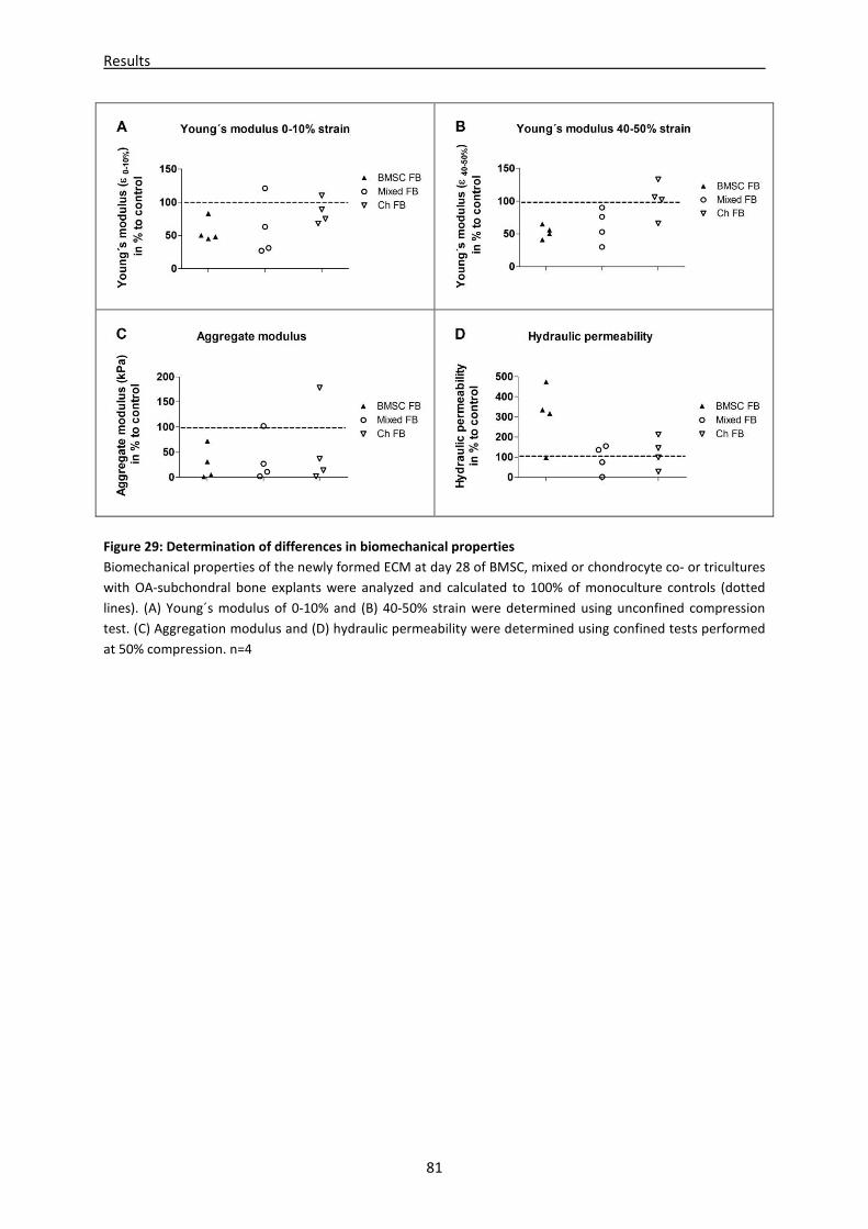
Results
81
Figure 29: Determination of differences in biomechanical properties
Biomechanical properties of the newly formed ECM at day 28 of BMSC, mixed or chondrocyte co- or tricultures
with OA-subchondral bone explants were analyzed and calculated to 100% of monoculture controls (dotted
lines). (A) Young´s modulus of 0-10% and (B) 40-50% strain were determined using unconfined compression
test. (C) Aggregation modulus and (D) hydraulic permeability were determined using confined tests performed
at 50% compression. n=4
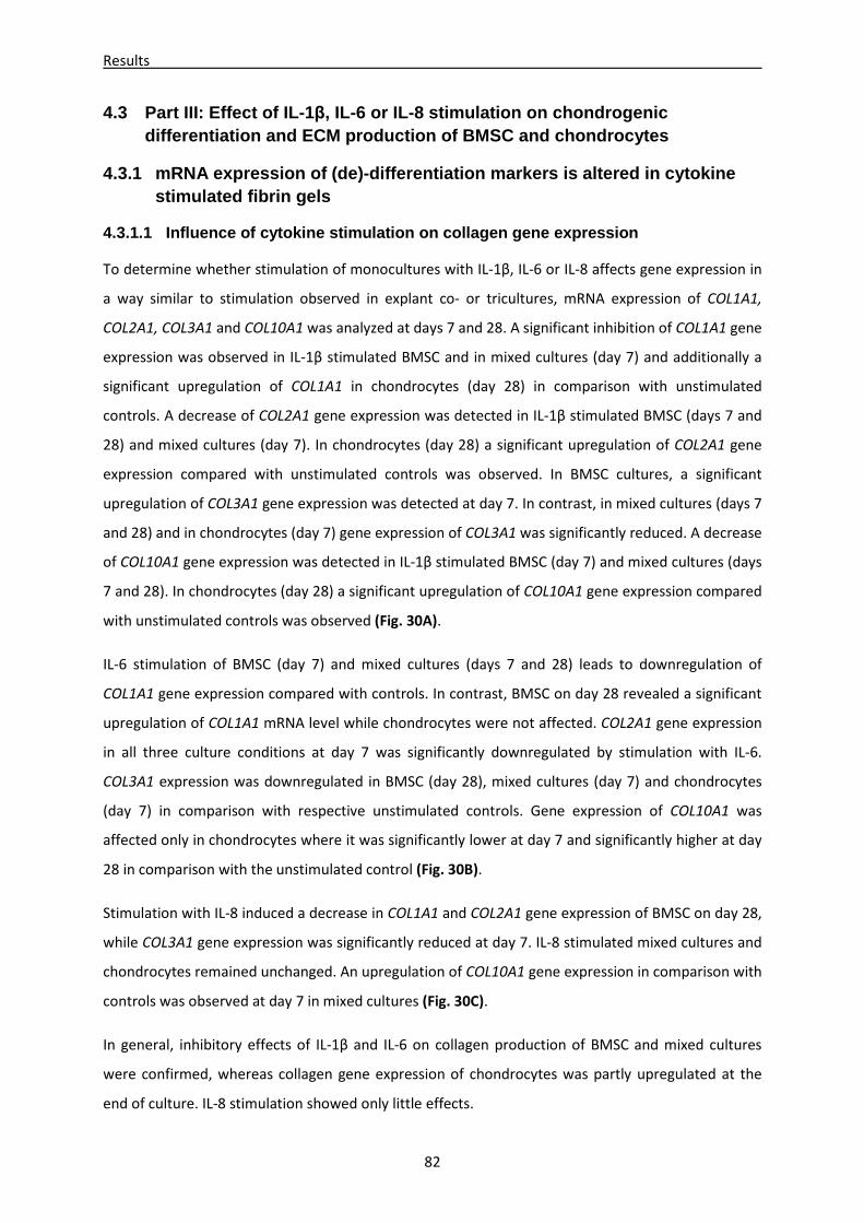
Results
82
4.3 Part III: Effect of IL-1 β, IL-6 or IL-8 stimulation on chondrogenic differentiation and ECM production of BMSC and chon drocytes
4.3.1 mRNA expression of (de)-differentiation marke rs is altered in cytokine stimulated fibrin gels
4.3.1.1 Influence of cytokine stimulation on collag en gene expression
To determine whether stimulation of monocultures with IL-1β, IL-6 or IL-8 affects gene expression in
a way similar to stimulation observed in explant co- or tricultures, mRNA expression of COL1A1,
COL2A1, COL3A1 and COL10A1 was analyzed at days 7 and 28. A significant inhibition of COL1A1 gene
expression was observed in IL-1β stimulated BMSC and in mixed cultures (day 7) and additionally a
significant upregulation of COL1A1 in chondrocytes (day 28) in comparison with unstimulated
controls. A decrease of COL2A1 gene expression was detected in IL-1β stimulated BMSC (days 7 and
28) and mixed cultures (day 7). In chondrocytes (day 28) a significant upregulation of COL2A1 gene
expression compared with unstimulated controls was observed. In BMSC cultures, a significant
upregulation of COL3A1 gene expression was detected at day 7. In contrast, in mixed cultures (days 7
and 28) and in chondrocytes (day 7) gene expression of COL3A1 was significantly reduced. A decrease
of COL10A1 gene expression was detected in IL-1β stimulated BMSC (day 7) and mixed cultures (days
7 and 28). In chondrocytes (day 28) a significant upregulation of COL10A1 gene expression compared
with unstimulated controls was observed (Fig. 30A).
IL-6 stimulation of BMSC (day 7) and mixed cultures (days 7 and 28) leads to downregulation of
COL1A1 gene expression compared with controls. In contrast, BMSC on day 28 revealed a significant
upregulation of COL1A1 mRNA level while chondrocytes were not affected. COL2A1 gene expression
in all three culture conditions at day 7 was significantly downregulated by stimulation with IL-6.
COL3A1 expression was downregulated in BMSC (day 28), mixed cultures (day 7) and chondrocytes
(day 7) in comparison with respective unstimulated controls. Gene expression of COL10A1 was
affected only in chondrocytes where it was significantly lower at day 7 and significantly higher at day
28 in comparison with the unstimulated control (Fig. 30B).
Stimulation with IL-8 induced a decrease in COL1A1 and COL2A1 gene expression of BMSC on day 28,
while COL3A1 gene expression was significantly reduced at day 7. IL-8 stimulated mixed cultures and
chondrocytes remained unchanged. An upregulation of COL10A1 gene expression in comparison with
controls was observed at day 7 in mixed cultures (Fig. 30C).
In general, inhibitory effects of IL-1β and IL-6 on collagen production of BMSC and mixed cultures
were confirmed, whereas collagen gene expression of chondrocytes was partly upregulated at the
end of culture. IL-8 stimulation showed only little effects.
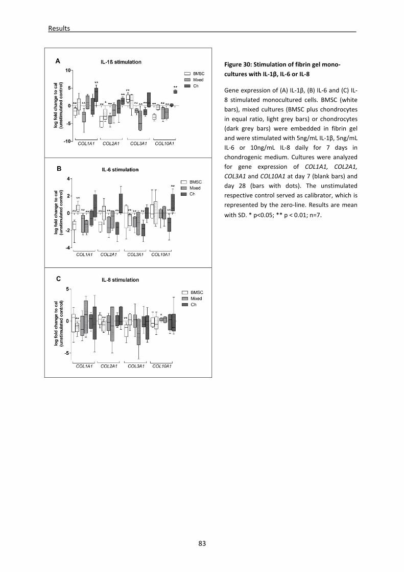
Results
83
Figure 30: Stimulation of fibrin gel mono-
cultures with IL-1β, IL-6 or IL-8
Gene expression of (A) IL-1β, (B) IL-6 and (C) IL-
8 stimulated monocultured cells. BMSC (white
bars), mixed cultures (BMSC plus chondrocytes
in equal ratio, light grey bars) or chondrocytes
(dark grey bars) were embedded in fibrin gel
and were stimulated with 5ng/mL IL-1β, 5ng/mL
IL-6 or 10ng/mL IL-8 daily for 7 days in
chondrogenic medium. Cultures were analyzed
for gene expression of COL1A1, COL2A1,
COL3A1 and COL10A1 at day 7 (blank bars) and
day 28 (bars with dots). The unstimulated
respective control served as calibrator, which is
represented by the zero-line. Results are mean
with SD. * p<0.05; ** p < 0.01; n=7.
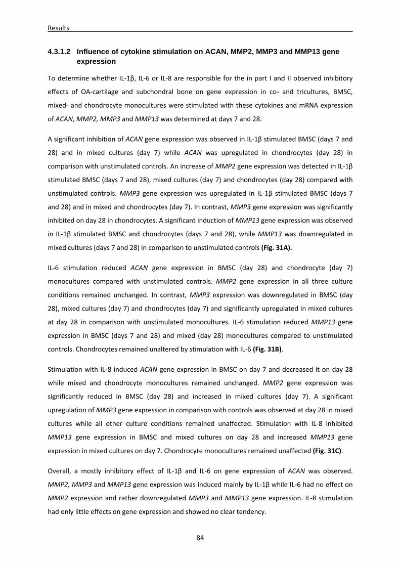
Results
84
4.3.1.2 Influence of cytokine stimulation on ACAN, MMP2, MMP3 and MMP13 gene expression
To determine whether IL-1β, IL-6 or IL-8 are responsible for the in part I and II observed inhibitory
effects of OA-cartilage and subchondral bone on gene expression in co- and tricultures, BMSC,
mixed- and chondrocyte monocultures were stimulated with these cytokines and mRNA expression
of ACAN, MMP2, MMP3 and MMP13 was determined at days 7 and 28.
A significant inhibition of ACAN gene expression was observed in IL-1β stimulated BMSC (days 7 and
28) and in mixed cultures (day 7) while ACAN was upregulated in chondrocytes (day 28) in
comparison with unstimulated controls. An increase of MMP2 gene expression was detected in IL-1β
stimulated BMSC (days 7 and 28), mixed cultures (day 7) and chondrocytes (day 28) compared with
unstimulated controls. MMP3 gene expression was upregulated in IL-1β stimulated BMSC (days 7
and 28) and in mixed and chondrocytes (day 7). In contrast, MMP3 gene expression was significantly
inhibited on day 28 in chondrocytes. A significant induction of MMP13 gene expression was observed
in IL-1β stimulated BMSC and chondrocytes (days 7 and 28), while MMP13 was downregulated in
mixed cultures (days 7 and 28) in comparison to unstimulated controls (Fig. 31A).
IL-6 stimulation reduced ACAN gene expression in BMSC (day 28) and chondrocyte (day 7)
monocultures compared with unstimulated controls. MMP2 gene expression in all three culture
conditions remained unchanged. In contrast, MMP3 expression was downregulated in BMSC (day
28), mixed cultures (day 7) and chondrocytes (day 7) and significantly upregulated in mixed cultures
at day 28 in comparison with unstimulated monocultures. IL-6 stimulation reduced MMP13 gene
expression in BMSC (days 7 and 28) and mixed (day 28) monocultures compared to unstimulated
controls. Chondrocytes remained unaltered by stimulation with IL-6 (Fig. 31B).
Stimulation with IL-8 induced ACAN gene expression in BMSC on day 7 and decreased it on day 28
while mixed and chondrocyte monocultures remained unchanged. MMP2 gene expression was
significantly reduced in BMSC (day 28) and increased in mixed cultures (day 7). A significant
upregulation of MMP3 gene expression in comparison with controls was observed at day 28 in mixed
cultures while all other culture conditions remained unaffected. Stimulation with IL-8 inhibited
MMP13 gene expression in BMSC and mixed cultures on day 28 and increased MMP13 gene
expression in mixed cultures on day 7. Chondrocyte monocultures remained unaffected (Fig. 31C).
Overall, a mostly inhibitory effect of IL-1β and IL-6 on gene expression of ACAN was observed.
MMP2, MMP3 and MMP13 gene expression was induced mainly by IL-1β while IL-6 had no effect on
MMP2 expression and rather downregulated MMP3 and MMP13 gene expression. IL-8 stimulation
had only little effects on gene expression and showed no clear tendency.
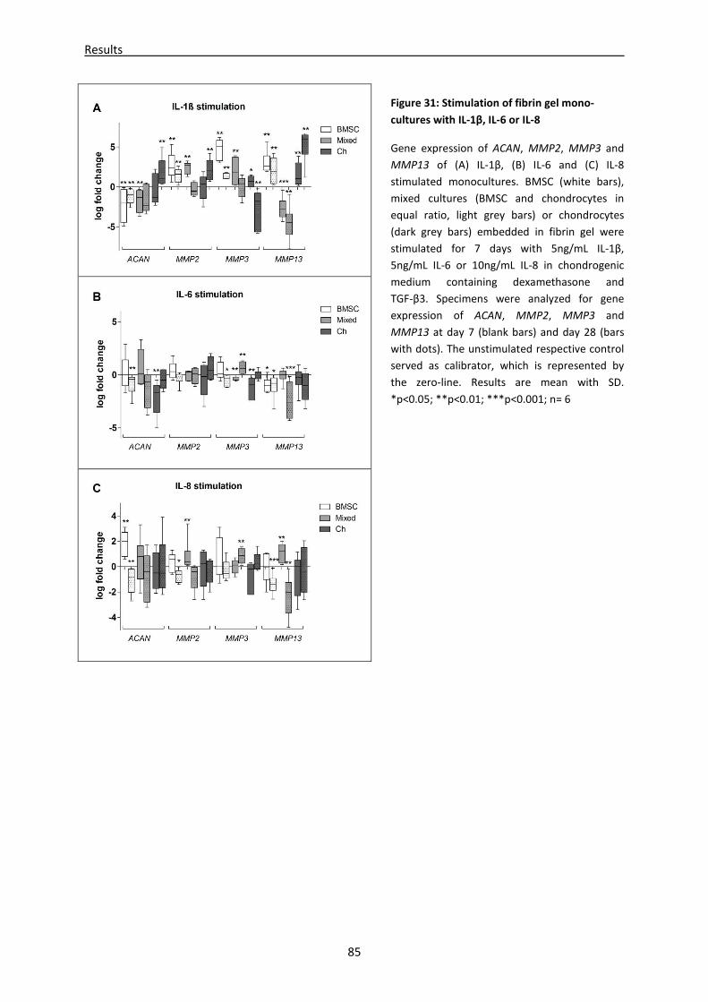
Results
85
Figure 31: Stimulation of fibrin gel mono-
cultures with IL-1β, IL-6 or IL-8
Gene expression of ACAN, MMP2, MMP3 and
MMP13 of (A) IL-1β, (B) IL-6 and (C) IL-8
stimulated monocultures. BMSC (white bars),
mixed cultures (BMSC and chondrocytes in
equal ratio, light grey bars) or chondrocytes
(dark grey bars) embedded in fibrin gel were
stimulated for 7 days with 5ng/mL IL-1β,
5ng/mL IL-6 or 10ng/mL IL-8 in chondrogenic
medium containing dexamethasone and
TGF-β3. Specimens were analyzed for gene
expression of ACAN, MMP2, MMP3 and
MMP13 at day 7 (blank bars) and day 28 (bars
with dots). The unstimulated respective control
served as calibrator, which is represented by
the zero-line. Results are mean with SD.
*p<0.05; **p<0.01; ***p<0.001; n= 6
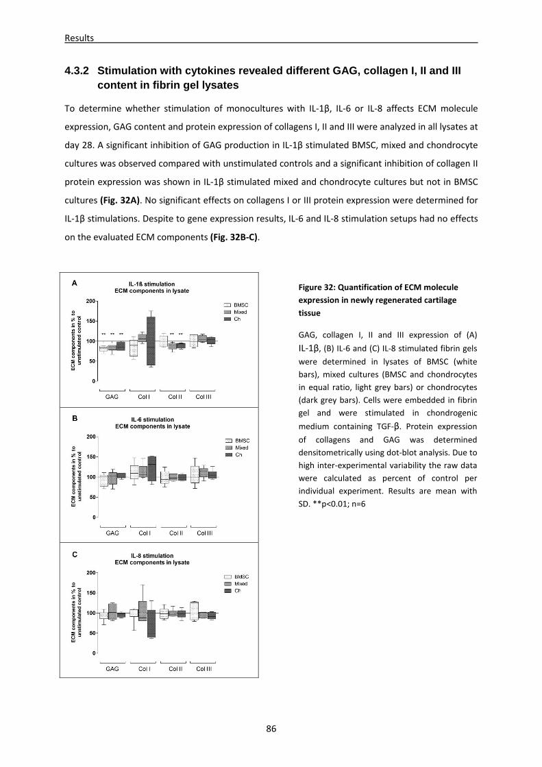
Results
86
4.3.2 Stimulation with cytokines revealed different GAG, collagen I, II and III content in fibrin gel lysates
To determine whether stimulation of monocultures with IL-1β, IL-6 or IL-8 affects ECM molecule
expression, GAG content and protein expression of collagens I, II and III were analyzed in all lysates at
day 28. A significant inhibition of GAG production in IL-1β stimulated BMSC, mixed and chondrocyte
cultures was observed compared with unstimulated controls and a significant inhibition of collagen II
protein expression was shown in IL-1β stimulated mixed and chondrocyte cultures but not in BMSC
cultures (Fig. 32A). No significant effects on collagens I or III protein expression were determined for
IL-1β stimulations. Despite to gene expression results, IL-6 and IL-8 stimulation setups had no effects
on the evaluated ECM components (Fig. 32B-C).
Figure 32: Quantification of ECM molecule
expression in newly regenerated cartilage
tissue
GAG, collagen I, II and III expression of (A)
IL-1β, (B) IL-6 and (C) IL-8 stimulated fibrin gels
were determined in lysates of BMSC (white
bars), mixed cultures (BMSC and chondrocytes
in equal ratio, light grey bars) or chondrocytes
(dark grey bars). Cells were embedded in fibrin
gel and were stimulated in chondrogenic
medium containing TGF-β. Protein expression
of collagens and GAG was determined
densitometrically using dot-blot analysis. Due to
high inter-experimental variability the raw data
were calculated as percent of control per
individual experiment. Results are mean with
SD. **p<0.01; n=6
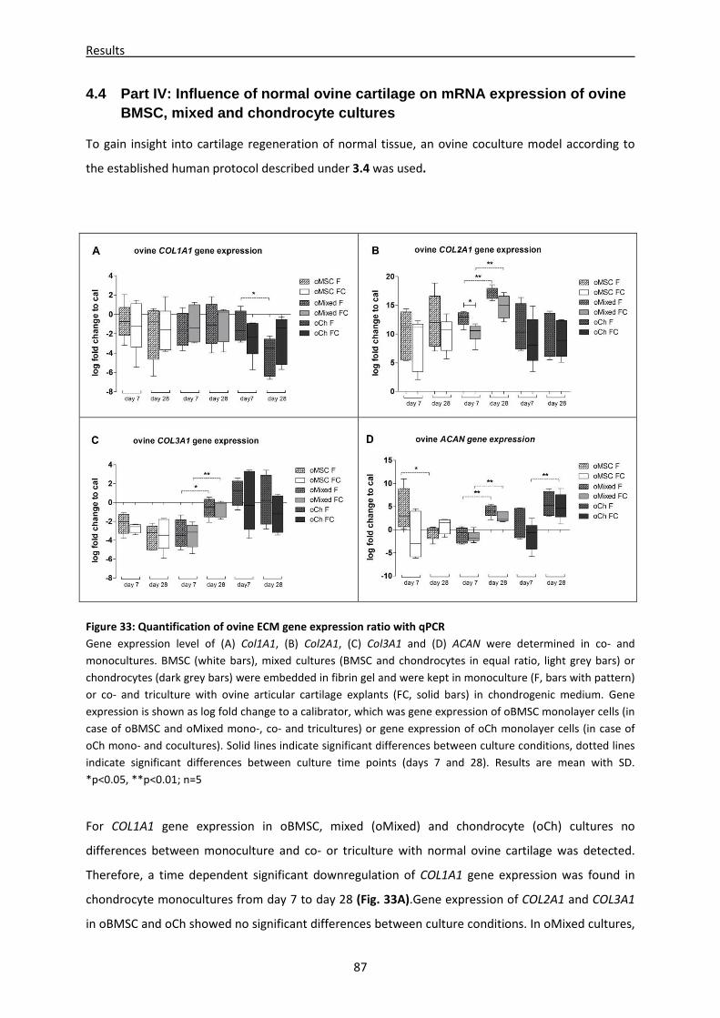
Results
87
4.4 Part IV: Influence of normal ovine cartilage on mRNA expression of ovine BMSC, mixed and chondrocyte cultures
To gain insight into cartilage regeneration of normal tissue, an ovine coculture model according to
the established human protocol described under 3.4 was used.
Figure 33: Quantification of ovine ECM gene expression ratio with qPCR
Gene expression level of (A) Col1A1, (B) Col2A1, (C) Col3A1 and (D) ACAN were determined in co- and
monocultures. BMSC (white bars), mixed cultures (BMSC and chondrocytes in equal ratio, light grey bars) or
chondrocytes (dark grey bars) were embedded in fibrin gel and were kept in monoculture (F, bars with pattern)
or co- and triculture with ovine articular cartilage explants (FC, solid bars) in chondrogenic medium. Gene
expression is shown as log fold change to a calibrator, which was gene expression of oBMSC monolayer cells (in
case of oBMSC and oMixed mono-, co- and tricultures) or gene expression of oCh monolayer cells (in case of
oCh mono- and cocultures). Solid lines indicate significant differences between culture conditions, dotted lines
indicate significant differences between culture time points (days 7 and 28). Results are mean with SD.
*p<0.05, **p<0.01; n=5
For COL1A1 gene expression in oBMSC, mixed (oMixed) and chondrocyte (oCh) cultures no
differences between monoculture and co- or triculture with normal ovine cartilage was detected.
Therefore, a time dependent significant downregulation of COL1A1 gene expression was found in
chondrocyte monocultures from day 7 to day 28 (Fig. 33A).Gene expression of COL2A1 and COL3A1
in oBMSC and oCh showed no significant differences between culture conditions. In oMixed cultures,
Results
88
COL2A1 was significantly reduced in ovine cartilage tricultures at day 7 compared with monocultures.
Additionally, there was a significant upregulation of COL2A1 and COL3A1 in both oMixed culture
conditions from day 7 to day 28. Gene expression of ACAN was downregulated in oBMSC
monocultures and upregulated in both oMixed cultures and oCh co- and tricultures from day 7 to 28
(Fig. 33B-D).
Overall, gene expression pattern of oBMSC, oMixed and oCh co- and tricultured with normal ovine
articular cartilage revealed no differences to respective monocultures.
Discussion
89
5 Discussion
5.1 General discussion
Since western population gets continuously older, the number of degenerative joint diseases like
osteoarthritis is steadily increasing (Hunter and Felson 2006). Various options for treatment of
symptoms are available, however there is only little chance to repair cartilage defects. Modern
medicine has opened BMSC as a potential source for tissue regeneration and plenty of new high-tech
biomaterials have been established in the last few years. Despite of numerous cell-based tissue-
engineering attempts, stem cell-based therapies are not jet venturous and artificial cartilage
reconstruction still needs improvement (Steinert, Ghivizzani et al. 2007; Grassel and Lorenz 2014).
Evidently, more intensive research in the field of stem cell-based OA reconstruction is needed.
Consequently, the focus of this study was on the usage of fibrin gel embedded BMSC and
chondrocytes, and the mixture of both cell types in equal ratio for cartilage regeneration in the
presence of OA-cartilage or subchondral bone.
BMSC are predicted to have a promising future in the field of tissue engineering, because they easily
can be isolated and expanded in order to be differentiated into repair cells and implanted in
respective sites of injury (Sylvester and Longaker 2004). In this study, stemness and differentiation
potential of isolated plastic adherent bone marrow cells was proven by flow cytometry with respect
to typical expression patterns of characteristic BMSC surface markers namely CD105pos, CD44pos,
CD34neg and CD19neg. As second criteria for their differentiation potential, BMSC were shown to
differentiate into chondrogenic, osteogenic and adipogenic lineages (Dominici, Le Blanc et al. 2006).
Even though these BMSC were obtained from OA-patients, they seemed not to be affected in their
differentiation capacity. These findings provide evidence to suggest that autologous obtained BMSC
from OA-patients contain the potential for cartilage tissue repair.
In order to discover differences between BMSC from normal and OA-donors, normal adipose derived
stem cells (ASC) were used for experiments with OA- or normal cartilage- and bone explants. These
ASC were previously shown by Schreml et al. to be capable for adipogenic, chondrogenic and
osteogenic differentiation and additionally expression pattern of characteristic BMSC surface
markers were analyzed by FACS (Schreml, Babilas et al. 2009). Therefore, ASC were a suitable
alternative cell source for verification of coculture results obtained with BMSC.
Human articular chondrocytes were isolated from different patients undergoing total knee
replacements due to OA. Despite critical selection of cartilage explants with similar OA grade, inter-
patient variations in cellular responses were not eliminated and resulted in a high variance of data
values. Not only different influences like genetics and previous drug therapy are affecting expression
Discussion
90
patterns – also the position of excised cartilage varies between patients. Symptoms of terminal stage
OA are heterogeneous in phenotype and underlying molecular mechanisms, in consequence
differences in key pathways or gene expression could be masked or misrepresented (Snelling, Rout et
al. 2014). As isolated chondrocytes were cultured up to 14 days in monolayer until they were
confluent, donors of chondrocytes and cartilage- or bone explants were not autologous. Additionally,
allogeneic BMSC were used for mixed coculture setups. Up to three different patients per approach
were combined resulting in even higher inter-patient variations, which impede generation of
statistical significances or trends. This phenomenon is widely known by usage of primarily patient
cells and can be countervailed by a high number of samples (Snelling, Rout et al. 2014).
Several studies indicate that BMSC alter the cytokine secretion profile of different cocultured
immune cells into a more anti-inflammatory or more tolerant phenotype (Aggarwal and Pittenger
2005). BMSC provide anti-inflammatory factors, which might interfere with inflammatory factors
released from OA-cartilage explants leading to a positive effect on differentiation and ECM
production. However, until now, immunosuppressive properties were not reported for chondrocytes
and thus explant cocultivation with chondrocytes was an important part for verification of BMSC
modulated effects.
To establish a reproducible coculture model for investigation of specifics in OA-cartilage repair, it was
necessary to verify survival and proliferation of cells in this setup. Analysis of supernatants for LDH, a
marker for cell-death, showed that neither cultivation of cells embedded in fibrin gel nor coculture
with cartilage or subchondral bone explants diminished cell vitality of BMSC, mixed or chondrocyte
cultures compared with respective control cells (cells differentiated in monolayer). This vitality test
revealed that local cells in OA-cartilage and OA-subchondral bone explants had a similar spontaneous
death rate than cells cultured in monolayer. Additionally, these cells are still metabolically active and
potentially paracrine regulative.
An important premise for chondrogenic differentiation is a moderate proliferative activity. Thereto
staining of PCNA, a marker for cell proliferation, in cryo sections of cell-fibrin gels was positive in
some nuclei equally distributed over the whole slice suggesting proliferation of a subpopulation of
fibrin gel embedded cells. Furthermore, no differences between monoculture and OA-cartilage co- or
triculture were observed indicating that fibrin gel or OA-cartilage had no influence on cell
proliferation in this culture set up.
Taken together, microenvironment of OA-cartilage or subchondral bone has no cytotoxic or
cytostatic features. Moreover, results from vitality tests with explants cultured without cell-fibrin
gels suggested that local cells in the explants are vital and metabolically active. Thus, it is likely, that
Discussion
91
these local cells in the explant release similar factors into the culture supernatant, as they would do
in vivo. In this study, usage of OA-tissues suggests mainly the release of pro-inflammatory cytokines
and degradation products of ECM molecules.
In detail, the coculture setups of this study combined two different conditions. At first, cells were
embedded in a fibrin gel matrix to provide a 3D chondrogenic surrounding which enhances
chondrogenic differentiation of BMSC, maintenance of articular phenotype and production of a
cartilage specific ECM (Lutolf and Hubbell 2005). Fibrin is the perfect biomaterial for this cast
because it occurs in the natural human healing system. In many studies it was successfully used
together with stem cells or chondrocytes for cartilage repair and negative influences on
chondrogenic differentiation are not known (Fussenegger, Meinhart et al. 2003; Park, Yang et al.
2009). Chondrogenic differentiation auxiliary was induced by chondrogenic medium containing
TGF-ß3 and dexamethasone (Ahmed, Dreier et al. 2007). Interestingly, dexamethasone is reported to
act chondroprotective and to decrease joint inflammation as well as joint tissue degradation
(Huebner, Shrive et al. 2014). A chondrogenic phenotype and matrix production was detected in all
monoculture conditions, with respect to present data. A second aspect of our setup was combination
of this chondrogenesis supporting fibrin gel and medium with OA-cartilage or subchondral bone
explants providing a diseased and inflamed microenvironment like in OA-patients (Tchetina, Squires
et al. 2005). Nevertheless, changes in the differentiation pattern of BMSC with respect to COL2A1
and COL10A1 expression as well as alterations in ECM production and composition in newly
generated cartilage like tissue were postulated by coculture with diseased OA-explants (Ahmed,
Dreier et al. 2007).
Readout parameters of this study were gene expression of typical marker genes namely COL1A1, a
de-differentiation marker (Marlovits, Hombauer et al. 2004; Cheng, Maddox et al. 2012), COL2A1, a
chondrogenic differentiation marker (Mendler, Eich-Bender et al. 1989; Bruckner and van der Rest
1994), COL3A1, a mesenchymal cell marker (Ku, Johnson et al. 2006; Juncosa-Melvin, Matlin et al.
2007)), COL10A1 (hypertrophic chondrocyte marker (Schmid and Linsenmayer 1985), and SOX9,
master transcription factor during chondrogenesis (de Crombrugghe, Lefebvre et al. 2000; Hattori,
Muller et al. 2010).
These marker genes indicate the functional status of BMSC and chondrocytes like de-differentiation
and chondrogenic- or terminal differentiation. Further production and functionality of newly
generated ECM was assayed by biochemical and biomechanical tests. Additionally, supernatants of
cocultures were analyzed. These supernatants are partly representing the unique OA-
microenvironment, which is characterized by soluble ECM fragments, newly generated ECM
precursor molecules and typical factors like pro-inflammatory cytokines, MMPs or proteases. These
Discussion
92
typical factors are either newly produced by local cells in the explant or released from the OA-
cartilage ECM to which they were previously bound. In this study, focus of research was restricted on
soluble molecules mainly components of the ECM as collagens, GAGs and fibronectin as well as
proinflammatory factors like cytokines and chemokines, which are known to be involved in cell
differentiation and alterations of ECM homeostasis. In addition, screening of supernatants for yet
unknown factors relevant for regulatory mechanisms during OA progression was performed via
LC-MS analysis to discover shifts in the secretome of BMSC mono- and cocultures with cartilage
explants.
Apparently, up to now, no other groups have analyzed the in vitro influence of OA-tissue on cartilage
regeneration with BMSC, chondrocytes or an equal mixture of both cell types embedded in fibrin gel.
This novel and innovative study gives new important insights in mechanisms, which occur during
BMSC differentiation and ECM production influenced by OA-tissue. In future, this knowledge could
be used for treatment of patients suffering from OA.
Discussion
93
5.2 Part I: Microenvironment of cartilage coculture influences BMSC differentiation and ECM production
Traumatic focal cartilage defects do not heal spontaneously, consequently stable long-term repair
and regeneration of destroyed articular cartilage needs innovative therapy strategies like cell-based
tissue engineering. At the site of injury, chondrocytes and in few cases BMSC were implanted, but
not much attention was paid to effects of neighboring cells and microenvironment provided by
remaining cartilage tissue. Treatment of BMSC with growth factors like TGF-ß resulted in induction of
mRNA and protein expression of several chondrogenic markers (Diederichs, Baral et al. 2012). Similar
reactions were observed for cocultures of BMSC with normal articular rat cartilage or human OA-
chondrocytes, which are known to induce chondrogenic differentiation by release of paracrine
factors (Ahmed, Dreier et al. 2007; Aung, Gupta et al. 2011). However, reports of proper OA-cartilage
regeneration resulting in an ECM with high biomechanical properties and a stable articular
phenotype are scarce.
Although many studies have examined effects of culture medium supplemented with well-defined
chondrogenic factors on differentiation capacity of BMSC, only few have addressed the influence of
OA-affected cartilage ECM or OA-chondrocytes on formation of matrix in cocultured cells and on
differentiation of BMSC. In order to identify culture conditions favoring proper matrix production,
chondrogenic differentiation and phenotype stability, a novel co- and triculture system was
established. BMSC, OA-chondrocytes and mixed cultures were embedded in a fibrin gel bio-matrix
providing a 3D environment, which mimics the natural habitat of chondrocytes and promotes
chondrogenic differentiation of BMSC (Lutolf and Hubbell 2005; Lee, Yu et al. 2008). To mimic an OA-
microenvironment, cell-fibrin gels were cocultured on top of OA-cartilage explants.
Achievement of the first part of this thesis was the realization that coculture with OA-cartilage
explants influenced gene expression and biosynthesis of collagens I, II, III and X in all co- and
triculture regimens and altered biomechanical properties presumable because of released regulatory
factors (Leyh, Seitz et al. 2014 a).
In detail, COL1A1, a marker for dedifferentiation was significantly reduced in all coculture conditions
compared with monoculture at day 7 possibly representing an initial diminished dedifferentiation
potential of cells cultured together with cartilage. Similar observations were made for COL3A1, a
mesenchymal collagen highly expressed in undifferentiated BMSC, which was also significantly
reduced in all cartilage co- and tricultures. A downregulation of COL3A1 in cocultured fibrin gels
during chondrogenic differentiation thus might denote a higher percentage of chondrogenic
Discussion
94
differentiated BMSC in presence of cartilage. With respect to chondrogenic differentiation of BMSC
suppression of collagen I and III gene expression via cartilage derived factors might be positive.
However, significant upregulation of COL2A1, a positive chondrogenic differentiation marker, over
time indicated positive chondrogenic differentiation in both mono- and coculture conditions of
BMSC, but cocultured BMSC exhibited significant less upregulation of COL2A1 than cells in
monoculture. In addition, COL2A1 gene expression of mixed- and chondrocyte cultures in presence
of OA-cartilage was significantly reduced at day 7, which could indicate a diminished or at least
delayed initiation of chondrogenic differentiation or collagen II production. Besides, mixed cultures
showed no significant upregulation of COL2A1 gene expression during culture time. Presumably,
chondrocytes produced large amounts of cartilage specific mRNA, mainly COL2A1, from the
beginning, which obscures the rising mRNA levels of BMSC in the mixed culture. A time dependent
downregulation of COL2A1 in chondrocyte monocultures from day 7 to day 28 might be a hint to
inhibition of matrix production induced in chondrocytes.
In this study, gene expression of the hypertrophic marker COL10A1 was strongly downregulated in
the presence of OA cartilage explants in all co- and triculture regimens. This inhibitory effect appears
not to be limited to OA-cartilage since it was previously described for normal BMSC-cartilage
cocultures (Ahmed, Dreier et al. 2007). One major problem in using BMSC for cell-based cartilage
defect repair is the instability of the chondrogenic phenotype, which tends to progress to a
hypertrophic phenotype with subsequent entering the endochondral ossification pathway. With
respect to COL10A1 gene expression coculture with cartilage could provide a protective effect, and
could promote stability of the chondrogenic versus hypertrophic phenotype.
Taken together, collagen gene expression of BMSC cocultures was inhibited on day 28 indicating an
inhibitory effect of OA-cartilage. In contrast, OA-chondrocytes showed signs of inhibition only for the
first days of OA-cartilage coculture. Furthermore, a time dependent downregulation of COL2A1 and
COL3A1 was observed for chondrocytes in monocultures. It seems that monocultured chondrocytes
start to produce plenty of ECM proteins (with respect to collagen mRNA) as soon as they are
unhinged from matrix, but once they are again trapped in the newly synthesized ECM gene
expression decreases. In addition, gene expression of collagens was significantly inhibited in
chondrocyte cocultures in comparison to monocultures of day 7, whereas at day 28 the ECM gene
expression pattern of chondrocytes in monoculture, which at that time were embedded by newly
generated matrix, is similar to chondrocytes in cartilage coculture. These results might be a hint to
soluble cartilage derived inhibitory factors which production is induced in chondrocytes by cell-ECM
contact to newly generated ECM (Leyh, Seitz et al. 2014 a).
Discussion
95
In concert with results from qPCR analysis, fibrin gels indicated matrix deposition in both culture
conditions, which was demonstrated by color changes of the fibrin gel from translucent to opaque
and also sensed by an explicit rise of stiffness. Analysis of biochemical composition of the newly
generated ECM via ELISA and immunofluorescent staining reassured in general inhibition of collagen
protein production by OA-microenvironment. In detail, BMSC revealed significant less collagen I and
II deposition in coculture with cartilage explants and mixed cocultures revealed significantly reduced
collagen I and III deposition. In contrast, no corresponding collagen X immunostaining in all
chondrocyte and mixed cultures and only little reactivity in BMSC monocultures was detectable.
Several studies showed hypertrophy-like changes in chondrocytes during OA (van der Kraan and van
den Berg 2012) and enhancement of hypertrophy induced in BMSC during chondrogenic
differentiation (Fischer, Aulmann et al. 2014). However, results of the present study indicated that
there is no increased hypertrophic activity in mono-, co- or tricultures after 28 days of differentiation
with respect to collagen X production.
Interestingly, inhibition of collagen protein biosynthesis was cell type dependent as chondrocytes
showed no significant inhibition of collagens on protein level approving that mono- and cocultured
chondrocytes have similar collagen expression patterns. In this line, Fan et al. showed that OA-
chondrocytes in 3D culture mimicked OA-aspects even after 3 weeks of culture in comparison with
normal chondrocytes (Fan, Bau et al. 2005). Probably because of this severely altered metabolism of
OA-chondrocytes, coculture of OA-chondrocytes with OA-cartilage might not induce additional
effects and supports the thesis that they are partly unresponsive to OA-cartilage derived factors.
Since a majority of cocultured chondrocytes has - at least at the beginning of culture time - no
contact to ECM molecules, soluble OA-cartilage released factor(s), possibly including GAG fragments,
are suggested to contribute to a reduced chondrogenic differentiation capacity of BMSC while
chondrocytes as fully differentiated cells are poorly responsive to these instructions.
Nevertheless, histochemical staining with alcian blue revealed uniform distribution of GAGs in all
fibrin gels of mono-, co- and tricultures and no sign for an influence of OA-cartilage explants like a
gradient in GAG staining was observed. Additionally, there was no hint to an induction of
proteoglycan degradation, since GAG content in fibrin gels of mono- and co- or tricultures of
chondrocytes and/or BMSC with OA-cartilage revealed no significant differences at the end of
culture, whereas at the beginning GAG production was reduced in cocultures.
In addition to GAG deposition also content of soluble GAG fragments was analyzed. All supernatants
of OA-cartilage cocultures showed significantly increased soluble GAG concentrations throughout the
culture time. Moreover, experiments with cell-free normal cartilage explants compared with cell-free
OA-cartilage explants showed no differences in soluble GAG release and revealed similar GAG levels
Discussion
96
like in co- and tricultures. GAGs were released from the explants into supernatant in a constant
manner over culture time, what might indicate physiological degradation of proteoglycans, especially
aggrecan with respect to recent literature (Little, Flannery et al. 1999; Kobayashi, Squires et al. 2005).
As a result, enhanced levels of soluble GAG fragments detected in the supernatant likely are released
by OA-cartilage explants and by co- or tricultured cells in an additive way (Leyh, Seitz et al. 2014 a).
Nonetheless, these soluble GAG fragments might stimulate degradation of other ECM components,
because GAG fragments released from articular cartilage are known to enhance catabolism of
collagen II (Aigner and McKenna 2002).
Overall, an inhibition of production of all collagens was investigated in every culture regimen in the
presence of OA-cartilage explants suggesting that no collagen type specific factor is responsible for
inhibition of collagen expression on gene and protein level. More likely, the effect must have some
specificity on inhibition of collagens in general and not on ECM production or deposition as GAG
production and deposition was mainly unaltered in OA-cartilage co- and tricultures compared with
monocultures.
In the present culture setup no differences in total soluble collagen concentration in supernatants
were found with respect to hydroxyproline, which is representative for all soluble collagens in
supernatants of cartilage mono-, co- and tricultures. This might be because collagen II degradation
products were not analyzed in particular and no distinction between newly synthesized or degraded
soluble collagens was made. It is also possible that other collagens like collagen I or III mask the OA-
typical collagen II catabolism. A specific collagen type II degradation product ELISA would be
indicated to clarify composition of total soluble collagens (Dahlberg, Billinghurst et al. 2000).
Assumedly, collagen contents in fibrin gel lysates are not reduced due to increased degradation but
presumably a priori by a decreased biosynthesis.
Alterations in ECM metabolism, for example an imbalance of proteoglycan and collagen II production
or degradation, occur during the progression of OA. The homeostasis of extracellular matrix
underlies a delicate balance of turnover and new formation of matrix in parallel (Tchetina 2011).
During OA-progression this balanced homeostasis is considerably disturbed towards a dramatically
enhanced degradation of articular ECM resulting in elevated contents of soluble ECM fragments,
which can serve as regulatory factors (Sofat 2009). Special attention was given to the question
whether degradation processes in the presence of cartilage explants contribute to reduction of
collagen contents in cells and ECM.
In order to study ECM turnover in fibrin gel cultures, analysis of soluble ECM components released
into the supernatants was performed. For that reason in addition to soluble GAG fragments,
Discussion
97
concentration of soluble collagens was analyzed via hydroxyproline-assay and soluble fibronectin
fragments in supernatants of mono-, co- and tricultures were determined via ELISA. OA-cartilage
explant culture setups showed no significant differences in concentration of soluble fibronectin
fragments between mono-, co- and tricultures. In contrast to soluble GAG, release of soluble
fibronectin fragments revealed no additive effect of OA-cartilage explants and fibrin gel embedded
cells. However, a decrease in fibronectin fragment concentration was observed for BMSC and mixed
cocultures during culture time. Since both culture conditions revealed similar fibronectin levels in
supernatants, soluble fragments have to be newly synthesized and released mainly by fibrin gel
embedded cells and not by OA-cartilage explants.
Sox9 is a major chondrogenic transcription factor (de Crombrugghe, Lefebvre et al. 2000; Hattori,
Muller et al. 2010), which is known to promote COL2A1 gene expression and thus chondrogenic
differentiation of BMSC during limb bud development. Furthermore, Sox9 is expressed in
differentiated chondrocytes where it maintains the articular phenotype (Lefebvre, Behringer et al.
2001; Akiyama, Chaboissier et al. 2002). Therefore, Sox9 gene and protein expression as well as
activity, with respect to phosphorylation status, were analyzed in mono-, co- and triculture
conditions. In general, co- and tricultures with OA-cartilage explants were without effect on Sox9
gene and protein expression or phosphorylation status. Because of this, a Sox9 independent signaling
system might be responsible for inhibitory effects on collagens and alterations in differentiation of
cocultured BMSC.
PTHrP was another important soluble factor, which was identified to be involved in chondrogenic
differentiation processes, especially in regulation of COL10A1. Fischer et al. could show that
supplementation of chondrogenic medium with PTHrP significantly decreased the COL10A1 mRNA
level in MSC pellets during chondrogenesis compared to pellets without supplementation (Fischer,
Dickhut et al. 2010). Consequently, in the present study PTHrP level was determined by ELISA and
was found to be strongly upregulated in the supernatant of all mono-, co- and tricultures during
culture time. Notably, co- and triculture conditions with OA-cartilage explants had no effect on PTHrP
compared with respective monocultures. Likewise, mixed cultures of BMSC and chondrocytes
revealed no differences in PTHrP level. This suggests, that in the present study COL10A1 gene
expression was regulated in a PTHrP-independent way mediated by other cartilage-released factors.
Possible candidates are growth factors (bFGF, IGF-1, PDGF) (Mastrogiacomo, Cancedda et al. 2001)
members of the CCN family (CYR61, CTGF, WISP-2 and -3) (Bohme, Conscience-Egli et al. 1992) or
hormones (thyroxin) (Schutze, Noth et al. 2005) that are also likely involved and that control
chondrocyte differentiation through an independent pathway in parallel to PTHrP.
Discussion
98
Another soluble factor analyzed in this study was bFGF, which revealed no differences between
mono- and cocultures of BMSC or chondrocytes and an upregulation in mixed tricultures for day 28
only. Thus, we suggest no crucial role of bFGF in OA-cartilage controlled regulation of chondrogenic
differentiation.
Screening for still unknown factors in the supernatant via LC-MS covers the whole secretome of cells
and opens a broad spectrum of possibilities for detection of factors responsible for alternative
regulation mechanisms involved in OA-progression and regulation. Consequently, analysis of
supernatants from BMSC mono- and cocultures and cell-free OA-cartilage with or without fibrin
was performed by LC-MS. Differentially expressed factors from coculture supernatants like
ribonuclease 4, MMP1, stromelysin (MMP3) (Murab, Chameettachal et al. 2013), serum amyloid (de
Seny, Cobraiville et al. 2013), lysozym C, titin, extracellular superoxid dismutase (Regan, Flannelly et
al. 2005), chondroadherin, fibronectin, prolargin and IL-8 were identified (Rosenthal, Gohr et al.
2011; Ikeda, Ageta et al. 2013). All of these factors were already known to be present in OA-diseased
microenvironment and some of them are reported to be involved in regulation of cartilage
homeostasis. These factors amongst others in the supernatant of OA-cartilage cocultures possibly
modulate chondrogenic differentiation of BMSC as well as ECM degradation. To clear questions like
why cartilage has no intrinsic healing capacity or why BMSC become hypertrophic in the end of
chondrogenic differentiation, several of the mentioned proteins were further analyzed in this study
like fibronectin (via ELISA of supernatants see 3.1.7), IL-8 (via ELISA of supernatants see 3.1.7 and
stimulation with IL-8 see 4.3) and MMP3 (gene expression after stimulation of monocultures see
4.3.1.2).
To gain insight into the microenvironment created in vitro by OA-cartilage explants, composition of
supernatants was analyzed for factors well known to be produced by OA-cartilage, such as
proinflammatory cytokines and chemokines. Additionally to ECM degradation products IL-1ß, IL-6
and IL-8 were inspected, which have been shown to be secreted by OA-chondrocytes and which are
considered to contribute to OA-pathogenesis (Goldring 2000).
OA-cartilage clearly increased the level of proinflammatory cytokines IL-1ß, IL-6 and IL-8 in
supernatants of all co- and triculture regimens. BMSC and mixed cultures revealed a significant
higher IL-1ß level in co- and tricultures with OA-cartilage as monocultures. Initial IL-1ß concentration
in supernatants of chondrocyte cultures was high in both culture conditions and exhibited a
significant downregulation only in monocultures. IL-1ß seemed to be either induced or produced by
OA-chondrocytes. To clarify the point whether cytokines were produced by the cells embedded in
fibrin gel or by local chondrocytes in the explants, analysis of supernatants from cell-free cartilage
explants were performed. Supernatants of cell-free normal and OA-cartilage revealed only low levels
Discussion
99
of IL-1ß, which however significantly increased in OA-cartilage supernatants during culture time.
Thus, a major part of IL-1ß must be either produced by co- and tricultured cells in the fibrin gel or
induced in resident cells of the explant. As IL-1ß is a known suppressor of COL2A1 gene expression in
OA-cartilage, it might be responsible for reduced COL2A1 gene expression in OA-cartilage co- and
tricultures (Fernandes, Martel-Pelletier et al. 2002).
Further, IL-6 and IL-8 were significantly increased at day 7 in all cartilage explant co- and tricultures
compared with monocultures. With exception of normal cartilage at day 7, IL-6 was not detectable in
cell-free cartilage explants. As a result, high IL-6 levels were either produced by co- and tricultured
cells in the fibrin gel or induced in local cells of OA-cartilage explants. It is known that traumatic
injury of joints causes an abrupt release of proinflammatory cytokines (for example IL-1ß, IL-6 and
IL-8) into the synovial fluid and increases the risk of developing osteoarthritis (Cameron, Buchgraber
et al. 1997; Irie, Uchiyama et al. 2003). In line with these reports, it is likely that IL-6 serves as a
mediator which coordinates responses to cartilage injury. For that reason, IL-6 may be beneficial in
the early phase of osteoarthritis, because it reduces proteoglycan and collagen loss in cartilage and
may support cartilage repair (van de Loo, Kuiper et al. 1997).
Initially, IL-8 was profoundly increased in supernatants of co- and tricultures and it was also secreted
in both cell-free explant cultures. During culture time, IL-8 secretion was reduced in all co- and
tricultures and cell-free OA-explants. In contrast, concentration of IL-8 in supernatants of cell-free
normal cartilage was significantly higher at day 28 compared to cell-free OA-cartilage. Nevertheless,
since IL-8 concentration was profoundly elevated in supernatants of co- and tricultures compared to
supernatants of cell-free explant cultures, IL-8 content in co- and triculture supernatants has to be
from explants and cocultured cells in an additive manner. This could be interpreted as a trial for
cartilage repair and suggests, that IL-8 is required to initiate tissue repair in vivo (Mishima and Lotz
2008). IL-8 is a known chemotactic chemokine, which induces metabolic activities in cells, and
initiates cell migration in chondrocytes and MSC. In addition to IL-6, IL-8 induces release of IL-1ß (Yu,
Sun et al. 1994), which might be in part the cause of rising IL-1ß concentrations in the supernatant of
cocultures during the culture time line. Thus, IL-6 and IL-8 were possibly replaced by IL-1ß during
establishment of chronic inflammation in OA.
In summary, OA-cartilage clearly increased the level of pro-inflammatory cytokines like IL-1ß, IL-6 and
IL-8 in supernatants of all culture regimens while IL-10 and TNF-α were not detectable. High IL-1ß,
IL-6 and IL-8 amounts were either produced by co- and tricultured cells embedded in fibrin gel or
induced in local cells of OA-cartilage explants. Further, levels of IL-1ß, IL-6 and IL-8 in cell-free normal
compared with OA-cartilage supernatants revealed no differences at the beginning of culture time.
This could hint to an OA-independent rise of pro-inflammatory cytokines induced by injury of the
Discussion
100
cartilage by punching the explants. Increase of IL-1ß in cell-free OA-cartilage explant supernatants
during culture time might be due to an altered metabolism of resident OA-chondrocytes, which
might mimic chronically inflammation in vitro. Additionally, alterations in IL-6 and IL-8 concentrations
might be one reason for diminished cartilage regeneration potential during OA (Tsuchida, Beekhuizen
et al. 2012). However, the underlying mechanisms, which induce increased release of cytokines, are
still unknown and further analysis is necessary to explain the influence of OA-cartilage on
chondrogenesis.
Despite to chondrogenesis inhibiting cytokines, it is theoretically possible that TGF-ß3, which is an
important initiator of chondrogenic differentiation in this experimental setup, is bound to soluble
ECM molecule fragments – especially GAGs – and thus is not or only in low amounts available for co-
and tricultured cells (Albro, Nims et al. 2013). To measure the concentration of
TGF-ß3, a modification of the present coculture model would be necessary. Chondrogenic medium
without TGF-ß3 could be analyzed for newly produced or released TGF-ß3 in supernatants via ELISA,
keeping in mind, removing of TGF-ß3 would severely change differentiation of BMSC. Therefore,
TGF-ß1, which is known to be released by chondrocytes, could be measured in supernatants of
mono-, co and tricultures. This approach would elucidate if TGF-ß is actively bound to soluble ECM
molecule fragments leading to a growth factor deficit, which influences differentiation of co- and
tricultured cells (Grassel, Rickert et al. 2010).
Biomechanical properties of the newly formed tissue are most important for successful cartilage
repair because abnormal mechanical loading is a major factor leading to alterations in chondrocyte
metabolism and OA promotion (Guilak 2011). OA-cartilage is even suggested to have a different
sensing of the mechanical environment compared with normal cartilage (Salter, Millward-Sadler et
al. 2002). Additionally, an inappropriate response to mechanical stress was shown by resident cell
populations what might be essential in disease progression (Millward-Sadler, Wright et al. 2000).
Several studies proved that collagen content of ECM determines biomechanical properties of
cartilage tissue. In this line, influence of OA-cartilage coculture on biomechanical properties of newly
generated matrix tissue was tested. Overall, the ECM of BMSC and mixed co- and tricultures with
articular OA-cartilage seemed to be of minor quality assigned by a diminished loading capacity of
matrix to withstand mechanical stress, i.e. load, increased porosity and fluid exchange as well as
stiffness. This was indicated by decreased Young´s modulus and Aggregate modulus at equilibrium
particularly in cocultures of BMSC and tricultures of mixed cells compared with respective controls.
Results of hydraulic permeability allowed hardly a conclusion suggesting that influence of OA-
cartilage coculture plays only a minor role on porosity. Viscoelastic properties are necessary for an
efficient and equal load distribution on hyaline cartilage and are provided by the very specific
Discussion
101
mixture of fluid (water) and ECM (collagens and proteoglycans). Under stress, load is carried by
interstitial fluid, which is then displaced from the matrix, which carries the load after water is gone
from the tissue. Water content and its retention in the matrix are physiologically important together
with the stability and integrity of the matrix. Fluid and ECM together determine biomechanical
properties of articular cartilage, as a result cartilage is best seen as a biphasic structure (Pearle,
Warren et al. 2005).
Cocultured chondrocytes showed increased Young´s modulus at 40% to 50% strain indicating a
matrix with better quality compared with BMSC and tricultures. In summary, coculture seemed to
produce a highly porous matrix, which is less resistant to load and had diminished biomechanical and
biochemical properties. Cartilage derived factors seemed to inhibit in particular the de novo collagen
protein expression in OA-cartilage cocultures, whereas GAG deposition remained unchanged.
OA-chondrocytes and OA-cartilage release a variety of regulatory growth and signaling molecules,
which create a specific hyaline OA-cartilage microenvironment that influences ECM production of
chondrocytes and might alter differentiation of BMSC (Grassel and Ahmed 2007). As results of this
study are not significantly pointing to clear effects on biomechanical properties induced by cartilage
coculture, more biomechanical tests are mandatory. Presumably, alterations in biomechanical
properties are mostly due to reduced biosynthesis of collagens, which impairs the formation of
proper interconnected fibrillar collagen networks and has a profound effect on the structural
integrity of the ECM (Leyh, Seitz et al. 2014 a).
Further it was of interest, whether differentiated chondrocytes in direct cell-to-cell contact with
undifferentiated BMSC improve or enhance effects of OA-cartilage explants on BMSC. In a mixed cell
population, BMSC contribute positive to total cell number and thus less chondrocytes are necessary,
what prevents de-differentiation of chondrocytes due to elongated expansion. Additionally, several
studies already reported a synergistic effect of mixed BMSC and chondrocyte cultures concerning
induction of differentiation, prevention of hypertrophy, reduction of fibrosis and anti-inflammatory
features (Bian, Zhai et al. 2011). A mixed OA-cartilage regimen (triculture) was thus included to the
present study to determine whether OA-chondrocytes residing in their native environment have
different effects than chondrocytes, which were isolated from their pathological environment (Aung,
Gupta et al. 2011). In general, collagen gene and protein expression as well as biomechanical
properties of tricultured mixed populations were more similar to cocultured BMSC than to
chondrocytes (Leyh, Seitz et al. 2014 a). It seemed that chondrocytes in the mixed tricultures with
OA-cartilage explants do not seriously alter BMSC metabolism. Therefore, it is suggested that
tricultures with OA-cartilage explants resemble rather the phenotype of cocultured BMSC than that
of cocultured chondrocytes.
Discussion
102
Results of the present study hint to soluble factors, present in the microenvironment of OA-cartilage
explants produced by resident chondrocytes and probably as well from fibrin gel embedded
chondrocytes in mixed tricultures, which influence matrix gene and protein expression. It was not
possible to determine whether secreted factors, or newly synthesized ECM components detected in
mixed culture setups were released by BMSC or chondrocytes. Anyhow, both cell types can function
as trophic sources what suggests that chondrocyte derived factors together with factors from BMSC
influence differentiation and matrix composition in mixed mono- and tricultures synergistically.
In summary, mRNA and ECM production was inhibited in co- and tricultures with OA-cartilage
compared with monocultures. Reduction of biochemical qualities resulted in diminished
biomechanical properties (like stiffness or elasticity) of the newly generated matrix. Screening with
LC-MS and analysis of supernatants confirmed alterations in concentration of cytokines/chemokines
and soluble ECM fragments. A profound upregulation of IL-1ß, IL-6 and IL-8 was detected in co-and
tricultures.
Though the inflammatory catabolic effects of OA-cartilage are prevalent, there might be some
benefits, like inhibition of collagens I, III and X with respect to the influence of cartilage on BMSC
metabolism.
Discussion
103
5.3 Part II: Microenvironment of subchondral bone c oculture influences BMSC differentiation and ECM production
Until now, it is not comprehensively understood why cell-based therapies especially in OA-joints have
only poor healing and repair outcome. Responsible are presumably altered microenvironmental cues
from surrounding cartilage and underlying subchondral bone tissue. Because of increased cartilage
loss during OA-progression denuded bone is exposed to factors from synovial fluid. So far, there is
only scarce knowledge about the impact of OA-subchondral bone on chondrogenic differentiation of
BMSC and their matrix forming capacity. On that account, the present study examined the impact of
subchondral bone from OA affected joints on BMSC and chondrocytes assuming that factors secreted
by OA-osteoblasts, osteocytes or osteoclasts modulate metabolic properties. Chondrogenic
differentiation potential and matrix forming capacity might be affected by these modulating factors.
To gain a detailed insight in mechanisms taking place when BMSC are implanted into a full thickness
osteochondral defect, a reproducible coculture model with subchondral bone was established.
Achievement of the second part of this thesis was that OA-subchondral bone influences gene
expression and protein production of collagens I, II, III and X in nearly all co- and triculture set ups
and alters biomechanical properties presumable because of released regulatory factors (Leyh, Seitz
et al. 2014 b).
In detail, gene expression of COL1A1 and COL2A1 was significantly reduced in BMSC cocultured with
OA-subchondral bone. Additionally gene expression of COL2A1 was significantly upregulated during
culture time in BMSC monocultures but not in cocultures with OA-subchondral bone, which could be
interpreted as an inhibition of chondrogenic differentiation of BMSC induced by OA-subchondral
bone explants. Further gene expression of COL3A1 was significantly reduced in BMSC and mixed co-
and tricultures with OA-subchondral bone explants compared to monocultures. With respect to
chondrogenic differentiation of BMSC, suppression of collagen I and III gene expression might be
positive. In addition, a specific effect on collagen X was observed as coculture of BMSC and
chondrocytes with subchondral bone significantly inhibits gene expression of COL10A1 compared to
monocultures. Anyhow, coculture with OA-subchondral bone did not prevent COL10A1 increase in
BMSC during culture time. Interestingly, chondrocyte mono- and cocultures showed a significant
downregulation of COL2A1, COL3A1 and COL10A1 (in coculture only) during culture time, indicating a
general downregulation of collagen gene expression.
As a key experiment, gene expression studies were repeated with normal ASC cocultured with OA-
and normal subchondral bone explants. Alterations in gene expression of collagens were confirmed
for ASC cocultured with OA-subchondral bone, where COL1A1, COL3A1 and COL10A1 gene
Discussion
104
expression was significantly inhibited compared to monocultures. Coculture experiments with
normal subchondral bone explants revealed an inhibition of COL3A1 and COL10A1 gene expression in
ASC. COL1A1 and COL2A1 gene expression of ASC cocultures with normal subchondral bone was
similar to ASC monocultures. This indicates that inhibition of COL1A1 and COL2A1 gene expression in
ASC cocultures is either related to factors specific for OA or to variations due to the age or trauma of
subchondral bone explant donors. In contrast, gene expression of COL10A1 was decreased in OA-
and normal subchondral bone co- and tricultures compared to monocultures, suggesting an OA
independent regulatory mechanism initiated by subchondral bone in general. Interestingly, COL10A1
gene expression was not induced in ASC cocultured with normal subchondral bone during culture
time. Since it is known that paracrine coculture of BMSC with normal articular cartilage results in a
specific collagen X suppression (Ahmed, Dreier et al. 2007) it appears that this effect is caused by yet
unidentified soluble factors, which are in common to both tissue types - cartilage and subchondral
bone.
Corresponding to gene expression, analysis of biochemical composition of newly generated ECM
confirmed inhibition of collagen protein production. In detail, synthesis of all investigated collagens
was suppressed in BMSC cocultures, collagen I and III in mixed and collagens II and III in chondrocyte
co- or tricultures with OA-subchondral bone. Notably, collagen X production was not quantified but
investigated via immunofluorescent staining and revealed only low staining in all culture conditions.
Taken together, these results hint to a general effect of subchondral bone co- and tricultures on
matrix macromolecule synthesis and not specifically on chondrogenic differentiation of BMSC.
The interaction between subchondral bone and cartilage as well as the interaction between
osteoblasts and chondrocytes is not completely understood until now. However, there are likely
important regulatory events during cartilage regeneration initiated by osteoblasts. Jiang et al.
revealed a suppression of certain specific markers in cocultures of chondrocytes and osteoblasts like
diminished GAG and collagen II expression, deposition or mineralization of ECM (Jiang, Nicoll et al.
2005). Since collagens are known to exhibit a half-life of more than 100 years in vivo (Verzijl, DeGroot
et al. 2000), the discovered decrease of deposited collagens is not likely due to enhanced
degradation of mature collagens. This was supported in the present study by a hydroxyproline assay,
which did not reveal increased levels of soluble collagen in culture supernatants. Thus, a reduced
synthesis was presumably the major factor for reduced collagen deposition and diminished
accumulation in the ECM during co- and triculture time. Collagen mRNA has a half-life of usually only
several hours (Dozin, Quarto et al. 1990) hence, collagen protein levels especially at the end of
differentiation time are different from mRNA level and not mandatory mirrored at the same time.
Discussion
105
Additionally to collagens, GAG content in lysates was determined. All three OA-subchondral bone
conditions revealed a significantly diminished GAG content at day 7 while at the end of culture time
no differences between mono and co- or tricultures were detectable. It is known that coculture of
OA-chondrocytes with OA-osteoblasts induces inhibition of aggrecan production and increases
significantly synthesis of MMP3 and MMP13 (Sanchez, Deberg et al. 2005). This might be an
explanation for the observed differences in GAG content of coculture lysates at day7. Anyhow, the
initial differences between mono- and co- or tricultures were compensated until day 28 where no
differences were detectable. This finding was assured by alcian blue histology, which did not reveal
differences between culture conditions at the end of culture (day 28).
Notably, despite GAG contents of co- and triculture lysates were reduced only at day 7, increased
levels of soluble GAGs were detected in supernatants of BMSC, chondrocyte and mixed co- and
tricultures with OA-subchondral bone compared to monocultures. Experiments with cell-free OA-
subchondral bone explants showed a similar soluble GAG level than co- and tricultures and normal
cell-free explants released even significantly higher amounts of soluble GAGs into their supernatant.
Thus, enhanced GAG levels in co- and tricultures might stem from both - explants and cells in the
fibrin gel. This means an additive effect is responsible for observed differences in GAG level and not a
true increase in degradation of proteoglycans.
To understand which factors are in detail responsible for the observed inhibition of ECM
components, further analysis of supernatants were performed. Degradation of ECM is typical for OA
progression and is the origin of high concentration of ECM molecule fragments in the tissue
microenvironment. These fragments on the other hand promote further cartilage loss.
No differences in total soluble collagen content with respect to hydroxyproline were found in
supernatants of mono-, co- or tricultures with OA-subchondral bone. Additionally, also no alterations
in soluble fibronectin fragments were detected.
One of the most important transcription factors during chondrogenesis is Sox9. Consequently, gene
expression of this factor was investigated. Osteoblasts from OA-subchondral bone are reported to
inhibit gene expression of SOX9 in cocultured chondrocytes (Sanchez, Deberg et al. 2005). This
appears to be in contrast to results from this study because no significant differences in Sox9 gene
and protein expression or activity were observed between mono-, co- and tricultures with OA-
subchondral bone. One possible reason for these discrepancies might be a different time line as
Sanchez et al. observed Sox9 inhibition only after 4 days of coculture while the effect was abolished
after 10 days (Sanchez, Deberg et al. 2005). Notably, in the present study SOX9 gene expression of
chondrocytes was downregulated during culture time along with COL2A1 gene expression, indicating
Discussion
106
an inhibitory effect of OA-subchondral bone on newly synthesized ECM molecules and proper matrix
formation. Very important was the observation that SOX9 gene expression in cocultures of ASC with
normal subchondral bone was significantly induced during culture time and was drastically increased
compared with mono- or OA-subchondral bone cocultures. This clearly hints to a positive,
chondrogenesis promoting effect of normal subchondral bone on ASC.
A known suppressor of hypertrophy in BMSC and chondrocytes is PTHrP (Jiang, Leong et al. 2008;
Aung, Gupta et al. 2011), which was not affected in the present study by co- or triculture with OA-
subchondral bone but clearly increased over culture time. An increase of PTHrP indicates induction of
chondrogenic differentiation in BMSC and promotes stabilization of a chondrogenic phenotype,
partly by regulation of COL10A1 (Fischer, Dickhut et al. 2010). With respect to downregulation of
COL10A1 gene expression and upregulation of PTHrP, the present study may indicate that coculture
with subchondral bone stabilizes a chondrogenic phenotype and does not enhance hypertrophy.
However, the collagen X inhibitory effect likely is unrelated to PTHrP, since no differences between
monocultures and subchondral bone co- and tricultures were observed. Nevertheless, a decrease in
PTHrP/PTH-receptor gene expression was reported to be induced in chondrocytes by cocultured
osteoblasts (Sanchez, Deberg et al. 2005). Further tests would be necessary to confirm whether a
regulation of the PTHrP/PTH-receptor is altered by co- or triculture with subchondral bone.
Another possible factor which might interfere with chondrogenic differentiation is bFGF, a negative
regulator of chondrogenesis and chondrocyte differentiation known to suppress chondrocyte
maturation and hypertrophy (Szuts, Mollers et al. 1998; Nagai and Aoki 2002). Cocultures of BMSC,
ASC and chondrocytes with OA-subchondral bone and cocultures of ASC with normal bone induced
bFGF release at the early phase of coculture. This is in line with a previous study, which showed that
bFGF secretion by articular chondrocytes was restricted to the first seven days of culture (Fischer,
Dickhut et al. 2010). During pathophysiological situations like injuries of tissue or during chronic
inflammation, bFGF which is bound to heparin- and chondroitin sulfate molecules of ECM
proteoglycans (Smith, West et al. 2007), might be released together with other growth factors
(D'Amore 1990).
Cell-free OA- and normal subchondral bone explants also released bFGF into their supernatant thus
in cocultures it might originate from both explants and fibrin gel embedded cells additionally.
Moreover, OA-subchondral bone explants released more bFGF compared to normal subchondral
bone explants indicating an induction of bFGF release either due to an altered disease status (OA vs.
trauma) or because of different mean age of respective tissue donors. Analysis of ASC cocultures
with OA- and normal subchondral bone confirmed induction of bFGF release specifically in the
presence of OA subchondral bone cells. Since Weiss et al. revealed that bFGF stimulation inhibits the
Discussion
107
TGF-ß responsive COL2A1 and COL10A1 gene expression of BMSC (Weiss, Hennig et al. 2010),
approaches should be performed to figure out whether these effects could be induced in
monocultures by bFGF supplementation.
Although many studies have examined effects of factors released from cartilage and chondrocytes on
chondrogenesis of BMSC, only few have focused on the influence of pro-inflammatory cytokines
from OA-subchondral bone on chondrogenic differentiation and matrix forming capacity of BMSC
(Dozin, Quarto et al. 1990; Sanchez, Deberg et al. 2005). Pro-inflammatory cytokines are also
produced by BMSC and OA-chondrocytes and are supposed to contribute to OA-pathogenesis
(Fernandes, Martel-Pelletier et al. 2002). Hence, pro-inflammatory cytokines like IL-1ß, IL-6 and IL-8
were analyzed in the present study. Coculture with OA-subchondral bone strongly induced the
release of these cytokines / chemokines into the culture supernatant in all culture conditions. This
was most prevalent in the early phase of culture except for IL-1ß, which remained induced
throughout the culture period. Notably, comparison of inflammatory factors released by cell-free OA-
and normal subchondral bone explants also revealed differences. Firstly, normal subchondral bone
released less IL-1ß than OA- subchondral bone and IL-1ß release was not induced in ASC by coculture
with normal subchondral bone. Secondly, release of IL-6 and to some extent IL-8 was higher in
cocultures of normal than of OA-subchondral bone.
One reason for these differences in cytokine expression between normal and OA-subchondral bone
might be diversity of age and health status of donors for both tissues. Normal subchondral bone
explants were obtained from younger trauma patients and reveal a differently composed
subchondral bone ECM and an increased osteoblast number with a different metabolism, which also
might lead to a higher bone-forming capacity, compared to elderly OA-subchondral bone donors
(Luder 1998; Kouri and Lavalle 2006). Therefore, it is likely that cytokine expression of osteoblasts
and importantly release of ECM bound cytokines is altered in normal osteoblasts and subchondral
bone compared to old OA-osteoblasts and OA-subchondral bone.
It was demonstrated that OA-osteoblasts clearly secreted a high level of mediators as IL-6 which are
involved in structural matrix changes, decrease aggrecan production in chondrocytes and play an
important role in bone remodeling. This suggests that these cytokines are partly responsible for
suppression of collagen and GAG synthesis in the present study, what would be in line with data from
literature (Sanchez, Deberg et al. 2005; Ryu and Chun 2006)
Reduced synthesis of matrix components likely influences mechanical properties of newly generated
matrix tissue (Poole, Kojima et al. 2001; Hollander, Dickinson et al. 2010). Therefore, biomechanical
properties of newly formed ECM of mono-, co- and tricultures with OA-subchondral bone were
Discussion
108
analyzed. BMSC and mixed cultures showed significantly reduced Young´s modulus in OA-
subchondral bone co- and tricultures whereas aggregate modulus was reduced in all co- and
tricultures compared to monocultures. No clear trend was observed for hydraulic permeability of
mixed tricultures and chondrocyte cocultures with OA-subchondral bone. In line with these findings,
a study from Erickson et al. showed that BMSC seeded hydrogel constructs possess significantly
lower mechanical properties than chondrocyte seeded hydrogel constructs (Erickson, Huang et al.
2009). OA-subchondral bone coculture of BMSC leads to reduced loading capacity of matrix and
increased permeability. In contrast, a trend to an increased Young´s modulus depending on applied
strain was detected for OA-subchondral bone cocultured chondrocytes while hydraulic permeability
and aggregate modulus remained unchanged.
A study of Li et al. revealed that OA-chondrocytes in a 3D culture demonstrate a significantly higher
expression of inflammatory genes even after 3 weeks of culture. Consequently, their collagen
network organization is perturbed resulting in a decreased stiffness and strength of matrix (Li,
Davison et al. 2012). This alteration of the OA-chondrocyte phenotype would explain why no
differences between mono- and cocultures of OA-chondrocytes with OA-subchondral bone were
detectable in this study. Both culture conditions seemed to produce a similar matrix with respect to
mechanical properties. Anyhow, this matrix is not comparable to an ECM synthesized by normal
chondrocytes.
According to present data, coculture with OA-subchondral bone leads to weaker mechanical
properties of BMSC and mixed cultures. In a previous study, our group indicated distinct modulating
influences of OA-cartilage explants, which affect collagen composition of ECM in co- and tri-cultured
cells leading to impaired biochemical and mechanical matrix properties caused by fibrillar network
alterations. In detail, Young´s modulus and Aggregate modulus were decreased particularly in
cocultures of BMSC and tricultures of mixed cells compared with respective controls, whereas
hydraulic permeability was not influenced (Leyh, Seitz et al. 2014 a).
Differentiated chondrocytes in close cell-to-cell contact with undifferentiated BMSC could improve or
enhance inhibitory effects of subchondral bone explants on BMSC. To answer this question mixed
mono- and tricultured populations of OA-chondrocytes and BMSC with or without OA-subchondral
bone were included. Overall, collagen gene expression was mainly not affected in OA-subchondral
bone tricultures compared with mixed monocultures, while protein expression and biomechanical
properties resembled rather the effects of BMSC cocultures than of chondrocyte cocultures.
Presumably, BMSC metabolism in mixed cultures is not altered by chondrocytes in tricultures with
subchondral bone explants.
Discussion
109
All BMSC were obtained from OA-affected donors, thus key experiments were repeated with normal
ASC as a reference. With respect to collagen gene expression together with cytokine, bFGF and GAG
release, no striking differences between subchondral bone cocultures of both cell types were
observed. As a result no effect of cell type (ASC opposed to BMSC), donor age or disease status
(normal opposed to OA) is suggested on chondrogenic differentiation and matrix forming capacity.
In summary, collagen mRNA expression and production of collagens and GAG (only temporary) was
inhibited in co- and tricultures with OA-subchondral bone compared with monocultures. Experiments
with normal ASC confirmed inhibitory effects for OA-subchondral bone, and co- and triculture with
normal cartilage or subchondral bone explants showed no or only reduced inhibitory effects on
chondrogenic differentiation or collagen gene expression which might either hint to disease status
induced effects (OA vs. trauma) or to a effect caused by different mean age of cell and tissue donors.
Further, repetition of key experiments with subchondral bone from trauma patients revealed a
relation of effects to OA age or trauma, because IL-1ß, bFGF and GAG release of cell-free OA-
subchondral bone and normal bone were different. This was confirmed by results of gene expression
and cytokine release of OA- versus normal subchondral bone cocultures. Therefore, at least some of
the observed effects might be due to OA, trauma or differences in mean age of donors.
Additionally a profoundly upregulation of IL-1ß, IL-6 and IL-8 was detected in co-and tricultures and
might partly mediate effects on newly formed extracellular matrix, leading to reduction of
biochemical properties and diminished biomechanical properties.
Discussion
110
5.4 Part III: Stimulation with IL-1 β, IL-6 and IL-8 during chondrogenesis
Cells in the neighborhood of OA-cartilage and subchondral bone are exposed to a chondrogenesis
inhibiting microenvironment. In part I and II inhibitory effects of OA-cartilage and OA-subchondral
bone on collagen gene and protein expression and in case of OA-subchondral bone also temporary
on GAG production were observed in addition to alterations in biomechanical properties.
Presumably, secreted factors, primarily high levels of pro-inflammatory cytokines mainly IL-1ß, IL-6
and IL-8 in the first days of coculture were - beside other factors - responsible for inhibition of
differentiation and ECM production. It is known that traumatic injury of joints causes an immediate
rise in pro-inflammatory cytokines like IL-1ß or IL-6 in synovial fluid (Lu, Evans et al. 2011). These
cytokine levels remained high for several days and decreased within 7 days to levels found in chronic
OA (Irie, Uchiyama et al. 2003). To create artificial OA-conditions, stimulation setups of fibrin gels
were performed with BMSC, mixed and chondrocyte monocultures. Monocultures were
supplemented with IL-1ß, IL-6 or IL-8 in concentrations giving best results in a previous tested dose-
response curve. Readout parameters were gene expression of COL1A1, COL2A1, COL3A1, COL10A1,
ACAN, MMP2, MMP3 and MMP13 at days 7 and 28 as well as ECM production at day 28, with respect
to deposited GAG, collagen I, II and III.
In detail, monocultures kept in chondrogenic medium containing TGF-ß3 and dexamethasone were
supplemented for the first 7 days of culture with IL-1ß, IL-6 or IL-8. Stimulation with IL-6 and IL-8 was
performed with similar concentrations previously found in co- and triculture supernatants, while
stimulation with IL-1ß was performed with a higher IL-1ß concentration. It was adapted because a
previous dose-response curve showed clearer defined changes in collagen gene expression patterns
for higher IL-1ß concentrations. Nevertheless, this previous dose-response study revealed no
differences in effects induced by stimulation with a higher IL-1ß concentration compared with a
lower IL-1ß concentration. Stimulation of monocultures was performed for the first 7 days of culture,
which was in contrast to the time dependent pattern of IL-1ß in the supernatants of co- and
triculture explants. IL-1ß increased in supernatants of co- and triculture explants during culture time
and was significant only at day 28. Notably, IL-1ß represents a factor of chronically inflammation,
which probably is present in supernatants of co- and tricultures during the whole culture time
(Maldonado and Nam 2013).
Stimulated monocultures revealed a similar phenotype like under OA-coculture conditions. In
general, stimulation of monocultures with IL-1ß resulted in an inhibition of COL1A1, COL2A1 and
COL10A1 gene expression in BMSC and mixed cultures whereas chondrocytes showed an induction
particularly at the end of the culture period. This confirms inhibitory effects of IL-1ß on BMSC and
mixed cell populations and could be interpreted as an anabolic effect of IL-1ß on OA-chondrocytes.
Discussion
111
They are known to exhibit a phenotype switch to repair mode in order to replace destroyed cartilage
accompanied with increased collagen synthesis, possibly in part induced by IL-1ß stimulation. Fan et
al. demonstrated that OA chondrocytes are less catabolically stimulated by cytokines than normal
cells – probably because of an initially high basal level of catabolic gene expression (Fan, Bau et al.
2005). In contrast, COL3A1 gene expression in IL-1ß stimulated BMSC monocultures was significantly
upregulated while it was significantly reduced in mixed and chondrocyte monocultures compared to
unstimulated controls. COL10A1 gene expression decreased in BMSC and mixed cultures induced by
IL-1ß supplementation. Contrary, COL10A1 gene expression was significantly upregulated in
chondrocytes what suggests an IL-1ß initiated shift of chondrocytes from a stable articular to a
hypertrophic phenotype. This discrepancy between BMSC and chondrocytes might be a result of the
IL-1ß induced inhibition, which prevents not only chondrogenic but also terminal differentiation of
BMSC.
In accordance with collagen gene expression, IL-1ß stimulation of BMSC and mixed monocultures
induced in general a significant downregulation of ACAN gene expression, which is in consent with
resent literature (Fernandes, Martel-Pelletier et al. 2002). However, ACAN gene expression in
stimulated chondrocytes was upregulated at day 28. This is in line with results from Salter et al.
suggesting involvement of IL-1ß in a chondroprotective signaling cascade, which is defined by
increasing ACAN mRNA in chondrocytes (Salter, Millward-Sadler et al. 2002).
Additionally, gene expression of MMP2, MMP3 and in case of BMSC and chondrocytes also MMP13
was principally induced during IL-1ß stimulation. This might hint to migration and inhibition of
differentiation.
Changes in gene expression induced by stimulation with IL-1ß showed most effects during
stimulation at day 7, but not at day 28, indicating that they were reversed at the end of cultivation
period. IL-1ß increased from the beginning of OA-coculture and kept a permanently high level.
Therefore, it is likely, that IL-1ß induced effects are continuing during the whole culture time in vitro
in coculture setups.
Overall, in this study stimulation with IL-6 resulted in a comparable outcome than stimulation with
IL-1ß. Further, gene regulation of OA-cocultures and stimulation setups of monocultures showed
similarities. In general, monocultures stimulated with IL-6 revealed a downregulation of COL1A1,
COL2A2 and COL3A1 gene expression in comparison to respective unstimulated controls. Again,
downregulation of COL2A2 could be interpreted as a differentiation inhibiting effect, while
downregulation of collagen gene expression in general might facilitate cell migration and could be a
hint for enhanced proliferation. Gene expression of COL10A1 was not affected in IL-6 stimulated
Discussion
112
BMSC and mixed monocultures. In contrast, gene expression of COL10A1 in chondrocytes was
significantly reduced during IL-6 stimulation compared to respective unstimulated controls. This
might hint to inhibition of hypertrophy in chondrocytes during IL-6 treatment (Goldring, Otero et al.
2008).
Stimulation of monocultures with IL-6 inhibited ACAN gene expression what is in concert with recent
literature. IL-6 is able to amplify effects of IL-1ß stimulation (Fernandes, Martel-Pelletier et al. 2002)
nevertheless, IL-6 initiated effects on aggrecan expression were less pronounced as after IL-1ß
stimulation. IL-6 was found to be involved in bone and cartilage crosstalk and studies showed that
IL-6 in combination with other cytokines could switch osteoblasts to a sclerotic phenotype (Sanchez,
Deberg et al. 2005). However, IL-6 was thought to be beneficial in the early phase of experimental
arthritis because it reduces cartilage proteoglycan loss (van de Loo, Kuiper et al. 1997).
MMP2 gene expression was not affected after IL-6 stimulation while MMP3 and MMP13 expression
were mainly downregulated in all monocultures. Auxiliary, IL-6 is reported to induce TIMP production
(Tsuchida, Beekhuizen et al. 2012) and thus is involved in feedback mechanisms by limiting
proteolytic damage of cartilage matrix. Taken together, these IL-6 induced effects can be interpreted
as a trial for tissue repair or at least for protection of ECM from degradation. Anyhow, the explicit
role of IL-6 during OA and inflammation is critically discussed in literature, because it can promote
both pro- and anti-inflammatory effects (Dinarello 2010).
Since IL-8 was detected in LC-MS analyses as one of the strongest differentially expressed factors in
co- and triculture supernatants with OA-cartilage and OA-subchondral bone, stimulation of
monocultures with IL-8 was performed. Taken together, IL-8 stimulation provoked only modest
changes in collagen gene expression. Controversial effects of IL-8 on gene expression were
demonstrated for BMSC and mixed cultures while chondrocytes were unaffected. In detail, IL-8
induced a decrease in COL1A1, COL2A1 and COL3A1 gene expression of BMSC, while COL10A1 was
only affected in mixed cultures. Additionally ACAN was significantly upregulated in BMSC
monocultures during stimulation with IL-8 and significantly downregulated at the end of culture time.
Mixed and chondrocyte monocultures did not respond on IL-8 stimulation. Possibly downregulation
of ECM production favors BMSC migration and hints to BMSC proliferation instead of differentiation.
Both would positively affect cartilage repair and suggests that IL-8 is required to initiate tissue repair
in vivo (Hollander, Dickinson et al. 2010).
Moreover, MMP2 and MMP13 gene expression was downregulated in IL-8 stimulated BMSC, while
they were elevated along with MMP3 in mixed monocultures. Chemotaxis in cells can be induced by
IL-8 stimulation and leads to cell migration associated with MMP upregulation. This could be seen as
Discussion
113
an attempt for cartilage repair with local BMSC and suggests that initiation of tissue repair in vivo
requires IL-8 (Mishima and Lotz 2008). Remarkably, chondrocytes were not affected by IL-8
stimulation perhaps, because OA-chondrocytes are less capable to cytokine stimulation than normal
cells (Fan, Bau et al. 2005).
Response of mixed cultures to proinflammatory factors like IL-1ß, IL-6 or IL-8 in general rather
resembled that of BMSC than that of chondrocyte monocultures, therefore chondrocytes in the
mixed cultures might not alter BMSC metabolism.
In addition to gene expression, the collagens and GAGs were quantified in stimulated fibrin gels after
28 days of culture. In contrast to IL-1ß, IL-6 or IL-8 induced inhibition of gene expression, stimulation
with these factors had only minimal effects on synthesis of ECM molecules. The newly generated
matrix of monocultures stimulated with IL-1ß revealed significantly diminished amounts of ECM
molecules (GAG and collagen II), what correlates with literature (Wehling, Palmer et al. 2009). No
significant effects on protein expression of collagens I or III were determined for stimulations with
IL-1ß. Contrary to gene expression, IL-6 or IL-8 stimulation setups had no significant effects on
collagen and GAG concentration.
Taken together, stimulation of monocultures with IL-1ß and also partly IL-6 but not IL-8 induced
similar gene expression patterns than observed in cocultures with OA-tissue and thus are suggested
to partially mediate observed effects.
Discussion
114
5.5 Part IV: Influence of coculture with normal ovi ne cartilage on BMSC differentiation
To gain insight in tissue intrinsic healing mechanisms cartilage defect models are the method of
choice. Unfortunately, in the majority of cases only diseased human cartilage is on hand for in vitro
studies. Normal human cartilage is hardly available, therefore cartilage from other species like sheep
are an alternative tissue source. Even though species specificities like joint size, different load and
weight distribution or cartilage thickness (McLure, Fisher et al. 2012), impedes comparison of human
and animal cartilage, an ovine model provides the opportunity to study influences of normal cartilage
on matrix production of chondrocytes and chondrogenic differentiated oBMSC in vitro. Soluble
factors derived from normal ovine cartilage or chondrocytes, might enhance chondrogenic
differentiation of oBMSC and influence ECM production of oCh and oBMSC in vitro.
Comparison of results gained from human OA-cartilage and subchondral bone co- and tricultures
with normal ovine cartilage co- and tricultures, is interesting since pro-inflammatory factors induced
by OA-tissue are partly responsible for inhibition of differentiation and ECM production what is in
line with literature. For example, inhibitory effects of an inflammatory ovine joint environment were
described for chondrogenic differentiation of oBMSC (Ando, Heard et al. 2012).
In this experimental setup, the microenvironment provided by normal ovine articular cartilage in
general seemed to have no influence on ECM gene expression (with respect to SOX9, ACAN, COL1A1,
COL2A1 and COL3A1) of fibrin gel embedded oBMSC, mixed cultures or oCh. Gene expression of
COL2A1, COL3A1 and ACAN in both mixed culture conditions was significantly upregulated during
culture time suggesting a positive synergistic effect of normal oBMSC with normal chondrocytes in
mixed cultures.
Tang et al. demonstrated positive chondrogenic differentiation of oBMSC cultivated in a 3D scaffold
supplemented with TGF-ß3 (Tang, Shakib et al. 2009). Therefore, normal ovine cartilage in this setup
was suggested to have no additional effect on chondrogenic differentiation or functional status of
cells than provided by a 3D environment and chondrogenic medium containing TGF-ß3.
In summary, no inhibitory or negative effects of normal ovine articular cartilage on differentiation or
ECM production of oBMSC, ovine chondrocytes or mixed cultures were detectable.
Summary and conclusion
115
6 Summary and conclusion
Positive chondrogenic differentiation was shown for fibrin gel embedded BMSC in monoculture and
co- or triculture with cartilage explants but not with subchondral bone explants with respect to
production of cartilage like ECM (collagen II and aggrecan). In contrast, chondrocytes, which served
as a control, had almost no collagen I and X but were primarily rich in collagen type II, III and
aggrecan. Furthermore, BMSC and chondrocyte monocultures exhibited an acceptable
biomechanical stability. Coculture of fibrin gel embedded BMSC and chondrocytes with OA-cartilage
resulted in an inhibition of collagen production and coculture with OA-subchondral bone resulted in
an inhibition of chondrogenic differentiation and ECM assembly with respect to collagen and GAG
(only temporary) production. Biomechanical properties of both OA-tissue cocultures were impaired
mainly due to reduced collagen synthesis and disturbed formation of a stable and well-
interconnected fibrillar collagen network.
Influences of known factors involved in chondrogenic differentiation (Sox9 and PTHrP) were
excluded. However, bFGF seemed to be altered in OA-subchondral bone and cocultures stimulation
attempts of monocultures with bFGF should be performed for verification.
Coculture with OA-cartilage and OA-subchondral bone induced release of proinflammatory cytokines
IL-1ß, IL-6 and IL-8 into the supernatant. Analyses of cell-free OA- and normal cartilage or OA- and
normal subchondral bone explant supernatants proved release of cytokines, GAGs and in case of
subchondral bone bFGF. Consequently, cytokines in cocultures were released from explants and
maybe additively from cocultured cells.
Verification of cytokines as origin for inhibitory effects was performed via stimulation of
monocultures with IL-1ß, IL-6 and IL-8. IL-1ß and also partly IL-6 but not IL-8 induced similar gene
expression patterns as observed in cocultures with OA-tissue and are suggested to partially mediate
observed effects.
Normal ovine cartilage revealed no influences on chondrogenic differentiation or matrix generation
of oBMSC or oCh with respect to collagen and ACAN gene expression. Additionally, repetition of key
experiments with ASC confirmed inhibitory effects for OA-subchondral bone but not for normal
subchondral bone cocultures. Normal cartilage or subchondral bone tissue in this experimental setup
showed no or only reduced inhibitory effects on chondrogenic differentiation or collagen gene
expression what might either hint to a disease status induced effect (OA vs. trauma) or to an effect
caused by different mean age of donors.
Summary and conclusion
116
To keep in mind, differentiation took place in a 3D surrounding in presence of TGF-ß3, which is a
potent promoter of chondrogenesis (Tang, Shakib et al. 2009). Therefore, positive effects of
coculture with normal ovine cartilage on oBMSC differentiation and matrix production might have
been masked.
It is important to consider the microenvironment provided by surrounding cartilage and subchondral
bone tissue especially before implanting cells into lesions and fissures of late stage OA-cartilage. The
microenvironment of OA-cartilage seems to provide both: Inhibiting signals for chondrogenic
differentiation of undifferentiated BMSC and promoting factors for phenotype stability of
differentiated chondrocytes and BMSC with respect to hypertrophic markers. This suggests that
balance of these factors determines the destiny of BMSC and chondrocytes and is of interest for
future therapeutic strategies to stop or reverse OA progression. Identification of differentially
expressed and released factors of OA- and normal cartilage- and subchondral bone explants should
be analyzed using a proteomic approach and might provide important information which could
essentially improve future treatment of OA pathology.
References
117
References
Abramson, S. B., M. Attur, et al. (2006). "Prospects for disease modification in osteoarthritis." Nat
Clin Pract Rheumatol 2(6): 304-312.
Aggarwal, S. and M. F. Pittenger (2005). "Human mesenchymal stem cells modulate allogeneic
immune cell responses." Blood 105(4): 1815-1822.
Ahmed, N., R. Dreier, et al. (2007). "Soluble signalling factors derived from differentiated cartilage
tissue affect chondrogenic differentiation of rat adult marrow stromal cells." Cell Physiol
Biochem 20(5): 665-678.
Aigner, T., M. Hemmel, et al. (2001). "Apoptotic cell death is not a widespread phenomenon in
normal aging and osteoarthritis human articular knee cartilage: a study of proliferation,
programmed cell death (apoptosis), and viability of chondrocytes in normal and osteoarthritic human knee cartilage." Arthritis Rheum 44(6): 1304-1312.
Aigner, T. and L. McKenna (2002). "Molecular pathology and pathobiology of osteoarthritic
cartilage." Cell Mol Life Sci 59(1): 5-18.
Akiyama, H., M. C. Chaboissier, et al. (2002). "The transcription factor Sox9 has essential roles in
successive steps of the chondrocyte differentiation pathway and is required for expression of
Sox5 and Sox6." Genes Dev 16(21): 2813-2828.
Akizuki, S., V. C. Mow, et al. (1987). "Tensile properties of human knee joint cartilage. II. Correlations
between weight bearing and tissue pathology and the kinetics of swelling." J Orthop Res 5(2):
173-186.
Albro, M. B., R. J. Nims, et al. (2013). "Dynamic mechanical compression of devitalized articular cartilage does not activate latent TGF-beta." J Biomech 46(8): 1433-1439.
Alsalameh, S., R. Amin, et al. (2004). "Identification of mesenchymal progenitor cells in normal and
osteoarthritic human articular cartilage." Arthritis Rheum 50(5): 1522-1532.
Amin, A. K., J. S. Huntley, et al. (2009). "Chondrocyte survival in articular cartilage: the influence of
subchondral bone in a bovine model." J Bone Joint Surg Br 91(5): 691-699.
Anderson, H. C., P. T. Hodges, et al. (2000). "Bone morphogenetic protein (BMP) localization in
developing human and rat growth plate, metaphysis, epiphysis, and articular cartilage." J
Histochem Cytochem 48(11): 1493-1502.
Ando, W., B. J. Heard, et al. (2012). "Ovine synovial membrane-derived mesenchymal progenitor cells retain the phenotype of the original tissue that was exposed to in-vivo inflammation:
evidence for a suppressed chondrogenic differentiation potential of the cells." Inflamm Res
61(6): 599-608.
Appleton, C. T., D. D. McErlain, et al. (2007). "Forced mobilization accelerates pathogenesis:
characterization of a preclinical surgical model of osteoarthritis." Arthritis Res Ther 9(1): R13.
Armstrong, S. J., R. A. Read, et al. (1995). "Topographical variation within the articular cartilage and
subchondral bone of the normal ovine knee joint: a histological approach." Osteoarthritis
Cartilage 3(1): 25-33.
Ashraf, S. and D. A. Walsh (2008). "Angiogenesis in osteoarthritis." Curr Opin Rheumatol 20(5): 573-
580. Aspenberg, P. and H. Van der Vis (1998). "Migration, particles, and fluid pressure. A discussion of
causes of prosthetic loosening." Clin Orthop Relat Res(352): 75-80.
Aung, A., G. Gupta, et al. (2011). "Osteoarthritic chondrocyte-secreted morphogens induce
chondrogenic differentiation of human mesenchymal stem cells." Arthritis Rheum 63(1): 148-
158.
Barry, F., R. E. Boynton, et al. (2001). "Chondrogenic differentiation of mesenchymal stem cells from
bone marrow: differentiation-dependent gene expression of matrix components." Exp Cell
Res 268(2): 189-200.
Bedi, A., B. T. Feeley, et al. (2010). "Management of articular cartilage defects of the knee." J Bone
Joint Surg Am 92(4): 994-1009.
References
118
Benedek, T. G. (2006). "A history of the understanding of cartilage." Osteoarthritis Cartilage 14(3):
203-209.
Berry, J. L., D. A. Thaeler-Oberdoerster, et al. (1986). "Subchondral pathways to the superior surface
of the human talus." Foot Ankle 7(1): 2-9. Bian, L., D. Y. Zhai, et al. (2011). "Coculture of human mesenchymal stem cells and articular
chondrocytes reduces hypertrophy and enhances functional properties of engineered
cartilage." Tissue Eng Part A 17(7-8): 1137-1145.
Bianco, P., F. D. Cancedda, et al. (1998). "Bone formation via cartilage models: the "borderline"
chondrocyte." Matrix Biol 17(3): 185-192.
Birmingham, E., G. L. Niebur, et al. (2012). "Osteogenic differentiation of mesenchymal stem cells is
regulated by osteocyte and osteoblast cells in a simplified bone niche." Eur Cell Mater 23: 13-
27.
Bohme, K., M. Conscience-Egli, et al. (1992). "Induction of proliferation or hypertrophy of
chondrocytes in serum-free culture: the role of insulin-like growth factor-I, insulin, or thyroxine." J Cell Biol 116(4): 1035-1042.
Bohme, K., K. H. Winterhalter, et al. (1995). "Terminal differentiation of chondrocytes in culture is a
spontaneous process and is arrested by transforming growth factor-beta 2 and basic
fibroblast growth factor in synergy." Exp Cell Res 216(1): 191-198.
Bonewald, L. F. (2002). "Osteocytes: a proposed multifunctional bone cell." J Musculoskelet Neuronal
Interact 2(3): 239-241.
Boyce, B. F., Z. Yao, et al. (2009). "Osteoclasts have multiple roles in bone in addition to bone
resorption." Crit Rev Eukaryot Gene Expr 19(3): 171-180.
Breedveld, F. C. (2004). "Osteoarthritis--the impact of a serious disease." Rheumatology (Oxford) 43
Suppl 1: i4-8.
Brittberg, M., A. Lindahl, et al. (1994). "Treatment of deep cartilage defects in the knee with
autologous chondrocyte transplantation." N Engl J Med 331(14): 889-895.
Brittberg, M., L. Peterson, et al. (2003). "Articular cartilage engineering with autologous chondrocyte
transplantation. A review of recent developments." J Bone Joint Surg Am 85-A Suppl 3: 109-
115.
Brochhausen, C., N. Sanchez, et al. (2013). "Phenotypic redifferentiation and cell cluster formation of
cultured human articular chondrocytes in a three-dimensional oriented gelatin scaffold in the
presence of PGE2--first results of a pilot study." J Biomed Mater Res A 101(8): 2374-2382.
Bruckner, P. and M. van der Rest (1994). "Structure and function of cartilage collagens." Microsc Res Tech 28(5): 378-384.
Buckwalter, J. A., D. D. Anderson, et al. (2013). "The Roles of Mechanical Stresses in the Pathogenesis
of Osteoarthritis: Implications for Treatment of Joint Injuries." Cartilage 4(4): 286-294.
Buckwalter, J. A. and H. J. Mankin (1998). "Articular cartilage repair and transplantation." Arthritis
Rheum 41(8): 1331-1342.
Bullough, P. G., P. S. Yawitz, et al. (1985). "Topographical variations in the morphology and
biochemistry of adult canine tibial plateau articular cartilage." J Orthop Res 3(1): 1-16.
Burdon, T. J., A. Paul, et al. (2011). "Bone marrow stem cell derived paracrine factors for regenerative
medicine: current perspectives and therapeutic potential." Bone Marrow Res 2011: 207326.
Burr, D. B. (2004). "Anatomy and physiology of the mineralized tissues: role in the pathogenesis of osteoarthrosis." Osteoarthritis Cartilage 12 Suppl A: S20-30.
Burr, D. B. (2004). "Bone quality: understanding what matters." J Musculoskelet Neuronal Interact
4(2): 184-186.
Burr, D. B. (2004). "The importance of subchondral bone in the progression of osteoarthritis." J
Rheumatol Suppl 70: 77-80.
Burr, D. B. and E. L. Radin (2003). "Microfractures and microcracks in subchondral bone: are they
relevant to osteoarthrosis?" Rheum Dis Clin North Am 29(4): 675-685.
Busby, G. A., M. H. Grant, et al. (2013). "Confined compression of collagen hydrogels." J Biomech
46(4): 837-840.
References
119
Cameron, M., A. Buchgraber, et al. (1997). "The natural history of the anterior cruciate ligament-
deficient knee. Changes in synovial fluid cytokine and keratan sulfate concentrations." Am J
Sports Med 25(6): 751-754.
Canalis, E., T. McCarthy, et al. (1988). "Isolation of growth factors from adult bovine bone." Calcif Tissue Int 43(6): 346-351.
Caplan, A. I. and J. E. Dennis (2006). "Mesenchymal stem cells as trophic mediators." J Cell Biochem
98(5): 1076-1084.
Caplan, A. I., M. Elyaderani, et al. (1997). "Principles of cartilage repair and regeneration." Clin
Orthop Relat Res(342): 254-269.
Chambers, M. G., L. Cox, et al. (2001). "Matrix metalloproteinases and aggrecanases cleave aggrecan
in different zones of normal cartilage but colocalize in the development of osteoarthritic
lesions in STR/ort mice." Arthritis Rheum 44(6): 1455-1465.
Cheng, A., T. E. Hardingham, et al. (2014). "Generating cartilage repair from pluripotent stem cells."
Tissue Eng Part B Rev 20(4): 257-266. Cheng, T., N. C. Maddox, et al. (2012). "Comparison of gene expression patterns in articular cartilage
and dedifferentiated articular chondrocytes." Journal of Orthopaedic Research 30(2): 234-
245.
Coimbra, I. B., S. A. Jimenez, et al. (2004). "Hypoxia inducible factor-1 alpha expression in human
normal and osteoarthritic chondrocytes." Osteoarthritis Cartilage 12(4): 336-345.
D'Amore, P. A. (1990). "Modes of FGF release in vivo and in vitro." Cancer Metastasis Rev 9(3): 227-
238.
Dahlberg, L., R. C. Billinghurst, et al. (2000). "Selective enhancement of collagenase-mediated
cleavage of resident type II collagen in cultured osteoarthritic cartilage and arrest with a synthetic inhibitor that spares collagenase 1 (matrix metalloproteinase 1)." Arthritis Rheum
43(3): 673-682.
Dallas, S. L., M. Prideaux, et al. (2013). "The osteocyte: an endocrine cell ... and more." Endocr Rev
34(5): 658-690.
Day, J. S., M. Ding, et al. (2001). "A decreased subchondral trabecular bone tissue elastic modulus is
associated with pre-arthritic cartilage damage." J Orthop Res 19(5): 914-918.
de Crombrugghe, B., V. Lefebvre, et al. (2000). "Transcriptional mechanisms of chondrocyte
differentiation." Matrix Biol 19(5): 389-394.
de Seny, D., G. Cobraiville, et al. (2013). "Acute-phase serum amyloid a in osteoarthritis: regulatory
mechanism and proinflammatory properties." PLoS One 8(6): e66769. Denker, A. E., S. B. Nicoll, et al. (1995). "Formation of cartilage-like spheroids by micromass cultures
of murine C3H10T1/2 cells upon treatment with transforming growth factor-beta 1."
Differentiation 59(1): 25-34.
Dickhut, A., E. Gottwald, et al. (2008). "Chondrogenesis of mesenchymal stem cells in gel-like
biomaterials in vitro and in vivo." Front Biosci 13: 4517-4528.
Diederichs, S., K. Baral, et al. (2012). "Interplay between local versus soluble transforming growth
factor-beta and fibrin scaffolds: role of cells and impact on human mesenchymal stem cell
chondrogenesis." Tissue Eng Part A 18(11-12): 1140-1150.
Dinarello, C. A. (2010). "Anti-inflammatory Agents: Present and Future." Cell 140(6): 935-950.
Djouad, F., B. Delorme, et al. (2007). "Microenvironmental changes during differentiation of mesenchymal stem cells towards chondrocytes." Arthritis Res Ther 9(2): R33.
Dominici, M., K. Le Blanc, et al. (2006). "Minimal criteria for defining multipotent mesenchymal
stromal cells. The International Society for Cellular Therapy position statement." Cytotherapy
8(4): 315-317.
Donnelly, E., D. X. Chen, et al. (2010). "Contribution of mineral to bone structural behavior and tissue
mechanical properties." Calcif Tissue Int 87(5): 450-460.
Dore, D., S. Quinn, et al. (2009). "Correlates of subchondral BMD: a cross-sectional study." J Bone
Miner Res 24(12): 2007-2015.
References
120
Dozin, B., R. Quarto, et al. (1990). "Stabilization of the mRNA follows transcriptional activation of
type II collagen gene in differentiating chicken chondrocyte." J Biol Chem 265(13): 7216-
7220.
Dreier, R. (2010). "Hypertrophic differentiation of chondrocytes in osteoarthritis: the developmental aspect of degenerative joint disorders." Arthritis Res Ther 12(5): 216.
Dreier, R., S. Grassel, et al. (2004). "Pro-MMP-9 is a specific macrophage product and is activated by
osteoarthritic chondrocytes via MMP-3 or a MT1-MMP/MMP-13 cascade." Exp Cell Res
297(2): 303-312.
Endres, M., K. Neumann, et al. (2012). "An ovine in vitro model for chondrocyte-based scaffold-
assisted cartilage grafts." J Orthop Surg Res 7: 37.
Erickson, I. E., A. H. Huang, et al. (2009). "Differential maturation and structure-function relationships
in mesenchymal stem cell- and chondrocyte-seeded hydrogels." Tissue Eng Part A 15(5):
1041-1052.
Fan, Z., B. Bau, et al. (2005). "Freshly isolated osteoarthritic chondrocytes are catabolically more active than normal chondrocytes, but less responsive to catabolic stimulation with
interleukin-1beta." Arthritis Rheum 52(1): 136-143.
Farndale, R. W., D. J. Buttle, et al. (1986). "Improved quantitation and discrimination of sulphated
glycosaminoglycans by use of dimethylmethylene blue." Biochim Biophys Acta 883(2): 173-
177.
Felson, D. T. (2004). "An update on the pathogenesis and epidemiology of osteoarthritis." Radiol Clin
North Am 42(1): 1-9, v.
Felson, D. T. and T. Neogi (2004). "Osteoarthritis: is it a disease of cartilage or of bone?" Arthritis
Rheum 50(2): 341-344. Fernandes, J. C., J. Martel-Pelletier, et al. (2002). "The role of cytokines in osteoarthritis
pathophysiology." Biorheology 39(1-2): 237-246.
Findlay, D. M. (2010). "If good things come from above, do bad things come from below?" Arthritis
Res Ther 12(3): 119.
Fischer, J., A. Aulmann, et al. (2014). "Intermittent PTHrP(1-34) exposure augments chondrogenesis
and reduces hypertrophy of mesenchymal stromal cells." Stem Cells Dev 23(20): 2513-2523.
Fischer, J., A. Dickhut, et al. (2010). "Human articular chondrocytes secrete parathyroid hormone-
related protein and inhibit hypertrophy of mesenchymal stem cells in coculture during
chondrogenesis." Arthritis Rheum 62(9): 2696-2706.
Francioli, S. E., C. Candrian, et al. (2010). "Effect of three-dimensional expansion and cell seeding density on the cartilage-forming capacity of human articular chondrocytes in type II collagen
sponges." J Biomed Mater Res A 95(3): 924-931.
Frank, R. S. and R. M. Hochmuth (1987). "An investigation of particle flow through capillary models
with the resistive pulse technique." J Biomech Eng 109(2): 103-109.
Fussenegger, M., J. Meinhart, et al. (2003). "Stabilized autologous fibrin-chondrocyte constructs for
cartilage repair in vivo." Ann Plast Surg 51(5): 493-498.
Gannon, A. R., T. Nagel, et al. (2012). "The role of the superficial region in determining the dynamic
properties of articular cartilage." Osteoarthritis Cartilage 20(11): 1417-1425.
Garner, B. C., A. M. Stoker, et al. (2011). "Using animal models in osteoarthritis biomarker research."
J Knee Surg 24(4): 251-264. Gehmert, S., M. Hidayat, et al. (2011). "Angiogenesis: the role of PDGF-BB on adipose-tissue derived
stem cells (ASCs)." Clin Hemorheol Microcirc 48(1): 5-13.
George, J., Y. Kuboki, et al. (2006). "Differentiation of mesenchymal stem cells into osteoblasts on
honeycomb collagen scaffolds." Biotechnol Bioeng 95(3): 404-411.
Geyer, M., S. Grassel, et al. (2009). "Differential transcriptome analysis of intraarticular lesional vs
intact cartilage reveals new candidate genes in osteoarthritis pathophysiology."
Osteoarthritis Cartilage 17(3): 328-335.
Goldring, M. B. (2000). "Osteoarthritis and cartilage: the role of cytokines." Curr Rheumatol Rep 2(6):
459-465.
References
121
Goldring, M. B. (2012). "Articular cartilage degradation in osteoarthritis." HSS J 8(1): 7-9.
Goldring, M. B. and S. R. Goldring (2007). "Osteoarthritis." J Cell Physiol 213(3): 626-634.
Goldring, M. B., M. Otero, et al. (2008). "Defining the roles of inflammatory and anabolic cytokines in
cartilage metabolism." Ann Rheum Dis 67 Suppl 3: iii75-82. Goldring, M. B., K. Tsuchimochi, et al. (2006). "The control of chondrogenesis." J Cell Biochem 97(1):
33-44.
Grabowski, P. (2009). "Physiology of bone." Endocr Dev 16: 32-48.
Grassel, S. (2014). "The role of peripheral nerve fibers and their neurotransmitters in cartilage and
bone physiology and pathophysiology." Arthritis Res Ther 16(6): 485.
Grassel, S. and N. Ahmed (2007). "Influence of cellular microenvironment and paracrine signals on
chondrogenic differentiation." Front Biosci 12: 4946-4956.
Grassel, S. and J. Lorenz (2014). "Tissue-engineering strategies to repair chondral and osteochondral
tissue in osteoarthritis: use of mesenchymal stem cells." Curr Rheumatol Rep 16(10): 452.
Grassel, S., M. Rickert, et al. (2010). "Coculture between periosteal explants and articular chondrocytes induces expression of TGF-beta1 and collagen I." Rheumatology (Oxford) 49(2):
218-230.
Greek, R. and A. Menache (2013). "Systematic reviews of animal models: methodology versus
epistemology." Int J Med Sci 10(3): 206-221.
Green, W. T., Jr., G. N. Martin, et al. (1970). "Microradiographic study of the calcified layer of
articular cartilage." Arch Pathol 90(2): 151-158.
Guevremont, M., J. Martel-Pelletier, et al. (2003). "Human adult chondrocytes express hepatocyte
growth factor (HGF) isoforms but not HgF: potential implication of osteoblasts on the
presence of HGF in cartilage." J Bone Miner Res 18(6): 1073-1081. Guilak, F. (2011). "Biomechanical factors in osteoarthritis." Best Pract Res Clin Rheumatol 25(6): 815-
823.
Hagg, R., P. Bruckner, et al. (1998). "Cartilage fibrils of mammals are biochemically heterogeneous:
differential distribution of decorin and collagen IX." J Cell Biol 142(1): 285-294.
Hamperl, S., C. R. Brown, et al. (2014). "Compositional and structural analysis of selected
chromosomal domains from Saccharomyces cerevisiae." Nucleic Acids Res 42(1): e2.
Hardingham, T. E., A. J. Fosang, et al. (1994). "The structure, function and turnover of aggrecan, the
large aggregating proteoglycan from cartilage." Eur J Clin Chem Clin Biochem 32(4): 249-257.
Harris, J. D., R. A. Siston, et al. (2010). "Autologous chondrocyte implantation: a systematic review." J
Bone Joint Surg Am 92(12): 2220-2233. Hatami-Marbini, H. and E. Etebu (2013). "A new method to determine rate-dependent material
parameters of corneal extracellular matrix." Ann Biomed Eng 41(11): 2399-2408.
Hattori, T., C. Muller, et al. (2010). "SOX9 is a major negative regulator of cartilage vascularization,
bone marrow formation and endochondral ossification." Development 137(6): 901-911.
Hennig, T., H. Lorenz, et al. (2007). "Reduced chondrogenic potential of adipose tissue derived
stromal cells correlates with an altered TGFbeta receptor and BMP profile and is overcome
by BMP-6." J Cell Physiol 211(3): 682-691.
Hiligsmann, M., C. Cooper, et al. (2013). "Health economics in the field of osteoarthritis: an expert's
consensus paper from the European Society for Clinical and Economic Aspects of
Osteoporosis and Osteoarthritis (ESCEO)." Semin Arthritis Rheum 43(3): 303-313. Hill, C. L., D. G. Gale, et al. (2001). "Knee effusions, popliteal cysts, and synovial thickening:
association with knee pain in osteoarthritis." J Rheumatol 28(6): 1330-1337.
Ho, S. T., S. M. Cool, et al. (2010). "The influence of fibrin based hydrogels on the chondrogenic
differentiation of human bone marrow stromal cells." Biomaterials 31(1): 38-47.
Hollander, A. P., S. C. Dickinson, et al. (2010). "Stem cells and cartilage development: complexities of
a simple tissue." Stem Cells 28(11): 1992-1996.
Holtzman, D. J., A. A. Theologis, et al. (2010). "T(1rho) and T(2) quantitative magnetic resonance
imaging analysis of cartilage regeneration following microfracture and mosaicplasty cartilage
resurfacing procedures." Journal of Magnetic Resonance Imaging 32(4): 914-923.
References
122
Homminga, G. N., P. Buma, et al. (1993). "Chondrocyte behavior in fibrin glue in vitro." Acta Orthop
Scand 64(4): 441-445.
Huang, K. and L. D. Wu (2008). "Aggrecanase and aggrecan degradation in osteoarthritis: a review." J
Int Med Res 36(6): 1149-1160. Huebner, K. D., N. G. Shrive, et al. (2014). "Dexamethasone inhibits inflammation and cartilage
damage in a new model of post-traumatic osteoarthritis." J Orthop Res 32(4): 566-572.
Hunter, D. J. and D. T. Felson (2006). "Osteoarthritis." BMJ 332(7542): 639-642.
Hwang, N. S., S. Varghese, et al. (2008). "Derivation of chondrogenically-committed cells from human
embryonic cells for cartilage tissue regeneration." PLoS One 3(6): e2498.
Ikeda, D., H. Ageta, et al. (2013). "iTRAQ-based proteomics reveals novel biomarkers of
osteoarthritis." Biomarkers 18(7): 565-572.
Inoue, H. (1981). "Alterations in the collagen framework of osteoarthritic cartilage and subchondral
bone." Int Orthop 5(1): 47-52.
Irie, K., E. Uchiyama, et al. (2003). "Intraarticular inflammatory cytokines in acute anterior cruciate ligament injured knee." Knee 10(1): 93-96.
Jazrawi, L. M., M. J. Alaia, et al. (2011). "Advances in magnetic resonance imaging of articular
cartilage." J Am Acad Orthop Surg 19(7): 420-429.
Jiang, J., N. L. Leong, et al. (2008). "Interaction between zonal populations of articular chondrocytes
suppresses chondrocyte mineralization and this process is mediated by PTHrP."
Osteoarthritis Cartilage 16(1): 70-82.
Jiang, J., S. B. Nicoll, et al. (2005). "Co-culture of osteoblasts and chondrocytes modulates cellular
differentiation in vitro." Biochem Biophys Res Commun 338(2): 762-770.
Jikko, A., Y. Kato, et al. (1999). "Inhibition of chondrocyte terminal differentiation and matrix calcification by soluble factors released by articular chondrocytes." Calcif Tissue Int 65(4):
276-279.
Juncosa-Melvin, N., K. S. Matlin, et al. (2007). "Mechanical stimulation increases collagen type I and
collagen type III gene expression of stem cell-collagen sponge constructs for patellar tendon
repair." Tissue Eng 13(6): 1219-1226.
Kang, N., X. Liu, et al. (2012). "Effects of co-culturing BMSCs and auricular chondrocytes on the elastic
modulus and hypertrophy of tissue engineered cartilage." Biomaterials 33(18): 4535-4544.
Karsdal, M. A., D. J. Leeming, et al. (2008). "Should subchondral bone turnover be targeted when
treating osteoarthritis?" Osteoarthritis Cartilage 16(6): 638-646.
Kim, M. K., S. W. Choi, et al. (2010). "Autologous chondrocyte implantation in the knee using fibrin." Knee Surg Sports Traumatol Arthrosc 18(4): 528-534.
Kobayashi, M., G. R. Squires, et al. (2005). "Role of interleukin-1 and tumor necrosis factor alpha in
matrix degradation of human osteoarthritic cartilage." Arthritis Rheum 52(1): 128-135.
Kolf, C. M., E. Cho, et al. (2007). "Mesenchymal stromal cells. Biology of adult mesenchymal stem
cells: regulation of niche, self-renewal and differentiation." Arthritis Res Ther 9(1): 204.
Kouri, J. B., J. M. Aguilera, et al. (2000). "Apoptotic chondrocytes from osteoarthrotic human articular
cartilage and abnormal calcification of subchondral bone." J Rheumatol 27(4): 1005-1019.
Kouri, J. B. and C. Lavalle (2006). "Do chondrocytes undergo "activation" and "transdifferentiation"
during the pathogenesis of osteoarthritis? A review of the ultrastructural and
immunohistochemical evidence." Histology and Histopathology 21(7): 793-802. Ku, C. H., P. H. Johnson, et al. (2006). "Collagen synthesis by mesenchymal stem cells and aortic valve
interstitial cells in response to mechanical stretch." Cardiovasc Res 71(3): 548-556.
Lai, W. M., J. S. Hou, et al. (1991). "A triphasic theory for the swelling and deformation behaviors of
articular cartilage." J Biomech Eng 113(3): 245-258.
Lane, L. B., A. Villacin, et al. (1977). "The vascularity and remodelling of subchondrial bone and
calcified cartilage in adult human femoral and humeral heads. An age- and stress-related
phenomenon." J Bone Joint Surg Br 59(3): 272-278.
Lee, H. J., C. Yu, et al. (2008). "Enhanced chondrogenesis of mesenchymal stem cells in collagen
mimetic peptide-mediated microenvironment." Tissue Eng Part A 14(11): 1843-1851.
References
123
Lefebvre, V., R. R. Behringer, et al. (2001). "L-Sox5, Sox6 and Sox9 control essential steps of the
chondrocyte differentiation pathway." Osteoarthritis Cartilage 9 Suppl A: S69-75.
Lefebvre, V. and B. de Crombrugghe (1998). "Toward understanding SOX9 function in chondrocyte
differentiation." Matrix Biol 16(9): 529-540. Lettry, V., K. Hosoya, et al. (2010). "Coculture of equine mesenchymal stem cells and mature equine
articular chondrocytes results in improved chondrogenic differentiation of the stem cells."
Jpn J Vet Res 58(1): 5-15.
Leyh, M., A. Seitz, et al. (2014 b). "Subchondral bone influences chondrogenic differentiation and
collagen production of human bone marrow-derived mesenchymal stem cells and articular
chondrocytes." Arthritis Res Ther 16(5): 453.
Leyh, M., A. Seitz, et al. (2014 a). "Osteoarthritic cartilage explants affect extracellular matrix
production and composition in cocultured bone marrow-derived mesenchymal stem cells
and articular chondrocytes." Stem Cell Res Ther 5(3): 77.
Li, F., N. Song, et al. (2014). "Pigment epithelium derived factor suppresses expression of Sost/sclerostin by osteocytes: Implication for its role in bone matrix mineralization." J Cell
Physiol.
Li, H., N. Davison, et al. (2012). "Evaluating Osteoarthritic Chondrocytes through a Novel 3-
Dimensional In Vitro System for Cartilage Tissue Engineering and Regeneration." Cartilage
3(2): 128-140.
Li, W. J., H. Chiang, et al. (2009). "Evaluation of articular cartilage repair using biodegradable
nanofibrous scaffolds in a swine model: a pilot study." J Tissue Eng Regen Med 3(1): 1-10.
Lin, Y. Y., N. Tanaka, et al. (2010). "Applying an excessive mechanical stress alters the effect of
subchondral osteoblasts on chondrocytes in a co-culture system." Eur J Oral Sci 118(2): 151-158.
Little, C. B., C. R. Flannery, et al. (1999). "Aggrecanase versus matrix metalloproteinases in the
catabolism of the interglobular domain of aggrecan in vitro." Biochem J 344 Pt 1: 61-68.
Lu, X. L. and V. C. Mow (2008). "Biomechanics of articular cartilage and determination of material
properties." Med Sci Sports Exerc 40(2): 193-199.
Lu, Y. H. C. S., C. H. Evans, et al. (2011). "Effects of short-term glucocorticoid treatment on changes in
cartilage matrix degradation and chondrocyte gene expression induced by mechanical injury
and inflammatory cytokines." Arthritis Res Ther 13(5).
Luder, H. U. (1998). "Age changes in the articular tissue of human mandibular condyles from
adolescence to old age: a semiquantitative light microscopic study." Anat Rec 251(4): 439-447.
Lutolf, M. P. and J. A. Hubbell (2005). "Synthetic biomaterials as instructive extracellular
microenvironments for morphogenesis in tissue engineering." Nat Biotechnol 23(1): 47-55.
Madry, H., C. N. van Dijk, et al. (2010). "The basic science of the subchondral bone." Knee Surg Sports
Traumatol Arthrosc 18(4): 419-433.
Majumdar, M. K., E. Wang, et al. (2001). "BMP-2 and BMP-9 promotes chondrogenic differentiation
of human multipotential mesenchymal cells and overcomes the inhibitory effect of IL-1." J
Cell Physiol 189(3): 275-284.
Malda, J., J. C. de Grauw, et al. (2013). "Of mice, men and elephants: the relation between articular
cartilage thickness and body mass." PLoS One 8(2): e57683. Maldonado, M. and J. Nam (2013). "The role of changes in extracellular matrix of cartilage in the
presence of inflammation on the pathology of osteoarthritis." Biomed Res Int 2013: 284873.
Mandelbaum, B., J. E. Browne, et al. (2007). "Treatment outcomes of autologous chondrocyte
implantation for full-thickness articular cartilage defects of the trochlea." Am J Sports Med
35(6): 915-921.
Mankin, H. J. (1982). "Alterations in the structure, chemistry, and metabolism of the articular
cartilage in osteoarthritis of the human hip." Hip: 126-145.
Mansell, J. P., J. F. Tarlton, et al. (1997). "Biochemical evidence for altered subchondral bone collagen
metabolism in osteoarthritis of the hip." Br J Rheumatol 36(1): 16-19.
References
124
Marlovits, S., M. Hombauer, et al. (2004). "Changes in the ratio of type-I and type-II collagen
expression during monolayer culture of human chondrocytes." J Bone Joint Surg Br 86(2):
286-295.
Maroudas, A., M. T. Bayliss, et al. (1980). "Further studies on the composition of human femoral head cartilage." Ann Rheum Dis 39(5): 514-523.
Maroudas, A. I. (1976). "Balance between swelling pressure and collagen tension in normal and
degenerate cartilage." Nature 260(5554): 808-809.
Massicotte, F., D. Lajeunesse, et al. (2002). "Can altered production of interleukin-1beta, interleukin-
6, transforming growth factor-beta and prostaglandin E(2) by isolated human subchondral
osteoblasts identify two subgroups of osteoarthritic patients." Osteoarthritis Cartilage 10(6):
491-500.
Mastrogiacomo, M., R. Cancedda, et al. (2001). "Effect of different growth factors on the
chondrogenic potential of human bone marrow stromal cells." Osteoarthritis Cartilage 9
Suppl A: S36-40. Mauck, R. L., X. Yuan, et al. (2006). "Chondrogenic differentiation and functional maturation of
bovine mesenchymal stem cells in long-term agarose culture." Osteoarthritis Cartilage 14(2):
179-189.
McCarty, R. C., S. Gronthos, et al. (2009). "Characterisation and developmental potential of ovine
bone marrow derived mesenchymal stem cells." J Cell Physiol 219(2): 324-333.
McLure, S. W., J. Fisher, et al. (2012). "Regional cartilage properties of three quadruped tibiofemoral
joints used in musculoskeletal research studies." Proc Inst Mech Eng H 226(8): 652-656.
Meachim, G. and R. Allibone (1984). "Topographical variation in the calcified zone of upper femoral
articular cartilage." J Anat 139 ( Pt 2): 341-352. Mendler, M., S. G. Eich-Bender, et al. (1989). "Cartilage contains mixed fibrils of collagen types II, IX,
and XI." J Cell Biol 108(1): 191-197.
Mesa, J. M., V. Zaporojan, et al. (2006). "Tissue engineering cartilage with aged articular
chondrocytes in vivo." Plast Reconstr Surg 118(1): 41-49; discussion 50-43.
Meunier, P. J. and G. Boivin (1997). "Bone mineral density reflects bone mass but also the degree of
mineralization of bone: therapeutic implications." Bone 21(5): 373-377.
Millward-Sadler, S. J., M. O. Wright, et al. (2000). "Mechanotransduction via integrins and
interleukin-4 results in altered aggrecan and matrix metalloproteinase 3 gene expression in
normal, but not osteoarthritic, human articular chondrocytes." Arthritis Rheum 43(9): 2091-
2099. Milz, S. and R. Putz (1994). "Quantitative morphology of the subchondral plate of the tibial plateau."
J Anat 185 ( Pt 1): 103-110.
Mishima, Y. and M. Lotz (2008). "Chemotaxis of human articular chondrocytes and mesenchymal
stem cells." J Orthop Res 26(10): 1407-1412.
Mo, X. T., S. C. Guo, et al. (2009). "Variations in the ratios of co-cultured mesenchymal stem cells and
chondrocytes regulate the expression of cartilaginous and osseous phenotype in alginate
constructs." Bone 45(1): 42-51.
Moldovan, F., J. P. Pelletier, et al. (2000). "Diacerhein and rhein reduce the ICE-induced IL-1beta and
IL-18 activation in human osteoarthritic cartilage." Osteoarthritis Cartilage 8(3): 186-196.
Mow, V. C. and X. E. Guo (2002). "Mechano-electrochemical properties of articular cartilage: their inhomogeneities and anisotropies." Annu Rev Biomed Eng 4: 175-209.
Mow, V. C., M. H. Holmes, et al. (1984). "Fluid transport and mechanical properties of articular
cartilage: a review." J Biomech 17(5): 377-394.
Mow, V. C., S. C. Kuei, et al. (1980). "Biphasic creep and stress relaxation of articular cartilage in
compression? Theory and experiments." J Biomech Eng 102(1): 73-84.
Mrugala, D., C. Bony, et al. (2008). "Phenotypic and functional characterisation of ovine
mesenchymal stem cells: application to a cartilage defect model." Ann Rheum Dis 67(3): 288-
295.
References
125
Mundlos, S. and B. R. Olsen (1997). "Heritable diseases of the skeleton. Part II: Molecular insights into
skeletal development-matrix components and their homeostasis." FASEB J 11(4): 227-233.
Munirah, S., O. C. Samsudin, et al. (2007). "Articular cartilage restoration in load-bearing
osteochondral defects by implantation of autologous chondrocyte-fibrin constructs: an experimental study in sheep." J Bone Joint Surg Br 89(8): 1099-1109.
Murab, S., S. Chameettachal, et al. (2013). "Matrix-embedded cytokines to simulate osteoarthritis-
like cartilage microenvironments." Tissue Eng Part A 19(15-16): 1733-1753.
Nagai, H. and M. Aoki (2002). "Inhibition of growth plate angiogenesis and endochondral ossification
with diminished expression of MMP-13 in hypertrophic chondrocytes in FGF-2-treated rats."
J Bone Miner Metab 20(3): 142-147.
Nakaoka, R., S. X. Hsiong, et al. (2006). "Regulation of chondrocyte differentiation level via co-culture
with osteoblasts." Tissue Eng 12(9): 2425-2433.
Neilson, M., A. White, et al. (2004). "Changes in bone architecture in the femoral head and neck in
osteoarthritis." Clin Anat 17(5): 378-391. Newberry, W. N., J. J. Garcia, et al. (1998). "Analysis of acute mechanical insult in an animal model of
post-traumatic osteoarthrosis." J Biomech Eng 120(6): 704-709.
Newberry, W. N., C. D. Mackenzie, et al. (1998). "Blunt impact causes changes in bone and cartilage
in a regularly exercised animal model." J Orthop Res 16(3): 348-354.
Osyczka, A. M., D. L. Diefenderfer, et al. (2004). "Different effects of BMP-2 on marrow stromal cells
from human and rat bone." Cells Tissues Organs 176(1-3): 109-119.
Otsuki, S., S. P. Grogan, et al. (2010). "Tissue neogenesis and STRO-1 expression in immature and
mature articular cartilage." J Orthop Res 28(1): 96-102.
Pan, J., X. Zhou, et al. (2009). "In situ measurement of transport between subchondral bone and articular cartilage." J Orthop Res 27(10): 1347-1352.
Park, J. S., H. N. Yang, et al. (2009). "In vitro and in vivo chondrogenesis of rabbit bone marrow-
derived stromal cells in fibrin matrix mixed with growth factor loaded in nanoparticles."
Tissue Eng Part A 15(8): 2163-2175.
Pearle, A. D., R. F. Warren, et al. (2005). "Basic science of articular cartilage and osteoarthritis."
Clinics in Sports Medicine 24(1): 1-+.
Pereira, D., B. Peleteiro, et al. (2011). "The effect of osteoarthritis definition on prevalence and
incidence estimates: a systematic review." Osteoarthritis Cartilage 19(11): 1270-1285.
Pfander, D., B. Swoboda, et al. (2001). "Expression of early and late differentiation markers
(proliferating cell nuclear antigen, syndecan-3, annexin VI, and alkaline phosphatase) by human osteoarthritic chondrocytes." Am J Pathol 159(5): 1777-1783.
Pitsillides, A. A. and F. Beier (2011). "Cartilage biology in osteoarthritis--lessons from developmental
biology." Nat Rev Rheumatol 7(11): 654-663.
Pittenger, M. F., A. M. Mackay, et al. (1999). "Multilineage potential of adult human mesenchymal
stem cells." Science 284(5411): 143-147.
Poole, A. R. (2003). "What type of cartilage repair are we attempting to attain?" J Bone Joint Surg Am
85-A Suppl 2: 40-44.
Poole, A. R., T. Kojima, et al. (2001). "Composition and structure of articular cartilage: a template for
tissue repair." Clin Orthop Relat Res(391 Suppl): S26-33.
Poole, C. A. (1997). "Articular cartilage chondrons: form, function and failure." J Anat 191 ( Pt 1): 1-13.
Pricola, K. L., N. Z. Kuhn, et al. (2009). "Interleukin-6 maintains bone marrow-derived mesenchymal
stem cell stemness by an ERK1/2-dependent mechanism." J Cell Biochem 108(3): 577-588.
Prockop, D. J. (1997). "Marrow stromal cells as stem cells for nonhematopoietic tissues." Science
276(5309): 71-74.
Regan, E., J. Flannelly, et al. (2005). "Extracellular superoxide dismutase and oxidant damage in
osteoarthritis." Arthritis Rheum 52(11): 3479-3491.
Reverte-Vinaixa, M. M., N. Joshi, et al. (2013). "Medium-term outcome of mosaicplasty for grade III-
IV cartilage defects of the knee." J Orthop Surg (Hong Kong) 21(1): 4-9.
References
126
Roddy, E. and M. Doherty (2006). "Changing life-styles and osteoarthritis: what is the evidence?" Best
Pract Res Clin Rheumatol 20(1): 81-97.
Rosenthal, A. K., C. M. Gohr, et al. (2011). "Proteomic analysis of articular cartilage vesicles from
normal and osteoarthritic cartilage." Arthritis Rheum 63(2): 401-411. Ryu, J. H. and J. S. Chun (2006). "Opposing roles of WNT-5A and WNT-11 in interleukin-1beta
regulation of type II collagen expression in articular chondrocytes." J Biol Chem 281(31):
22039-22047.
Salter, D. M., D. E. Hughes, et al. (1992). "Integrin expression by human articular chondrocytes." Br J
Rheumatol 31(4): 231-234.
Salter, D. M., S. J. Millward-Sadler, et al. (2002). "Differential responses of chondrocytes from normal
and osteoarthritic human articular cartilage to mechanical stimulation." Biorheology 39(1-2):
97-108.
Sanchez, C., M. A. Deberg, et al. (2008). "Phenotypic characterization of osteoblasts from the
sclerotic zones of osteoarthritic subchondral bone." Arthritis Rheum 58(2): 442-455. Sanchez, C., M. A. Deberg, et al. (2005). "Osteoblasts from the sclerotic subchondral bone
downregulate aggrecan but upregulate metalloproteinases expression by chondrocytes. This
effect is mimicked by interleukin-6, -1beta and oncostatin M pre-treated non-sclerotic
osteoblasts." Osteoarthritis Cartilage 13(11): 979-987.
Sanchez, C., M. A. Deberg, et al. (2005). "Subchondral bone osteoblasts induce phenotypic changes in
human osteoarthritic chondrocytes." Osteoarthritis Cartilage 13(11): 988-997.
Schagemann, J. C., E. H. Mrosek, et al. (2006). "Morphology and function of ovine articular cartilage
chondrocytes in 3-d hydrogel culture." Cells Tissues Organs 182(2): 89-97.
Schmid, T. M. and T. F. Linsenmayer (1985). "Immunohistochemical localization of short chain cartilage collagen (type X) in avian tissues." J Cell Biol 100(2): 598-605.
Schreml, S., P. Babilas, et al. (2009). "Harvesting human adipose tissue-derived adult stem cells:
resection versus liposuction." Cytotherapy 11(7): 947-957.
Schulze-Tanzil, G., R. D. Muller, et al. (2009). "Differing in vitro biology of equine, ovine, porcine and
human articular chondrocytes derived from the knee joint: an immunomorphological study."
Histochem Cell Biol 131(2): 219-229.
Schutze, N., U. Noth, et al. (2005). "Differential expression of CCN-family members in primary human
bone marrow-derived mesenchymal stem cells during osteogenic, chondrogenic and
adipogenic differentiation." Cell Commun Signal 3(1): 5.
Sekiya, I., B. L. Larson, et al. (2005). "Comparison of effect of BMP-2, -4, and -6 on in vitro cartilage formation of human adult stem cells from bone marrow stroma." Cell Tissue Res 320(2): 269-
276.
Sekiya, I., K. Tsuji, et al. (2000). "SOX9 enhances aggrecan gene promoter/enhancer activity and is up-
regulated by retinoic acid in a cartilage-derived cell line, TC6." J Biol Chem 275(15): 10738-
10744.
Setton, L. A., V. C. Mow, et al. (1994). "Mechanical properties of canine articular cartilage are
significantly altered following transection of the anterior cruciate ligament." J Orthop Res
12(4): 451-463.
Sims, C. D., P. E. Butler, et al. (1998). "Tissue engineered neocartilage using plasma derived polymer
substrates and chondrocytes." Plast Reconstr Surg 101(6): 1580-1585. Singh, I. (1978). "The architecture of cancellous bone." J Anat 127(Pt 2): 305-310.
Smith, S. M., L. A. West, et al. (2007). "Heparan and chondroitin sulfate on growth plate perlecan
mediate binding and delivery of FGF-2 to FGF receptors." Matrix Biol 26(3): 175-184.
Snelling, S., R. Rout, et al. (2014). "A gene expression study of normal and damaged cartilage in
anteromedial gonarthrosis, a phenotype of osteoarthritis." Osteoarthritis Cartilage 22(2):
334-343.
Sofat, N. (2009). "Analysing the role of endogenous matrix molecules in the development of
osteoarthritis." Int J Exp Pathol 90(5): 463-479.
References
127
Soltz, M. A. and G. A. Ateshian (1998). "Experimental verification and theoretical prediction of
cartilage interstitial fluid pressurization at an impermeable contact interface in confined
compression." J Biomech 31(10): 927-934.
Steinert, A. F., S. C. Ghivizzani, et al. (2007). "Major biological obstacles for persistent cell-based regeneration of articular cartilage." Arthritis Res Ther 9(3): 213.
Stockwell, R. A. (1971). "The interrelationship of cell density and cartilage thickness in mammalian
articular cartilage." J Anat 109(Pt 3): 411-421.
Suri, S., S. E. Gill, et al. (2007). "Neurovascular invasion at the osteochondral junction and in
osteophytes in osteoarthritis." Ann Rheum Dis 66(11): 1423-1428.
Syftestad, G. T. and A. I. Caplan (1984). "A fraction from extracts of demineralized adult bone
stimulates the conversion of mesenchymal cells into chondrocytes." Dev Biol 104(2): 348-
356.
Sylvester, K. G. and M. T. Longaker (2004). "Stem cells: review and update." Arch Surg 139(1): 93-99.
Szuts, V., U. Mollers, et al. (1998). "Terminal differentiation of chondrocytes is arrested at distinct stages identified by their expression repertoire of marker genes." Matrix Biol 17(6): 435-448.
Tang, Q. O., K. Shakib, et al. (2009). "TGF-beta3: A potential biological therapy for enhancing
chondrogenesis." Expert Opin Biol Ther 9(6): 689-701.
Taylor, D. W., N. Ahmed, et al. (2010). "Proteoglycan and collagen accumulation by passaged
chondrocytes can be enhanced through side-by-side culture with primary chondrocytes."
Tissue Eng Part A 16(2): 643-651.
Tchetina, E. V. (2011). "Developmental mechanisms in articular cartilage degradation in
osteoarthritis." Arthritis 2011: 683970.
Tchetina, E. V., M. Kobayashi, et al. (2007). "Chondrocyte hypertrophy can be induced by a cryptic sequence of type II collagen and is accompanied by the induction of MMP-13 and
collagenase activity: implications for development and arthritis." Matrix Biol 26(4): 247-258.
Tchetina, E. V., G. Squires, et al. (2005). "Increased type II collagen degradation and very early focal
cartilage degeneration is associated with upregulation of chondrocyte differentiation related
genes in early human articular cartilage lesions." J Rheumatol 32(5): 876-886.
Tetlow, L. C., D. J. Adlam, et al. (2001). "Matrix metalloproteinase and proinflammatory cytokine
production by chondrocytes of human osteoarthritic cartilage: associations with
degenerative changes." Arthritis Rheum 44(3): 585-594.
Ting, V., C. D. Sims, et al. (1998). "In vitro prefabrication of human cartilage shapes using fibrin glue
and human chondrocytes." Ann Plast Surg 40(4): 413-420; discussion 420-411. Torzilli, P. A., R. Grigiene, et al. (1999). "Effect of impact load on articular cartilage: cell metabolism
and viability, and matrix water content." J Biomech Eng 121(5): 433-441.
Tsuchida, A. I., M. Beekhuizen, et al. (2012). "Interleukin-6 is elevated in synovial fluid of patients
with focal cartilage defects and stimulates cartilage matrix production in an in vitro
regeneration model." Arthritis Res Ther 14(6): R262.
van de Loo, F. A., S. Kuiper, et al. (1997). "Interleukin-6 reduces cartilage destruction during
experimental arthritis. A study in interleukin-6-deficient mice." Am J Pathol 151(1): 177-191.
van den Berg, W. B. (2011). "Osteoarthritis year 2010 in review: pathomechanisms." Osteoarthritis
Cartilage 19(4): 338-341.
van den Berg, W. B., L. A. Joosten, et al. (1999). "Role of tumour necrosis factor alpha in experimental arthritis: separate activity of interleukin 1beta in chronicity and cartilage destruction." Ann
Rheum Dis 58 Suppl 1: I40-48.
van der Harst, M. R., P. A. Brama, et al. (2004). "An integral biochemical analysis of the main
constituents of articular cartilage, subchondral and trabecular bone." Osteoarthritis Cartilage
12(9): 752-761.
van der Kraan, P. M. and W. B. van den Berg (2012). "Chondrocyte hypertrophy and osteoarthritis:
role in initiation and progression of cartilage degeneration?" Osteoarthritis Cartilage 20(3):
223-232.
References
128
Van der Vis, H. M., P. Aspenberg, et al. (1998). "Fluid pressure causes bone resorption in a rabbit
model of prosthetic loosening." Clin Orthop Relat Res(350): 201-208.
van Susante, J. L., P. Buma, et al. (1999). "Resurfacing potential of heterologous chondrocytes
suspended in fibrin glue in large full-thickness defects of femoral articular cartilage: an experimental study in the goat." Biomaterials 20(13): 1167-1175.
Vats, A., R. C. Bielby, et al. (2006). "Chondrogenic differentiation of human embryonic stem cells: the
effect of the micro-environment." Tissue Eng 12(6): 1687-1697.
Verzijl, N., J. DeGroot, et al. (2000). "Effect of collagen turnover on the accumulation of advanced
glycation end products." J Biol Chem 275(50): 39027-39031.
Walsh, D. A., C. S. Bonnet, et al. (2007). "Angiogenesis in the synovium and at the osteochondral
junction in osteoarthritis." Osteoarthritis Cartilage 15(7): 743-751.
Wehling, N., G. D. Palmer, et al. (2009). "Interleukin-1beta and tumor necrosis factor alpha inhibit
chondrogenesis by human mesenchymal stem cells through NF-kappaB-dependent
pathways." Arthritis Rheum 60(3): 801-812. Weiss, S., T. Hennig, et al. (2010). "Impact of growth factors and PTHrP on early and late
chondrogenic differentiation of human mesenchymal stem cells." J Cell Physiol 223(1): 84-93.
Westacott, C. I., G. R. Webb, et al. (1997). "Alteration of cartilage metabolism by cells from
osteoarthritic bone." Arthritis Rheum 40(7): 1282-1291.
Wiegant, K., P. M. van Roermund, et al. (2013). "Sustained clinical and structural benefit after joint
distraction in the treatment of severe knee osteoarthritis." Osteoarthritis Cartilage 21(11):
1660-1667.
Williams, C. G., T. K. Kim, et al. (2003). "In vitro chondrogenesis of bone marrow-derived
mesenchymal stem cells in a photopolymerizing hydrogel." Tissue Eng 9(4): 679-688. Yamane, S., E. Cheng, et al. (2007). "Gene expression profiling of mouse articular and growth plate
cartilage." Tissue Eng 13(9): 2163-2173.
Yan, D., G. Zhou, et al. (2009). "The impact of low levels of collagen IX and pyridinoline on the
mechanical properties of in vitro engineered cartilage." Biomaterials 30(5): 814-821.
Yu, C. L., K. H. Sun, et al. (1994). "Interleukin 8 modulates interleukin-1 beta, interleukin-6 and tumor
necrosis factor-alpha release from normal human mononuclear cells." Immunopharmacology
27(3): 207-214.
Zscharnack, M., P. Hepp, et al. (2010). "Repair of chronic osteochondral defects using
predifferentiated mesenchymal stem cells in an ovine model." Am J Sports Med 38(9): 1857-
1869.
Publications, Awards, Posters and Presentations
129
Publications, Awards, Posters and Presentations
Publications
Leyh, M., Seitz, A., Dürselen, L., Schaumburger, J., Ignatius, A., et al. Subchondral bone influences
chondrogenic differentiation and collagen production of human bone marrow-derived mesenchymal
stem cells and articular chondrocytes. Arthritis Research & Therapy. 10/2014; 16(5):453
Leyh, M., Seitz, A., Dürselen, L., Springorum, HR., Angele, P., et al. Osteoarthritic cartilage explants
affect extracellular matrix production and composition in cocultured bone marrow-derived
mesenchymal stem cells and articular chondrocytes. Stem Cell Res Ther (2014); 5: 77.
Haubner, F., Leyh, M., Ohmann, E., Pohl, F., Prantl, L., Gassner, HG. Effects of external radiation in a
co-culture model of endothelial cells and adipose-derived stem cells. Radiat Oncol (2013); 8(1):66.
Haubner, F., Leyh, M., Ohmann, E., Sadick, H., Gassner, HG. Effects of botulinum toxin A on patient
specific keloid fibroblasts in vitro. Laryngoscope (2013)
Awards
Highest Broicher Award of the "Deutsche Gesellschaft für Hals-Nasen-Ohren-Heilkunde, Kopf- und
Hals-Chirurgie, e.V. " (May 2014) for: Michaela Leyh, Regensburg, with research group H. G. Gassner,
F. Pohl, F. Haubner, Regensburg; Poster: "Modulation radiogener Effekte durch plättchenreiches
Plasma auf Zellen der kutanen Wundheilung in vitro"
Posters and Presentations:
17 April 2009 Forschungssymposium (Bad Abbach) presentation
3 - 5 June 2009 Joint Meeting of the French and German Societies for
Connestive Tissue (DGBF, Reims), poster
grant: Reisekostenbeihilfe der FAS Frauenförderung
4 - 6 October 2009 Ernst Klenk Symposium (Köln), poster
grant: Reisestipendium der Freunde der Uni Regensburg
4 - 5 December 2009 German Cartilage Club Meeting der ICRS (Bad Abbach)
10 - 13 March 2010 33rd Annual Meeting of the German Society for Cell Biology
(GSCB, Regensburg), poster
18 - 20 March 2010 Joint Meeting of the British and German Societies for
Publications, Awards, Posters and Presentations
130
Matrix Biology (DGBF, Frankfurt), poster
16 April 2010 Forschungssymposium (Bad Abbach) presentation
26 - 27 November 2010 Translation in Regenerative Medicine
(TIRM, Regensburg)
23 - 26 September 2010 OARSI World Congress on Osteoarthritis (Brüssel), poster
grant: Reisekostenbeihilfe der FAS Frauenförderung
3 - 7 July 2010 FECTS Meeting (Davos), poster
grant: Reisekostenbeihilfe der GlaxoSmithKline Stiftung
25 March 2011 Forschungssymposium (Bad Abbach), presentation
31 March -2 April 2011 Jahrestagung der Deutschen Gesellschaft für Bindegewebs-
forschung (DGBF, Köln), poster
2 - 3 September 2011 Symposium of AO Exploratory Research (Davos), presentation
grant: Reisekostenbeihilfe der FAS Frauenförderung
6 - 8 October 2011 Münster DFG Abschluss- Symposium
2 - 3 March 2012 Münchner Symposium für exp. Orthopädie, Unfallchirurgie
und muskuloskelettale Forschung (München), presentation
23 - 25 May 2012 Würzburger Initiative Tissue Engineering (WITE, Würzburg), presentation
28 - 31 May 2014 85. Jahrestagung der Deutschen Gesellschaft für Hals- Nasen-
Ohren-Heilkunde, Kopf- und Hals-Chirurgie e.V. (Dortmund) poster &
grant: Deutsche Gesellschaft für HNO e.V. presentation