Embed Size (px)
Citation preview

Diversity, topographic differentiation, and positionalmemory in human fibroblastsHoward Y. Chang*†, Jen-Tsan Chi†, Sandrine Dudoit‡, Chanda Bondre†, Matt van de Rijn§, David Botstein¶,and Patrick O. Brown†�**
Departments of *Dermatology, †Biochemistry, §Pathology, and ¶Genetics, and �Howard Hughes Medical Institute, Stanford University School of Medicine,Stanford, CA 94305; and ‡Division of Biostatistics, School of Public Health, University of California, Berkeley, CA 94720
Contributed by Patrick O. Brown, August 14, 2002
A fundamental feature of the architecture and functional design ofvertebrate animals is a stroma, composed of extracellular matrixand mesenchymal cells, which provides a structural scaffold andconduit for blood and lymphatic vessels, nerves, and leukocytes.Reciprocal interactions between mesenchymal and epithelial cellsare known to play a critical role in orchestrating the developmentand morphogenesis of tissues and organs, but the roles played byspecific stromal cells in controlling the design and function oftissues remain poorly understood. The principal cells of stromaltissue are called fibroblasts, a catch-all designation that belies theirdiversity. We characterized genome-wide patterns of gene expres-sion in cultured fetal and adult human fibroblasts derived from skinat different anatomical sites. Fibroblasts from each site displayeddistinct and characteristic transcriptional patterns, suggesting thatfibroblasts at different locations in the body should be considereddistinct differentiated cell types. Notable groups of differentiallyexpressed genes included some implicated in extracellular matrixsynthesis, lipid metabolism, and cell signaling pathways thatcontrol proliferation, cell migration, and fate determination. Sev-eral genes implicated in genetic diseases were found to be ex-pressed in fibroblasts in an anatomic pattern that paralleled thephenotypic defects. Finally, adult fibroblasts maintained key fea-tures of HOX gene expression patterns established during embry-ogenesis, suggesting that HOX genes may direct topographicdifferentiation and underlie the detailed positional memory infibroblasts.
F ibroblasts are mesenchymal cells with many vital functionsduring development and in adult organisms. They are re-
sponsible for much of the synthesis of extracellular matrix(ECM) in connective tissues and play major roles in woundhealing. Many diseases are associated with fibroblasts, eitherbecause fibroblasts are implicated in their etiology or because ofthe fibrosis that accompanies damage to other cell types intissues.
During development, reciprocal epithelial-mesenchymal in-teractions are required for the development of many organs,including the skin, eyes, lung, and other visceral organs. Het-erotopic recombination experiments with tissue explants showedthat mesenchymal identity determines the epidermal append-ages that subsequently develop, indicating that the mesenchymehas an essential role in establishing positional identity (1). Insome instances, purified human fibroblasts can substitute formesenchymal tissue in epithelial induction (2). The molecularsignals that underlie epithelial-mesenchymal interaction in manyinstances remain incompletely understood; the precision andextent of their roles in specifying the differentiation and archi-tecture of epithelial tissues remain to be defined.
Although fibroblasts are among the most accessible normalmammalian cell types and still the most amenable to culture invitro, they remain poorly defined in molecular terms. In practice,fibroblasts are usually identified by their spindle-shaped mor-phology, ability to adhere to plastic culture vessels, and theabsence of markers for other cell lineages. Despite the evidencethat the cells we call fibroblasts from different stromal sites may
comprise a host of distinct differentiated cell types, neither thediversity of these cells nor the extent or nature of local specificityin their differentiation has been systematically examined.
Recent advances in microarray technology and bioinformaticshave made it possible to appreciate previously unknown hetero-geneity in cells and diseases based on the genome-wide geneexpression profiles (3, 4). We hypothesized that fibroblasts, asthey are traditionally defined, consist of several distinct, func-tional cell types separable based on their gene expressionprofiles. In this article, we assess the intrinsic diversity of fetaland adult fibroblasts derived from different sites by culturingthem in standardized in vitro conditions. Comparison of thegenome-wide mRNA expression programs provides a muchricher portrait of fibroblast physiology than was previouslypossible and indicates that fibroblasts of distinct anatomicalorigin can be distinct differentiated cell types.
Materials and MethodsReagents and Cells. Human foreskin fibroblasts were gifts from J.Brooks (Stanford University). Other primary human fibroblastswere obtained from Coriell Cell Repositories, Camden, NJ orderived from autopsy skin samples after removal of keratino-cytes and endothelial cells as described (5). The demographicinformation of fibroblasts is summarized in Table 1, which ispublished as supporting information on the PNAS web site,www.pnas.org.
In Vitro Propagation of Fibroblasts. Fibroblasts were propagated inDMEM supplemented with 10% FBS (HyClone), glutamine,and 100 units penicillin-streptomycin (GIBCO). Cells werepassaged for at least 10 population doublings in vitro beforemRNA harvest. A total of 5 � 106 cells were harvested 48 hafter the last passage for asynchronously growing cells or after48 h in DMEM with 0.1% FBS for serum-starved samples asdescribed (6).
Immunofluorescence. Cells (104) were plated in 8-well chamberslides (Lab-Tek II, Nalge Nunc). Cells were fixed in 4% para-formaldehyde and stained with the indicated antibodies and4�,6-diamidino-2-phenylindole as described (7).
Microarray Procedures. Human cDNA microarray constructionand hybridization were as described (3). mRNA was purified byusing FastTrack according to the manufacturer’s instructions(Invitrogen). A standard reference mixture of mRNAs derivedfrom 11 cell lines was used in all experiments as an internalstandard for quantitative measurement (4). Primary data andsupplemental figures are available at http:��genome-www.stanford.edu�fibroblast�.
Abbreviations: ECM, extracellular matrix; SAM, Significance Analysis of Microarrays; EDS,Ehler–Danlos disease; LDL, low density lipoprotein; SREBP, sterol regulatory element-binding protein; OMIM, Online Mendelian Inheritance in Man.
**To whom reprint requests should be addressed. E-mail: [email protected].
www.pnas.org�cgi�doi�10.1073�pnas.162488599 PNAS � October 1, 2002 � vol. 99 � no. 20 � 12877–12882
DEV
ELO
PMEN
TAL
BIO
LOG
Y
Statistical Analysis. Hierarchical clustering with array-weightedaverage linkage clustering (8) and Significance Analysis ofMicroarrays (SAM) (9) were performed as described. For SAM,14 classes (fetal lung, fetal skin, abdomen, arm, foreskin, toe, andgum in either asynchronous or serum-starved condition) wherereplicate samples were available were used for multiclass analysis(9). The genes identified by SAM were then analyzed from allsamples. The similarity score among clustering results is calcu-lated as follows. The known sites of origin identify k classes.Fibroblast samples are clustered based on the expression levelsof varying sets of genes by using the Partitioning AroundMedoids algorithm (10), implemented in the R function pamfrom the cluster package. For n samples and k clusters, eachapplication of the clustering algorithm produces a vector of ninteger labels ranging from 1 through k. The similarity score forcomparing two clusterings is defined as the maximum overlap ofthe two vectors of labels. More precisely, consider all possiblepermutations of the integers 1 through k for one of the vectorsof cluster labels. For each such permutation, compute thenumber of entries at which the two vectors agree, and then takethe maximum over permutations.
Results and DiscussionFifty primary human fibroblast cultures obtained from 10 dif-ferent sites in 16 donors were propagated in vitro. We chosefibroblasts from adult arm, abdomen, back, scalp, foreskin, thigh,gum, and toe, as well as fetal lung and skin to highlight potentialdifferences among fetal vs. adult, dermal vs. visceral and mu-cosal, proximal vs. acral, and glabrous vs. hair bearing sites. Thegenome-wide gene expression program of each fibroblast culturewas determined in two different culture conditions: asynchro-nously growing or serum-starved. Cultured fibroblasts from thediverse sites showed similar morphology, appearing as elon-gated, spindle-shaped cells; immunofluorescence microscopyconfirmed that the fibroblast cultures were uniformly positive forvimentin, a mesenchymal marker, but negative for markers ofepithelial, smooth muscle, endothelial, perineural, and histio-
cytic cells (Fig. 6, which is published as supporting informationon the PNAS web site).
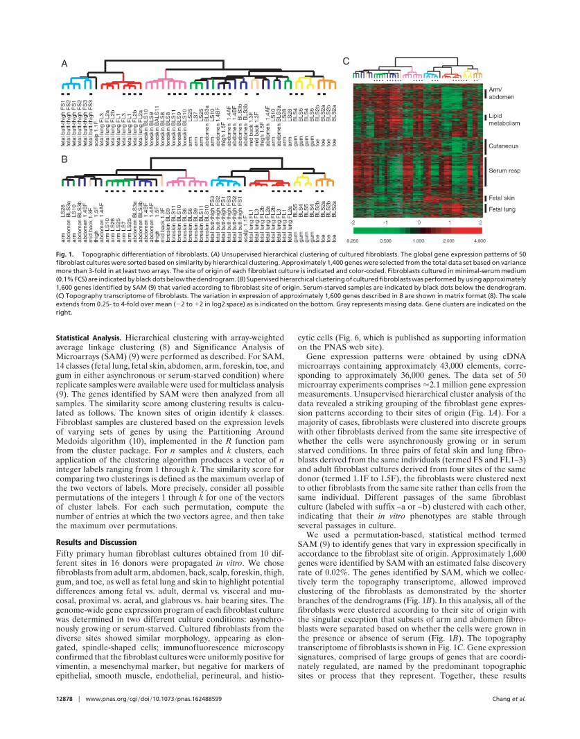
Gene expression patterns were obtained by using cDNAmicroarrays containing approximately 43,000 elements, corre-sponding to approximately 36,000 genes. The data set of 50microarray experiments comprises �2.1 million gene expressionmeasurements. Unsupervised hierarchical cluster analysis of thedata revealed a striking grouping of the fibroblast gene expres-sion patterns according to their sites of origin (Fig. 1A). For amajority of cases, fibroblasts were clustered into discrete groupswith other fibroblasts derived from the same site irrespective ofwhether the cells were asynchronously growing or in serumstarved conditions. In three pairs of fetal skin and lung fibro-blasts derived from the same individuals (termed FS and FL1–3)and adult fibroblast cultures derived from four sites of the samedonor (termed 1.1F to 1.5F), the fibroblasts were clustered nextto other fibroblasts from the same site rather than cells from thesame individual. Different passages of the same fibroblastculture (labeled with suffix –a or –b) clustered with each other,indicating that their in vitro phenotypes are stable throughseveral passages in culture.
We used a permutation-based, statistical method termedSAM (9) to identify genes that vary in expression specifically inaccordance to the fibroblast site of origin. Approximately 1,600genes were identified by SAM with an estimated false discoveryrate of 0.02%. The genes identified by SAM, which we collec-tively term the topography transcriptome, allowed improvedclustering of the fibroblasts as demonstrated by the shorterbranches of the dendrograms (Fig. 1B). In this analysis, all of thefibroblasts were clustered according to their site of origin withthe singular exception that subsets of arm and abdomen fibro-blasts were separated based on whether the cells were grown inthe presence or absence of serum (Fig. 1B). The topographytranscriptome of fibroblasts is shown in Fig. 1C. Gene expressionsignatures, comprised of large groups of genes that are coordi-nately regulated, are named by the predominant topographicsites or process that they represent. Together, these results
Fig. 1. Topographic differentiation of fibroblasts. (A) Unsupervised hierarchical clustering of cultured fibroblasts. The global gene expression patterns of 50fibroblast cultures were sorted based on similarity by hierarchical clustering. Approximately 1,400 genes were selected from the total data set based on variancemore than 3-fold in at least two arrays. The site of origin of each fibroblast culture is indicated and color-coded. Fibroblasts cultured in minimal-serum medium(0.1% FCS) are indicated by black dots below the dendrogram. (B) Supervised hierarchical clustering of cultured fibroblasts was performed by using approximately1,600 genes identified by SAM (9) that varied according to fibroblast site of origin. Serum-starved samples are indicated by black dots below the dendrogram.(C) Topography transcriptome of fibroblasts. The variation in expression of approximately 1,600 genes described in B are shown in matrix format (8). The scaleextends from 0.25- to 4-fold over mean (�2 to �2 in log2 space) as is indicated on the bottom. Gray represents missing data. Gene clusters are indicated on theright.
12878 � www.pnas.org�cgi�doi�10.1073�pnas.162488599 Chang et al.
demonstrate that fibroblasts from different anatomic sites havecharacteristic, distinct phenotypes, a phenomenon we termtopographic differentiation. Moreover, topographic differenti-ation is maintained in vitro when fibroblasts are isolated from theinfluence of other cell types.
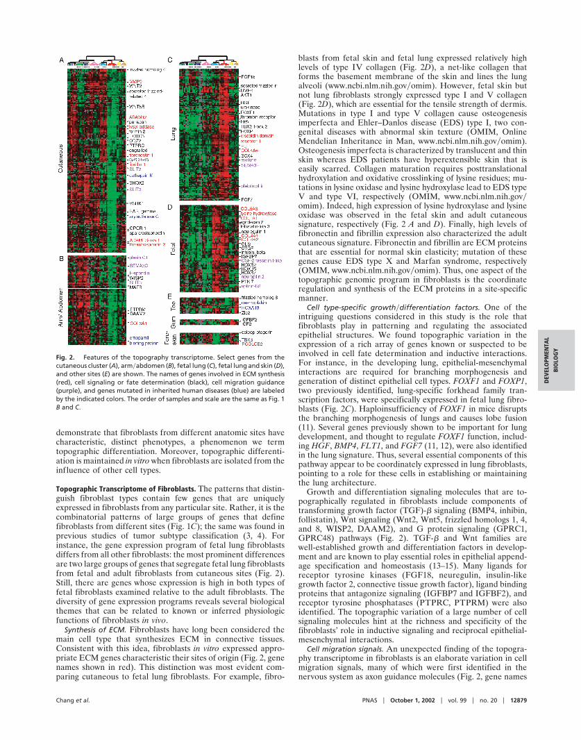
Topographic Transcriptome of Fibroblasts. The patterns that distin-guish fibroblast types contain few genes that are uniquelyexpressed in fibroblasts from any particular site. Rather, it is thecombinatorial patterns of large groups of genes that definefibroblasts from different sites (Fig. 1C); the same was found inprevious studies of tumor subtype classification (3, 4). Forinstance, the gene expression program of fetal lung fibroblastsdiffers from all other fibroblasts: the most prominent differencesare two large groups of genes that segregate fetal lung fibroblastsfrom fetal and adult fibroblasts from cutaneous sites (Fig. 2).Still, there are genes whose expression is high in both types offetal fibroblasts examined relative to the adult fibroblasts. Thediversity of gene expression programs reveals several biologicalthemes that can be related to known or inferred physiologicfunctions of fibroblasts in vivo.
Synthesis of ECM. Fibroblasts have long been considered themain cell type that synthesizes ECM in connective tissues.Consistent with this idea, fibroblasts in vitro expressed appro-priate ECM genes characteristic their sites of origin (Fig. 2, genenames shown in red). This distinction was most evident com-paring cutaneous to fetal lung fibroblasts. For example, fibro-
blasts from fetal skin and fetal lung expressed relatively highlevels of type IV collagen (Fig. 2D), a net-like collagen thatforms the basement membrane of the skin and lines the lungalveoli (www.ncbi.nlm.nih.gov�omim). However, fetal skin butnot lung fibroblasts strongly expressed type I and V collagen(Fig. 2D), which are essential for the tensile strength of dermis.Mutations in type I and type V collagen cause osteogenesisimperfecta and Ehler–Danlos disease (EDS) type I, two con-genital diseases with abnormal skin texture (OMIM, OnlineMendelian Inheritance in Man, www.ncbi.nlm.nih.gov�omim).Osteogenesis imperfecta is characterized by translucent and thinskin whereas EDS patients have hyperextensible skin that iseasily scarred. Collagen maturation requires posttranslationalhydroxylation and oxidative crosslinking of lysine residues; mu-tations in lysine oxidase and lysine hydroxylase lead to EDS typeV and type VI, respectively (OMIM, www.ncbi.nlm.nih.gov�omim). Indeed, high expression of lysine hydroxylase and lysineoxidase was observed in the fetal skin and adult cutaneoussignature, respectively (Fig. 2 A and D). Finally, high levels offibronectin and fibrillin expression also characterized the adultcutaneous signature. Fibronectin and fibrillin are ECM proteinsthat are essential for normal skin elasticity; mutation of thesegenes cause EDS type X and Marfan syndrome, respectively(OMIM, www.ncbi.nlm.nih.gov�omim). Thus, one aspect of thetopographic genomic program in fibroblasts is the coordinateregulation and synthesis of the ECM proteins in a site-specificmanner.
Cell type-specific growth�differentiation factors. One of theintriguing questions considered in this study is the role thatfibroblasts play in patterning and regulating the associatedepithelial structures. We found topographic variation in theexpression of a rich array of genes known or suspected to beinvolved in cell fate determination and inductive interactions.For instance, in the developing lung, epithelial-mesenchymalinteractions are required for branching morphogenesis andgeneration of distinct epithelial cell types. FOXF1 and FOXP1,two previously identified, lung-specific forkhead family tran-scription factors, were specifically expressed in fetal lung fibro-blasts (Fig. 2C). Haploinsufficiency of FOXF1 in mice disruptsthe branching morphogenesis of lungs and causes lobe fusion(11). Several genes previously shown to be important for lungdevelopment, and thought to regulate FOXF1 function, includ-ing HGF, BMP4, FLT1, and FGF7 (11, 12), were also identifiedin the lung signature. Thus, several essential components of thispathway appear to be coordinately expressed in lung fibroblasts,pointing to a role for these cells in establishing or maintainingthe lung architecture.
Growth and differentiation signaling molecules that are to-pographically regulated in fibroblasts include components oftransforming growth factor (TGF)-� signaling (BMP4, inhibin,follistatin), Wnt signaling (Wnt2, Wnt5, frizzled homologs 1, 4,and 8, WISP2, DAAM2), and G protein signaling (GPRC1,GPRC48) pathways (Fig. 2). TGF-� and Wnt families arewell-established growth and differentiation factors in develop-ment and are known to play essential roles in epithelial append-age specification and homeostasis (13–15). Many ligands forreceptor tyrosine kinases (FGF18, neuregulin, insulin-likegrowth factor 2, connective tissue growth factor), ligand bindingproteins that antagonize signaling (IGFBP7 and IGFBF2), andreceptor tyrosine phosphatases (PTPRC, PTPRM) were alsoidentified. The topographic variation of a large number of cellsignaling molecules hint at the richness and specificity of thefibroblasts’ role in inductive signaling and reciprocal epithelial-mesenchymal interactions.
Cell migration signals. An unexpected finding of the topogra-phy transcriptome in fibroblasts is an elaborate variation in cellmigration signals, many of which were first identified in thenervous system as axon guidance molecules (Fig. 2, gene names
Fig. 2. Features of the topography transcriptome. Select genes from thecutaneous cluster (A), arm�abdomen (B), fetal lung (C), fetal lung and skin (D),and other sites (E) are shown. The names of genes involved in ECM synthesis(red), cell signaling or fate determination (black), cell migration guidance(purple), and genes mutated in inherited human diseases (blue) are labeledby the indicated colors. The order of samples and scale are the same as Fig. 1B and C.
Chang et al. PNAS � October 1, 2002 � vol. 99 � no. 20 � 12879
DEV
ELO
PMEN
TAL
BIO
LOG
Y
shown in purple). Many of these molecules are now appreciatedas general cell migration guidance molecules, which can alsopattern mesodermal and epidermal cell migration (16, 17). Thetopographically regulated guidance molecules we found in fi-broblasts include semaphorins (SEMA3F, SEMA3C), receptorsfor semaphorins (neuropilin 2 and plexin C1), Slit proteins(SLIT2, SLIT3), ephrin B2, F-spondin, midkine, and pleiotro-phin (Fig. 2). Many of these proteins were expressed constitu-tively in both adult and fetal cells and regardless of the prolif-eration state of fibroblasts (Fig. 2). The topographic patterningof many migration guidance molecules in fibroblasts indicatesthat fibroblasts may play a critical role in posting ‘‘road signs’’that guide axons and possibly the migration of vasculature andmany other cell types.
Expression of Genes Associated with Genetic Syndromes. We ob-served that several genes defective in genetic syndromes wereprominently expressed in fibroblasts originating from sites mostaffected in those diseases (summarized in Table 2, which ispublished as supporting information on the PNAS web site). Asdescribed above, genes underlying 6 of 10 types of EDS syn-dromes were identified by comparing genes expressed in dermalversus lung fibroblasts. Moreover, HoxA13 is expressed in toeand foreskin fibroblasts, and mutation of HOXA13 causes hand-foot-genital syndrome, characterized by hypoplastic distal pha-langes and malformation of the urogenital system (OMIM,www.ncbi.nlm.nih.gov�omim). Desmoplakin I, a cytoskeletallinker protein mutated in striatal palmar-plantar keratoderma,is also highly expressed in toe fibroblasts. Similarly, emopamilbinding protein and arylsulfatase C, two enzymes involved inepidermal cholesterol metabolism, were identified in the armand cutaneous clusters, respectively. Mutation of emopamilbinding protein causes X-linked Conradi–Hunerman syndrome,characterized by rhizomelic shortening of limbs and whorledichthyosis, and mutation of arylsulfatase C causes X-linkedgeneralized ichthyosis (OMIM, www.ncbi.nlm.nih.gov�omim).The correspondence of gene expression and phenotype mayprovide an explanation for the localization of various defects inthese syndromes and suggests that genes that are topographicallyregulated in fibroblasts may present a fertile ground for findinggenes underlying disorders and diseases of skin, the musculo-skeletal system and even visceral organs.
Several of the proteins encoded by disease genes are thoughtto act in the epidermis but not in the dermis. For example,desmoplakin I has important structural functions connectingintermediate filaments to desmosomes in keratinocytes (OMIM,www.ncbi.nlm.nih.gov�omim). The parallel between the topo-graphic regulation of genes in dermal fibroblasts and the patternin which mutations in those genes affect the epidermis suggeststhat those genes may be expressed in the skin in a graded,segmental fashion that cuts across the germ layers.
Altered Lipid Metabolism in Serum-Starved Fetal Lung Fibroblasts.Although many genes are topographically regulated, a largegroup of genes was coordinately regulated in response to serumin all fibroblasts. Many features of this program have beendescribed (6), and detailed analysis of the common response offibroblasts to serum will be presented elsewhere. Because serumcontains low density lipoprotein (LDL), fibroblasts grown inserum-containing media can take up LDL, suppressing endog-enous production of cholesterol and fatty acids. When fibroblastsare cultured in limited serum (and thus low exogenous sterol),they induce expression of genes involved in cholesterol and fattyacid synthesis pathways (6). Sterol and fatty acid synthesis inmammalian cells is coordinately regulated at the transcriptionallevel by the sterol regulatory element-binding protein (SREBP)pathway (reviewed in ref. 18). SREBPs are transcriptionalfactors that are normally tethered to the endoplasmic reticulum;
when sterol levels are low, regulated proteolysis of SREBPallows the NH2-terminal, active fragments of SREBP to enterthe nucleus and activate the transcription of sterol and fatty acidbiosynthetic genes (18). More recently, SREBPs have also beenimplicated in adipogenesis, but their exact roles are incompletelyunderstood (19, 20).
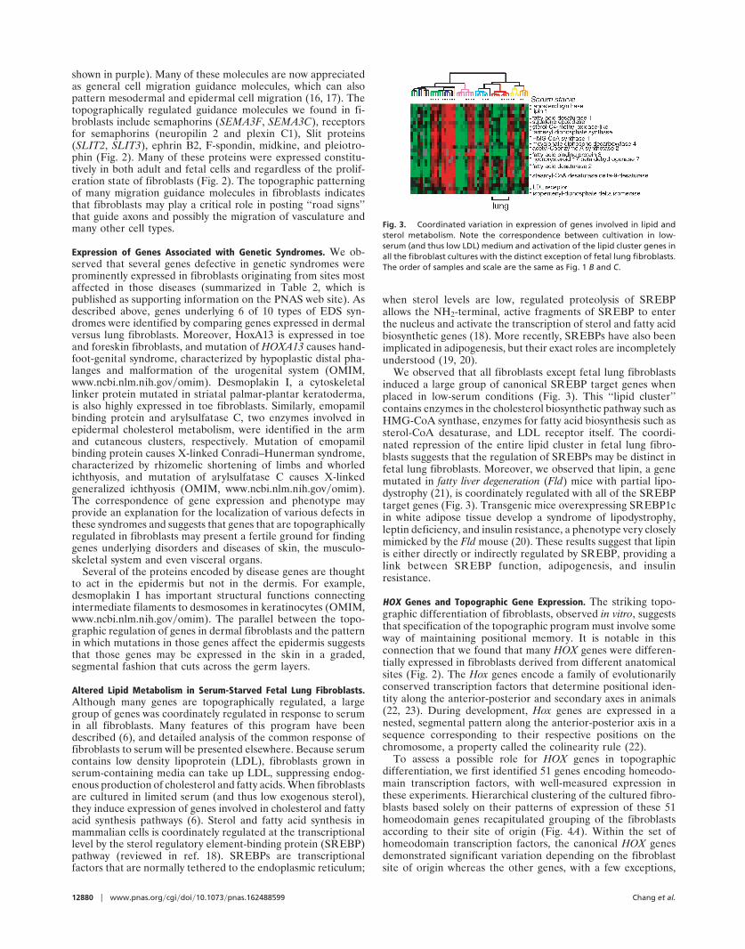
We observed that all fibroblasts except fetal lung fibroblastsinduced a large group of canonical SREBP target genes whenplaced in low-serum conditions (Fig. 3). This ‘‘lipid cluster’’contains enzymes in the cholesterol biosynthetic pathway such asHMG-CoA synthase, enzymes for fatty acid biosynthesis such assterol-CoA desaturase, and LDL receptor itself. The coordi-nated repression of the entire lipid cluster in fetal lung fibro-blasts suggests that the regulation of SREBPs may be distinct infetal lung fibroblasts. Moreover, we observed that lipin, a genemutated in fatty liver degeneration (Fld) mice with partial lipo-dystrophy (21), is coordinately regulated with all of the SREBPtarget genes (Fig. 3). Transgenic mice overexpressing SREBP1cin white adipose tissue develop a syndrome of lipodystrophy,leptin deficiency, and insulin resistance, a phenotype very closelymimicked by the Fld mouse (20). These results suggest that lipinis either directly or indirectly regulated by SREBP, providing alink between SREBP function, adipogenesis, and insulinresistance.
HOX Genes and Topographic Gene Expression. The striking topo-graphic differentiation of fibroblasts, observed in vitro, suggeststhat specification of the topographic program must involve someway of maintaining positional memory. It is notable in thisconnection that we found that many HOX genes were differen-tially expressed in fibroblasts derived from different anatomicalsites (Fig. 2). The Hox genes encode a family of evolutionarilyconserved transcription factors that determine positional iden-tity along the anterior-posterior and secondary axes in animals(22, 23). During development, Hox genes are expressed in anested, segmental pattern along the anterior-posterior axis in asequence corresponding to their respective positions on thechromosome, a property called the colinearity rule (22).
To assess a possible role for HOX genes in topographicdifferentiation, we first identified 51 genes encoding homeodo-main transcription factors, with well-measured expression inthese experiments. Hierarchical clustering of the cultured fibro-blasts based solely on their patterns of expression of these 51homeodomain genes recapitulated grouping of the fibroblastsaccording to their site of origin (Fig. 4A). Within the set ofhomeodomain transcription factors, the canonical HOX genesdemonstrated significant variation depending on the fibroblastsite of origin whereas the other genes, with a few exceptions,
Fig. 3. Coordinated variation in expression of genes involved in lipid andsterol metabolism. Note the correspondence between cultivation in low-serum (and thus low LDL) medium and activation of the lipid cluster genes inall the fibroblast cultures with the distinct exception of fetal lung fibroblasts.The order of samples and scale are the same as Fig. 1 B and C.
12880 � www.pnas.org�cgi�doi�10.1073�pnas.162488599 Chang et al.
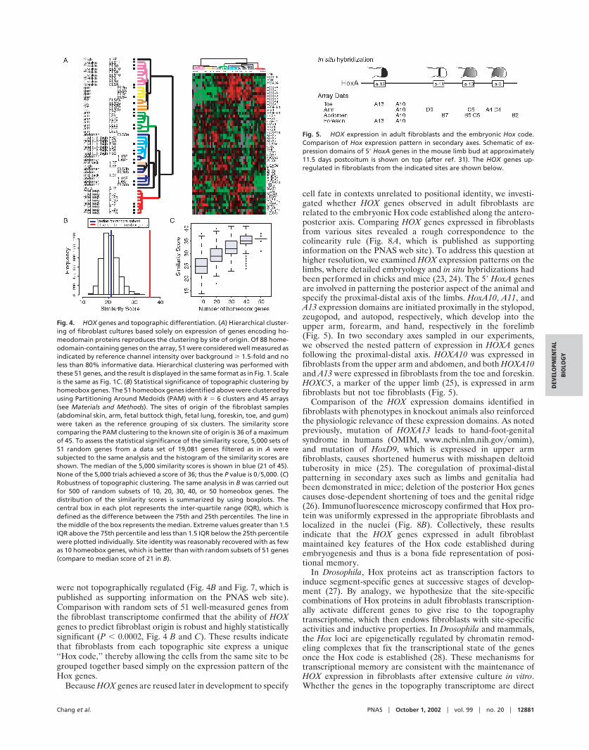
were not topographically regulated (Fig. 4B and Fig. 7, which ispublished as supporting information on the PNAS web site).Comparison with random sets of 51 well-measured genes fromthe fibroblast transcriptome confirmed that the ability of HOXgenes to predict fibroblast origin is robust and highly statisticallysignificant (P � 0.0002, Fig. 4 B and C). These results indicatethat fibroblasts from each topographic site express a unique‘‘Hox code,’’ thereby allowing the cells from the same site to begrouped together based simply on the expression pattern of theHox genes.
Because HOX genes are reused later in development to specify
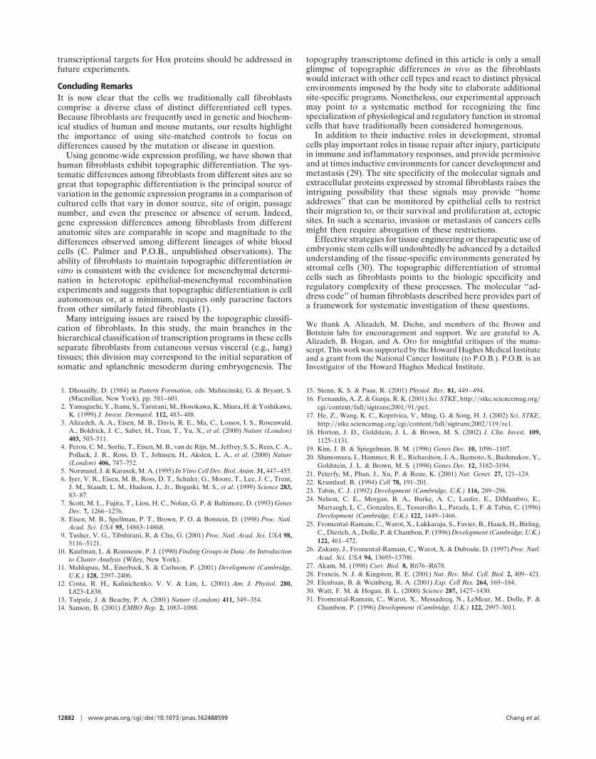
cell fate in contexts unrelated to positional identity, we investi-gated whether HOX genes observed in adult fibroblasts arerelated to the embryonic Hox code established along the antero-posterior axis. Comparing HOX genes expressed in fibroblastsfrom various sites revealed a rough correspondence to thecolinearity rule (Fig. 8A, which is published as supportinginformation on the PNAS web site). To address this question athigher resolution, we examined HOX expression patterns on thelimbs, where detailed embryology and in situ hybridizations hadbeen performed in chicks and mice (23, 24). The 5� HoxA genesare involved in patterning the posterior aspect of the animal andspecify the proximal-distal axis of the limbs. HoxA10, A11, andA13 expression domains are initiated proximally in the stylopod,zeugopod, and autopod, respectively, which develop into theupper arm, forearm, and hand, respectively in the forelimb(Fig. 5). In two secondary axes sampled in our experiments,we observed the nested pattern of expression in HOXA genesfollowing the proximal-distal axis. HOXA10 was expressed infibroblasts from the upper arm and abdomen, and both HOXA10and A13 were expressed in fibroblasts from the toe and foreskin.HOXC5, a marker of the upper limb (25), is expressed in armfibroblasts but not toe fibroblasts (Fig. 5).
Comparison of the HOX expression domains identified infibroblasts with phenotypes in knockout animals also reinforcedthe physiologic relevance of these expression domains. As notedpreviously, mutation of HOXA13 leads to hand-foot-genitalsyndrome in humans (OMIM, www.ncbi.nlm.nih.gov�omim),and mutation of HoxD9, which is expressed in upper armfibroblasts, causes shortened humerus with misshapen deltoidtuberosity in mice (25). The coregulation of proximal-distalpatterning in secondary axes such as limbs and genitalia hadbeen demonstrated in mice; deletion of the posterior Hox genescauses dose-dependent shortening of toes and the genital ridge(26). Immunofluorescence microscopy confirmed that Hox pro-tein was uniformly expressed in the appropriate fibroblasts andlocalized in the nuclei (Fig. 8B). Collectively, these resultsindicate that the HOX genes expressed in adult fibroblastmaintained key features of the Hox code established duringembryogenesis and thus is a bona fide representation of posi-tional memory.
In Drosophila, Hox proteins act as transcription factors toinduce segment-specific genes at successive stages of develop-ment (27). By analogy, we hypothesize that the site-specificcombinations of Hox proteins in adult fibroblasts transcription-ally activate different genes to give rise to the topographytranscriptome, which then endows fibroblasts with site-specificactivities and inductive properties. In Drosophila and mammals,the Hox loci are epigenetically regulated by chromatin remod-eling complexes that fix the transcriptional state of the genesonce the Hox code is established (28). These mechanisms fortranscriptional memory are consistent with the maintenance ofHOX expression in fibroblasts after extensive culture in vitro.Whether the genes in the topography transcriptome are direct
Fig. 4. HOX genes and topographic differentiation. (A) Hierarchical cluster-ing of fibroblast cultures based solely on expression of genes encoding ho-meodomain proteins reproduces the clustering by site of origin. Of 88 home-odomain-containing genes on the array, 51 were considered well measured asindicated by reference channel intensity over background � 1.5-fold and noless than 80% informative data. Hierarchical clustering was performed withthese 51 genes, and the result is displayed in the same format as in Fig. 1. Scaleis the same as Fig. 1C. (B) Statistical significance of topographic clustering byhomeobox genes. The 51 homeobox genes identified above were clustered byusing Partitioning Around Medoids (PAM) with k � 6 clusters and 45 arrays(see Materials and Methods). The sites of origin of the fibroblast samples(abdominal skin, arm, fetal buttock thigh, fetal lung, foreskin, toe, and gum)were taken as the reference grouping of six clusters. The similarity scorecomparing the PAM clustering to the known site of origin is 36 of a maximumof 45. To assess the statistical significance of the similarity score, 5,000 sets of51 random genes from a data set of 19,081 genes filtered as in A weresubjected to the same analysis and the histogram of the similarity scores areshown. The median of the 5,000 similarity scores is shown in blue (21 of 45).None of the 5,000 trials achieved a score of 36; thus the P value is 0�5,000. (C)Robustness of topographic clustering. The same analysis in B was carried outfor 500 of random subsets of 10, 20, 30, 40, or 50 homeobox genes. Thedistribution of the similarity scores is summarized by using boxplots. Thecentral box in each plot represents the inter-quartile range (IQR), which isdefined as the difference between the 75th and 25th percentiles. The line inthe middle of the box represents the median. Extreme values greater than 1.5IQR above the 75th percentile and less than 1.5 IQR below the 25th percentilewere plotted individually. Site identity was reasonably recovered with as fewas 10 homeobox genes, which is better than with random subsets of 51 genes(compare to median score of 21 in B).
Fig. 5. HOX expression in adult fibroblasts and the embryonic Hox code.Comparison of Hox expression pattern in secondary axes. Schematic of ex-pression domains of 5� HoxA genes in the mouse limb bud at approximately11.5 days postcoitum is shown on top (after ref. 31). The HOX genes up-regulated in fibroblasts from the indicated sites are shown below.
Chang et al. PNAS � October 1, 2002 � vol. 99 � no. 20 � 12881
DEV
ELO
PMEN
TAL
BIO
LOG
Y
transcriptional targets for Hox proteins should be addressed infuture experiments.
Concluding RemarksIt is now clear that the cells we traditionally call fibroblastscomprise a diverse class of distinct differentiated cell types.Because fibroblasts are frequently used in genetic and biochem-ical studies of human and mouse mutants, our results highlightthe importance of using site-matched controls to focus ondifferences caused by the mutation or disease in question.
Using genome-wide expression profiling, we have shown thathuman fibroblasts exhibit topographic differentiation. The sys-tematic differences among fibroblasts from different sites are sogreat that topographic differentiation is the principal source ofvariation in the genomic expression programs in a comparison ofcultured cells that vary in donor source, site of origin, passagenumber, and even the presence or absence of serum. Indeed,gene expression differences among fibroblasts from differentanatomic sites are comparable in scope and magnitude to thedifferences observed among different lineages of white bloodcells (C. Palmer and P.O.B., unpublished observations). Theability of fibroblasts to maintain topographic differentiation invitro is consistent with the evidence for mesenchymal determi-nation in heterotopic epithelial-mesenchymal recombinationexperiments and suggests that topographic differentiation is cellautonomous or, at a minimum, requires only paracrine factorsfrom other similarly fated fibroblasts (1).
Many intriguing issues are raised by the topographic classifi-cation of fibroblasts. In this study, the main branches in thehierarchical classification of transcription programs in these cellsseparate fibroblasts from cutaneous versus visceral (e.g., lung)tissues; this division may correspond to the initial separation ofsomatic and splanchnic mesoderm during embryogenesis. The
topography transcriptome defined in this article is only a smallglimpse of topographic differences in vivo as the fibroblastswould interact with other cell types and react to distinct physicalenvironments imposed by the body site to elaborate additionalsite-specific programs. Nonetheless, our experimental approachmay point to a systematic method for recognizing the finespecialization of physiological and regulatory function in stromalcells that have traditionally been considered homogenous.
In addition to their inductive roles in development, stromalcells play important roles in tissue repair after injury, participatein immune and inflammatory responses, and provide permissiveand at times inductive environments for cancer development andmetastasis (29). The site specificity of the molecular signals andextracellular proteins expressed by stromal fibroblasts raises theintriguing possibility that these signals may provide ‘‘homeaddresses’’ that can be monitored by epithelial cells to restricttheir migration to, or their survival and proliferation at, ectopicsites. In such a scenario, invasion or metastasis of cancers cellsmight then require abrogation of these restrictions.
Effective strategies for tissue engineering or therapeutic use ofembryonic stem cells will undoubtedly be advanced by a detailedunderstanding of the tissue-specific environments generated bystromal cells (30). The topographic differentiation of stromalcells such as fibroblasts points to the biologic specificity andregulatory complexity of these processes. The molecular ‘‘ad-dress code’’ of human fibroblasts described here provides part ofa framework for systematic investigation of these questions.
We thank A. Alizadeh, M. Diehn, and members of the Brown andBotstein labs for encouragement and support. We are grateful to A.Alizadeh, B. Hogan, and A. Oro for insightful critiques of the manu-script. This work was supported by the Howard Hughes Medical Instituteand a grant from the National Cancer Institute (to P.O.B.). P.O.B. is anInvestigator of the Howard Hughes Medical Institute.
1. Dhouailly, D. (1984) in Pattern Formation, eds. Malincinski, G. & Bryant, S.(Macmillan, New York), pp. 581–601.
2. Yamaguchi, Y., Itami, S., Tarutani, M., Hosokawa, K., Miura, H. & Yoshikawa,K. (1999) J. Invest. Dermatol. 112, 483–488.
3. Alizadeh, A. A., Eisen, M. B., Davis, R. E., Ma, C., Lossos, I. S., Rosenwald,A., Boldrick, J. C., Sabet, H., Tran, T., Yu, X., et al. (2000) Nature (London)403, 503–511.
4. Perou, C. M., Sorlie, T., Eisen, M. B., van de Rijn, M., Jeffrey, S. S., Rees, C. A.,Pollack, J. R., Ross, D. T., Johnsen, H., Akslen, L. A., et al. (2000) Nature(London) 406, 747–752.
5. Normand, J. & Karasek, M. A. (1995) In Vitro Cell Dev. Biol. Anim. 31, 447–455.6. Iyer, V. R., Eisen, M. B., Ross, D. T., Schuler, G., Moore, T., Lee, J. C., Trent,
J. M., Staudt, L. M., Hudson, J., Jr., Boguski, M. S., et al. (1999) Science 283,83–87.
7. Scott, M. L., Fujita, T., Liou, H. C., Nolan, G. P. & Baltimore, D. (1993) GenesDev. 7, 1266–1276.
8. Eisen, M. B., Spellman, P. T., Brown, P. O. & Botstein, D. (1998) Proc. Natl.Acad. Sci. USA 95, 14863–14868.
9. Tusher, V. G., Tibshirani, R. & Chu, G. (2001) Proc. Natl. Acad. Sci. USA 98,5116–5121.
10. Kaufman, L. & Rousseuw, P. J. (1990) Finding Groups in Data: An Introductionto Cluster Analysis (Wiley, New York).
11. Mahlapuu, M., Enerback, S. & Carlsson, P. (2001) Development (Cambridge,U.K.) 128, 2397–2406.
12. Costa, R. H., Kalinichenko, V. V. & Lim, L. (2001) Am. J. Physiol. 280,L823–L838.
13. Taipale, J. & Beachy, P. A. (2001) Nature (London) 411, 349–354.14. Sanson, B. (2001) EMBO Rep. 2, 1083–1088.
15. Stenn, K. S. & Paus, R. (2001) Physiol. Rev. 81, 449–494.16. Fernandis, A. Z. & Ganju, R. K. (2001) Sci. STKE, http:��stke.sciencemag.org�
cgi�content�full�sigtrans;2001�91�pe1.17. He, Z., Wang, K. C., Koprivica, V., Ming, G. & Song, H. J. (2002) Sci. STKE,
http:��stke.sciencemag.org�cgi�content�full�sigtrans;2002�119�re1.18. Horton, J. D., Goldstein, J. L. & Brown, M. S. (2002) J. Clin. Invest. 109,
1125–1131.19. Kim, J. B. & Spiegelman, B. M. (1996) Genes Dev. 10, 1096–1107.20. Shimomura, I., Hammer, R. E., Richardson, J. A., Ikemoto, S., Bashmakov, Y.,
Goldstein, J. L. & Brown, M. S. (1998) Genes Dev. 12, 3182–3194.21. Peterfy, M., Phan, J., Xu, P. & Reue, K. (2001) Nat. Genet. 27, 121–124.22. Krumlauf, R. (1994) Cell 78, 191–201.23. Tabin, C. J. (1992) Development (Cambridge, U.K.) 116, 289–296.24. Nelson, C. E., Morgan, B. A., Burke, A. C., Laufer, E., DiMambro, E.,
Murtaugh, L. C., Gonzales, E., Tessarollo, L., Parada, L. F. & Tabin, C. (1996)Development (Cambridge, U.K.) 122, 1449–1466.
25. Fromental-Ramain, C., Warot, X., Lakkaraju, S., Favier, B., Haack, H., Birling,C., Dierich, A., Dolle, P. & Chambon, P. (1996) Development (Cambridge, U.K.)122, 461–472.
26. Zakany, J., Fromental-Ramain, C., Warot, X. & Duboule, D. (1997) Proc. Natl.Acad. Sci. USA 94, 13695–13700.
27. Akam, M. (1998) Curr. Biol. 8, R676–R678.28. Francis, N. J. & Kingston, R. E. (2001) Nat. Rev. Mol. Cell. Biol. 2, 409–421.29. Elenbaas, B. & Weinberg, R. A. (2001) Exp. Cell Res. 264, 169–184.30. Watt, F. M. & Hogan, B. L. (2000) Science 287, 1427–1430.31. Fromental-Ramain, C., Warot, X., Messadecq, N., LeMeur, M., Dolle, P. &
Chambon, P. (1996) Development (Cambridge, U.K.) 122, 2997–3011.
12882 � www.pnas.org�cgi�doi�10.1073�pnas.162488599 Chang et al.