Embed Size (px)
Citation preview
United States Patent [191 Heinemann et a1.
[54] DNA ENCODING NEURONAL NICOTINIC ACETYLCHOLINE RECEPTOR COMPOSITIONS CONTAINING THE BETA 4 SUBUNIT
[75] Inventors: Stephen F. Heinemann, La J olla; Robert M. Duvoisin, Del Mar; Evan S. Deneris, La J 011a, all of Calif.; James W. Patrick, Houston, Tex.
The Salk Institute for Biological Studies, La Jolla, Calif.
[21] Appl. No.: 59,502
[22] Filed: May 6,1993
[73] Assignee:
Related U.S. Application Data
[63] Continuation of Ser. No. 492,555, Mar. 12, 1990, aban doned, which is a continuation-in-part of Ser. No. 170,295, Mar. 18, 1988, abandoned, and a continuation in-part of Ser. No. 321,384, Mar. 14, 1989, abandoned.
[51] Int. Cl.6 ............................................ .. C12N 15/12
[52] U.S. Cl. ..................................... .. 435/6; 435/ 64.1; 435/2523; 435/3201; 536/235
[58] Field of Search ............... .. 435/691, 252.3, 220.1; 436/501; 536/27
[56] References Cited U.S. PATENT DOCUMENTS
4,518,527 5/1985 Numa et a1. ...................... .. 530/327
OTHER PUBLICATIONS
Nature 311:626-631, (18 Oct. 1984) Leonard et al. Mo lecular Cloning and expression of cDNAs for the human interneural receptor. Nature, vol. 328, 221-227, 16 Jul. 1987, Scho?eld et a1. Sequence and functional expression of the GABAA receptor shows a ligand-gated receptor superfamily. Nucleic Acids Research, vol. 13, 4739-4749, 1985, Hauptmann et al., A novel class of human type I inter ferons. Nature, vol. 313, 806-810, 28 Feb. 1985, Jacobs et al., isolation and characterization of genomic and cDNA clones of human erythropoietin. Anand et al., “Neuronal Nicotinic Acetylcholine Re ceptors Expressed in Xenopus Oocytes Have a Pentam
lllllllllllllllllllllllllllllllllllllllllllllllllllllllllllllllllllllllllll US005449606A
[11] Patent Number:
[45] Date of Patent: 5,449,606
Sep. 12, 1995
eric Quaternary Structure,” The Journal of Biological Chemistry 266(17):l1192—1ll98 (1991). Boulter et al., '“Isolation of a cDNA Clone Coding for a Possible Neural Nicotinic Acetylcholine Receptor a-Subunit,” Nature 319:368-374 (1986). Cooper et al., “Pentameric Structure and Subunit Stoi chiometry of a Neuronal Nicotinic Acetylcholine Re ceptor,” Nature 350:235-238 (1991). Deneris et al., “Primary Structure and Expression of B: A Novel subunit of Neuronal Nicotinic Acetylcholine Receptors,” Neuron 1:45-54 (1988). Duvoisin et al., “The Functional Diversity of the Neue ronal Nicotinic Acetylcholine Receptors is Increased by a Novel Subunit: B4,” Neuron 3:487-496 (1989). Figl et al., “Regions of [84 (1B2 Subunit Chimeras that Contribute to the Agonist Selectivity of Neuronal Nico tinic Receptors,” FEBS 308:245-248 (1992). Goldman et al., “Members of a Nicotinic Acetylcholine Receptor Gene Family Are Expressed in DifferentRe gions of the Mammalian Central Nervous System,” Cell 48:965-973 (1987). Halvorsen and‘ Berg, “Af?nity Labeling of Neuronal Acetylcholine Receptor Subunits with an a-Neuro toxin that Blocks Receptor Function,” The Journal of Neuroscience 7(8):2547—2555 (1987). Luetje and Patrick, “Both a-and B-Subunits Contrib-> .
(List continued on next page.)
Primary Examiner—Garnette D. Draper Assistant Examiner—John D. Ulm Attorney, Agent, or Firm—Pretty, Schroeder, Brueggemann & Clark; Stephen E. Reiter
I [57] ABSTRACT
The present invention discloses a new neuronal nico tinic acetylcholine receptor subunit, B4. The new sub unit can form functional combinations with other neu ronal nicotinic acetylcholine receptor subunits, includ ing, but not limited to, alpha2, alpha3, alpha4 and beta2. A cDNA clone containing the DNA sequences that encode the novel receptor subunit of the invention has been deposited with the American Type Culture Col lection for patent purposes.
21 Claims, 11 Drawing Sheets
5,449,606 Page 2
OTHER PUBLICATIONS ute to the Agonist Sensitivity of NeuronalgNicotinic Acetylcholine Receptors,” The Journal of Neuroscience ll(3):837-845 (1991). Mauron, et a1, “Structure of Chicken Genes Encoding the Nicotinic Acetylcholine Receptor Subunits and Their Variants,” Society for Neuroscience Abstracts 55.10, p. 171 (1985). Nef et al., “Genes Expressed in the Brain De?ne Three Distinct Neuronal Nicotinic Acetylcholine Receptors,” EMBO J. 7:595-601 (1988). Papke and Heinemann, “The Role of the B4 -Subunit in Determining the Kinetic Properties of Rat Neuronal Nicotinic Acetylcholine x3 -Receptors,” Journal of Physiology 444:95-112 (1990). Schoepfer et al., “cDNA Clones Coding for the Struc tural Subunit of a Chicken Brain Nicotinic Acetylcho line Receptor,” Neuron 1:241-248 (1988). Schoepfer et al., “Brain a-Bungarotoxin Binding Pro tein cDNAs and MAbs Reveal Subtypes of This Branch of the Ligand-Gated Ion Channel Gene Superfamily,” Neuron 5:35-38 (1990). Vernallis et al., “Achr Gene Products in Chick Ciliary Ganglia: Transcripts, Subunits, and Receptor Sub types,” Society for Neuroscience Abstracts 14.9 17:23 (1991). Wada et al., “Functional Expression of a New Pharma cological Subtype of Brain Nicotinic Acetylcholine Receptor,” Science 240:330-334 (1988). Wada et al., “Distribution of Alpha2, ALpha3, Alpha4, and Beta2 Neuronal Nicotinic Receptor Subunit mRNAs in the Central Nervous System: A Hybridiza tion Histochemical Study in the Rat,” The Journal of Comparative Neurology 284:314-335 (1989). Whiting et al., “Neuronal Nicotinic Acetylcholine Re ceptor ,B-Subunit Is Coded for by the cDNA Clone B4,” FEBS 219(2):459-463 (1987). Whiting et al., “structurally Different Neuronal Nico
tinic Acetylcholine Receptor Subtypes Puri?ed and Characterized Using Monoclonal Antibodies,” The journal of Neuroscience 7(l2):4005-40l6 (1987). Whiting et al., “Expression of Nicotinic Acetylcholine Receptor Subtypes in Brain and Retina,” Molecular Brain Research 10:61-70 (1991). Whiting and Lindstrom, “Af?nity Labelling of Neuro nal Acetylcholine Receptors Localizes Acetyl choline-Binding Sites to Their B-Subunits,” FEBS 213:(l):55-6O (1987). Whiting and Lindstrorn, “Puri?cation and Character ization of a Nicotinic Acetylcholine Receptor from Rat Brain,” Proc. Natl. Acad. Sci. USA 84:595-599 (1987). Whiting et al., “Structural and Pharmacological Characterizations of the major Nicotinic Acetylcholine Receptor Subtype Stably Expressed in Mouse Fibro
_ blasts,” Molecular Pharmacology 40:463-472 (1991). ' Mishina, et al., “Expression of Functional Acetylcho
line Receptor From Cloned cDNAs”, Nature 307, 604-608 (1984). Goldman, et al., “Members of a Nicotinic Acetylcho line Receptor Gene Family Are Expressed in Diffrent Regions of the Mammalian Central Nervous System”, Cell 48: 965-973 (1987). Whiting et al., “Functional Acetylcholine Receptor in PC12 Cells Reacts With A Monoclonal Antibody to Brain Nicotine Receptors”, Nature, 327, 515-518 (1987). .
Whiting, et al., “Functional Acetylcholine Receptor in PC12 Cells Reacts With A Monoclonal Antibody to Brain Nicotine Receptors”, Nature 3272515-518 (1987). Deneris, et al., “Primary Structure and Expression of A Novel Subunit of Neuronal Nicotinic Acetylcholine Receptors”, Neuron 1, 45-54 (1988). Boulter, J., Connolly, J., Deneris E., Goldman D., Heinemann S., and Patrick, 1., “Functional Expression of Two Neuronal Nicotinic Acetylcholine Receptors
' From cDNA Clones Identi?es a Gene Family”, Proc. Natl. Acad. Sci. USA 84, 7763-7767, 1987).
US. Patent Sep. 12, 1995 Sheet 2 of 11 5,449,606
4m .QI a’

US. Patent Sep. 12, 1995 Sheet 7 of 11 5,449,606
m..m .QE UUHHFdUHHHUF?UUdUUUUUHHdUQHU60UQHUO?UUUHHdUHUHUU08094000400 HovN HvmN HmNN HNNN HwHN HOHN ?vom HmmH

US. Patent Sep. 12, 1995 Sheet 10 0f 11 5,449,606
FIG. 5 A (1254
T T 5 pM A01 5 pM Nic
FIG. 5 B (1354
.W M
T T 5 pM ACh 5 pM Nic
Fl 6 5 D 0.5pM ACh 0.5}1M Nic
0115476
k [L
T T ML l pM ACh I00 uM Nic 30 sec
US. Patent Sep. 12, 1995 Sheet 11 of 11 5,449,606
“.3 om 5< 21 H 6< 21 Q
||_ >8 8 h h
1 a 1 1 1 1 >5 v.8
2111‘ “mm 3583 .JL‘II .
Ema mm .QI
h 1 1 | 1 1 1 1 w >53?
“$3558 / \
8% 4w .oI
5,449,606 1
DNA ENCODING NEURONAL NICOTlNIC ACE'I'YLCHOLINE RECEPTOR COMPOSITIONS
CONTAINING THE BETA 4 SUBUNIT
ACKNOWLEDGMENT
This invention was made with government support under several grants from the National Institutes of Health and the Muscular Dystrophy Association.
RELATED APPLICATION
This application is a continuation of application Ser. No. 07/492,555, ?led Mar. 12, 1990, now abandoned, which is a continuation-in-part of application Ser. No. 07/170,295, ?led Mar. 18, 1988, now abandoned, and application Ser. No. 07/321,384, ?led Mar. 14, 1989, now abandoned.
FIELD OF THE INVENTION
The present invention relates generally to neuronal nicotinic acetylcholine receptor genes and proteins. More particularly, the invention relates to a new sub unit member of the family of neuronal nicotinic acetyl choline receptors. The new subunit, named beta4 (B4), can participate with known subunits, including alpha type subunits B2, B3 and B4, and the beta-type subunit, B2, (in the presence of at least one alpha subunit), to form previously unknown functional receptors.
BACKGROUND
The diversity of neurotransmitter receptors is far greater than had been anticipated by pharmacological and physiological studies. Two gene superfamilies en code all cloned neurotransmitter receptors: one is the G protein-coupled receptor superfamily, and the other is the ligand-gated ion channel superfamily (for reviews see Hall, 1987; Barnard, et al., 1987). All of the recep tors that act through a G protein, for example, the mus carinic acetylcholine receptors, the dopamine recep tors, and the B-adrenergic receptors, are formed by a single polypeptide chain that is postulated to span the plasma membrane seven times. For each class of recep tor, a gene family encodes closely related variants that have different pharmacological and physiological char acteristics and different patterns of distribution in the nervous system.
In contrast to the G protein superfamily, ligand-gated ion channels are composed of more than one subunit. Expression studies have shown that the diversity of the ligand-gated receptors is due to the presence of the same subunits in different combinations. In addition, multiple genes encode each type of subunit. For exam ple, three types of GABAA receptor subunits, a, B, and 7, have been described, and at least for the a and B subunits, several genes encode variant subtypes (Scho ?eld, et al., 1987; Levitan, et al., 1988; Pritchett, et al., 1989). In transient transfection assays, functional recep tors can be formed by either the a or the B subunit alone or in pairwise combinations (Pritchett, et al., 1988). The presence of the 7 subunit, however, appears to be re quired for benzodiazepine sensitivity (Pritchett, et al., 1989). One way to unravel the functional diversity of the
nicotinic acetylcholine receptors (NACHRS) in the nervous system is to identify at the molecular level all of the different nAChR subunits. The pharmacology and single-channel characteristics of cloned subunits, ex pressed in various combinations in transfected cells or in
10
20
25
35
45
50
55
65
2 Xenopus oocytes, can then be analyzed. By comparing these results with similar studies performed in vivo, a better understanding of the neuronal nicotinic pathways will become available. In pursuing this effort, the Mo lecular Neurobiology Laboratory at the Salk Institute for Biological Studies has isolated rat cDNA clones that identify four distinct neuronal nAChR subunits: a2, a3, a4 and B2 (see US. Ser. Nos. 07/170,295, abandoned, 07/ 321,384, abandoned and Wada, et al., 1988; Boulter, etal., 1986; Goldman, et al., 1987; Deneris, et al., 1988). Recently, two additional clones that are closely related to the neuronal nAChRs but for which no function has yet been found have been identi?ed. They are referred to as a5 an B3 (U.S. Ser. Nos. 07/170,295, abandoned and 07/321,384, abandoned, and Boulter, et al., 1990; Deneris, et al., 1989). The present speci?cation discloses another distinct neuronal nAChR subunit, beta4. In identifying the gene that encodes this subunit, rat geno mic libraries were screened with neuronal nAChR cDNA probes. The genes for several of the previously described nAChR subunits were isolated, and their restriction maps were determined. A recombinant phage containing part of the gene for a novel nAChR subunit was also isolated. The primary structure of this subunit was deduced from the nucleotide sequence of a cDNA clone. Expression studies using Xenopus oo cytes have shown that this subunit can combine with each of the neuronal a2, a3, and a4 subunits to form functional nAChRs. The neuronal nAChR subunits are closely related and
form a subgroup of the nAChR gene family that also includes the nicotinic receptors present in the Torpedo electric organ and at the vertebrate neuromuscular junction. The latter are formed by the pentameric as sembly of four homologous subunits: two a ligand bind ing subunits and one each of the B, 'y, and 5 subunits (Reynolds and Karlin, 1978). There is evidence that during development, new nicotinic receptors contain ing an 6 subunit instead of a 7 subunit are inserted at the neuromuscular endplate (Gu and Hall, 1988). Concomi tant with this repopulation, an increase in channel con ductance is observed (Sakmann and Brenner, 1978; Mishina, et al., 1986). Functional neuronal nAChRs can be produced by the coinjection into Xenopus oocytes of RNA encoding a B2 subunit and one of either an (12, an (13 or an a4 subunit (US. Ser. Nos. 07/170,295, aban doned and 07/321,384, abandoned, and Boulter, et al., 1987; Wada, et al., 1988). Whiting and Lindstrom (1986, 1987) have proposed that the nicotinic receptors found in the peripheral and central nervous system are assem bled from only two different subunits, an a and a B subunit, in a yet undetermined stoichiometry. However, the possibility remains that neuronal nAChRs are com posed of more than two distinct subunits. As those skilled in the art will appreciate, an under
standing of the molecular mechanisms involved in neurotransmission in the central nervous system is lim ited by the complexity of the system. The cells are small, have extensive processes, and often have thou sands of synapses deriving from inputs from many dif ferent parts of the brain. In addition, the actual number of neurotransmitter receptors is low, making their puri ?cation dif?cult, even under the best of circumstances. Consequently, neither cellular nor biochemical ap proaches to studying neurotransmission in the central nervous system has been particularly fruitful. This is unfortunate because it is quite probable that the treat
5,449,606 3
ment of dementia, Alzheimer’s disease and other forms of mental illness will involve modi?cation of synaptic transmission with speci?c drugs. The realization that the nicotinic acetylcholine recep
tors are much more diverse than previously expected offers an opportunity for a level of pharmaceutical in tervention and a chance to design new drugs that affect speci?c receptor subunits. Such subtypes make it possi ble to observe the effect of a drug substance on a partic ular subtype. Information derived from these observa tions will allow the development of new drugs that are more speci?c, and therefore have fewer unwanted side effects.
In addition, the availability of these neuronal recep tors makes it possible to perform initial in vitro screen ing of the drug substance. While it is true that the drug eventually has to work in the whole animal, it is proba ble that useful drugs are being missed because conven tional screening is limited to average composite effects. Consequently, the ability to screen drug substances in vitro on a speci?c receptor subtype(s) is likely to be more informative than merely screening the drug sub stance in whole animals. Both the receptor subunit DNA and the encoded
protein(s) of the present invention can be used for drug design and screening. For example, the cDNA clone encoding the beta4 subunit, alone, or in combination with various alpha subunit clones or other subunit clones, now known or later to be discovered, can be transcribed in vitro to produce mRNA. This mRNA, either from a single subunit clone or from a combination of clones, can then be injected into oocytes where the mRNA will direct the synthesis of the receptor protein. Alternatively, the clones may be placed downstream from appropriate gene regulatory elements and inserted into the genome of eukaryotic cells. This will result in transfected cell lines expressing a speci?c receptor sub type, or speci?c combinations of subtypes. The derived cell lines can then be produced in quantity for reproduc ible quantitative analysis of the effects of drugs on re ceptor function.
BRIEF DESCRIPTION OF THE DRAWINGS
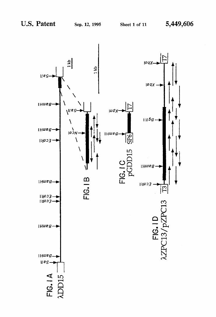
The drawing comprise six ?gures, of which: FIG. 1 is a schematic drawing showing the restriction
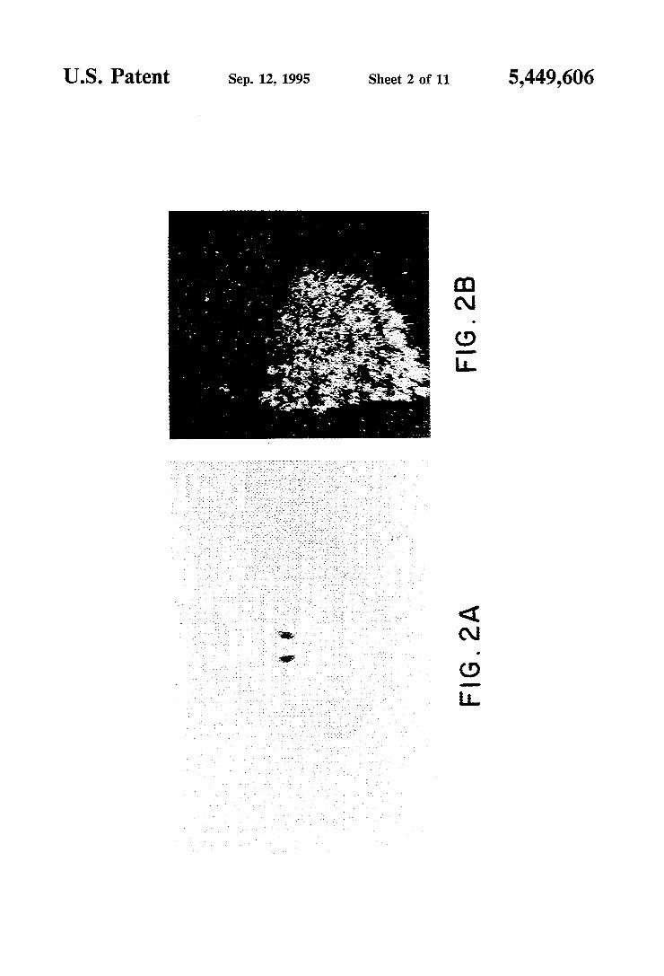
maps and sequencing strategies. FIG. 1A is the partial restriction map of the genomic insert of ADD15. The solid bar indicates the ?fth exon coding region. FIG. 1B is an enlargement of this exon with arrows referring to the extent and direction of nucleotide sequencing reac tions. FIG. 1C shows pGDDl5, a plasmid used for preparing B4-speci?c probes; pGDDl5 contains an NcoI-SalI fragment of ADDlS encoding a noncon served segment of the cytoplasmic domain. FIG. 1D shows a partial restriction map and sequencing strategy of the cDNA insert of 7tZPCl3 and its in vivo excised plasmid version pZPCl3. See Experimental Procedures section of this speci?cation. The solid bar identi?es the coding region, and the ?ne line represents the 5’ and 3’ untranslated regions. SP6, T3 and T7 refer to the SP6, T3 and T7 transcription promoters. FIG. 2 is a photograph showing an analysis by in situ
hybridization histochemistry of the distribution of B4 transcripts. Adult rat brain coronal sections (30 pm thick) were hybridized with 35S-radiolabeled sense and antisense strands probes derived from KpnI- and Bam HI-linearized pGDDl5, respectively (see FIG. 1C). FIG. 2A shows that only sections across the thalamus,
20
25
30
35
40
45
50
55
65
4 as the one shown here, gave above background signals by X-ray ?lm autoradiography. In parallel experiments, a sense probe was hybridized to adjacent sections and gave background levels of hybridization (data not shown). FIG. 2B is a dark-?eld photomicrograph of a medial habenula form a section identical to that shown in FIG. 2A after emulsion dipping. FIGS. 3-1 through 3-5 is a schematic drawing show
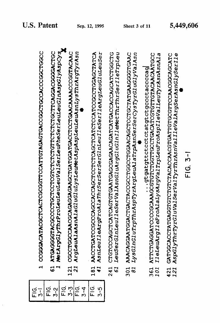
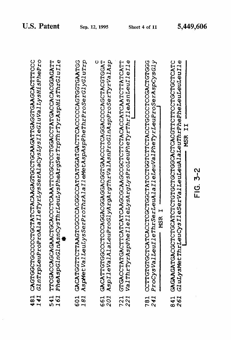
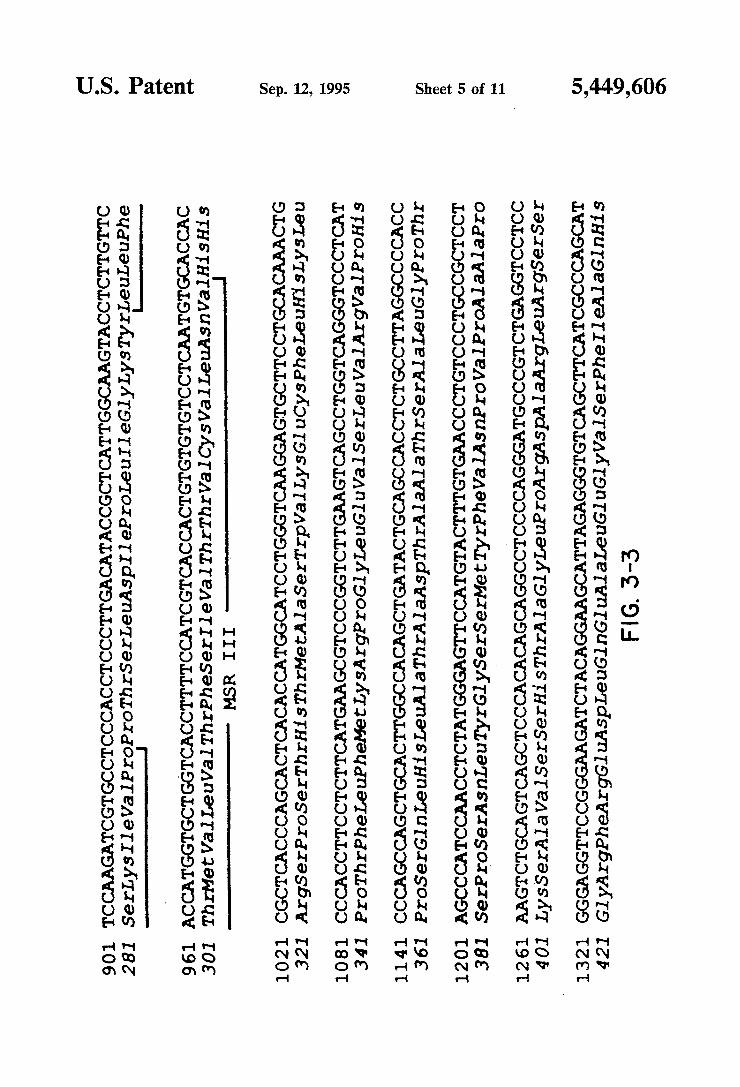
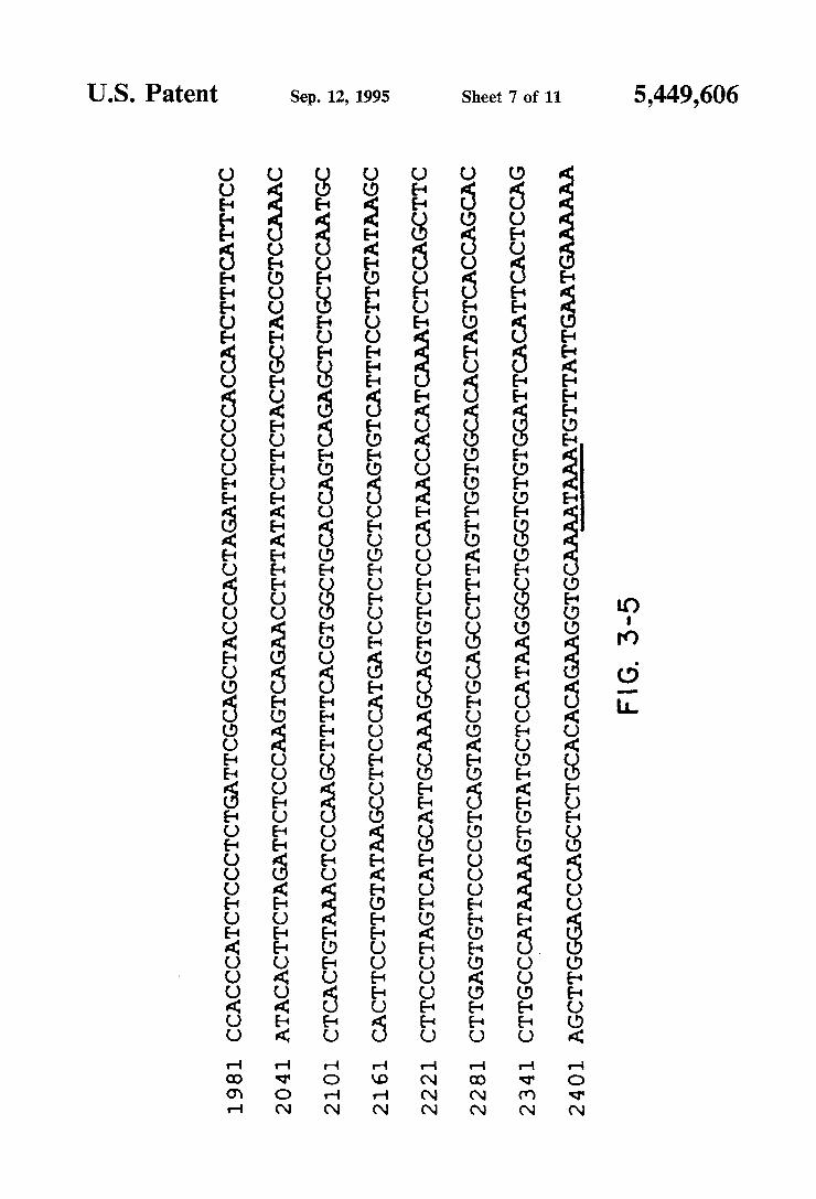
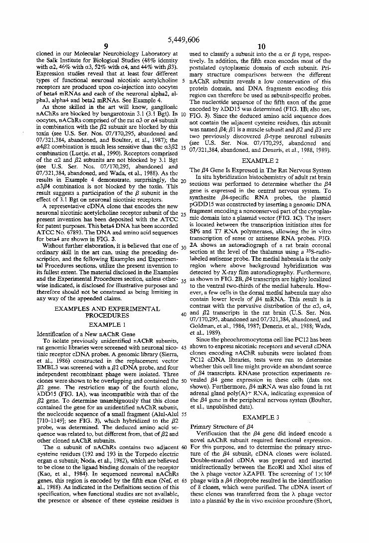
ing the nucleotide sequence and deduced amino acid sequence of AZPC13. The genomic nucleotide sequen ces at the splice junctions and at the substitution posi tion (720) are shown above the cDNA sequence in lowercase letters. Vertical bars indicate the predicted splice sites. The core position of a DNA sequence re peated three times is boxed in gray. Putative membrane spanning regions (MSRs) are indicated. The polyadeny lation signal is underlined. A scissors indicates predicted signal peptide cleavage site. An asterisk indicates puta tive N-linked glycosylation sites. FIGS. 4-1 and 4-2 is a schematic drawing showing
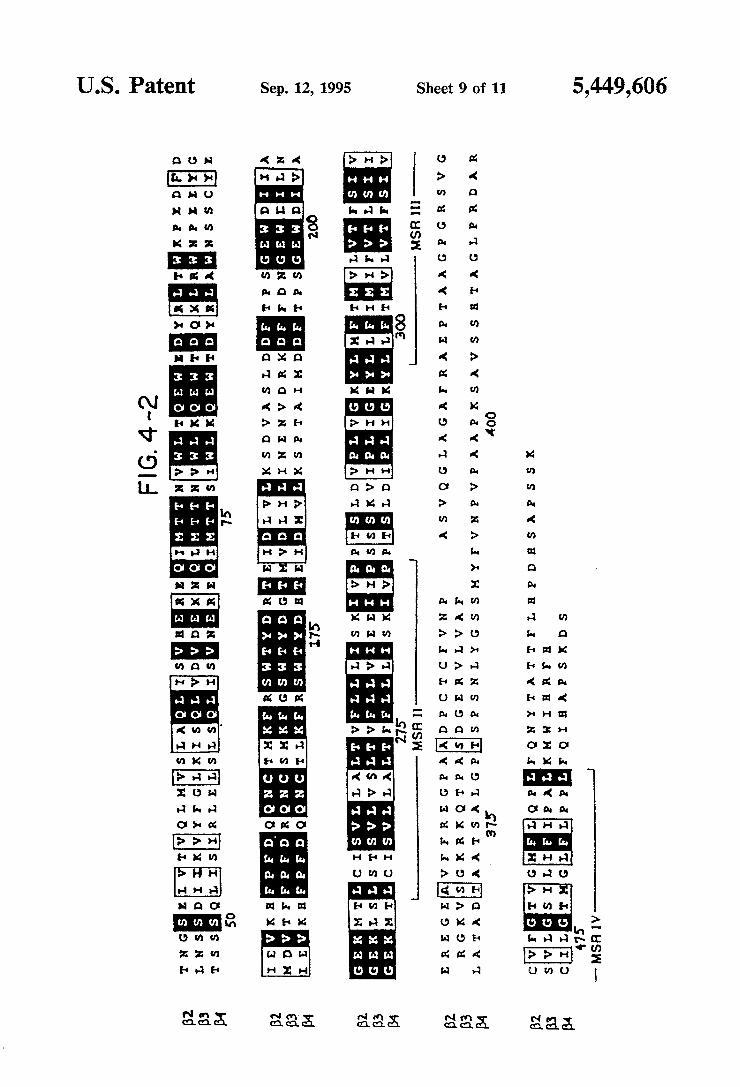
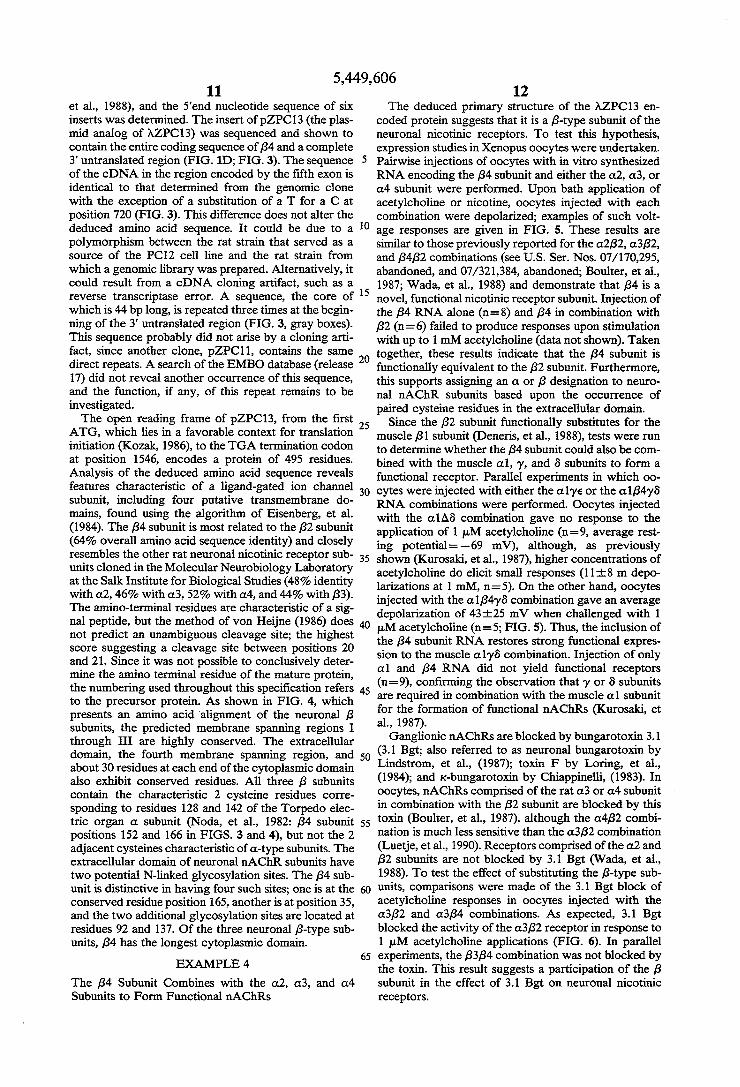
amino acid alignment of the rat neuronal B-type sub units. Aligned with the B4 subunit are the B2 (U.S. Ser. Nos. 170,295 and 321,384 and Deneris, et al., 1988) and B3 (U.S. Ser. Nos. 170,295 and 321,384 and Deneris, et al., 1989) sequences. Identical residues in all three sub units are shown on a black background. Conservative changes are indicated by a gray background. Putative signal peptides and membrane spanning regions are identi?ed below the sequences. The region referred to as the extracellular domain is located between the amino terminus and MSR I, and the putative cytoplas mic domain is located between MSR III and MSR IV. The numbering is that of the precursor B4 subunit. FIGS. 5A through 5D shows electrophysiological
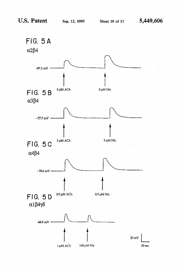
recordings of Xenopus oocytes injected with in vitro synthesized RNA encoding the nAChR subunits in the indicated combinations. Representative responses in duced by acetylcholine and nicotine stimulations at the given concentrations are shown. Potential measure ments were monitored on a digital voltmeter and re corded on a Gould pen recorder. Voltage traces were scanned and prepared for printing using a personal computer. FIGS. 6A and 6B shows voltage recordings of Xeno
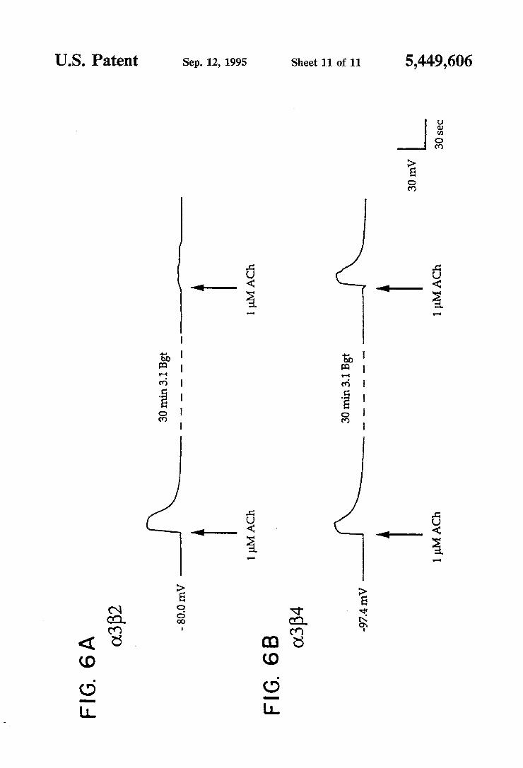
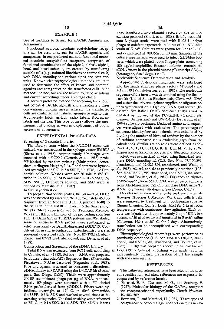
pus oocytes injected with (13 and either B2 or ,84 before and after exposure to ~0.1 uM 3.1 Bgt. Representative responses induced by acetylcholine and nicotine stimu lations at the given concentrations are shown. Potential measurements were monitored on a digital voltmeter and recorded on a Gould pen recorder. Voltage traces were scanned and prepared for printing using a personal computer.
DEFINITIONS
In the present speci?cation and claims, reference will be made to phrases and terms of art which are expressly de?ned for use herein as follows: As used herein, nAChR means nicotinic acetylcho
line receptor. As used herein, an agonist binding subunit is a subunit
of an acetylcholine receptor that contains a binding site for the neurotransmitter, acetylcholine and its analogs. A putative neuronal nAChR subunit isolated by
cDNA cloning is'identi?ed as an “alpha” ((1) subunit if the Torpedo alpha subunit cysteines 128, 142, 192, and 193 are conserved. Some alpha subunits are known to be agonist binding subunits, others are suspected of being agonist binding subunits based on the fact that they
5,449,606 5
contain the conserved cysteines at positions 128, 142, 192, and 193. Known a-type agonist binding subunits include alphal, and alpha4 (alpha4.l and alpha4.2); suspected a-type agonist binding subunits include al pha2, alpha3, and alpha5. A putative neuronal nAChR subunit isolated by
cDNA cloning is identi?ed as a “beta” (B) subunit if only the Torpedo 128 and 142 cysteines are conserved. Known B-subunits include betal, beta2 and beta3. The novel beta subunit of the invention is beta4. As used herein, the term antagonist refers to a sub
stance that interferes with receptor function. Antago nists are of two types: competitive and non-competitive. A competitive antagonist (or competitive blocker) com petes with the neurotransmitter for the same binding site. In the case of acetylcholine, an example of such an antagonist is 3.1 bungarotoxin. A non-competitive an tagonist or blocker inactivates the functioning of the receptor by binding to a site other than the acetylcho line binding site. As used herein, alphal refers to a DNA segment
which encodes an agonist binding subunit of the same name. This subunit is expressed in skeletal muscle and Torpedo electric organ. As used herein, alpha2 refers to a DNA segment,
which has been identi?ed in chick and rat, that encodes a neuronal nAChR subunit of the same name. DNA encoding the alpha2 subunit has been deposited with the ATCC; the DNA (designated as pHYP16) has been accorded ATCC No. 67646. As used herein, alpha3 refers to a DNA segment that
encodes a neuronal nAChR subunit of the same name. This subunit is expressed in the PC12 cell line and vari ous regions of the mammalian brain. DNA encoding the alpha3 subunit has been deposited with the ATCC; the DNA (designated as pPCA48) has been accorded ATCC No. 67642. As used herein, alpha4 refers to a DNA segment that
encodes a neuronal agonist binding subunit of the same name. The cDNA clones encoding the proteins referred to herein as alpha4.1 and 4.2 are both derived from the alpha4 gene. DNAs coding for the alpha4.1 and 4.2 trancripts have been deposited with the ATCC. The alpha4.1 DNA (designated as pI-IYA23-1(E)1) has been accorded ATCC No. 67644; the a1pha4.2 DNA (desig nated as pHIP3C(3) has been accorded ATCC No. 67645. '
As used herein, alphaS refers to a DNA segment encoding a neuronal nAChR subunit of the same name. DNA encoding the alphaS subunit has been deposited with the ATCC; the DNA (designated as PC1321) has been accorded ATCC No. 67652. As used herein, betal refers to a DNA segment en
coding a nAChR subunit of the same name. This subunit is expressed in the Torpedo electric organ and mamma lian muscle receptors. As used herein, beta2 refers to a DNA segment en
coding a neuronal nAChR subunit of the same name. See U.S. Ser. Nos. 07/170,295, abandoned and 07/321,384, abandoned, and Deneris, et al., (1988). DNA encoding beta2 has been deposited with the ATCC; the DNA (designated as pPCX49) has been accorded ATCC No. 67643. As used herein, beta3 refers to a DNA segment en
coding a neuronal nAChR subunit of the same name. See U.S. Ser. Nos. 07/ 170,295, abandoned and 07/321,384, abandoned, and Deneris, et al., (1989). DNA encoding beta3 has been deposited with the
10
5
25
35
40
45
50
55
65
6 ATCC; the DNA (designated as EDS76) has been ac corded ATCC No. 67653. As used herein, beta4 refers to a DNA segment en
coding a neuronal nAChR subunit of the same name. DNA encoding the beta4 subunit has been deposited with the ATCC; the DNA (designated as pZPC13) has been accorded ATCC No. 67893. As used herein, PC12 refers to the rat adrenal chro~
maf?n tumor cell line, PC12. This cell line expresses presses a “ganglionic” nicotinic acetylcholine receptor of the type found in sympathetic neurons. Use of the phrase “substantial sequence homology” in
the present speci?cation and claims means that DNA, RNA or amino acid sequences which have slight and non-consequential sequence variations from the actual sequences disclosed and claimed herein are considered to be equivalent to the sequences of the present inven tion, and as such are within the scope of the appended claims. In this regard, “slight and non-consequential sequence variations” mean that “homologous” sequen ces (i.e., the sequences that have substantial sequence homology with the DNA, RNA, or proteins disclosed and claimed herein) will be functionally equivalent to the sequences disclosed and claimed in the present in vention. Functionally equivalent sequences will func tion in substantially the same manner to produce sub stantially the same compositions as the nucleic acid and amino acid compositions disclosed and claimed herein. Use of the phrase “substantially pure” in the present
speci?cation and claims as a modi?er of DNA, RNA, polypeptides or proteins means that the DNA, RNA, polypeptides or proteins so designated have been sepa rated from their in vivo cellular environments through the efforts of human beings; as a result of this separation, the substantially pure DNAs, RNAs, polypeptides and proteins are useful in ways that the non-separated, im pure DNAs, RNAs, polypeptides or proteins are not. The amino acids which comprise the various amino
acid sequences appearing herein may be identi?ed ac cording to the following three-letter or one-letter ab breviations:
3 Letter 1 Letter Amino Acid Abbreviation Abbreviation
L-Alanine Ala A L-Arginine Arg R L-Asparagine Asn N L-Aspartic Acid Asp D L-Cysteine Cys C L-Glutamine Gln Q L-Glutamic Acid Glu E L-Glycine Gly G L-Histidine His H L-Isoleucine Ile I L<Leucine Leu L L-Lysine . Lys K L-Methionine Met M L-Phenylalanine Phe F L-Proline Pro P L-Serine Ser S L-Threonine Thr T L-Tryptophan Trp W L~Tyrosine Tyr Y L-Valine Val V
The nucleotides which comprise the various nucleo tide sequences appearing herein have their usual single letter designations (A, G, T, C or U) used routinely in the art.
5,449,606 7
In present speci?cation and claims, references to Greek letters are written as both as alpha, beta, gamma, delta, epsilon, etc., and as a, u, 'y, 8, 6, etc.
In the present speci?cation, temperatures are in de grees Centigrade unless speci?ed otherwise.
DEPOSITS
cDNA clones comprising neuronal nicotinic acetyl choline receptor subunits a1pha2 (clone pI-IYP16), al pha3 (clone pPCA48), alpha4.l (clone pHYA23-1(E)l), alpha4.2 (clone pHIP3C(E)3), alphaS (clone PC1321), beta2 (clone pPCX49), beta3 (clone EDS76) and beta4 (clone pZPC13), all of which are in E. call‘ HBlOl, have been deposited at the American Type Culture Collec tion, Rockville, Md., U.S.A. (ATCC) under the terms of the Budapest Treaty on the International Recogni tion of Deposits of Microorganisms for Purposes of Patent Procedure and the Regulations promulgated under this Treaty. Samples of the cloned subunits are and will be available to industrial property of?ces and other persons legally entitled to receive them under the terms of said Treaty and Regulations and otherwise in compliance with the patent laws and regulations of the United States of America and all other nations or inter national organizations in which this application, or an application claiming priority of this application, is ?led or in which any patent granted on any such application is granted. The ATCC Deposit Numbers for the various deposits
are as follows:
alpha2 clone pHYPl6 ATCC No. 67646 alpha3 clone pPCA48 ATCC No. 67642 alpha4.l clone pHYA23-1(E)l ATCC No. 67644 alpha4.2 clone pHIP3C(3) ATCC No. 67645 alphaS clone PC1321 ATCC No. 67652 beta2 clone pPCX49 ATCC No. 67643 beta3 clone EDS76 ATCC No. 67653 beta4 clone pZPCl3 ATCC No. 67893
SUMMARY OF THE INVENTION
The invention discloses a new subunit member of the family of mammalian neuronal nicotinic acetylcholine receptors. The new subunit, named B4, can combine with known subunits, including a-type subunits (12, a3 and a4, and the B-type subunit, B2, in the presence of at least one alpha, to form previously unknown functional receptors. More speci?cally, in one aspect, the present invention
is a substantially pure functional neuronal nicotinic acetylcholine receptor comprised of at least one beta4 subunit.
In another aspect, the present invention is a substan tially pure functional neuronal nicotinic acetylcholine receptor comprised of at least one beta4 subunit and at least one non-beta4 subunit wherein at least one of the non-beta4 subunits will be an alpha subunit.
In another aspect, the invention is a substantially pure neuronal nicotinic acetylcholine receptor subunit, beta4.
In another aspect, the invention is a DNA segment comprising substantially pure double-stranded DNA sequences wherein the sense strand encodes the amino acid sequence of the mammalian neuronal nicotinic acetylcholine receptor subunit, beta4.
In another aspect, the invention comprises substan tially pure single-stranded DNA and mRNA tran scribed therefrom wherein the sequences encode the
15
25
35
45
50
65
8 amino acid sequence of the mammalian neuronal nico tinic acetylcholine receptor subunit, beta4.
In another aspect, the invention comprises substan tially pure DNA sequences encoding the neuronal nico tinic acetylcholine receptor beta4 subunit of the present invention. A cDNA clone comprised of such sequences has been deposited with the American Type Culture Collection for patent purposes. The cDNA of the in vention is identi?ed as beta4 (clone pZPCl3, ATCC No. 67893). DNA sequences from the clone can be used as probes to identify and isolate other neuronal nicotinic acetylcholine receptors from cDNA libraries.
In still another aspect, the invention comprises a cell, preferably a mammalian cell, transfected with DNA sequences of the invention.
Still further, the invention comprises novel neuronal nicotinic acetylcholine receptors made by expression of DNA sequences of the invention, or translation of the corresponding mRNAs. Such novel receptors include receptors that contain only the beta4 subunit, plus func tional combinations that contain at least one beta4 sub unit and at least one other non-beta4 subunit. Functional combinations of the present invention include, but are not limited to, receptors that contain: at least one beta4 subunit and at least one alpha2 subunit; at least one beta4 subunit and at least one alpha3 subunit; at least one beta4 subunit and at least one alpha4; and at least one beta4 subunit, at least one beta2, and at least one alpha subunit.
Still further the invention comprises DNA, RNA and proteins that are functionally equivalent to the DNAs, RNAs and proteins of the present invention. Such func tionally equivalent DNAs, RNAs and proteins will function in substantially the same manner as the DNAs, RNAs and proteins of the invention.
In yet another aspect, the invention comprises use of substantially pure functional neuronal nicotinic acetyl choline receptors, comprised of at least one beta4 sub unit, to screen for neuronal nicotinic acetylcholine re ceptor agonists and antagonists.
DETAILED DESCRIPTION OF THE INVENTION
The present invention is the discovery and isolation of DNA segments that encode a new neuronal nicotinic receptor subunit, beta4 (B4). The new subunit is ex pressed in the central and peripheral nervous systems and in PCl2 cells, and can participate in the formation of functional nAChRs with previously described alpha (a) and beta (B) subunits. To gain access to the new neuronal receptor subunit,
mammalian (rat) genomic libraries were screened with neuronal nAChR probes. A recombinant phage con taining DNA sequences encoding a novel nAChR sub unit was isolated. The primary structure of this subunit was deduced from the nucleotide sequence of a cDNA clone. Expression studies using Xenopus oocytes showed that this subunit can combine with each of the neuronal a2, a3, a4 subunits, and the B2 subunit, in combination with at least one alpha subunit, to form functional nAChRs. As the results in the following Examples demon
strate, the beta4 subunit is expressed in the mammalian central and peripheral nervous systems. The results also show that B4 subunit is most related to the B2 subunit (64% overall amino acid sequence identity) and closely resembles the other neuronal nicotinic receptor subunits
5,449,606 9
cloned in our Molecular Neurobiology Laboratory at the Salk Institute for Biological Studies (48% identity with a2, 46% with a3, 52% with a4, and 44% with B3). Expression studies reveal that at least four different types of functional neuronal nicotinic acetylcholine receptors are produced upon co-injection into oocytes of beta4 mRNAs and each of the neuronal alpha2, al pha3, alpha4 and beta2 mRNAs. See Example 4. As those skilled in the art will know, ganglionic
nAChRs are blocked by bungarotoxin 3.1 (3.1 Bgt). In oocytes, nAChRs comprised of the rat (13 or (14 subunit in combination with the B2 subunit are blocked by this toxin (see US Ser. Nos. 07/ 170,295, abandoned and 07/321,384, abandoned, and Boulter, et al., 1987); the a4B2 combination is much less sensitive than the cL3B2 combination (Luetje, et al., 1990). Receptors comprised of the Q2 and B2 subunits are not blocked by 3.1 Bgt (see US. Ser. Nos. 07/ 170,295, abandoned and O7/32l,384, abandoned, and Wada, et al., 1988). As the results in Example 4 demonstrate, surprisingly, the 0.3B4 combination is not blocked by the toxin. This result suggests a participation of the B subunit in the effect of 3.1 Bgt on neuronal nicotinic receptors. A representative cDNA clone that encodes the new
neuronal nicotinic acetylcholine receptor subunit of the present invention has been deposited with the ATCC for patent purposes. This beta4 DNA has been accorded ATCC No. 67893. The DNA and amino acid sequences for beta4 are shown in FIG. 3.
20
25
Without further elaboration, it is believed that one of 30 ordinary skill in the art can, using the preceding de scription, and the following Examples and Experimen tal Procedures sections, utilize the present invention to its fullest extent. The material disclosed in the Examples and the Experimental Procedures section, unless other wise indicated, is disclosed for illustrative purposes and therefore should not be construed as being limiting in any way of the appended claims.
EXAMPLES AND EXPERIMENTAL PROCEDURES
EXAMPLE 1
Identification of a New nAChR Gene To isolate previously unidenti?ed nAChR subunits,
rat genomic libraries were screened with neuronal nico tinic receptor cDNA probes. A genomic library (Sierra, et al., 1986) constructed in the replacement vector EMBL3 was screened with a B2 cDNA probe, and four independent recombinant phage were isolated. Three clones were shown to be overlapping and contained the B2 gene. The restriction map of the fourth clone, )tDDl5 (FIG. 1A), was incompatible with that of the B2 gene. To determine unambiguously that this clone contained the gene for an unidenti?ed nAChR subunit, the nucleotide sequence of a small fragment (Alul-Alul [710-1149]; see FIG. 3), which hybridized to the B2 probe, was determined. The deduced amino acid se quence was related to, but different from, that of B2 and other cloned nAChR subunits. The ct subunit of nAChRs contains two adjacent
cysteine residues (192 and 193 in the Torpedo electric organ a subunit; Noda, et al., 1982), which are believed to be close to the ligand binding domain of the receptor (Kao, et al., 1984). In sequenced neuronal nAChRs genes, this region is encoded by the ?fth exon (N ef, et al., 1988). As indicated in the Definitions section of this speci?cation, when functional studies are not available, the presence or absence of these cysteine residues is
45
60
65
10 used to classify a subunit into the (1 or B type, respec tively. In addition, the ?fth exon encodes most of the postulated cytoplasmic domain of each subunit. Pri mary structure comparisons between the different nAChR subunits reveals a low conservation of this protein domain, and DNA fragments encoding this region can therefore be used as subunit-speci?c probes. The nucleotide sequence of the ?fth exon of the gene encoded by )tDDIS was determined (FIG. 1B; also see, FIG. 3). Since the deduced amino acid sequence does not contain the adjacent cysteine residues, this subunit was named B4; B l is a muscle subunit and B2 and B3 are two previously discovered B-type neuronal subunits (see US Ser. Nos. 07/170,295, abandoned and O7/32l,384, abandoned, and Deneris, et al., 1988, 1989).
EXAMPLE 2
The B4 Gene Is Expressed in The Rat Nervous System In situ hybridization histochemistry of adult rat brain
sections was performed to determine whether the B4 gene is expressed in the central nervous system. To synthesize B4-speci?c RNA probes, the plasmid pGDDlS was constructed by inserting a genomic DNA fragment encoding a nonconserved part of the cytoplas mic domain into a plasmid vector (FIG. 1C). The insert is located between the transcription initiation sites for SP6 and T7 RNA polymerases, allowing the in vitro transcription of sense or antisense RNA probes. FIG. 2A shows an autoradiograph of a rat brain coronal section at the level of the thalamus using a 35S-radio labeled antisense probe. The medial habenula is the only region where above background hybridization was detected by X-ray ?lm autoradiography. Furthermore, as shown in FIG. 2B, B4 transcripts are highly localized to the ventral two-thirds of the medial habenula. How ever, a few cells in the dorsal medial habenula may also contain lower levels of B4 mRNA. This result is in contrast with the pervasive distribution of the a3, a4, and B2 transcripts in the rat brain (U .5. Ser. Nos. 07/ 170,295, abandoned and 07/ 321,384, abandoned, and Goldman, et al., 1986, 1987; Deneris, et al., 1988; Wada, et al., 1989).
Since the pheochromocytoma cell line PC12 has been shown to express nicotinic receptors and several cDNA clones encoding nAChR subunits were isolated from PC12 cDNA libraries, tests were run to determine whether this cell line might provide an abundant source of B4 transcripts. RNAase protection experiments re vealed B4 gene expression in these cells (data not shown). Furthermore, B4 mRNA was also found in rat adrenal gland poly(A)+ RNA, indicating expression of the B4 gene in the peripheral nervous system (Boulter, et al., unpublished data).
EXAMPLE 3
Primary Structure of B4 Veri?cation that the B4 gene did indeed encode a
novel nAChR subunit required functional expression. For this purpose, and to determine the primary struc ture of the B4 subunit, cDNA clones were isolated. Double-stranded cDNA was prepared and inserted unidirectionally between the EcoRI and Xhol sites of the A phage vector AZAPII. The screening of 1x10⁶ phage with a B4 riboprobe resulted in the identi?cation of 8 clones, which were puri?ed. The cDNA insert of these clones was transferred from the A phage vector into a plasmid by the in vivo excision procedure (Short,
5,449,606 11
et al., 1988), and the 5’end nucleotide sequence of six inserts was determined. The insert of pZPC13 (the plas mid analog of AZPC13) was sequenced and shown to contain the entire coding sequence of B4 and a complete 3’ untranslated region (FIG. 1D; FIG. 3). The sequence of the cDNA in the region encoded by the ?fth exon is identical to that determined from the genomic clone with the exception of a substitution of a T for a C at position 720 (FIG. 3). This difference does not alter the deduced amino acid sequence. It could be due to a polymorphism between the rat strain that served as a source of the PC12 cell line and the rat strain from which a genomic library was prepared. Alternatively, it could result from a cDNA cloning artifact, such as a reverse transcriptase error. A sequence, the core of which is 44 bp long, is repeated three times at the begin ning of the 3' untranslated region (FIG. 3, gray boxes). This sequence probably did not arise by a cloning arti fact, since another clone, pZPCll, contains the same direct repeats. A search of the EMBO database (release 17) did not reveal another occurrence of this sequence, and the function, if any, of this repeat remains to be investigated. The open reading frame of pZPC13, from the ?rst
ATG, which lies in a favorable context for translation initiation (Kozak, 1986), to the TGA termination codon at position 1546, encodes a protein of 495 residues. Analysis of the deduced amino acid sequence reveals features characteristic of a ligand-gated ion channel subunit, including four putative transmembrane do mains, found using the algorithm of Eisenberg, et al. (1984). The B4 subunit is most related to the B2 subunit (64% overall amino acid sequence identity) and closely resembles the other rat neuronal nicotinic receptor sub units cloned in the Molecular Neurobiology Laboratory at the Salk Institute for Biological Studies (48% identity with a2, 46% with a3, 52% with (14, and 44% with B3). The amino-terminal residues are characteristic of a sig nal peptide, but the method of von Heijne (1986) does not predict an unambiguous cleavage site; the highest score suggesting a cleavage site between positions 20 and 21. Since it was not possible to conclusively deter mine the amino terminal residue of the mature protein, the numbering used throughout this speci?cation refers to the precursor protein. As shown in FIG. 4, which presents an amino acid alignment of the neuronal B subunits, the predicted membrane spanning regions I through III are highly conserved. The extracellular domain, the fourth membrane spanning region, and about 30 residues at each end of the cytoplasmic domain also exhibit conserved residues. All three B subunits contain the characteristic 2 cysteine residues corre sponding to residues 128 and 142 of the Torpedo elec tric organ (1 subunit (Noda, et al., 1982: B4 subunit positions 152 and 166 in FIGS. 3 and 4), but not the 2 adjacent cysteines characteristic of a-type subunits. The extracellular domain of neuronal nAChR subunits have two potential N-linked glycosylation sites. The B4 sub unit is distinctive in having four such sites; one is at the conserved residue position 165, another is at position 35, and the two additional glycosylation sites are located at residues 92 and 137. Of the three neuronal B-type sub units, B4 has the longest cytoplasmic domain.
EXAMPLE 4
The B4 Subunit Combines with the (12, a3, and a4 Subunits to Form Functional nAChRs
45
55
60
65
12 The deduced primary structure of the 7tZPC13 en
coded protein suggests that it is a B-type subunit of the neuronal nicotinic receptors. To test this hypothesis, expression studies in Xenopus oocytes were undertaken. Pairwise injections of oocytes with in vitro synthesized RNA encoding the B4 subunit and either the (12, a3, or (:4 subunit were performed. Upon bath application of acetylcholine or nicotine, oocytes injected with each combination were depolarized; examples of such volt age responses are given in FIG. 5. These results are similar to those previously reported for the a2B2, 0.3B2, and B4B2 combinations (see US. Ser. Nos. 07/170,295, abandoned, and 07/321,384, abandoned; Boulter, et al., 1987; Wada, et al., 1988) and demonstrate that B4 is a novel, functional nicotinic receptor subunit. Injection of the B4 RNA alone (n=8) and B4 in combination with B2 (n=6) failed to produce responses upon stimulation with up to 1 mM acetylcholine (data not shown). Taken together, these results indicate that the B4 subunit is functionally equivalent to the B2 subunit. Furthermore, this supports assigning an a or B designation to neuro nal nAChR subunits based upon the occurrence of paired cysteine residues in the extracellular domain.
Since the B2 subunit functionally substitutes for the muscle B1 subunit (Deneris, et al., 1988), tests were run to determine whether the B4 subunit could also be com bined with the muscle a1, 7, and 8 subunits to form a functional receptor. Parallel experiments in which 00 cytes were injected with either the (1175 or the a1B4'y8 RNA combinations were performed. Oocytes injected with the alA6 combination gave no response to the application of l p.M acetylcholine (n=9, average rest ing potential: —-69 mV), although, as previously shown (Kurosaki, et al., 1987), higher concentrations of acetylcholine do elicit small responses (11:8 m depo larizations at 1 mM, n=5). On the other hand, oocytes injected with the a1B4'y8 combination gave an average depolarization of 43 :25 mV when challenged with 1 uM acetylcholine (n=5; FIG. 5). Thus, the inclusion of the B4 subunit RNA restores strong functional expres sion to the muscle a1'y8 combination. Injection of only a1 and B4 RNA did not yield functional receptors (n=9), con?rming the observation that 'y or 6 subunits are required in combination with the muscle (11 subunit for the formation of functional nAChRs (Kurosaki, et al., 1987).
Ganglionic nAChRs are blocked by bungarotoxin 3.1 (3.1 Bgt; also referred to as neuronal bungarotoxin by Lindstrom, et al., (1987); toxin F by Loring, et al., (1984); and K-bungarotoxin by Chiappinelli, (1983). In oocytes, nAChRs comprised of the rat a3 or (14 subunit in combination with the B2 subunit are blocked by this toxin (Boulter, et al., 1987), although the a4B2 combi nation is much less sensitive than the a3B2 combination (Luetje, et al., 1990). Receptors comprised of the (12 and B2 subunits are not blocked by 3.1 Bgt (Wada, et al., 1988). To test the effect of substituting the B-type sub units, comparisons were made of the 3.1 Bgt block of acetylcholine responses in oocytes injected with the a3B2 and a3B4 combinations. As expected, 3.1 Bgt blocked the activity of the a3B2 receptor in response to 1 p.M acetylcholine applications (FIG. 6). In parallel experiments, the B3B4 combination was not blocked by the toxin. This result suggests a participation of the B subunit in the effect of 3.1 Bgt on neuronal nicotinic receptors.
5,449,606 13
EXAMPLE 5
Use of nAChRs to Screen for nAChR Agonists and Antagonists
Functional neuronal nicotinic acetylcholine recep tors can be used to screen for nAChR agonists and antagonists. In one preferred method, functional neuro nal nicotinic acetylcholine receptors, comprised of functional combinations of the alpha2, alpha3, alpha4, beta2 and beta4 subunits, are created by transfecting suitable cells (e. g., cultured fibroblasts or neuronal cells) with DNA encoding the various alpha and beta sub units. Known electrophysiological methods are then used to determine the effect of known and potential agonists and antagonists on the transfected cells. Such methods include, but are not limited to, depolarizations and current recordings under a voltage clamp. A second preferred method for screening for known
and potential nAChR agonists and antagonists utilizes conventional binding assays wherein the transfected cells are contacted with labeled agonists or antagonists. Appropriate labels include radio labels, ?uorescent labels and the like. This type of assay allows the mea surement of binding as well as displacement of bound agonists or antagonists.
EXPERIMENTAL PROCEDURES
Screening of Genomic Libraries The library, from which the AXDDlS clone was
isolated, was constructed in the A phage vector EMBL3 (Sierra et al., 1986). Approximately 106 phage were screened with a PCX49 (Deneris et al., 1988) probe 32P-1abeled by random priming (Multi-prime, Amer sham, Arlington Heights, Ill.). The ?lters were hybrid ized overnight at 65° C. in SXSSPE, 1% SDS, lXDen hardt’s solution. Washes were for 30 min at 65° C., twice in 2><SSC, 1% SDS and once in 0.2XSSC, 1% SDS. SSPE, Denhardt’s solution, and SSC were as de?ned by Maniatis, et al. (1982). In Situ Hybridization To prepare B4-speci?c probes, the plasmid pGDDlS
was constructed by inserting the approximately 450 bp fragment from an NcoI site (FIG. 3, position 1040) to the SalI site in the EMBL3 linker into the SmaI site of the pGEM-3Z expression vector (Promega, Madison, Wis.) after Klenow ?lling-in of the protruding ends (see FIG. 1). Using SP6 or T7 RNA polymerase, 35S-labeled sense or antisense RNA probes were synthesized in vitro from KpnI- or BamHI-linearized pGDDl5. Con ditions for in situ hybridization histochemistry were as previously described (U.S. Ser. Nos. 07/ 170,295, aban doned, and 07/321,384, abandoned, and Deneris, et al., 1988). Construction and Screening of the cDNA Library
Total RNA was extracted from PC12 cells according to Cathala, et al., (1983). Poly(A)+ RNA was prepared batchwise using oligo(dT) Sepharose ?nes (Pharmacia, Piscataway, NJ.) as described (Nagamine et al., 1983). Five micrograms was used to construct a directional cDNA library in AZAPII using the UniZAP kit (Strata gene, San Diego, Calif). Yields were approximately 5X10⁶ recombinant phage per pg of RNA. Approxi mately 106 phage were screened with a 32P-labeled RNA probe derived from pGDDlS. Filters were hy bridized overnight at 42° C. in 50% formamide, SXSSPE, 0.5% SDS and washed several times at in creasing stringencies. The final washing was performed at 75° C. in O.l><SSC, 0.1% SDS. The cDNA inserts
20
25
30
35
45
50
55
60
65
14 were transferred into plasmid vectors by the in vivo excision protocol (Short, et al., 1988). Brie?y, recombi nant >\ phage stocks were used with R408 fl helper phage to coinfect exponential cultures of the XLl-blue strain of E. coli. Cultures were grown for 6 hr at 37° C. and centrifuged at 7000>< g for 10 min. Samples of the culture supernatants were used to infect XLl-blue bac teria, which were plated out on L-agar plates containing 100 ug/ml ampicillin. Resistant colonies contain the cDNA insert in the plasmid vector pBluescript SK(—) (Stratagene, San Diego, Calif). Nucleotide Sequence Determination and Analysis Appropriate restriction fragments were subcloned
into the single stranded phage vectors M13mp18 and M13mp19 (Y anish-Perron, et al., 1985). The nucleotide sequence of the inserts was determined using the Seque nase kit (United States Biochemicals, Cleveland, Ohio) and either the universal primer supplied or o1igonucleo~ tides synthesized on a Cyclone DNA synthesizer (Bi osearch, San Rafael, Calif). Sequence analysis was fa cilitated by the use of the PC/GENE (Geno?t SA, Geneva, Switzerland) and UW-GCG (Devereux, et al., 1984) software packages. Deduced amino acid sequen ces were aligned with each other. The percentage of sequence identity between subunits was calculated by dividing the number of identical residues by the number of residues compared (thus excluding gaps from the calculation). Similar amino acids were de?ned as fol lows: A, S, T; D, E; N, Q; R, K; I, L, M, V; F, Y, W. Expression in Xenopus oocytes and Electrophysiology RNA was synthesized in vitro using linearized tem
plate DNA encoding a2 (U.S. Ser. Nos. 07/170,295, abandoned, and 07/321,384, abandoned, and Wada, et al., 1988), :13, a4, and B2 as previously described (U .5. Ser. Nos. 07/170,295, abandoned, and 07/ 321,384, aban doned, and Boulter, et al., 1987). Diguanosine triphos phate-capped B4 encoding RNA was prepared similarly from Xhol-linearized pZPCl3 template DNA using T3 RNA polymerase (Stratagene, San Diego, Calif). Oocytes were taken from anesthetized, mature female
Xenopus (Xenopus I, Madison, Wis.), and follicle cells were removed by treatment with collagenase type IA (Sigma Chemical Co., St. Louis, M0.) for 2 hr at room temperature with continuous slow agitation. Each 00 cyte was injected with approximately 5 ng of RNA in a volume of 50 n1 of water and incubated in Barth’s saline (Coleman, 1984) at 20° C. for 2 days. Alternatively, transfection can be accomplished with corresponding DNA sequences.
Electrophysiological recordings were performed as previously described (U .8. Ser. Nos. 07/ 170,295, aban doned, and 07/321,384, abandoned, and Boulter, et al., 1987). 3.1 Bgt was prepared according to Ravdin and Berg (1979). Several recordings were made using an independently puri?ed preparation of 3.1 Bgt sample with the same results.
REFERENCES
The following references have been cited in the pres ent speci?cation. All cited references are expressly in corporated by reference herein. 1. Barnard, E. A., Darlison, M. G., and Seeburg, P.
(1987). Molecular biology of the GABAA receptor: the receptor/channel superfamily. Trends Neurosci 10, 502-509.
2. Borrnann, J., and Matthaei, H. (1983). Three types of acetylcholine-induced single channel currents in clo

![Human a4b2 Nicotinic Acetylcholine Receptor as a Novel ......nicotine through the activation of nicotinic acetylcholine receptors (nAChRs) [22,23,24,25]. Previous studies indicate](https://img.pdfslide.net/doc/110x75/5f0f0a627e708231d442317c/human-a4b2-nicotinic-acetylcholine-receptor-as-a-novel-nicotine-through.jpg)


![18F]Flubatine as a novel α4β2 nicotinic acetylcholine](https://img.pdfslide.net/doc/110x75/629737326d4e5a451c0d4cae/18fflubatine-as-a-novel-42-nicotinic-acetylcholine-.jpg)