Embed Size (px)
Citation preview
J. JALOUDI: ALCOHOL BINGE DRINKING
- 1 -
DOES ALCOHOL BINGE DRINKING AFFECT LIVER FUNCTIONS?
Author: Jumana Jaloudi Faculty Sponsor: W. S. Klug, Department of Biology ABSTRACT
Alcohol is the most commonly abused substance worldwide. While alcohol abuse causes damage to several organ systems including the gastrointestinal, immune, circulatory and reproductive systems, the dynamics and mechanisms of alcohol-induced injury are largely unknown. Since any chronic abuse starts with repeated single alcohol binges, it was hypothesized that binge (acute) alcohol abuse would damage the liver. It was further hypothesized that alcohol-induced liver damage could be dependent on TNFα, a potent pro-inflammatory cytokine. The experiment aimed to identify a) the relationship between the quantity/frequency of alcohol intake and liver damage, and b) the mechanisms of acute alcohol-induced liver injury. TNFR knockout mice which lack receptors for TNFα were used as a control because TNFα’s known role in apoptosis. Wild-type (wt) and TNFR KO mice were fed alcohol (or caloric/volumetric sugar equivalent), followed by intraperitoneal lipopolysaccharide (LPS) or saline control injection. Alcohol elevated the serum ALT, a marker indicative of liver damage, in a time dependent fashion; liver damage was similar after one binge compared to repeated binges. Twenty-four hours were sufficient for the liver to recover from damage, independent of the number of binges. Further, alcohol exacerbated LPS-induced ALT elevation and triggered liver lipid accumulation. In contrast to wt animals, the TNFR KO mice were protected against increased serum ALT and liver steatosis. LPS triggered comparable levels of serum TNFα in both wt and TNFR KO animals; alcohol inhibited this process in all animals. It was demonstrated that acute alcohol abuse caused liver damage, initiated steatosis and exacerbated LPS-induced liver damage. Further, alcohol impaired LPS-induced TNFα production; the effects of alcohol on liver seemed to be TNF-receptor dependent. INTRODUCTION
According to a survey released by the Department of Health and Human Services in 2007, about 42 percent of persons aged 18 to 25 engage in binge drinking. Further, nearly 90% of alcohol consumed by persons under the age of 21 is in the form of binge drinking (Miller et al., 2007). The National Institute of Alcohol Abuse and Alcoholism defines binge drinking as ‘a pattern of drinking that brings a person’s blood alcohol concentration (BAC) to 0.08 gram percent or above (2004). Chronic alcohol abuse has been shown to affect many different organ systems including the nervous, gastrointestinal, reproductive, and circulatory systems. The most prevalent disease associated with alcohol abuse is alcoholic liver disease (ALD). ALD is characterized by the accumulation of fat also known as steatosis, progression towards cirrhosis and a chronic inflammatory response. Autophagy plays an important role in the innate immune response because it is responsible for the catabolic degradation of cellular components (Seay and Dinesh-Kumar, 2007). Signaling pathways and cytokine production involved in the inflammatory response are modified in the presence of alcohol (Hines and Wheeler, 2004). Chronic alcohol ingestion alters lipid metabolism by modifying the hepatic lipogenesis pathway (Siler, Neese and Hellerstein, 1999). Autophagy also controls lipolysis in hepatocytes (Weidberg, Shvets and Elazar, 2009) and when inhibited, results in increased lipid storage (Singh, Susmita and Wang, 2009). Comprehensive research has identified the effects of chronic alcohol abuse, but little is known about the health effects of binge drinking; meanwhile more than 75% of the total volume of alcohol consumed in the United States is in the form of binge drinking (Miller et al. 2007). Moreover, chronic
J. JALOUDI: ALCOHOL BINGE DRINKING
- 2 -
alcohol abuse begins with repeated episodes of acute alcohol abuse. The relationship between the quantity/frequency of alcohol intake and liver damage are not completely understood. Therefore, it was hypothesized that acute binge drinking would cause damage to liver functions and modulate the effectiveness of the liver’s response to bacteria in wt mice while there would be no pathological effects on TNFR knockout mice. TNFR knockout mice are genetically engineered mice that do not express the gene for the TNFα receptor.
Tumor necrosis factor α (TNFα) is the most commonly studied cytokine in ALD because of its known effects of inducing apoptosis and inflammation in hepatocytes (Jaeschke, Gores, Cederbaum, Hinson, Pessayre, & Lemasters, 2002). It has been demonstrated that chronic ethanol exposure is characterized by increased TNFα production and is correlated with the development of ALD (Nagy, 2003). Effects of TNFα in TNFR knockout mice would be masked since the cytokine could never be activated due to the lack of receptors. Therefore, TNFR knockout mice were used to determine whether this cytokine played a similar role in acute alcohol abuse. The experiment aimed to determine whether alcohol binge drinking affects liver functions, with special focus on hepatocyte health. Six-to-eight-week old C57BL/6J and TNFR-1/2/-/- mice from Jackson Laboratory (Bar Harbor, ME) received either ethanol (6g/kg i.g.) or isocaloric/isovolumetric maltose-dextrin by gastric gavage followed by lipopolysaccharide (E.coli, serotype 055:B5; Sigma, St. Louis, MO; 10mg/kg i.p.) or saline injection at varying time lapses. Liver tissue was snap frozen in liquid nitrogen, OCT-compound (Sakura Finetek, Torrance, CA), and fixed in 10% neutral buffered formalin for analysis. Citrated plasma was used to measure serum levels of aminotransferases, cytokines, and tissue triglycerides with standard kits (Thermotrace, Melbourne, Australia). Levels of Tumor necrosis factor alpha (TNF-α), a pro-inflammatory cytokine that causes hepatocellular inflammation and/or death, were monitored. RNA was extracted using Qiagen kits and then converted to cDNA (Promega) to amplify specific genes in order to study any modifications made transcriptionally. Sections were stained for hematoxylin and eosin (H&E) and analyzed using Oil Red O staining to examine and score pathology. Samples homogenized in RIPA buffer were loaded on SDS-polyacrylamide gels followed by Western blotting onto PVDF membranes (Hybond P, GE Healthcare, Piscataway, NJ). Although acute alcohol exposure did result in liver damage, the mechanism by which this was done is not similar to that of chronic alcohol exposure. MATERIALS AND METHODS
Six-to-eight-week-old female C57BL/6J and TNFR -1/2/-/- mice were purchased from the Jackson Laboratory (Bar Harbor, ME). Female mice were used since they are more sensitive to alcohol and male mice are aggressive hence would they need additional containment structures. Six-to-eight-week-old mice are comparable to human age at adolescence. Mice were housed in a pathogen-free barrier facility; all procedures were approved by the University of Massachusetts Medical School IACUC. Food and tap water were allowed ad libitum. Animals received ethanol (6 g/kg i.g.) or isocaloric/isovolumetric maltose-dextrin solution in a binge regiment: one time/day by gastric gavage for 3 days. LPS (E. coli, serotype 055:B5; Sigma, St Louis, MO; 10 mg/kg i.p.) was injected 24 hours after the last ethanol administration.
Blood was collected just prior to sacrifice and the citrated plasma was stored at -80°C for further analysis. The animals were sacrificed and portions of liver tissue were snap-frozen in liquid nitrogen, frozen-fixed in OCT-Compound (Sakura Finetek, Torrance, CA), and fixed in 10% neutral buffered formalin for subsequent sectioning and mounting on microscope slides.
Plasma was collected and levels of aminotransferases (alanine aminotransferase [ALT] and aspartate aminotransferase [AST]) and hepatic levels of triglycerides were determined using standard kits (Thermotrace, Melbourne, Australia). Paraffin-embedded sections of liver tissue were stained for hematoxylin and eosin (H&E); frozen sections were analyzed after Oil Red O staining.
RNA extraction from liver samples was performed using Qiagen Mini kits from RNA-ice-fixed samples; RNA was quantified by specrophotometry, and was converted to cDNA using reverse transcription kit (Promega). CyberGreen-based real-time RT-PCR was performed using specific primers for genes of interest and housekeeping controls; PCR primers and probes were designed using Primer 3 (Whitehead Institute for Biomedical Research, Cambridge, MA). Primers crossed introns to ensure that
J. JALOUDI: ALCOHOL BINGE DRINKING
- 3 -
only complementary (c)DNA and not genomic DNA was amplified. Genes of interest were autophagy related genes (Atg), specifically the Bcl-2 gene family which is known to be involved in outer membrane permeability and apoptotic events (Adams & Cory, 1998). Beclin 1 was also studied because of its interaction with the Bcl-2 gene family (Pattingre, et al., 2005).
Liver samples were homogenized in RIPA buffer (20 mM Tris/Cl, pH 7.5, 150 mM NaCl, 1 mM EDTA, 1 mM EGTA, 1% [wt/vol] Triton X-100), containing protease and phosphatase inhibitor cocktails (Sigma). Samples were loaded onto sodium dodecyl sulfate (SDS)-polyacrylamide gels of 10% (wt/vol) acrylamide followed by electrophoresis and Western blotting onto PVDF membranes (Hybond P, GE Healthcare, Piscataway, NJ). Primary antibodies followed by secondary-HRP were applied. Bands were visualized using an ECL kit (Pierce, Rockford, IL) and Hyperfilm (GE Healthcare). Densitometric analysis was performed using ImageQuant (GE Healthcare) software.
Analysis of variance (ANOVA) with Bonferroni's post-hoc test or the Mann-Whitney Rank Sum test was used for the determination of statistical significance among treatment groups, as appropriate. Results were reported as means ± SEM (n = 4-6). RESULTS
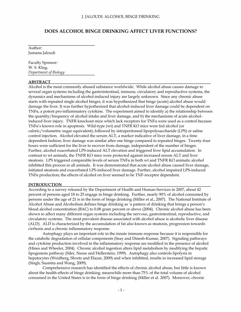
In order to examine levels of LPS induces liver damage, wt and TNFR knockout mice received alcohol for three days followed by IP injection of LPS or saline. Twelve hours after IP injection, serum ALT levels were collected and analyzed. While wt alcohol binging-individuals demonstrated high levels of initial phase LPS-induced liver damage at 12 hours, TNFR knockout mice did not show elevated serum ALT levels (Figure 1). TNFR knockout mice were protected from liver damage because TNFα is essential for ethanol induced pathology; however, TNFα was never activated.
Figure 1. Alcohol binge causes and exacerbates LPS-induced liver damage. C57BL/6J and TNFR -1/2- -/- mice were fed alcohol intra-gastrically for 3 days (1 time/day, 500µl of 10% 200 proof alcohol diluted
J. JALOUDI: ALCOHOL BINGE DRINKING
- 4 -
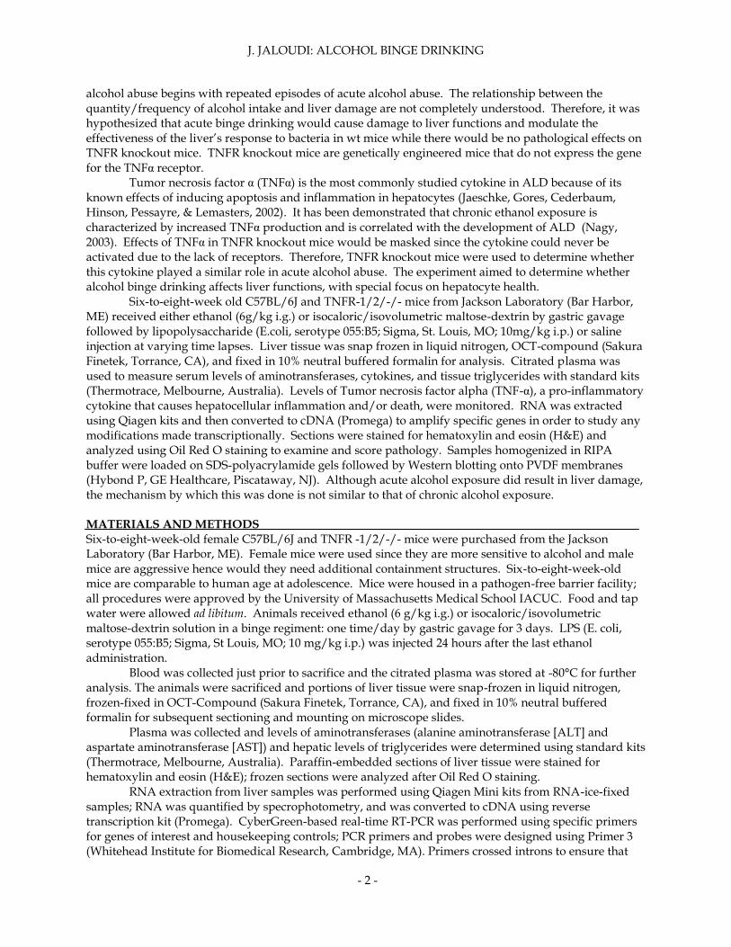
in acidified water), followed by intra-peritoneal injection of either LPS (5µg/kg b/w) or saline control. Twelve hours after intra-peritoneal injection, the serum ALT levels were measured by kinetic assay (n=6/group). Individuals that received binge alcohol treatments were then examined to study alcohol’s effects on LPS triggered TNFα production. Both wild type and TNFR mice displayed impaired LPS-triggered TNFα production (figure 2). Wt mice fed with saline had an increase in LPS-triggered TNFα production when injected with LPS. When wt mice received alcohol treatments followed by LPS injections, they displayed a decrease in LPS-triggered TNFα production. When TNFR knockout mice followed the same treatments, their LPS-triggered TNFα serum levels had a comparable pattern but were generally lower. Alcohol did have a significant effect on pathology by inhibiting an immune response to LPS.
Figure 2. Alcohol binge drinking impairs LPS-triggered TNFα production in vivo. C57BL/6J and TNF-1/2- -/- mice were fed alcohol intra-gastrically for 3 days (1time/day, 500µl of 10% 200 proof alcohol diluted in acidified water), followed by intra-peritoneal injection of either LPS (5µg/kg b/w) or saline
control. Twelve hours later serum TNFα levels were analyzed by ELISA (n=6/group).
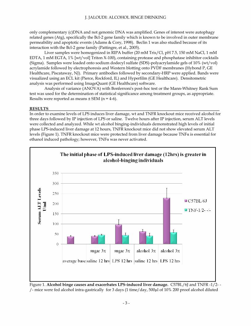
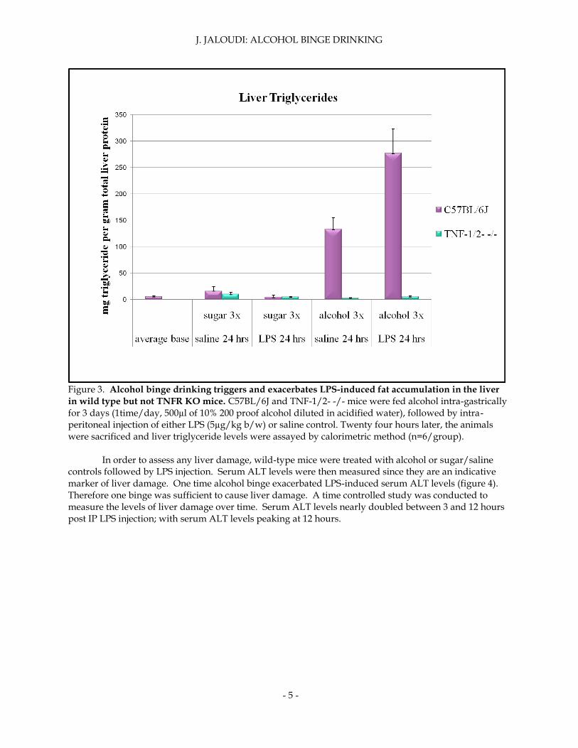
Liver triglycerides were then analyzed to examine if alcohol had other inhibitory effects on autophagy. Wild-type mice that received alcohol treatments showed a significant increase in liver triglycerides, especially when injected with LPS (figure 3). Liver triglycerides were highest in I.G. alcohol and I.P. LPS treated mice. TNFR knockout mice displayed constant levels of liver triglycerides despite treatment given. TNFR knockout mice were protected from liver lipid accumulation.
J. JALOUDI: ALCOHOL BINGE DRINKING
- 5 -
Figure 3. Alcohol binge drinking triggers and exacerbates LPS-induced fat accumulation in the liver
in wild type but not TNFR KO mice. C57BL/6J and TNF-1/2- -/- mice were fed alcohol intra-gastrically for 3 days (1time/day, 500µl of 10% 200 proof alcohol diluted in acidified water), followed by intra-peritoneal injection of either LPS (5µg/kg b/w) or saline control. Twenty four hours later, the animals were sacrificed and liver triglyceride levels were assayed by calorimetric method (n=6/group).
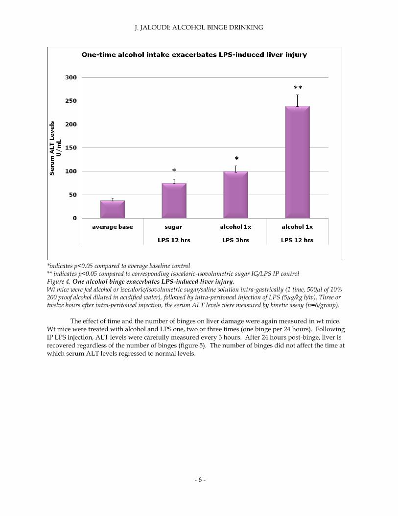
In order to assess any liver damage, wild-type mice were treated with alcohol or sugar/saline controls followed by LPS injection. Serum ALT levels were then measured since they are an indicative marker of liver damage. One time alcohol binge exacerbated LPS-induced serum ALT levels (figure 4). Therefore one binge was sufficient to cause liver damage. A time controlled study was conducted to measure the levels of liver damage over time. Serum ALT levels nearly doubled between 3 and 12 hours post IP LPS injection; with serum ALT levels peaking at 12 hours.
J. JALOUDI: ALCOHOL BINGE DRINKING
- 6 -
* *indicates p<0.05 compared to average baseline control ** indicates p<0.05 compared to corresponding isocaloric-isovolumetric sugar IG/LPS IP control Figure 4. One alcohol binge exacerbates LPS-induced liver injury. Wt mice were fed alcohol or isocaloric/isovolumetric sugar/saline solution intra-gastrically (1 time, 500µl of 10% 200 proof alcohol diluted in acidified water), followed by intra-peritoneal injection of LPS (5µg/kg b/w). Three or twelve hours after intra-peritoneal injection, the serum ALT levels were measured by kinetic assay (n=6/group).
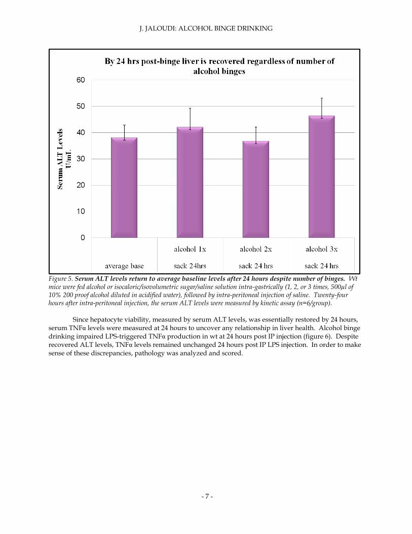
The effect of time and the number of binges on liver damage were again measured in wt mice. Wt mice were treated with alcohol and LPS one, two or three times (one binge per 24 hours). Following IP LPS injection, ALT levels were carefully measured every 3 hours. After 24 hours post-binge, liver is recovered regardless of the number of binges (figure 5). The number of binges did not affect the time at which serum ALT levels regressed to normal levels.
J. JALOUDI: ALCOHOL BINGE DRINKING
- 7 -
Figure 5. Serum ALT levels return to average baseline levels after 24 hours despite number of binges. Wt mice were fed alcohol or isocaloric/isovolumetric sugar/saline solution intra-gastrically (1, 2, or 3 times, 500µl of 10% 200 proof alcohol diluted in acidified water), followed by intra-peritoneal injection of saline. Twenty-four hours after intra-peritoneal injection, the serum ALT levels were measured by kinetic assay (n=6/group).
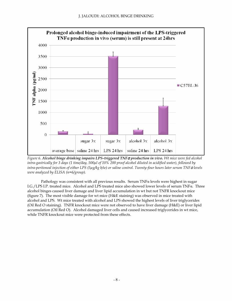
Since hepatocyte viability, measured by serum ALT levels, was essentially restored by 24 hours, serum TNFα levels were measured at 24 hours to uncover any relationship in liver health. Alcohol binge drinking impaired LPS-triggered TNFα production in wt at 24 hours post IP injection (figure 6). Despite recovered ALT levels, TNFα levels remained unchanged 24 hours post IP LPS injection. In order to make sense of these discrepancies, pathology was analyzed and scored.
J. JALOUDI: ALCOHOL BINGE DRINKING
- 8 -
Figure 6. Alcohol binge drinking impairs LPS-triggered TNFα production in vivo. Wt mice were fed alcohol intra-gastrically for 3 days (1 time/day, 500µl of 10% 200 proof alcohol diluted in acidified water), followed by
intra-peritoneal injection of either LPS (5µg/kg b/w) or saline control. Twenty-four hours later serum TNFα levels were analyzed by ELISA (n=6/group).
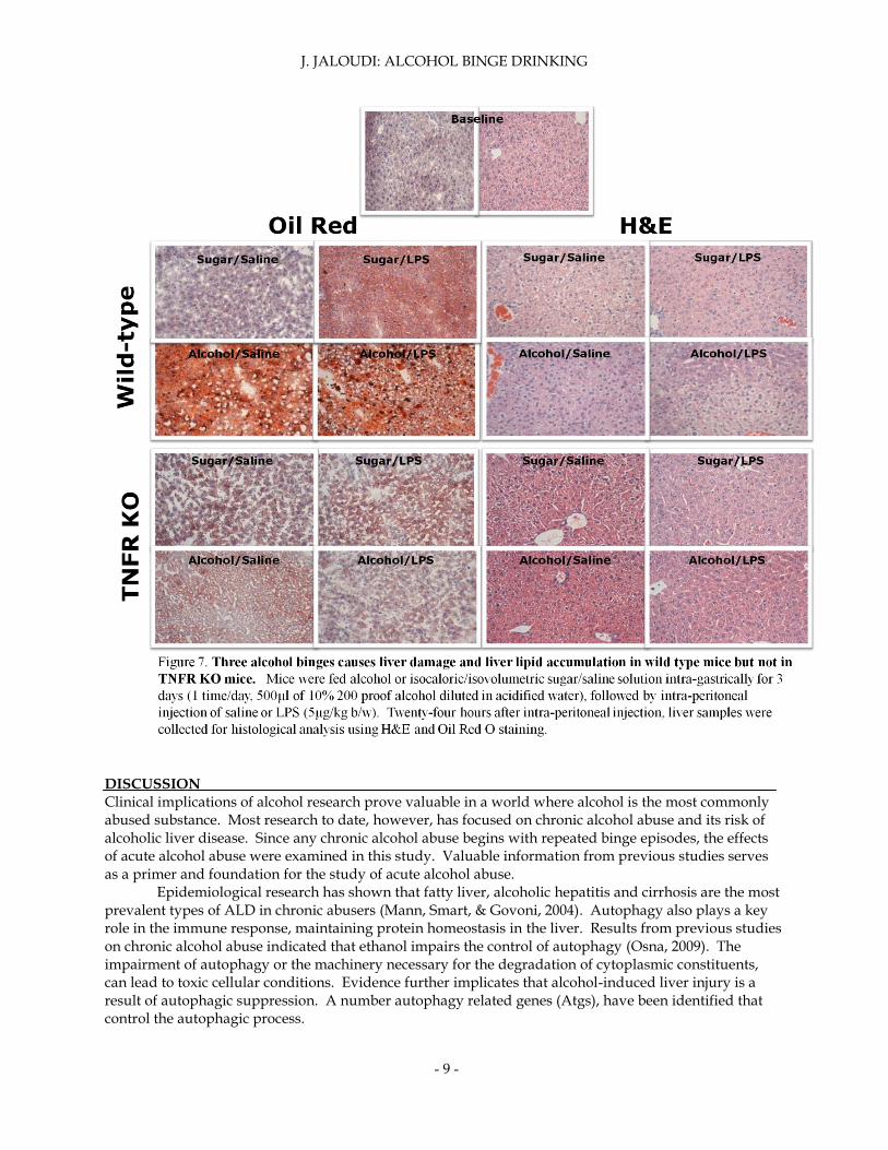
Pathology was consistent with all previous results. Serum TNFα levels were highest in sugar I.G./LPS I.P. treated mice. Alcohol and LPS treated mice also showed lower levels of serum TNFα. Three alcohol binges caused liver damage and liver lipid accumulation in wt but not TNFR knockout mice (figure 7). The most visible damage for wt mice (H&E staining) was observed in mice treated with alcohol and LPS. Wt mice treated with alcohol and LPS showed the highest levels of liver triglycerides (Oil Red O staining). TNFR knockout mice were not observed to have liver damage (H&E) or liver lipid accumulation (Oil Red O). Alcohol damaged liver cells and caused increased triglycerides in wt mice, while TNFR knockout mice were protected from these effects.
J. JALOUDI: ALCOHOL BINGE DRINKING
- 9 -
DISCUSSION
Clinical implications of alcohol research prove valuable in a world where alcohol is the most commonly abused substance. Most research to date, however, has focused on chronic alcohol abuse and its risk of alcoholic liver disease. Since any chronic alcohol abuse begins with repeated binge episodes, the effects of acute alcohol abuse were examined in this study. Valuable information from previous studies serves as a primer and foundation for the study of acute alcohol abuse.
Epidemiological research has shown that fatty liver, alcoholic hepatitis and cirrhosis are the most prevalent types of ALD in chronic abusers (Mann, Smart, & Govoni, 2004). Autophagy also plays a key role in the immune response, maintaining protein homeostasis in the liver. Results from previous studies on chronic alcohol abuse indicated that ethanol impairs the control of autophagy (Osna, 2009). The impairment of autophagy or the machinery necessary for the degradation of cytoplasmic constituents, can lead to toxic cellular conditions. Evidence further implicates that alcohol-induced liver injury is a result of autophagic suppression. A number autophagy related genes (Atgs), have been identified that control the autophagic process.
J. JALOUDI: ALCOHOL BINGE DRINKING
- 10 -
Autophagy has also been shown to regulate lipid metabolism (Singh, Susmita and Wang, 2009). Lipid droplet metabolism also known as lipolysis, is autophagy dependent in hepatocytes (Weidberg, Shvets, & Elazar, 2009). Since chronic ethanol metabolism leads to fatty liver, it was hypothesized that acute alcholic abuse would do the same in an autophagy dependent manner.
The liver plays an essential function in the innate immune response by releasing Kupffer cells, resident macrophages, when activated by an endotoxin. Additionally, the liver recruits neutrophils in a proinflammatory response to microbes (Nagy, 2003). Signaling pathways in the immune response are modified as a result of alcohol metabolism. One pathway by which apoptosis occurs, is dependent on a pro-inflammatory cytokine, TNFα (Jaeschke, Gores, Cederbaum, Hinson, Pessayre, & Lemasters, 2002). When TNFR knockout mice metabolized alcohol, hepatocytes were still able to undergo apoptosis, signifying that the hepatocyte immune response is dependent on a TNFα mechanism. TNFR knockout mice showed no pathological changes due to ethanol, results consistent with the hypothesis that TNFα is necessary for liver damage in mice (Nagy, 2003). Therefore TNFR knockout mice were used in the study as a control and to assess if this cytokine plays a similar role in binge drinking.
Other studies have confirmed these findings: livers with depleted Kupffer cells have decreased liver damage (Sozio & Crabb, 2008). Alcohol treated animals further demonstrated sensitivity to endotoxin LPS, a component of the outer wall of gram-negative bacteria. Patients with ALD have been shown to contain higher levels of serum ALT, a marker indicative of liver damage (Hines and Wheeler, 2004). Wt and TNFR knockout mice were treated in order to uncover the mechanism behind ethanol induced liver damage. Wt mice were hypothesized to display liver damage and a suppressed immune response to bacteria. TNFR knockout mice were hypothesized to show no changes in pathology due to alcohol. The results were consistent with previous studies on the pathology of chronic ethanol exposure; however, the mechanisms by which these processes occur are different in acute and chronic alcohol abuse.
Wt mice that were fed alcohol via I.G. once a day for three days, followed by LPS I.P. injection displayed exacerbated LPS-induced liver damage. Alcohol and saline treated mice had comparable ALT levels to saline and sugar treated mice. Alcohol in combination with LPS nearly tripled serum ALT levels in wt mice. Serum ALT levels were not statistically significant in varying treatments in TNFR knockout mice, consistent with the hypothesis that TNFR knockout mice are protected from changes in pathology. TNFR knockout mice were protected from LPS-induced liver damage despite similar TNFα production levels compared to wt mice. Twelve hours after I.P. injection of either LPS or saline control, both wt and TNFR knockout mice displayed down-regulation of LPS-induced TNFα production. In both wt and TNFR knockout mice, TNFα levels were highest in sugar/LPS treated mice. Alcohol in the presence of LPS significantly decreased the production of TNFα. The effects of alcohol on TNFα serum levels were still present at 24 hours post I.P. injection. TNFα levels remained elevated in LPS treated groups until 24 hours post I.P., demonstrating a prolonged alcohol-induced impairment of the LPS-triggered TNFα production in vivo. Down-regulation of TNFα production due to alcohol is also still present at 24 hours. One alcohol binge episode was sufficient to exacerbate LPS-induced liver injury in wt mice; serum ALT levels continually increased until reaching its peak at 12 hours (figure 4). Moreover, despite the number of binges, serum ALT levels recovered to baselines at 24 hours. TNFR knockout mice were protected from LPS-induced liver damage and increased liver triglycerides. Liver triglycerides were highest in the I.G. alcohol/ I.P. LPS treated knockout mice. TNFR knockout treatment groups did not have statistically significant differences in liver triglycerides. Histology was consistent with the reported data. TNFR knockout mice were protected from liver lipid accumulation and liver damage. Wt mice treated with alcohol and LPS showed the highest liver lipid levels and most liver damage. Acute alcohol abuse exacerbated lipid accumulation in the liver and LPS-induced liver damage in
wt mice. Alcohol binge drinking down-regulated LPS-induced TNFα production in both wt and TNFR knockout mice; the effects of alcohol on liver seem to be TNF-receptor dependent. The quantity and frequency of alcohol intake and liver damage were closely examined in this study but questions regarding the mechanisms of acute alcohol induced liver injury remain. Numerous
J. JALOUDI: ALCOHOL BINGE DRINKING
- 11 -
studies have demonstrated the importance of the signal transduction intermediates in the development of alcohol induced liver damage. The key to discovering treatment strategies for liver injury are to be found when molecular mechanisms by which acute alcohol abuse dysregulates the immune system are fully understood. *Submitted in partial fulfillment of credit in BIO 399-Biology Research Internship, Department of Biology, The College of New Jersey, Ewing, NJ. Summer research conducted at Department of Medicine, University of Massachusetts Medical School, Worcester, MA. REFERENCES Adams, J. M., & Cory, S. (1998). The Bcl-2 Protein Family: Arbiters of Cell Survival. Science , 1322-1333. Hines, Ian and Michael Wheeler (2004). Recent Advances in Alcoholic Liver Disease. American Journal of Physiology- Gastrointestinal and Liver Physiology: G310-G314. Jaeschke, H., Gores, G. J., Cederbaum, A. I., Hinson, J. A., Pessayre, D., & Lemasters, J. J. (2002). Mechanisms of Hepatotoxicity. Toxicological Sciences, 166-176. Mann, R. E., Smart, R. G., & Govoni, R. (2004). The Epidemiology of Alcoholic Liver Disease. National Institute on Alcohol Abuse and Alcoholism. Miller, J. (2007). Binge Drinking and Associated Health Risk Behaviors among High School Students. Pediatrics. Nagy, L. E. (2003). Recent Insights into the Role of the Innate Immune System in the Development of Alcoholic Liver Disease. Cleveland: Society for Experimental Biology and Medicine. National Institute of Alcohol Abuse and Alcoholism (2004). NIAAA council approves definition of binge drinking. NIAAA Newsletter: No. 3:3. Osna, N. (2009). Autophagy and ethanol-induced liver injury. World Journal of Gastroenterology , 1178-1185. Pattingre, S., Tassa, A., Qu, X., Garuti, R., Liang, X. H., Mizushima, N., et al. (2005). Bcl-2 Antiapoptotic Proteins Inhibit Beclin 1- Dependent Autophagy. Cell . Seay, Montrell and S.P Dinesh-Kumar (2007). Autophagy Takes its Toll on Innate Immunity. Cell Host & Microbe: 69. Siler, Scott, Richard Neese and Marc Hellerstein (1999). De novo lipogensis, lipid kinetics, and whole-body lipid balances in humans after acute alcohol consumption. American Journal of Clinical Nutrition: 928-933. Singh, Rajat, et al (2009). Autophagy regulates lipid metabolism. Nature: 1131-1135. Sozio, M., & Crabb, D. W. (2008). Alcohol and lipid metabolism. American Journal of Physiology- Endocrinology and Metabolism , E10-E16. Substance Abuse and Mental Health Services Administration, Office of Applied Studies (2008). Results from the 2007 National Survey on Drug Use and Health: National Findings (NSDUH Series H-34, DHHS Publication No. SMA 08-4343). <http://www.oas.samhsa.gov/NSDUH/2k7NSDUH/2k7results.cfm#Ch3>.
J. JALOUDI: ALCOHOL BINGE DRINKING
- 12 -
Weidberg, Hilla, Elena Shvets and Zvulun Elazar (2009). Lipophagy: Selective Catabolism Designed for Lipids. Developmental Cell: 628-630. ACKNOWLEDGEMENTS I would like to thank Professor Steve Klug for the Biology internship opportunity and the NIH for funding the project. I am grateful to Dr. Deborah Harmon Hines and Dr. Gary Stein for providing me with this research opportunity. Special thanks to Karen Zirpola-Miller, Judith Verdini and Linhelle Charles for making my experience here very enjoyable. Thanks also to Dr. Jan Petrasek and Dr. Amalene Cooper-Morgan for sharing their knowledge. Last, not least, I am grateful to Dr. Timea Csak and Mrs. Karen Kodys for their support and enthusiasm in the lab.