Embed Size (px)
Citation preview
Review ArticleDoes Posterior Capsule Opacification Affectthe Results of Diagnostic Technologies to Evaluatethe Retina and the Optic Disc?
Jose Javier Garcia-Medina,1,2,3,4 Monica del Rio-Vellosillo,5
Vicente Zanon-Moreno,3,4,6 Enrique Santos-Bueso,7 Roberto Gallego-Pinazo,3,4,8
Antonio Ferreras,4,9 and Maria Dolores Pinazo-Duran3,4,10
1 Department of Ophthalmology, General University Hospital Reina Sofia, Avenida Intendente Jorge Palacios 1, 30003Murcia, Spain2 Department of Ophthalmology and Optometry, School of Medicine, University of Murcia, Avenida Intendente Jorge Palacios 1,30003Murcia, Spain
3 Ophthalmology Research Unit “Santiago Grisolia”, Avenida Gaspar Aguilar, 90, 46017 Valencia, Spain4 Oftared-Retics, Instituto de Salud Carlos III, 28029Madrid, Spain5 Department of Anesthesia, University Hospital Virgen de la Arrixaca, Ctra. Madrid-Cartagena, s/n, El Palmar, 30120Murcia, Spain6 Department of Preventive Medicine & Public Health and CIBER Physiopathology of Obesity and Nutrition, School of Medicine,University of Valencia, Avenida Blasco Ibanez 15-17, 46010 Valencia, Spain
7 Department of Ophthalmology, San Carlos University Hospital, Calle Profesor Martın Lagos, S/N, 28040Madrid, Spain8 Department of Ophthalmology, University Hospital La Fe, Bulevar del Sur, 46026 Valencia, Spain9 Miguel Servet University Hospital, Aragon Health Sciences Institute, Paseo Isabel la Catolica, 1-3, 50009 Zaragoza, Spain10Department of Ophthalmology, University School of Medicine, University of Valencia,Avenida Blasco Ibanez 15-17, 46010 Valencia, Spain
Correspondence should be addressed to Jose Javier Garcia-Medina; [email protected]
Received 14 July 2014; Accepted 1 February 2015
Academic Editor: Paolo Frezzotti
Copyright © 2015 Jose Javier Garcia-Medina et al. This is an open access article distributed under the Creative CommonsAttribution License, which permits unrestricted use, distribution, and reproduction in any medium, provided the original work isproperly cited.
The visual outcome obtained after cataract removal may progressively decline because of posterior capsular opacification (PCO).This condition can be treated by creating an opening in the posterior lens capsule by Nd:YAG laser capsulotomy. PCO opticalimperfections cause several light reflection, refraction, and diffraction phenomena, which may interfere with the functionaland structural tests performed in different ocular locations for the diagnosis and follow-up of ocular disease, like macular andoptic nerve diseases. Some parameters measured by visual field examinations, scanning laser polarimetry, and optical coherencetomography (OCT) have changed after PCO removal. Imaging quality also changes following capsulotomy. Consequently, theresults of ancillary tests in pseudophakic eyes for studying ocular diseases like glaucoma or maculopathies should be correlatedwith other clinical examinations, for example, slit-lamp biomicroscopy or funduscopy. If PCO is clinically significant, a new baselineshould be set for future comparisons following capsulotomy when using automated perimetry and scanning laser polarimetry. ToperformOCT in the presence of PCO, reliable examinations (considering signal strength) apparently guarantee that measurementsare not influenced by PCO.
1. IntroductionPhacoemulsification with implantation of intraocular lensin the capsular bag is the most frequent surgical proce-dure performed in ophthalmology. However, the visual gain
obtained after cataract removalmay progressively decline dueto posterior capsular opacification (PCO) [1].
Despite variations in surgical techniques, intraocularlens material or design, implantation of additional devices,
Hindawi Publishing Corporation
BioMed Research International
Volume 2015, Article ID 813242, 8 pages
http://dx.doi.org/10.1155/2015/813242
2 BioMed Research International
and pharmacological interventions, PCO remains the mostfrequent long term complication after cataract surgery [1].Published PCO rates are variable. However, a meta-analysisconcluded that approximately 25% of patients operated fromextracapsular cataract surgery suffered visually significantPCO within 5 years of the operation [2].
PCO is due to the proliferation of lens residual epithelialcells from the lens equator following cataract extraction,which induces visual alteration by direct interaction withlight passing through the visual axis [3]. According to thedistribution of proliferation, the resulting opacities mayadopt a morphologic pattern or two or even a combination ofboth: (a) fibrous-type PCO (fibrous epithelial layers) and (b)pearl-type PCO (groups of swollen, optically active, opacifiedgrape-like epithelial growth) [4]. In clinical terms, thesechanges can diminish visual acuity significantly, alter contrastsensitivity, and cause glare and monocular diplopia [5–7].
Metaplasia of epithelial cells may also induce capsularfolds because of mechanical forces. In general terms, theseepithelial cells may transform into myofibroblasts, whichhave contractile properties and allow the posterior capsuleto wrinkle [4]. These phenomena may create visual distor-tions, including a Maddox rod effect, metamorphopsia-likephenomena, or glare [8].
All of these effects together, these being irregular for-mations of fibrous proliferation, pearls, and puckers of theposterior capsule generate special properties that affect lightreflection, refraction, and diffraction, which may interferenot only with patient vision (Figure 1), but also with func-tional and structural ocular diagnostic tests [2].
PCO-induced visual affection can be solved by laserNd:YAG capsulotomy by producing an opening in the pos-terior capsule and avoiding distortion of light in its passage[1, 4].
An observation made before and after capsulotomy ofthe outcome of different diagnostic examinations to estimateocular diseases provides a better understanding of the effectsof PCO on such technologies. Recent research suggests thatPCO may affect the appropriate ocular disease assessmentsmade by automated perimetry, scanning laser polarimetry, oroptical coherence tomography, as seen in Table 1.
2. Effect of PCO on Automated Perimetry
Avisualfield test throughwhite-on-white automated perime-try is a widely used technique and is a useful tool for thediagnosis and follow-up of several ocular disorders, suchas glaucoma and neuroophthalmological diseases [9, 10].Translucent irregularities in the anterior ocular pole, forexample, cataracts or PCO, can be a confusing factor thatmay lead to an inappropriate interpretation of automatedperimetry, even when it is not uncommon to encounterpatients who are affected or are suspected of being affected,by concurrent entities, for example, PCO and glaucoma orPCO and macular oedema.The ophthalmologist must assesshow much visual impairment is due to PCO and how muchis related to the other concomitant disorder.
The effect that cataract has on automated perimetry hasbeen well investigated [11–13]. Our research group completed
(a)
(b)
Figure 1: Simulation of vision in a PCO-affected eye before (a) andafter Nd:YAG capsulotomy (b).
a study in eyes of patients with PCO who underwent awhite-on-white automated perimetry test (Humphrey SITAstandard programme 24-2) immediately before Nd:YAG cap-sulotomy and between postsurgery weeks 1 and 8 [14]. Thecompared pre- and postlaser perimetric indices were meandefect (MD) and pattern standard deviation (PSD).
MD is the average measure of how depressed the patient’svisual field is (compared with a control of the same age).Several researchers have reported that MD improved aftercataract surgery [11–13]. Similarly according to our results,amelioration of MD occurred after capsulotomy [14].
PSD is a measure of how the different adjacent pointson a visual field are. If an area is focally depressed, PSDwill rise given the major difference between the points inscotoma and their normal adjacent points. PSD remainsunchanged after cataract removal [11–13]. Compared to PCO,however, the PSD in our study improved significantly aftercapsulotomy.This change could be explained by optical PCOfeatures. PCO translucent opacities apparently induce erraticlight-scatter within the eye, which results in a combinationof underilluminated retinal areas and in an increased PSD.Yet when these irregularities have been eliminated throughcapsulotomy, retinal illumination can be more uniform, soPSD lowers [14].
As the clinical slit-lamp examination aspect can besomewhat guesswork-related and as the automated perimetryanalysis corroborates, cataracts depress an automated visual

BioMed Research International 3
Table 1: Studies considering the influence of PCO on test results.
Author (year) [referencenumber] Test ! Precapsulotomy
BCVA(mean ± SD) Postcapsulotomy
BCVA(mean ± SD) Results after capsulotomy
Garcıa-Medina et al.(2006) [14]. AP 26 0.35 ± 0.11
(decimal scale)0.84 ± 0.14
(decimal scale) MD and PSD improved.
Garcıa-Medina et al.(2006) [23] SLP 28 0.41 ± 0.12
(decimal scale)0.85 ± 0.13
(decimal scale)NFI and TSS increased. Significantdecreases of all absolute parameters.
Vetrugno et al. (2007)[24] SLP 158 0.3 ± 0.6
(LogMar)0.05 ± 0.2(LogMar)
Inferior ratio and TSNIT SDdecreased. Superior/nasal increased.
Brittain et al. (2007) [25] SLP 20 0.32 ± un(LogMar)
0.14 ± un(LogMar)
TSS and TSNIT SD increased. TSNITscore decreased.
Arraes et al. (2008) [26] SLP 37 0.2 ± un(decimal scale)
0.8 ± un(decimal scale)
No significant difference betweenparameters.
Hougaard et al. (2001)[35] TD-OCT 13 0.29 ± un
(decimal scale)0.39 ± un
(decimal scale)Signal-to-noise ratio increased but nochanges in macular thickness.
Garcia-Medina et al.(2007) [32] TD-OCT 32 0.25 ± 0.17
(decimal scale)0.77 ± 0.22
(decimal scale)SS increased but no changes in pRNFLthicknesses (in reliable exams).
Gonzalez-Ocampo-Dorta et al. (2008)[40]
TD-OCT 32 0.25 ± 0.17(decimal scale)
0.77 ± 0.22(decimal scale)
SS increased but no changes inmacular thicknesses (in reliableexams).
Altiparmak et al. (2010)[36] TD-OCT 54 0.47 ± 0.3
(decimal scale)0.91 ± 0.14
(decimal scale) No change of the foveal thickness.
Giocanti-Auregan et al.(2011) [37] TD-OCT 30 0.6 ± 0.3
(LogMar)0.1 ± 0.3(LogMar) No change of the foveal thickness.
Wroblewska-Czajka et al.(2012) [38] TD-OCT 55 NA NA No change of the central macular
thickness.
Kara et al. (2012) [31] TD-OCT 98 0.49 ± 0.28(LogMar)
0.09 ± 0.11(LogMar) SS and pRNFL thicknesses increased.
Garcia-Medina et al.(2013) [34] SD-OCT 37 0.27 ± 0.19
(decimal scale)0.83 ± 0.18
(decimal scale)All pRNFL thickness parametersincreased. No changes whenconsidering reliable examinations.
Garcia-Medina et al.(2013) [41] SD-OCT 35 0.23 ± 0.28
(decimal scale)0.81 ± 0.16
(decimal scale)All macular thickness parametersincreased. No changes whenconsidering reliable examinations.
Ruiz-Casas et al. (2013)[39] SD-OCT 31 0.4 ± NA 0.8 ± NA No change of the foveal thickness.
BCVA: best-corrected visual acuity, SD: standard deviation, AP: automated perimetry, MD: mean deviation, PSD: pattern standard deviation, SLP: scanninglaser polarimetry, NFI: nerve fiber indicator, TSS: typical scan score, TSNIT: temporal-superior-nasal-inferior-temporal, NA: not available, TD-OCT: timedomain optical coherence tomography, SD-OCT: spectral domain optical coherence tomography, SS: signal strength, and pRNFL = peripapillary retinal nervefiber layer.
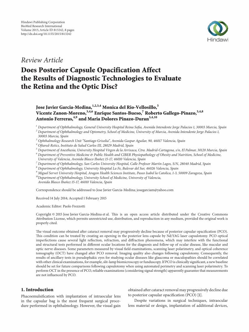
field quite uniformly. So they constitute homogeneous opaci-ties. However, PCOs depress the visual field heterogeneously.Therefore, they have been demonstrated as being polymor-phous opacities that may even mimic pathological patterns[14], such as glaucoma arcuate scotoma, which are susceptibleto elimination after capsulotomy (Figure 2).
In conclusion, PCO has proven to be a heterogeneousmean opacity. This polymorphism may alter visual fieldresults. For practical purposes, a perimetric defect producedby a PCO can be confused in some cases with a pathologicperimetric defect (false-positive). Consequently, presence ofPCO should be taken into account while evaluating anyautomated perimetry in eyes operated from cataracts.
3. Influence of PosteriorCapsular Opacification on ScanningLaser Polarimetry
Scanning laser polarimetry (SLP) is a technology for esti-mating retinal nerve fibre layer (RNFL) thickness in vivoat a specific location [15]. It is based on the principle thata polarised laser beam changes its polarisation status whenpassing through a birefringent tissue. The RNFL is madeup of highly ordered parallel axon bundles that containmicrotubules, which is the source of its birefringence [15].As polarised light passes through the RNFL and is reflectedback, it undergoes a phase shift. This change in polarisation
4 BioMed Research International
(a) (b)
Figure 2: Perimetric defect thatmimics inferior arcuate scotoma in a PCO-affected eye (a).The defect partially disappeared after capsulotomy(b).
(retardation), as measured by SLP, correlates with RNFLthickness [16, 17].Therefore, SLP allows a quantitative assess-ment of the degree of thinning of the peripapillary RNFL.Such information has been demonstrated as being clinicallyuseful in screening and following up both glaucoma [18–20]and nonglaucomatous optic neuropathies, such as anteriorischaemic optic neuropathy, optic nerve head drusen, anddemyelinating optic neuritis [21].
Nevertheless, the RNFL is not the only birefringent struc-ture in the eye. The anterior segment also has birefringentproperties, mainly the cornea. Therefore, total retardationof a subject’s eye is the sum of both the anterior segmentand RNFL birefringence. Accuracy of SLP measurementsdepends on the ability to isolate RNFL retardation from totalocular retardation [21].
To reduce the effect of anterior segment polarisation, thenewest GDx generation incorporates a variable corneal com-pensator (VCC) that enables compensation of the anteriorsegment birefringence (ASB) in each individual eye [21].
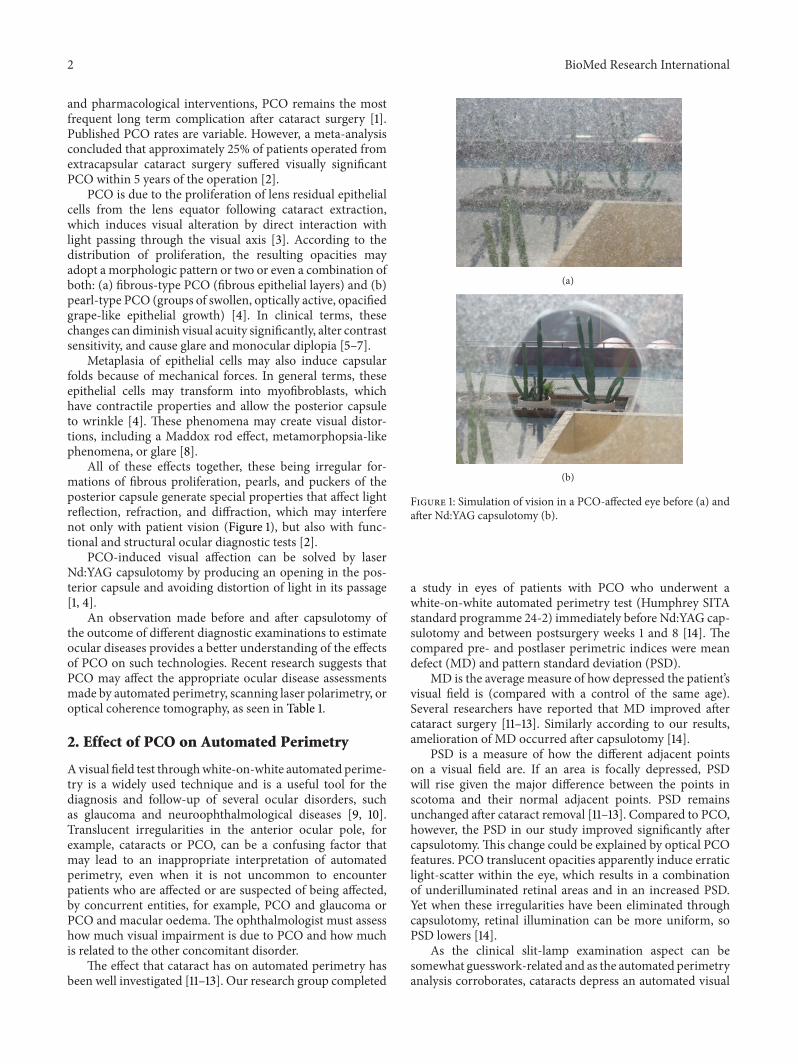
Several researchworks into the effect that PCOand subse-quent Nd:YAG capsulotomy have on the SLP results of RNFLretardation measurements have been conducted [22–26].With this purpose in mind, our research group performeda study into PCO-affected eyes and SLP, selected using GDxVCC, on each patient before and after capsulotomy. Wecompensated ASB before doing any SLP examination. Wecompared the SLP parameters before and after PCO removal.We concluded that PCO removal is associated with remark-ably significant changes in all the SLP measurements. Briefly,our results suggest that thickness parameters are higherbefore than after capsulotomy. In other words, SLP exami-nation with GDx VCC may overestimate RNFL retardationmeasurements in PCO-affected eyes.Therefore, the glaucomadiagnosis in PCO can be underestimated on the basis of theSLP results (false-negatives) (Figure 3) [22, 23]. Furthermore,some SLP measurements (nasal average and nerve fibre indi-cator) have been significantly associated with best-corrected
Nerve fiber layer
(a)
Nerve fiber layer (microns)0 20 40 60 80 100 120
(b)
Figure 3: Scanning laser polarimetry examination before (a) andafter Nd:YAG capsulotomy (b). Note that the thickness measure-ments reduce after PCO removal.
visual acuity (BCVA) before capsulotomy,which suggests thatthis technology may be useful for quantifying the degreeof PCO [23]. However other authors have not found asmany changes in the GDx parameters before and after
BioMed Research International 5
PCO removal. Vetrugno et al. [24] reported modificationsin symmetry, inferior ratio, superior, nasal, and temporal-superior-nasal-inferior-temporal SD whereas Brittain et al.[25] showed significant changes in the typical scan scoreand temporal-superior-nasal-inferior-temporal average. Inaddition, Arraes et al. did not show any significant differencebetween the thickness parameters before and after posteriorcapsulotomy in patients with moderate degrees of PCO [26].The variability of the results can be related to the fact thatanterior segment birefringence is only assessed before andafter laser capsulotomy [23] or only before capsulotomy [25].The characteristics of the population included in these studiescan also be related to this variability noted in the results [23–25].
We also performed a study on a new series of PCOaffected eyes that supports our previous conclusions of GDxVCC measurements. In this study we also observed thatcorneal polarisation axis and corneal polarisation magnitude(the two parameters that determine ASB) changed signifi-cantly after PCO removal [27, 28].
Although the results of different studies in the literatureare not fully coincident, it is advisable to not only repeat theSLP examination after capsulotomy to serve as a new baselinefor the future but also recompensate ASB after Nd:YAG laserapplication to obtain reliablemeasurements using GDxVCC.
4. Effect of PCO on OpticalCoherence Tomography
Optical coherence tomography (OCT) generates high reso-lution, 2-dimensional cross-sectional images of the internalmicrostructure of ocular structures. Transverse images of thedevice are produced using low coherence tomography, anoptical measuring technique that is analogous to a B-scanultrasound, but instead of sound waves, OCT uses a laser-generated beam of light. Two kinds of OCT are available todate: time domain OCT and, more recently, spectral domainOCT. Although both types of OCT use the same basicworking principles, the scan rate and axial resolution haveimproved in spectral domain OCT [29].
OCT explored structures like the peripapillary RNFLand the central retina (including total macular thickness).RNFL thickness, measured by OCT, has been used to studyglaucomatous neuropathy, anterior ischaemic optic neuropa-thy, optic nerve head drusen, demyelinating optic neuritis,traumatic optic tract lesion, Leber hereditary optic neuropa-thy, and toxic optic neuropathy [21]. Macular assessment byOCT has proved to be a very useful tool for studying thevitreoretinal interface, intraretinal oedema, neuroepithelialdetachment, impairments in normal retina architectonics,and its pigment epithelium or choroidal disorders, no matterwhat its aetiology is [30].
In theory, PCOoptical translucent imperfections can alterthis beam of light and can, consequently, induce artifactualresults in OCT thickness and quality parameters. Severalstudies have been performed to answer this question.
In relation to peripapillary RNFL measurements, Karaet al. [31] recently investigated the effect that PCO has on
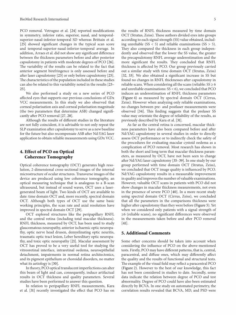
the results of RNFL thickness measured by time domainOCT (Stratus, Zeiss).These authors divided eyes into groupsaccording to each signal strength (SS) value obtained, includ-ing unreliable (SS < 5) and reliable examinations (SS > 5).They also compared the thickness in each group indepen-dently and observed that the lower the SS value, the greaterthe precapsulotomy RNFL average underestimation and themore significant the results. They concluded that RNFLthickness is affected by PCO. Our group previously carriedout a similar study with time domain OCT (Stratus, Zeiss)[32, 33]. We also obtained a significant increase in SS butfound no changes in RNFL thicknesses after capsulotomy inreliable scans.When considering all the scans (reliable: SS≥ 6and unreliable examinations: SS < 6), we concluded that PCOinduces an underestimation of RNFL thickness parameters(Figure 4) as measured by spectral domain OCT (Cirrus,Zeiss). However when analyzing only reliable examinations,no changes between pre- and postlaser measurements wereobserved [34]. This finding suggests that the prelaser SSvalue may orientate the degree of reliability of the results, aspreviously described by Kara et al. [31].
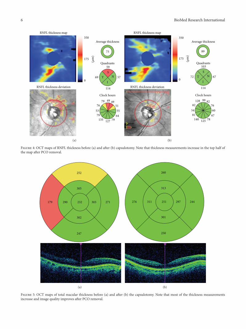
As far as the central retina is concerned, macular thick-ness parameters have also been compared before and afterNd:YAG capsulotomy in several studies in order to directlyassess OCT performance or to indirectly check the safety ofthe procedures for evaluating macular cystoid oedema as acomplication of PCO removal. Most research has shown inboth the short and long term that macular thickness param-eters, as measured by OCT, have not been seen to changeafter Nd:YAG laser capsulotomy [35–39]. In one study by ourgroup performed with time domain OCT (Stratus, Zeiss),we concluded that OCT image quality is influenced by PCO.Nd:YAG capsulotomy results in a measurable improvementin quality and improves the number of valuable examinations.However, valuable OCT scans in patients with PCO did notshow changes in macular thickness measurements, not evenin the presence of severe PCO [40]. In a more recent studyusing spectral domain OCT (Cirrus, Zeiss), we concludedthat all the parameters in the comparisons thickness werehigher after capsulotomy than theywere before (Figure 5). Yetwhen we considered only patients with a signal strength of≥6 (reliable scans), no significant differences were observedin the measurements taken before and after PCO removal[41].
5. Additional Comments
Some other concerns should be taken into account whenconsidering the influence of PCO on the above-mentionedtests. Firstly, PCOmay have different patterns, that is, central,paracentral, and diffuse ones, which may differently affectthe quality and the results of functional and structural tests.The example of the visual field may reflect a paracentral PCO(Figure 2). However to the best of our knowledge, this facthas not been considered in studies to date. Secondly, somedata indicate the relation between degree of PCO and testabnormality. Degree of PCO could have also been estimateddirectly by BCVA. In one study on automated perimetry, thecorrelation results revealed that BCVA, MD, and PSD were
6 BioMed Research International
RNFL thickness map350
175
0
(!m
)
RNFL thickness deviation
Average thickness
75
Quadrants59
69
114
57T NS
I
Clock hours49 49
515564
79127135795376
79
(a)
350
175
0
(!m
)
RNFL thickness map
RNFL thickness deviation
Average thickness
89
Quadrants
Clock hours
103
72
114
67T NS
I
99 82766067
78125140815482128
(b)
Figure 4: OCT maps of RNFL thickness before (a) and after (b) capsulotomy. Note that thickness measurements increase in the top half ofthe map after PCO removal.
252
179 290
305
303232 271
302
247
(a)
260
276 311
313
297251 244
301
250
(b)
Figure 5: OCT maps of total macular thickness before (a) and after (b) the capsulotomy. Note that most of the thickness measurementsincrease and image quality improves after PCO removal.
BioMed Research International 7
significantly associated both before and after capsulotomy[14]. Another study showed some SLP measurements asso-ciated significantly with BCVA before capsulotomy, whichindicates that this technology may be useful for quantifyingdegree of PCO [23]. In relation to OCT, the correlation foundbetween BCVA and SS before capsulotomy suggests that SScould be considered an objective indicator of degree of PCO[40]. Kara et al. [31] also found a significant correlationbetween, on the one hand, preoperative BCVA and SS and,on the other hand, between preoperative BCVA and degreeof PCO. Finally, differences between instruments may be dueto, at least in part, differences in inclusion criteria betweenstudies and characteristics of included eyes.
6. Conclusion
Optical translucent imperfections of PCO induce specialproperties relating to reflection, refraction, and diffractionthat may alter the ancillary tests used in the diagnosis andfollow-up of different optic nerve diseases.
In fact the results of automated perimetry and SLP havebeen shown to change after capsulotomy. In addition, OCTquality imaging of RNFL thickness is influenced by PCO.However, no change has been observed after PCO removal inthe retinal nerve fibre layer parameters of pseudophakic eyesby reliable examinations before capsulotomy, as measured byOCT.
Thus, features of ancillary tests in pseudophakic eyes forstudying optic nerve diseases should be well-interpreted andshould correlate with other clinical examinations, such asslit-lamp biomicroscopy. If a clinically significant PCO isdetected, newmeasurements should be considered after PCOremoval to serve as a baseline for future comparisons, espe-cially when using automated perimetry and SLP. As for OCTin the presence of PCO, reliable examinations (consideringsignal strength) apparently guarantee that the measurementstaken before and after capsulotomy are similar.
Conflict of Interests
The authors declare that there is no conflict of interestsregarding the publication of this paper.
References
[1] O. Findl, W. Buehl, P. Bauer, and T. Sycha, “Interventions forpreventing posterior capsule opacification,” Cochrane Databaseof Systematic Reviews, no. 3, Article ID CD003738, 2007.
[2] D. A. Schaumberg, M. R. Dana,W. G. Christen, and R. J. Glynn,“A systematic overview of the incidence of posterior capsuleopacification,” Ophthalmology, vol. 105, no. 7, pp. 1213–1221,1998.
[3] D. J. Apple, K. D. Solomon, M. R. Tetz et al., “Posterior capsuleopacification,” Survey of Ophthalmology, vol. 37, no. 2, pp. 73–116, 1992.
[4] I. M. Wormstone, L. Wang, and C. S. C. Liu, “Posterior capsuleopacification,”Experimental Eye Research, vol. 88, no. 2, pp. 257–269, 2009.
[5] M. Claesson, L. Klaren, C. Beckman, and J. Sjostrand, “Glareand contrast sensitivity before and after Nd:YAG laser capsulo-tomy,” Acta Ophthalmologica, vol. 72, no. 1, pp. 27–32, 1994.
[6] P. Sunderraj, J. R. Villada, P. W. Joyce, and A. Watson, “Glaretesting in pseudophakes with posterior capsule opacification,”Eye, vol. 6, no. 4, pp. 411–413, 1992.
[7] J. J. Garcıa Medina, M. Garcıa Medina, M. D. Pinazo Duran,and M. Morales Suarez-Varela, “Monocular diplopia afterneodymium: YAG laser capsulotomy,”Graefe’s Archive for Clini-cal and Experimental Ophthalmology, vol. 243, no. 12, pp. 1288–1290, 2005.
[8] J. T. Holladay, J. E. Bishop, and J. W. Lewis, “Diagnosis andtreatment of mysterious light streaks seen by patients followingextacapsular cataract extraction,” The American Intra-OcularImplant Society Journal, vol. 11, no. 1, pp. 21–23, 1985.
[9] S. P.Donahue, “Perimetry techniques in neuro-ophthalmology,”Current Opinion in Ophthalmology, vol. 10, no. 6, pp. 420–428,1999.
[10] M. Wall, S. G. Punke, T. L. Stickney, C. F. Brito, K. R. Withrow,and R. H. Kardon, “SITA standard in optic neuropathiesand hemianopias: a comparison with full threshold testing,”Investigative Ophthalmology and Visual Science, vol. 42, no. 2,pp. 528–537, 2001.
[11] B. L. Lam,W. L. M. Alward, and H. E. Kolder, “Effect of cataracton automated perimetry,” Ophthalmology, vol. 98, no. 7, pp.1066–1070, 1991.
[12] S. D. Smith, J. Katz, and H. A. Quigley, “Effect of cataractextraction on the results of automated perimetry in glaucoma,”Archives of Ophthalmology, vol. 115, no. 12, pp. 1515–1519, 1997.
[13] Y. Y. Kim, J. S. Kim, D. H. Shin, B. C. Kim, and H. R. Jung,“Effect of cataract extraction on blue-on-yellow visual field,”TheAmerican Journal of Ophthalmology, vol. 132, no. 2, pp. 217–220,2001.
[14] J. J. Garcıa-Medina, M. Garcıa-Medina, M. T. Arbona-Nadal,and M. D. Pinazo-Duran, “Effect of posterior capsular opaci-fication removal on automated perimetry,” Eye, vol. 20, no. 5,pp. 537–545, 2006.
[15] Q. Zhou and R. W. Knighton, “Light scattering and form bire-fringence of parallel cylindrical arrays that represent cellularorganelles of the retinal nerve fiber layer,” Applied Optics, vol.36, no. 10, pp. 2273–2285, 1997.
[16] R. N. Weinreb, A. W. Dreher, A. Coleman, H. Quigley, B.Shaw, and K. Reiter, “Histopathologic validation of Fourier-ellipsometry measurements of retinal nerve fiber layer thick-ness,” Archives of Ophthalmology, vol. 108, no. 4, pp. 557–560,1990.
[17] A. W. Dreher, K. Reiter, and R. N. Weinreb, “Spatially resolvedbirefringence of the retinal nerve fiber layer assessed with aretinal laser ellipsometer,”AppliedOptics, vol. 3, no. 19, pp. 3730–3735, 1992.
[18] N. T. Choplin and D. C. Lundy, “The sensitivity and specificityof scanning laser polarimetry in the detection of glaucoma ina clinical setting,” Ophthalmology, vol. 108, no. 5, pp. 899–904,2001.
[19] R. N. Weinreb, C. Bowd, and L. M. Zangwill, “Glaucoma detec-tion using scanning laser polarimetry with variable cornealpolarization compensation,”Archives of Ophthalmology, vol. 121,no. 2, pp. 218–224, 2003.
[20] N. J. Reus and H. G. Lemij, “Diagnostic accuracy of the GDxVCC for glaucoma,” Ophthalmology, vol. 111, no. 10, pp. 1860–1865, 2004.
8 BioMed Research International
[21] G. L. Trick, F. Y. Calotti, and B. Skarf, “Advances in imaging ofthe optic disc and retinal nerve fiber layer,” Journal of Neuro-Ophthalmology, vol. 26, no. 4, pp. 284–295, 2006.
[22] J. J. Garcıa Medina, M. Garcıa Medina, M. Shahin, and M.D. Pinazo Duran, “Posterior capsular opacification affectsscanning laser polarimetry examination,” Graefe’s Archive forClinical and Experimental Ophthalmology, vol. 244, no. 4, pp.520–523, 2006.
[23] J. J. Garcıa-Medina, M. Garcıa-Medina, S. G.-O. Dorta, M. D.Pinazo-Duran, R. Gallego-Pinazo, and V. C. Zanon-Moreno,“Effect of posterior capsular opacification removal on scanninglaser polarimetry measurements,” Graefe’s Archive for Clinicaland Experimental Ophthalmology, vol. 244, no. 11, pp. 1398–1405, 2006.
[24] M. Vetrugno, F. Masselli, G. Greco et al., “The influence ofposterior capsule opacification on scanning laser polarimetry,”Eye, vol. 21, no. 6, pp. 760–763, 2007.
[25] C. J. Brittain, K. C. S. Fong, C. C. Hull, and I. H. Gillespie,“Changes in scanning laser polarimetry before and after lasercapsulotomy for posterior capsular opacification,” Journal ofGlaucoma, vol. 16, no. 1, pp. 112–116, 2007.
[26] T. A. Arraes, H. D. Cavalcanti, J. Arraes, A. C. de Souza Leao,and M. F. Sena, “Analysis of the nerve fiber layer using GDxin pseudophakic patients with posterior capsular opacification,”Arquivos Brasileiros de Oftalmologia, vol. 71, no. 1, pp. 75–78,2008.
[27] J. J. Garcia-Medina, M. Garcia-Medina, V. C. Zanon-Moreno etal., “The influence of posterior capsular opacification removalon anterior segment birefringence parameters as measured byscanning laser polarimetry,”Clinical and Experimental Ophthal-mology, vol. 35, no. 5, pp. 414–420, 2007.
[28] J. J. Garcia-Medina, S. Gonzalez-Ocampo, and M. Garcia-Medina, “Scanning laser polarimetry: what could wemeasure?”Clinical andExperimentalOphthalmology, vol. 36, no. 1, pp. 100–101, 2008.
[29] D. S. Grewal and A. P. Tanna, “Diagnosis of glaucoma anddetection of glaucoma progression using spectral domain opti-cal coherence tomography,” Current Opinion in Ophthalmology,vol. 24, no. 2, pp. 150–161, 2013.
[30] J. J. Wong, T. C. Chen, L. Q. Shen, and L. R. Pasquale, “Macularimaging for glaucoma using spectral-domain optical coherencetomography: a review,” Seminars in ophthalmology, vol. 27, no.5-6, pp. 160–166, 2012.
[31] N. Kara, H. Altinkaynak, K. Yuksel, T. Kurt, and A. Demirok,“Effects of posterior capsular opacification on the evaluationof retinal nerve fiber layer as measured by stratus opticalcoherence tomography,” Canadian Journal of Ophthalmology,vol. 47, no. 2, pp. 176–180, 2012.
[32] J. J. Garcia-Medina, S. Gonzalez-Ocampo-Dorta, A. Feliciano-Sanchez et al., “Changes in optical coherence tomographyafter posterior capsular opacification removal. poster commu-nication number 95,” in Proceedings of the Congress of theAmerican Academy of Ophthalmology, New Orleans, La, USA,2007, http://aao.scientificposters.com/epsSearchAAO.cfm.
[33] J. J. Garcıa-Medina, M. Garcıa-Medina, and S. Gonzalez-Ocampo-Dorta, “Posterior capsular opacification: one factor tobe considered for the study of the optic nerve,” Archivos de laSociedad Espanola de Oftalmologia, vol. 84, no. 1, pp. 1–4, 2009.
[34] J. J. Garcia Medina, J. J. Gomez Fernandez, S. Valentino etal., “Influence of the posterior capsular opacification in themeasurement of thickness of macular ganglion cell complexand retinal nerve fiber layer by means of spectral domain
OCT,” in Proceedings of the 8th Congress of the Spanish Societyof Glaucoma, Cordoba, Spain, March 2013, http://www.soci-edadglaucoma.com/nova/NNws ShwNewDup?codigo=3567&cod primaria=1453&cod secundaria=100765.
[35] J. L. Hougaard, M. Wang, B. Sander, and M. Larsen, “Effects ofpseudophakic lens capsule opacification on optical coherencetomography of the macula,” Current Eye Research, vol. 23, no. 6,pp. 415–421, 2001.
[36] U. E. Altiparmak, I. Ersoz, D. Hazirolan, B. Koklu, R. Kasim,and S. Duman, “The impact of Nd:YAG capsulotomy on fovealthickness measurement by optical coherence tomography,”Ophthalmic Surgery Lasers and Imaging, vol. 41, no. 1, pp. 67–71, 2010.
[37] A. Giocanti-Auregan, J. Tilleul, C. Rohart et al., “OCT mea-surement of the impact of Nd:YAG laser capsulotomy on fovealthickness,” Journal Francais d’Ophtalmologie, vol. 34, no. 9, pp.641–646, 2011.
[38] E. Wroblewska-Czajka, E. Wylegała, D. Tarnawska, A. Now-inska, and D. Dobrowolski, “Assessment of retinal thicknessobtain by optical coherence tomography after Nd: YAG capsu-lotomy,” Klinika Oczna, vol. 114, no. 3, pp. 194–197, 2012.
[39] D. Ruiz-Casas, C. Barrancos, J. L. Alio II, M. Ruiz-Guerrero,and F. J. Munoz-Negrete, “Effect of posterior neodymium:YAGcapsulotomy. Safety evaluation of macular foveal thickness,intraocular pressure and endothelial cell loss in pseudophakicpatients with posterior capsule opacification,” Archivos de laSociedad Espanola de Oftalmologia, vol. 88, no. 11, pp. 415–422,2013.
[40] S. Gonzalez-Ocampo-Dorta, J. J. Garcıa-Medina, A. Feliciano-Sanchez, and G. Scalerandi, “Effect of posterior capsular opaci-fication removal on macular optical coherence tomography,”European Journal of Ophthalmology, vol. 18, no. 3, pp. 435–441,2008.
[41] J. J. Garcia-Medina, J. J. Gomez-Fernandez, S. Valentino,M. Morcillo-Guardiola, J. C. Villada-Sanchez, and A. PastorGrau, “Effects of posterior capsule opacification on macularthickness measurements by spectral domain OCT. Interna-tional SIRCOVA-OFTARED Congress Abstract,” OphthalmicResearch, vol. 50, pp. 27–53, 2013, http://www.karger.com/Article/Pdf/351623.


![Sulforaphane promotes ER stress, autophagy, and cell death: … · 2017-08-25 · drain on healthcare providers [3, 4]. Posterior capsule opacification(PCO) isthemostcommoncomplication](https://img.pdfslide.net/doc/110x75/5e5b933fdabba4232e4e223b/sulforaphane-promotes-er-stress-autophagy-and-cell-death-2017-08-25-drain-on.jpg)