Embed Size (px)
DESCRIPTION
2011
Citation preview
Discussion Paper
The role of occipitotemporal body-selective regions inperson perception
Paul E. Downing1 and Marius V. Peelen2
1Wales Institute of Cognitive Neuroscience, School of Psychology, Bangor University, Bangor, UK2Center for Mind/Brain Sciences, University of Trento, Rovereto, Italy
The visual appearance of others’ bodies is a powerful source of information about the people around us. Thisinformation is implicit in the stimulus and must be extracted and made explicit by the coordination of activity inmultiple cortical areas. Here we consider the contribution to this process of two strongly body-selectiveoccipitotemporal regions identified in human neuroimaging experiments: the extrastriate body area (EBA) andthe fusiform body area (FBA). We address the evidence and arguments behind numerous recent proposals that EBAand FBA build explicit representations of identity, emotion, body movements, or goal-directed actions from thevisual appearance of bodies, and also explore the contribution of these regions to motor control. We argue that thecurrent evidence does not support a model in which EBA and FBA directly perform any of these higher-levelfunctions. Instead, we argue that these regions comprise populations of neurons that encode fine details of the shapeand posture of the bodies of people in the current percept. In doing so, they provide a powerful but cognitivelyunelaborated perceptual framework that allows other cortical systems to exploit the rich, socially relevantinformation that is conveyed by the body.
Keywords: Body perception; Extrastriate body area; Fusiform body area; Action perception; Emotional gestures;Self–other distinction.
The appearance of the human body provides importantvisual signals. We see some of our own movements,and to an extent these are guided visually. Visual bodyinformation also provides myriad social signals aboutother people––who they are, what they are doing, howthey are feeling, and the like. Accordingly, the braindevotes considerable cortical resources to representingvisual information about the human body (Berlucchi &Aglioti, 2010; Minnebusch & Daum, 2009; Peelen &Downing, 2007a; Schwoebel & Coslett, 2005). Clearevidence for specialized visual representations of thebody––and a link between these and the temporallobes––extends well into the previous century, andcan be found in neuropsychology (Konorski, 1967)and in some of the earliest work on the visual response
properties of single units in the macaque temporalcortex (Gross, 1992).
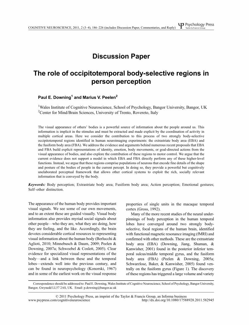
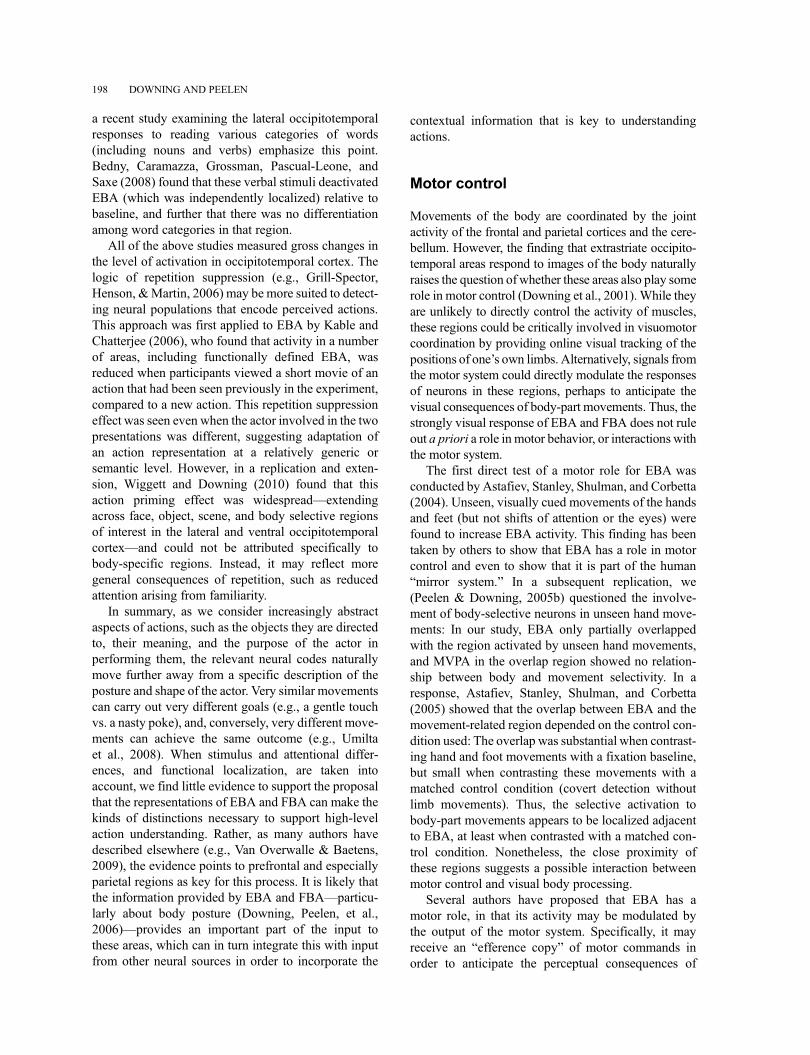
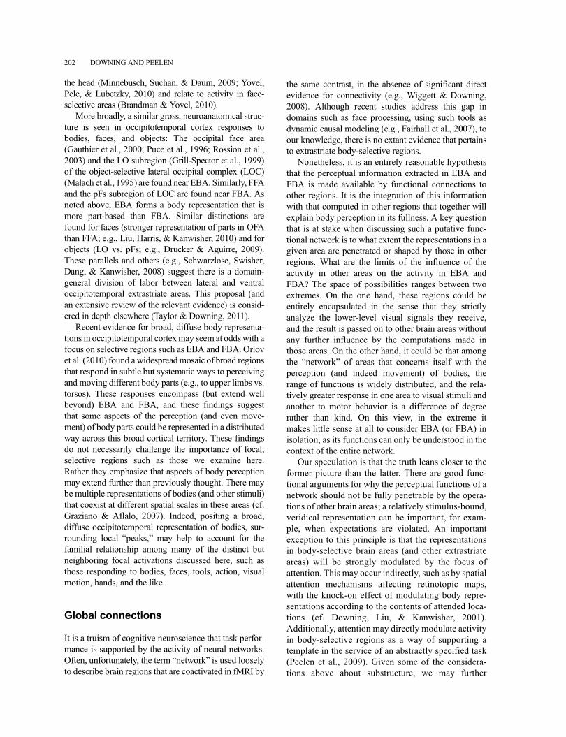
Many of the more recent studies of the neural under-pinnings of body perception in the human temporallobes have converged around two strongly body-selective, focal regions of the human brain, identifiedwith functional magnetic resonance imaging (fMRI) andconfirmed with other methods. These are the extrastriatebody area (EBA) (Downing, Jiang, Shuman, &Kanwisher, 2001) found in the posterior inferior tem-poral sulcus/middle temporal gyrus, and the fusiformbody area (FBA) (Peelen & Downing, 2005a;Schwarzlose, Baker, & Kanwisher, 2005) found ven-trally on the fusiform gyrus (Figure 1). The discoveryof these regions has triggered a large volume and variety
COGNITIVE NEUROSCIENCE, 2011, 2 (3–4), 186–226 (includes Discussion Paper, Commentaries, and Reply)
Correspondence should be addressed to: Paul E. Downing,Wales Institute of Cognitive Neuroscience, School of Psychology, Bangor University,Bangor, Gwynedd LL57 2AS, UK. Email: [email protected]
© 2011 Psychology Press, an imprint of the Taylor & Francis Group, an Informa businesswww.psypress.com/cognitiveneuroscience http://dx.doi.org/10.1080/17588928.2011.582945
of subsequent work. This includes fMRI studies ofhealthy human populations and of psychiatric popula-tions, and extends to investigations with transcranialmagnetic stimulation (TMS), patients with neurologicaldamage, event-related potential (ERP) and magneto-encephalography (MEG) techniques, and nonhumanprimates.
We focus the present discussion on these tworegions because they are clearly identifiable areasinvolved in visual body analysis, while acknowledgingthat they may be further subdivided by functional and/or anatomical criteria (e.g., Bracci, Ietswaart, Peelen, &Cavina-Pratesi, 2010; Weiner & Grill-Spector, 2011),and that they must operate not in isolation but rather inconcert with other brain areas. (We will return to thesepoints later.) Now that there is a rich set of data aboutthe properties of EBA and FBA, multiple researchgroups have proposed that their neural activity directlyunderpins a plethora of functions (Table 1). Theseinclude identifying other individuals, perceiving emo-tions, perceiving body movements, understanding themeaning of others’ actions, and even the control ofmotor movements.
In our view, many of these proposals extend too farpast the data. Here, we critically consider current evi-dence on the possible function(s) of EBA and FBA,with reference to a more parsimonious model: Theextrastriate and fusiform body areas jointly create adetailed but cognitively unelaborated visual represen-tation of the appearance of the human body. Thisrepresentation makes explicit the aspects of the imagethat contain bodies or body parts, and represents theirshape and posture in some detail. It does not, we argue,make explicit high-level information about other peo-ple, such as their identities, actions, or emotional states.Thus, this representation provides a general perceptualinfrastructure that contributes to the information pro-cessing in other interconnected brain areas that extractcomplex and often context-dependent informationabout the people we observe.
BACKGROUND
Downing et al. (2001) reported new evidence fromhuman fMRI for a lateral occipitotemporal region that
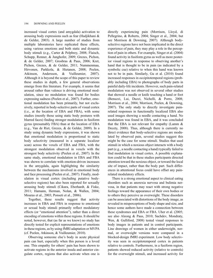
Figure 1. Location of occipitotemporal body-selective regions (extrastriate body area: EBA; fusiform body area: FBA). (A) EBA (left) and FBA(right) in one participant, defined by a contrast of bodies – (chairs + scenes), p < .00005. Z values refer to position of axial slices in Talairach space. (B)Results of a fixed-effects, group-average contrast of (bodies – chairs), n¼ 8, p< .00001, rendered on the inflated cortical surface of a single participant.
ROLE OF BODY-SELECTIVE REGIONS 187
responds strongly and selectively to images of humanbodies and body parts. This followed earlier findings ofsimilar face-selective (Kanwisher, McDermott, &Chun, 1997; Puce, Allison, Asgari, Gore, &McCarthy, 1996) and scene-selective (Aguirre,Zarahn, & D’Esposito, 1998; Epstein & Kanwisher,
1998) areas, and was part of a program to determinethe extent of such apparently specialized regions(e.g., Downing, Chan, Peelen, Dodds, & Kanwisher,2006). Several years after identifying EBA, we (Peelen& Downing, 2005a) identified a second highly body-selective region in the fusiform gyrus, overlapping, but
TABLE 1The discovery of highly body-selective regions in the lateral (extrastriate body area; EBA) and ventral (fusiform body area; FBA)
occipitotemporal cortex has led to a wide diversity of proposals for the functional role(s) that these regions play. The quotations beloware taken out of context and are simply meant to illustrate this diversity. In this article, we argue, contrary to many (but not all) of theseproposals, that these regions perform a role that is largely restricted to representing the shape and posture of the body, and it is not
elaborated with information about identity, emotion, motion, action goals, or motor control
� Arzy et al. (2006): “Collectively, these data show that distributed brain activity at the EBA and TPJ as well as their timing are crucial for thecoding of the self as embodied and as spatially situated within the human body.”
� Astafiev et al. (2004): “Our results indicate that in addition to this visual recognition function, the EBA integrates visual, spatial attention, andsensory-motor signals involved in the representation of the observer’s body.”
� Blanke et al. (2010): “Our data show that the EBA is also involved in mental imagery of human bodies.”� Calvo-Merino et al. (2010): “Our results suggest that the EBA and vPMC may be two complementary components of the aesthetic perception
network for bodies.”� Chan et al. (2004): “We propose that the EBA plays a relatively early role in social vision.”�Costantini et al. (2005): “The higher BOLD signal during observation of impossible movements . . . not only suggests that EBAmay be activated
during action observation but also that this area codes body and action related stimuli multimodally.”� Cross et al. (2010): “A key function of EBAmight be to extract body-form cues that are either unrelated, impossible or beyond what the viewer’s
body can do.”�David et al. (2007): “Our results suggest that the EBA represents the human body in a more integrative and dynamic manner, being able to detect
an incongruence of internal body or action representations and external visual signals.”� De Lange et al. (2008): “Our data show that EBA activity is further influenced by the motoric context in which the body part is presented.”�Downing, Peelen et al. (2006): “We speculate that the EBA computes a static representation of the human body and is not involved in analysis of
biological motion per se.”�Hodzic, Muckli et al. (2009): “[Identity analysis] appears to be accomplished by a network comprising the right FBA, positioned ventrally to the
EBA, regions of the superior parietal lobe, the inferior parietal cortex, and the middle frontal gyrus.”� Jackson et al. (2006): “It is thus likely that the EBA is important not only for the visual processing of body parts, but also for . . . automatically
mapping the visual representation of another’s body to one’s own body.”� Jastorff & Orban (2009): “Our results suggest that the EBA and the FBA correspond to the initial stages in visual action analysis, in which the
performed action is linked to the body of the actor.”�Kable& Chatterjee (2006): “Representations in the pSTS,MT/MST, and EBA abstract actions from the agents involved and distinguish between
different particular actions.”� Kokal et al. (2009): “The presence of these regions [EBA and pSTS] in our [joint action] networks suggests that the process of integrating
observed and executed actions . . . may also occur at a more sensory level.”� Kontaris et al. (2009): “In contrast to pSTS, EBA and FBA are decoupled from motor systems.”�Kuhn et al. (2011): “Our study suggests that both EBA and FFA play a role in the representation of one’s own body and in the control of voluntary
action.”�Marsh et al. (2010): “The extrastriate body area may be geared to take into account the social meaning of actions, so that actions can be
understood with reference to the person executing them.”�Moro et al. (2008): “Visual analysis of human body stimuli is based on the division of labor into two cortical systems, with EBA and FBA
representing the actors’ identity and vPMc mapping the observed action in a neutral format with respect to the identity of the acting bodies.”�Myers & Sowden (2008): “We argue that the right EBA may perform an important sorting of body part images by identity (including self-
recognition) and may interact both with brain areas involved in sensory processing and social cognition.”� Newman-Norlund et al. (2007): “EBA ... may represent the brain basis of our ability to relate the movements of others directly with our own
movements in joint-action situations.”� Pierno et al. (2009): “EBA . . . [has] a role concerned with the discrimination of specific aspects characterizing the observed actions.”� Pitcher et al. (2009): “Our results extend earlier findings by showing that rEBA represents bodies in their most common configuration.”� Pourtois et al. (2007): “[There is ] a major role for EBA in the initial perceptual analysis of body shapes.”� Ramsey & Hamilton (2010): “Our data suggest that [FFA, EBA, FBA] are also recruited in more social and dynamic contexts; they distinguish
between two intentional agents who are acting in a goal-directed fashion.”� Saxe et al. (2006): “[There is] a role for the right EBA in the perception of other people per se, perhaps as input to subsequent perception of
others’ actions and reasoning about other minds.”� Suchan et al. (2010): “Body image distortion is related at least in part to structural alteration in the EBA.”� Urgesi, Candidi et al. (2007): “Thus, the present data clearly show that EBA is crucial in processing bodily forms but not bodily actions.”
188 DOWNING AND PEELEN
distinct from, the fusiform face area (FFA). Shortlyafterwards, Schwarzlose et al. (2005) reported thathigh-resolution fMRI revealed a double dissociationbetween FBA and FFA.
fMRI studies have demonstrated that EBA and FBArespond to photorealistic depictions of whole humanbodies or body parts, significantly more than to faces,face parts, objects, object parts, scenes, visual motion,and other control stimuli (Downing, Chan et al., 2006;Downing et al., 2001; Peelen & Downing, 2005a;Schwarzlose et al., 2005; Spiridon, Fischl, &Kanwisher, 2006; Weiner & Grill-Spector, 2010). Theresponses of these regions are not limited to full-cuestimuli: Their body selectivity extends to line drawings,“stick figures,” and silhouettes (Downing et al., 2001;Peelen&Downing, 2005a). The body representations inthese regions appear to be viewpoint-dependent––in anfMRI adaptation design, changes in view greater than45� result in release from adaptation to the level of a newstimulus (Taylor, Wiggett, & Downing, 2010). There isevidence from intracranial ERP for a body-selectivevisual response originating at the approximate site ofEBA (Pourtois, Peelen, Spinelli, Seeck, & Vuilleumier,2007), and this is confirmed by scalp ERP (Thierry et al.,2006) andMEG (Ishizu, Amemiya, Yumoto, & Kojima,2010) studies, and near infrared spectroscopy (Ishizu,Noguchi, Ito, Ayabe, & Kojima, 2009). Notably, fMRI,TMS, and ERP studies indicate that the representation ofbodies in EBA is more strongly part-based than in FBA(Costantini, Urgesi, Galati, Romani, & Aglioti, 2011;Taylor, Wiggett, & Downing, 2007; Taylor, Roberts,Downing, & Thierry, 2010; Urgesi, Calvo-Merino,Haggard, & Aglioti, 2007). Finally, recent fMRI studiesof macaques show body-selective activation patches inthe temporal cortex (Pinsk et al., 2005, 2009; Tsao,Freiwald, Knutsen, Mandeville, & Tootell, 2003), mak-ing an important link between human studies and pre-vious single-unit work that reported strong selectiveneural responses to hands or whole bodies (Desimone,Albright, Gross, & Bruce, 1984; Gross, Bender, &Rocha-Miranda, 1969; Gross, Rocha-Miranda, &Bender, 1972; Oram & Perrett, 1996; Wachsmuth,Oram, & Perrett, 1994).
Is the response to human bodies and body parts inEBA and FBA functionally relevant for body percep-tion? At least five published studies show a selectivedisruption to body- or body-part perceptual tasks fol-lowing TMS over EBA (Calvo-Merino, Urgesi, Orgs,Aglioti, & Haggard, 2010; Pitcher, Charles, Devlin,Walsh, & Duchaine, 2009; Urgesi, Berlucchi, &Aglioti, 2004; Urgesi, Calvo-Merino et al., 2007;Urgesi, Candidi, Ionta, & Aglioti, 2007). These effectsare specific to stimulus type and stimulation location,affecting only body perception (and not other
categories) and only when TMS is applied to EBA(and not other nearby areas). Further, a recent studyof neuropsychological patients (Moro et al., 2008)found that impaired performance on body perceptiontasks was associated with lesions to areas that wereconsistent with EBA and FBA, and/or connectionsbetween these two regions (see also Kemmerer &Tranel, 2008; Schwoebel & Coslett, 2005). Takentogether, these findings provide evidence that the activ-ity in EBA and FBA plays a selective and causal func-tional role in some aspect(s) of body perception.
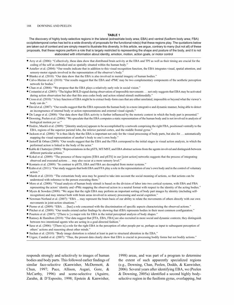
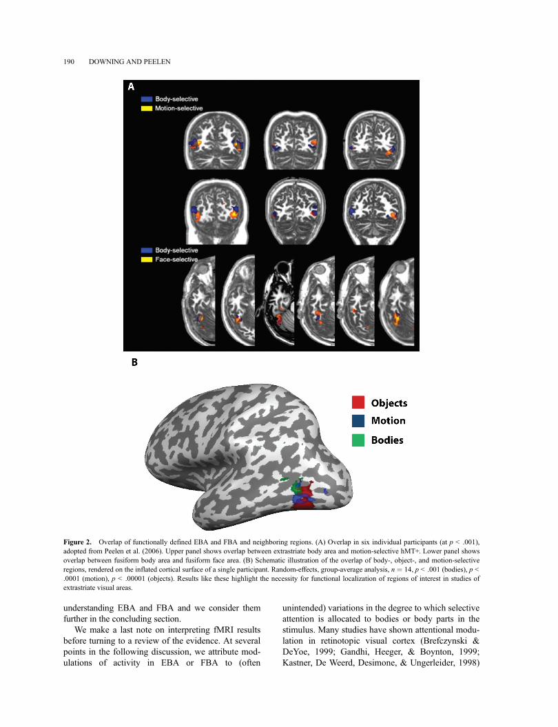
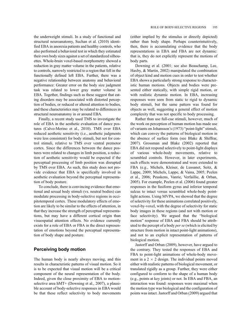
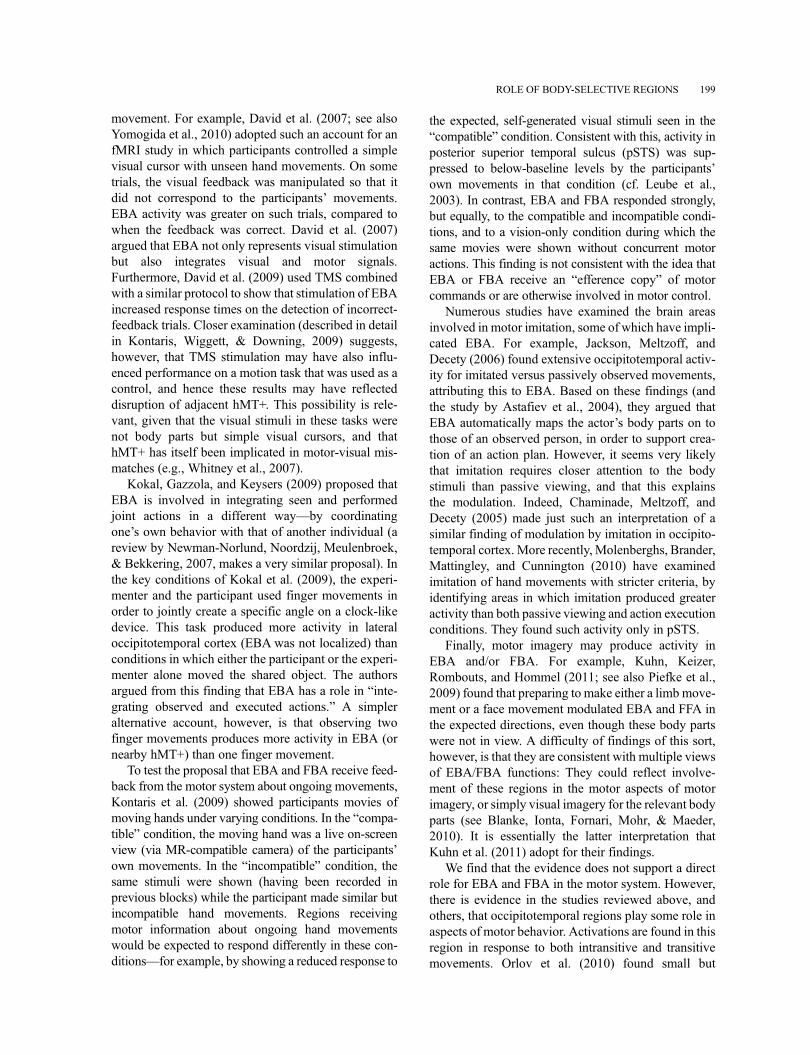
EBA and FBA are found close to, or overlappingwith, other known regions. Thus, previous research hashad to functionally distinguish these regions from theirneighbors. For example, Downing et al. (2001) reporteda dissociation between the responses of EBA and nearbymotion-selective region hMT+ in the responses to mov-ing objects and bodies (see also Pitcher et al., 2009;Spiridon et al., 2006; Valyear & Culham, 2010).Similarly, Peelen, Wiggett, and Downing (2006) usedmultivoxel pattern analysis (MVPA) (Haynes & Rees,2006; Kamitani & Tong, 2005; Mur, Bandettini, &Kriegeskorte, 2009; Oosterhof, Wiggett, Diedrichsen,Tipper, & Downing, 2010; Peelen & Downing, 2007b)to distinguish the responses of motion-selective hMT+and EBA to point-light biological motion stimuli (seealso Downing,Wiggett, & Peelen, 2007). Most recently,Weiner and Grill-Spector (2011) used high-resolutionfMRI to anatomically dissociate hMT+ and EBA. Withrespect to FBA, MVPA (Peelen et al., 2006), high-resolution fMRI (Schwarzlose et al., 2005), and devel-opmental time course (Peelen, Glaser, Vuilleumier, &Eliez, 2009) show that the body-selective and face-selective responses in the fusiform gyrus can be disso-ciated. Importantly, this overlap means that occipitotem-poral activations in the general region of EBA or FBAcan be difficult to interpret without careful localizationwithin-studies and within-participants (Figure 2).
The overlap and close proximity of EBA and FBA toother functional regions has further implications as well.For one, the nature of the neighboring areas may provideclues about the properties of EBA and FBA. For exam-ple, the proximity of EBA to motion-selective regionssuggests that it interacts with dynamic visual representa-tions, although we argue below that the body represen-tation in EBA itself is not dynamic. Furthermore, veryrecent evidence suggests that the highly body selectiveresponses we define as EBA and FBAmay be situated ina much broader, more weakly body-sensitive expanse ofcortex. Specifically, Orlov, Makin, and Zohary (2010)find large occipitotemporal regions that respond subtlybut reliably more to specific body parts (e.g., upper orlower limbs) than to other body parts. Both of theseobservations provide an important context for
ROLE OF BODY-SELECTIVE REGIONS 189
understanding EBA and FBA and we consider themfurther in the concluding section.
We make a last note on interpreting fMRI resultsbefore turning to a review of the evidence. At severalpoints in the following discussion, we attribute mod-ulations of activity in EBA or FBA to (often
unintended) variations in the degree to which selectiveattention is allocated to bodies or body parts in thestimulus. Many studies have shown attentional modu-lation in retinotopic visual cortex (Brefczynski &DeYoe, 1999; Gandhi, Heeger, & Boynton, 1999;Kastner, De Weerd, Desimone, & Ungerleider, 1998)
Figure 2. Overlap of functionally defined EBA and FBA and neighboring regions. (A) Overlap in six individual participants (at p < .001),adopted from Peelen et al. (2006). Upper panel shows overlap between extrastriate body area and motion-selective hMT+. Lower panel showsoverlap between fusiform body area and fusiform face area. (B) Schematic illustration of the overlap of body-, object-, and motion-selectiveregions, rendered on the inflated cortical surface of a single participant. Random-effects, group-average analysis, n ¼ 14, p < .001 (bodies), p <.0001 (motion), p < .00001 (objects). Results like these highlight the necessity for functional localization of regions of interest in studies ofextrastriate visual areas.
190 DOWNING AND PEELEN
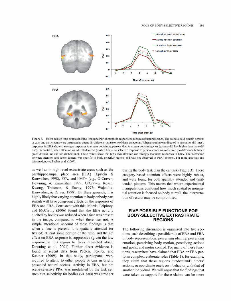
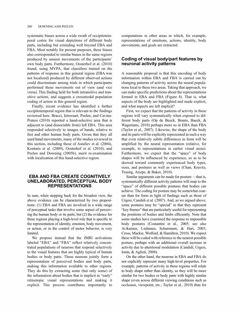
as well as in high-level extrastriate areas such as theparahippocampal place area (PPA) (Epstein &Kanwisher, 1998), FFA, and hMT+ (e.g., O’Craven,Downing, & Kanwisher, 1999; O’Craven, Rosen,Kwong, Treisman, & Savoy, 1997; Wojciulik,Kanwisher, & Driver, 1998). On these grounds, it ishighly likely that varying attention to body or body partstimuli will have congruent effects on the responses ofEBA and FBA. Consistent with this, Morris, Pelphrey,and McCarthy (2006) found that the EBA activityelicited by bodies was reduced when a face was presentin the image, compared to when there was not. Asimple attentional account of these findings is thatwhen a face is present, it is spatially attended (orfixated) at least some portion of the time, and the neteffect on EBA response is suppressive (given the lowresponse in this region to faces presented alone;Downing et al., 2001). Further direct evidence isfound in recent data from Peelen, Fei-Fei, andKastner (2009). In that study, participants wererequired to attend to either people or cars in brieflypresented natural scenes. Activity in EBA, but notscene-selective PPA, was modulated by the task set,such that selectivity for bodies (vs. cars) was stronger
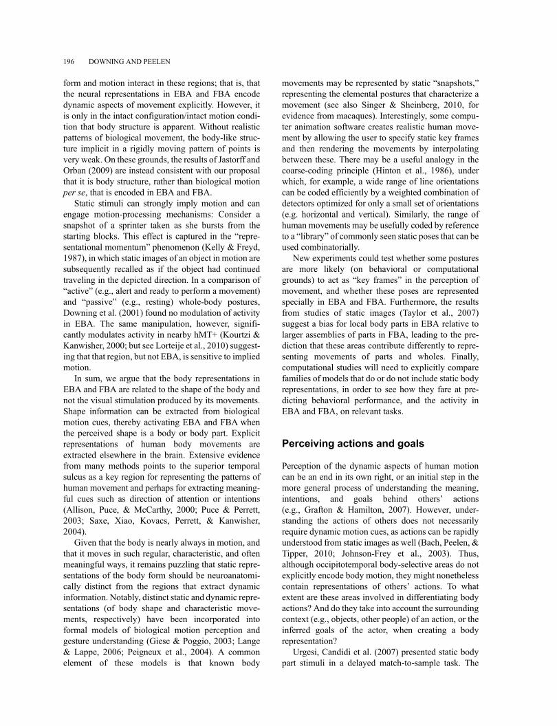
during the body task than the car task (Figure 3). Thesecategory-based attention effects were highly robust,and were found for both spatially attended and unat-tended pictures. This means that where experimentalmanipulations confound how much spatial or nonspa-tial attention is focused on body stimuli, the interpreta-tion of results may be compromised.
FIVE POSSIBLE FUNCTIONS FORBODY-SELECTIVE EXTRASTRIATE
REGIONS
The following discussion is organized into five sec-tions, each describing a possible role of EBA and FBAin body representation: perceiving identity, perceivingemotion, perceiving body motion, perceiving actionsand goals, and motor control. For many of these func-tions, researchers have claimed that EBA or FBA per-form complex, elaborate roles (Table 1); for example,they claim that these regions “understand” others’actions, or coordinate one’s own behavior with that ofanother individual. We will argue that the findings thatwere taken as support for these claims can be more
Figure 3. Event-related time courses in EBA (top) and PPA (bottom) in response to pictures of natural scenes. The scenes could contain personsor cars, and participants were instructed to attend (in different runs) to one of these categories. When attention was directed to persons (solid lines),responses in EBA showed stronger responses to scenes containing persons than to scenes containing cars (green solid line higher than red solidline). By contrast, when attention was directed to cars (dashed lines), no selective response to person scenes was observed (no difference betweengreen dashed line and red dashed line). These results show that top-down attention can strongly modulate responses in EBA. The interactionbetween attention and scene content was specific to body-selective regions and was not observed in PPA (bottom). For more analyses andinformation, see Peelen et al. (2009).
ROLE OF BODY-SELECTIVE REGIONS 191
parsimoniously explained by a model in which EBAand FBA represent visual features of the body such asshape and posture without further elaboration.
Perceiving identity
Although person identity is often perceived from facialcues, the rest of the body also provides importantinformation about identity. Body posture, shape, andgait can all be used to determine person identity, andmay be particularly useful when the face is poorlyvisible such as when viewing someone from behindor at a distance. Since recognizing identity from bodiesrequires the perceptual analysis of body stimuli, it islikely that EBA and FBA play an important role in thisprocess. Below, we discuss studies that have investi-gated the role of EBA and FBA in extracting personidentity, and indicate how a perceptual account of theseregions can accommodate these findings.
Several studies have investigated whether EBA andFBA respond differentially to images of one’s own ver-sus another person’s body, as this is perhaps the mostfundamental identity distinction. Chan, Peelen, andDowning (2004) reported no difference between EBAresponses elicited by viewing the bodies of the selfversus familiar others in a blocked design fMRI experi-ment. Subsequent studies that contrasted the self withfamiliar others similarly found no difference in EBA orFBA (Devue et al., 2007; Hodzic, Kaas, Muckli, Stirn, &Singer, 2009), although one study (that did not localizeEBA/FBA) reported a broad swath of occipitotemporalcortex that responded more to self than to familiar otherimages (Sugiura et al., 2006). The results of studies thatcontrasted images of the self with unfamiliar others areless consistent: Hodzic, Kaas et al. (2009) found nodifference in right EBA, but a stronger response to selfthan other bodies in left EBA and right FBA. However,in another study, these authors did not find a differencebetween self and unfamiliar other in EBA or FBA(Hodzic, Muckli, Singer, & Stirn, 2009). Finally, Vockset al. (2010) reported small but significant increases toown bodies relative to unknown others’ bodies in rightEBA and FBA.
Thus, while most studies reported no difference inEBA and/or FBA between self and other bodies, somestudies reported modest increases in one or both of theseregions. It should be noted that these studies comparedthe overall BOLD signal in EBA and/or FBA to imagesof the self versus a familiar or unfamiliar other. It isunlikely, however, that a region that extracts identityfrom perceived bodies would show gross differences inthe overall response magnitude to different identities.
More likely, different identities would elicit differentpatterns of activity resulting in roughly the same grosslevel of response (Kriegeskorte, Formisano, Sorger, &Goebel, 2007). So these studiesmay not be ideally suitedto test whether EBA/FBA represents identity. Rather,they test whether EBA and FBA are primarily involvedin representing the self (e.g., for guiding actions) or theother (e.g., for social cognition)–– and their results do notpoint to a uniquely strong role in either process.
Another approach to test whether EBA and/or FBAprimarily encode the self or the other is to test the effectof viewpoint: If EBA/FBA would preferentiallyrespond to one’s own body parts, we would expect astronger response to body parts presented in a firstperson viewpoint (camera at eye level), while strongerresponses to third person views would be expected ifthese regions are primarily involved in the encoding ofothers’ bodies. The two studies that have manipulatedviewpoint both found slightly stronger responses inright EBA to bodies presented in third person view(Chan et al., 2004; Saxe, Jamal, & Powell, 2006),inconsistent with the preference for one’s own bodyreported by some of the studies reviewed above.
Using a blocked-design adaptation approach, Myersand Sowden (2008) reported significantly strongerresponses (i.e., less adaptation) in right, but not left,EBA (FBA was not tested) to blocks in which imagesof the participants’ own hand were alternated withimages of an unknown other’s hand, relative to blockswhere two other individuals’ hands were alternated.The authors concluded that right EBA contains differ-ent neural populations to represent the appearance ofone’s own versus others’ body parts. These results mayalternatively be explained by differences in attention: Itis not unlikely that participants paid more attention totheir own hands than to a stranger’s hand, especiallybecause the participants’ own body parts appearedrelatively infrequently in the experiment. Such atten-tion effects would be expected to primarily modulatehigher-level visual areas strongly responsive to thehand stimuli, rather than weakly responsive areas orlower-level visual cortex.
Perceiving identity is, of course, not limited to dis-tinguishing the self from others. Surprisingly, only afew studies have investigated the role of EBA or FBAin representing body identity beyond the self/otherdistinction. Kable and Chatterjee (2006) used a long-term, event-related adaptation design, with dynamicwhole-body/face stimuli, to investigate adaptation toactor identity. No significant identity adaptation wasfound in EBA. In a subsequent extension of this study,the absence of significant identity-related adaptationeffects in EBA and FBA was confirmed (Wiggett &Downing, 2010). Ramsey and Hamilton (2010) also
192 DOWNING AND PEELEN
used an adaptation design with the similar aim ofexamining representations of actors. Functional locali-zation was not performed, but anatomical regions ofinterest around fusiform and middle temporal gyri (andother areas) were tested with small-volume correctionanalyses. Both areas showed significant adaptation forrepeated versus nonrepeated actors, which the authorsattributed to “person identity processes” taking place inEBA and FBA. However, it is not clear which aspect ofthe stimulus drove this adaptation––whether it was therepetition of actor identity, the repetition of body shape,or the repetition of low-level visual aspects such as thecolor of the clothes.
Together, these results suggest that EBA and FBAare involved in the encoding of body parts belonging tothe self as well as to others. Small modulations reportedin some of the studies reviewed here may be explainedby attentional differences––looking at a picture ofyourself may simply be more interesting than lookingat a picture of a stranger. Notably, identity effects tendto be found when identity is either an explicit part of theparticipants’ task (Hodzic, Kaas et al., 2009; Ramsey &Hamilton, 2010; Sugiura et al., 2006) or else is madeexplicit to participants by direct cues (Vocks et al.,2010). This pattern points strongly to the idea thatidentity modulation in EBA or FBA is the result oftop-down influences, including attentional biases.
What could be the role of EBA and FBA in theperception of body identity? We argue that the role ofthese regions consists in creating a perceptual repre-sentation of the shape and posture of the body and itsparts, which may then be used by other brain regions(e.g., the anterior temporal lobes; Kriegeskorte et al.,2007) to represent person identity explicitly. On thisaccount, EBA and FBA encode body identity only inthe sense that these regions differentiate between indi-viduals with different body shapes. We argue that EBAand FBA do not, however, explicitly represent personidentity beyond body shape, and as such may notspecifically differentiate between the bodies of theself and others. While identity can be extracted frombody shape, identity and shape can be dissociated(people age, grow, lose or gain weight, undergo plasticsurgery, etc.). Furthermore, identity can be extractedfrom multiple body parts (e.g., lower and upper body).We expect responses in EBA/FBA to follow the per-ceived body shape (or body part) rather than the per-ceived body identity.
Note that our account does not exclude the possibilitythat responses in EBA and FBA can be influenced byidentity through top-down modulation: The sight of aromantic partner (or, indeed, the self) may increaseresponses in EBA/FBA due to increases in attentionand arousal. Also, we expect that familiarity with
particular body shapes (e.g., growing up in a countrywith tall people) may change the tuning of body-selective neurons, such that their discrimination ofbody shapes is optimal for the current environment.Finally, the knowledge of another person’s body shape(available after identifying the person, whether throughvisual or nonvisual cues) may modulate responses inEBA and FBA, as, for example, in cases where clothesobscure most of the body. In other words, responses inEBA and FBA are not merely a copy of the visual image,but more closely correspond to the subjective percept,which is influenced by identity (and other factors). Thecritical test of our account would require an experimentthat dissociates body shape and body identity in combi-nation with suitable techniques (e.g., MVPA).Distributed activity in EBA and FBA should conformto a “space” inwhich different body shapes/parts, but notnecessarily different individuals, are represented by sys-tematically different corresponding patterns.
Perceiving emotion
Basic emotions, such as fear, anger, and happiness, areassociated with characteristic body postures and move-ments (Atkinson, Dittrich, Gemmell, & Young, 2004).Apart from providing information about others’ emo-tional states, bodies may also evoke emotions in theobserver––for example, through empathy (e.g., withothers’ pain), admiration of beauty (e.g., in dance), orsexual arousal. What is the role of occipitotemporalbody-selective areas in these processes? Here we con-sider the evidence from studies that have implicatedEBA or FBA in the processing of emotion from bodystimuli.
Research on emotional body perception has largelyaddressed questions that had earlier been addressed inthe field of emotional face perception, such as compar-ing fMRI responses to emotional expressions withresponses to neutral expressions. Employing thisapproach, research on face perception indicates thatemotional expressions modulate responses in visualcortex, particularly the fusiform face area (for a review,see Vuilleumier, 2005). These emotional modulationsare similar to attentional modulations but are thought tobe mediated by the amygdala, rather than by frontopar-ietal attention networks (Amaral, Behniea, & Kelly,2003; Morris et al., 1998; Vuilleumier, Richardson,Armony, Driver, & Dolan, 2004). Such activityincreases in visual cortex may correspond to theenhanced perceptual processing of emotionally salientstimuli (e.g., Anderson, 2005).
Similar to emotional faces, emotional bodies havebeen shown to modulate perceptual responses, with
ROLE OF BODY-SELECTIVE REGIONS 193
increased visual cortex (and amygdala) activation toarousing body expressions such as fear (Hadjikhani &de Gelder, 2003). A large number of studies frommultiple laboratories have replicated these effects,using various emotions and both static and dynamicbody stimuli (e.g., Carter & Pelphrey, 2008; Flaisch,Schupp, Renner, & Junghofer, 2009; Grezes, Pichon,& de Gelder, 2007; Grosbras & Paus, 2006; Kret,Pichon, Grezes, & de Gelder, 2011; Nummenmaa,Hirvonen, Parkkola, & Hietanen, 2008; Peelen,Atkinson, Andersson, & Vuilleumier, 2007).Although it is beyond the scope of this paper to reviewthese studies in depth, a few general observationsemerge from this literature. For example, it seems thatarousal rather than valence is driving emotional mod-ulation, since no modulation was found for bodiesexpressing sadness (Peelen et al., 2007). Further, emo-tional modulation has been primarily, but not exclu-sively, reported in body-selective parts of visual cortex(i.e., at the location of EBA and FBA), with somestudies (mostly those using static body postures withblurred faces) finding stronger modulation in fusiformgyrus (presumably FBA) than at the location of EBA(e.g., Van de Riet, Grezes, & de Gelder, 2009). In astudy using dynamic body expressions, it was shownthat emotional modulation is proportional to staticbody selectivity (measured in a separate localizerscan) across the voxels of EBA and FBA, with thestrongest modulation observed in voxels with thestrongest body selectivity (Peelen et al., 2007). In thesame study, emotional modulation in EBA and FBAwas shown to correlate with emotion-driven increasesin the amygdala, again highlighting the similaritybetween the mechanisms involved in emotional bodyand face processing (Peelen et al., 2007). Finally, mod-ulation in visual cortex (including putative body-selective regions) has also been reported for sexuallyarousing body stimuli (Cikara, Eberhardt, & Fiske,2011; Hamann, Herman, Nolan, & Wallen, 2004;Mouras et al., 2003; Ponseti et al., 2006).
Together, these results suggest that activityincreases in EBA and FBA in response to emotionalor sexual body stimuli primarily reflect modulatoryeffects (or “emotional attention”), rather than a directencoding of emotions within these regions. It should benoted, however, that (as far as we know) no study hasdirectly tested for explicit representations of emotionsin these regions, as by using fMRI adaptation orMVPA(cf. Peelen, Atkinson, & Vuilleumier, 2010).
Observing someone else’s body in acute physicalpain can hurt, especially when this person is a lovedone. This empathy for others’ pain has been shown toactivate regions in the anterior insula and anterior cin-gulate cortex, regions that also activate when one is
directly experiencing pain (Morrison, Lloyd, diPellegrino, & Roberts, 2004; Singer et al., 2004; butsee Morrison & Downing, 2007). Although body-selective regions have not been implicated in the directexperience of pain, they may play a role in the percep-tion of pain in others. For example, Singer et al. (2004)found activity in fusiform gyrus as well as more poster-ior visual regions in response to observing another’shand that is thought to be in pain (as indicated by asymbolic cue) relative to when this hand was knownnot to be in pain. Similarly, Gu et al. (2010) foundincreased responses in occipitotemporal regions (prob-ably including EBA) to photographs of body parts inpainful daily-life incidents. However, such pain-relatedmodulation was not observed in several other studiesthat showed a needle or knife touching a hand or foot(Benuzzi, Lui, Duzzi, Nichelli, & Porro, 2008;Morrison et al., 2004; Morrison, Peelen, & Downing,2007). The only study to directly investigate pain-related responses in functionally localized EBA alsoused images showing a needle contacting a hand. Nomodulation was found in EBA, and it was concludedthat the EBA is not relevant for empathy (Lamm &Decety, 2008). Thus, although there is currently nodirect evidence that body-selective regions are modu-lated by observed pain, several studies suggest thismight be the case for some stimuli. Studies that usedstimuli in which a noxious object interacts with a bodypart (e.g., a needle contacting a hand) typically failed tofind modulation in visual cortex. A possible explana-tion could be that in these studies participants directedattention toward the noxious object, or toward the localsite of impact, rather than the body part. Such differ-ences in attentional focus could have offset any pain-related modulatory effects.
There is a strong emotional aspect to clinical eatingdisorders such as anorexia nervosa and bulimia ner-vosa, in that patients may react with strong negativefeelings toward the appearance of their own bodies orto others they perceive as overweight. These disorderscan be associated with distortions of the body image, asrevealed in misperceptions of body shape and size, andhence some authors have made a connection betweenthese syndromes and EBA or FBA. Uher et al. (2005;see also Aleong & Paus, 2010; Sachdev, Mondraty,Wen, & Gulliford, 2008) tested visual responses tobody images in patients and in control participants.Line drawings of women in either underweight, nor-mal, or overweight versions were compared in ablocked-design fMRI experiment. Low body selectiv-ity was seen in occipitotemporal cortex in patientsrelative to controls. Furthermore, in a fusiform region,patients showed reduced activity (relative to controls)for the overweight stimuli, and increased activity for
194 DOWNING AND PEELEN
the underweight stimuli. In a study of functional andstructural neuroanatomy, Suchan et al. (2010) identi-fied EBA in anorexia patients and healthy controls, whoalso performed a behavioral test in which they estimatedtheir own body sizes against a set of standardized silhou-ettes. Whole-brain voxel-based morphometry showed areduction in gray matter volume in the patients, relativeto controls, narrowly restricted to a region that fell in thefunctionally defined left EBA. Further, there was anegative relationship between anatomy and behavioralperformance: Greater error on the body size judgmenttask was related to lower gray matter volume inEBA. Together, findings such as these suggest that eat-ing disorders may be associated with distorted percep-tion of bodies, or reduced or altered attention to bodies,and these characteristics may be related to differences instructural neuroanatomy in or around EBA.
Finally, a recent study used TMS to investigate therole of EBA in the aesthetic evaluation of dance pos-tures (Calvo-Merino et al., 2010). TMS over EBAreduced aesthetic sensitivity (i.e., aesthetic judgmentswere less consistent) for body stimuli, but not for con-trol stimuli, relative to TMS over ventral premotorcortex. Since the differences between the dance pos-tures were related to changes in limb position, a reduc-tion of aesthetic sensitivity would be expected if theperceptual processing of limb position was disruptedby TMS over EBA. As such, this study does not pro-vide evidence that EBA is specifically involved inaesthetic evaluation beyond the perceptual representa-tion of body posture.
To conclude, there is convincing evidence that emo-tional and sexual body stimuli (vs. neutral bodies) canmodulate processing in body-selective regions in occi-pitotemporal cortex. These modulatory effects of emo-tion are likely to be similar to the effects of attention, inthat they increase the strength of perceptual representa-tions, but may have a different cortical origin thanvisuospatial attention effects. No evidence currentlyexists for a role of EBA or FBA in the direct represen-tation of emotions beyond the perceptual representa-tion of body shape and posture.
Perceiving body motion
The human body is nearly always moving, and thisresults in characteristic patterns of visual motion. So itis to be expected that visual motion will be a criticalcomponent of the neural representation of the body.Indeed, given the close proximity of EBA to motion-selective area hMT+ (Downing et al., 2007), a plausi-ble account of body-selective responses in EBAwouldbe that these reflect selectivity to body movements
(either implied by the stimulus or directly depicted)rather than body shape. Perhaps counterintuitively,then, there is accumulating evidence that the bodyrepresentations in EBA and FBA are not dynamic:that is, they do not explicitly represent the motions ofbody parts.
Downing et al. (2001; see also Beauchamp, Lee,Haxby, & Martin, 2002) manipulated the combinationof object kind and motion cues in order to test whetherEBA shows a particularly strong response to character-istic human motions. Objects and bodies were pre-sented either statically, with simple rigid motion, orwith realistic dynamic motion. In EBA, increasingresponses were seen from static to rigid to dynamicbody stimuli, but the same pattern was found forobjects as well, suggesting a general effect of motioncomplexity that was not specific to body processing.
Rather than use full-cue stimuli, however, much ofthe work on perception of human motion has made useof variants on Johansson’s (1973) “point-light” stimuli,which can convey the patterns of biological motion inthe absence of surface form (see Blake & Shiffrar,2007). Grossman and Blake (2002) reported thatEBA did not respond selectively to point-light displaysof various whole-body movements, relative toscrambled controls. However, in later experiments,such effects were demonstrated and were extended toFBA (e.g., Michels, Kleiser, de Lussanet, Seitz, &Lappe, 2009; Michels, Lappe, & Vaina, 2005; Peelenet al., 2006; Peuskens, Vanrie, Verfaillie, & Orban,2005). For example, Peelen et al. (2006) found greaterresponses in the fusiform gyrus and inferior temporalsulcus to intact versus scrambled whole-body point-light actions. Using MVPA, we showed that the degreeof selectivity for these animations correlated positively,voxel-by-voxel, with the degree of selectivity for staticbody images in these regions (and not with motion orface selectivity). We argued that the “biologicalmotion” response of EBA and FBA should be attrib-uted to the percept of a body per se (which is elicited bystructure from motion in intact point-light animations),and not to an explicit representation of patterns ofbiological motion.
Jastorff and Orban (2009), however, have argued tothe contrary. They tested the responses of EBA andFBA to point-light animations of whole-body move-ment in a 2 � 2 design. The individual points movedeither with realistic patterns of biological movement, ortranslated rigidly as a group. Further, they were eitherconfigured to conform to the shape of a human body(e.g., points at key joints) or not. In EBA and FBA, aninteraction was found: responses were maximal whenthe motion type was biological and the configuration ofpoints was intact. Jastorff and Orban (2009) argued that
ROLE OF BODY-SELECTIVE REGIONS 195
form and motion interact in these regions; that is, thatthe neural representations in EBA and FBA encodedynamic aspects of movement explicitly. However, itis only in the intact configuration/intact motion condi-tion that body structure is apparent. Without realisticpatterns of biological movement, the body-like struc-ture implicit in a rigidly moving pattern of points isvery weak. On these grounds, the results of Jastorff andOrban (2009) are instead consistent with our proposalthat it is body structure, rather than biological motionper se, that is encoded in EBA and FBA.
Static stimuli can strongly imply motion and canengage motion-processing mechanisms: Consider asnapshot of a sprinter taken as she bursts from thestarting blocks. This effect is captured in the “repre-sentational momentum” phenomenon (Kelly & Freyd,1987), in which static images of an object in motion aresubsequently recalled as if the object had continuedtraveling in the depicted direction. In a comparison of“active” (e.g., alert and ready to perform a movement)and “passive” (e.g., resting) whole-body postures,Downing et al. (2001) found no modulation of activityin EBA. The same manipulation, however, signifi-cantly modulates activity in nearby hMT+ (Kourtzi &Kanwisher, 2000; but see Lorteije et al., 2010) suggest-ing that that region, but not EBA, is sensitive to impliedmotion.
In sum, we argue that the body representations inEBA and FBA are related to the shape of the body andnot the visual stimulation produced by its movements.Shape information can be extracted from biologicalmotion cues, thereby activating EBA and FBA whenthe perceived shape is a body or body part. Explicitrepresentations of human body movements areextracted elsewhere in the brain. Extensive evidencefrom many methods points to the superior temporalsulcus as a key region for representing the patterns ofhuman movement and perhaps for extracting meaning-ful cues such as direction of attention or intentions(Allison, Puce, & McCarthy, 2000; Puce & Perrett,2003; Saxe, Xiao, Kovacs, Perrett, & Kanwisher,2004).
Given that the body is nearly always in motion, andthat it moves in such regular, characteristic, and oftenmeaningful ways, it remains puzzling that static repre-sentations of the body form should be neuroanatomi-cally distinct from the regions that extract dynamicinformation. Notably, distinct static and dynamic repre-sentations (of body shape and characteristic move-ments, respectively) have been incorporated intoformal models of biological motion perception andgesture understanding (Giese & Poggio, 2003; Lange& Lappe, 2006; Peigneux et al., 2004). A commonelement of these models is that known body
movements may be represented by static “snapshots,”representing the elemental postures that characterize amovement (see also Singer & Sheinberg, 2010, forevidence from macaques). Interestingly, some compu-ter animation software creates realistic human move-ment by allowing the user to specify static key framesand then rendering the movements by interpolatingbetween these. There may be a useful analogy in thecoarse-coding principle (Hinton et al., 1986), underwhich, for example, a wide range of line orientationscan be coded efficiently by a weighted combination ofdetectors optimized for only a small set of orientations(e.g. horizontal and vertical). Similarly, the range ofhuman movements may be usefully coded by referenceto a “library” of commonly seen static poses that can beused combinatorially.
New experiments could test whether some posturesare more likely (on behavioral or computationalgrounds) to act as “key frames” in the perception ofmovement, and whether these poses are representedspecially in EBA and FBA. Furthermore, the resultsfrom studies of static images (Taylor et al., 2007)suggest a bias for local body parts in EBA relative tolarger assemblies of parts in FBA, leading to the pre-diction that these areas contribute differently to repre-senting movements of parts and wholes. Finally,computational studies will need to explicitly comparefamilies of models that do or do not include static bodyrepresentations, in order to see how they fare at pre-dicting behavioral performance, and the activity inEBA and FBA, on relevant tasks.
Perceiving actions and goals
Perception of the dynamic aspects of human motioncan be an end in its own right, or an initial step in themore general process of understanding the meaning,intentions, and goals behind others’ actions(e.g., Grafton & Hamilton, 2007). However, under-standing the actions of others does not necessarilyrequire dynamic motion cues, as actions can be rapidlyunderstood from static images as well (Bach, Peelen, &Tipper, 2010; Johnson-Frey et al., 2003). Thus,although occipitotemporal body-selective areas do notexplicitly encode body motion, they might nonethelesscontain representations of others’ actions. To whatextent are these areas involved in differentiating bodyactions? And do they take into account the surroundingcontext (e.g., objects, other people) of an action, or theinferred goals of the actor, when creating a bodyrepresentation?
Urgesi, Candidi et al. (2007) presented static bodypart stimuli in a delayed match-to-sample task. The
196 DOWNING AND PEELEN
images could vary either in the action they implied(subtle variations in the position of appendages) orform (subtle variations in their shape). TMS overEBA impaired performance on the form task, but notthe action task, while TMS over ventral premotor cor-tex produced the opposite pattern (see also Pitcheret al., 2009). These findings suggest that EBA doesnot critically contribute to representing actions impliedfrom static images. This is consistent with the findingsof Downing, Peelen, Wiggett, & Tew (2006), whobroke movies of whole-body movements into staticframes and manipulated the order in which these werepresented. In the coherent condition, the frames werepresented in the original order, to create a meaningfulaction sequence without motion cues. In the incoherentcondition, frames from different actions were mixed,leading to a disjointed percept and to relatively largeframe-to-frame differences. Posterior STS and fronto-parietal areas responded more to the coherent series,suggesting they represented the unfolding coherentactions. However, EBA (and to some extent FBA)responded more strongly to the incoherent than to thecoherent series. These results again suggest that EBAand FBA do not form an integrated representation ofbody actions.
Many everyday actions are object directed, as in theuse of tools or other implements. Does activity in EBAor FBA reflect the relationship between the movementand the object? De Lange, Spronk, Willems, Toni, andBekkering (2008) found increased bilateral activity in aregion they attributed to EBA (not localized) whenparticipants observed atypical versus typical object-directed actions (e.g., drinking from a cup with a typi-cal grasp or a clumsy, backwards grip). They argue thatthis shows EBA is sensitive to the “motoric context” inwhich body parts are viewed. When Valyear andCulham (2010) took a similar approach––comparingthe observation of typical versus atypical grasps oftools––they found a left posterior middle temporalgyrus region that was sensitive to grasp. Notably, thiseffect did not extend to functionally defined EBA. Inthat study, however, both types of grasp did produce agreater response in right EBA than a control conditionthat showed an object simply being touched. Likewise,Pierno et al. (2009) found an increased occipitotem-poral response to grasping (over pointing) movements,which they attributed to EBA. Both effects could sim-ply reflect competition between stimuli––in grasp, butnot point, conditions, less of the target object is visible,and hence that object makes a weaker competitor forattention with the hand stimulus. Moreover, graspactions may direct attention to the object more stronglythan pointing (Bekkering & Neggers, 2002), with thesame result on activity in EBA.
Several recent studies compared the effects ofattending to different aspects of actions. For example,Spunt, Satpute, and Lieberman (2011) had participantsview movies of short, object-directed actions underdifferent task instructions. Increased activity was seenin left lateral occipitotemporal cortex when participantscovertly attended to the mechanics of the actions(“how”) rather than to the descriptive (“what”) orintentional (“why”) aspects. The authors argue (andwe agree) that this could reflect increased attention tothe body under “how” instructions (since perceivingthe body accurately is essential to understanding themechanism of an action). In contrast, understanding“why” an action takes place will naturally requirewider attention to other aspects of the surroundingscene.
A visual-attention account does not explain, how-ever, why similar lateral occipitotemporal activationsare found in studies that use purely verbal material.Such studies aim to test an “embodied” view of actionknowledge, whereby even abstractly specified con-cepts (e.g., words about actions) are rooted in theactivity of visuomotor brain areas. Spunt, Falk, andLieberman (2010) had participants make “how” (rela-tive to “why”) judgments about actions described verb-ally. Likewise, Van Dam, Rueschemeyer, andBekkering (2010) compared the reading of verbs thatwere generic with respect to a motor program (e.g., “toclean”) and verbs that were more closely tied to actions(e.g., “to wipe”). And Rueschemeyer, Pfeiffer, andBekkering (2010) compared nouns related to actionsmade toward the body (e.g., cup, comb, sweater) with“world-” related items that typically involve actionsaway from the body (e.g., wrench, pen, frisbee). Allof these studies identified lateral occipitotemporal acti-vations (variably on the left or right), and these wereattributed directly to EBA by Van Dam et al. (2010)and Rueschemeyer et al. (2010).
Such results could reflect semantic or crossmodalaction or body knowledge in EBA. Alternatively, theycould reflect top-down activation of perceptual bodyrepresentations––for example, through mental imageryor automatic association processes––after the meaningof the words has been processed elsewhere.Furthermore, the relationship of these activations toEBA is unclear, as none of these studies localized thisarea functionally––and the candidate regions in VanDam et al. (2010; Figure 1, Table 3) make a particularlypoor match to EBA on anatomical grounds. This isimportant, as noted above and illustrated in this contextby the findings of Valyear and Culham (2010), becausewithout precise localization, EBA may be confusedwith nearby areas with entirely different functionalproperties (see also Takahashi et al., 2008). Results of
ROLE OF BODY-SELECTIVE REGIONS 197
a recent study examining the lateral occipitotemporalresponses to reading various categories of words(including nouns and verbs) emphasize this point.Bedny, Caramazza, Grossman, Pascual-Leone, andSaxe (2008) found that these verbal stimuli deactivatedEBA (which was independently localized) relative tobaseline, and further that there was no differentiationamong word categories in that region.
All of the above studies measured gross changes inthe level of activation in occipitotemporal cortex. Thelogic of repetition suppression (e.g., Grill-Spector,Henson, &Martin, 2006) may be more suited to detect-ing neural populations that encode perceived actions.This approach was first applied to EBA by Kable andChatterjee (2006), who found that activity in a numberof areas, including functionally defined EBA, wasreduced when participants viewed a short movie of anaction that had been seen previously in the experiment,compared to a new action. This repetition suppressioneffect was seen even when the actor involved in the twopresentations was different, suggesting adaptation ofan action representation at a relatively generic orsemantic level. However, in a replication and exten-sion, Wiggett and Downing (2010) found that thisaction priming effect was widespread––extendingacross face, object, scene, and body selective regionsof interest in the lateral and ventral occipitotemporalcortex––and could not be attributed specifically tobody-specific regions. Instead, it may reflect moregeneral consequences of repetition, such as reducedattention arising from familiarity.
In summary, as we consider increasingly abstractaspects of actions, such as the objects they are directedto, their meaning, and the purpose of the actor inperforming them, the relevant neural codes naturallymove further away from a specific description of theposture and shape of the actor. Very similar movementscan carry out very different goals (e.g., a gentle touchvs. a nasty poke), and, conversely, very different move-ments can achieve the same outcome (e.g., Umiltaet al., 2008). When stimulus and attentional differ-ences, and functional localization, are taken intoaccount, we find little evidence to support the proposalthat the representations of EBA and FBA can make thekinds of distinctions necessary to support high-levelaction understanding. Rather, as many authors havedescribed elsewhere (e.g., Van Overwalle & Baetens,2009), the evidence points to prefrontal and especiallyparietal regions as key for this process. It is likely thatthe information provided by EBA and FBA––particu-larly about body posture (Downing, Peelen, et al.,2006)––provides an important part of the input tothese areas, which can in turn integrate this with inputfrom other neural sources in order to incorporate the
contextual information that is key to understandingactions.
Motor control
Movements of the body are coordinated by the jointactivity of the frontal and parietal cortices and the cere-bellum. However, the finding that extrastriate occipito-temporal areas respond to images of the body naturallyraises the question of whether these areas also play somerole in motor control (Downing et al., 2001). While theyare unlikely to directly control the activity of muscles,these regions could be critically involved in visuomotorcoordination by providing online visual tracking of thepositions of one’s own limbs. Alternatively, signals fromthe motor system could directly modulate the responsesof neurons in these regions, perhaps to anticipate thevisual consequences of body-part movements. Thus, thestrongly visual response of EBA and FBA does not ruleout a priori a role in motor behavior, or interactions withthe motor system.
The first direct test of a motor role for EBA wasconducted by Astafiev, Stanley, Shulman, and Corbetta(2004). Unseen, visually cued movements of the handsand feet (but not shifts of attention or the eyes) werefound to increase EBA activity. This finding has beentaken by others to show that EBA has a role in motorcontrol and even to show that it is part of the human“mirror system.” In a subsequent replication, we(Peelen & Downing, 2005b) questioned the involve-ment of body-selective neurons in unseen hand move-ments: In our study, EBA only partially overlappedwith the region activated by unseen hand movements,and MVPA in the overlap region showed no relation-ship between body and movement selectivity. In aresponse, Astafiev, Stanley, Shulman, and Corbetta(2005) showed that the overlap between EBA and themovement-related region depended on the control con-dition used: The overlap was substantial when contrast-ing hand and foot movements with a fixation baseline,but small when contrasting these movements with amatched control condition (covert detection withoutlimb movements). Thus, the selective activation tobody-part movements appears to be localized adjacentto EBA, at least when contrasted with a matched con-trol condition. Nonetheless, the close proximity ofthese regions suggests a possible interaction betweenmotor control and visual body processing.
Several authors have proposed that EBA has amotor role, in that its activity may be modulated bythe output of the motor system. Specifically, it mayreceive an “efference copy” of motor commands inorder to anticipate the perceptual consequences of
198 DOWNING AND PEELEN
movement. For example, David et al. (2007; see alsoYomogida et al., 2010) adopted such an account for anfMRI study in which participants controlled a simplevisual cursor with unseen hand movements. On sometrials, the visual feedback was manipulated so that itdid not correspond to the participants’ movements.EBA activity was greater on such trials, compared towhen the feedback was correct. David et al. (2007)argued that EBA not only represents visual stimulationbut also integrates visual and motor signals.Furthermore, David et al. (2009) used TMS combinedwith a similar protocol to show that stimulation of EBAincreased response times on the detection of incorrect-feedback trials. Closer examination (described in detailin Kontaris, Wiggett, & Downing, 2009) suggests,however, that TMS stimulation may have also influ-enced performance on a motion task that was used as acontrol, and hence these results may have reflecteddisruption of adjacent hMT+. This possibility is rele-vant, given that the visual stimuli in these tasks werenot body parts but simple visual cursors, and thathMT+ has itself been implicated in motor-visual mis-matches (e.g., Whitney et al., 2007).
Kokal, Gazzola, and Keysers (2009) proposed thatEBA is involved in integrating seen and performedjoint actions in a different way––by coordinatingone’s own behavior with that of another individual (areview by Newman-Norlund, Noordzij, Meulenbroek,& Bekkering, 2007, makes a very similar proposal). Inthe key conditions of Kokal et al. (2009), the experi-menter and the participant used finger movements inorder to jointly create a specific angle on a clock-likedevice. This task produced more activity in lateraloccipitotemporal cortex (EBA was not localized) thanconditions in which either the participant or the experi-menter alone moved the shared object. The authorsargued from this finding that EBA has a role in “inte-grating observed and executed actions.” A simpleralternative account, however, is that observing twofinger movements produces more activity in EBA (ornearby hMT+) than one finger movement.
To test the proposal that EBA and FBA receive feed-back from the motor system about ongoing movements,Kontaris et al. (2009) showed participants movies ofmoving hands under varying conditions. In the “compa-tible” condition, the moving hand was a live on-screenview (via MR-compatible camera) of the participants’own movements. In the “incompatible” condition, thesame stimuli were shown (having been recorded inprevious blocks) while the participant made similar butincompatible hand movements. Regions receivingmotor information about ongoing hand movementswould be expected to respond differently in these con-ditions––for example, by showing a reduced response to
the expected, self-generated visual stimuli seen in the“compatible” condition. Consistent with this, activity inposterior superior temporal sulcus (pSTS) was sup-pressed to below-baseline levels by the participants’own movements in that condition (cf. Leube et al.,2003). In contrast, EBA and FBA responded strongly,but equally, to the compatible and incompatible condi-tions, and to a vision-only condition during which thesame movies were shown without concurrent motoractions. This finding is not consistent with the idea thatEBA or FBA receive an “efference copy” of motorcommands or are otherwise involved in motor control.
Numerous studies have examined the brain areasinvolved in motor imitation, some of which have impli-cated EBA. For example, Jackson, Meltzoff, andDecety (2006) found extensive occipitotemporal activ-ity for imitated versus passively observed movements,attributing this to EBA. Based on these findings (andthe study by Astafiev et al., 2004), they argued thatEBA automatically maps the actor’s body parts on tothose of an observed person, in order to support crea-tion of an action plan. However, it seems very likelythat imitation requires closer attention to the bodystimuli than passive viewing, and that this explainsthe modulation. Indeed, Chaminade, Meltzoff, andDecety (2005) made just such an interpretation of asimilar finding of modulation by imitation in occipito-temporal cortex. More recently, Molenberghs, Brander,Mattingley, and Cunnington (2010) have examinedimitation of hand movements with stricter criteria, byidentifying areas in which imitation produced greateractivity than both passive viewing and action executionconditions. They found such activity only in pSTS.
Finally, motor imagery may produce activity inEBA and/or FBA. For example, Kuhn, Keizer,Rombouts, and Hommel (2011; see also Piefke et al.,2009) found that preparing to make either a limb move-ment or a face movement modulated EBA and FFA inthe expected directions, even though these body partswere not in view. A difficulty of findings of this sort,however, is that they are consistent with multiple viewsof EBA/FBA functions: They could reflect involve-ment of these regions in the motor aspects of motorimagery, or simply visual imagery for the relevant bodyparts (see Blanke, Ionta, Fornari, Mohr, & Maeder,2010). It is essentially the latter interpretation thatKuhn et al. (2011) adopt for their findings.
We find that the evidence does not support a directrole for EBA and FBA in the motor system. However,there is evidence in the studies reviewed above, andothers, that occipitotemporal regions play some role inaspects of motor behavior. Activations are found in thisregion in response to both intransitive and transitivemovements. Orlov et al. (2010) found small but
ROLE OF BODY-SELECTIVE REGIONS 199
systematic biases across a wide swath of occipitotem-poral cortex for visual depictions of different bodyparts, including but extending well beyond EBA andFBA. Most notably for present purposes, these biasesalso corresponded to similar biases in the same regionsproduced by unseen movements of the participants’own body parts. Furthermore, Oosterhof et al. (2010)found, using MVPA, that classifiers trained on thepatterns of response in this general region (EBA wasnot localized) produced by different observed actionscould discriminate among trials in which participantsperformed those movements out of view (and viceversa). This finding held for both intransitive and tran-sitive actions, and suggests a crossmodal populationcoding of action in this general region.
Finally, recent evidence has identified a furtheroccipitotemporal region that is relevant to the findingsreviewed here. Bracci, Ietswaart, Peelen, and Cavina-Pratesi (2010) reported a hand-selective area that isadjacent to (and dissociable from) left EBA. This arearesponded selectively to images of hands, relative tofeet and other human body parts. Given that they allused hand movements, many of the studies reviewed inthis section, including those of Astafiev et al. (2004),Kontaris et al. (2009), Oosterhof et al. (2010), andPeelen and Downing (2005b), merit re-examinationwith localization of this hand-selective region.
EBA AND FBA CREATE COGNITIVELYUNELABORATED, PERCEPTUAL BODY
REPRESENTATIONS
In sum, when stepping back for the broadest view, theabove evidence can be characterized by two proposi-tions: (1) EBA and FBA are involved in a wide rangeof perceptual tasks that involve some aspect of perceiv-ing the human body or its parts; but (2) the evidence forthese regions playing a high-level role that is specific tothe representation of identity, emotion, body movement,or action, or in the control of motor behavior, is verylimited.
We propose instead that the fMRI activationslabeled “EBA” and “FBA” reflect relatively concen-trated populations of neurons that respond selectivelyto the visual features that are highly typical of humanbodies or body parts. These neurons jointly form arepresentation of perceived bodies and body parts,making this information available to other regions.They do this by extracting some (but only some) ofthe information about bodies that is implicit in “early”retinotopic visual representations and making itexplicit. This process contributes importantly to
computations in other areas in which, for example,representations of emotions, actions, identity, bodymovements, and goals are extracted.
Coding of visual body/part features byneuronal activity patterns
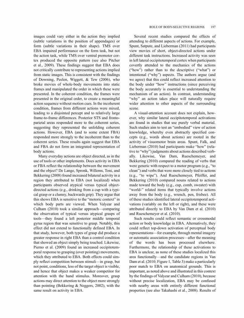
A reasonable proposal is that this encoding of bodyinformation within EBA and FBA is carried out bychanging patterns of activity across the neural popula-tions local to these two areas. Taking that approach, wecan make specific predictions about the representationsformed in EBA and FBA (Figure 4). That is, whataspects of the body are highlighted and made explicit,and what aspects are left implicit?
First, we expect that the patterns of activity in theseregions will vary systematically when exposed to dif-ferent body parts (Op de Beeck, Brants, Baeck, &Wagemans, 2010) perhaps more so in EBA than FBA(Taylor et al., 2007). Likewise, the shape of the bodyand its parts will be explicitly represented in such a waythat even relatively subtle differences in form will beamplified by the neural representation (relative, forexample, to representations in earlier visual areas).Furthermore, we expect that the “space” of bodyshapes will be influenced by experience, so as to beskewed toward commonly experienced body types,sizes, and postures as well as views (Chan, Kravitz,Truong, Arizpe, & Baker, 2010).
Similar arguments can be made for posture ––that is,systematically different activity patterns will map to the“space” of different possible postures that bodies canachieve. The coding for posture may be somewhat coar-ser than for form in light of findings such as those ofUrgesi, Candidi et al. (2007). And, as we argued above,some postures may be “special” in that they represent“key frames” that are particularly useful for representingthe positions of bodies and limbs efficiently. Note thatsome studies have examined the response to impossiblebody postures (Costantini et al., 2005; see alsoAvikainen, Liuhanen, Schurmann, & Hari, 2003;Cross, Mackie, Wolford, & Hamilton, 2010). We expectthese will be coded with reference to the nearest possibleposture, perhaps with an additional overall increase inactivity due to attentional modulation (Candidi, Urgesi,Ionta, & Aglioti, 2008).
On the other hand, the neurons in EBA and FBA donot explicitly represent many high-level properties. Forexample, patterns of activity in these regions will relateto body shape rather than identity, so they will be moresimilar for two bodies or body parts with highly similarshape (even across different viewing conditions such asocclusion, viewpoint, etc.; Taylor et al., 2010) than for
200 DOWNING AND PEELEN
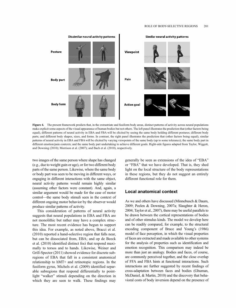
two images of the same person where shape has changed(e.g., due toweight gain or age), or for two different bodyparts of the same person. Likewise, where the same bodyor body part was seen to be moving in different ways, orengaging in different interactions with the same object,neural activity patterns would remain highly similar(assuming other factors were constant). And, again, asimilar argument would be made for the case of motorcontrol––the same body stimuli seen in the context ofdifferent ongoing motor behavior by the observer wouldproduce similar patterns of activity.
This consideration of patterns of neural activitysuggests that neural populations in EBA and FBA arenot monolithic but rather may have a complex struc-ture. The most recent evidence has begun to supportthis idea. For example, as noted above, Bracci et al.(2010) reported a hand-selective region that falls near,but can be dissociated from, EBA; and op de Beecket al. (2010) identified distinct foci that respond maxi-mally to torsos and to hands. Likewise, Weiner andGrill-Spector (2011) showed evidence for discrete sub-regions of EBA that fall in a consistent anatomicalrelationship to hMT+ and retinotopic regions. In thefusiform gyrus, Michels et al. (2009) identified separ-able subregions that respond differentially to point-light “walker” stimuli depending on the direction inwhich they are seen to walk. These findings may
generally be seen as extensions of the idea of “EBA”or “FBA” that we have developed. That is, they shedlight on the local structure of the body representationsin these regions, but they do not suggest an entirelydifferent functional role for them.
Local anatomical context
As we and others have discussed (Minnebusch & Daum,2009; Peelen & Downing, 2007a; Slaughter & Heron,2004; Taylor et al., 2007), there may be useful parallels tobe drawn between the cortical representations of bodiesand of other stimulus kinds. The model we develop herecan be readily compared, for example, to the structuralencoding component of Bruce and Young’s (1986)model of face perception, in which the visual propertiesof faces are extracted andmade available to other systemsfor the analysis of properties such as identification andemotion recognition. This comparison may indeed bemore than just an analogy. Bodies and faces, of course,are commonly perceived together, and the close overlapof FFA and FBA hints at functional interactions. Suchinteractions are further suggested by recent findings ofcross-adaptation between faces and bodies (Ghuman,McDaniel, & Martin, 2010) and the discovery that beha-vioral costs of body inversion depend on the presence of
Figure 4. The present framework predicts that, in the extrastriate and fusiform body areas, distinct patterns of activity across neural populationsmake explicit some aspects of the visual appearance of human bodies but not others. The left panel illustrates the prediction that (other factors beingequal), different patterns of neural activity in EBA and FBAwill be elicited by seeing the same body holding different postures; different bodyparts; and different body shapes, sizes, and forms. In contrast, the right panel illustrates the prediction that (other factors being equal), similarpatterns of neural activity in EBA and FBAwill be elicited by varying viewpoints of the same body (up to some tolerance); the same body part indifferent emotion/pain contexts; and the same body part undertaking to achieve different goals. Right-side figures adapted from Taylor, Wiggett,and Downing (2010); Morrison et al. (2007); and Bach et al. (2010), respectively.
ROLE OF BODY-SELECTIVE REGIONS 201
the head (Minnebusch, Suchan, & Daum, 2009; Yovel,Pelc, & Lubetzky, 2010) and relate to activity in face-selective areas (Brandman & Yovel, 2010).
More broadly, a similar gross, neuroanatomical struc-ture is seen in occipitotemporal cortex responses tobodies, faces, and objects: The occipital face area(Gauthier et al., 2000; Puce et al., 1996; Rossion et al.,2003) and the LO subregion (Grill-Spector et al., 1999)of the object-selective lateral occipital complex (LOC)(Malach et al., 1995) are found near EBA. Similarly, FFAand the pFs subregion of LOC are found near FBA. Asnoted above, EBA forms a body representation that ismore part-based than FBA. Similar distinctions arefound for faces (stronger representation of parts in OFAthan FFA; e.g., Liu, Harris, & Kanwisher, 2010) and forobjects (LO vs. pFs; e.g., Drucker & Aguirre, 2009).These parallels and others (e.g., Schwarzlose, Swisher,Dang, & Kanwisher, 2008) suggest there is a domain-general division of labor between lateral and ventraloccipitotemporal extrastriate areas. This proposal (andan extensive review of the relevant evidence) is consid-ered in depth elsewhere (Taylor & Downing, 2011).
Recent evidence for broad, diffuse body representa-tions in occipitotemporal cortexmay seem at oddswith afocus on selective regions such as EBA and FBA. Orlovet al. (2010) found a widespreadmosaic of broad regionsthat respond in subtle but systematic ways to perceivingand moving different body parts (e.g., to upper limbs vs.torsos). These responses encompass (but extend wellbeyond) EBA and FBA, and these findings suggestthat some aspects of the perception (and even move-ment) of body parts could be represented in a distributedway across this broad cortical territory. These findingsdo not necessarily challenge the importance of focal,selective regions such as those we examine here.Rather they emphasize that aspects of body perceptionmay extend further than previously thought. There maybe multiple representations of bodies (and other stimuli)that coexist at different spatial scales in these areas (cf.Graziano & Aflalo, 2007). Indeed, positing a broad,diffuse occipitotemporal representation of bodies, sur-rounding local “peaks,” may help to account for thefamilial relationship among many of the distinct butneighboring focal activations discussed here, such asthose responding to bodies, faces, tools, action, visualmotion, hands, and the like.
Global connections
It is a truism of cognitive neuroscience that task perfor-mance is supported by the activity of neural networks.Often, unfortunately, the term “network” is used looselyto describe brain regions that are coactivated in fMRI by
the same contrast, in the absence of significant directevidence for connectivity (e.g., Wiggett & Downing,2008). Although recent studies address this gap indomains such as face processing, using such tools asdynamic causal modeling (e.g., Fairhall et al., 2007), toour knowledge, there is no extant evidence that pertainsto extrastriate body-selective regions.
Nonetheless, it is an entirely reasonable hypothesisthat the perceptual information extracted in EBA andFBA is made available by functional connections toother regions. It is the integration of this informationwith that computed in other regions that together willexplain body perception in its fullness. A key questionthat is at stake when discussing such a putative func-tional network is to what extent the representations in agiven area are penetrated or shaped by those in otherregions. What are the limits of the influence of theactivity in other areas on the activity in EBA andFBA? The space of possibilities ranges between twoextremes. On the one hand, these regions could beentirely encapsulated in the sense that they strictlyanalyze the lower-level visual signals they receive,and the result is passed on to other brain areas withoutany further influence by the computations made inthose areas. On the other hand, it could be that amongthe “network” of areas that concerns itself with theperception (and indeed movement) of bodies, therange of functions is widely distributed, and the rela-tively greater response in one area to visual stimuli andanother to motor behavior is a difference of degreerather than kind. On this view, in the extreme itmakes little sense at all to consider EBA (or FBA) inisolation, as its functions can only be understood in thecontext of the entire network.
Our speculation is that the truth leans closer to theformer picture than the latter. There are good func-tional arguments for why the perceptual functions of anetwork should not be fully penetrable by the opera-tions of other brain areas; a relatively stimulus-bound,veridical representation can be important, for exam-ple, when expectations are violated. An importantexception to this principle is that the representationsin body-selective brain areas (and other extrastriateareas) will be strongly modulated by the focus ofattention. This may occur indirectly, such as by spatialattention mechanisms affecting retinotopic maps,with the knock-on effect of modulating body repre-sentations according to the contents of attended loca-tions (cf. Downing, Liu, & Kanwisher, 2001).Additionally, attention may directly modulate activityin body-selective regions as a way of supporting atemplate in the service of an abstractly specified task(Peelen et al., 2009). Given some of the considera-tions above about substructure, we may further
202 DOWNING AND PEELEN
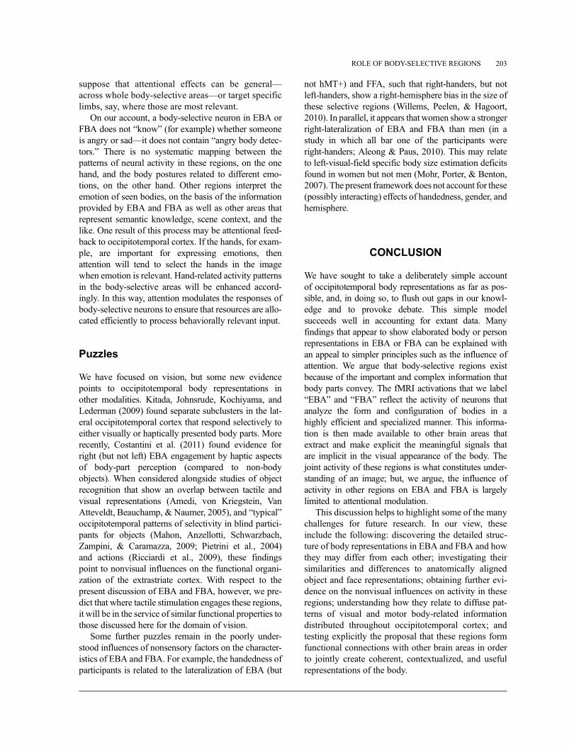
suppose that attentional effects can be general––across whole body-selective areas––or target specificlimbs, say, where those are most relevant.
On our account, a body-selective neuron in EBA orFBA does not “know” (for example) whether someoneis angry or sad––it does not contain “angry body detec-tors.” There is no systematic mapping between thepatterns of neural activity in these regions, on the onehand, and the body postures related to different emo-tions, on the other hand. Other regions interpret theemotion of seen bodies, on the basis of the informationprovided by EBA and FBA as well as other areas thatrepresent semantic knowledge, scene context, and thelike. One result of this process may be attentional feed-back to occipitotemporal cortex. If the hands, for exam-ple, are important for expressing emotions, thenattention will tend to select the hands in the imagewhen emotion is relevant. Hand-related activity patternsin the body-selective areas will be enhanced accord-ingly. In this way, attention modulates the responses ofbody-selective neurons to ensure that resources are allo-cated efficiently to process behaviorally relevant input.
Puzzles
We have focused on vision, but some new evidencepoints to occipitotemporal body representations inother modalities. Kitada, Johnsrude, Kochiyama, andLederman (2009) found separate subclusters in the lat-eral occipitotemporal cortex that respond selectively toeither visually or haptically presented body parts. Morerecently, Costantini et al. (2011) found evidence forright (but not left) EBA engagement by haptic aspectsof body-part perception (compared to non-bodyobjects). When considered alongside studies of objectrecognition that show an overlap between tactile andvisual representations (Amedi, von Kriegstein, VanAtteveldt, Beauchamp, & Naumer, 2005), and “typical”occipitotemporal patterns of selectivity in blind partici-pants for objects (Mahon, Anzellotti, Schwarzbach,Zampini, & Caramazza, 2009; Pietrini et al., 2004)and actions (Ricciardi et al., 2009), these findingspoint to nonvisual influences on the functional organi-zation of the extrastriate cortex. With respect to thepresent discussion of EBA and FBA, however, we pre-dict that where tactile stimulation engages these regions,it will be in the service of similar functional properties tothose discussed here for the domain of vision.
Some further puzzles remain in the poorly under-stood influences of nonsensory factors on the character-istics of EBA and FBA. For example, the handedness ofparticipants is related to the lateralization of EBA (but
not hMT+) and FFA, such that right-handers, but notleft-handers, show a right-hemisphere bias in the size ofthese selective regions (Willems, Peelen, & Hagoort,2010). In parallel, it appears that women show a strongerright-lateralization of EBA and FBA than men (in astudy in which all bar one of the participants wereright-handers; Aleong & Paus, 2010). This may relateto left-visual-field specific body size estimation deficitsfound in women but not men (Mohr, Porter, & Benton,2007). The present framework does not account for these(possibly interacting) effects of handedness, gender, andhemisphere.
CONCLUSION
We have sought to take a deliberately simple accountof occipitotemporal body representations as far as pos-sible, and, in doing so, to flush out gaps in our knowl-edge and to provoke debate. This simple modelsucceeds well in accounting for extant data. Manyfindings that appear to show elaborated body or personrepresentations in EBA or FBA can be explained withan appeal to simpler principles such as the influence ofattention. We argue that body-selective regions existbecause of the important and complex information thatbody parts convey. The fMRI activations that we label“EBA” and “FBA” reflect the activity of neurons thatanalyze the form and configuration of bodies in ahighly efficient and specialized manner. This informa-tion is then made available to other brain areas thatextract and make explicit the meaningful signals thatare implicit in the visual appearance of the body. Thejoint activity of these regions is what constitutes under-standing of an image; but, we argue, the influence ofactivity in other regions on EBA and FBA is largelylimited to attentional modulation.
This discussion helps to highlight some of the manychallenges for future research. In our view, theseinclude the following: discovering the detailed struc-ture of body representations in EBA and FBA and howthey may differ from each other; investigating theirsimilarities and differences to anatomically alignedobject and face representations; obtaining further evi-dence on the nonvisual influences on activity in theseregions; understanding how they relate to diffuse pat-terns of visual and motor body-related informationdistributed throughout occipitotemporal cortex; andtesting explicitly the proposal that these regions formfunctional connections with other brain areas in orderto jointly create coherent, contextualized, and usefulrepresentations of the body.
ROLE OF BODY-SELECTIVE REGIONS 203
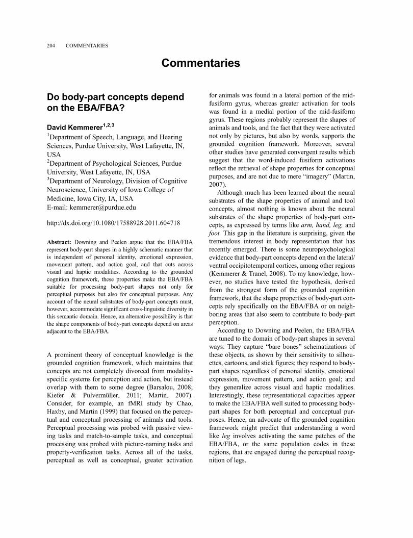
Commentaries
Do body-part concepts dependon the EBA/FBA?
David Kemmerer1,2,31Department of Speech, Language, and HearingSciences, Purdue University, West Lafayette, IN,USA2Department of Psychological Sciences, PurdueUniversity, West Lafayette, IN, USA3Department of Neurology, Division of CognitiveNeuroscience, University of Iowa College ofMedicine, Iowa City, IA, USAE-mail: [email protected]
http://dx.doi.org/10.1080/17588928.2011.604718
Abstract: Downing and Peelen argue that the EBA/FBArepresent body-part shapes in a highly schematic manner thatis independent of personal identity, emotional expression,movement pattern, and action goal, and that cuts acrossvisual and haptic modalities. According to the groundedcognition framework, these properties make the EBA/FBAsuitable for processing body-part shapes not only forperceptual purposes but also for conceptual purposes. Anyaccount of the neural substrates of body-part concepts must,however, accommodate significant cross-linguistic diversity inthis semantic domain. Hence, an alternative possibility is thatthe shape components of body-part concepts depend on areasadjacent to the EBA/FBA.
A prominent theory of conceptual knowledge is thegrounded cognition framework, which maintains thatconcepts are not completely divorced from modality-specific systems for perception and action, but insteadoverlap with them to some degree (Barsalou, 2008;Kiefer & Pulvermüller, 2011; Martin, 2007).Consider, for example, an fMRI study by Chao,Haxby, and Martin (1999) that focused on the percep-tual and conceptual processing of animals and tools.Perceptual processing was probed with passive view-ing tasks and match-to-sample tasks, and conceptualprocessing was probed with picture-naming tasks andproperty-verification tasks. Across all of the tasks,perceptual as well as conceptual, greater activation
for animals was found in a lateral portion of the mid-fusiform gyrus, whereas greater activation for toolswas found in a medial portion of the mid-fusiformgyrus. These regions probably represent the shapes ofanimals and tools, and the fact that they were activatednot only by pictures, but also by words, supports thegrounded cognition framework. Moreover, severalother studies have generated convergent results whichsuggest that the word-induced fusiform activationsreflect the retrieval of shape properties for conceptualpurposes, and are not due to mere “imagery” (Martin,2007).
Although much has been learned about the neuralsubstrates of the shape properties of animal and toolconcepts, almost nothing is known about the neuralsubstrates of the shape properties of body-part con-cepts, as expressed by terms like arm, hand, leg, andfoot. This gap in the literature is surprising, given thetremendous interest in body representation that hasrecently emerged. There is some neuropsychologicalevidence that body-part concepts depend on the lateral/ventral occipitotemporal cortices, among other regions(Kemmerer & Tranel, 2008). To my knowledge, how-ever, no studies have tested the hypothesis, derivedfrom the strongest form of the grounded cognitionframework, that the shape properties of body-part con-cepts rely specifically on the EBA/FBA or on neigh-boring areas that also seem to contribute to body-partperception.
According to Downing and Peelen, the EBA/FBAare tuned to the domain of body-part shapes in severalways: They capture “bare bones” schematizations ofthese objects, as shown by their sensitivity to silhou-ettes, cartoons, and stick figures; they respond to body-part shapes regardless of personal identity, emotionalexpression, movement pattern, and action goal; andthey generalize across visual and haptic modalities.Interestingly, these representational capacities appearto make the EBA/FBAwell suited to processing body-part shapes for both perceptual and conceptual pur-poses. Hence, an advocate of the grounded cognitionframework might predict that understanding a wordlike leg involves activating the same patches of theEBA/FBA, or the same population codes in theseregions, that are engaged during the perceptual recog-nition of legs.
204 COMMENTARIES
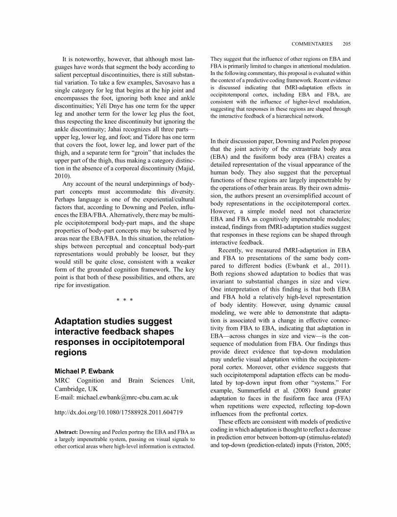
It is noteworthy, however, that although most lan-guages have words that segment the body according tosalient perceptual discontinuities, there is still substan-tial variation. To take a few examples, Savosavo has asingle category for leg that begins at the hip joint andencompasses the foot, ignoring both knee and anklediscontinuities; Yél̂� Dnye has one term for the upperleg and another term for the lower leg plus the foot,thus respecting the knee discontinuity but ignoring theankle discontinuity; Jahai recognizes all three parts—upper leg, lower leg, and foot; and Tidore has one termthat covers the foot, lower leg, and lower part of thethigh, and a separate term for “groin” that includes theupper part of the thigh, thus making a category distinc-tion in the absence of a corporeal discontinuity (Majid,2010).
Any account of the neural underpinnings of body-part concepts must accommodate this diversity.Perhaps language is one of the experiential/culturalfactors that, according to Downing and Peelen, influ-ences the EBA/FBA. Alternatively, there may bemulti-ple occipitotemporal body-part maps, and the shapeproperties of body-part concepts may be subserved byareas near the EBA/FBA. In this situation, the relation-ships between perceptual and conceptual body-partrepresentations would probably be looser, but theywould still be quite close, consistent with a weakerform of the grounded cognition framework. The keypoint is that both of these possibilities, and others, areripe for investigation.
* * *
Adaptation studies suggestinteractive feedback shapesresponses in occipitotemporalregions
Michael P. EwbankMRC Cognition and Brain Sciences Unit,Cambridge, UKE-mail: [email protected]
http://dx.doi.org/10.1080/17588928.2011.604719
Abstract: Downing and Peelen portray the EBA and FBA asa largely impenetrable system, passing on visual signals toother cortical areas where high-level information is extracted.
They suggest that the influence of other regions on EBA andFBA is primarily limited to changes in attentional modulation.In the following commentary, this proposal is evaluated withinthe context of a predictive coding framework. Recent evidenceis discussed indicating that fMRI-adaptation effects inoccipitotemporal cortex, including EBA and FBA, areconsistent with the influence of higher-level modulation,suggesting that responses in these regions are shaped throughthe interactive feedback of a hierarchical network.
In their discussion paper, Downing and Peelen proposethat the joint activity of the extrastriate body area(EBA) and the fusiform body area (FBA) creates adetailed representation of the visual appearance of thehuman body. They also suggest that the perceptualfunctions of these regions are largely impenetrable bythe operations of other brain areas. By their own admis-sion, the authors present an oversimplified account ofbody representations in the occipitotemporal cortex.However, a simple model need not characterizeEBA and FBA as cognitively impenetrable modules;instead, findings from fMRI-adaptation studies suggestthat responses in these regions can be shaped throughinteractive feedback.
Recently, we measured fMRI-adaptation in EBAand FBA to presentations of the same body com-pared to different bodies (Ewbank et al., 2011).Both regions showed adaptation to bodies that wasinvariant to substantial changes in size and view.One interpretation of this finding is that both EBAand FBA hold a relatively high-level representationof body identity. However, using dynamic causalmodeling, we were able to demonstrate that adapta-tion is associated with a change in effective connec-tivity from FBA to EBA, indicating that adaptation inEBA––across changes in size and view––is the con-sequence of modulation from FBA. Our findings thusprovide direct evidence that top-down modulationmay underlie visual adaptation within the occipitotem-poral cortex. Moreover, other evidence suggests thatsuch occipitotemporal adaptation effects can be modu-lated by top-down input from other “systems.” Forexample, Summerfield et al. (2008) found greateradaptation to faces in the fusiform face area (FFA)when repetitions were expected, reflecting top-downinfluences from the prefrontal cortex.
These effects are consistent with models of predictivecoding inwhich adaptation is thought to reflect a decreasein prediction error between bottom-up (stimulus-related)and top-down (prediction-related) inputs (Friston, 2005;
COMMENTARIES 205
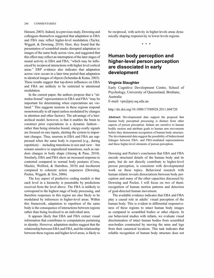
Henson, 2003). Indeed, in a previous study,Downing andcolleagues themselves suggested that adaptation in EBAand FBA may reflect higher-level modulation (Taylor,Wiggett, & Downing, 2010). Here, they found that thepresentation of scrambled masks disrupted adaptation toimages of the same body across view, and suggested thatthis effect may reflect an interruption of the later stages ofneural activity in EBA and FBA, “which may be influ-enced by reciprocal interactions with higher level corticalareas.” ERP evidence also indicates that adaptationacross view occurs in a later time period than adaptationto identical images of objects (Schendan &Kutas, 2003).These results suggest that top-down influences on EBAand FBA are unlikely to be restricted to attentionalmodulation.
In the current paper, the authors propose that a “sti-mulus-bound” representation in EBA and FBA “may beimportant for determining when expectations are vio-lated.” This suggests neurons in these regions respondmonotonically to all input (unless modulated by changesin attention and other factors). The advantage of a hier-archical model, however, is that it enables the brain toconstruct prior expectations in a dynamic fashion––rather than being stimulus bound, energy-costly signalsare focused on rare inputs, alerting the system to impor-tant changes. Thus, neurons in EBA and FBA are sup-pressed when the same body is expected (e.g., duringrepetition)–– including transitions in size and view––butremain sensitive to unpredicted transitions, such as ran-dom changes in body shape (Aleong & Paus, 2010).Similarly, EBA and FBA show an increased response tocontorted compared to normal body postures (Cross,Mackie, Wolford, & Hamilton, 2010) and incoherentcompared to coherent action sequences (Downing,Peelen, Wiggett, & Tew, 2006).
The key aspect of predictive coding models is thateach level in a hierarchy is penetrable by predictionsreceived from the level above. The FBA is unlikely tocorrespond to the highest stage of body processing, andtherefore responses in this region are also likely to bemodulated by inferences in higher-level areas. Withinthis framework, adaptation to repetition of the samebody is the consequence of interactions between regionsrather than being localized to an individual area.
It appears likely that EBA and FBA extract visualinformation that contributes to computations pertainingto identity. However, adaptation studies suggest that therelationship betweenEBAandFBA, and the relationshipbetween these regions and higher-level areas, is likely to
be reciprocal, with activity in higher-levels areas dyna-mically shaping responsivity in lower-levels regions.
* * *
Human body perception andhigher-level person perceptionare dissociated in earlydevelopment
Virginia SlaughterEarly Cognitive Development Centre, School ofPsychology, University of Queensland, Brisbane,AustraliaE-mail: [email protected]
http://dx.doi.org/10.1080/17588928.2011.604720
Abstract: Developmental data support the proposal thathuman body perceptual processing is distinct from otheraspects of person perception. Infants are sensitive to humanbodily motion and attribute goals to human arm movementsbefore they demonstrate recognition of human body structure.The developmental data suggest the possibility of bidirectionallinkages between EBA- and FBA-mediated representationsand these higher-level elements of person perception.
Downing and Peelen’s conclusion that EBA and FBAencode structural details of the human body and itsparts, but do not directly contribute to higher-levelperson perception, is consistent with developmentalwork on these topics. Behavioral research withhuman infants reveals dissociations between body per-ception and many of the other capacities discussed byDowning and Peelen. I will focus on two of them:recognition of human motion patterns and detectionof goal-directed human movement.
The available evidence indicates that EBA and FBAplay a causal role in adults’ visual perception of thehuman body. This is evident in differential responsive-ness of these regions to intact human body imagesas compared to scrambled bodies or other objects. Inour behavioral studies with infants, we evaluate visualdiscrimination of intact human bodies from scramblednon-bodies constructed by moving the arms and legsfrom their canonical locations. This task indicates thatreliable recognition of human body structure does not
206 COMMENTARIES
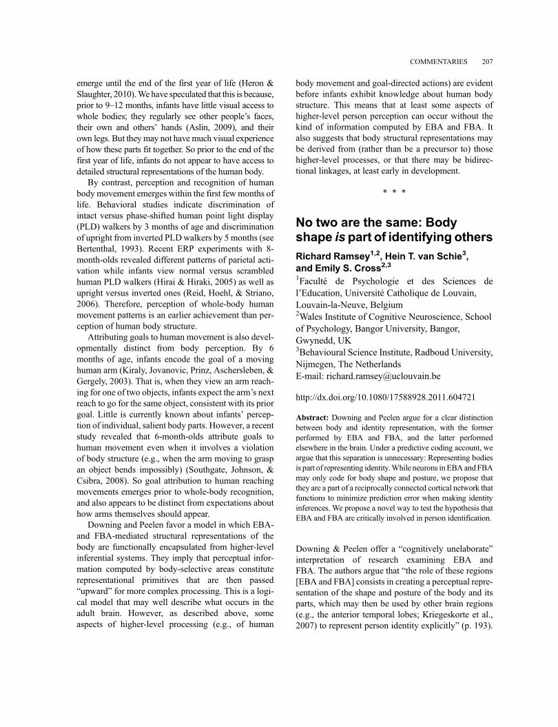
emerge until the end of the first year of life (Heron &Slaughter, 2010).We have speculated that this is because,prior to 9–12 months, infants have little visual access towhole bodies; they regularly see other people’s faces,their own and others’ hands (Aslin, 2009), and theirown legs. But they may not have much visual experienceof how these parts fit together. So prior to the end of thefirst year of life, infants do not appear to have access todetailed structural representations of the human body.
By contrast, perception and recognition of humanbody movement emerges within the first few months oflife. Behavioral studies indicate discrimination ofintact versus phase-shifted human point light display(PLD) walkers by 3 months of age and discriminationof upright from inverted PLDwalkers by 5 months (seeBertenthal, 1993). Recent ERP experiments with 8-month-olds revealed different patterns of parietal acti-vation while infants view normal versus scrambledhuman PLD walkers (Hirai & Hiraki, 2005) as well asupright versus inverted ones (Reid, Hoehl, & Striano,2006). Therefore, perception of whole-body humanmovement patterns is an earlier achievement than per-ception of human body structure.
Attributing goals to human movement is also devel-opmentally distinct from body perception. By 6months of age, infants encode the goal of a movinghuman arm (Kiraly, Jovanovic, Prinz, Aschersleben, &Gergely, 2003). That is, when they view an arm reach-ing for one of two objects, infants expect the arm’s nextreach to go for the same object, consistent with its priorgoal. Little is currently known about infants’ percep-tion of individual, salient body parts. However, a recentstudy revealed that 6-month-olds attribute goals tohuman movement even when it involves a violationof body structure (e.g., when the arm moving to graspan object bends impossibly) (Southgate, Johnson, &Csibra, 2008). So goal attribution to human reachingmovements emerges prior to whole-body recognition,and also appears to be distinct from expectations abouthow arms themselves should appear.
Downing and Peelen favor a model in which EBA-and FBA-mediated structural representations of thebody are functionally encapsulated from higher-levelinferential systems. They imply that perceptual infor-mation computed by body-selective areas constituterepresentational primitives that are then passed“upward” for more complex processing. This is a logi-cal model that may well describe what occurs in theadult brain. However, as described above, someaspects of higher-level processing (e.g., of human
body movement and goal-directed actions) are evidentbefore infants exhibit knowledge about human bodystructure. This means that at least some aspects ofhigher-level person perception can occur without thekind of information computed by EBA and FBA. Italso suggests that body structural representations maybe derived from (rather than be a precursor to) thosehigher-level processes, or that there may be bidirec-tional linkages, at least early in development.
* * *
No two are the same: Bodyshape is part of identifying others
Richard Ramsey1,2, Hein T. van Schie3,and Emily S. Cross2,31Faculté de Psychologie et des Sciences del’Education, Université Catholique de Louvain,Louvain-la-Neuve, Belgium2Wales Institute of Cognitive Neuroscience, Schoolof Psychology, Bangor University, Bangor,Gwynedd, UK3Behavioural Science Institute, Radboud University,Nijmegen, The NetherlandsE-mail: [email protected]
http://dx.doi.org/10.1080/17588928.2011.604721
Abstract: Downing and Peelen argue for a clear distinctionbetween body and identity representation, with the formerperformed by EBA and FBA, and the latter performedelsewhere in the brain. Under a predictive coding account, weargue that this separation is unnecessary: Representing bodiesis part of representing identity.While neurons inEBA andFBAmay only code for body shape and posture, we propose thatthey are a part of a reciprocally connected cortical network thatfunctions to minimize prediction error when making identityinferences. We propose a novel way to test the hypothesis thatEBA and FBA are critically involved in person identification.
Downing & Peelen offer a “cognitively unelaborate”interpretation of research examining EBA andFBA. The authors argue that “the role of these regions[EBA and FBA] consists in creating a perceptual repre-sentation of the shape and posture of the body and itsparts, which may then be used by other brain regions(e.g., the anterior temporal lobes; Kriegeskorte et al.,2007) to represent person identity explicitly” (p. 193).
COMMENTARIES 207
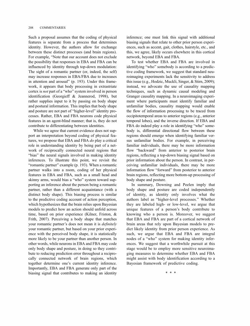
Such a proposal assumes that the coding of physicalfeatures is separate from a process that determinesidentity. However, the authors allow for exchangebetween these distinct processes (and brain regions).For example, “Note that our account does not excludethe possibility that responses in EBA and FBA can beinfluenced by identity through top-down modulation:The sight of a romantic partner (or, indeed, the self)may increase responses in EBA/FBA due to increasesin attention and arousal” (p. 193). Under this frame-work, it appears that body processing in extrastriatecortex is not part of a “who” system involved in personidentification (Georgieff & Jeannerod, 1998), butrather supplies input to it by passing on body shapeand postural information. This implies that body shapeand posture are not part of “higher-level” identity pro-cesses. Rather, EBA and FBA neurons code physicalfeatures in an agent-blind manner; that is, they do notcontribute to differentiating between identities.
While we agree that current evidence does not sup-port an interpretation beyond coding of physical fea-tures, we propose that EBA and FBA do play a criticalrole in understanding identity by being part of a net-work of reciprocally connected neural regions that“bias” the neural signals involved in making identityinferences. To illustrate this point, we revisit the“romantic partner” example (p. 193). When a romanticpartner walks into a room, coding of her physicalfeatures in EBA and FBA, such as a small head andskinny arms, would bias a “who” system toward sup-porting an inference about the person being a romanticpartner, rather than a different acquaintance (with adistinct body shape). This biasing process is similarto the predictive coding account of action perception,which hypothesizes that the brain relies upon Bayesianmodels to predict how an action should unfold acrosstime, based on prior experience (Kilner, Friston, &Frith, 2007). Perceiving a body shape that matchesyour romantic partner’s does not mean it is definitelyyour romantic partner, but based on your prior experi-ence with the perceived body shape, it is statisticallymore likely to be your partner than another person. Inother words, while neurons in EBA and FBAmay codeonly body shape and posture, in doing so they contri-bute to reducing prediction error throughout a recipro-cally connected network of brain regions, whichtogether determine one’s current identity inference.Importantly, EBA and FBA generate only part of thebiasing signal that contributes to making an identity
inference; one must link this signal with additionalbiasing signals that relate to other prior person experi-ences, such as accent, gait, clothes, hairstyle, etc., andthis, we agree, likely occurs elsewhere in this corticalnetwork, beyond EBA and FBA.
To test whether EBA and FBA are involved inidentifying “who” somebody is according to a predic-tive coding framework, we suggest that standard neu-roimaging experiments lack the sensitivity to addressthis issue (e.g., Hodzic, Muckli, Singer, & Stirn, 2009);instead, we advocate the use of causality mappingtechniques, such as dynamic causal modeling andGranger causality mapping. In a neuroimaging experi-ment where participants must identify familiar andunfamiliar bodies, causality mapping would enablethe flow of information processing to be traced fromoccipitotemporal areas to anterior regions (e.g., anteriortemporal lobes), and the inverse direction. If EBA andFBA do indeed play a role in identifying “who” some-body is, differential directional flow between theseregions should emerge when identifying familiar ver-sus unfamiliar bodies. For example, in perceivingfamiliar individuals, there may be more informationflow “backward” from anterior to posterior brainregions, reflecting a top-down biasing signal based onprior information about the person. In contrast, in per-ceiving unfamiliar individuals, there may be moreinformation flow “forward” from posterior to anteriorbrain regions, reflecting more bottom-up processing ofbody shape and posture.
In summary, Downing and Peelen imply thatbody shape and posture are coded independentlyof identity, as identity only involves what theauthors label as “higher-level processes.” Whetherthey are labeled high- or low-level, we argue thatunique features of a person’s body contribute toknowing who a person is. Moreover, we suggestthat EBA and FBA are part of a cortical network ofbrain areas that rely upon Bayesian models to pre-dict likely identity from prior person experience. Assuch, we argue that EBA and FBA are integralnodes of a “who” system for making identity infer-ences. We suggest that a worthwhile pursuit at thisstage would be to employ more sensitive neuroima-ging measures to determine whether EBA and FBAmight assist with body identification according to aBayesian framework of predictive coding.
* * *
208 COMMENTARIES
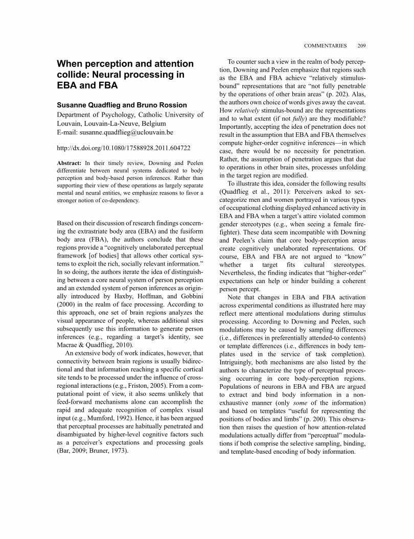
When perception and attentioncollide: Neural processing inEBA and FBA
Susanne Quadflieg and Bruno RossionDepartment of Psychology, Catholic University ofLouvain, Louvain-La-Neuve, BelgiumE-mail: [email protected]
http://dx.doi.org/10.1080/17588928.2011.604722
Abstract: In their timely review, Downing and Peelendifferentiate between neural systems dedicated to bodyperception and body-based person inferences. Rather thansupporting their view of these operations as largely separatemental and neural entities, we emphasize reasons to favor astronger notion of co-dependency.
Based on their discussion of research findings concern-ing the extrastriate body area (EBA) and the fusiformbody area (FBA), the authors conclude that theseregions provide a “cognitively unelaborated perceptualframework [of bodies] that allows other cortical sys-tems to exploit the rich, socially relevant information.”In so doing, the authors iterate the idea of distinguish-ing between a core neural system of person perceptionand an extended system of person inferences as origin-ally introduced by Haxby, Hoffman, and Gobbini(2000) in the realm of face processing. According tothis approach, one set of brain regions analyzes thevisual appearance of people, whereas additional sitessubsequently use this information to generate personinferences (e.g., regarding a target’s identity, seeMacrae & Quadflieg, 2010).
An extensive body of work indicates, however, thatconnectivity between brain regions is usually bidirec-tional and that information reaching a specific corticalsite tends to be processed under the influence of cross-regional interactions (e.g., Friston, 2005). From a com-putational point of view, it also seems unlikely thatfeed-forward mechanisms alone can accomplish therapid and adequate recognition of complex visualinput (e.g., Mumford, 1992). Hence, it has been arguedthat perceptual processes are habitually penetrated anddisambiguated by higher-level cognitive factors suchas a perceiver’s expectations and processing goals(Bar, 2009; Bruner, 1973).
To counter such a view in the realm of body percep-tion, Downing and Peelen emphasize that regions suchas the EBA and FBA achieve “relatively stimulus-bound” representations that are “not fully penetrableby the operations of other brain areas” (p. 202). Alas,the authors own choice of words gives away the caveat.How relatively stimulus-bound are the representationsand to what extent (if not fully) are they modifiable?Importantly, accepting the idea of penetration does notresult in the assumption that EBA and FBA themselvescompute higher-order cognitive inferences––in whichcase, there would be no necessity for penetration.Rather, the assumption of penetration argues that dueto operations in other brain sites, processes unfoldingin the target region are modified.
To illustrate this idea, consider the following results(Quadflieg et al., 2011): Perceivers asked to sex-categorize men and women portrayed in various typesof occupational clothing displayed enhanced activity inEBA and FBAwhen a target’s attire violated commongender stereotypes (e.g., when seeing a female fire-fighter). These data seem incompatible with Downingand Peelen’s claim that core body-perception areascreate cognitively unelaborated representations. Ofcourse, EBA and FBA are not argued to “know”whether a target fits cultural stereotypes.Nevertheless, the finding indicates that “higher-order”expectations can help or hinder building a coherentperson percept.
Note that changes in EBA and FBA activationacross experimental conditions as illustrated here mayreflect mere attentional modulations during stimulusprocessing. According to Downing and Peelen, suchmodulations may be caused by sampling differences(i.e., differences in preferentially attended-to contents)or template differences (i.e., differences in body tem-plates used in the service of task completion).Intriguingly, both mechanisms are also listed by theauthors to characterize the type of perceptual proces-sing occurring in core body-perception regions.Populations of neurons in EBA and FBA are arguedto extract and bind body information in a non-exhaustive manner (only some of the information)and based on templates “useful for representing thepositions of bodies and limbs” (p. 200). This observa-tion then raises the question of how attention-relatedmodulations actually differ from “perceptual” modula-tions if both comprise the selective sampling, binding,and template-based encoding of body information.
COMMENTARIES 209
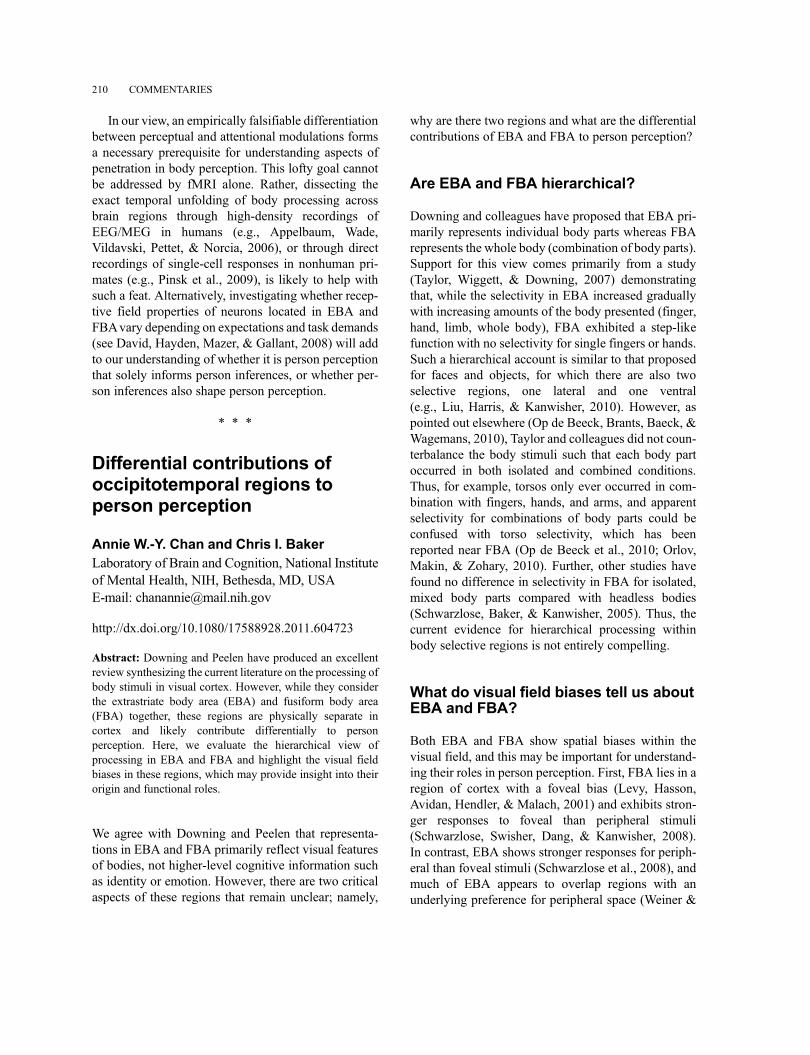
In our view, an empirically falsifiable differentiationbetween perceptual and attentional modulations formsa necessary prerequisite for understanding aspects ofpenetration in body perception. This lofty goal cannotbe addressed by fMRI alone. Rather, dissecting theexact temporal unfolding of body processing acrossbrain regions through high-density recordings ofEEG/MEG in humans (e.g., Appelbaum, Wade,Vildavski, Pettet, & Norcia, 2006), or through directrecordings of single-cell responses in nonhuman pri-mates (e.g., Pinsk et al., 2009), is likely to help withsuch a feat. Alternatively, investigating whether recep-tive field properties of neurons located in EBA andFBAvary depending on expectations and task demands(see David, Hayden, Mazer, & Gallant, 2008) will addto our understanding of whether it is person perceptionthat solely informs person inferences, or whether per-son inferences also shape person perception.
* * *
Differential contributions ofoccipitotemporal regions toperson perception
Annie W.-Y. Chan and Chris I. BakerLaboratory of Brain and Cognition, National Instituteof Mental Health, NIH, Bethesda, MD, USAE-mail: [email protected]
http://dx.doi.org/10.1080/17588928.2011.604723
Abstract: Downing and Peelen have produced an excellentreview synthesizing the current literature on the processing ofbody stimuli in visual cortex. However, while they considerthe extrastriate body area (EBA) and fusiform body area(FBA) together, these regions are physically separate incortex and likely contribute differentially to personperception. Here, we evaluate the hierarchical view ofprocessing in EBA and FBA and highlight the visual fieldbiases in these regions, which may provide insight into theirorigin and functional roles.
We agree with Downing and Peelen that representa-tions in EBA and FBA primarily reflect visual featuresof bodies, not higher-level cognitive information suchas identity or emotion. However, there are two criticalaspects of these regions that remain unclear; namely,
why are there two regions and what are the differentialcontributions of EBA and FBA to person perception?
Are EBA and FBA hierarchical?
Downing and colleagues have proposed that EBA pri-marily represents individual body parts whereas FBArepresents the whole body (combination of body parts).Support for this view comes primarily from a study(Taylor, Wiggett, & Downing, 2007) demonstratingthat, while the selectivity in EBA increased graduallywith increasing amounts of the body presented (finger,hand, limb, whole body), FBA exhibited a step-likefunction with no selectivity for single fingers or hands.Such a hierarchical account is similar to that proposedfor faces and objects, for which there are also twoselective regions, one lateral and one ventral(e.g., Liu, Harris, & Kanwisher, 2010). However, aspointed out elsewhere (Op de Beeck, Brants, Baeck, &Wagemans, 2010), Taylor and colleagues did not coun-terbalance the body stimuli such that each body partoccurred in both isolated and combined conditions.Thus, for example, torsos only ever occurred in com-bination with fingers, hands, and arms, and apparentselectivity for combinations of body parts could beconfused with torso selectivity, which has beenreported near FBA (Op de Beeck et al., 2010; Orlov,Makin, & Zohary, 2010). Further, other studies havefound no difference in selectivity in FBA for isolated,mixed body parts compared with headless bodies(Schwarzlose, Baker, & Kanwisher, 2005). Thus, thecurrent evidence for hierarchical processing withinbody selective regions is not entirely compelling.
What do visual field biases tell us aboutEBA and FBA?
Both EBA and FBA show spatial biases within thevisual field, and this may be important for understand-ing their roles in person perception. First, FBA lies in aregion of cortex with a foveal bias (Levy, Hasson,Avidan, Hendler, & Malach, 2001) and exhibits stron-ger responses to foveal than peripheral stimuli(Schwarzlose, Swisher, Dang, & Kanwisher, 2008).In contrast, EBA shows stronger responses for periph-eral than foveal stimuli (Schwarzlose et al., 2008), andmuch of EBA appears to overlap regions with anunderlying preference for peripheral space (Weiner &
210 COMMENTARIES
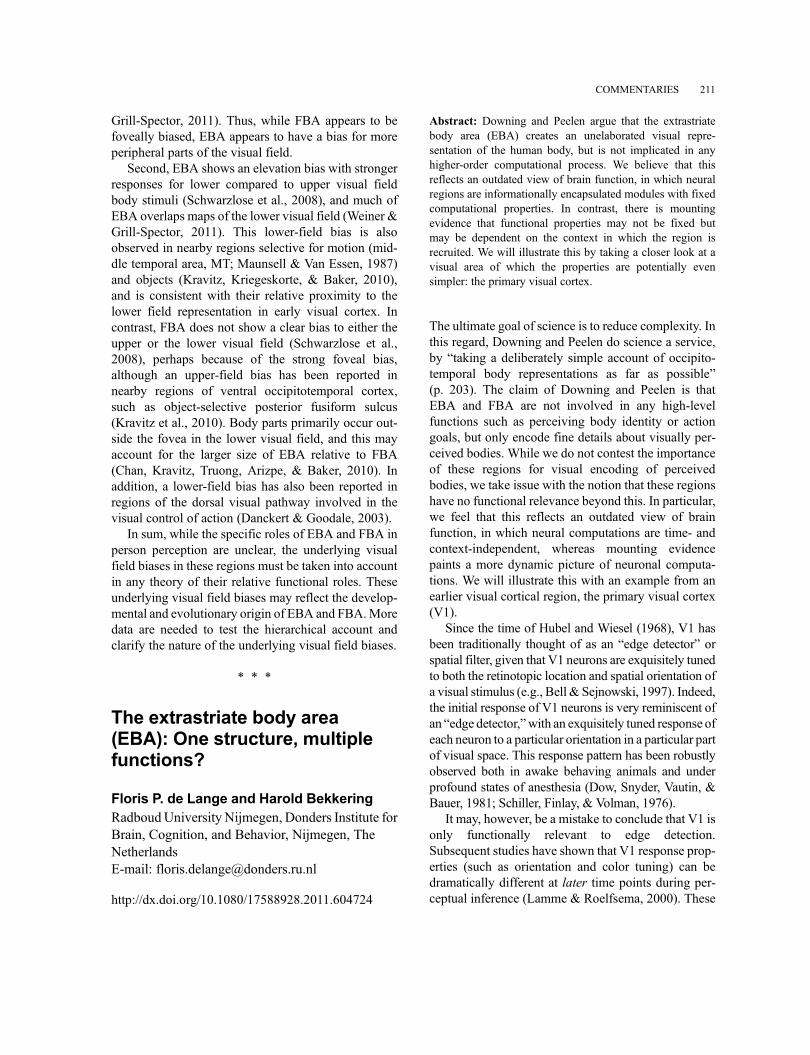
Grill-Spector, 2011). Thus, while FBA appears to befoveally biased, EBA appears to have a bias for moreperipheral parts of the visual field.
Second, EBA shows an elevation bias with strongerresponses for lower compared to upper visual fieldbody stimuli (Schwarzlose et al., 2008), and much ofEBA overlaps maps of the lower visual field (Weiner &Grill-Spector, 2011). This lower-field bias is alsoobserved in nearby regions selective for motion (mid-dle temporal area, MT; Maunsell & Van Essen, 1987)and objects (Kravitz, Kriegeskorte, & Baker, 2010),and is consistent with their relative proximity to thelower field representation in early visual cortex. Incontrast, FBA does not show a clear bias to either theupper or the lower visual field (Schwarzlose et al.,2008), perhaps because of the strong foveal bias,although an upper-field bias has been reported innearby regions of ventral occipitotemporal cortex,such as object-selective posterior fusiform sulcus(Kravitz et al., 2010). Body parts primarily occur out-side the fovea in the lower visual field, and this mayaccount for the larger size of EBA relative to FBA(Chan, Kravitz, Truong, Arizpe, & Baker, 2010). Inaddition, a lower-field bias has also been reported inregions of the dorsal visual pathway involved in thevisual control of action (Danckert & Goodale, 2003).
In sum, while the specific roles of EBA and FBA inperson perception are unclear, the underlying visualfield biases in these regions must be taken into accountin any theory of their relative functional roles. Theseunderlying visual field biases may reflect the develop-mental and evolutionary origin of EBA and FBA.Moredata are needed to test the hierarchical account andclarify the nature of the underlying visual field biases.
* * *
The extrastriate body area(EBA): One structure, multiplefunctions?
Floris P. de Lange and Harold BekkeringRadboudUniversity Nijmegen, Donders Institute forBrain, Cognition, and Behavior, Nijmegen, TheNetherlandsE-mail: [email protected]
http://dx.doi.org/10.1080/17588928.2011.604724
Abstract: Downing and Peelen argue that the extrastriatebody area (EBA) creates an unelaborated visual repre-sentation of the human body, but is not implicated in anyhigher-order computational process. We believe that thisreflects an outdated view of brain function, in which neuralregions are informationally encapsulated modules with fixedcomputational properties. In contrast, there is mountingevidence that functional properties may not be fixed butmay be dependent on the context in which the region isrecruited. We will illustrate this by taking a closer look at avisual area of which the properties are potentially evensimpler: the primary visual cortex.
The ultimate goal of science is to reduce complexity. Inthis regard, Downing and Peelen do science a service,by “taking a deliberately simple account of occipito-temporal body representations as far as possible”(p. 203). The claim of Downing and Peelen is thatEBA and FBA are not involved in any high-levelfunctions such as perceiving body identity or actiongoals, but only encode fine details about visually per-ceived bodies. While we do not contest the importanceof these regions for visual encoding of perceivedbodies, we take issue with the notion that these regionshave no functional relevance beyond this. In particular,we feel that this reflects an outdated view of brainfunction, in which neural computations are time- andcontext-independent, whereas mounting evidencepaints a more dynamic picture of neuronal computa-tions. We will illustrate this with an example from anearlier visual cortical region, the primary visual cortex(V1).
Since the time of Hubel and Wiesel (1968), V1 hasbeen traditionally thought of as an “edge detector” orspatial filter, given that V1 neurons are exquisitely tunedto both the retinotopic location and spatial orientation ofa visual stimulus (e.g., Bell & Sejnowski, 1997). Indeed,the initial response of V1 neurons is very reminiscent ofan “edge detector,”with an exquisitely tuned response ofeach neuron to a particular orientation in a particular partof visual space. This response pattern has been robustlyobserved both in awake behaving animals and underprofound states of anesthesia (Dow, Snyder, Vautin, &Bauer, 1981; Schiller, Finlay, & Volman, 1976).
It may, however, be a mistake to conclude that V1 isonly functionally relevant to edge detection.Subsequent studies have shown that V1 response prop-erties (such as orientation and color tuning) can bedramatically different at later time points during per-ceptual inference (Lamme & Roelfsema, 2000). These
COMMENTARIES 211
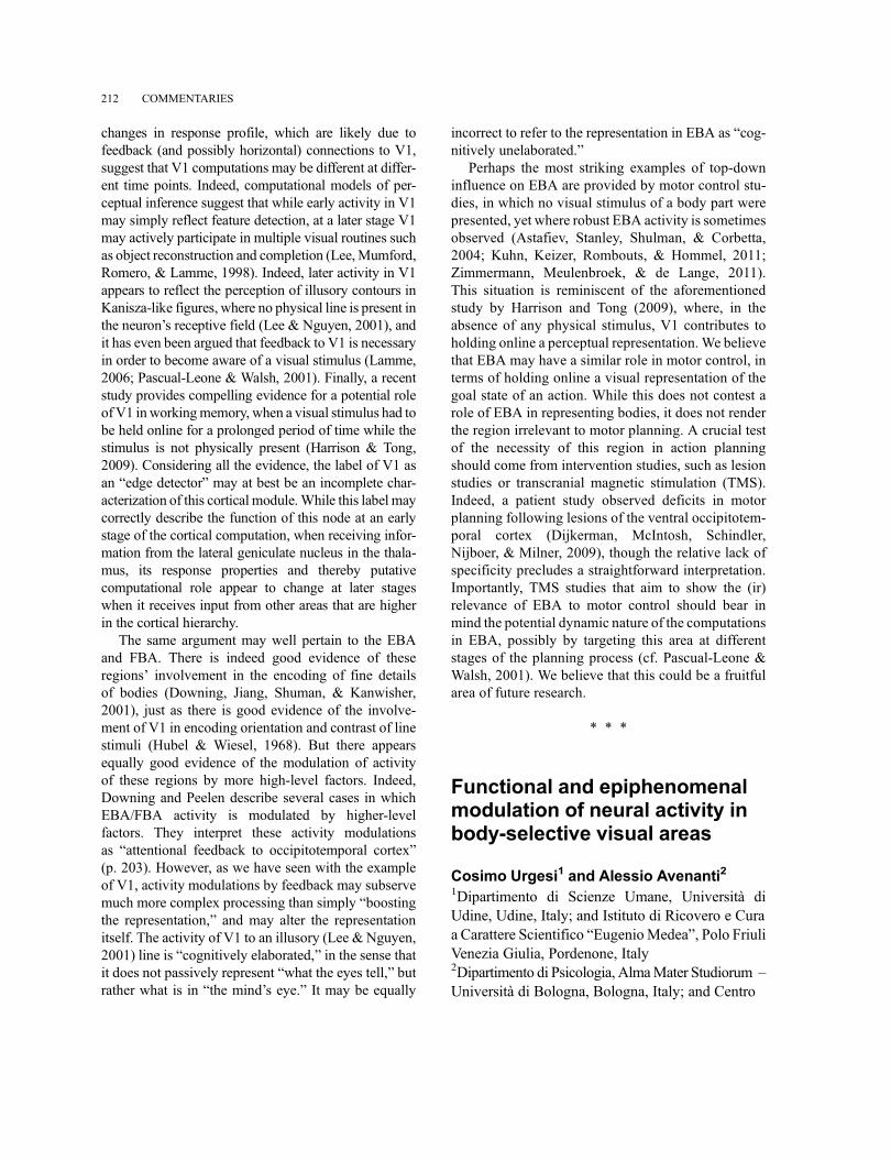
changes in response profile, which are likely due tofeedback (and possibly horizontal) connections to V1,suggest that V1 computations may be different at differ-ent time points. Indeed, computational models of per-ceptual inference suggest that while early activity in V1may simply reflect feature detection, at a later stage V1may actively participate in multiple visual routines suchas object reconstruction and completion (Lee,Mumford,Romero, & Lamme, 1998). Indeed, later activity in V1appears to reflect the perception of illusory contours inKanisza-like figures, where no physical line is present inthe neuron’s receptive field (Lee & Nguyen, 2001), andit has even been argued that feedback to V1 is necessaryin order to become aware of a visual stimulus (Lamme,2006; Pascual-Leone & Walsh, 2001). Finally, a recentstudy provides compelling evidence for a potential roleof V1 inworkingmemory, when a visual stimulus had tobe held online for a prolonged period of time while thestimulus is not physically present (Harrison & Tong,2009). Considering all the evidence, the label of V1 asan “edge detector” may at best be an incomplete char-acterization of this cortical module.While this label maycorrectly describe the function of this node at an earlystage of the cortical computation, when receiving infor-mation from the lateral geniculate nucleus in the thala-mus, its response properties and thereby putativecomputational role appear to change at later stageswhen it receives input from other areas that are higherin the cortical hierarchy.
The same argument may well pertain to the EBAand FBA. There is indeed good evidence of theseregions’ involvement in the encoding of fine detailsof bodies (Downing, Jiang, Shuman, & Kanwisher,2001), just as there is good evidence of the involve-ment of V1 in encoding orientation and contrast of linestimuli (Hubel & Wiesel, 1968). But there appearsequally good evidence of the modulation of activityof these regions by more high-level factors. Indeed,Downing and Peelen describe several cases in whichEBA/FBA activity is modulated by higher-levelfactors. They interpret these activity modulationsas “attentional feedback to occipitotemporal cortex”(p. 203). However, as we have seen with the exampleof V1, activity modulations by feedback may subservemuch more complex processing than simply “boostingthe representation,” and may alter the representationitself. The activity of V1 to an illusory (Lee & Nguyen,2001) line is “cognitively elaborated,” in the sense thatit does not passively represent “what the eyes tell,” butrather what is in “the mind’s eye.” It may be equally
incorrect to refer to the representation in EBA as “cog-nitively unelaborated.”
Perhaps the most striking examples of top-downinfluence on EBA are provided by motor control stu-dies, in which no visual stimulus of a body part werepresented, yet where robust EBA activity is sometimesobserved (Astafiev, Stanley, Shulman, & Corbetta,2004; Kuhn, Keizer, Rombouts, & Hommel, 2011;Zimmermann, Meulenbroek, & de Lange, 2011).This situation is reminiscent of the aforementionedstudy by Harrison and Tong (2009), where, in theabsence of any physical stimulus, V1 contributes toholding online a perceptual representation. We believethat EBA may have a similar role in motor control, interms of holding online a visual representation of thegoal state of an action. While this does not contest arole of EBA in representing bodies, it does not renderthe region irrelevant to motor planning. A crucial testof the necessity of this region in action planningshould come from intervention studies, such as lesionstudies or transcranial magnetic stimulation (TMS).Indeed, a patient study observed deficits in motorplanning following lesions of the ventral occipitotem-poral cortex (Dijkerman, McIntosh, Schindler,Nijboer, & Milner, 2009), though the relative lack ofspecificity precludes a straightforward interpretation.Importantly, TMS studies that aim to show the (ir)relevance of EBA to motor control should bear inmind the potential dynamic nature of the computationsin EBA, possibly by targeting this area at differentstages of the planning process (cf. Pascual-Leone &Walsh, 2001). We believe that this could be a fruitfularea of future research.
* * *
Functional and epiphenomenalmodulation of neural activity inbody-selective visual areas
Cosimo Urgesi1 and Alessio Avenanti21Dipartimento di Scienze Umane, Università diUdine, Udine, Italy; and Istituto di Ricovero e Curaa Carattere Scientifico “EugenioMedea”, Polo FriuliVenezia Giulia, Pordenone, Italy2Dipartimento di Psicologia, AlmaMater Studiorum –Università di Bologna, Bologna, Italy; and Centro
212 COMMENTARIES
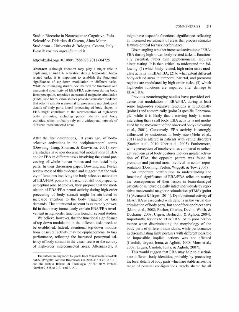
Studi e Ricerche in Neuroscienze Cognitive, PoloScientifico-Didattico di Cesena, Alma MaterStudiorum – Università di Bologna, Cesena, ItalyE-mail: [email protected]
http://dx.doi.org/10.1080/17588928.2011.604725
Abstract: Although attention may play a major role inexplaining EBA/FBA activation during high-order, body-related tasks, it is important to establish the functionalsignificance of top-down modulation in different tasks.While neuroimaging studies documented the functional andanatomical specificity of EBA/FBA activation during bodyform perception, repetitive transcranial magnetic stimulation(rTMS) and brain-lesion studies provided causative evidencethat activity in EBA is essential for processing morphologicaldetails of body parts. Local processing of body shapes inEBA might contribute to the representation of high-orderbody attributes, including person identity and bodyesthetics, which probably rely on a widespread network ofdifferent interconnected areas.
After the first descriptions, 10 years ago, of body-selective activations in the occipitotemporal cortex(Downing, Jiang, Shuman, & Kanwisher, 2001), sev-eral studies have now documented modulations of EBAand/or FBA in different tasks involving the visual pro-cessing of whole human bodies and non-facial bodyparts. In their discussion paper, Downing and Peelenreview most of this evidence and suggest that the vari-ety of functions involving the body-selective activationof EBA/FBA points to a basic, but still body-specific,perceptual role. Moreover, they propose that the mod-ulation of EBA/FBA neural activity during high-orderprocessing of body stimuli might be attributed toincreased attention to the body triggered by taskdemands. The attentional account is extremely power-ful in that it may immediately explain EBA/FBA invol-vement in high-order functions found in several studies.
We believe, however, that the functional significanceof top-down modulation in the different tasks needs tobe established. Indeed, attentional top-down modula-tions of neural activity may be epiphenomenal to taskperformance, reflecting the increased perceptual sal-iency of body stimuli in the visual scene or the activityof high-order interconnected areas. Alternatively, it
might have a specific functional significance, reflectingan increased recruitment of areas that process stimulusfeatures critical for task performance.
Disentangling whether increased activation of EBA/FBA during high-order, body-related tasks is function-ally essential, rather than epiphenomenal, requiresdirect testing. It is then critical to understand the fol-lowing: (1) which body-related, high-order tasks mod-ulate activity in EBA/FBA; (2) to what extent differentbody-related areas in temporal, parietal, and premotorregions are modulated by high-order tasks; (3) whichhigh-order functions are impaired after damage toEBA/FBA.
Previous neuroimaging studies have provided evi-dence that modulation of EBA/FBA during at leastsome high-order cognitive functions is functionally(point 1) and anatomically (point 2) specific. For exam-ple, while it is likely that a moving body is moreinteresting than a still body, EBA activity is not modu-lated by the movement of the observed body (Downinget al., 2001). Conversely, EBA activity is stronglyinfluenced by distortions in body size (Mohr et al.,2011) and is altered in patients with eating disorders(Suchan et al., 2010; Uher et al., 2005). Furthermore,while perception of incoherent, as compared to coher-ent, sequences of body postures induces greater activa-tion of EBA, the opposite pattern was found inpremotor and parietal areas involved in action repre-sentation (Downing, Peelen, Wiggett, & Tew, 2006).
An important contribution to understanding thefunctional significance of EBA/FBA relies on testingthe consequences of their lesion in brain-damagedpatients or in neurologically intact individuals by repe-titive transcranial magnetic stimulation (rTMS) (point3) (Avenanti &Urgesi, 2011). Dysfunctional activity ofEBA/FBA is associated with deficits in the visual dis-crimination of body parts, but not of face or object parts(Moro et al., 2008; Pitcher, Charles, Devlin, Walsh, &Duchaine, 2009; Urgesi, Berlucchi, & Aglioti, 2004).Importantly, lesions to EBA/FBA led to poor perfor-mance when discriminating the morphology of thebody parts of different individuals, while performancein discriminating limb postures with different possibleor impossible implied actions was not affected(Candidi, Urgesi, Ionta, & Aglioti, 2008; Moro et al.,2008; Urgesi, Candidi, Ionta, & Aglioti, 2007).
This would suggest that EBA may help to discrimi-nate different body identities, probably by processingthe local details of body parts which are stable across therange of postural configurations largely shared by all
The authors are supported by grants fromMinistero Italiano dellaSalute (Progetto Giovani Ricercatori GR-2008-1137139; to C.U.)and the Istituto Italiano di Tecnologia (SEED 2009 ProtocolNumber 21538 to C. U. and A. A.).
COMMENTARIES 213
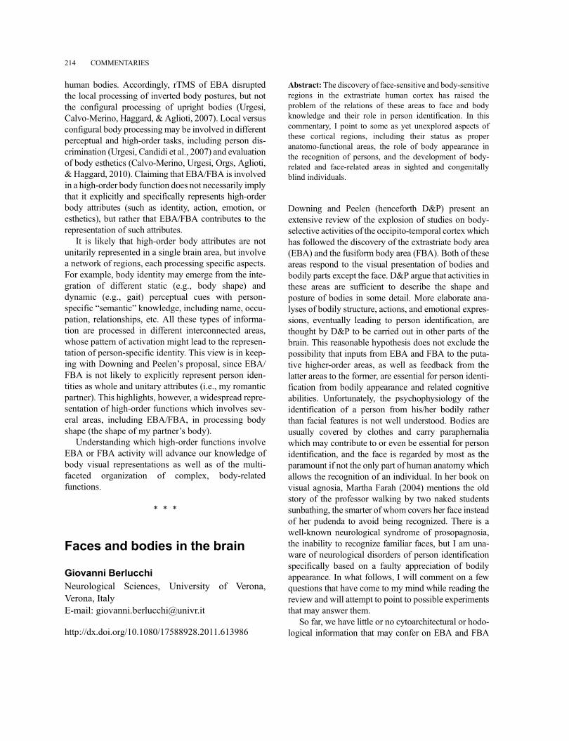
human bodies. Accordingly, rTMS of EBA disruptedthe local processing of inverted body postures, but notthe configural processing of upright bodies (Urgesi,Calvo-Merino, Haggard, & Aglioti, 2007). Local versusconfigural body processing may be involved in differentperceptual and high-order tasks, including person dis-crimination (Urgesi, Candidi et al., 2007) and evaluationof body esthetics (Calvo-Merino, Urgesi, Orgs, Aglioti,& Haggard, 2010). Claiming that EBA/FBA is involvedin a high-order body function does not necessarily implythat it explicitly and specifically represents high-orderbody attributes (such as identity, action, emotion, oresthetics), but rather that EBA/FBA contributes to therepresentation of such attributes.
It is likely that high-order body attributes are notunitarily represented in a single brain area, but involvea network of regions, each processing specific aspects.For example, body identity may emerge from the inte-gration of different static (e.g., body shape) anddynamic (e.g., gait) perceptual cues with person-specific “semantic” knowledge, including name, occu-pation, relationships, etc. All these types of informa-tion are processed in different interconnected areas,whose pattern of activation might lead to the represen-tation of person-specific identity. This view is in keep-ing with Downing and Peelen’s proposal, since EBA/FBA is not likely to explicitly represent person iden-tities as whole and unitary attributes (i.e., my romanticpartner). This highlights, however, a widespread repre-sentation of high-order functions which involves sev-eral areas, including EBA/FBA, in processing bodyshape (the shape of my partner’s body).
Understanding which high-order functions involveEBA or FBA activity will advance our knowledge ofbody visual representations as well as of the multi-faceted organization of complex, body-relatedfunctions.
* * *
Faces and bodies in the brain
Giovanni BerlucchiNeurological Sciences, University of Verona,Verona, ItalyE-mail: [email protected]
http://dx.doi.org/10.1080/17588928.2011.613986
Abstract: The discovery of face-sensitive and body-sensitiveregions in the extrastriate human cortex has raised theproblem of the relations of these areas to face and bodyknowledge and their role in person identification. In thiscommentary, I point to some as yet unexplored aspects ofthese cortical regions, including their status as properanatomo-functional areas, the role of body appearance inthe recognition of persons, and the development of body-related and face-related areas in sighted and congenitallyblind individuals.
Downing and Peelen (henceforth D&P) present anextensive review of the explosion of studies on body-selective activities of the occipito-temporal cortexwhichhas followed the discovery of the extrastriate body area(EBA) and the fusiform body area (FBA). Both of theseareas respond to the visual presentation of bodies andbodily parts except the face. D&P argue that activities inthese areas are sufficient to describe the shape andposture of bodies in some detail. More elaborate ana-lyses of bodily structure, actions, and emotional expres-sions, eventually leading to person identification, arethought by D&P to be carried out in other parts of thebrain. This reasonable hypothesis does not exclude thepossibility that inputs from EBA and FBA to the puta-tive higher-order areas, as well as feedback from thelatter areas to the former, are essential for person identi-fication from bodily appearance and related cognitiveabilities. Unfortunately, the psychophysiology of theidentification of a person from his/her bodily ratherthan facial features is not well understood. Bodies areusually covered by clothes and carry paraphernaliawhich may contribute to or even be essential for personidentification, and the face is regarded by most as theparamount if not the only part of human anatomy whichallows the recognition of an individual. In her book onvisual agnosia, Martha Farah (2004) mentions the oldstory of the professor walking by two naked studentssunbathing, the smarter of whom covers her face insteadof her pudenda to avoid being recognized. There is awell-known neurological syndrome of prosopagnosia,the inability to recognize familiar faces, but I am una-ware of neurological disorders of person identificationspecifically based on a faulty appreciation of bodilyappearance. In what follows, I will comment on a fewquestions that have come to my mind while reading thereview and will attempt to point to possible experimentsthat may answer them.
So far, we have little or no cytoarchitectural or hodo-logical information that may confer on EBA and FBA
214 COMMENTARIES
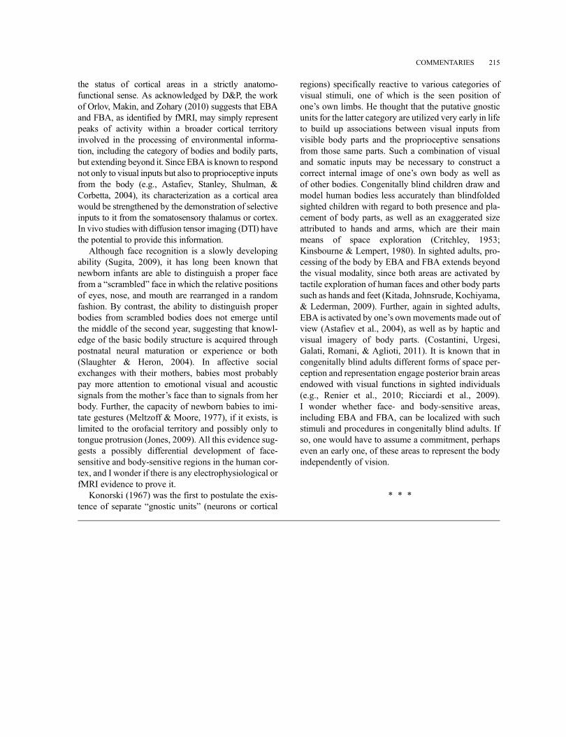
the status of cortical areas in a strictly anatomo-functional sense. As acknowledged by D&P, the workof Orlov, Makin, and Zohary (2010) suggests that EBAand FBA, as identified by fMRI, may simply representpeaks of activity within a broader cortical territoryinvolved in the processing of environmental informa-tion, including the category of bodies and bodily parts,but extending beyond it. Since EBA is known to respondnot only to visual inputs but also to proprioceptive inputsfrom the body (e.g., Astafiev, Stanley, Shulman, &Corbetta, 2004), its characterization as a cortical areawould be strengthened by the demonstration of selectiveinputs to it from the somatosensory thalamus or cortex.In vivo studies with diffusion tensor imaging (DTI) havethe potential to provide this information.
Although face recognition is a slowly developingability (Sugita, 2009), it has long been known thatnewborn infants are able to distinguish a proper facefrom a “scrambled” face in which the relative positionsof eyes, nose, and mouth are rearranged in a randomfashion. By contrast, the ability to distinguish properbodies from scrambled bodies does not emerge untilthe middle of the second year, suggesting that knowl-edge of the basic bodily structure is acquired throughpostnatal neural maturation or experience or both(Slaughter & Heron, 2004). In affective socialexchanges with their mothers, babies most probablypay more attention to emotional visual and acousticsignals from the mother’s face than to signals from herbody. Further, the capacity of newborn babies to imi-tate gestures (Meltzoff & Moore, 1977), if it exists, islimited to the orofacial territory and possibly only totongue protrusion (Jones, 2009). All this evidence sug-gests a possibly differential development of face-sensitive and body-sensitive regions in the human cor-tex, and I wonder if there is any electrophysiological orfMRI evidence to prove it.
Konorski (1967) was the first to postulate the exis-tence of separate “gnostic units” (neurons or cortical
regions) specifically reactive to various categories ofvisual stimuli, one of which is the seen position ofone’s own limbs. He thought that the putative gnosticunits for the latter category are utilized very early in lifeto build up associations between visual inputs fromvisible body parts and the proprioceptive sensationsfrom those same parts. Such a combination of visualand somatic inputs may be necessary to construct acorrect internal image of one’s own body as well asof other bodies. Congenitally blind children draw andmodel human bodies less accurately than blindfoldedsighted children with regard to both presence and pla-cement of body parts, as well as an exaggerated sizeattributed to hands and arms, which are their mainmeans of space exploration (Critchley, 1953;Kinsbourne & Lempert, 1980). In sighted adults, pro-cessing of the body by EBA and FBA extends beyondthe visual modality, since both areas are activated bytactile exploration of human faces and other body partssuch as hands and feet (Kitada, Johnsrude, Kochiyama,& Lederman, 2009). Further, again in sighted adults,EBA is activated by one’s own movements made out ofview (Astafiev et al., 2004), as well as by haptic andvisual imagery of body parts. (Costantini, Urgesi,Galati, Romani, & Aglioti, 2011). It is known that incongenitally blind adults different forms of space per-ception and representation engage posterior brain areasendowed with visual functions in sighted individuals(e.g., Renier et al., 2010; Ricciardi et al., 2009).I wonder whether face- and body-sensitive areas,including EBA and FBA, can be localized with suchstimuli and procedures in congenitally blind adults. Ifso, one would have to assume a commitment, perhapseven an early one, of these areas to represent the bodyindependently of vision.
* * *
COMMENTARIES 215
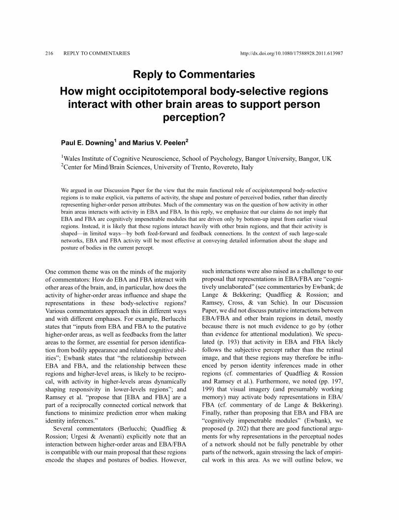
216 REPLY TO COMMENTARIES http://dx.doi.org/10.1080/17588928.2011.613987
Reply to Commentaries
How might occipitotemporal body-selective regionsinteract with other brain areas to support person
perception?
Paul E. Downing1 and Marius V. Peelen2
1Wales Institute of Cognitive Neuroscience, School of Psychology, Bangor University, Bangor, UK2Center for Mind/Brain Sciences, University of Trento, Rovereto, Italy
We argued in our Discussion Paper for the view that the main functional role of occipitotemporal body-selectiveregions is to make explicit, via patterns of activity, the shape and posture of perceived bodies, rather than directlyrepresenting higher-order person attributes. Much of the commentary was on the question of how activity in otherbrain areas interacts with activity in EBA and FBA. In this reply, we emphasize that our claims do not imply thatEBA and FBA are cognitively impenetrable modules that are driven only by bottom-up input from earlier visualregions. Instead, it is likely that these regions interact heavily with other brain regions, and that their activity isshaped––in limited ways––by both feed-forward and feedback connections. In the context of such large-scalenetworks, EBA and FBA activity will be most effective at conveying detailed information about the shape andposture of bodies in the current percept.
One common theme was on the minds of the majorityof commentators: How do EBA and FBA interact withother areas of the brain, and, in particular, how does theactivity of higher-order areas influence and shape therepresentations in these body-selective regions?Various commentators approach this in different waysand with different emphases. For example, Berlucchistates that “inputs from EBA and FBA to the putativehigher-order areas, as well as feedbacks from the latterareas to the former, are essential for person identifica-tion from bodily appearance and related cognitive abil-ities”; Ewbank states that “the relationship betweenEBA and FBA, and the relationship between theseregions and higher-level areas, is likely to be recipro-cal, with activity in higher-levels areas dynamicallyshaping responsivity in lower-levels regions”; andRamsey et al. “propose that [EBA and FBA] are apart of a reciprocally connected cortical network thatfunctions to minimize prediction error when makingidentity inferences.”
Several commentators (Berlucchi; Quadflieg &Rossion; Urgesi & Avenanti) explicitly note that aninteraction between higher-order areas and EBA/FBAis compatible with our main proposal that these regionsencode the shapes and postures of bodies. However,
such interactions were also raised as a challenge to ourproposal that representations in EBA/FBA are “cogni-tively unelaborated” (see commentaries by Ewbank; deLange & Bekkering; Quadflieg & Rossion; andRamsey, Cross, & van Schie). In our DiscussionPaper, we did not discuss putative interactions betweenEBA/FBA and other brain regions in detail, mostlybecause there is not much evidence to go by (otherthan evidence for attentional modulation). We specu-lated (p. 193) that activity in EBA and FBA likelyfollows the subjective percept rather than the retinalimage, and that these regions may therefore be influ-enced by person identity inferences made in otherregions (cf. commentaries of Quadflieg & Rossionand Ramsey et al.). Furthermore, we noted (pp. 197,199) that visual imagery (and presumably workingmemory) may activate body representations in EBA/FBA (cf. commentary of de Lange & Bekkering).Finally, rather than proposing that EBA and FBA are“cognitively impenetrable modules” (Ewbank), weproposed (p. 202) that there are good functional argu-ments for why representations in the perceptual nodesof a network should not be fully penetrable by otherparts of the network, again stressing the lack of empiri-cal work in this area. As we will outline below, we
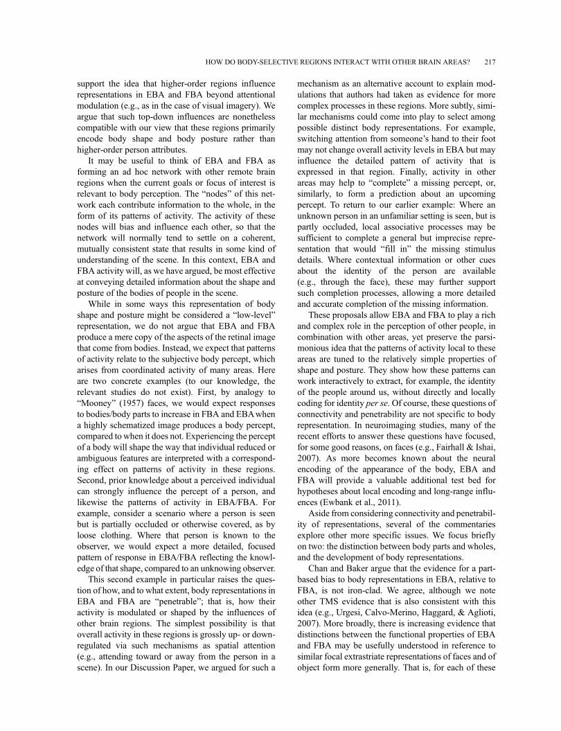
support the idea that higher-order regions influencerepresentations in EBA and FBA beyond attentionalmodulation (e.g., as in the case of visual imagery). Weargue that such top-down influences are nonethelesscompatible with our view that these regions primarilyencode body shape and body posture rather thanhigher-order person attributes.
It may be useful to think of EBA and FBA asforming an ad hoc network with other remote brainregions when the current goals or focus of interest isrelevant to body perception. The “nodes” of this net-work each contribute information to the whole, in theform of its patterns of activity. The activity of thesenodes will bias and influence each other, so that thenetwork will normally tend to settle on a coherent,mutually consistent state that results in some kind ofunderstanding of the scene. In this context, EBA andFBA activity will, as we have argued, be most effectiveat conveying detailed information about the shape andposture of the bodies of people in the scene.
While in some ways this representation of bodyshape and posture might be considered a “low-level”representation, we do not argue that EBA and FBAproduce a mere copy of the aspects of the retinal imagethat come from bodies. Instead, we expect that patternsof activity relate to the subjective body percept, whicharises from coordinated activity of many areas. Hereare two concrete examples (to our knowledge, therelevant studies do not exist). First, by analogy to“Mooney” (1957) faces, we would expect responsesto bodies/body parts to increase in FBA and EBAwhena highly schematized image produces a body percept,compared to when it does not. Experiencing the perceptof a body will shape the way that individual reduced orambiguous features are interpreted with a correspond-ing effect on patterns of activity in these regions.Second, prior knowledge about a perceived individualcan strongly influence the percept of a person, andlikewise the patterns of activity in EBA/FBA. Forexample, consider a scenario where a person is seenbut is partially occluded or otherwise covered, as byloose clothing. Where that person is known to theobserver, we would expect a more detailed, focusedpattern of response in EBA/FBA reflecting the knowl-edge of that shape, compared to an unknowing observer.
This second example in particular raises the ques-tion of how, and to what extent, body representations inEBA and FBA are “penetrable”; that is, how theiractivity is modulated or shaped by the influences ofother brain regions. The simplest possibility is thatoverall activity in these regions is grossly up- or down-regulated via such mechanisms as spatial attention(e.g., attending toward or away from the person in ascene). In our Discussion Paper, we argued for such a
mechanism as an alternative account to explain mod-ulations that authors had taken as evidence for morecomplex processes in these regions. More subtly, simi-lar mechanisms could come into play to select amongpossible distinct body representations. For example,switching attention from someone’s hand to their footmay not change overall activity levels in EBA but mayinfluence the detailed pattern of activity that isexpressed in that region. Finally, activity in otherareas may help to “complete” a missing percept, or,similarly, to form a prediction about an upcomingpercept. To return to our earlier example: Where anunknown person in an unfamiliar setting is seen, but ispartly occluded, local associative processes may besufficient to complete a general but imprecise repre-sentation that would “fill in” the missing stimulusdetails. Where contextual information or other cuesabout the identity of the person are available(e.g., through the face), these may further supportsuch completion processes, allowing a more detailedand accurate completion of the missing information.
These proposals allow EBA and FBA to play a richand complex role in the perception of other people, incombination with other areas, yet preserve the parsi-monious idea that the patterns of activity local to theseareas are tuned to the relatively simple properties ofshape and posture. They show how these patterns canwork interactively to extract, for example, the identityof the people around us, without directly and locallycoding for identity per se. Of course, these questions ofconnectivity and penetrability are not specific to bodyrepresentation. In neuroimaging studies, many of therecent efforts to answer these questions have focused,for some good reasons, on faces (e.g., Fairhall & Ishai,2007). As more becomes known about the neuralencoding of the appearance of the body, EBA andFBA will provide a valuable additional test bed forhypotheses about local encoding and long-range influ-ences (Ewbank et al., 2011).
Aside from considering connectivity and penetrabil-ity of representations, several of the commentariesexplore other more specific issues. We focus brieflyon two: the distinction between body parts and wholes,and the development of body representations.
Chan and Baker argue that the evidence for a part-based bias to body representations in EBA, relative toFBA, is not iron-clad. We agree, although we noteother TMS evidence that is also consistent with thisidea (e.g., Urgesi, Calvo-Merino, Haggard, & Aglioti,2007). More broadly, there is increasing evidence thatdistinctions between the functional properties of EBAand FBA may be usefully understood in reference tosimilar focal extrastriate representations of faces and ofobject form more generally. That is, for each of these
HOW DO BODY-SELECTIVE REGIONS INTERACT WITH OTHER BRAIN AREAS? 217
kinds, fMRI studies show focal, selective activationsthat are relatively posterior-lateral (EBA, face-selectiveOFA, and object-form selective LO) and distinct focithat are relatively ventro-medial (FBA, face-selectiveFFA, and object-form selective pFs). Across cate-gories, these representations appear to be distinguishedin several ways, including among them a bias for partover whole representations in the posterior-lateralregions, and the reverse bias in ventro-medial repre-sentations (Taylor & Downing, 2011).
Further, in an earlier paper, Taylor, Wiggett, andDowning (2007) noted that the hierarchical organiza-tion of the body into parts is systematic (“fingers,”“arms,” etc.) and asked whether EBA body representa-tions might respect these boundaries, carving at thejoints, as it were. Kemmerer valuably points out thatlinguistic partitions of the body are not culturally uni-versal, so that, at some level at least, what is a natural“part” of the body for one individual may not be so foranother. This provokes an interesting question aboutthe susceptibility of part representations in EBA tosemantic/linguistic influences. In the spirit of our pro-posed framework, we might expect no such influence.However, it is possible that these semantic /linguisticinfluences shape the way that individuals attend to thebodies of others in daily life. If these biases are regularand consistent, it is conceivable that they would in turnshape, over time, the “space” of neural patterns repre-senting the body in these regions.
Slaughter and Berlucchi both comment on the devel-opment of body structure knowledge in infants.Evidence for late development of body structure knowl-edge comes from studies measuring the time infantslook at intact versus scrambled bodies (e.g., a bodywith arms attached to the hips). Initial studies estimatedthat it is not before 15 months of age that infants’looking behavior discriminates between intact andscrambled bodies (Slaughter & Heron, 2004). In a laterstudy (Heron & Slaughter, 2010), this was shown todepend on the realism of the stimuli, with the earliestage of discriminating realistic intact from realisticscrambled bodies being around 9 months of age. By
contrast, sensitivity to body motion and goal-directedaction appears to develop earlier in life (Bertenthal,1993; Király, Jovanovic, Prinz, Aschersleben, &Gergely, 2003). According to Slaughter, this develop-mental dissociation is consistent with our proposed dis-sociation between EBA and FBA (involved in theencoding of body shape) and other regions involved inthe encoding of body motion and action goals. Shefurther suggests that the developmental dissociationimplies that certain aspects of higher-level person per-ception can occur without the information computed byEBA and FBA. While this is an interesting idea, whatneeds to be shown to complete the link that Slaughtermakes is that developing knowledge of body structurerelates systematically to increasing sophistication of thebody shape/posture representations in EBA/FBA. Without this, it is still unclear whether the rela-tively late development of body structure knowledge ininfants should be interpreted as providing evidence thathigh-level person perception, such as perceiving actiongoals, can bypass EBA and FBA. Finally, it is notunlikely that future studies, using more sensitive meth-ods, may reveal body structure knowledge in infants atincreasingly younger ages.
To sum up, the simple model of body encoding thatwe proposed in our Discussion Paper explains a greatdeal of the current literature and does so parsimoniously.When considering how EBA and FBA work interac-tively with other brain areas, as they must, we can seehow their shape and posture representations could con-tribute critically to many high-level functions of personperception, such as the identification of others. Muchremains to be learned about how these two areas makedistinct contributions to person perception. More gener-ally, it is clear that in the next few years there will be a lotof energy directed at the broad question of how thenodes of the brain’s interconnected networks influenceeach other. The highly focal, well-characterized natureof extrastriate body representations, and the many cog-nitive and perceptual functions for which the perceptionof other people is relevant, make these areas worthy of afocus in these ongoing studies.
218 DOWNING AND PEELEN
References from the Discussion Paper,the Commentaries, and the Reply
Aguirre, G. K., Zarahn, E., & D’Esposito, M. (1998). An areawithin human ventral cortex sensitive to “building”stimuli: Evidence and implications.Neuron,21(2), 373–383.
Aleong, R., & Paus, T. (2010). Neural correlates of humanbody perception. Journal of Cognitive Neuroscience, 22(3),482–495.
Allison, T., Puce, A., & McCarthy, G. (2000). Social percep-tion from visual cues: Role of the STS region. Trends inCognitive Sciences, 4(7), 267–278.
Amaral, D. G., Behniea, H., & Kelly, J. L. (2003).Topographic organization of projections from the amyg-dala to the visual cortex in the macaque monkey.Neuroscience, 118(4), 1099–1120.
Amedi, A., von Kriegstein, K., Van Atteveldt, N. M.,Beauchamp, M. S., & Naumer, M. J. (2005). Functionalimaging of human crossmodal identification and objectrecognition. Experimental Brain Research, 166(3–4),559–571.
Anderson, A. K. (2005). Affective influences on the atten-tional dynamics supporting awareness. Journal ofExperimental Psychology: General, 134(2), 258–281.
Appelbaum, L. G., Wade, A. R., Vildavski, V. Y., Pettet,M. W., & Norcia, A. M. (2006). Cue-invariant networksfor figure and background processing in human visualcortex. Journal of Neuroscience, 26, 11695–11708.
Arzy, S., Thut, G., Mohr, C., Michel, C. M., & Blanke, O.(2006). Neural basis of embodiment: Distinct contribu-tions of temporoparietal junction and extrastriate bodyarea. Journal of Neuroscience, 26(31), 8074–8081.
Aslin, R. (2009). How infants view natural scenes gatheredfrom a head-mounted camera. Optometry and VisionScience, 86, 561–565.
Astafiev, S. V., Stanley, C. M., Shulman, G. L., & Corbetta,M. (2004). Extrastriate body area in human occipitalcortex responds to the performance of motor actions.Nature Neuroscience, 7(5), 542–548.
Astafiev, S. V., Stanley, C. M., Shulman, G. L., & Corbetta,M. (2005). Is the extrastriate body area involved in motoractions? Reply. Nature Neuroscience, 8(2), 125–126.
Atkinson, A. P., Dittrich, W. H., Gemmell, A. J., & Young,A. W. (2004). Emotion perception from dynamic andstatic body expressions in point-light and full-lightdisplays. Perception, 33(6), 717–746.
Avenanti, A., & Urgesi, C. (2011). Understanding ‘what’others do: Mirror mechanisms play a crucial role in actionperception. Social Cognitive and Affective Neuroscience,6, 257–259.
Avikainen, S., Liuhanen, S., Schurmann, M., & Hari, R.(2003). Enhanced extrastriate activation during observa-tion of distorted finger postures. Journal of CognitiveNeuroscience, 15(5), 658–663.
Bach, P., Peelen, M. V., & Tipper, S. P. (2010). On the role ofobject information in action observation: An fMRI study.Cerebral Cortex, 20(12), 2798–2809.
Bar, M. (2009). The proactive brain: Memory for predictions.Philosophical Transactions of the Royal Society ofLondon, Series B: Biological Sciences, 364, 1235–1243.
Barsalou, L.W. (2008). Grounded cognition. Annual Reviewof Psychology, 59, 617–645.
Beauchamp, M. S., Lee, K. E., Haxby, J. V., & Martin, A.(2002). Parallel visual motion processing streams formanipulable objects and human movements. Neuron,34(1), 149–159.
Bedny,M., Caramazza, A., Grossman, E., Pascual-Leone, A.,& Saxe, R. (2008). Concepts are more than percepts: Thecase of action verbs. Journal of Neuroscience, 28(44),11347–11353.
Bekkering, H., & Neggers, S. F. (2002). Visual search ismodulated by action intentions. Psychological Science,13(4), 370–374.
Bell, A. J., & Sejnowski, T. J. (1997). The “independentcomponents” of natural scenes are edge filters. VisionResearch, 377, 3327–3338.
Benuzzi, F., Lui, F., Duzzi, D., Nichelli, P. F., & Porro, C. A.(2008). Does it look painful or disgusting? Ask yourparietal and cingulate cortex. Journal of Neuroscience,28(4), 923–931.
Berlucchi, G., & Aglioti, S. M. (2010). The body in the brainrevisited. Experimental Brain Research, 200(1), 25–35.
Bertenthal, B. (1993). Infants’ perception of biomechanicalmotions: Intrinsic image and knowledge-based con-straints. In C. E. Granrud (Ed.), Visual perception andcognition in infancy (pp. 175–214). Hillsdale, NJ:Lawrence Erlbaum Associates.
Blake, R., & Shiffrar, M. (2007). Perception of humanmotion. Annual Review of Psychology, 58, 47–73.
Blanke, O., Ionta, S., Fornari, E., Mohr, C., & Maeder, P.(2010). Mental imagery for full and upper humanbodies: Common right hemisphere activations and dis-tinct extrastriate activations. Brain Topography, 23(3),321–332.
Bracci, S., Ietswaart, M., Peelen, M. V., & Cavina-Pratesi, C.(2010). Dissociable neural responses to hands and non-hand body parts in human left extrastriate visual cortex.Journal of Neurophysiology, 103(6), 3389–3397.
Brandman, T., & Yovel, G. (2010). The body inversion effectis mediated by face-selective, not body-selective, mechan-isms. Journal of Neuroscience, 30(31), 10534–10540.
Brefczynski, J. A., & DeYoe, E. A. (1999). A physiologicalcorrelate of the ‘spotlight’ of visual attention. NatureNeuroscience, 2(4), 370–374.
Bruce, V., & Young, A. (1986). Understanding face recogni-tion. British Journal of Psychology, 77(Pt 3), 305–327.
Bruner, J. (1973). Going beyond the information given. NewYork, NY: Norton.
Calvo-Merino, B., Urgesi, C., Orgs, G., Aglioti, S. M., &Haggard, P. (2010). Extrastriate body area underlies aes-thetic evaluation of body stimuli. Experimental BrainResearch, 204(3), 447–456.
Candidi, M., Urgesi, C., Ionta, S., & Aglioti, S. M.(2008). Virtual lesion of ventral premotor corteximpairs visual perception of biomechanically possiblebut not impossible actions. Social Neuroscience,3(3–4), 388–400.
Carter, E. J., & Pelphrey, K. A. (2008). Friend or foe? Brainsystems involved in the perception of dynamic signals ofmenacing and friendly social approaches. SocialNeuroscience, 3(2), 151–163.
REFERENCES 219
Chaminade, T., Meltzoff, A. N., & Decety, J. (2005). AnfMRI study of imitation: Action representation and bodyschema. Neuropsychologia, 43(1), 115–127.
Chan, A. W., Kravitz, D. J., Truong, S., Arizpe, J., & Baker,C. I. (2010). Cortical representations of bodies and facesare strongest in commonly experienced configurations.Nature Neuroscience, 13(4), 417–418.
Chan, A. W., Peelen, M. V., & Downing, P. E. (2004). Theeffect of viewpoint on body representation in the extra-striate body area. Neuroreport, 15(15), 2407–2410.
Chao, L. L., Haxby, J. V., &Martin, A. (1999). Attribute-basedneural substrates in temporal cortex for perceiving andknowing about objects. Nature Neuroscience, 2, 913–919.
Cikara, M., Eberhardt, J. L., & Fiske, S. T. (2011). Fromagents to objects: Sexist attitudes and neural responses tosexualized targets. Journal of Cognitive Neuroscience,23(3), 540–551.
Costantini, M., Galati, G., Ferretti, A., Caulo, M., Tartaro, A.,Romani, G. L., et al. (2005). Neural systems underlyingobservation of humanly impossible movements: An fMRIstudy. Cerebral Cortex, 15(11), 1761–1767.
Costantini, M., Urgesi, C., Galati, G., Romani, G. L., &Aglioti, S. M. (2011). Haptic perception and body repre-sentation in lateral and medial occipito-temporal cortices.Neuropsychologia, 49(5), 821–829.
Critchley, M. (1953). Tactile thought, with special referenceto the blind. Brain, 76, 19–35.
Cross, E. S., Mackie, E. C., Wolford, G., & Hamilton, A. F.(2010). Contorted and ordinary body postures in thehuman brain. Experimental Brain Research, 204(3),397–407.
Danckert, J. A., & Goodale, M. A. (2003). Ups and downs inthe visual control of action. In S. H. Johnson-Frey (Ed.),Taking action: Cognitive neuroscience perspectives onintentional acts (pp. 29–64). Cambridge, MA: MIT Press.
David, N., Cohen, M. X., Newen, A., Bewernick, B. H., Shah,N. J., Fink, G. R., et al. (2007). The extrastriate cortexdistinguishes between the consequences of one’s own andothers’ behavior. NeuroImage, 36(3), 1004–1014.
David, N., Jansen, M., Cohen, M. X., Osswald, K., Molnar-Szakacs, I., Newen, A., et al. (2009). Disturbances of self–other distinction after stimulation of the extrastriate bodyarea in the human brain. Social Neuroscience, 4(1), 40–48.
David, S. V., Hayden, B. Y., Mazer, J. A., & Gallant, J. L.(2008). Attention to stimulus features shifts spectraltuning of V4 neurons during natural vision. Neuron, 59,509–521.
De Lange, F. P., Spronk, M., Willems, R. M., Toni, I., &Bekkering, H. (2008). Complementary systems for under-standing action intentions. Current Biology, 18(6),454–457.
Desimone, R., Albright, T. D., Gross, C. G., & Bruce, C.(1984). Stimulus-selective properties of inferior temporalneurons in the macaque. Journal of Neuroscience, 4(8),2051–2062.
Devue, C., Collette, F., Balteau, E., Dequeldre, C., Luxen, A.,Maquet, P., et al. (2007). Here I am: The cortical correlatesof visual self-recognition. Brain Research, 1143, 169–182.
Dijkerman, H. C., McIntosh, R. D., Schindler, I., Nijboer,T. C. W., & Milner, A. D. (2009). Choosing between
alternative wrist postures: Action planning needs percep-tion. Neuropsychologia, 477, 1476–1482.
Dow, B.M., Snyder, A. Z., Vautin, R. G., & Bauer, R. (1981).Magnification factor and receptive field size in fovealstriate cortex of the monkey. Experimental BrainResearch, 447, 213–228.
Downing, P., Liu, J., & Kanwisher, N. (2001). Testing cog-nitive models of visual attention with fMRI and MEG.Neuropsychologia, 39(12), 1329–1342.
Downing, P. E., Chan, A. W., Peelen, M. V., Dodds, C. M., &Kanwisher, N. (2006). Domain specificity in visual cor-tex. Cerebral Cortex, 16(10), 1453–1461.
Downing, P. E., Jiang, Y., Shuman, M., & Kanwisher, N. G.(2001). A cortical area selective for visual processing ofthe human body. Science, 293(5539), 2470–2473.
Downing, P. E., Peelen, M. V., Wiggett, A. J., & Tew, B. D.(2006). The role of the extrastriate body area in actionperception. Social Neuroscience, 1(1), 52–62.
Downing, P. E., Wiggett, A. J., & Peelen, M. V. (2007).Functional magnetic resonance imaging investigation ofoverlapping lateral occipitotemporal activations usingmulti-voxel pattern analysis. Journal of Neuroscience,27(1), 226–233.
Drucker, D. M., & Aguirre, G. K. (2009). Different spatialscales of shape similarity representation in lateral andventral LOC. Cerebral Cortex, 19(10), 2269–2280.
Epstein, R., & Kanwisher, N. (1998). A cortical representa-tion of the local visual environment. Nature, 392(6676),598–601.
Ewbank, M. P., Lawson, R. P., Henson, R. N., Rowe, J. B.,Passamonti, L., & Calder, A. J. (2011). Changes in “top-down” connectivity underlie repetition suppression in theventral visual pathway. Journal of Neuroscience, 31(15),5635–5642.
Fairhall, S. L., & Ishai, A. (2007). Effective connectivitywithin the distributed cortical network for face perception.Cerebral Cortex, 17(10), 2400–2406.
Farah, M. J. (2004). Visual agnosia. Cambridge, MA: MITPress.
Flaisch, T., Schupp, H. T., Renner, B., & Junghofer, M.(2009). Neural systems of visual attention responding toemotional gestures. NeuroImage, 45(4), 1339–1346.
Friston, K. J. (2005). A theory of cortical responses.Philosophical Transactions of the Royal Society ofLondon, Series B: Biological Sciences, 360(1456),815–836.
Gandhi, S. P., Heeger, D. J., & Boynton, G. M. (1999). Spatialattention affects brain activity in human primary visualcortex. Proceedings of the National Academy of Sciencesof the United States of America, 96(6), 3314–3319.
Gauthier, I., Tarr, M. J., Moylan, J., Skudlarski, P., Gore, J. C.,& Anderson, A.W. (2000). The fusiform “face area” is partof a network that processes faces at the individual level.Journal of Cognitive Neuroscience, 12(3), 495–504.
Georgieff, N., & Jeannerod, M. (1998). Beyond conscious-ness of external reality: A “who” system for conscious-ness of action and self-consciousness. Consciousness andCognition, 7(3), 465–477.
Ghuman, A. S., McDaniel, J. R., & Martin, A. (2010). Faceadaptation without a face. Current Biology, 20(1), 32–36.
220 REFERENCES
Giese, M. A., & Poggio, T. (2003). Neural mechanisms forthe recognition of biological movements. Nature ReviewsNeuroscience, 4(3), 179–192.
Grafton, S. T., & Hamilton, A. F. (2007). Evidence for adistributed hierarchy of action representation in thebrain. Human Movement Science, 26(4), 590–616.
Graziano, M. S., & Aflalo, T. N. (2007). Rethinking corticalorganization: Moving away from discrete areas arrangedin hierarchies. Neuroscientist, 13(2), 138–147.
Grezes, J., Pichon, S., & de Gelder, B. (2007). Perceiving fearin dynamic body expressions. NeuroImage, 35(2),959–967.
Grill-Spector, K., Henson, R., &Martin, A. (2006). Repetitionand the brain: Neural models of stimulus-specific effects.Trends in Cognitive Science, 10(1), 14–23.
Grill-Spector, K., Kushnir, T., Edelman, S., Avidan, G.,Itzchak, Y., & Malach, R. (1999). Differential processingof objects under various viewing conditions in the humanlateral occipital complex. Neuron, 24(1), 187–203.
Grosbras, M. H., & Paus, T. (2006). Brain networks involvedin viewing angry hands or faces. Cerebral Cortex, 16(8),1087–1096.
Gross, C. G. (1992). Representation of visual stimuli ininferior temporal cortex. Philosophical Transactions ofthe Royal Society of London. Series B, BiologicalSciences, 335(1273), 3–10.
Gross, C. G., Bender, D. B., & Rocha-Miranda, C. E. (1969).Visual receptive fields of neurons in inferotemporal cortexof the monkey. Science, 166(910), 1303–1306.
Gross, C. G., Rocha-Miranda, C. E., & Bender, D. B. (1972).Visual properties of neurons in inferotemporal cortex ofthe macaque. Journal of Neurophysiology, 35(1), 96–111.
Grossman, E. D., & Blake, R. (2002). Brain areas activeduring visual perception of biological motion. Neuron,35(6), 1167–1175.
Gu, X., Liu, X., Guise, K. G., Naidich, T. P., Hof, P. R., &Fan, J. (2010). Functional dissociation of the frontoinsularand anterior cingulate cortices in empathy for pain.Journal of Neuroscience, 30(10), 3739–3744.
Hadjikhani, N., & de Gelder, B. (2003). Seeing fearful bodyexpressions activates the fusiform cortex and amygdala.Current Biology, 13(24), 2201–2205.
Hamann, S., Herman, R. A., Nolan, C. L., & Wallen, K.(2004). Men and women differ in amygdala response tovisual sexual stimuli. Nature Neuroscience, 7(4), 411–416.
Harrison, S. A., & Tong, F. (2009). Decoding reveals thecontents of visual working memory in early visual areas.Nature, 458(7238), 632–635.
Haxby, J. V., Gobbini, M. I., Furey, M. L., Ishai, A.,Schouten, J. L., & Pietrini, P. (2001). Distributed andoverlapping representations of faces and objects in ventraltemporal cortex. Science, 293(5539), 2425–2430.
Haxby, J. V., Hoffman, E. A., & Gobbini, M. A. (2000). Thedistributed human neural system for face perception.Trends in Cognitive Sciences, 4, 223–233.
Haynes, J. D., & Rees, G. (2006). Decoding mental statesfrom brain activity in humans. Nature ReviewsNeuroscience, 7(7), 523–534.
Henson, R. N. (2003). Neuroimaging studies of priming.Progress in Neurobiology, 70(1), 53–81.
Heron, M., & Slaughter, V. (2010). Infants’ responses to realhumans and representations of humans. InternationalJournal of Behavioral Development, 34(1), 34–45.
Hinton, G. E., McClelland, J. L., & Rumelhart, D. E. (1986).Distributed representations. In D. E. Rumelhart & J. L.McClelland (Eds.), Parallel distributed processing:Explorations in the microstructure of cognition(pp. 77–109). Cambridge, MA: MIT Press.
Hirai, M., & Hiraki, K. (2005). An event-related potentialsstudy of biological motion perception in human infants.Brain Research. Cognitive Brain Research, 22, 301–304.
Hodzic, A., Kaas, A., Muckli, L., Stirn, A., & Singer, W.(2009). Distinct cortical networks for the detection andidentification of human body. NeuroImage, 45(4),1264–1271.
Hodzic, A., Muckli, L., Singer, W., & Stirn, A. (2009).Cortical responses to self and others. Human BrainMapping, 30(3), 951–962.
Hubel, D. H., & Wiesel, T. N. (1968). Receptive fields andfunctional architecture of monkey striate cortex. Journalof Physiology, 1957, 215–243.
Ishizu, T., Amemiya, K., Yumoto, M., & Kojima, S. (2010).Magnetoencephalographic study of the neural responsesin body perception. Neuroscience Letters, 481(1), 36–40.
Ishizu, T., Noguchi, A., Ito, Y., Ayabe, T., & Kojima, S.(2009). Motor activity and imagery modulate the body-selective region in the occipital-temporal area: A near-infrared spectroscopy study. Neuroscience Letters,465(1), 85–89.
Jackson, P. L., Meltzoff, A. N., & Decety, J. (2006). Neuralcircuits involved in imitation and perspective-taking.NeuroImage, 31(1), 429–439.
Jastorff, J., & Orban, G. A. (2009). Human functional mag-netic resonance imaging reveals separation and integra-tion of shape and motion cues in biological motionprocessing. Journal of Neuroscience, 29(22), 7315–7329.
Johansson, G. (1973). Visual perception of biological motionand a model for its analysis. Perception & Psychophysics,14, 201–211.
Johnson-Frey, S. H., Maloof, F. R., Newman-Norlund, R.,Farrer, C., Inati, S., & Grafton, S. T. (2003). Actions orhand–object interactions? Human inferior frontal cortexand action observation. Neuron, 39(6), 1053–1058.
Jones, S. S. (2009). The development of imitation in infancy.Philosophical Transactions of the Royal Society ofLondon, Series B: Biological Sciences, 364, 2325–2335.
Kable, J. W., & Chatterjee, A. (2006). Specificity of actionrepresentations in the lateral occipitotemporal cortex.Journal of Cognitive Neuroscience, 18(9), 1498–1517.
Kamitani, Y., & Tong, F. (2005). Decoding the visual andsubjective contents of the human brain. NatureNeuroscience, 8(5), 679–685.
Kanwisher, N., McDermott, J., & Chun, M. M. (1997). Thefusiform face area: A module in human extrastriate cortexspecialized for face perception. Journal of Neuroscience,17(11), 4302–4311.
Kastner, S., De Weerd, P., Desimone, R., & Ungerleider,L. G. (1998). Mechanisms of directed attention in thehuman extrastriate cortex as revealed by functional MRI.Science, 282(5386), 108–111.
REFERENCES 221
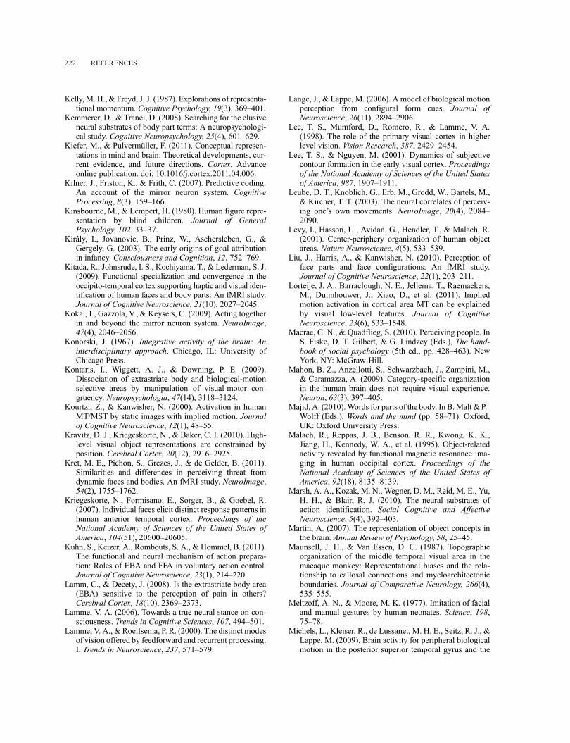
Kelly, M. H., & Freyd, J. J. (1987). Explorations of representa-tional momentum. Cognitive Psychology, 19(3), 369–401.
Kemmerer, D., & Tranel, D. (2008). Searching for the elusiveneural substrates of body part terms: A neuropsychologi-cal study. Cognitive Neuropsychology, 25(4), 601–629.
Kiefer, M., & Pulvermüller, F. (2011). Conceptual represen-tations in mind and brain: Theoretical developments, cur-rent evidence, and future directions. Cortex. Advanceonline publication. doi: 10.1016/j.cortex.2011.04.006.
Kilner, J., Friston, K., & Frith, C. (2007). Predictive coding:An account of the mirror neuron system. CognitiveProcessing, 8(3), 159–166.
Kinsbourne, M., & Lempert, H. (1980). Human figure repre-sentation by blind children. Journal of GeneralPsychology, 102, 33–37.
Király, I., Jovanovic, B., Prinz, W., Aschersleben, G., &Gergely, G. (2003). The early origins of goal attributionin infancy. Consciousness and Cognition, 12, 752–769.
Kitada, R., Johnsrude, I. S., Kochiyama, T., & Lederman, S. J.(2009). Functional specialization and convergence in theoccipito-temporal cortex supporting haptic and visual iden-tification of human faces and body parts: An fMRI study.Journal of Cognitive Neuroscience, 21(10), 2027–2045.
Kokal, I., Gazzola, V., & Keysers, C. (2009). Acting togetherin and beyond the mirror neuron system. NeuroImage,47(4), 2046–2056.
Konorski, J. (1967). Integrative activity of the brain: Aninterdisciplinary approach. Chicago, IL: University ofChicago Press.
Kontaris, I., Wiggett, A. J., & Downing, P. E. (2009).Dissociation of extrastriate body and biological-motionselective areas by manipulation of visual-motor con-gruency. Neuropsychologia, 47(14), 3118–3124.
Kourtzi, Z., & Kanwisher, N. (2000). Activation in humanMT/MST by static images with implied motion. Journalof Cognitive Neuroscience, 12(1), 48–55.
Kravitz, D. J., Kriegeskorte, N., & Baker, C. I. (2010). High-level visual object representations are constrained byposition. Cerebral Cortex, 20(12), 2916–2925.
Kret, M. E., Pichon, S., Grezes, J., & de Gelder, B. (2011).Similarities and differences in perceiving threat fromdynamic faces and bodies. An fMRI study. NeuroImage,54(2), 1755–1762.
Kriegeskorte, N., Formisano, E., Sorger, B., & Goebel, R.(2007). Individual faces elicit distinct response patterns inhuman anterior temporal cortex. Proceedings of theNational Academy of Sciences of the United States ofAmerica, 104(51), 20600–20605.
Kuhn, S., Keizer, A., Rombouts, S. A., & Hommel, B. (2011).The functional and neural mechanism of action prepara-tion: Roles of EBA and FFA in voluntary action control.Journal of Cognitive Neuroscience, 23(1), 214–220.
Lamm, C., & Decety, J. (2008). Is the extrastriate body area(EBA) sensitive to the perception of pain in others?Cerebral Cortex, 18(10), 2369–2373.
Lamme, V. A. (2006). Towards a true neural stance on con-sciousness. Trends in Cognitive Sciences, 107, 494–501.
Lamme, V. A., & Roelfsema, P. R. (2000). The distinct modesof vision offered by feedforward and recurrent processing.I. Trends in Neuroscience, 237, 571–579.
Lange, J., & Lappe, M. (2006). A model of biological motionperception from configural form cues. Journal ofNeuroscience, 26(11), 2894–2906.
Lee, T. S., Mumford, D., Romero, R., & Lamme, V. A.(1998). The role of the primary visual cortex in higherlevel vision. Vision Research, 387, 2429–2454.
Lee, T. S., & Nguyen, M. (2001). Dynamics of subjectivecontour formation in the early visual cortex. Proceedingsof the National Academy of Sciences of the United Statesof America, 987, 1907–1911.
Leube, D. T., Knoblich, G., Erb, M., Grodd, W., Bartels, M.,& Kircher, T. T. (2003). The neural correlates of perceiv-ing one’s own movements. NeuroImage, 20(4), 2084–2090.
Levy, I., Hasson, U., Avidan, G., Hendler, T., & Malach, R.(2001). Center-periphery organization of human objectareas. Nature Neuroscience, 4(5), 533–539.
Liu, J., Harris, A., & Kanwisher, N. (2010). Perception offace parts and face configurations: An fMRI study.Journal of Cognitive Neuroscience, 22(1), 203–211.
Lorteije, J. A., Barraclough, N. E., Jellema, T., Raemaekers,M., Duijnhouwer, J., Xiao, D., et al. (2011). Impliedmotion activation in cortical area MT can be explainedby visual low-level features. Journal of CognitiveNeuroscience, 23(6), 533–1548.
Macrae, C. N., & Quadflieg, S. (2010). Perceiving people. InS. Fiske, D. T. Gilbert, & G. Lindzey (Eds.), The hand-book of social psychology (5th ed., pp. 428–463). NewYork, NY: McGraw-Hill.
Mahon, B. Z., Anzellotti, S., Schwarzbach, J., Zampini, M.,& Caramazza, A. (2009). Category-specific organizationin the human brain does not require visual experience.Neuron, 63(3), 397–405.
Majid, A. (2010).Words for parts of the body. In B.Malt & P.Wolff (Eds.), Words and the mind (pp. 58–71). Oxford,UK: Oxford University Press.
Malach, R., Reppas, J. B., Benson, R. R., Kwong, K. K.,Jiang, H., Kennedy, W. A., et al. (1995). Object-relatedactivity revealed by functional magnetic resonance ima-ging in human occipital cortex. Proceedings of theNational Academy of Sciences of the United States ofAmerica, 92(18), 8135–8139.
Marsh, A. A., Kozak, M. N., Wegner, D. M., Reid, M. E., Yu,H. H., & Blair, R. J. (2010). The neural substrates ofaction identification. Social Cognitive and AffectiveNeuroscience, 5(4), 392–403.
Martin, A. (2007). The representation of object concepts inthe brain. Annual Review of Psychology, 58, 25–45.
Maunsell, J. H., & Van Essen, D. C. (1987). Topographicorganization of the middle temporal visual area in themacaque monkey: Representational biases and the rela-tionship to callosal connections and myeloarchitectonicboundaries. Journal of Comparative Neurology, 266(4),535–555.
Meltzoff, A. N., & Moore, M. K. (1977). Imitation of facialand manual gestures by human neonates. Science, 198,75–78.
Michels, L., Kleiser, R., de Lussanet, M. H. E., Seitz, R. J., &Lappe, M. (2009). Brain activity for peripheral biologicalmotion in the posterior superior temporal gyrus and the
222 REFERENCES
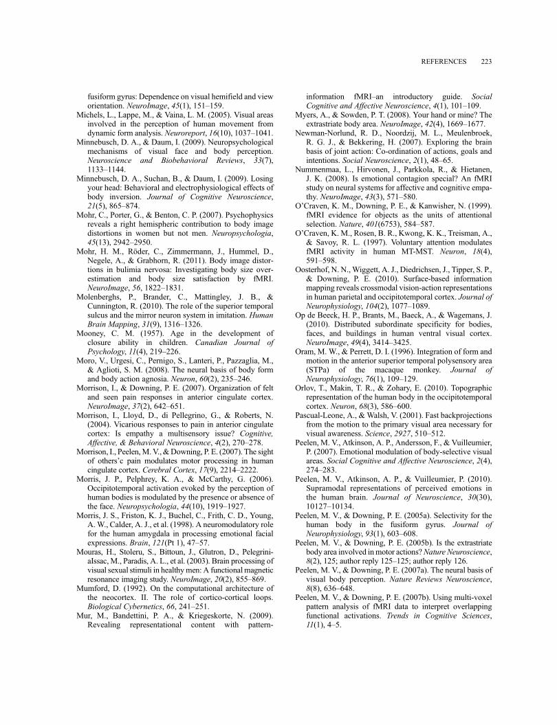
fusiform gyrus: Dependence on visual hemifield and vieworientation. NeuroImage, 45(1), 151–159.
Michels, L., Lappe, M., & Vaina, L. M. (2005). Visual areasinvolved in the perception of human movement fromdynamic form analysis. Neuroreport, 16(10), 1037–1041.
Minnebusch, D. A., & Daum, I. (2009). Neuropsychologicalmechanisms of visual face and body perception.Neuroscience and Biobehavioral Reviews, 33(7),1133–1144.
Minnebusch, D. A., Suchan, B., & Daum, I. (2009). Losingyour head: Behavioral and electrophysiological effects ofbody inversion. Journal of Cognitive Neuroscience,21(5), 865–874.
Mohr, C., Porter, G., & Benton, C. P. (2007). Psychophysicsreveals a right hemispheric contribution to body imagedistortions in women but not men. Neuropsychologia,45(13), 2942–2950.
Mohr, H. M., Röder, C., Zimmermann, J., Hummel, D.,Negele, A., & Grabhorn, R. (2011). Body image distor-tions in bulimia nervosa: Investigating body size over-estimation and body size satisfaction by fMRI.NeuroImage, 56, 1822–1831.
Molenberghs, P., Brander, C., Mattingley, J. B., &Cunnington, R. (2010). The role of the superior temporalsulcus and the mirror neuron system in imitation. HumanBrain Mapping, 31(9), 1316–1326.
Mooney, C. M. (1957). Age in the development ofclosure ability in children. Canadian Journal ofPsychology, 11(4), 219–226.
Moro, V., Urgesi, C., Pernigo, S., Lanteri, P., Pazzaglia, M.,& Aglioti, S. M. (2008). The neural basis of body formand body action agnosia. Neuron, 60(2), 235–246.
Morrison, I., & Downing, P. E. (2007). Organization of feltand seen pain responses in anterior cingulate cortex.NeuroImage, 37(2), 642–651.
Morrison, I., Lloyd, D., di Pellegrino, G., & Roberts, N.(2004). Vicarious responses to pain in anterior cingulatecortex: Is empathy a multisensory issue? Cognitive,Affective, & Behavioral Neuroscience, 4(2), 270–278.
Morrison, I., Peelen,M.V.,&Downing, P. E. (2007). The sightof others’c pain modulates motor processing in humancingulate cortex. Cerebral Cortex, 17(9), 2214–2222.
Morris, J. P., Pelphrey, K. A., & McCarthy, G. (2006).Occipitotemporal activation evoked by the perception ofhuman bodies is modulated by the presence or absence ofthe face. Neuropsychologia, 44(10), 1919–1927.
Morris, J. S., Friston, K. J., Buchel, C., Frith, C. D., Young,A. W., Calder, A. J., et al. (1998). A neuromodulatory rolefor the human amygdala in processing emotional facialexpressions. Brain, 121(Pt 1), 47–57.
Mouras, H., Stoleru, S., Bittoun, J., Glutron, D., Pelegrini-aIssac, M., Paradis, A. L., et al. (2003). Brain processing ofvisual sexual stimuli in healthymen: A functional magneticresonance imaging study. NeuroImage, 20(2), 855–869.
Mumford, D. (1992). On the computational architecture ofthe neocortex. II. The role of cortico-cortical loops.Biological Cybernetics, 66, 241–251.
Mur, M., Bandettini, P. A., & Kriegeskorte, N. (2009).Revealing representational content with pattern-
information fMRI–an introductory guide. SocialCognitive and Affective Neuroscience, 4(1), 101–109.
Myers, A., & Sowden, P. T. (2008). Your hand or mine? Theextrastriate body area. NeuroImage, 42(4), 1669–1677.
Newman-Norlund, R. D., Noordzij, M. L., Meulenbroek,R. G. J., & Bekkering, H. (2007). Exploring the brainbasis of joint action: Co-ordination of actions, goals andintentions. Social Neuroscience, 2(1), 48–65.
Nummenmaa, L., Hirvonen, J., Parkkola, R., & Hietanen,J. K. (2008). Is emotional contagion special? An fMRIstudy on neural systems for affective and cognitive empa-thy. NeuroImage, 43(3), 571–580.
O’Craven, K. M., Downing, P. E., & Kanwisher, N. (1999).fMRI evidence for objects as the units of attentionalselection. Nature, 401(6753), 584–587.
O’Craven, K. M., Rosen, B. R., Kwong, K. K., Treisman, A.,& Savoy, R. L. (1997). Voluntary attention modulatesfMRI activity in human MT-MST. Neuron, 18(4),591–598.
Oosterhof, N. N.,Wiggett, A. J., Diedrichsen, J., Tipper, S. P.,& Downing, P. E. (2010). Surface-based informationmapping reveals crossmodal vision-action representationsin human parietal and occipitotemporal cortex. Journal ofNeurophysiology, 104(2), 1077–1089.
Op de Beeck, H. P., Brants, M., Baeck, A., & Wagemans, J.(2010). Distributed subordinate specificity for bodies,faces, and buildings in human ventral visual cortex.NeuroImage, 49(4), 3414–3425.
Oram, M. W., & Perrett, D. I. (1996). Integration of form andmotion in the anterior superior temporal polysensory area(STPa) of the macaque monkey. Journal ofNeurophysiology, 76(1), 109–129.
Orlov, T., Makin, T. R., & Zohary, E. (2010). Topographicrepresentation of the human body in the occipitotemporalcortex. Neuron, 68(3), 586–600.
Pascual-Leone, A., & Walsh, V. (2001). Fast backprojectionsfrom the motion to the primary visual area necessary forvisual awareness. Science, 2927, 510–512.
Peelen, M. V., Atkinson, A. P., Andersson, F., & Vuilleumier,P. (2007). Emotional modulation of body-selective visualareas. Social Cognitive and Affective Neuroscience, 2(4),274–283.
Peelen, M. V., Atkinson, A. P., & Vuilleumier, P. (2010).Supramodal representations of perceived emotions inthe human brain. Journal of Neuroscience, 30(30),10127–10134.
Peelen, M. V., & Downing, P. E. (2005a). Selectivity for thehuman body in the fusiform gyrus. Journal ofNeurophysiology, 93(1), 603–608.
Peelen, M. V., & Downing, P. E. (2005b). Is the extrastriatebody area involved inmotor actions?Nature Neuroscience,8(2), 125; author reply 125–125; author reply 126.
Peelen, M. V., & Downing, P. E. (2007a). The neural basis ofvisual body perception. Nature Reviews Neuroscience,8(8), 636–648.
Peelen, M. V., & Downing, P. E. (2007b). Using multi-voxelpattern analysis of fMRI data to interpret overlappingfunctional activations. Trends in Cognitive Sciences,11(1), 4–5.
REFERENCES 223
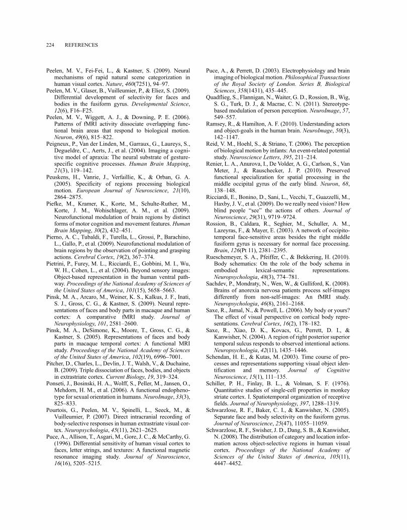
Peelen, M. V., Fei-Fei, L., & Kastner, S. (2009). Neuralmechanisms of rapid natural scene categorization inhuman visual cortex. Nature, 460(7251), 94–97.
Peelen, M. V., Glaser, B., Vuilleumier, P., & Eliez, S. (2009).Differential development of selectivity for faces andbodies in the fusiform gyrus. Developmental Science,12(6), F16–F25.
Peelen, M. V., Wiggett, A. J., & Downing, P. E. (2006).Patterns of fMRI activity dissociate overlapping func-tional brain areas that respond to biological motion.Neuron, 49(6), 815–822.
Peigneux, P., Van der Linden, M., Garraux, G., Laureys, S.,Degueldre, C., Aerts, J., et al. (2004). Imaging a cogni-tive model of apraxia: The neural substrate of gesture-specific cognitive processes. Human Brain Mapping,21(3), 119–142.
Peuskens, H., Vanrie, J., Verfaillie, K., & Orban, G. A.(2005). Specificity of regions processing biologicalmotion. European Journal of Neuroscience, 21(10),2864–2875.
Piefke, M., Kramer, K., Korte, M., Schulte-Ruther, M.,Korte, J. M., Wohischlager, A. M., et al. (2009).Neurofunctional modulation of brain regions by distinctforms of motor cognition and movement features. HumanBrain Mapping, 30(2), 432–451.
Pierno, A. C., Tubaldi, F., Turella, L., Grossi, P., Barachino,L., Gallo, P., et al. (2009). Neurofunctional modulation ofbrain regions by the observation of pointing and graspingactions. Cerebral Cortex, 19(2), 367–374.
Pietrini, P., Furey, M. L., Ricciardi, E., Gobbini, M. I., Wu,W. H., Cohen, L., et al. (2004). Beyond sensory images:Object-based representation in the human ventral path-way. Proceedings of the National Academy of Sciences ofthe United States of America, 101(15), 5658–5663.
Pinsk, M. A., Arcaro, M., Weiner, K. S., Kalkus, J. F., Inati,S. J., Gross, C. G., & Kastner, S. (2009). Neural repre-sentations of faces and body parts in macaque and humancortex: A comparative fMRI study. Journal ofNeurophysiology, 101, 2581–2600.
Pinsk, M. A., DeSimone, K., Moore, T., Gross, C. G., &Kastner, S. (2005). Representations of faces and bodyparts in macaque temporal cortex: A functional MRIstudy. Proceedings of the National Academy of Sciencesof the United States of America, 102(19), 6996–7001.
Pitcher, D., Charles, L., Devlin, J. T., Walsh, V., & Duchaine,B. (2009). Triple dissociation of faces, bodies, and objectsin extrastriate cortex. Current Biology, 19, 319–324.
Ponseti, J., Bosinski, H. A., Wolff, S., Peller, M., Jansen, O.,Mehdorn, H. M., et al. (2006). A functional endopheno-type for sexual orientation in humans.NeuroImage, 33(3),825–833.
Pourtois, G., Peelen, M. V., Spinelli, L., Seeck, M., &Vuilleumier, P. (2007). Direct intracranial recording ofbody-selective responses in human extrastriate visual cor-tex. Neuropsychologia, 45(11), 2621–2625.
Puce, A., Allison, T., Asgari, M., Gore, J. C., &McCarthy, G.(1996). Differential sensitivity of human visual cortex tofaces, letter strings, and textures: A functional magneticresonance imaging study. Journal of Neuroscience,16(16), 5205–5215.
Puce, A., & Perrett, D. (2003). Electrophysiology and brainimaging of biological motion. Philosophical Transactionsof the Royal Society of London. Series B, BiologicalSciences, 358(1431), 435–445.
Quadflieg, S., Flannigan, N., Waiter, G. D., Rossion, B., Wig,S. G., Turk, D. J., & Macrae, C. N. (2011). Stereotype-based modulation of person perception. NeuroImage, 57,549–557.
Ramsey, R., & Hamilton, A. F. (2010). Understanding actorsand object-goals in the human brain. NeuroImage, 50(3),142–1147.
Reid, V. M., Hoehl, S., & Striano, T. (2006). The perceptionof biological motion by infants: An event-related potentialstudy. Neuroscience Letters, 395, 211–214.
Renier, L. A., Anurova, I., De Volder, A. G., Carlson, S., VanMeter, J., & Rauschecker, J. P. (2010). Preservedfunctional specialization for spatial processing in themiddle occipital gyrus of the early blind. Neuron, 68,138–148.
Ricciardi, E., Bonino, D., Sani, L., Vecchi, T., Guazzelli, M.,Haxby, J. V., et al. (2009). Do we really need vision? Howblind people “see” the actions of others. Journal ofNeuroscience, 29(31), 9719–9724.
Rossion, B., Caldara, R., Seghier, M., Schuller, A. M.,Lazeyras, F., & Mayer, E. (2003). A network of occipito-temporal face-sensitive areas besides the right middlefusiform gyrus is necessary for normal face processing.Brain, 126(Pt 11), 2381–2395.
Rueschemeyer, S. A., Pfeiffer, C., & Bekkering, H. (2010).Body schematics: On the role of the body schema inembodied lexical-semantic representations.Neuropsychologia, 48(3), 774–781.
Sachdev, P., Mondraty, N., Wen, W., & Gulliford, K. (2008).Brains of anorexia nervosa patients process self-imagesdifferently from non-self-images: An fMRI study.Neuropsychologia, 46(8), 2161–2168.
Saxe, R., Jamal, N., & Powell, L. (2006). My body or yours?The effect of visual perspective on cortical body repre-sentations. Cerebral Cortex, 16(2), 178–182.
Saxe, R., Xiao, D. K., Kovacs, G., Perrett, D. I., &Kanwisher, N. (2004). A region of right posterior superiortemporal sulcus responds to observed intentional actions.Neuropsychologia, 42(11), 1435–1446.
Schendan, H. E., & Kutas, M. (2003). Time course of pro-cesses and representations supporting visual object iden-tification and memory. Journal of CognitiveNeuroscience, 15(1), 111–135.
Schiller, P. H., Finlay, B. L., & Volman, S. F. (1976).Quantitative studies of single-cell properties in monkeystriate cortex. I. Spatiotemporal organization of receptivefields. Journal of Neurophysiology, 397, 1288–1319.
Schwarzlose, R. F., Baker, C. I., & Kanwisher, N. (2005).Separate face and body selectivity on the fusiform gyrus.Journal of Neuroscience, 25(47), 11055–11059.
Schwarzlose, R. F., Swisher, J. D., Dang, S. B., & Kanwisher,N. (2008). The distribution of category and location infor-mation across object-selective regions in human visualcortex. Proceedings of the National Academy ofSciences of the United States of America, 105(11),4447–4452.
224 REFERENCES
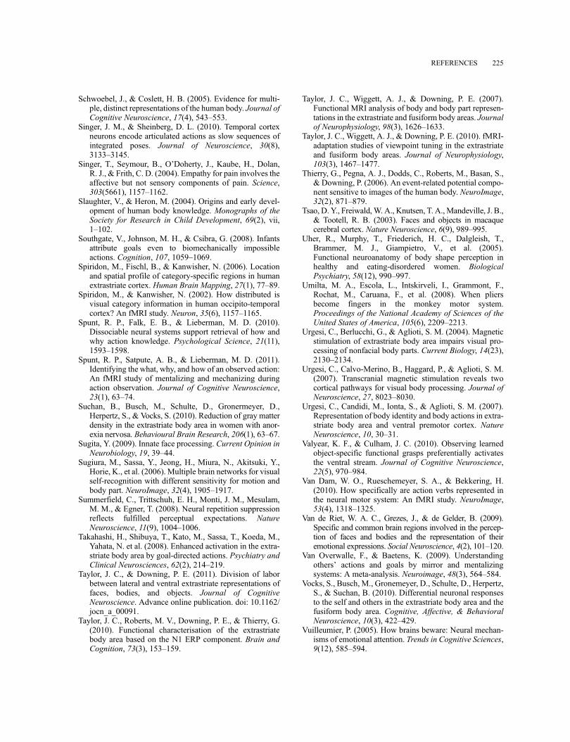
Schwoebel, J., & Coslett, H. B. (2005). Evidence for multi-ple, distinct representations of the human body. Journal ofCognitive Neuroscience, 17(4), 543–553.
Singer, J. M., & Sheinberg, D. L. (2010). Temporal cortexneurons encode articulated actions as slow sequences ofintegrated poses. Journal of Neuroscience, 30(8),3133–3145.
Singer, T., Seymour, B., O’Doherty, J., Kaube, H., Dolan,R. J., & Frith, C. D. (2004). Empathy for pain involves theaffective but not sensory components of pain. Science,303(5661), 1157–1162.
Slaughter, V., & Heron, M. (2004). Origins and early devel-opment of human body knowledge. Monographs of theSociety for Research in Child Development, 69(2), vii,1–102.
Southgate, V., Johnson, M. H., & Csibra, G. (2008). Infantsattribute goals even to biomechanically impossibleactions. Cognition, 107, 1059–1069.
Spiridon, M., Fischl, B., & Kanwisher, N. (2006). Locationand spatial profile of category-specific regions in humanextrastriate cortex. Human Brain Mapping, 27(1), 77–89.
Spiridon, M., & Kanwisher, N. (2002). How distributed isvisual category information in human occipito-temporalcortex? An fMRI study. Neuron, 35(6), 1157–1165.
Spunt, R. P., Falk, E. B., & Lieberman, M. D. (2010).Dissociable neural systems support retrieval of how andwhy action knowledge. Psychological Science, 21(11),1593–1598.
Spunt, R. P., Satpute, A. B., & Lieberman, M. D. (2011).Identifying the what, why, and how of an observed action:An fMRI study of mentalizing and mechanizing duringaction observation. Journal of Cognitive Neuroscience,23(1), 63–74.
Suchan, B., Busch, M., Schulte, D., Gronermeyer, D.,Herpertz, S., & Vocks, S. (2010). Reduction of gray matterdensity in the extrastriate body area in women with anor-exia nervosa. Behavioural Brain Research, 206(1), 63–67.
Sugita, Y. (2009). Innate face processing. Current Opinion inNeurobiology, 19, 39–44.
Sugiura, M., Sassa, Y., Jeong, H., Miura, N., Akitsuki, Y.,Horie, K., et al. (2006). Multiple brain networks for visualself-recognition with different sensitivity for motion andbody part. NeuroImage, 32(4), 1905–1917.
Summerfield, C., Trittschuh, E. H., Monti, J. M., Mesulam,M. M., & Egner, T. (2008). Neural repetition suppressionreflects fulfilled perceptual expectations. NatureNeuroscience, 11(9), 1004–1006.
Takahashi, H., Shibuya, T., Kato, M., Sassa, T., Koeda, M.,Yahata, N. et al. (2008). Enhanced activation in the extra-striate body area by goal-directed actions. Psychiatry andClinical Neurosciences, 62(2), 214–219.
Taylor, J. C., & Downing, P. E. (2011). Division of laborbetween lateral and ventral extrastriate representations offaces, bodies, and objects. Journal of CognitiveNeuroscience. Advance online publication. doi: 10.1162/jocn_a_00091.
Taylor, J. C., Roberts, M. V., Downing, P. E., & Thierry, G.(2010). Functional characterisation of the extrastriatebody area based on the N1 ERP component. Brain andCognition, 73(3), 153–159.
Taylor, J. C., Wiggett, A. J., & Downing, P. E. (2007).Functional MRI analysis of body and body part represen-tations in the extrastriate and fusiform body areas. Journalof Neurophysiology, 98(3), 1626–1633.
Taylor, J. C., Wiggett, A. J., & Downing, P. E. (2010). fMRI-adaptation studies of viewpoint tuning in the extrastriateand fusiform body areas. Journal of Neurophysiology,103(3), 1467–1477.
Thierry, G., Pegna, A. J., Dodds, C., Roberts, M., Basan, S.,& Downing, P. (2006). An event-related potential compo-nent sensitive to images of the human body. NeuroImage,32(2), 871–879.
Tsao, D. Y., Freiwald,W. A., Knutsen, T. A., Mandeville, J. B.,& Tootell, R. B. (2003). Faces and objects in macaquecerebral cortex. Nature Neuroscience, 6(9), 989–995.
Uher, R., Murphy, T., Friederich, H. C., Dalgleish, T.,Brammer, M. J., Giampietro, V., et al. (2005).Functional neuroanatomy of body shape perception inhealthy and eating-disordered women. BiologicalPsychiatry, 58(12), 990–997.
Umilta, M. A., Escola, L., Intskirveli, I., Grammont, F.,Rochat, M., Caruana, F., et al. (2008). When pliersbecome fingers in the monkey motor system.Proceedings of the National Academy of Sciences of theUnited States of America, 105(6), 2209–2213.
Urgesi, C., Berlucchi, G., & Aglioti, S. M. (2004). Magneticstimulation of extrastriate body area impairs visual pro-cessing of nonfacial body parts. Current Biology, 14(23),2130–2134.
Urgesi, C., Calvo-Merino, B., Haggard, P., & Aglioti, S. M.(2007). Transcranial magnetic stimulation reveals twocortical pathways for visual body processing. Journal ofNeuroscience, 27, 8023–8030.
Urgesi, C., Candidi, M., Ionta, S., & Aglioti, S. M. (2007).Representation of body identity and body actions in extra-striate body area and ventral premotor cortex. NatureNeuroscience, 10, 30–31.
Valyear, K. F., & Culham, J. C. (2010). Observing learnedobject-specific functional grasps preferentially activatesthe ventral stream. Journal of Cognitive Neuroscience,22(5), 970–984.
Van Dam, W. O., Rueschemeyer, S. A., & Bekkering, H.(2010). How specifically are action verbs represented inthe neural motor system: An fMRI study. NeuroImage,53(4), 1318–1325.
Van de Riet, W. A. C., Grezes, J., & de Gelder, B. (2009).Specific and common brain regions involved in the percep-tion of faces and bodies and the representation of theiremotional expressions. Social Neuroscience, 4(2), 101–120.
Van Overwalle, F., & Baetens, K. (2009). Understandingothers’ actions and goals by mirror and mentalizingsystems: A meta-analysis. Neuroimage, 48(3), 564–584.
Vocks, S., Busch, M., Gronemeyer, D., Schulte, D., Herpertz,S., & Suchan, B. (2010). Differential neuronal responsesto the self and others in the extrastriate body area and thefusiform body area. Cognitive, Affective, & BehavioralNeuroscience, 10(3), 422–429.
Vuilleumier, P. (2005). How brains beware: Neural mechan-isms of emotional attention. Trends in Cognitive Sciences,9(12), 585–594.
REFERENCES 225

Vuilleumier, P., Richardson, M. P., Armony, J. L., Driver, J.,& Dolan, R. J. (2004). Distant influences of amygdalalesion on visual cortical activation during emotional faceprocessing. Nature Neuroscience, 7(11), 1271–1278.
Wachsmuth, E., Oram, M. W., & Perrett, D. I. (1994).Recognition of objects and their component parts:Responses of single units in the temporal cortex of themacaque. Cerebral Cortex, 4(5), 509–522.
Weiner, K. S., & Grill-Spector, K. (2010). Sparsely-distributedorganization of face and limb activations in human ventraltemporal cortex. NeuroImage, 52(4), 1559–1573.
Weiner, K. S., & Grill-Spector, K. (2011). Not one extrastri-ate body area: Using anatomical landmarks, hMT+, andvisual field maps to parcellate limb-selective activationsin human lateral occipitotemporal cortex. NeuroImage,56(4), 2183–2199.
Whitney, D., Ellison, A., Rice, N. J., Arnold, D., Goodale,M., Walsh, V., et al. (2007). Visually guided reachingdepends on motion area MT+. Cerebral Cortex, 17(11),2644–2649.
Wiggett, A. J., & Downing, P. E. (2008). The face network:Overextended? (Comment on: “Let’s face it: It’s a corticalnetwork” by Alumit Ishai). NeuroImage, 40(2), 420–422.
Wiggett, A. J., & Downing, P. E. (2011). Representation ofaction in occipito-temporal cortex. Journal of CognitiveNeuroscience, 23(7), 1765–1780.
Willems, R. M., Peelen, M. V., & Hagoort, P. (2010).Cerebral lateralization of face-selective and body-selective visual areas depends on handedness. CerebralCortex, 20(7), 1719–1725.
Wojciulik, E., Kanwisher, N., & Driver, J. (1998). Covertvisual attention modulates face-specific activity in thehuman fusiform gyrus: fMRI study. Journal ofNeurophysiology, 79(3), 1574–1578.
Yomogida, Y., Sugiura, M., Sassa, Y., Wakusawa, K.,Sekiguchi, A., Fukushima, A., et al. (2010). The neuralbasis of agency: An fMRI study. NeuroImage, 50(1),198–207.
Yovel, G., Pelc, T., & Lubetzky, I. (2010). It’s all in yourhead: Why is the body inversion effect abolished forheadless bodies? Journal of Experimental Psychology:Human Perception and Performance, 36(3), 759–767.
Zimmermann, M., Meulenbroek, R. G., & de Lange, F. P.(2011). Motor planning is facilitated by adopting anaction’s goal posture: An fMRI study. Cerebral Cortex.Advance online publication. doi: 10.1093/cercor/bhr098.
226 REFERENCES


![[XLS]30th Infantry Division Roster30thinfantry.org/history_docs/30throster_080901.xls · Web viewMortar Sqd Downey, Francis L. 120 Medics Downing, (unknown) Downing, Doyle B. Downing,](https://img.pdfslide.net/doc/110x75/5ad459287f8b9a571e8c190b/xls30th-infantry-division-viewmortar-sqd-downey-francis-l-120-medics-downing.jpg)