Embed Size (px)
Citation preview
ONCOGENES AND HUMAN CANCER
(Oncogenen en Humane Kanker)
PROEFSCHRIFT
ter verkrijging van de graad van doctor in de geneeskunde
aan de Erasmus Universiteit te Rotterdam op gezag van de rector magnificus
Prof. Dr. M.W. van Hof en volgens besluit van bet college van dekanen.
De openbare verdediging-zal plaatsvinden op woensdag 30 mei 1984 te 14.00 uur.
door
Eleonora Classina Petronella Heisterkamp
geboren te Diemen
J-1 dischP. Bibliotheek __ ..., _, ...
Promotor: Prof. Dr. D. Bootsma
Gedrukt bij Offsetdrukkerij Kanters B. V., Alblasserdam.
Dit proefschrift werd bewerkt in de Laboratory of Viral Carcinogenesis, FCRF, NIH, Frederick, MD., USA.
ONCOGENES AND HUMAN CANCER
(Oncogenen en Humane Kanker)
PROEFSCHRIFT
ter verkrijging van de graad van doctor in de geneeskunde
aan de Erasmus Universiteit te Rotterdam op gezag van de rector magnificus
Prof. Dr. M.W. van Hof en volgens besluit van het college van dekanen.
De openbare verdediging zal plaatsvinden op woensdag 30 mei 1984 te 15.15 uur.
door
Johannes Hendrikus Comelis Groffen
geboren te Vlaardingen
Medische Bibliotheek E.L;.f"!..

Promotor: Prof. Dr. D. Bootsma
Dit proefschrift werd bewerkt in de Laboratory of Viral Carcinogenesis, FCRF, NIH, Frederick, MD., USA.
CONTENTS
PREFACE - VOORWOORD. • • • . • • • • . • • • • . • • • • • • • • 6
l. INTRODUCTION
1.1 General Introduction.
1.2 Scope of the thesis
1.3 Type C RNA Viruses.
1.4 Cellular Homologs of Viral Oncogenes.
• 9
.10
.14
.22
1.5 Human Sequences with Potential Transforming Activity •
a. Overview • 26
b. Human c-abl. .29
c. Human c-fes. .31
d. Human c-fms. .32
e. Human c-fos. .32
f. Human c-mos. .32
g. Human c-myc. .33
h. Human c-myb, .37
i. Human c-sis. .38
j. Human c-Ha-ras. c-Ki-ras and other transforming
human sequences detected by transfection .39
2. CONCLUSION • • • . • . • • . • . . • • • . . • • • • . . . 43
SUMMARY •••..•.•.••••.•.•••.•••••••.. 49
SAMENVATTING • • . • . . . • • . • . • . . • • • • . • . • • • . 51
REFERENCES • . • . • • • • • • . • • • • • • • • . • . • . • • • 54
CURRICULA VITAE ••••••..•••...•..•••..• 66
APPENDIX PAPERS, I-XII •.•.••••••..••.••••.. 73
Paper I:
Paper II:
Paper III:
Paper IV:
Paper V:
Paper VI:
Paper VII:
Isolation of v-fms and its Cellular Homolog
N. Heisterkamp~ J. Groffen, J.R. Stephenson
Virology 126 (1983):248-258
Chromosomal Localization of the Human c-fms Oncogene
J. Groffen, N, Heisterkamp, N. Spurr, S. Dana,
J. Wasmuth, J.R. Stephenson.
Nucleic Acids Research ll (1983) : 6331-6341
Isolation of Human Oncogene Sequences (v-fes homolog)
from a Cosmid Library
J.
J. Groffen, N. Heisterkamp, F. Grosveld, W. van de Ven.
J.R. Stephenson.
Science 216 (1982): 1136-1138
Transforming Genes of Avian (v-~) and Mammalian
v-fes) Retroviruses Correspond to a Common Cellular
Locus
J. Groffen, N. Heisterkamp, M. Shibuya, H. Hanafusa,
J.R. Stephenson.
Virology 125 (1983):480-486
Chromosomal Localization of Human Cellular Homologues
of two Viral Oncogenes
N. Heisterkamp, J. Groffen, J.R. Stephenson,
N.K. Spurr, P.N. Goodfellow, E. Solomon, B. Carritt,
W.F. Bodmer.
Nature 299 (1982): 747-749
Genetic Analysis of the 15:17 Chromosome Translocation
Associated with Acute Promyelocytic Leukemia
D. Sheer, L.R. Hiorns, K.F. Stanley, P.N. Goodfellow,
D.M. Swallow, S. Pavey, N. Heisterkamp, J. Groffen,
J.R. Stephenson, E. Solomon.
Proc. Natl. Acad. Sci. USA 80 (1983): 5007-5011
The Human v-abl Cellular Homolog
N. Heisterkamp, J. Groffen, J.R. Stephenson.
J. Mol. Appl. Genetics 2 (1983): 57-68
Paper VIII: Homology between Phosphotyrosine Acceptor Site of Human
c-abl and Viral Oncogene Products
Paper IX:
Paper X:
Paper XI:
Paper XII:
J. Groffen, N. Heisterkamp, F.H. Reynolds, Jr.,
J.R. Stephenson.
Nature 304 (1983): 167-169
A Cellular Oncogene is Translocated to the Philadelphia
Chromosome in Chronic Nyelocytic Leukemia
A. de Klein, A. Geurts van Kessel, G. Grosveld,
C.R. Bartram, A. Hagemeijer, D. Bootsma, N.K. Spurr,
N. Heisterkamp, J. Groffen, J.R. Stephenson.
Nature 300 (1982): 765-767
C-sis is Translocated from Chromosome 22 to
Chromosome 9 in Chronic Myelocytic Leukemia
J. Groffen, N. Heisterkamp, J.R. Stephenson, A. Geurts
van Kessel, A. de Klein, G. Grosveld, D. Bootsma.
J. Exper. Med 158 (1983): 9-15
Localization of the c-abl Oncogene Adjacent to a
Translocation Breakpoint in Chronic Myelocytic
Leukemia.
N. Heisterkamp, J.R. Stephenson, J. Groffen,
P.F. Hansen, A. de Klein, C.R. Bartram, G. Grosveld.
Nature 306 (1983): 239-242.
Philadelphia Chromosomal Breakpoints are clustered
within a Limited Region, bcr, on Chromosome 22.
J. Groffen, J.R. Stephenson, N. Heisterkamp,
A. de Klein, C.R. Bartram, G. Grosveld.
Cell ~ (1984): 93-99
PREFACE - VOORWOORD
Dit proefschrift is tot stand gekomen op een voor Nederland
ongebruikelijke wijze: bet wetenschappelijke werk dat ervoor door
ons is gedaan is uitgevoerd in Amerika, in bet Laboratory of Viral
Carcinogenesis van de National Institutes of Health te Frederick,
M.D. Dat wij in de gelegenheid zijn gesteld om daar een aantal jaren
onderzoek te doen is, als we teruggaan naar onze studententijd,
indirect te danken aan Prof. Dr. M. Gruber: hij was ten zeerste
bereid om mij, John (die niet eens een doctoraal onderzoek in zijn
afdeling had gedaan) te helpen met mijn wens om 11 te leren
kloneren". Het resultaat was, dat ik een periode in Landen kon
doorbrengen in de Laboratory of Gene Structure and Expression te
Mill Hill, waar ik een enorme hoeveelheid kennis en ervaring heb
opgedaan. Een interesse in het kankeronderzoek, de mogelijkheid om
de opgedane kloneringservaring toe te passen en bet feit, dat wij
onderzoeksplaatsen niet al te ver van elkaar zochten leidde ertoe
dat wij uiteindelijk in de NIH zijn terechtgekomen. Hierbij zijn wij
veel dank verschuldigd aan Dr. W.J.M. van de Ven, Elly van de Ven en
Dr. H.P.J. Bloemers.
Het contact met de mensen in Mill Hill is nag steeds aanwezig;
hoewel vele van de oude groep naar elders zijn verhuisd geldt het
gezegde "uit het oog, uit bet hart" bepaald niet. Dick Flavell en
Frank Grosveld willen we in de eerste plaats bedanken voor alles wat
ze in het verleden en heden voor ons hebben gedaan. Zander hun
bereidheid, om een student uit Nederland een kans te geven was dit
alles niet mogelijk geweest.Ook een aantal andere mensen van de Mill
Hill groep, ondanks bet feit dat zij niet direct betrakken zijn
geweest bij dit praefschrift, hebben tach in zekere zin een invloed
gehad. Er moet dan speciaal worden gedacht aan Cora, Dimitris,
Tarben, Titia, Chris, Andy, Elizabeth, Stephanie, Rene, Henrik, Lex
en Ernie. Oak Gerard Grosveld en Annelies de Klein hebben een grate
ral gehad in bet tot stand komen van dit proefschrift; wie had aoit
6
gedacht dat bet oversturen van een "proobje" zulke spectaculaire
resultaten zou opleveren! Wij willen jullie bedanken voor jullie
enthousiasme en voor de hele plezierige samenwerking. Oak de andere
mensen van de vakgroep Celbiologie en Genetica in Rotterdam zijn
wij zeer erkentelijk.
Tot onze promotor, Prof. Dr. Dick Bootsma willen we een
speciaal woord van dank richten: bet is al een opgave, om promotor
te zijn voor promovendi binnen je vakgroep, laat staan als de
promovendi niet tot je vakgroep behoren en hun onderzoek verrichten
op een heel ander continent. Ondanks deze belemmering hebben we bet
contact als bijzonder prettig en probleemloos ervaren; wij willen je
nogmaals be dan ken voor alles, wat j e voor ons hebt gedaan en bet
feit, dat je bereid bent geweest als promotor op te treden.
We also wish to thank all our colleagues from the Laboratory of
Viral Carcinogenesis; first of all, Dr. John Stephenson, who
initially did not believe in the great virtues of molecular cloning.
However, we presume he has changed his mind since then, if we
interpret his threat that "one day he will run a DNA sequencing gel"
correctly. We would like to thank Dr. Fred Reynolds, Dr. Fulvia
Veronese and their technicians Chris, Dick and Bob, the latter who
did some of the photography work for our manuscripts. A special
word of thanks goes to Siobahn, who did all the tissue culture work
and kept John's weird cell lines alive.
Last but not least we want to thank Gail Blennerhasset and
Pamela Hansen, our technicians. Although DNA work is certainly not
among the easiest, you have both done your very best and thanks to
you we have been able to set up a well-running DNA laboratory. Dear
Pam and Gail, we have very much appreciated your technical
assistance, your patience and you involvement in the work.
Finally, in America, we thank all the secretaries who have typed and
7
retyped different parts of the manuscript even though they were
busy: Mrs. Barbara Eader and Mrs. Beverly Bales in Frederick, Mrs.
Ann Newson, Mrs. Peggy Brandeburg and Ms. Marianne Lewis in Mineola.
Tot slot willen wij nog een speciaal woord van dank aan onze ouders
richten, hoewel zij zich misschien hieronder een beetje
ongemakkelijk voelen. Dankzij de opvoeding die ik, Nora, heb
gekregen heb ik ten volle gebruik kunnen maken van het beste wat het
Nederlandse onderwijssysteem te bieden heeft; mijn ouders hebben de
term "emancipatie" in mijn opvoeding in de practijk toegepast lang
voordat de term mode werd. Ook ik, John, ben ervan overtuigd dat
mijn ouders heel belangrijk zijn geweest voor mijn geestelijke
vorming en moet daarbij speciaal denken aan de vele weekeinden die
in de "natuur" werden doorgebracht.
Mineola, maart 1984
8
Nora Heisterkamp
John Groffen
I. INTRODUCTION
1.1 General Introduction
The first demonstrations that cancer could have an infectious
nature was by Ellerman and Bang (1) ~ who showed that leukemia in
chickens was transmissible with cell-free extracts and by Rous (2),
who found in a similar fashion that naturally occurring chicken
sarcomas were transmissible. Although they were able to show that
these cell-free extracts contained a transmissible agent~ the idea
that this induced cancer was received by the scientific world at that
time with great skepticism. The interest in oncogenic viruses was
strongly enhanced in the early 60's by the isolation of mammalian
tumor viruses and the general acceptance that at least some of these
viruses were tumorigenic. The discovery of the reverse transcriptase
enzyme in RNA tumor viruses (3,4), gave a logical explanation for how
these viruses became integrated in the chromosomes of eukaryotic
cells.
Taxonomically, oncogenic viruses are members of diverse
families. DNA viruses (herpes-, adeno- and papovaviruses) as well as
many members of the retrovirus family (containing RNA such as the
type C RNA viruses) are capable of inducing tumors. For the
retroviruses two different routes to become transforming (oncogenic)
have become clear. The majority of these viruses (the acute type C
RNA transforming viruses) 11 acquire11 certain genetic sequences
(oncogenes) from their host, which are necessary to initiate and
maintain the malignant transformation of the cell by the virus.
Other retroviruses integrate their genome nearby oncogenic sequences
in the chromosome of their host. Independent of the exact mechanism,
these viruses share the capability of inducing tumorigenesis by
triggering the transcription of certain sequences, and it is the
proteins encoded by these sequences which are necessary to maintain
the neoplastic phenotype of the infected cell.
9
The accumulating number of independent isolates of tumorigenic
retroviruses induced in the mid-70's a worldwide search for these
viruses in humans. Only very recently the isolation of a human tumor
T-cell leukemia retrovirus (HTLV) was reported (5,6).
Another approach was initiated in the beginning of the 80's,
with the finding that the acquired sequences of retroviruses are
strongly conserved among species. In general, the cellular homologs
of these sequences were easily detectable and could be studied in
more detail by molecular cloning, using the oncogenic acquired
sequences of retroviruses as probes. This approach seems to be very
fruitful and will be discussed in more detail below.
Although the oncogenic potential of the acquired sequences in a
number of these viruses in vertebrates is well estabished, the
involvement of their human cellular homologs in human tumorigenesis
has been and will be a rich source for discussion. However, at the
moment they provide us the best available model for the induction of
human cancer at the molecular level.
1.2 Scope of the Thesis
The detailed structure of type C RNA viruses became clear once
they had been molecularly cloned: as discussed before, most acute
type C RNA transforming viruses had acquired DNA sequences from their
host (e.g. chicken, cat, mouse, rat) which were responsible for the
tumorigenic properties of the virus. A new approach, in the research
of human cancer as a molecular biological discipline, became evident;
by using the acquired cellular sequences of, for example, a cat acute
type C RNA transforming virus, as a molecular probe, one could
investigate the possible existence of homologous sequences in man.
If the evolutionary divergence between these feline sequences and the
homologous sequences in human were sufficiently low, the human
10
cellular homologs could be molecularly cloned and their possible
involvement in human cancer could be studied in a detailed way. We
have chosen that approach in the research described in this thesis:
initially, three distinct acute type C RNA transforming viruses (see
Introduction 1. 3 for a more detailed description of these viruses)
were chosen as a source for molecular probes. Two of these, the
Snyder-Theilen Feline Sarcoma virus (ST-FeSV) and the Sarma-McDonough
Feline Sarcoma virus (SM-FeSV) were molecularly cloned from
transformed cell lines containing a complete integrated provirus.
The third, Abelson Murine Leukemia virus (Ab-MuLV) was obtained as a
gift from P. Reddy. Each of these viruses contains different
acquired cellular sequences, v-fes in ST-FeSV, v-fms in SM-FeSV and
v-abl in Ab-MuLV. Molecular probes specific for the acquired
cellular sequences of each hybridized strongly to sequences in human
DNA, indicating that these sequences were strongly conserved during
evolution. The human cellular homologs of v-fes, v-fms and v-abl
were subsequently molecularly cloned (!Japer I, III and VII) and
research on the potential involvement of these human oncogenes in
cancer was initiated. As a preliminary characterization we made
detailed restriction maps of these human oncogenes and the position
of sequences homologous to the viral acquired cellular sequences were
determined; extensive homology between the v-abl tyrosine
phosphorylation acceptor region and a corresponding region in human
c-abl was established more precisely by nucleic acid sequencing
(paper VIII). Additionally, human c-fes was examined for homology
with the acquired cellular sequences (v-fps) of Fujinami Sarcoma
virus (FSV); earlier reports (7) had indicated, that v-fes (from cat)
and v-fps (from chicken) were related. Our results indicated that
one locus exists in man having homology both to v-fes and v-~
(paper IV).
One straightforward method of examining the transforming poten
tial of the human cellular homologs is, to introduce these sequences
11
into rodent cells in culture and to investigate, if they can trans
form the cells to a malignant phenotype (see Introduction 1.4)
However, the c-fes, c-fms or c-abl human oncogenes were incapable of
transforming cells in culture. This is in concordance with the
results of others obtained with other human oncogenes: generally,
human oncogenes do not have transforming ability unless they are
manipulated extensively in vitro before introduction into cells, We
have chosen not to pursue the effort to attempt to modify the cloned
oncogenes in vitro in such a way that they might become transforming:
one major obstacle in such an effort would have been the large size
of the genes encoding human c-abl and c-fms. Rather, we have ex
amined the possible involvement of human c-fes, c-fms and c-abl in
existing human malignancies. As a first step, the chromosomal
localization of each of these human oncogenes was determined. Such
information might be of value in view of the accumulating evidence,
that specific chromosomal aberrations are associated with specific
types of neoplasia (See Introduction 1.5). The localization of human
c-fms to chromosome 5 (paper II) and human c-fes to chromosome 15
(paper V) has to date been of moderate value. Aberrations on chromo
some 5 have not been intimately associated with one type of
neoplasia. Although c-fes is translocated from chromosome 15 to
chromosome 17 in patients with acute promyelocytic leukemia (APL) in
which a t (15; 17) is characteristic (paper VI), we have not found a
chromosomal breakpoint in the proximity of c-fes.
Human c-abl, on chromosome 9 (paper V) has provided us with an
excellent tool to study leukemogenesis at the molecular level: in the
t(9;22) characteristic of chronic myelogenous leukemia (GML) human
c-abl is translocated to chromosome 22, the Philadelphia (Ph')
chromosome (paper IX). As another human oncogene, c-sis, is located
on chromosome 22, we examined if c-sis was also involved in the
Philadelphia translocation. Although we could demonstrate the
translocation of c-sis to chromosome 9 (paper X) human c-sis was
12
subsequently localized far from the Philadelphia translocation
breakpoint (8). Therefore~ our attention concentrated on the human
c-abl oncogene; in one CML patient~ we found~ after an elaborate
search, a chromosomal breakpoint 5' to the human c-abl locus (paper
XI) . Furthermore, we were able to clone a region from chromosome 22
in which the breakpoints occur in all Ph'-positive CML DNAs examined
to date (paper XII).
The three acute type C transforming viruses utilized in our
research represent only part of the many type C RNA viruses isolated
to date (see table I). We have not concentrated our research on the
viral oncogenes but rather, have used them as a tool in characteriz
ing human oncogenes. However, the molecular organization, protein
products, relatedness and transforming mechanism of the viral
oncogenes has been studied extensively by others. Although this does
not directly bear on the question if human oncogenes are involved in
human tumorigenesis, studies on viral oncogenes may ultimately
explain how cellular sequences can become transforming. Therefore,
we have included research regarding type C RNA viruses (Introduction
!. 3). Cellular oncogenes in animal models have also yielded much
information (Introduction 1.4).
Many human oncogenes have been molecularly cloned and charac
terized as we have done (Introduction 1.5). We have attempted to
provide a limited amount of structural information for each. Of the
human oncogenes, c-abl, c-myc and the ~ gene family have been most
strongly implicated in neoplastic diseases. The putative transform
ing mechanism of c-abl and c-myc seem to be similar. We have done
most of the research on c-abl; the research on c-myc is very elabo
rate and we have attempted to summarize this information.
·In the above described research projects, Drs. N. Heisterkamp
has concentrated on the human c-fms oncogene (papers I and II) and
13
human c-abl (papers V, VII, IX and XI). Drs. J. Groffen has had a
major contribution in research concerning human c-fes (papers III, IV
and VI), sequencing of human c-abl (paper VIII) and sequences on
chromosome 22 (papers X and XII). However, all projects were per
formed in close collaboration between both authors.
1.3 Type C RNA Viruses
Type C RNA viruses can be divided into two classes on the basis
of tumorigenicity. The first class (the chronic type C RNA viruses)
cannot transform cells in culture. in vivo, however, these viruses,
such as Feline leukemia virus (FeLV) and Moloney murine leukemia
virus (MoMuLV), can induce leukemias in infected animals after a
relatively long period of latency ranging from 6 to 12 months. The
viral genome consists of several genes, which together are necessary
for the life cycle of the virus. After infection of the cell, the
RNA genome is converted to a double stranded DNA copy by the vi
ral-encoded enzyme reverse transcriptase.
The DNA copy of the viral genome is integrated into the genome
of the infected cell; this provirus carries two identical LTR's (long
terminal repeats), one at either end of the viral genome. The LTR
contains promoter sequences, signals for transcription termination
and polyadenylation signals (9) . Transcription is initiated from the
5' LTR; RNA is transported to the cytoplasm and translated into the
proteins utilized for the formation of a new virus particle. The
order of the genes on the linear double stranded DNA copy of the
viral RNA is:
5' LTR ---- gag pol env ---- LTR 3'
The ~ gene encodes the structural proteins of the viral core.
These proteins are synthesized as part of a polyprotein, which is
14
processed into the mature viral proteins. Pol encodes the viral
reverse transcriptase and~ the viral envelope glycoproteins.
The second class of type C RNA viruses (the acute type C RNA
viruses) is called transforming because of their ability to transform
cells in culture. In general, they cause sarcomas and acute
leukemias in vivo after a relatively short latency period (a few days
or weeks). What has been described for the "chronic" viruses also
applies to these viruses; the latter, however, are generally replica
tion deficient and require the presence of a helper virus for the
production of infectious viral particles. Acute type C transforming
RNA viruses are thought to have originated from chronic type C
viruses through a recombination event between the latter viruses and
host cellular sequences. As a consequence, part of the structural
information of the viral genome has been deleted and replaced by
"non-structural11 or "acquired" sequences of host cellular origin.
These acquired sequences have imparted a new ability on the virus,
enabling it to transform fibroblasts in vitro and to have an acute
oncogenic potential in vivo (10, 11).
At least 15 different type C transforming viruses have been
isolated, from species as diverse as chicken and baboon. Each
isolate contains acquired cellular sequences. To distinguish the
cellular homologs from their viral counterparts a standard nomencla
ture (12) is accepted: viral oncogenes are denoted by "v" as in
v-~, the cellular progenitors by "c" as in c-src.
The chicken has been a particularly rich source for the iso
lation of type C transforming viruses (Table I). Rous sarcoma virus
(RSV), in contrast with the other virus isolates, is not replication
deficient: chicken cellular sequences have been inserted 3' to the
structural genes of avian leukosis virus, resulting in the formation
of a replication competent transforming virus. The acquired cellular
15
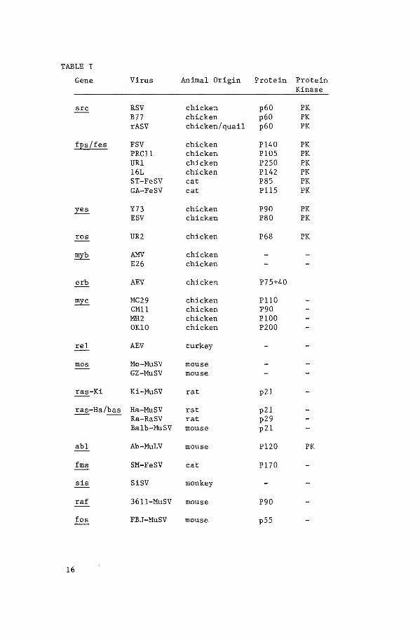
TABLE I
Gene Virus Animal Origin Protein Protein Kinase
src RSV chicken p60 PK B77 chicken p60 PK rASV chicken/quail p60 PK
fEs/fes FSV chicken Pl40 PK PRCll chicken Pl05 PK URl chicken P250 PK 16L chicken Pl42 PK ST-FeSV cat P85 PK GA-FeSV cat Pll5 PK
~ Y73 chicken P90 PK ESV chicken P80 PK
ros UR2 chicken P68 PK
myb liliV chicken E26 chicken
erb AEV chicken P75+40
myc MC29 chicken PllO CMll chicken P90 MH2 chicken PlOO OKlO chicken P200
rel AEV turkey
mos Mo-MuSV mouse GZ-MuSV mouse
ras-Ki Ki-MuSV rat p21
ras-Ha/bas Ha-MuSV rat p21 Ra-RaSV rat p29 Balb-MuSV mouse p21
abl Ab-MuLV mouse Pl20 PK
fms SM-FeSV cat Pl70
sis SiSV monkey
ra£ 3611-MuSV mouse P90
fos FBJ-MuSV mouse pSS
16
sequences of RSV are called ~· Two different strains of avian
sarcoma virus, Y73 (13) and Esh (14) have independently recombined
with the same cellular sequence, e-yes (15). Fujinami, PRCII and
URl avian sarcoma viruses all contain a strongly related oncogene.
v-~ (7, 16). A new avian sarcoma virus isolate, UR2 (17),
contains a unique oncogene, v-ros ( 18). Avia~ erythroblastosis
virus contains two 11 domains" of acquired cellular sequences, erbA
and B. Each encodes a protein: erbA encodes a ~ fusion protein
(see later) of MW 75,000 (19). ErbB, which consists entirely of
acquired cellular sequences, encodes a 40,000 MW protein (20) which
is thought to be responsible for the transforming activity of the
virus (21). The acquired cellular sequences of avian myeloblastosis
virus (AMV) have been called both amv and myb (22, 12). Finally,
MC29 (myelocytomatosis virus) is one of a series of avian type C
transforming viruses that have transduced a cellular oncogene called
myc from chicken. One species of transforming type C virus has been
isolated from turkey; the reticuloendotheliosis virus strain (REV-T)
contains the rel oncogene (23).
In mammals, the isolation of type C transforming viruses has
been reported from mouse, rat, cat and baboon., In mouse, murine
leukemia virus has recombined with different cellular oncogenes,
giving rise to Abelson murine leukemia virus (v-abl) (24,25),
:Holoney murine sarcoma virus (v-mos) (26), 3611-:HuSV (v-raf) (27),
and Finkel-Biskis-Jinkins murine osteosarcoma virus (v-fos) (28).
Through passage of a murine leukemia virus on rat cells Harvey and
Kirsten murine sarcoma virus were obtained (29, 30). Both contain
an oncogene (v-Ha-ras (31) and v-Ki-~) flanked at either end by
rat "30S" DNA sequences, which are retrovirus-like but without
transforming capacity. v-Ha-ras and v-Ki-ras are related but not
identical: they contain homologous and unique sequences (32). From
cat, two strains of feline sarcoma virus have been independently
isolated which both contain portions of the same cellular oncogene
17
c-fes: Snyder-Theilen FeSV and Gardner FeSV (33, 34, paper IV).
Another cat isolate yielded McDonough FeSV which carries a unique
oncogene, v-fms (35, paper I). One type C transforming RNA virus
has been isolated from primates, the simian sarcoma virus (v-sis
oncogene) (36, 37).
From this summary it is clear that a large variety of type C
transforming viruses has been isolated. In this context it should
be mentioned that in most cases the viruses have been molecularly
cloned, allowing a detailed comparison at the nucleic acid level
with respect to the relatedness of various isolates; most references
in this thesis refer to the molecular cloning of the virus and not
the original isolation. From a comparison of the fes oncogene in
ST-FeSV and GA-FeSV with the fps oncogene, in Fujinami sarcoma
virus, it became clear that fps and fes represent the same oncogene
from chicken and from cat (38, 39, paper IV). New viral isolates do
not always yield new oncogenes: a type C RNA virus isolated from
cat contains a sis-like oncogene (40). The acquired cellular
sequences from Balb murine sarcoma virus (bas) seem to be analogous
to ras from rats (41). Therefore, the amount of different oncogenes
transduced by type C RNA viruses may be quite limited. This may
reflect a situation wherein the number of oncogenes in the genome is
low; on the other hand, it is possible that certain oncogenes cannot
fulfill certain "requirements 11 necessary to become integrated in the
retroviral genome. At the moment, it is not known if this type of
requirement exists; however, with the development of retroviral
vectors for eukaryotic cells, analogous to the E. coli cloning
vectors, this type of information should become available. The
isolation of new oncogenes with DNA-mediated transfection
experiments (see Introduction 1. 5, j) lends support to the
hypothesis that there may be still a large repertoire of sequences
with oncogenic potential in the genome not yet isolated,
18
The protein products of the type C transforming RNA viruses are
very diverse in size. As mentioned, the cellular sequences have
been integrated in phase in the structural genes of the virus.
Where this recombination has taken place differs in various viral
isolates. Fuj inami sarcoma virus has the structure 5 '-Llgag-~-3'
(38), indicating that part of the~ gene and the entire~ and
~ gene have been deleted and replaced by ~ sequences. As a
consequence, ~ is expressed as a polyprotein, with amino
acid-terminus encoded structural components covalently linked to the
acquired cellular sequence encoded carboxy-terminus component: Pl40
5'-b gag-fps
Both ST-FeSV and Gardner FeSV have the structure
They express fes
polyprotein; the difference in molecular
Gardner FeSV, P85gag-fes for ST-FeSV) is
as part of a gag-fes
weight (Pll5gag-fes for
caused by the different
size of the ~ portion and differences in the acquired sequence
component (39). MC29 also encodes a gag-~ fusion protein, of MW
llO,OOO (PllOgag-myc); the genomic organization is similar to that
of STand Gardner FeSV: 5!..,6. gag-myc-.6.~-3' (42). Simian sarcoma
virus has a variant structure~ in which the sis sequences are
situated in the env gene (36). As mentioned before, avian
erythroblastosis virus encodes two proteins, a gag-erbA fusion
protein and a protein completely encoded by the acquired sequence
component (20,21). A number of other type C transforming viruses
have oncogenes, which are not expressed as part of a polyprotein;
Rous sarcoma virus (in which structural genes have not been replaced
by an oncogene) encodes a pp60src ( 43). Both Ha-MuSV and Ki-MuSV
encode a p21 protein from the acquired cellular sequences (32).
The protein products of many of these viral oncogene sequences
have been identified; in many cases, however, the putative enzymatic
activity or function remains obscure. Even though for some an
enzymatic activity has been found it is difficult to unravel the
pathway by which the viral oncogene mediates its transforming
effect,
19
However, some retroviruses have acquired sequences that encode
proteins, which all possess a tyrosine-specific protein kinase
activity. Protein kinases are thought to have a regulatory function
in the cell through the activation and deactivation of cellular
enzymes by phosphorylation. That pp60src (Rous sarcoma virus)
possessed a protein kinase activity with specificity for tyrosine
residues (44) came somewhat as a surprise; until then it was thought
that phosphorylation involved only serine or threonine residues. In
normal cells only one of the 3000 phosphorylated residues is a
tyrosine; in RSV transformed cells this amount increases five-to
tenfold. Soon after this enzymatic activity had been discovered it
was found for a number of other viral oncogene products (Table I):
v-fes/fps (45,46), v-abl (47 ,48), v-~ (49) and v-E.'?.'!_ (SO). This
enzymatic activity seems required for the maintenance of the trans
formed state; temperature sensitive mutants of RSV (51) and FSV (52)
are no longer able to transform cells and do not exhibit protein
kinase activity at the non-permissive temperature. Transformation
deficient mutants of AbMuLV lack protein kinase activity (53, 54).
Although phosphotyrosine residues in the protein kinases themselves
are thought to be of importance for their enzymatic activity (55),
their exact functional significance remains to be determined.
Nucleic acid sequencing data have shown that the different
oncogene encoded protein kinases may be related and have evolved
from a common ancestor: v-fps and v-~ are 40% homologous in the
carboxy terminal 280 amino acids (38). Although v-yes and v-src did
not exhibit significant homology in nucleic acid hybridization
experiments (13), the amino acid sequence predicted on the base of
nucleic acid sequencing data revealed that both oncogenes were
strikingly similar (56). That v-abl and v-src are related stems
from two lines of evidence. Firstly, in Drosophila melanogaster
sequences have been cloned that exhibit nucleic acid homology to
both v-abl and v-src (which might represent a common ancestor), and
20
ones that have exclusive homology to ~ (57, 58). Interestingly,
the regions of v-abl and v-src that show cross homology with this
putative progenitor sequence (called c-Dash) are those that are
essential for the protein kinase activity in v-abl (25) and v-src
(59). Secondly, nucleic acid sequence data have shown that v-~,
v-fes, v-abl and human c-abl contain a common region that is highly
conserved (paper VIII).
Even v-~ appears to have limited homology with v-src at the
amino acid level (60). The phosphorylation of tyrosine residues
does not seem to be an exclusive property of viral oncogene
products. The cell surface receptor for epidermal growth factor
(EGF) is phosphorylated in tyrosine after binding EGF, a small
polypeptide with growth promoting activity (61,, 62). Furthermore,
the same reaction is found after the binding of insulin to the
insulin receptor (63) and the binding of platelet derived growth
factor (PDGF) to its receptor (64). Although it seems plausible
that these receptors are themselves protein kinases specific for
tyrosine residues, it remains to be established if this is the case
(65) or that such protein kinases are intimately associated with
them. Interestingly, cells transformed by either AbMuLV (v-abl) or
ST-FeSV (v-fes) and some human tumor derived cell lines show an
overall increase in phosphotyrosine level and a reduced binding of
exogenously added EGF to its receptor. The cause of this reduced
binding capability seems to be, that in cells transformed by these
viruses, a new growth factor (TGF = transforming growth factor) is
produced, that competes with EGF for the binding of its receptor
( 66). That a connection exists between some oncogene encoded
products and growth factors became clear, when protein sequencing of
a part of human PDGF revealed complete concordance in 104 amino acid
residues with the deduced protein sequence of v-sis (67, 68).
21
The information regarding the other viral oncogene products is
less elaborate. None encode proteins with tyrosine specific kinase
activity. v-myc codes for a DNA-binding nuclear protein (69, 70).
No protein product of v-rel has been reported to date. v-fms and
v-raf both encode identifiable products of unknown function (71,
72). v-erbB seems to be a cell membrane glycoprotein (73). The p21
of Harvey and Kirsten MuSV (v-Ha-~ and v-Ki-ras) has a protein
kinase activity; it specifically binds guanine containing
nucleotides and can phosphorylate itself by transferring a phosphate
of GTP to a threonine residue on its own molecule (74).
1.4 Cellular Progenitors of Viral Oncogenes
With the isolation of new oncogenic retroviruses, the possibil
ity emerged that these viruses, during passage in animals,
recombined with the genome of the host and that this recombination
resulted in the formation of an acutely transforming virus.
Molecular hybridization and the ability to prepare radioactive cDNAs
were the tools used to show that the Rous sarcoma virus had acquired
specific sequences (c-src) from the chicken genome (75).
Subsequently it was possible to show that the presence of these
sequences was not restricted to chicken; v-src homologous sequences
were found in fish and human with a nucleotide divergence possibly
as low as 15%, indicating that this gene is evolutionarily conserved
(76). Very recently (57, 77) v-~ related sequences were found in
the insect Drosophila, which extends the conservation of this gene
over an even longer evolutionary period.
With the introduction of molecular cloning it became possible
to specifically clone the acquired sequences of tumorigenic
retroviruses and to perform these studies in a much more
straightforward and detailed way. These types of studies
established that the majority of the retroviral oncogenes are
22

acquired from the cellular genome and that the extent of
evolutionary conservation varies among the different oncogenes.
The cellular homologs of most of the known viral oncogenes
exist as single loci within a given species. Although in some cases
additional related sequences can be detected (78) these cellular
sequences are only partly homologous to viral oncogenes, which makes
it unlikely that viral oncogenes are derived from them. More likely
these related sequences are derivatives of the original cellular
gene and unable to encode a functional protein ("pseudo-genes"),
"processed genes, 11 or related loci. The latter possibility is
illustrated by v-ras, of which two distinctive forms have been
acquired, one by the Harvey and one by the Kirsten strain of sarcoma
virus (79,80). Although the acquired sequences of these viruses are
related they are derived from different loci. The acquired
sequences of the transforming viruses do not exceed a stretch of
more than 3 kb of DNA. However their cellular progenitors are in
most cases found to be distributed over a much larger stretch Of
DNA, indicating the presence of intervening sequences. The finding
that most of the cellular homologs contain one or more intervening
sequences unambiguously demonstrates that their origin is not viral.
That the cellular oncogenes are strongly conserved in evolution
implies that these sequences encode mRNAs and that their
translational products are essential for the cell (i.e.~ "housekeep
ing" enzymes). First experimental evidence for their presence was
obtained with the discovery of RNA homologous to v-src in uninfected
fibroblasts of several avian species (81). Similar findings were
subsequently made for other cellular oncogenes. Exceptions were,
however, found; no c-fes transcript so far has been detected; more
over, a detailed search for c-mos mRNAs in many different tissues
has not revealed any transcripts.
23
Detection of cellular oncogene proteins has not been easy. To
date, three proteins have been identified. The most thoroughly
studied is pp60c-src, a 60,000 MW phosphoprotein; p21c-ras, a 21,000
MW protein encoded by Harvey e-ras and P150c-abl• a 150,000 MW
protein most probably encoded by c-abl.
The translation products of v-src and c-src are remarkably
identical; moreover. deletion mutants of v-src can be restored by
recombination with c-~ resulting in a functional oncogene (82).
The DNA-mediated transfection procedure, introduced by Graham
and van der Eb in 1973 (83), permits transfer of genetic traits to
cells in culture by exposing these cells to DNA containing the genes
encoding the characteristics to be transferred. In this way, for
instance, cells lacking the activity of thymidine kinase (TK) can be
made TK+ by transfecting DNA of Herpes simplex type II virus, which
carries a gene for thymidine kinase. In some cases, the donor DNA
is stably integrated into the genome of the recipient.
This procedure provides a new approach to investigate if DNA
sequences have oncogenic activity; this DNA can be transferred to
recipient non-transformed cells through DNA-mediated transfection
procedures. The successful transfer of an oncogene would be
apparent by the formation of foci of transformed recipient cells,
which are usually NIH 3T3 cells.
The finding that the viral oncogenes were derivatives of cellu
lar progenitors introduced the tentative possibility that the cellu
lar oncogenes could be induced to cause malignant transformation in
other ways than through integration in retroviruses. Data
concerning this question accumulated rapidly after the molecular
cloning of the first cellular oncogenes: introduction of viral
transcriptional regulatory sequences (LTR) 5 1 of normal mouse c-mos
24
and rat c-~ by ligation, subsequently followed by transfection to
NIH 3T3 cells resulted in the activation of the transforming
potential of these cellular genes (84, 85), The number of foci was
comparable to that obtained upon transfection of the homologous
sarcoma virus DNA's. The cellular~ and~ genes do not exihibit
transforming activity on their own. However, removal of cellular
sequences 5' of the mouse c-mos gene results in a very low
transforming activity (86). The hypothesis that this low
transforming activity is a consequence of the integration of this
gene nearby strong cellular promoters is very plausible. Further
evidence that cellular oncogene expression could be responsible for
at least some forms of tumorigenesis was obtained from the study of
avian leukosis virus (ALV). This virus has no acquired sequences
(oncogene) yet it is capable of inducing B-cell lymphoma in
chickens. Its tumorigenicity was explained by the finding that in
these ALV-induced lymphomas, ALV is integrated adjacent to the c-myc
gene and that transcription, initiated from the viral promotor of
ALV, causes enhanced expression of c-myc, leading to neoplastic
transformation (87), These data showed that a carcinogenic agent
(ALV) acts by altering the expression of a cellular oncogene, c-myc.
Recently the DNA transfection procedure was employed in a new
way to detect DNA sequences possibly involved in tumorigenesis.
Instead of transfection of a particular DNA segment to recipient
contact inhibited eukaryotic cells~ the cells were exposed to total
cellular DNA. This type of experiment resulted in the
identification of oncogenes homologous to the acquired sequences of
retroviruses, However, in some experiments the results were not in
agreement with the expectations. When DNA of an ALV induced chicken
lymphoma was transfected to NIH 3T3 cells a number of foci were
obtained. Upon cloning, DNA sequences responsible for focus
formation were distinct from the expected c-myc gene, Nucleotide
sequencing identified the transforming sequence to be coding for a
25
small protein characterized by partial homology with transferrin
(88) 0 In similar experiments, by the same group, the DNA of
lymphoid neoplasms induced by Abelson murine leukaemia virus
(AbMuLV) was used to transfect NIH 3T3 cells. Again it was shown
that the transforming sequences were distinct from AbMuLV sequences
(89). This apparent contradiction of results could be explained if
some forms of tumorigenesis are multiple step events.
Strong evidence for the involvement of cellular oncogenes in
tumorigenesis was recently obtained from mouse myelomas and
plasmacytomas. In a mouse myeloma it was found that 5' of the c-mos
gene normal cellular sequences were substituted by an insertion
sequence (IS)-like element. Upon cloning of this rearranged gene it
was subsequently shown that in transfection experiments it was
capable of transforming mouse fibroblasts (90). Many mouse
plasmacytomas were found to have rearranged immunoglobulin heavy
chain constant regions. A certain DNA sequence (called LyR, NIARD
or NIRD by different investigators) was found to be translocated
into this region; (91, 92, 93) subsequently it could be shown that
this sequence represented c-myc sequences (94, 95, 96). As a result
of these findings an analogous translocation was found with human
c-myc in Burkitt lymphoma. In the next chapter, the involvement of
c-myc in tumorigenesis will be discussed in more detail.
1.5 Human sequences with potential transforming activity.
a. Overview
An increasing number of human sequences with a potential
to transform cells in culture has been isolated. Using viral
oncogene sequences as probes, the following human cellular homologs
have been isolated: c-abl (paper VII), c-fes (97, 98, paper III),
c-fms (paper I), c-fos (99), c-~ (100, 101), c-myb (102), c-myc
(103, 104), c-Ha-ras (lOS), c-Ki-ras (105), and c-sis (106). The
26
isolation of the human cellular homologs of v-~, v-erb and v-rel
has not yet been reported, This may reflect difficulty using avian
probes to isolate human sequences that are evolutionary distant,
For instance, although v-fps (chicken) and v-fes (cat) both detect
an identical locus in human (paper IV) the strength of hybridization
is much less when v-fps sequences are used as a probe, Sequences
homologous to v-rel seem not to be highly conserved in non-avian
species (23, 107),
Other human sequences, with transforming activity, have been
shown to exist by means of DNA-mediated transfection experiments;
DNA from either human tumor tissue or cell lines established from
human tumors transfected to mouse NIH 3T3 cells induces focus
formation at a low frequency. These foci contain human sequences,
detectable by an Alu-repeat probe, which is specific for human DNA.
Some of these transforming sequences have been molecularly cloned.
What could be regarded as evidence that these human oncogenes
represent causative agents in tumorigenesis? Because the protein
products of many human oncogenes have not yet been identified,
expression has mostly been studied on the RNA level, using either
viral or human probes, An increased level of RNA production of a
particular oncogene in transformed cells is thought to be an in
dication that the oncogene may have taken part in the neoplastic
process, However, elevated levels of oncogene RNA may also be a
phenomenon accompanying neoplastic transformation but not
necessarily causing it: altered expression of many enzymes and
proteins in tumors has been reported (for a review, see 108),
reflecting changes in metabolism of the transformed cells.
Hypomethylation of human growth hormone, o( -globin and ~-globin
sequences in cancerous tissues in comparison with healthy tissue of
the same patient has also been reported (109); the tissues studied
normally do not express these genes,
27
Increased mRNA production can be caused by an amplification of
oncogene sequences, for example by duplication of the chromosome on
which the oncogene is situated. Alternatively, only limited regions
of a chromosome may be amplified. Alterations in the signals
responsible for the regulation of transcription can also cause an
increase in mRNA production. These alterations can be local, by the
insertion or deletion of sequences near the oncogene or by mutations
in the promoter sequences of an oncogene. Or as a part of a gross
rearrangement~ oncogene sequences can be translocated to other
chromosomes and either activated through position effects (e.g., an
oncogene that is normally not transcribed is translocated into a
region of high transcriptional activity) or through replacement of
its own promoter by a promoter at the breakpoint on the recipient
chromosome. Novel species of mRNA could be synthesized in this
case.
One firmly established fact is, that human neoplasms do very
often contain chromosomal aberrations. Double minute chromosomes
(DM), which contain as yet unidentified portions of chromosomes have
been found in tumor cell lines; they could represent amplification
of one limited large stretch of DNA. Translocations between
chromosomes in human neoplasms, especially in leukemias and
lymphomas, have been reported in an increasing number (110). The
first discovered and most well known example is the Philadelphia
translocation, t(9;22), associated with chronic myelogenous leukemia
(CML) (111). In this translocation a piece of the long arm of
chromosome 22 (qll-qter) is translocated to chromosome 9 (112); a
very small piece of chromosome 9 is translocated to chromosome 22
(paper IX). The 22q- chromosome (the Philadelphia chromosome) is
present in the leukemic cells of about 96% of all CML patients; this
generally appears to be the only chromosomal abnormality prior to
the blast crisis during which other abnormalities become apparent
(110).
28
In certain forms of acute promyelocytic leukemia (APL) a
t (15; 17) is characteristic (113, 114). Recently, a correlation has
been found between Burkitt lymphoma and translocations involving
chromosome 2, 14 or 22 with chromosome 8 (115). With the
improvement of cytogenetic techniques, the number of specific
chromosomal aberrations associated with specific neoplastic disease
is increasing rapidly (116). For example, among the solid tumors
small lung cell carcinoma ( 117) is correlated with a deletion in
chromosome 3 and Wilms tumor with a deletion of chromosome 11 at
band pl3 (ll8), However, if a chromosomal translocation is the
direct cause of a neoplastic disease or if the disease causes
chromosomal aberrations is a question difficult to answer but
important to the debate if human oncogenes have oncogenic potential.
Another mechanism, by which an oncogene might acquire
tumorigenic properties is by a mutation, for example in the coding
sequences; the oncogene sequences could then code for a protein with
activity or characteristics different from the normal protein, which
makes it turmorigenic. This will be discussed in the chapter
concerning human c-Ki-ras and c-Ha-ras.
Models have been proposed which catagorize all the above de
scribed mechanisms by which an oncogene might become tumorigenic.
One model states that the overproduction of an oncogene product may
cause malignancy; the second model supposes that a mutation in the
coding region of an oncogene leads to the production of a new
oncogenic protein. A third alternative is that a normal oncogene
product is produced in normal quantities, but at the wrong moment in
a developmental process.
b. Human c-abl
Human c-abl has been molecularly cloned using a cosmid
vector system because of the presence of an extensive region of DNA
29
homologous to v-abl (paper VII). Apart from the main human c-abl
locus, other regions of more limited homology to v-abl were found,
encompassed by two separate EcoRI fragments of 4.1 and 12.5 kb
(paper VII). The 12.5 kb EcoRI fragment has been molecularly cloned
in phage (unpublished results). Homology only to a central part of
v-abl could be detected; in v-abl, this region contains the putative
tyrosine phosphorylation acceptor site which is highly conserved
between v-abl, v-src and v-fes. It is possible that sequences in
the 12.5 kb EcoRI fragment code for a protein with tyrosine-specific
protein kinase activity. That these sequences represent a
"processed gene 11 is not likely: the region of homology to v-abl
stretches out over 7.4 kb whereas v-abl itself is approximately 3.0
kb in length. Therefore these human sequences may contain introns.
The human v-abl homologue is located on chromosome 9q34 (paper
V). In CML, it is translocated to chromosome 22 in the standard
t(9;22) Philadelphia translocation (paper IX) and in complex
translocations such as the t(l;9;22) and the t(9;11;22) (ref. 141).
In one CML patient, a chromosomal breakpoint was found 5' to the
human c-abl locus; a restriction enzyme fragment containing
sequences from chromosome 9 and 22 was molecularly cloned from the
DNA of this patient (paper XI). Using a probe isolated from the
chromosome 22-specific sequences of this fragment, an extended
region of chromosome 22 DNA was cloned. In the DNAs of all
Ph' -positive CML patients examined a chromosomal breakpoint was
found within a limited region of approximately 5 kb on chromosome 22
(paper XII). The human CML cell line K562 appears to be
representative of CML in that a chromosomal breakpoint is present
within that same region on chromosome 22 (unpublished observations);
furthermore, the five fold amplification of the immunoglobulin~
light chain constant region on chromosome 22 (paper XI), of the
breakpoint region remaining on chromosome 22 (unpublished
observations) and of the human v-abl homologue (paper XI) suggests
that at least part of the Philadelphia chromosome has selectively
been amplified in this cell line.
30
Human c-abl is expressed in various human cell lines; at least
four RNA species of different molecular weights (7.2~ 6.4~ 3.8 and
2.0 kb) were found using a v-abl probe, both in hematopoietic and in
tumor cell lines; normal fibroblasts contained only two species of
high molecular weight (119, 120). Others, however, have reported
the presence of only two species of v-abl homologous RNA, of 5.4 and
5. 2 kb molecular weight in all human cells examined (121). Two
additional RNA species of 6.8 and 6.2 kb were found in a human cell
line estabished from a patient with a pre B acute lymphoblastic
leukemia (ALL). However, transfection of this DNA to NIH 3T3 cells
yielded foci which did not contain detectable human v-abl homologous
sequences. Production of a transforming growth factor was evident
both in the original ALL cell line and in NIH 3T3 cells transfected
by DNA of this cell line. It was suggested, that sequences
responsible for the induction of foci in NIH 3T3 either coded for a
(human) transforming growth factor or induced the production of a
transforming growth factor in the NIH 3T3 cells (121).
c. Human c-fes
RNA transcripts of human c-fes have not been found in
detectable quantities in 36 human cell lines examined (unpublished
observations). Molecularly cloned human c-fes does not exhibit
transforming activity, even if a viral LTR is ligated 5' to the
putative coding region (unpublished observations). Human c-fes maps
on chromosome 15 (paper V, 102) in the region q25-26 (122). In the
t(15;17) characteristic of some forms of APL, c-fes was not found on
the 15q+ chromosome in one patient (paper VI), suggesting that it
had been translocated to chromosome 17. However no sequences up to
30 kb 5' and 12 kb 3' of human v-fes homologous sequences were found
to be retained on the 15q+ chromosome from the same patient
indicating the absence of a chromosomal breakpoint near human c-fes
(unpublished observations) . These observations are concordant with
the localization of human c-fes to 15q25-26 (122) and the breakpoint
31
·an chromosome 15 in APL to band q22 (paper VI).
d. Human c-fms
Human c-fms, as human c-abl, appears to be a very large
gene; sequences homologous to v-fms stretch out over a region of at
least 30 kb. Human c-fms exhibits no transforming ability when it
is transfected to Rat-2 cells, even when all v-fms homologous
sequences are joined together in one cosmid clone. However, as with
human c-abl, the possibility must be considered that coding
sequences at the 5' region, which are not represented in v-abl or
v-fms, are missing from the clones. Human c-fms has been mapped on
chromosome 5, band q34 (paper II). A relatively high level of c-fms
transcripts was found in human placenta (123).
e. Human c-fos
Human c-fos has been cloned as a 9 kb EcoRI fragment (99).
Sequencing of the human c-fos gene has shown, that it contains
introns and can encode a protein of 380 aminoacids. Human c-fos
transcripts of 2.2 kb have been found in human term fetal membranes
(123). Human c-fos does not transform NIH 3T3 cells; however, if
the carboxy-terminus encoding region is replaced by that of v-f os
(human c-fos and v-fos differ in that region by a 104 bp sequence
not present in v-fos) the hybrid gene is able to induce the
formation of foci (124).
f, Human c-mos
Human c-mos was cloned as part of a 9 kb BamHI restriction
fragment ( 100); the homology to v-~ proved to be confined to a
very limited region (an 0. 96 kb EcoRI/Bglii restriction fragment)
with no apparent intervening sequences. Human c-mos and v-mos are
77% homologous in nucleic acid sequence. Although the mouse c-mos
32
readily transforms cells in culture when an LTR is ligated 5' to the
putative coding region, human c-mos has no detectable transforming
activity in a similar transfection assay (100). RNA transcripts of
human c-mos have not been detected in undifferentiated B-cell
lymphomas (125); however, others have reported expression in
Burkitt lymphoma cell lines (126). Human c-mos has been localized
to chromosome 8 (96, 127) in band q22.
g. Human c-myc
Human c-myc has been isolated in two overlapping phage
clones spanning a region of more than 20 kb (103) and as a 20 kb
EcoRI restriction fragment from a partial EcoRI human library (104).
Homology to v-myc was restricted to two stretches of DNA smaller
than 1 kb, separated by an apparent intervening sequence of
approximately 1. 4 kb (104). The sequences mentioned above
hybridized to probes specific for both the 5' and 3' end of v-myc.
On a genomic blot, more sequences with exclusive homology to a probe
specific for the 5' part of v-myc are seen. Three such regions,
representing apparent non-contiguous DNA sequences have been cloned
(103); heteroduplex mapping showed that only 0.2-0.4 kb of these
clones contained homology to v-myc (the central region); no
intervening sequences were apparent. Since no transcript other than
a 2. 7 kb RNA species, presumably encoded by the main c-myc locus
(see below) has been found, the regions with less homology to v-myc
could represent pseudogenes.
Sequencing of the main human c-myc locus has revealed an open
reading frame, which, (with omission of the putative intron) could
code for a mRNA of 2.3 kb (104). This size is reasonably concordant
with the size of an RNA of 2.7 kb found in human tumor cell lines,
in normal human fibroblasts and in haematopoietic cells (119, 120),
33
In the acute promyelocytic leukemia cell line HL-60 (this cell
line does not contain the t (15; 17) often found in APL (128)) the
level of transcription of the 2.7 kb mRNA was found to be approxi
mately tenfold enhanced (120), Moreover~ c-myc was amplified
8-32-fold in this cell line (129, 130). These results imply that
the increased level of c-myc expression in HL-60 is due to
amplification of c-myc sequences. Because amplifications of the
c-myc locus was also found in primary leukemic cells of the patient
from which HL-60 was established before chemotherapeutic treatment,
(129), this phenomenon does not seem to have occurred during passage
of the cells in tissue culture. However, it is difficult to
evaluate the possible role of c-myc in the causation of this
leukemia because no non-leukemic cells of the same patient could be
examined, Moreoever, no amplification of c-~ was found in cells
from the peripheral blood of three different patients with APL. The
correlation between c-myc and APL is therefore unclear.
The same applies to another study, in which an elevated (20 to
30-fold) level of c-myc expression was found in two cell lines
established from a colon carcinoma with characteristics of a
neuroendocrine tumor. By in situ hybridization the presence of
v-myc homologous sequences could be demonstrated in the
heterogenously staining region of a marker chromosome; the human
c-myc locus was amplified in both cell lines 16 to 32-fold (131).
Human c-mos and c-myc have both been mapped to chromosome 8;
c-mos is located at position 8q22 (127) and c-myc at 8q24 (96, 132).
Chromosome 8 is frequently involved in translocations associated
with neoplastic disease; in Burkitt lymphomas for instance,
translocations of chromosome 8 with chromosome 2, 14 and 22 have
been described (115). In all cases the breakpoint on chromosme 8 is
8q24; those on chromosome 2, 14 and 22 are 2p12, 14q32 and 22q11
(133, 134, 96).
34
Chromosomes 2, 14 and 22 each contain sequences coding for
immunoglobulins; the immunoglobulin heavy chain locus maps to 14q32
( 135), exactly in the region that contains the breakpoint in some
forms of Burkitt lymphoma. Using somatic cell hybrids of the human
Burkitt lymphoma cell line Daudi with murine cells, a chromosomal
breakpoint in the immunoglobulin heavy chain variable region was
demonstrated (136). Subsequently, human c-myc sequences were
detected in somatic cell hybrids carrying the 14q+ chromosome of
Daudi and of another Burkitt lymphoma cell line, P3
-HRI, indicating
that c-myc had been translocated from chromosome 8 to 14 (132).
Rearrangement of the c-myc locus has been found in 5 out of 15
B-cell lymphoma cell lines tested, both of the Burkitt and
non-Burkitt type. In three, c-myc and heavy chain immunoglobulin
sequences were found on the same restriction enzyme fragment. The
rearrangements had occurred 5' to c-myc, resulting in an apparent
head-to-head joining of c-myc with immunoglobulin sequences (137).
In another study (96), one allelic copy of c-myc was rearranged in 8
out of 12 Burkitt lymphoma cell lines studied, including the Raj i
cell line,
Before the connection between c-myc and Burkitt lymphoma had
been made, various groups had found non-immunoglobulin DNA sequences
near the heavy chain constant region in murine plasmacytomas and in
some B-lymphomas which contain a 15:12 translocation (chromosome 12
contains the murine heavy chain locus). These sequences, named LyR
(91), NIARD (92) or NIRD (93) were molecularly cloned and
subsequently shown to be murine c-myc sequences originating from
chromosome 15 (94-96). By testing the hybridization of probes from
various regions of the murine c-myc to murine RNA, two additional
coding regions were noted other than the two identified on basis of
hybridization to v-myc: one stretch of DNA was separated by
sequences which did not hybridize to RNA (the apparent first intron)
from the two known exons; an additional putative coding region was
35
present 3' to the already identified exons. In murine
plasmacytoma's, however, a probe from the 5 1 exon does not detect
homologous RNA, indicating that the translocation has altered or
removed the 5 1 exon and the S' regulatory sequences. A probe from
the first intron does detect multiple RNA species. This may
indicate, that a cryptic promotor in the first intron has been
activated (95). In two mouse myelomas, Ml67 and M603, breaks had
occurred in the first c-~ intron but at different positions. In
MPC-11 the translocation had occurred in the first c-myc exon,
without any visible rearrangements in the immunoglobulin heavy chain
constant region. In all cases, the S' regulatory sequences and the
first exon had been disrupted, which could possibly result in the
activation of several putative transcription initiation sites within
the first intron. The first (non v-myc homologous) exon contains
stop codons in every reading frame and could represent a
non-translated leader sequence; by removal of these sequences as a
consequence of the translocation a shorter transcript could be
synthesized which could have a lower probability of degradation and
thus a prolonged half-life. The actual cause of the translocation
remains unknown, as the recombination sites in J558, Ml67 and M603
do not share any obvious sequence-homology; furthermore, mouse c-myc
does not contain the repetitive sequences found in the }l switch
region (138).
In human c-myc, an ex on 5 1 to the v-myc homologous exons was
detected using a probe from the murine S' exon. In contrast to the
mouse plasmacytomas. no altered RNA product could be found or a
significant increased level of transcription. The breakpoint seemed
to be near immunoglobulin switch regions in two Burkitt lines (W1
,
w2). In the c-myc locus, translocation breakpoints were all in the
5' region, at different sites: for the Lou and w1
lines, this was
1-2 kb 5' from the second exon; in Raji, the breakpoint seemed to be
3-9 kb S' from the second exon. Of importance was that non-Burkitt
cell lines of the lines w1
, w2
and Lou were available and showed no
36
rearrangements in the human c-myc locus (95). The breakpoint in the
Raji cell line was defined more accurately by the molecular cloning
of a DNA fragment containing c-myc sequences and immunoglobulin
constant region sequences. The presence of an exon 5' to the two
defined on basis of hybridization to v-myc was confirmed, No
increase in the level of c-myc mRNA could be found specifically in
Burkitt lymphoma cell lines. Although two species of mRNA (2.4 and
2.2 kb) were detected, both seem to be transcribed starting at the
most 5 1 exon and neither is expressed preferentially in Burkitt
lymphoma (139). In contrast, others (126) have claimed a
significant increase in the levels of c-myc RNA in Burkitt lymphoma
cell lines compared to control lymphoblastoid cell lines.
Although no translocation of c-myc to chromosome 22 has been
demonstrated yet in the t(8;22), immunoglobulin A light chain
sequences are certainly involved; as mentioned, the breakpoint on
chromosome 22 is at qll and the A light chain constant locus has
recently been mapped to band qll using in situ hybridizing
techniques (140). In one Burkitt lymphoma cell line, sequences
hybridizing to the 8q+ and to the 22q chromosomes were detected
using a \constant region probe, indicating a breakpoint in the A
constant region. In another cell line, only the 8q + and not the
22q chromosome retained A-homologous sequences (140).
h. Human c-myb
v-myb has been used as a molecular probe to investigate
expression of human v-myb homologous RNA. None of 18 human cell
lines established from solid tumors exhibited expression of v-myb
homologous RNA sequences in detectable amounts. Mature T- and B
cells also were negative in this respect. However, an RNA species
of 4. 5 kb was detected in more immature cells and lymphoid cells.
As with human c-myc expression in HL-60, the expression of the 4.5
kb v-myb homologous RNA was abolished if the cell line was induced
37
to differentiate with Me2so or retinoic acid (22).
Using a human probe isolated from a 2. 0 kb v-myb homologous
EcoRI restriction fragment, human c-myb has been localized on
chromosme 6 (102) in the region q22-24 (122).
i. Human c-sis
Human v-sis homologous sequences have been molecularly
cloned as a 14 kb EcoRI restriction fragment (106) from a human
partial EcoRI library. These sequences seem to be present in one
single locus in man, contained on large Hindiii and EcoRI
restriction enzyme fragments. Sequences hybridizing to v-sis are
distributed over 5 apparent exons.
Although in this study (106) no polymorphisms were found in all
12 human DNA samples examined, we found a polymorphism for Hindiii
to be rather common; this polymorphism was exhibited by the presence
of an extra Hindiii site near the second intervening sequence in
human c-sis (unpublished observations).
Human c-sis has been mapped on chromosme 22 (101), distal to
band q11, since it is translocated from chromosome 22 to chromosome
9 in CML patients carrying the Philadelphia chromosome (paper X).
However, it is not localized in close proximity to the breakpoint on
chromosome 22 (8) and is translocated to other chromosomes than
chromosome 9 in complex Philadelphia translocations (141).
Expression of human c-sis is not common; in 8 out of 23 tumor
cell lines tested, a 4.2 kb v-sis homologous transcript was found.
The highest level of expression was in the glioblastoma cell line A
172 (119); expression was found in one of the haematopoietic cell
lines tested, the T-cell lymphoma cell line HUT 102, which produces
the human T-cell leukemia/lymphoma retrovirus HTLV (120).
38
j. Human c-Ha-~, c-Ki-~ and other
Transforming Human Sequences Detected by
Transfection
Using probes specific for the acquired cellular sequences
of Harvey and Kirsten MuSV, the human cellular homologs of v-Ha-~
and v-Ki-ras have been molecularly cloned from normal human DNA
(105). Two non-overlapping stretches of DNA were isolated with
homology to v-Ha-~, designated c-Ha-~-1 and c-Ha-~-2. Two
distinct v-Ki-ras homologous clones, containing c-Ki-ras-1 and
c-Ki-ras-2 were also isolated. Human c-Ha-~-1 has four apparent
coding regions, separated by intervening sequences. In contrast·,
c-Ha-ras-2 lacked intervening sequences and exhibited a limited
homology to v-Ha-ras. Human c-Ki-ras-1 has 0.9 kb of sequence
homologous to the viral probe, organized in two exons separated by
one intron; c-Ki-ras-2 has one region of 30G bp homologous to
v-Ki-ras without apparent introns. As both c-Ha-ras-2 and
c-Ki-~-2 hybridize to a limited region, that is conserved between
v-Ha-~ and v-Ki-ras, they may represent functional, related genes
(lOS). Human c-Ha-ras-1 has been mapped on the short arm of
chromosome 11 in the region p11-p15 (105, 142, 143); human
c-Ki-ras-2 is localized on chromosome 12 (144),
and c-Ki-ras-1 are on chromosome 6 and X (145).
Human c-Ha-ras-2
Human c-Ha-ras-1 has no transforming activity when it is
transfected to NIH 3T3 cells without further manipulation.
However, if a viral LTR is ligated to it, it can transform cells at
high frequency and a p21 protein can be immunoprecipitated from
these cells (146). Human v-~ homologous transcripts have been
commonly found in all samples tested (120).
Using DNA-mediated transfection experiments, a transforming
gene had been identified in the T24 and EJ bladder carcinoma cell
lines (147, 148). Once these genes had been cloned (147-149)-, they
39
were examined for sequence homology to other human and viral
oncogenes; soon it was reported that the T24 and EJ bladder
carcinoma genes were highly homologous to c-Ha-ras-1 (150) and v-bas
(151), an oncogene of murine origin analogous to v-Ha-ras from rats
(41). On restriction enzyme level, no obvious differences were
observed between the T24 oncogene and a molecularly cloned human
c-Ha-ras-1 except for a 200 bp deletion in the latter (151).
However, the T24 oncogene transformed NIH 3T3 cells efficiently,
whereas the human c-Ha-ras-1 did not. Two proteins (a p21 and a
p23) could be immunoprecipitated from cells transfected with the T24
oncogene (151).
Since no gross differences could be detected between the T24
oncogene and its normal cellular counterpart, the transforming
element in the T24 oncogene was localized more precisely by exchang
ing restriction enzyme fragments between the transforming and
nontransforming gene and assaying the hybrid genomes for fo
cus-forming ability. In this way. the transforming activity was
localized to a small region encompassing the first exon (encoding
the first 37 amino acids) and sequences immediately 5 1 and 3' to it
(152-154). This region was subsequently sequenced. Only one essen
tial change could be detected: in the codon for the twelfth amino
acid, a T was found in the bladder carcinoma gene and a G in the
normal gene, resulting in a valine at position 12 in the former and
a glycine in the latter (152-154). Interestingly, Kirsten, Harvey
and Balb MuSV also differ in the amino acid at position 12; v-Ha-ras
has an arginine, v-bas has a lysine and v-Ki-ras has a serine. Like
the normal human c-Ha-ras-1, rat c-Ha-ras-1 has a glycine at
position 12 (153). The change of one amino acid residue can be
detected on
(collectively
protein level; in
described as p21)
v-Ha-~-1 p21 specific antiserum.
normal cells two proteins
are im.munoprecipitated with
In cells transfected by DNA of
the EJ bladder carcinoma cell line, four proteins can be detected,
two of which are identical to those found in normal cells. The
other two migrate slightly slower on gels and are encoded by the
40
transforming allele of the c-Ha-ras-1 in the EJ cell line (152).
Other studies have confirmed that only the single basepair change in
the transforming oncogene distinguishes it from its nontransforming
counterpart (155).
An independent~ apparently spontaneous, mutation was found in
an originally non-transforming c-Ha-ras-1 allele; it had become
transforming after transfection to NIH 3T3 cells. In this case, a
point mutation had also occurred in the twelfth codon, but the
normally occurring glycine was replaced by an aspartic acid (156).
A transforming c-Ha-ras-1 gene has also been cloned from a lung
carcinoma cell line, Hs242. In contrast to the T24 oncogene, the
transforming sequences were not localized to the region encoding
amino acid residue 12, but to a region more to the 3' in the gene; a
single basepair change had occurred in codon 6 l, resulting in the
substitution of a leucine for a glutamine (41).
Transfection experiments with DNA isolated from solid tumors
has shown (157) that some of these also contain transforming
oncogenes detectable through transfection; this gave answer to an
important question, namely, if the transforming oncogenes in the
cell lines could have been active in the original tumor and were not
merely the result of an activation process during tissue culture
propagation. However, only 5 of 28 DNA samples from solid tumors
contained an oncogene, detectable with the transfection assay. DNA
of second-cycle transformants were examined for similarity in
restriction enzyme patterns after hybridization with total human DNA
and for homology to a number of viral oncogenes. Many of these
transforming DNAs contained similar sequences that had been
transferred to the NIH 3T3 cells, sequences that hybridized to a
v-Ki-ras probe (157).
The oncogenes transmissable from the SH 480 colon carcinoma
cell line and the small lung carcinoma cell line LX-1 apparently
also are homologous to v-Ki-ras (158, 159). Since two v-Ki-ras
41
homologous loci exist in man (105), c-Ki-ras-1 and c-Ki-ras-2, each
was examined for identity with the colon and lung carcinoma genes;
these genes hybridized to a probe specific for c-Ki-ras-2, (159).
The SW 480 sequences necessary for transformation stretch out over
an extended region of DNA; c-Ki-~-2 is a very large gene (30-35
kb) ~ comparable to human c-abl and human c-fms (160, 161). The
normal c-Ki-ras-2 allele does not transform NIH 3T3 cells; the
transforming gene has apparently undergone a point mutation (162).
The transforming gene from the neuroblastoma cell line SK-N-SH
has been molecularly cloned (163); the transforming activity is
situated on two EcoRI fragments of 9.2 and 7.0 kb. This
neuroblastoma oncogene seems to be related to the human ras gene
family (163, 164). A part of the gene has been cloned from
secondary transfectants of the fibrosarcoma cell line, HT-1080; it
is apparently identical to the SK-N-SH transforming gene and can be
demonstrated in transfectants of DNA from the cell lines RD
(rhabdomyosarcoma) and HL-60 {a promyelocytic line); as this N-ras
gene is located on chromosome 1, it is clearly distinct from the
other members of the ~ gene family. RNA transcripts of 2.2 kb
were detected in comparable amounts in HT-1080 cells, in EJ cells
and in normal human fibroblasts {165).
Tumorigenesis is generally regarded to be a multi-stage
process, That members of the ras gene family are able to transform
NIH 3T3 cells with single hit kinetics is an argument against the
possibility that these cells are representative of actual target
cells in vivo. The ras genes are not capable of transforming normal
fibroblasts in the transfection assay; however, upon cotransfection
with a myc gene or the polyoma large-T antigen normal fibroblasts
can be made tumorigenic (166). A similar phenomenon was reported by
others; neither the T24 oncogene (Ha-ras) nor the polyoma middle-T
antigen can transform primary cells alone; however, co-transfection
with adenovirus early region 1A yielded transformed cells (167).
42
Additionally, the EJ oncogene (Ha-~) can only transform normal
fibroblasts after they have been immortalized by carcinogens (168).
These results have led to the proposal that certain complementation
groups exist among oncogenic sequences: large T-antigen, adenovirus
early region lA and myc would belong to one group, inducing
immortality. The ras gene family and polyoma middle-T would be
implicated in events leading to anchorage independent growth and
morphological change (166),
2. CONCLUSION
The connection between human oncogenes and cancer has to date
not been clarified for any human oncogene. Since all viral
oncogenes cause transformation, the potential to transform may be
present in these particular homologous sequences in human. However,
it is clear that the viral oncogenes are in no case identical to
their own direct cellular progenitors: viral oncogenes lack all
intervening sequences and in some instances exons present in their
cellular counterparts. Point mutations and deletions further
distinguish them from the cellular oncogenes (for a review, see
169). However, similar critical pTocesses which may have changed a
virally transduced, non-transforming sequence into a transforming
gene may take place in an individual. Therefore, it is possible
that every human oncogene is ultimately found to be connected with
some kind of malignancy. Only a limited number of human oncogenes
have been implicated to date in vivo in neoplastic diseases.
Human c-myc appears to be closely associated with Burkitt
lymphoma and with the type of APL represented by the cell line
HL-60. In the first case, c-myc homologous RNA is not found in
significantly higher amount, indicating that the transformation is
not caused by elevated transcription. Moreover, no human c-myc RNAs
of an abnormal size have been found in Burkitt lymphomas. Recently,
it was shown that the c-myc allele translocated to chromosome 14 is
43
expressed, whereas the other allele is not; this led to the proposal
that the translocated oncogene somehow escapes normal regulation as
a consequence of the translocation (170). The breakpoints on both
chromosome 8 and 14 seem to be variable: in some cell lines (such as
Daudi) no breakpoint anywhere in the neighborhood of c-myc is
detectable although it is translocated to chromosome 14. In the
immunoglobulin locus, breaks in the variable region and in the
switch region of the heavy chain have been reported. The mechanism
by which human c-myc may cause transformation in Burkitt lymphoma
may therefore be called unclear. However. the strong correlation
between Burkitt lymphoma and the presence of a translocation of
c-myc to chromosome 14 in the t(8;14) indicates that this oncogene
must somehow be involved in this disease.
The oncogenes detected by transfection do not provide a consis
tent linkage between one type of malignancy and one type of
oncogene; both c-Ha-ras and c-Ki-ras have been implicated in the
transformation in bladder carcinomas. The N-ras gene has been
activated in four different types of malignancies (see Table II).
Some of these oncogenes are activated by single point mutations.
How common such events are in vivo remains to be determined, as none
of 10 primary bladder carcinomas, 9 colon carcinomas or 10 lung
carcinomas had a point mutation in the codon for the twelfth amino
acid (171), although a mutation in other codons may be possible.
The involvement of human c-fes in human tumorigenesis is at the
moment unclear. Since the fes gene has been "acquired"
independently by four type C transforming viruses in chicken and two
in cat, the oncogenicity of these sequences seems unquestionable.
Nonetheless, it is possible that in the evolutionary period between
man and cat, safeguards have evolved to prevent the human homolog
from being expressed at the wrong moment. The highly conserved
nature of fes suggests that it has an important function in the
cell, but, since it has not been found expressed in any cell line
44
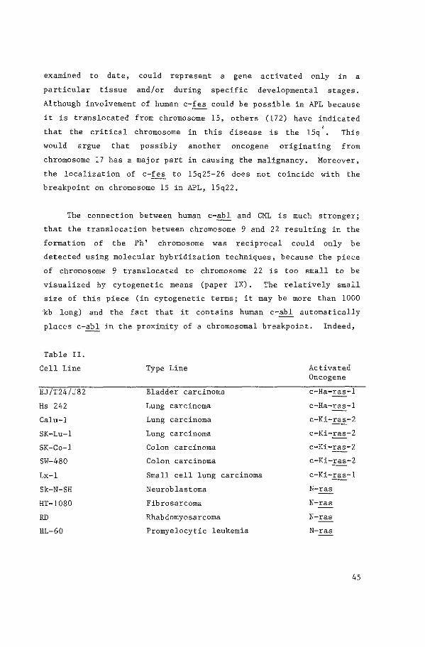
examined to date, could represent a gene activated only in a
particular tissue and/or during specific developmental stages.
Although involvement of human c-fes could be possible in APL because
it is translocated from chromosome 15, others (172) have indicated
that the critical chromosome in this disease is the 15q +. This
would argue that possibly another oncogene originating from
chromosome 17 has a major part in causing the malignancy. Moreover,
the localization of c-fes to 15q25-26 does not coincide with the
breakpoint on chromosome 15 in APL, 15q22.
The connection between human c-abl and CML is much stronger;
that the translocation between chromosome 9 and 22 resulting in the
formation of the Ph' chromosome was reciprocal could only be
detected using molecular hybridization techniques, because the piece
of chromosome 9 translocated to chromosome 22 is too small to be
visualized by cytogenetic means (paper IX). The relatively small
size of this piece (in cytogenetic terms; it may be more than 1000
kb long) and the fact that it contains human c-abl automatically
places c-abl in the proximity of a chromosomal breakpoint. Indeed,
Table II.
Cell Line
EJ/T24/J82
Hs 242
Calu-1
SK-Lu-1
SK-Co-1
SW-480
Lx-1
Sk-N-SH
HT-1080
RD
HL-60
Type Line
Bladder carcinoma
Lung carcinoma
Lung carcinoma
Lung carcinoma
Colon carcinoma
Colon carcinoma
Small cell lung carcinoma
Neuroblastoma
Fibrosarcoma
Rhabdomyosarcoma
Promyelocytic leukemia
Activated Oncogene
c-Ha-ras-1
c-Ha-ras-1
c-Ki-ras-2
c-Ki-ras-2
c-Ki-ras-2
c-Ki-ras-2
c-Ki-ras-1
N-ras
N-ras
N-ras
N-ras
45
we have found a chromosomal breakpoint in one CML patient located
within 14 kb 5' from the most 5' v-abl homologous sequence (paper
XI). That such a proximity is fortituous seems unlikely: recently,
we have found a chromosomal breakpoint in a second CML DNA in the
relative neighborhood of human c-abl (unpublished observations). If
the break actually occurs in the human c-abl gene itself is at the
moment unresolved; v-abl is approximately 3 kb long whereas v-abl
homologous human RNA's of around 6 kb have been detected (unpublished
observations), suggesting the presence of as of yet unidentified
exons. Furthermore~ studies with the mouse c-abl gene have indicated
that it contains sequences to the 5', not present in v-abl (173). If
the break is outside the human c-abl locus itself, a similar
situation is found as in Burkitt lymphoma, in which a breakpoint is
not found in c-myc coding sequences.
In contrast to the t(8;14) in Burkitt lymphoma, the "recipient"
chromosome of the human oncogene in CML, chromosome 22, contains
breakpoints within a narrowly defined region (4.2- 5.8 kb). Prelim
inary results indicate that this region contains protein encoding
sequences not related to the A immunoglobulin region. The breakpoints
seem to occur within
defined if they are
these sequences. although we have not
in exons or introns. All Ph '-positive
yet
CML
patients seem to share a common breakpoint region on chromosome 22;
Ph' -negative CML seems to be a different type of leukemia in this
respect.
We believe that at the moment we have a strong model system to
examine the possible involvement of genetic sequences in the causa-
tion of human cancer: on chromosome 22 a region is present that
consistently contains Ph' breakpoints and in addition c-abl is
translocated into its relative vicinity, even in the DNAs of patients
having complex translocations such as a t(9;11;22) and a t(1;9;22).
The specific amplification in a human CML cell line of both the c-abl
oncogene and chromosome 22 sequences in the breakpoint region (unpub-
46
lished observations) further implies that these genetic sequences~ if
any, may be responsible for the causation of CML.
In general) it seems reasonable to expect that in the near
future discoveries on the molecular basis of human cancer will result
in the development of new technologies for early diagnosis and
ultimately in new approaches to cure this disease. A great amount of
attention has been focused on oncogenes in particular and this thesis
is a reflection of the expectation, that oncogenes do have a role in
human cancer. Obviously, the correlation between oncogene expression
and human tumorigenesis has yet to be established firmly. The
practical value of the knowledge~ that such a correlation exists, is
large.
In diagnostics, tests could be developed based on abnormal
oncogene expression either at the mRNA (detecting overproduction of,
or abnormal size of mRNA with Northern blot) or at the protein level
(detecting products with monoclonal antibodies). DNA probes may be
used for the detection of gross DNA rearrangements in specific types
of human cancer: for example, in Ph'-positive CML the presence of the
breakpoint on chromosome 22 and thus the Philadelphia chromosome can
be detected with DNA probes. Possibly these tests could be used for
diagnosis of cancer at a very early stage. Knowledge of the type(s)
of oncogene(s) activated in a certain neoplastic disease could lead
to more accurate prognosis.
In patients with a certain type of cancer, identification of the
oncogene responsible could result in a more specific treatment. If
the biochemical pathway by which the oncogene causes cancer has
become evident, very effective treatments may be possible. Even if
such data were not available, monoclonal antibodies against specific
oncogene products could be of potential value in controlling the
disease and perhaps curing it.
47
In existing "cancer families". in which individual members have
an increased probability of developing a certain type of cancer {such
as hereditary retinoblastoma), identification of e.g. the
retinoblastoma-causative oncogene(s) could be invaluable for genetic
counselling • using DNA probes derived from these genes. If
tumorigenesis requires the activation of more than one oncogene, some
individuals could be members of a "high risk group 11 (e.g. highly
susceptable to the development of lung cancer) because they were born
with one activated oncogene.
early and they could adapt
These individuals could be identified
their life-style (e.g. refrain from
smoking) with the foreknowledge of an increased risk of developing
that type of cancer. Even tumor-promoting substances in the environ
ment could be identified based on their ability to mutate certain
oncogenes (such as causing base-changes in the ras gene family).
Taken together, the identification of oncogenes as the causative
agents of tumorigenesis is not only valuable for improved cancer
diagnosis, prognosis, treatment and cure, but also for a more pro
found understanding of the mechanisms in the cell which dictate
growth and differentiation.
48
SUMMARY
The human oncogene field is a relatively new area investigating
the molecular basis of human tumorigenesis, Since the discovery,
that acute type C RNA transforming viruses have acquired sequences
(oncogenes) of cellular origin, many human homologs of these viral
oncogenes have been molecularily cloned. Although the viral
oncogenes unequivocally have transforming activity, there is at the
moment no definite proof available that human oncogenes cause human
cancer. Because of their homology to the viral oncogenes, a con-
nection between these human cellular sequences and tumorigenesis is
not unlikely and evidence that this may be the case is accumulating;
this thesis forms a contribution to that evidence,
A first step in the process of investigating the possible
tumorigenic properties of human genetic sequences is their molecular
cloning. In research presented in this thesis, we have molecularly
cloned the human oncogenes c-abl, c-fes and c-fms from a human cosmid
library using viral oncogene sequences as molecular probes. These
oncogenes were further characterized by nucleic acid hybridization
and restriction enzyme mapping. Nucleic acid sequencing of a
homologous part of human c-abl and v-abl (from mouse) revealed a
striking conservation on both nucleic acid and arninoacid level; a
strong homology between the v-fes/v-fps, v-src oncogenes was also
observed in the same region, as well as a more distant relatedness to
the catalytic chain of the bovine cAMP protein kinase.
Furthermore, v-fps (from chicken) and v-fes (from cat) were shown to
be represented by one single locus in man, the human c-fes oncogene.
Certain
chromosomal
types of human
aberrations. To
cancer are
determine,
strongly
if the
correlated with
isolated human
oncogenes could be connected with known chromosomal aberrations and
to further characterize them, we determined the chromosomal locali
zation of each; all three oncogenes were localized to different
49
chromosomes, c-abl to chromosome 9 band q34, c-fes to chromosome 15
and c-fms to chromosome 5 band q34. Although human c-fes was found
to be translocated from chromosome 15 in the t(15;17) associated with
some forms of acute promyelocytic leukemia, we could detect no
chromosomal breakpoint in the relative vicinity of this oncogene,
Human c-ab! however, was consistently translocated to chromosome
22 in the Philadelphia translocation occuring in the leukemic cells
of a high percentage of all cases of chronic myelocytic leukemia
(CML). The human c-sis oncogene, normally located on chromosome 22,
was also involved in the translocation but was not situated in close
proximity to the breakpoint on this chromosome. In one CML patient,
a break on chromosome 9 had occurred only 15kb upstream of the most
5' located v-abl homologous sequences. The isolation of a chimeric
restriction enzyme breakpoint fragment containing sequences
originating both from chromosome 9 and 22 enabled us to moleculary
clone the breakpoint region of chromosome 22. Not only the DNA of
the patient examined originally, but all Ph'-positive CML DNAs were
found to contain a chromosomal breakpoint within a very limited
region on chromosome 22.
We have molecularly cloned human genetic sequences from both
chromosomes involved in the Philadelphia translocation, immediately
adjacent to the chromosomal breakpoints. It will therefore be
possible to examine the significance of the Philadelphia translo-
cation in the development of CML on a genetic level. Similar
approaches have been used by others to study human oncogenes,
resulting in the strong implication of especially human e-ras and
c-myc in human tumorigenesis. There is no doubt that the study of
human oncogenes will result in a better understanding of cancer.
50
SAMENVATTING
Ret onderzoek aan menselijke oncogenen is een betrekkelijk nieuw
gebied, dat zich bezighoudt met de moleculaire basis van menselijke
tumorgenesis. Sinds de ontdekking, dat acuut type C RNA
transformerende virus sen sequenties van cellulaire oorsprong
(oncogenen) hebben verkregen, zijn vele menselijke homologen van deze
virale oncogenen gekloneerd. Alhoewel de virale oncogenen zonder
twij fel transformerende activiteit bezitten is er momenteel geen
definitief bewijs beschikbaar dat menselijke oncogenen kanker
veroorzaken. Echter, vanwege hun homologie met de virale oncogenen is
bet niet onwaarschijnlijk dat er een verband bestaat tussen deze
menselijke oncogenen en tumorgenesis. Het a an tal aanwij zing en~ dat
een dusdanig verband aanwezig is neemt toe en dit proefschrift !evert
daarin een bijdrage.
Het kloneren van deze menselijke DNA sequenties is een eerste
stap van het onderzoek naar hun kankerverwekkende eigenschappen. rn het onderzoek beschreven in dit proefschrift hebben wij,
gebruikmakend van de virale oncogen sequenties als moleculaire
probes, de menselijke c-abl, c-fes en c-fms oncogenen gekloneerd uit
een menselijke cosmide bank. Verdere karakterisatie van deze
oncogenen vend plaats door middel van DNA hybridisaties en het in
kaart brengen met restrictie enzymen. Na het bepalen van de
nucleotide-volgorde van een homoloog gedeelte van het menselijke
c-abl en het virale v-abl gen (dat van muis afkomstig is) bleek zowel
de aminozuurvolgorde als de DNA sequentie sterk geconserveerd te
zijn; oak de v-fes/v-~ en v-src oncogenen vertoonden in hetzelfde
gebied een sterke homologie, evenals, in mindere mate, de
katalytische keten van cAMP -afhankelijk protein kinase. Voorts kon
er aangetoond worden, dat v-fps (uit de kip) en v-fes (uit de kat)
voorkomen als een enkele genetische locus in de mens, het menselijke
c-fes oncogen.
51
Er bestaat een sterke correlatie tussen bepaalde soorten
menselijke kanker en de aanwezigheid van chromosomale afwijkingen,
Wij hebben de chromosomale lokatie van elk der geisoleerde menselijke
oncogenen bepaald, teneinde deze verder te karakteriseren en om te
bepalen of er een verband kon worden gevonden met bekende
chromosomale afwijkingen; elk oncogen kon worden geplaatst op een
ander chromosoom, c-abl op chromosoom 9 band q34, c-fes op chromosoom
15 en c-fms op chromosoom 5 band q34. Hoewel wij konden aantonen, dat
c-fes in de t (15; 17) die voorkomt in sommige soorten van acute
promyelocytic leukemie van chromosoom 15 naar chromosoom 17 wordt
getransloceerd, konden wij geen chromosomale breukpunt vinden in de
nabije omgeving van dit oncogen.
Het menselijke c-abl oncogen werd echter consequent
getransloceerd naar chromosoom 22 in de Philadelphia translocatie;
deze translocatie vindt plaats in de leukemische cellen van een groat
percentage van alle gevallen van chronische myeloide leukemie (CML).
Ret menselijke oncogen c-sis, dat normaliter aanwezig is op
chromosoom 22, was ook betrokken bij de translocatie maar bleek niet
dichtbij bet breukpunt op dit chromosoom te liggen.In een CML patient
werd de chromosomale breuk op chromosoom 9 slechts 15 kb
stroomopwaarts van de meest 5' gelegen v-abl homologe sequenties
gevonden. Dankzij de isolatie van een restrictie enzym fragment dat
sequenties bevatte van zowel chromosoom 9 als 22, konden wij het
breukpunt gebied van chromosoom 22 kloneren. Niet alleen het DNA van
de patient, dat wij aanvankelijk hadden onderzocht, maar alle
Ph'-positieve CML DNAs bleken een chromosomaal breukpunt te hebben
binnen een zeer beperkt gebied van de gekloneerde DNA sequenties op
chromosoom 22,
Wij hebben menselijke sequenties gekloneerd die direct grenzen
aan de chromosomale breukpunten van de beide chromosomen, die
betrokken zijn bij de Philadelphia translocatie. Derhalve zal bet
mogelijk zijn om de betekenis van de Philadelphia translocatie in bet
52
ontstaan van CML op een moleculair niveau te onderzoeken. Anderen
hebben een soortgelijke benadering gebruikt om menselijke oncogenen
te bestuderen, waardoor de betrokkenheid van vooral het menselijke
e-ras en c-myc oncogen in menselijke tumorgenesis sterk wordt
gesuggereerd. Zander twijfel zal de studie aan menselijke oncogenen
leiden tot het beter begrijpen van kanker.
53
REFERENCES
1. Ellermann, V., Bang, 0. (1908). Zentralbl. Bakteriol.
Parasitenkd. Infektionskr, Hyg., Abt. 1:46, 595-609.
2. Rous, P. (1910) .:!_. Exp. Med. 12:696-705.
3. Temin, H. M., Mizutani, S. (1970). Nature (London) 226:
1211-1213.
4. Baltimore, D. (1970). Nature (London) 226:1209-1211.
5. Poiesz, B. J., Ruscetti, F. W., Gazdar, A. F., Bunn, P. A.,
Minna, J. D., Gallo, R. C. (1980). Proc. Natl. Acad. Sci. USA
77:7415-7419.
6. Yoshida, M., Miyoshi, I., Hinuma, Y. (1982). Proc. Natl. Acad.
Sci. USA 79: 2031-2035.
7. Shibuya, M., Hanafusa, T., Hanafusa, H., Stephenson, J. R.
(1980). Proc. Natl. Sci. USA 77: 6563-6540.
8. Bartram, C. R., de Klein, A., Hagemeijer, A., Grosveld, G.,
Heisterkamp, N., Groffen, J. (1983) Blood 63:223-225.
9. For an overview, see Temin H.M. (1981). Cell 27: 1-3.
10. Bishop, J. M. (1978). Retroviruses. Ann. Rev. Biochem.
11.
12.
13.
14.
47: 35-88.
Bishop, J. M. (1981). Cell 23: 5-6.
Coffin, J. M., Vogt, P. K. (1981). .:!__,_ Virol. 40: 953-957.
Yoshida, M., Kawai, S., Toyoshima, K. (1980). Nature 287:
653-654.
Wallbank, A. M., Sperling, F. G., Hubben, K., Stubbs, E. 1.
(1966). Nature 209: 1265.
15. Ghysdael, J., Neil, J. C., Vogt, P. K. (198 ), Proc. Natl. Acad.
Sci. USA. 768: 2611-2615.
16. Wang, 1-H., Feldman, R., Shibuya, M., Hanafusa, H., Notter, M.
F. D., Balduzzi, P. C. (1981). J. Virol. 40: 258-267.
17. Wang, 1-H., Hanafusa, H., Notter, M. F. D., Balduzzi, P. C.
(1982). J. Virol. 41: 833-841.
54
18. Sibuya, M., Hanafusa, H., Balduzzi, P. C. (1982). J. Virol.
42: 143-152.
19. Hayman, M. J.' Royer-Pokora, B.' Graf, T. (1979). Virology
92: 31-45.
20. Lai, M. M. c., Hu, s. s. F., Vogt, P. K. (1979). Virology
97: 366-377.
21. Frykberg, L., Palmieri, S., Beug, H., Graf, T., Hayman, M. J.,
Vennstrom, B. (1982). Cell 32: 227-238.
22. Westin, E. H., Gallo, R. C., Arya, S. K., Eva, A., Souza, L. M.,
Baluda, M. A., Aaronson, S. A., Wong-Staal, F. (1982). Proc.
Natl. Acad. Sci. USA 79: 2194-2198.
23. Chen, I. S. Y., Mak, T. W., O'Rear, J. J., Temin, H. M. (1981).
J. Virol. 40: 800-811.
24. Srinivasan, A., Premkumar Reddy, E., Aaronson, S. A. (1981),
Proc. Natl. Acad. Sci. USA 78: 2077-2081.
25. Goff, S. P., Gilboa, E., Witte, 0, N., Baltimore, D. (1980),
Cell 22: 777-785.
26. Tronick, S. R., Robbins, K. C., Canaani, E., Devare, S. G.,
Andersen, P. R., Aaronson, S. A. (1979). Proc. Natl. Acad. Sci.
USA 76: 6314-6318.
27. Rapp, U. R., Goldsborough, M. D. , Mark, G. E., Bonner, T. I.,
Groffen, J., Reynolds, F. H. Jr., Stephenson, J. R. (1983).
Proc. Natl. Acad. Sci. USA 80: 4218-4222.
28. Curran, T., Peters, G., van Beveren, C., Teich, N. M., Verma, I.
H. (1982). J. Virol. 44: 674-682.
29. Shih, T. Y., Williams, D. R., Weeks, M. 0,, Maryak, J. M., Vass,
W. c., Scolnick, E. M. (1978) J. Virol. 27: 45-55.
30. Chien, Y. H., Lai, Mq Shih, T. Y., Verma, I. M., Scolnick, E.
M., Roy-Burrnan, P., Davidson, N. (1979) I.:_ Virol. 31: 752-560.
31. Ellis, R. W., deFeo, D. Maryak, J. M., Young, H. A., Shih, T.
Y., Chang, E. H., Lowy, D. R., Scolnick, E. M. (1980) J.Virol.
36: 408-420.
55
32. Ellis, R. W., De Feo, D., Shih, T. Y., Gonda, M. A., Young,
H.A., Tsuchida, N., Lowy, D. R., Scolnick, E. M. (1981). Nature
292: 506-511.
33. Sherr, C. J., Fedele, 1. A., Oskarson, M., Mmaizel, J., Van de
Woude, G. (1980). ~ Virol. 34: 200-212.
34. Fedele, L. A., Even, J., Garon, C. F., Donner, L., Sherr, C.J.
(1981) Proc. Natl. Acad. Sci. USA 78: 4036-4040.
35. Donner, L., Fedele, L. A., Garon, C. F., Anderson, S. J., Sherr,
C. J. (1982). ~ Virol. 41: 489-500.
36. Robbins, K. C., Devare, S. G., Aaronson, S. A. (1981).
Proc. Natl. Acad. Sci. USA 78: 2918-2922.
37. Devare, S. G., Reddy, E. P., Robbins, K. C., Anderson, P.R.,
Tronick, S. R., Aaronson, S. A. (1982). Pro c. Natl. Acad. Sci.
USA 79: 3179-3182.
38. Shibuya, M., Hanafusa, H. (1982). Cell 30: 787-795.
39. Hampe, A., Laprevotte, I., Galibert, F., Fedele, L. A., Sherr,
C. J., (1982). Cell 30: 775-785.
40. Besmer, P., Synder, H. W .. Jr., Murphy, J. E., Hardy, W. D.,
Jr., Parodi, A., .::!...:.._Viral. 46: 606-613.
41. Yuasa, Y., Srinivastava, S. K., Dunn, C. Y., Rhim, J. S., Reddy,
E. P., Aaronson, S. A. (1983). Nature 303: 775-779.
42. Alitalo, K., Bishop, J. M., Smith, D. H., Chen, E. Y.,
43.
44.
Colby, W. W., Levinson, A. D. (1983). Proc. Natl. Acad. Sci.
USA 80: 100-104.
Brugge, J. S., Erikson, R. L. (1977). Nature 269: ---Collett, M. S., Purchio, A. F., Erikson, R. L.
285: 167-169.
346-347.
(1980) Nature
45. Reynolds, F. H., Jr., Van de Ven, W. J. M., Stephenson, J. R.
(1980). ~ Biol. 255: 11040-11047.
46. Feldman, R. A., Hanafusa, T., Hanafusa, H. (1980), Cell 22:
757-765.
47. Van de Ven, W. J. M., Reynolds, F. H., Jr., Stephenson, J. R.
(1980). Virology 101: 185-197.
56
48~ Vntte, 0, N., Dasgupta, A., Baltimore, D. (1980). Nature 283:
826-831.
49. Kawai, S., Yoshida, M., Segawa, K., Sugiyama, H., Ishizaki, R.,
Toyoshima, K. (1980). Proc. Natl. Acad. Sci. USA 77: 6199-6203.
50. Feldman, R. A., Wang, L-H., Hanafusa, H., Balduzzi, P. c. (1982). ~Viral. 42: 228-236.
51. Sefton, B. M., Hunter, T., Beeman, K., Eckhart, W. (1980). Cell.
20: 807-816.
52. Pawson, T., Guyden, J., Kung, T-H, Radke, K., Gilmore, T.,
Martin, G. S. (1980). Cell 22: 767-775.
53. Reynolds, F. H. Jr., Van de Ven, W. J. M., Stephenson, J. R. (19
) . ~ Viral. 36: 374-386.
54. Witte, 0, N., Goff, S., Rosenberg, N., Baltimore, D. (1980).
Proc. Natl. Acad. Sci. USA 77: 4933-4997.
55. Snyder, M. A., Bishop, J. M., Colby, W. tv., Levinson, A. D.
(1983). Cell 32: 891-901.
56. Kitamura, N., Kitamura, A., Toyoshima, K., Hirayama, Y.,
Yoshida, M. (1982). Nature 297: 205-208.
57. Hoffman-Falk, H., Einat, P., Shilo, B-Z, Hoffman, F. M. (1983).
Cell 32: 589-598.
58. Simon, M. A., Kornberg, T. B., Bishop, J. M. (1983).
302: 837-839.
59. Levinson, A. D., Courtnenidge, S. A., Bishop, J. M.
Proc. Natl. Acad. Sci. USA 78: 1624-1628. --- --- --- --- --
Nature
(1981).
60. Van Beveren, C., Galleshaw, J. A., Jonas, V., Berns, A. J. M.,
Doolittle, R. F., Donoghue, D. J., Verma, I. (1981). Nature
289: 258-262.
61. Cohen, S., Carpenter, G., King, L. (1980).
4837-4842.
J. Biol. Chern. 255: -------
62. King, L. E., Carpenter, G., Cohen, S. (1980). Biochemistry 19:
1524-1528.
63. Kasuga, M., Fugita-Yamaguchi, Y., Blithe, D. L., Kahn, C.R.
(1983). Proc. Natl. Acad. Sci. USA 80: 2137-2141.
57
64. Ek, B., Westermark, B., Wasteson, A., Heldin, C-H. (1982).
Nature 295: 419-421,
65. Rosen, 0. M., Herrera, R., Olowe, Y., Petruzzelli, L. M,, Cobb,
M. H. (1983). Proc. Natl. Acad. Sci. USA 80: 3237-3240.
66. De Larco, J. E., Todaro, G. J. (1980) . .:.!..:_ Cell Physiol.
102: 267-277.
67. Doolittle, R. F., Hunkapiller, M. W., Hood, 1. E., Devare,
S. G., Robbins, K. C., Aaronson, S. A., Antoniades, H. N.
(1983). Science 221: 275-277.
68, Water~ield, M. D., Scrace, G. Tq Whittle, N., Stroobant, P.,
Johnsson, A., Wasteson, A., Westermark, B., Heldin, C-H., Huang,
J. s., Deuel, T. F. (1983). Nature 304: 35-39.
69. Donner, P., Greiser-Wilke, I., Moelling, K. (1982). Nature 296:
262-266.
70. Abrams, H. D., Rohrschneider, L. R., Eisenman, R. N. (1982).
Cell 29: 427-439.
71. Reynolds, F. H., Jr., Vande Ven, W. J. M., Blomberg, J.,
Stephenson, J. R. (1981). J. Virol. 41: 1084-1089. ----72. Rapp, U. R., Reynolds, F. H., Jr., Stephenson, J. R. (1983). J.
Virol. 45: 914-924.
73. Hayman, M. J., Ramsay, G. M., Savin, K., Kitchener, G.,
Graf, T., Beug, H. (1983). Cell 32: 579-588.
74. Shih, T. Y., Papageorge, A. G., Stokes, P. E., Weeks, M. 0.,
Scolnick, E. M. (1980). Nature 287: 686-691.
75. Stehelin, D., Guntaka, R. V., Varmus, H. E., Bishop, J. M.
(1976). _,l_,_ Mole. Biol. 101: 349-365.
76. Stehelin, D., Varmus, H. E., Bishop, J. M., Vogt, P. K. (1976).
Nature (London) 260: 170-173.
77. Simon, M. A., Kornberg, T. B., Bishop, J. H. (1983), Nature
302: 837-839.
78. Parker, R. C., Varmus, H. E., Bishop, J. M. (1981). Proc. Natl.
Acad. Sci. USA 78: 6842-5846. --------79. Gross (1970) "Oncogene Viruses 11 Pergamon, Oxford.
58
80. Young, H. A., Rasheed, S., Sowder, R., Benton, C. V. Henderson,
1. E. (1981). l:_ Virol. 38: 286-293,
81. Wang, S. Y., Hayward, W. S., Hanafusa, H. (1977), J. Virol.
24: 64-73.
82. Hanafusa, H., Halpern, C. C., Buchhagen, D. L., Kawai, S.
(1977). l:_~Med, 146: 1735:1737.
83. Graham, F. 1., Vander Eb, A. J. (1973). Virology 54: 536-539.
84. Oskarsson, M., McClements, W. L., Blair, D. G., Maize!, J.V.,
Van de Woude, G. (1980). Science 207: 1222.
85. DeFeo, D., Gonda, M. A., Young, H. A., Chang, D. R., Lowy, E.
M., Scolnick, E. M., Ellis, R. W. (1981), Proc. Natl. Acad.
Sci. USA 78: 3328. ----86. Blair, D. G., Oskarsson, M., Wood, T. G., McClements, W.L.,
Fischinger, P. J., Van de Woude, G. (1981). Science 212: 941.
87. Hayward, W. S., Neel, B. G., Astrin, S. M. (1981). Nature
290: 475-480.
88. Goubin, G., Goldman, D. S., Luce, J., Neiman, P. E., Cooper,
89.
90.
G.M. (1983). Nature 302: 114-119.
Lane, M-A, Neary, D., Cooper, G. M. (1982). Nature 300:
Rechavi, G., Givol, D., Canaani, E. (1982).
300: 607-611.
659-66!.
Nature ---
91. Adams, J. M., Gerondakis, S., Webb, E., Mitchell, J., Bernard,
0., Cory, S. (1982). Proc. Natl. Acad. Sci. USA 79: 6966-6970.
92. Harris, L. J., D'Eustachio, P., Ruddle, F. H., Marcu, K.B.
(1982). Proc. Natl, Acad, Sci. USA. 79: 6622-6626.
93. Calame, K., Kim, S., Lalley, P., Hill, R., Davis, M., Hood, L.
(1982). Proc. Natl. Acad. Sci. USA 79: 6994-6998.
94. Crews, S., Barth, R., Hood, L., Prehn, J., Calame, K. (1982).
Science 218: 1319-l32l.
95, Adams, J. Mq Gerondakis, S., Webb, E., Corcoran, L. Mq
59
96. Taub, R., Kirsch, J., Morton, C., Lenoir, G., Swan, D.,
Tronick, S., Aaronson, S., Leder, P. (1982). Proc. Natl. Acad.
Sci, USA 79: 7837-7841. ----97. Franchini, G., Gelmann, E. P., Dalla-Favera, R., Gallo, R. C.,
Wong-Staal, F. (1982). Mole Cell Biol. 2: 1014-1019. -------98. Trus, M. D., Sodroski, J. G., Haseltine, W.A. (1982).
Chem. 257: 2730-2733,
J. Biol. ----
99. Curran, T., MacConnell, W. P., van Straaten, F., Verma, I. M.
(1983). Mole. Cell Biol. (in press),
100. Watson, R., Oskarsson, M., Van de Woude, G. F. (1982). Proc.
Natl. Acad, Sci. USA 79: 4078-4082.
101. Prakash, K., McBride, 0. W., Swan, D. C., Devare, S. G.,
Tronick, S. R., Aaronson, S. A. (1982). Proc. Natl. Acad. Sci.
USA 79: 5210-5214.
102. Dalla-Favera, R., Franchini. G., Martinotti, s., Wong-Staal, F., Gallo, R. c., Croce, c. M. (1982) 0 Proc. Natl. ------Acad. Sci. USA 79: 4714-4717 0 --------
103. Dalla-Favera, R.' Gelmann, E. P., Martinotti, S.,
Franchini, G., Papas, T. s 0. Gallo, R. c .• Wong- Staal, F.
(1982), Pro c. Natl, Acad. Sci. USA 79: 6497-6501.
104. Colby, W. W., Chen, E. Y., Smith, D. H., Levinson, A. D,
(1983). Nature 301: 722-725.
105. Chang, E. H., Gonda, M. A., Ellis, R. W., Scolnick, E. M.,
Lowy, D. R. (1982). Proc, Natl, Acad, Sci. USA 79: 4848-4852.
106. Dalla-Favera, R., Gelmann, E. P., Gallo, R. C., Wong-Staal, F.
(1981). Nature 292: 31-35.
107, Wong, T. C., Lai, M. M. C. (1981). Virology 11: 289-293,
108. Honey, N. K., Shows, T. B. (1983). Cancer Genet. Cytogenet:
287-310.
109. Feinberg, A. P., Vogelstein, B. (1983). Nature 301: 89-92.
llO. Sandberg, A. A. (1980). The chromosomes in human cancer and
leukemia. Elsevier, North Holland, New York.
111. Rowley, J. D. (1973). Nature 243: 290-293,
60
112.
113.
114.
Lawler, s. D. (1977). Clin. Haemat. 6: 55-75.
Rowley, J. D.' Golomb, H. M., Dougherty, c. (1977). Lancet 1:
549-550.
The Second International Workshop on Chromosomes
Leuven, Belgium, October 2-6, 1979 (1980).
Cytogenet, 2: 89-113.
---
in Leukemia,
Cancer Genet ---
115. Manolov, G., Manolova, Y. (1972). Nature 237: 33-34.
116. Yunis, J. (1983). Science 221: 227-236.
117. Whang-Peng, J., Kao-Shan, C-S, Lee, E. C., Bunn, P. A., Carney,
D. N., Gazdar, A. F., Minna, J. D. (1982).
181-182.
Science 215:
118. Francke, U., Holmes, L. B., Atkins, L., Riccardi, V. W.
(1979). Cytogenet Cell Genet. 24: 185.
119. Eva, A., Robbins, K. C., Andersen, P. R., Srinivasan, A.,
Tronick, S. R., Reddy, E. P., Ellmore, N. W., Galen, A. T.,
Lautenberger, J. A., Papas, T. S., Westin, E. H., Wong-Staal,
F., Gallo, R. C., Aaronson, S. A. (1982). Nature 295: 116-119.
120. Westin, E. H., Wong-Staal, F., Gelman, E. P., Dalla-Favera, R.,
Papas, T. S., Lautenberger, J. A., Eva, A., Reddy, E. P.,
Tronick, S. R., Aaronson, S. A., Gallo, R. C. (1982). Proc.
Natl. Acad. Sci. USA. 79: 2490-2494. ----------121. Ozanne, B., Wheeler, T., Zack, J., Smith, G., Dale, B. (1982).
Nature 299: 744-747.
122. Harper, M. E., Fra.nehird, G., Love, J., Simon, M. I.,
Gallo, R.C., Hong-Staal, F. (1983), Nature 304: 169-171.
123. Muller, R., Tremblay, J. M., Adamson, E. D., Verma, I. M.
(1983). Nature 304: 454-456.
124. van Straaten, F., Muller, R., Curran, T., van Beveren, C.,
Venna, I. M. (1983). Proc, Natl. Acad. Sci. USA 80: 3183-3187.
125. Maguire, R. T.' Robins, T. s.' Thorgeirsson, s. s.' Heilman, C. A. (1983). Proc. Natl. Acad. Sci. USA 80:
1947-1950.
61
126. Nishikura, K., ar-Rushdi, A., Erikson, J., Watt, R.,
Rovera, G., Croce, C. M. (1983). Proc. Natl. Acad. Sci. USA 80:
4822-4826.
127. Prakash, K., McBride, 0. W .• Swan, D. ~...., Devare, S. G .•
Tronick, S. R., Aaronson, S. A. (1982). Proc. Natl. Acad. Sci.
USA 79: 5210-5214.
128. Gallagher, R. E. (1979). Blood 54: 713-733.
129. Dalla-Favera, R., Wong-Staal, F .• Gallo, R. C. (1982) Nature
299: 61-63.
130. Collins, S., Groudine, M. (1982). Nature 298: 679-681.
131. Alitalo, K., Schwab, Mq Lin, C. C., Varmus, H. E., Bishop,
J.M. (!983). Proc. Natl. Acad. Sci. USA 80: 1707-171!.
132. Dalla-Favera, R., Bregni, M., Erikson, J., Patterson, D .•
Gallo, R. C., Croce, C. M. (1982). Proc. Natl. Acad. Sci
USA: 7824-7827.
133. Kirsch, I. R., Morton, C. C., Nakahara, K., Leder, P. (1982).
Science 216: 301-303.
134. Malcolm, S., Barton, P., Murphy, C., Ferguson-Smith, M.A.,
Bentley, D. L., Rabbits, T. H. (1982).
USA 79: 4957-4961.
Proc. Natl. Acad. Sci. ----- ----- ----- ----
135. Kirsch, I. R., Morton, C. C., Nakahara, K., Leder, P. (1982).
Science 216: 301-303.
136. Erikson, J., Finan, J., Nowell, P. C., Croce, C. M. (1982).
Proc. Natl. Acad. Sci. USA 79: 5611-5615. ----- ---- ----- ---- --137. Dalla-Favera, R., Martinotti, S., Gallo, R. C., Erikson, J.,
Croce, C. M. (1983). Science: 963-967.
138. Stanton, L. W., Watt, R., Marcu, K. B. (!983) Nature
303: 401-406.
139. Hamyln, P. H., Rabbits, T. H. (1983). Nature 304: 135-139.
140. de La Chapelle, A., Lenoir, G., Boue, J., Boue, A.,
Gallano, P., Huerre, C., Szajner, M-F., Jeanpierre, M., Laloue,
J-H., Kaplan, J-C. (1983). Nucleic Acid Res. 11: 1133-1141.
62
141. Bartram, C. R., de Klein, A., Hagemeijer, A., van Agthoven, T.,
van Kessel, A. G., Bootsma, D., Grosveld, G., Ferguson-
Smith, M.A., Davies, T.' Stone, M., Heisterkamp, N.'
Stephenson, J.R., Groffen, J. (1983). Nature 306: 277-280.
142. Rampino, M. R., Reynolds, R. C. (1983). Science 219: 498-501.
143. McBride, 0. W., Swan, D. C., Santos, E., Barbacid, M.,
Tronick, S.R., Aaronson, S. A. (1982). Nature 300: 773-774.
144. Sakaguchi, A. Y., Naylor, S. L., Shows, T. B., Toole, J.J.,
McCoy, M., Weinberg, R. A. (1983) Science 219: 1081-1082.
145. O'Brien, S. J., Nash, W. G., Goodwin, J. L., Lowy, D.R.,
Chang, E. H. (1983). Nature 302: 839-842.
146. Chang, E. H.' Furth, M. E.' Scolnick, E. M., Lowy, D.R. (1982).
Nature 297: 474-478.
147. Goldfarb, M., Shimizu, K.' Perucho, M., Wigler, M. (1982).
Nature 296: 404-409. ---148. Pulcian, s.' Santos, E.' Lauver, A. v.' Long, L.K.,
Robbins, K.C., Barbacid, M. (1982). Proc. Natl. Acad. Sci. --- ---- --- ---USA : 2845-2849.
149. Shih, C., Weinberg, R. A. (1982). Cell 29: 161-169.
150. Parada, L. F., Tabin, C. J., Shih, C., Weinberg, R.A. (1982).
Nature 297: 474-478.
151. Santos, E., Troni:-:k, S. R., Aaronson, S. A., Pulciani, S.,
Barbacid, M. (1982). Nature 298: 343-347.
152. Tab in, C. J., Bradley, S. M., Bargmann, C. I., Weinberg, R.A.,
Papageorge, A. G., Scolnick, E.M., Dhar, R-D., Lowy, D.R.,
Chang, E.H. (1982). Nature 300: 143-149.
153. Reddy, E. P., Reynolds, R. K., Santos, E., Barbacid, M. (1982).
Nature 300: 149-152.
154. Taparowsky, E.' Suard, Y. Fasano, 0.' Shimizu, K.'
Goldfarb, M., Wigler, M. (1982). Nature 300: 762-765.
155. Capon, D. J., Chen, E. Y., Levinson, A. D., Seeburg, P. H.,
Goeddel, D. V. (1983). Nature 301: 33-37.
63
156. Santos, E., Reddy, E. P., Pulciani, S., Feldmann, R.J.,
Barbacid, M. (1983). Proc. Natl. Acad. Sci. USA 80: 4679-4683.
157. Pulciani, s .• Santos, E.' Lauver, A. v .• Long, L.K.,
Aaronson, S.A., Barbacid, M. (1982). Nature 300: 539-542. ---158. Der, c. J .• Krontiris, T. G.' Cooper, G. M. (1982). Proc.
Natl. Acad. Sci. USA 79: 3637-3640.
159. McCoy, M. S., Toole, J. J., Cunningham, J. M., Chang, E.H.,
Lowy, D.R., Weinberg, R. A. (1983). Nature 302: 79-81.
160. Shimizu, K., Birnbaum, D., Ruley, M. A., Fasano, 0., Suard, Y.,
Edlund, L •• Taparowsky, E., Goldfarb, M., Wigler, M. (1983).
Nature 304: 497-500.
161. McGrath, J. P., Capon, D. J., Smith, D. H., Chen, E.Y.,
Seeburg, P.H., Goeddel, D. V., Levinson, A. D. (1983). Nature
304: 501-506.
162. Capon, D. J., Seeburg, P. H., McGrath, J. P., Hayflick, J.S.,
Edman, U., Levinson, A.D., Goeddel, D. V. (1983). Nature
304: 507-512.
163. Shimizu, K., Goldfarb, M., Perucho, M., Wigler, M. (1983).
Proc. Natl. Acad. Sci. USA 80: 383-387. --- --- --- --- --164. Shimizu, K., Goldfarb, M., Suard, Y., Perucho, M., Li, Y.,
Kamata, T., Feramisco, J.' Stavnezer, E.' Fogh, J. ,
Wigler, M.H. (1983). Proc. Natl. Acad. Sci. USA. 80: 2112-2116.
165. Hall, A., Marshall, C. J., Spurr, N, K., Weiss, R. A. (1983).
Nature 303: 396-400.
166. Land, H., Parada, L. F., Weinberg, R. A. (1983). Nature 304:
596-602.
167. Ruley, H. E. (1983). Nature 304: 602-606.
168. Newbold, R. F., Overall, R. W. (1983). Nature 304: 648-651.
169. Duesberg, P. H. (1983). Nature 301: 219-225.
170. Nishikura, K.' ar-Rushdi, A., Erikson, J •• Watt, R.,
Rovera, G., Croce, C. M. (1983). Proc. Natl. Acad. Sci. USA 80:
4822-4826.
64
171. Feinberg, A. P.' Vogelstein, B.' Droller, M. J., Bay lin, s.B., Nelkin, B. D. (1983). Science 220: 1175-1177.
172. Rowley, J. D. (1982). Science 220: 749-751.
173. Wang, J. Y. J.' Baltimore, D. (1983). Mole. Cell Biol.
3: 773-779.
65
CURRICULUM VITAE
Name: Eleonora C. P. Heisterkamp
Date and Place of Birth: March 22, 1954; Diemen, The Netherlands
Citizenship: Dutch
Marital Status: Single
Education:
1973
1977
1981
Start university studies, University of Groningen, The Netherlands.
"Kandidaats Bl" degree, University of Groningen.
"Doctoraal'' degree (cum laude), Specialization Molecular Genetics, University of Groningen, The Netherlands. Start of the research documented in this thesis.
Brief Chronology of Employment:
1977 - 1981
May 1981-June 1981
July 1981-Aug 1981
Aug 1981-Nov 1983
66
Full time "Doctoraal" research in the following laboratories: Laboratory of Molecular Genetics, University of Groningen, studied the restriction enzyme cleavage resistance of several Bacillus subtilis phages. Laboratory of Biochemistry, studied the binding interaction of E.coli RNA polymerase with rRNA cistron promoters. Laboratory of Developmental Biology, research on RNA populations during the differentiation of the fungus Schizophyllum commune.
Guest Worker, Carcinogenesis 11echanisms and Control Section, Laboratory of Viral Carcinogenesis, NCI, NIH, Frederick, MD.
Consultant, Litton Bionetics, Inc., Frederick, MD
Visiting Fellow, Carcinogenesis Mechanisms and Control Section, Laboratory of Viral Carcinogenesis, Chief Dr. J. R. Stephenson, NCI, NIH, Frederick, MD. During this period, collaborative projects with the department of Cellbiology and Genetics in Rotterdam (Head Prof. Dr. D. Bootsma) were initiated.
Dec 1983-Present
Senior Scientist, Laboratory of Molecular Genetics, Oncogene Science, Inc., Mineola, New York.
67
CURRICULUM VITAE
Name: Johannes H. C. Groffen
Date and Place of Birth: February 16, 1954; Vlaardingen, The Netherlands
Citizenship: Dutch
Marital Status: Single
Education:
1973
1977
1981
Start university studies, University of Groningen, The Netherlands,
"Kandidaats Bl" degree, University of Groningen.
"Doctoraal" degree (cum laude), Specialization Molecular Genetics, University of Groningen, The Netherlands. Start of the research documented in this thesis.
Brief Chronology of Employment:
1977 - 1981
Hay 1981-June 1981
July 1981-Aug 1981
Sept 1981-Nov 1983
68
Full time 11 Doctoraal" research in the following laboratories: Laboratory of Molecular Genetics, University of Groningen, studied the restriction enzyme cleavage resistance of several Bacillus subtilis phages. Laboratory of Developmental Biology, University of Groningen, studied mitochondrial DNA and the genes coding for rRNA of the fungus Schizophyllum commune. Laboratory of Gene Structure and Expression, National Institute of Medical Research, Mill Hill, London, molecular cloning of human -globin in phage and cosmid vectors in order to study repetitive DNA sequences and methylation patterns,
Guest \\Torker, Carcinogenesis Mechanisms and Control Section, Laboratory of Viral Carcinogenesis, NCI, NIH, Frederick, MD.
Consultant, Litton Bionetics, Inc., Frederick, MD
Visiting Fellow, Carcinogenesis Mechanisms and Control Section, Laboratory of Viral Carcinogenesis, Chief Dr. J. R. Stephenson, NCI, NIH, Frederick, MD. During this period, collaborative projects with the department of Cellbiology and Genetics in Rotterdam (Head Prof. Dr. D. Bootsma) were initiated,

Dec 1983-Present
Senior Scientist, Laboratory of Molecular Genetics, Oncogene Science, Inc., Mineola, New York.
69

APPENDIX PAPER I

Isolation of v-fms and Its Human Cellular Homolog
NORA HEISTERKAMP, JOHN GROFFEN, AND JOHN R. STEPHENSON'
Laboratory of Viral Carcinogenesis, National Cancer Institute, Frederick, Maryland :!1701
Received October 28, 1982; accepted December 20. 1982
The integrated form of McDonough FeSV proviral DNA, including cellular flanking sequences, was molecularly cloned from nonproductively transformed Fisher rat cells. Acquired cellular-derived (v-fms) sequences within the cloned proviral DNA were mapped from between 2.6 and 5.5 kb from the 5' LTR. Upon transfection, the cloned proviral DNA was biologically active; it caused induction of the transformed phenotype and the resulting transformed cells expressed the major McDonough FeSV translational product, Pl70'"'~~1;,, at high level. Using a series of molecular probes representing subgenomic regions of the viral v-jms gene, a cosmid library of human lung carcinoma DNA was screened for v-jms homologous sequences. Three cosmid clones containing overlapping v-jms homologous cellular DNA inserts, representing a contiguous region of cellular DNA sequence of approximately 64 kb in length, were isolated. Within this region of human genomic DNA, v-jms homologous sequences are dispersed over a total region of around 32 kb. These represent the entire human cellular homolog of v-jms, are colin ear with the viral v-jms transforming gene, and contain a minimum of four intervening sequences. At least 12 regions of highly repetitive DNA sequences have been mapped in close proximity to c-jms coding sequences.
INTRODUCTION
Approximately 16 independent type C retrovirus "oncogenes" have now been described (Coffin et al., 1981; Varmus, 1982). These were initially identified as transformation-specific sequences within the genomes of various transforming retrovirus isolates of both avian and mammalian origin. The human cellular homologs of several such viral oncogenes have been molecularly cloned and studies initiated to determine their possible involvement in naturally occurring tumors of man (Klein, 1982). Based on comparison to transforming sequences identified by transfection techniques using in vitro cultured tumor cells as a DNA source, one oncogene, crasHa, has been implicated in the induction of human bladder carcinomas (Der et al., 1982; Parada eta!., 1982; Santos et al., 1982) while a second, c-rasK1
, appears to be associated with carcinomas of the lung (Der et aL, 1982). Other cellular homologs of viral oncogenes, including c-fes (Dalla-
1 To whom reprint requests should be addressed.
Faver a et aL, 1982; Heisterkamp et aL, 1982) and c-abl (Heisterkamp et al., 1982; de Klein et al., 1982), have been mapped on chromosomes involved in translocations frequently associated with human lymphoid neoplasms. These findings indicate that studies of cellular homologs of type C viral oncogenes may provide new insights into mechanisms involved in malignant transformation.
The McDonough strain of feline sarcoma virus (FeSV) was initially isolated from a naturally occurring fibrosarcoma of the domestic cat (McDonough et al., 1971). By molecular hybridization it was shown to represent a recombinant between feline leukemia virus (FeLV) and cellular sequences of cat origin (Donner et al., 1982). McDonough FeSV resembles the majority of other mammalian and avian transforming retroviruses in that it is replication-defective, requiring a helper virus for in vitro and in vivo propagation (Barbacid et al., 1980; Van de Ven et al., 1980). The major McDonough FeSV translational product has been identified as a 170,000 Mr polyprotein (P17()1Jar;·/m~· consist-
73
ing of amino terminal FeLV gag gene encoded structural components, p15, p12, and p30, covalently linked to an acquired sequence encoded nonstructural component (Barbacid et aL, 1980; Van de Ven et aL, 1980; Ruscetti et al., 1980). By tryptic peptide analysis (Van de Ven et aL, 1980) and on the basis of immunologic criteria using both polyvalent (Barbacid et aL, 1980; Van de Ven et aL, 1980) and monoclonal antisera (Veronese et al., 1982; Anderson et al., 1982), the v-fms specific region of P17fY'aa-fms has been shown to be distinct from that encoded by v-fes, an oncogene common to the Snyder-Theilen and Gardner isolates-of FeSV (Barbacid et al., 1980; Van de Ven et al., 1980; Fedele et aL, 1981). Analogous results has been obtained upon direct comparison of v-fms and v-fes by molecular hybridization (Donner et aL, 1982). Functionally, P17QY<lfJ·/ms differs from the Gardner and Snyder-Theilen FeSV encoded transforming proteins in that it lacks tyrosine-specific protein kinase activity (Reynolds et al., 1981).
We have recently applied a cosmid vector system to the molecular cloning of the human cellular homo logs of two retroviral oncogenes, v-jes (Groffen et al., 1982) and v-abl (Heisterkamp et aL, 1983). The use of a cosmid vector for this purpose was of particular value in the case of the human c-abl gene because of its overall size and relatively large intervening sequences. Using a similar approach in the present study we have molecularly cloned the McDonough FeSV proviral DNA from a transformed rat cell line and, by use of a v-jms specific probe, identified and isolated its cellular homolog from a cosmid library of human lung carcinoma DNA. Our results establish the c-fms gene to resemble c-abl in that it is distributed over a relatively large region (>30 kb) of human genomic DNA and contains multiple regions of noncoding (intervening) sequence.
MATERIALS AND METHODS
Cells and viruses. Cells were grown in Dulbecco's modification of Eagle's medium supplemented with 10% calf serum. Cell lines include a highly contact inhibited
74
Fisher rat embryo line, FRE 3A, and several previously described subclones of FRE 3A nonproductively transformed by the McDonough strain of FeSV (Van de Ven et aL, 1980). A TK- rat cell line, Rat-2 (Topp, 1981), was kindly provided by W. Topp.
Bacteriophages and plasmids. AHF60, a recombinant of Charon 4A containing an EcoRI restriction fragment consisting of an entire infectious FeLV provirus (Gardner-Arnstein B) with human host flanking sequences (Mullins et al., 1981), was a generous gift of J. L Mullins. A recombinant plasmid clone, designated pHEPSTH, containing the complete Snyder-Theilen FeSV proviral DNA, including FRE 3A rat cellular flanking sequences, within the Hindiii site of plasmid pREP, and other vectors used including AgtWESAB and pBR328, have previously been described (Groffen et aL, 1982).
Gel electrophoresis and hybridization. Restriction enzymes were purchased from either New England Biolabs or Bethesda Research Laboratories (BRL) and were used according to the suppliers' specifications. DNAs were digested with restriction enzymes, subjected to electrophoresis through 0.75% agarose gels and transferred to nitrocellulose (Schleicher and Schuell, PH79) as described by Southern (1975). Nick translation of probes and filter hybridization were as described (Bernards and Flavell, 1980; Flavell et aL, 1978). Specific activity of probes was 2-5 X 10⁸
cpm/p.g. After hybridization, filters were exposed to XAR-2 film (Kodak) for up to 5 days at -70° with Dupont Lightening Plus intensifying screens.
Preparation of DNA probes. DNA probes were prepared by digesting 150 p.g of DNA with appropriate restriction enzymes, followed by electrophoresis through low melting point agarose (BRL). Desired bands were excised from gels and brought into solution by heating at 65° for 30 min. Agarose was removed by two extractions with phenol equilibrated with 0.3 M NaOAc, pH 5.0, and one extraction with phenol:chloroform:isoamylalcohol = 25:24:1. DNA was precipitated with ethanol and 0.2 M NaOAc, pH 4.8, in the
presence of 20 ,ug/ml Dextran T-500 as carrier.
Isolation of human v-jms homologous sequences. For isolation of v-fms homologous cellular sequences, a previously described cosmid library of human lung carcinoma DNA partially digested with Mboi was used. This library is highly representative and contains relatively large (35-45 kb) cellular inserts. Construction of the cosmid library, as well as procedures for the screening, isolation, and growth of recombinants were as described (Grosveld et aL, 1981).
DNA-mediated transfection. Transfection of TK- Rat-2 cells was based on the methods of Graham and Vander Eb (1973) as modified by Wigler et al. (1978).
RESULTS
UrLTR Probe/or Detection of McDonough FeSV Proviral DNA
As an initial approach to the identification and molecular cloning of the McDonough FeSV proviral DNA from nonproductively transformed rat cells, a series of molecular probes containing FeL V -specific sequences were analyzed. These included Smai and Xhoi restriction fragments of around 3.7 and 5.0 kb, respectively, from the 5' half of the FeLV genome (Mullins et al., 1981) and a probe corresponding to the complete SnyderTheilen FeSV proviral DNA containing both 5' and 3' FeLV-derived sequences (Groffen et al., 1982). The DNA restriction fragments were purified by preparative gel electrophoresis and 32P labeled by nicked translation as. described under Materials and Methods. To test the potential use of these probes for detection of the integrated McDonough FeSV provirus, highmolecular-weight DNAs were prepared from each of several independently transformed and non transformed control FRE 3A Fisher rat clones, digested with EcoRI and subjected to hybridization analysis by the method of Southern (1975). With all three :12P-labeled probes, multiple bands of hybridization were observed in DNAs from both transformed and control cells (data not shown), thus precluding their use for
molecular cloning the McDonough FeSV provirus.
As an alternative approach to identification of the integrated McDonough FeSV proviral DNA, we undertook construction of a probe specific for the u3 region of the Snyder-Theilen FeSV LTR. We reasoned that the McDonough FeSV proviral DNA might hybridize strongly with this probe by virtue of the fact that it contains two copies of the LTR sequences. Moreover, an FeLV-derived probe of this nature was previously shown to be of value for the identification of newly integrated exogenous FeLV proviral DNAs in cat cells (Casey et aL, 1981). As shown in Fig. 1, plasmid pHEPSTH, which contains a single complete copy of Snyder-Theilen FeSV proviral DNA including both 5' and 3' cellular DNA flanking sequences, was digested with Kpni and religated generating a new plasmid, pHEPLTR, from which all of the Snyder-Theilen FeSV proviral DNA sequences, with the exception of a single LTR, had been eliminated. This construction was possible due to the presence of corresponding Kpni restriction sites within the 5' and 3' proviral LTRs, thus generating a single hybrid LTR in pHEPLTR. For preparation of a Us-LTR specific probe, pHEPLTR was digested with BamHI and Kpni, and a 0.9-kb restriction fragment containing the U3 region of the viral LTR and approximately 0.5 kb of mink cellular flanking sequence was purified by gel electrophoresis.
To test the potential use of the U3-LTR probe for detection of the integrated McDonough FeSV provirus, high-molecular-weight DNAs were prepared from each of five independently transformed and one control clone of FRE 3A rat cells, digested with EcoRI and subjected to hybridization analysis. As shown in Fig. 2, single U3-
LTR cross-reactive restriction fragments were observed at different molecular weights in DNAs from transformed clones which were not present in control FRE 3A cellular DNAs. These results are consistent with those of a previous study in which the McDonough FeSV proviral DNA was shown to lack an internal EcoRI restriction site (Donner et al., 1982). The differ-
75
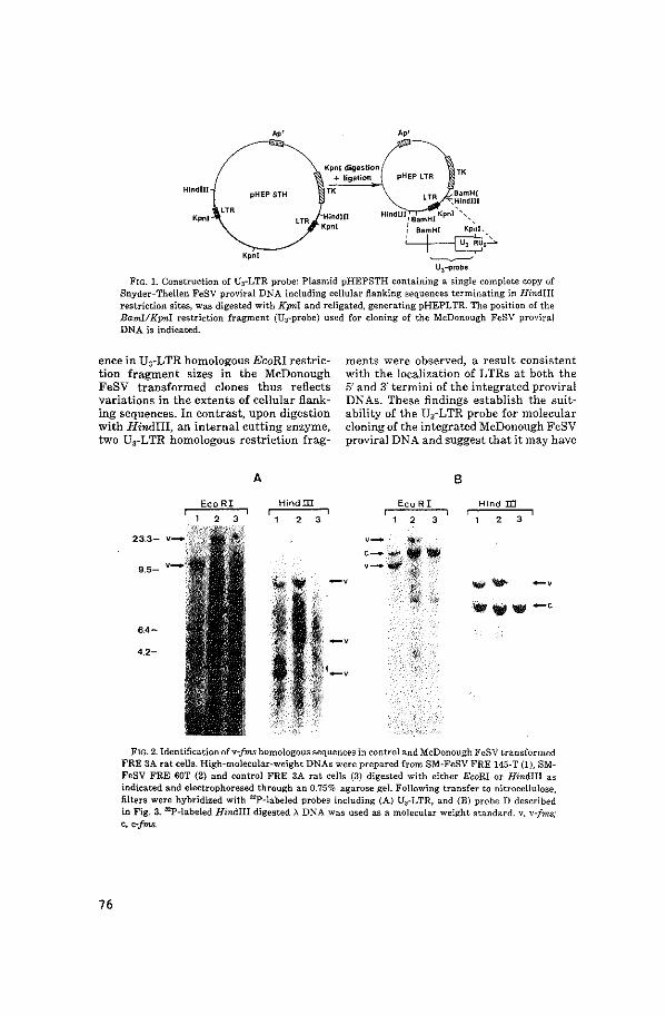
"'
Hindlll pHEP STH
Kpnl
Kpnl
Ao'
pHEPLTR
LTR ,~~~WI Hind'n',-, ,~,-m•,•,'Kpnl' ',,
' ' 1 BamHI Kpnl,
~,C. ~
U3-probe
FrG. 1. Construction of U3-LTR probe: Plasmid pHEPSTH containing a single complete copy of Snyder-Theilen FeSV proviral DNA including cellular flanking sequences terminating in Hindiii restriction sites, was digested with Kpni and religated, generating pHEPLTR. The position of the Bami!Kpnl restriction fragment (U~-probe) used for cloning of the McDonough FeSV proviral DNA is indicated.
ence in U3-LTR homologous EcoRI restriction fragment sizes in the McDonough FeSV transformed clones thus reflects variations in the extents of cellular flanking sequences. In contrast, upon digestion with Hindiii, an internal cutting enzyme, two U3-LTR homologous restriction frag-
ments were observed, a result consistent with the localization of LTRs at both the 5' and 3' termini of the integrated proviral DNAs. These findings establish the suitability of the U3-LTR probe for molecular cloning of the integrated McDonough FeSV proviral DNA and suggest that it may have
76
A 8
Hindm Eco R I Hind ID
2 3 2 3 2 3
FIG. 2. ldentificationofv-frns homologous sequences in control and McDonough FeSV transformed FRE 3A rat cells. High-molecular-weight DNAs were prepared from SM-FeSV FRE 145-T (1}, SMFeSV FRE 60T (2) and control FRE 3A rat cells (3) digested with either EcoRI or Hindlll as indicated and electrophoresed through an 0.75% agarose gel. Following transfer to nitrocellulose, filters were hybridized with 32P-labe\ed probes including (A) U0-LTR, and (B) probe D described in Fig. 3. 9>-\abeled Hindiii digested ;:>., DNA was used as a molecular weight standard. v, v-Jrns; c, c-fms.
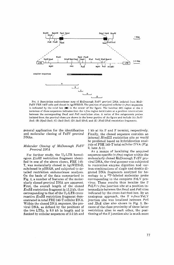
EcoRI BamHI Kpnl Smal
I '"i'' ( ,..---1 II !Ill'
Smal Kpnl EcoRI
::;j.l
Kpnl Pvul[ Smal Hpal Sa~ j Sac I j Xht Pou[l Xhol BamHI --.
I ' ) Ps\1 Ps!l
acquired sequences
'
FIG. 3. Restriction endonuclease map of McDonough FeSV proviral DNA isolated from McDFeSV FRE 145T cells and cloned in AgtWESAB. The position of acquired cellular {v-fm.s) sequences is indicated by the solid box {B) in the center of the figure. The hatched {GJ) region at the 5' terminus of these sequences indicates that the v-frns region terminates at a position intermediate between the corresponding Srnai and Psll restriction sites. A series of five subgenomic probes isolated from the proviral clone are shown in the lower portion of the figure and include {Al Socii Sacl; {B) Hpai!Sacl; {C) Saci/Sacl; {D) Saci/Xhoi; and {E) Xhol!.X7wi restriction fragments.
general application for the identification and molecular cloning of FeSV proviral DNAs.
Molecular Cloning of McDonough FeSV Proviral DNA
For further study, the UrLTR homologous EcoRI restriction fragment identified in one of the above clones, FRE 145-T, was molecularly cloned in AgtWESAB, subcloned in pBR328, and subjected to detailed restriction endonuclease analysis. On the basis of the data summarized in Fig. 3, a number of features of the molecularly cloned proviral DNA are apparent. First, the overall length of the cloned EcoRI restriction fragment is 11.2 kb, thus corresponding to that of the U3-LTR crossreactive EcoRI restriction fragment demonstrated in total FRE 145-T cellular DNA. Within the cloned DNA sequence, the proviral DNA, as defined by the positions of the two LTRs, is 8.0 kb in length and is flanked by cellular sequences of 2.2 kb and
1 kb at its 5' and 3' termini, respectively. Finally, the cloned sequence contains an internal Hindiii restriction site as would be predicted based on hybridization analysis of FRE 145-T total cellular DNA (Fig. 2, lane A-1).
As a means of localizing the acquired sequence-specific (v-fms) region within the molecularly cloned McDonough FeSV proviral DNA, the viral genome was subjected to restriction enzyme digestion and various combinations of single and double digested DNA fragments analyzed for homology to a 32P-labeled molecular probe corresponding to the complete FeLV provirus. These results thus localize the 5' FeLV/v-fms junction site at a position intermediate between the Smai and Psti sites indicated by the cross-hat::hed box. By an analogous approach, the 3' v-fms/FeL V junction site was localized between Psti and JOwl sites also shown in Fig. 3. Because of the close proximity of these latter restriction sites to each other, the positioning of the 3' junction site is much more
77
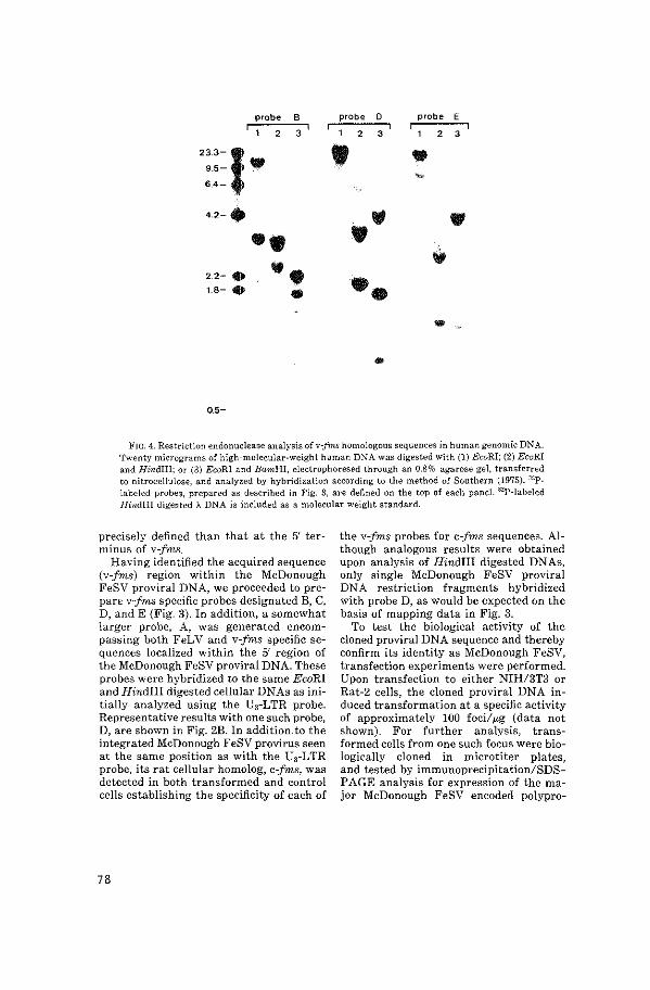
probe B probe 0 probe E
2 3 2 3 2 3
233-l • • 9.5- •
"' 6.4-
42-· ., • •• • ., 2.2- • •• • 1.8- • • • ... .
.. 0.5-
FIG. 4. Restriction endonuclease analysis ofv-frns homologous sequences in human genomic DNA. Twenty micrograms of high-molecular-weight human DNA was digested with (1) EcoRI; (2) EcoRI and Hindiii; or (3) EcoRI and BamHI, electrophoresed through an 0.8% agarose gel, transferred to nitrocellulose, and analyzed by hybridization according to the method of Southern (1975). :rop_ labeled probes, prepared as described in Fig. 3, are defined on the top of each paneL 32P-labeled Hindiii digested A DNA is included as a molecular weight standard.
precisely defined than that at the 5' terminus of v-fms.
Having identified the acquired sequence (v-fms) region within the McDonough FeSV proviral DNA, we proceeded to prepare v-fms specific probes designated B, C, D, and E (Fig. 3). In addition, a somewhat larger probe, A, was generated encompassing both FeLV and v-fms specific sequences localized within the 5' region of the McDonough FeSV proviral DNA. These probes were hybridized to the same EcoRI and Hindiii digested cellular DNAs as initially analyzed using the Us-LTR probe. Representative results with one such probe, D, are shown in Fig. 2B. In addition. to the integrated McDonough FeSV provirus seen at the same position as with the U3-LTR probe, its rat cellular homolog, c-fms, was detected in both transformed and control cells establishing the specificity of each of
78
the v-fms probes for c-fms sequences. Although analogous results were obtained upon analysis of Hindiii digested DNAs, only single McDonough FeSV proviral DNA restriction fragments hybridized with probe D, as would be expected on the basis of mapping data in Fig. 3.
To test the biological activity of the cloned proviral DNA sequence and thereby confirm its identity as McDonough FeSV, transfection experiments were performed. Upon transfection to either NIH/3T3 or Rat-2 cells, the cloned proviral DNA induced transformation at a specific activity of approximately 100 foci/~g (data not shown). For further analysis, transformed cells from one such focus were biologically cloned in microtiter plates, and tested by immunoprecipitation/SDSPAGE analysis for expression of the major McDonough FeSV encoded polypro-
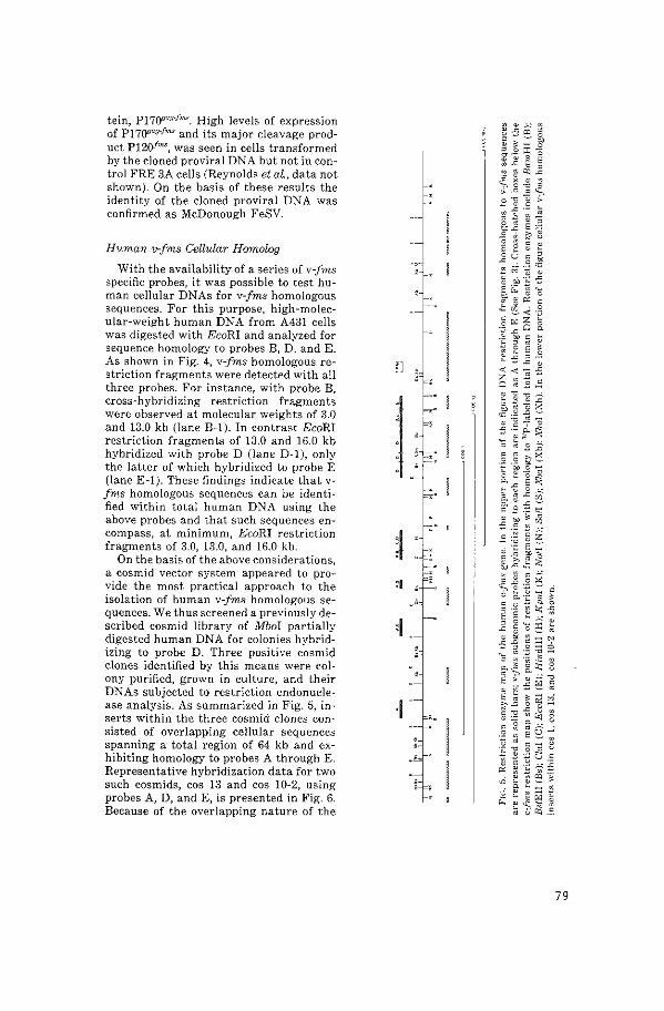
tein, P17fY'agfnw. High levels of expression of P11()flagfms and its major cleavage product P120f""', was seen in cells transformed by the cloned proviral DNA but not in control FRE 3A cells (Reynolds et aL, data not shown). On the basis of these results the identity of the cloned proviral DNA was confirmed as McDonough FeSV.
Human v-fms Cellular Hmnolog
With the availability of a series ofv-fms specific probes, it was possible to test human cellular DNAs for v-fms homologous sequences. For this purpose, high-molecular-weight human DNA from A431 cells was digested with EcoRI and analyzed for sequence homology to probes B, D, and E. As shown in Fig. 4, v-fms homologous restriction fragments were detected with all three probes. For instance, with probe B, cross-hybridizing restriction fragments were observed at molecular weights of 3.0 and 13.0 kb (lane B-1). In contrast EcoRI restriction fragments of 13.0 and 16.0 kb hybridized with probeD (lane D-1), only the latter of which hybridized to probe E (lane E-1). These findings indicate that vfms homologous sequences can be identified within total human DNA using the above probes and that such sequences encompass, at minimum, EcoRI restriction fragments of 3.0, 13.0, and 16.0 kb.
On the basis of the above considerations, a cosmid vector system appeared to provide the most practical approach to the isolation of human v-fms homologous sequences. We thus screened a previously described cosmid library of Mboi partially digested human DNA for colonies hybridizing to probe D. Three positive cosmid clones identified by this means were colony purified, grown in culture, and their DNAs subjected to restriction endonuclease analysis. As summarized in Fig. 5, inserts within the three cosmid clones consisted of overlapping cellular sequences spanning a total region of 64 kb and exhibiting homology to probes A through E. Representative hybridization data for two such cosmids, cos 13 and cos 10-2, using probes A, D, and E, is presented in Fig. 6. Because of the overlapping nature of the
1]
l ~l :I
:I
J
i
J' I '
'l c-·
u-1=~
c-•
79
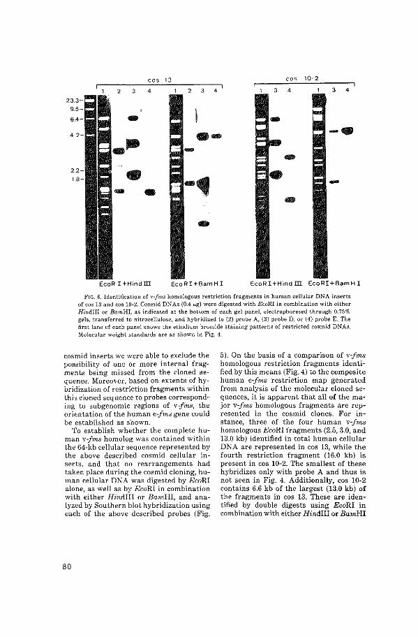
cos 13 cos 10·2
2 3 4 2 3 4 4 3 4
·- -· - --
EcoRI+BamHI EcoRI+Hind m EcoRI+Bam HI
FIG. 6. Identification of v-fms homologous restriction fragments in human cellular DNA inserts of cos 13 and cos 10-2. Cos mid DNAs (0.4 ..,g) were digested with EcoRI in combination with either Hindiii or BamHI, as indicated at the bottom of each gel panel, electrophoresed through 0.75% gels, transferred to nitrocellulose, and hybridized to (2) probe A, (3) probeD, or (4) probe E. The first lane of each panel shows the ethidium bromide staining patterns of restricted cosmid DNAs. Molecular weight standards are as shown in Fig. 4.
cos mid inserts we were able to exclude the possibility of one or more internal fragments being missed from the cloned sequence. Moreover, based on extents of hybridization of restriction fragments within this cloned sequence to probes corresponding to subgenomic regions of v-fms, the orientation of the human c-fms gene could be established as shown.
To establish whether the complete human v-fms homolog was contained within the 64-kb cellular sequence represented by the above described cosmid cellular inserts, and that no rearrangements had taken place during the cos mid cloning, human cellular DNA was digested by EcoRI alone, as well as by EcoRI in combination with either Hindiii or BamHI, and analyzed by Southern blot hybridization using each of the above described probes (Fig.
80
5). On the basis of a comparison of v-fms homologous restriction fragments identified by this means (Fig. 4) to the composite human c-fms restriction map generated from analysis of the molecular cloned sequences, it is apparent that all of the major v-fms homologous fragments are represented in the cosmid clones. For instance, three of the four human v-fms homologous EcoRI fragments (2.5, 3.0, and 13.0 kb) identified in total human cellular DNA are represented in cos 13, while the fourth restriction fragment (16.0 kb) is present in cos 10-2. The smallest of these hybridizes only with probe A and thus is not seen in Fig. 4. Additionally, cos 10-2 contains 6.6 kb of the largest (13.0 kb) of the fragments in cos 13. These are identified by double digests using EcoRI in combination with either Hindiii or BamHI
and hybridization with probes corresponding to various regions of v-fms (Fig. 6). In addition, several minor restriction fragments were observed which hybridized to much lesser extents with one or more of the v-fm.s specific probes. These weakly hybridizing fragments could either reflect secondary loci within the human genome containing v-fms homologous sequences or may reflect low level cellular DNA contaminants of the v-fms probes.
We have previously demonstrated large numbers of highly repetitive sequences within the human c-a,bl cellular homolog, using as a molecular probe, :32P-labeled total human cellular DNA (Heisterkamp et al., 1983). These have been primarily localized within the noncoding (intron) regions of c-abl. To explore the possibility that c·-fms may contain analogous repeats, various combinations of restriction enzyme digests of each of the cosmid clones were assayed for sequence homology to total human cellular DNA. As summarized in Fig. 5, c-fm.s was shown by this means to contain a minimum of 12 separate regions of repetitive sequence. Although the majority of these could be identified within non-v-frns homologous regions of the human c-fms locus, their precise localization will require further study.
DISCUSSION
In the present study we report the molecular cloning of a biologically active McDonough FeSV provirus from non productively transformed Fisher rat embryo cells. This was accomplished by use of a probe specific for the U3 region of the Snyder-Theilen FeSV LTR. In addition, using a molecular probe prepared from the acquired sequence-specific region of the McDonough FeSV genome (v-fms), the human cellular homolog of v-fms has been isolated. It is of interest that the McDonough FeSV proviral DNA isolated in our study, although similar to, shows significant differences from a clone of McDonough FeSV described previously (Donner et al., 1982). These differences are primarily manifested with respect to numbers and positions of particular restriction sites.
For instance McDonough FeSV proviral DNA isolated in the present study lacks one of the Hpai sites identified in the previously described clone and contains additional Sacl, Pstl, and Bglii sites. Such variation may reflect evolutionary drift during the different passage histories of these virus isolates. With respect to their critical properties, however, the two clones appear highly related. Both are of the same overall size, the positioning of their acquired sequence (v-fms) components is similar, and upon transfection to appropriate recipient cell lines, both induce morphologic transformation at high efficiency. Moreover, both encode polyprotein gene products of the same size and immunologic properties. These include a major translational product of around 170,000 Mr and an acquired sequence specific cleavage product of 120,000 Mr.
Using a subgenomic probe corresponding to the acquired sequence-specific (vfms) region of the molecularly cloned McDonough FeSV proviral DNA, we have extended the above studies to molecular cloning of the human v-fms cellular homolog. For this purpose a cosmid vector system was utilized because of the need to obtain cellular DNA inserts of relatively large (30-40 kb) size. The human c-fms gene isolated by this approach encompasses a total length of over 30 kb and contains numerous regions of noncoding (intervening) sequence. Colinearity with v-fms was established on the basis of relative extents of c-fms hybridization to subgenomic probes corresponding to different regions of the McDonough FeSV genome. It should be noted that the mapping procedure used provides an underestimate of the number of introns and overestimate of the size of v-fms homologous regions (exons) within the c-fms human locus. Moreover, when originally generated, the viral v-fms gene may have differed significantly from the cat c-fms and subsequently may have diverged further. Finally, differences in the cat v-fms and human c-fms gene may reflect differences in their species of origin. Thus conclusions regarding the positions and extents of human c-fms coding sequences must at present be considered
81
preliminary.lt is, however, of interest that the c-fms gene contains extensive regions of highly repetitive DNA within what appears to be the noncoding (intron) regions of the genome. Similar sequences have been reported within the noncoding regions of a number of other eukaryotic genes including c-abl (Heisterkamp et aL, 1983). The nature of such sequences, their exact locations and significance will require further study.
With respect to overall size and extent of noncoding regions (introns), the human c-fms locus resembles c-abl which is also large (>25 kb) and consists of highly dispersed coding sequences (Heisterkamp et aL, 1983). Certain human cellular oncogenes such as c-mos lack detectable noncoding sequences (introns) (Prakash et al., 1982; Watson et al., 1982). Although the human homologs of many other viral oncogenes, including c-rasKi, c-rasHa (Chang et aL, 1982), and c-fes (Trus et aL, 1982; Groffen et aL, 1982; Franchini et al., 1982), have been found to contain intervening sequences, the regions of homology have been localized within relatively small DNA fragments. Molecular cloning of each of the latter loci has been accomplished, using conventional A phage vector systems. This approach, however, proved difficult for c-abl and c-fms because of their size, a problem which was to a large extent circumvented by use of the cos mid vector system. An additional advantage of the cosmid system for isolation of the cellular homo logs of these viral oncogenes is that relatively extensive regions are obtained at their 5' and 3' termini, thus strengthening the possibility that the complete cellular homologs of the viral genes, including putative regulatory sequences, are represented. The availability of molecular clones of c-fms should now make possible studies to explore the possible involvement of this cellular oncogene in naturally occurring human tumors.
ACKNOWLEDGMENTS
The authors thank F. H. Reynolds, Jr. for analysis of P170"""f""' expression in v-fms transformed cells and for helpful discussions. The excellent technical assistance of G. T. Blennerhassett and P. Hansen is
82
also gratefully acknowledged. This work was supported under Contract No. NOI-C0-76380 from the National Cancer Institute, Bethesda, Maryland.
REFERENCES
ANDERSON, S. J., FURTH, M., WOLFF, L., RUSCE"ITI, S. K., and SHERR, C. J. (1982). Monoclonal antibodies to the transformation-specific glycoprotein encoded by the feline retroviral oncogene v-fms. J. ViroL 44, 696-702.
BARBAC!D, M., LAUVER, A. V., and DEVARE, S. G. (1980). Biochemical and immunological characterization of polyproteins coded for by the McDonough, Gardner-Arnstein, and Snyder-Theilen strains of feline sarcoma virus. J. ViroL 33, 196-207.
BERNARDS, R., and FLAVELL, R. A. (1980). Physical mapping of the globin gene deletion in hereditary persistance of foetal haemoglobin (HPFH). Nucleic Acids Res. 8, 1521-1534.
CASEY, J. W., ROACH, A., MULLINS, J. 1., BURCK, K. B., NICOLSON, M. Q., GARDNER, M. B., and DAVIDSON, N. (1981}. The U3 portion of feline leukemia virus DNA identifies horizontally acquired proviruses in leukemic cats. Proc. Nat. Acad. Sci. USA 78, 7778-7782.
COFFI~, J. M., VARMUS, H. E., BISHOP, J. M., ESSEX, M., HARDY, W. D., JR., MARTIN, G. S., ROSENBERG, N. E., SCOLNICK, E. M., WEINBERG, R. A., and VOGT, P. K. (1981}. Proposal for naming host cell-derived inserts in retrovirus genomes. J. ViroL 40, 953-957.
DALLA-FA VERA, R., FRANCHINI, G., MARTINOTTI, S., WONG-STAAL, F., GALLO, R. C., and CROCE, C. M. {1982}. Chromosomal assignment of the human homologues of feline sarcoma virus and avian myeloblastosis virus one genes. Proc. Nat. Acad. Sci. USA 79,4714-4717.
DE KLEIN, A., GEURTS VAN KESSEL, A., GROSVELD, G., BARTRAM, C. R., HAGEMEIJER, A., BOOTSMA, D., SPURR, N. K., HEISTERKAMP, N., GROFFEN, J., and STEPHENSON, J. R. (1982). A cellular oncogene (co.bl) is translocated to the Philadelphia chromosome in chronic myelocytic leukemia. Nature (London) 300, 765-767.
DER, C. J., KRO:-<TIR!S, T. G., and COOPER, G. M. (1982). Transforming genes of human bladder and lung carcinoma cell lines are homologous to the ras genes of Harvey and Kirsten sarCO!l1a viruses. Proc Nat. Acad. Sci. USA 79, 3637-3640.
DONNER, L., FEDELE, L.A., GARON, C. F., ANDERSON, S. J., and SHERR, C. J. (1982). McDonough feline sarcoma virus: Characterization of the molecularly cloned provirus and its feline oncogene (v-fms). J. ViraL 41, 489-500.
FEDELE, L. A., EVEN, J., GARON, C. F., DONNER, L., and SHERR, C. J. (1981). Recombinant bacteriophages containing the integrated, transforming provirus of Gardner-Arnstein feline sarcoma virus. Proc. Nat. A cad. Sci. USA 78, 4036-4040.
FLAVELL, R. A., KOOTER, J. M., DE BOER, E., LITTLE, P. F. R., and WILLIAMSON, R. (1978). Analysis of the -y-,8-globin gene loci in normal and Hb Lepore DNA: Direct determination of gene linkage and intergene distance. GelliS, 25-41.
FRANCHINI, G., GELMANN, E. P., DALLA-FA VERA, R., GALLO, R. C., and WONG-STAAL, F. (1982). Human gene (c-fes) related to the one sequences of SnyderTheilen feline sarcoma virus. Mot Cell. BioL 2, 1014-1019.
FURTH, M. E., SCOLNICK, E. M., and LoWY, D. R. (1982). Tumorigenic transformation of mammalian cells induced by a normal human gene homologous to the oncogene of Harvey murine sarcoma virus. Nature 297, 479-483.
GRAHAM, F. L., and VANDER EB, A. J. (1973). Transformation of rat cells by DNA of human adenovirus 2. Virology 54, 536-539.
GROFFEN, J., HEISTERKAMP, N., BLENNERHASSETI, G. T., and STEPHENSON, J. R. (1983). Regulation of viral and cellular oncogene expression by cytosine methylation. Virology, in press.
GROFFEN, J., HEISTERKAMP, N., GROSVELD, F., VAN DE VEN, W., and STEPHENSON, J. R. (1982). Isolation of human oncogene sequences (v-fes homolog) from a cosmid library. Science 216, 1136-1138.
GROSVELD, F. G., DAHL, H-H. M., DE BOER, E., and FLAVELL, R. A. (1981). Isolation of ,8-globin-related genes from a human cosmid library. Gene 13, 227-237.
HEISTERKAMP, N., GROFFEN, J., and STEPHENSON, J. R. (1983). The human v-abl cellular homologue. J. MoL AppL GeMt., in press.
HElSTERKAMP, N., GROFFEN, J., STEPHENSON, J. R., SPURR, N. K., GOODFELLOW, P. N., SOLOMON, E., CARRITT, B., and BODMER, W. F. (1982). Chromosomal localisation of human cellular homo\ogues of two viral oncogenes (c-fes and c-ahl). Nature (London) 299, 747-749.
KLEIN, G. (1982). "Advances in Viral Oncology." Raven, New York.
MULLINS, J. I., CASEY, J. W., BURCK, K. B., DAVIDSON, N., and NICOLSON, M. 0. (1981). The sequence arrangement and biological activity of cloned FeLV proviruses from a virus productive human cell line. J. ViroL 38, 688-703.
McDONOUGH, S. K., LARSEN, S., BRODEY, R. S., STOCK, N. D., and HARDY, W. D., JR. (1971). A transmissible feline fibrosarcoma of viral origin. Cancer Res. 31, 953-956.
PARADA, L. F., TABIN, C. J., SHIH, C., and WEINBERG, R. A. (1982). Human EJ bladder carcinoma oncogene is homologue of Harvey sarcoma virus ras gene. Nature (London) 297, 474-478.
PRAKASH, K., MCBRIDE, Q. W .. SWAN, D. C., DEVARE,
S. G., TRONICK, S. R., and AARONSON, S. A. (1982). Molecular cloning and chromosomal mapping of a human locus related to the transforming gene of Moloney murine sarcoma virus. Proc. Nat. Acad. Sci. USA 79, 5210-5214.
REYNOLDS, F. H., JR., VAN DE VEN, W. J. M., BLOMBERG, J., and STEPHENSON, J. R. (1981). Differences in mechanisms of transformation by independent feline sarcoma virus isolates. J. ViroL 41, 1084-1089.
RuSCETTI, S. K., TUREK, L. P., and SHERR, C. J. (1980). Three independent isolates of feline sarcoma virus code for three distinct gag-x polyproteins. J. ViroL 35, 259-264.
SANTOS, E., TRONICK, S. R., AARONSON, S. A., PULCIANI, S., and BARBACID, M. (1982). T24 human bladder carcinoma oncogene is an activated form of the normal human homologue of BALE- and Harvey-MSV transforming genes. Nature (London) 298, 343-347.
SOUTHERN, E. M. (1975). Detection of specific sequences among DNA fragments separated by gel electrophoresis. J. MoL BioL 98,503-517.
SWAN, D. C., McBRIDE, 0. W., ROBBINS, K. C., KEITHLEY, D. A., REDDY, E. P., and AARONSON, S. A. (1982). Chromosomal mapping of the simian sarcoma virus one gene analogue in human cells. Proc. Nat. Acad. Sci. USA 79,4691-4695.
TOPP, W. C. (1981). Normal rat cell lines deficient in nuclear thymidine kinase. Virology 113, 408-411.
TRUS, M.D., SODROSKI, J. G., and HASELTINE, W. A. (1982). Isolation and characterization of a human locus homologous to the transforming gene (v-fes) of feline sarcoma virus. J. Bio[ Chem. 257, 2730-2733.
VARMUS, H. E. (1982). Form and function ofretroviral proviruses. Science 216, 812-820.
VAN DE VEN, W. J. M., REYNOLDS, F. H., NALEWAIK, R., and STEPHENSON, J. R. (1980). Characterization of a 170,000-dalton polyprotein encoded by the McDonough strain of feline sarcoma virus. J. ViroL 35,165-175.
VERONESE, F., KELLOFF, G. J., REYNOLDS, F. H., JR., HILL, R. W., and STEPHENSON, J. R. (1982). Monoclonal antibodies specific to transforming polyproteins encoded by independent isolates of feline sarcoma virus. J. ViroL 43, 896-904.
WATSON, R., 0SKARSSON, M., and VANDE WOUDE, G. F. (1982). Human DNA sequence homologue to the transforming gene (mos) of Moloney murine sarcoma virus. Proc. Nat. Acad. Sci. USA 79, 4078-4082.
WIGLER, M., PELLICER, A., SILVERSTEIN, S., and AXEL, R. (1978). Biochemical transfer of single-copy genes using total cellular DNA as donor. Celll4, 725-731.
83


APPENDIX PAPER II

Chromosomal localization of the human c-fms oncogene
John Groffen 1, Nora Heisterkampl, Nigel Spurr2 , Sharon Dana3 , John J.Wasmuth 3 and John R.Stephenson1
1Lab. Viral Carcinogenesis, National Cancer Institute-FCRF, Frederick, MD 21701, USA, 2Imperial Cancer Research Fund, P.O. Box 123, Lincoln's Inn Fields, London WC2A 3PX, UK, and 3Dept. Biological Chemistry, California College of Medicine, Univ. California, Irvine, CA 92717, USA
Received 27 June 1983; Revised and Accepted 10 August 1983
ABSTRACT A molecular probe was prepared >Nith specificity for the human cellular
homologue of transforming sequences represented within the HcDonough strain of feline sarcoma virus (v-fms). By analysis of a series of mouse-human somatic cell hybrids containing--;ariable complements of human chromosomes it was possible to assign this human oncogene, designated c-fms, to chromsome 5. Regional localization of c-fms to band q34 on chromosome-s-was accomplished by analysis of Chinese hamster-human cell hybrids containing as their only human components, terminal and interstitial deleted forms of chromosome 5. The localization of c-fms to chromosome 5 (q34) is of interest in view of reports of a specific, apparently interstitial, deletion involving approximately two thirds of the q arm of chromosome 5 in acute myelogenous leukemia cells.
INTRODUCTION
A number of well defined genetic sequences, designated "oncogenes," have
been identified within the human genome ( 1 ,2). Such sequences were demon-
strated by use of molecular probes corresponding to transforming sequences of
oncogenic retrovirus isolates of various avian and mammalian species. In
general, cellular oncogenes have remained highly conserved throughout
vertebrate evolution although their functional significance is not known. The
viral counterparts of many of the oncogenes studied to date are known to
encode tyrosine specific protein kinases while enzymatic functions have not
been ascribed to transforming proteins encoded by the others (3). Support for
the possible involvement of cellular oncogenes in human cancer has recently
been derived by the demonstraton that under certain conditions, members of the
ras family, can transform cells in culture (4-7). Moreover, two other human
cellular oncogenes, c-abl and c-myc, are involved in translocations associated
with chronic myelogenous leukemia (8) and Burkitt's lymphoma (9,10),
respectively.
The human c-fms oncogene represents a cellular sequence initially
identified on the basis of homology to the transforming gene (v-fms) of the
87
McDonough strain of feline sarcoma virus (11,12). We have recently applied a
cosmid vector system for the molecular cloning of this gene (12). Although the
c-fms gene product has not been identified, the corresponding viral gene, v
fms, has been shown to encode a protein with transforming function (13,14) but
lacking protein kinase activity (15). In the present study we have developed a
specific probe for the human c-fms gene. Using this reagent, studies were
performed to determine the chromosomal localization of c-fms and its relation
to chromosomal deletions and/or rearrangements associated with specific forms
of human cancers.
MATERIALS AND METHODS
Cells and Viruses: Cells were grown in Dulbecco's modification of Eagles
medium supplemented with 10% calf serum and included NIH/3T3 mouse cells (15),
A673 human cells (15) and the human-mouse somatic cell hybrids described in
Table 1. A series of human-Chinese hamster cell hybrids, including HHW105
containing chromosome 5 as its only human component and segregants of HHW105,
designated HHW207, HHW'209, ffilW212, HHW213, HHW224, characterized by a variety
of terminal and interstitial deletions of chromosome 5, have been described
(16,17).
Preparation of a Human c-fms Specific Probe: The isolation of a cosmid
clone with a cellular insert of approximately 38 kb containing v-fms homo
logous sequences (cos 13) from a library of human lung carcinoma DNA has been
described (12). For use as a probe, an 0.9 kb Kpn-1 restriction fragment, was
isolated from cos 13 according to previously described methods (12).
Molecular Hybridization: Restriction enzymes were purchased from New
England Biolabs and Bethesda Research Laboratories and were used according to
the suppliers' specifications. DNAs were digested with restriction enzymes,
subjected to electrophoresis through 0. 75% agarose gels and transferred to
nitrocellulose essentially as described by Southern (18). Nick translation of
were as described (19), Specific activity of probes and filter hybridization
the probes was 2-5xl08
cpm/ug. After hybridization, filters were washed under
high stringency conditions (0.1 xSSC,65°C) and exposed to XAR-2 film (Kodak)
for up to 5 days at -70°C with Dupont Lightning Plus intensifying screens.
RESULTS
Identification of Human c fms Specific Sequences: As a means of
discriminating human c-fms sequences from related sequences in the mouse
cellular genome, a series of probes were prepared from cos 13 (Fig. 1), a
88
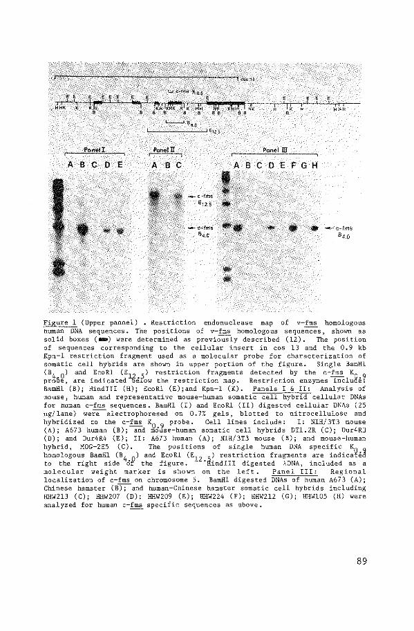
~KG.!< E,E,EEEEEE, E
"J'JJK;"-~·I;--~ln·~~L~~·~~~~~--~L--r.~~~ntt~~-k~~rrtr;-:"~~~~··M~l_.x~t~J~I·~-----,-~o--r~-"--L-M-r-~ a & a a a e ae sa
• • • • I
I I' I I I I II ' H •' H HHH
-~1!4.0
Pone! I PoneiU Panel m A B C 0 E A B C ABCDEFGH
Figure 1 (Upper pannel) • Restriction endonuclease map of v-fms homologous human DNA sequences. The positions of v-fms homologous sequences, shown as solid boxes ( IBD) were determined as previously described (12). The position of sequences corresponding to the cellular insert in cos 13 and the 0.9 kb Kpn-1 restriction fragment used as a molecular probe for characterization of somatic cell hybrids are shown in upper portion of the figure. Single BamHl (B
4 0) and EcoRl (E 12 'l) restriction fragments detected by the c-frr:.s K0 9
proOe, are indicatedbe:Iow the restriction map. Restriction enzymes inclua:e: BamHl (B); tlindiii (H); EcoRl (E);and Kpn-1 (K). Panels I & II: Analysis of mouse, human and representative mouse-human somatic cell hybrid cellular DNAs for human c-fms sequences. BamHl (I) and EcoRl (II) digested cellular DNAs (25 )..lg/lane) wer:e-electrophoresed on 0.7% gels, blotted to nitrocellulose and hybridized to the c-fms K
0 9 probe. Cell lines include: I: NIH/3T3 mouse
(A); A673 human (B); and mOuse-human somatic cell hybrids DT1.2R (C); Dur4R3 (D); and Dur4R4 (E); II: A673 human (A); NIH/3T3 mouse (B); and mouse-human hybrid, MOG-2E5 (C). The positions of single human DNA specific K homologous BamHl (B
4 0) and EcoRl (E
12 'i) restriction fragments are indica~ed
to the right side Of the figure. "Hindiii digested :\DNA, included as a molecular weight marker is shown on the left. Panel III: Regional localization of c-fms on chromosome 5. BamHl digested DNAs of human A673 (A); Chinese hamster (B~ and human-Chinese hamster somatic cell hybrids including HHW213 (C); HHW207 (D); HHW209 (E); HHW224 (F); HHW212 (G); HHW105 (H) were analyzed for human c-fms specific sequences as above.
89
previously described cosmid clone containing v-fms homologous human cellular
DNA sequences (12). One such probe, c-fms K0 , 9 was suitable for the purpose
of the present study in that it lacked detectable repetitive sequences and
hybridized specifically to single 4.0 kb BamHl and 12.5 kb EcoRl human DNA
restriction fragments (Fig. l, Panels I & II). The sizes of both restriction
fragments correspond to those predicted on the basis of restriction endonu
clease map analysis of the v-fms homologous region of the human genome. No
detectable cross-homology between this probe and mouse cellular DNA sequences
was observed when high stringency washing conditions were used as described in
Methods.
c-fms Maps on Human Chromosome 5: To determine the chromosomal location
of the human cellular homologue of v-fms, EcoRl and BamHl restriction
endonuclease-digested cellular DNAs of human-mouse somatic cell hybrids were
screened for human c-fms specific sequences. The above described c-fms K0
•9
probe was used for this purpose. These hybrids each contain a full complement
of mouse chromosomes but have retained only limited numbers of human
chromosomes. As shown in Fig, l, Panels I & II, it was possible, by this
means, to distinguish between hybrids containing the human c-fms gene and
those lacking such sequences, As shown in Table 1, six of the fifteen somatic
cell hybrids analyzed contained c-fms K0
•9
homologous sequences. Only four
chromosomes (5 ,12 ,14 and 17) are common to all six c-fms positive hybrids.
These findings localize c-fms to human chromosomes 5,12,14 or 17. To further
localize c-fms we examined the chromosomal content of the series of nine
hybrids which were non-crossreactive with the c-fms K0
•9
probe. As summarized
in the lower portion of Table 1, only human chromosome 5 was missing from each
of these nine hybrids. This excludes the possibility of c-fms mapping on
chromosomes 12,14 or 17, each of which were present in at least three of the
c-fms negative hybrids, and thus localizes c-fms to human chromosome 5.
Regional Localization of c-fms to Chromosome 5 (q34): The recent
isolation of a series of segregants of Chinese hamster-human somatic cell
hybrids expressing variable numbers of genes mapping on chromosome 5 (16 ,17)
provided a means of regionally localizing c-fms. The majority of these
segregants are characterized by terminal deletions involving well defined
regions of the long arm of chromosome 5; these were isolated by positive
selection for leuS, which maps on the p arm or very near the centromere on the
q arm of the chromosome, and negative selection directed against markers on
the distal half of the q arm (emtB and chr), DNAs prepared from Chinese
hamster-human hybrid cells were analyzed for c-fms K0
•9
homologous sequences
90
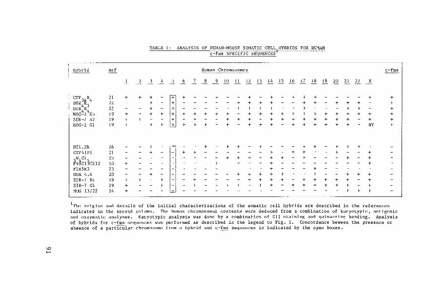
TABLE 1: ANALYSIS OF HUHAN~MOUSE SOMATIC CELL HYBRIDS FOR HUr1AN c fms SPECIFIC SEQUENCES
Hybrid Ref Human Chromosomes c~fms
l 2 3 4 5 6 7 8 9 10 ll 12 13 14 15 16 17 18 19 20 21 22 X
CTP 3~a4 21 + + +
[ + > + > + + + + +
OUR 22 + + + + + + + + + + + DUR
4R
3 22 + + + + + + + + + MOG~2 4E5 19 + + + + + + + + + + + + + + + + + + + + + SIR-7 A2 19 + + + + + + + + + + + + + + + + t10G-2 Gl 19 + + + + + + + + + + + + + + + + NT +
DTl. 2R 26 + + + + t + + t + + CTP41Pl 21 + + + + + + + + w Cl 25 + + + + + + + + h~c1Jcuz lO + + + + FlR5R3 23 + + DUR 4.4 20 + + + + + + + + + + SIR-7 Dl 19 + + + + + + + + + + + + + + SIR-7 Gl 19 + + + + + + + + + + + + + + MOG U/22 24 + + + +
1The origins and details of the initial characterizations of the somatic cell hybrids are described in the references indicated in the second column. The human chromosomal contents were deduced from a combination of karyotypic, antigenic and enzymatic analyses. Karyotypic analysis was done by a combination of Gll staining and quinacrine banding. Analysis of hybrids for c-fms sequences was performed as described in the legend to Fig. !. Concordance beween the presence or absence of a particul~r chromosome from a hybrid and c-fms sequences is indicated by the open boxes.
"' ,....
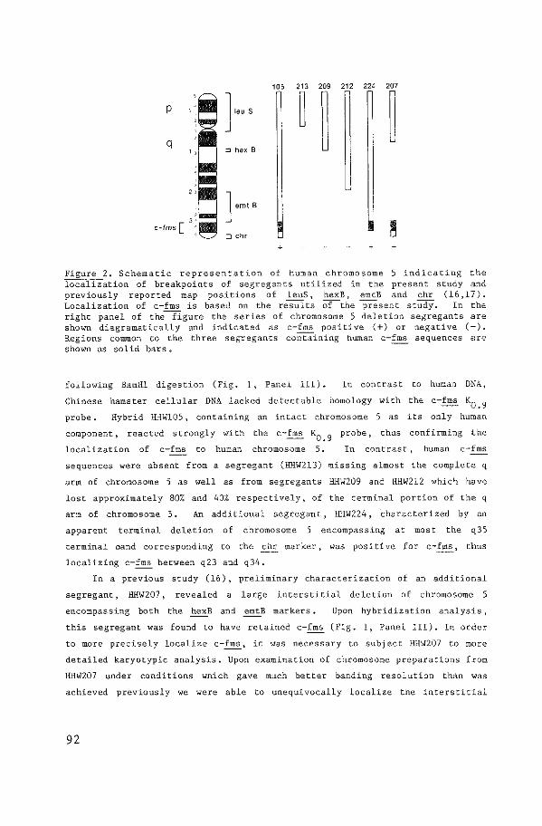
105 213 209 212 224 207
p
q q ~
' J emt B
~ e-lms [ ;:J chr
+ + +
Figure 2. Schematic representation of human chromosome 5 indicating the localization of breakpoints of segregants utilized in the present study and previously reported map positions of leuS, hexB, emtB and chr (16,17). Localization of c.-fms is based on the results or-the present study. In the right panel of thefigure the series of chromosome 5 deletion se6regants are shown diagramatically and indicated as c-fms positive ( +) or negative (-). Regions common to the three segregants containing human c-fms sequences are shown as solid bars.
following BamHl. digestion (Fig. 1, Panel III). In contrast to human DNA,
Chinese hamster cellular DNA lacked detectable homology with the c-fms K0
_9
probe. Hybrid IDIW105, containing an intact chromosome 5 as its only human
component, reacted strongly with
localization of c-fms to human
the c-fms
chromosome
probe, thus confirming th.e
In contr~st, human c-fms
sequences were absent from a segregant (HHW213) missing almost the complete q
arm of chromosome 5 as well as from segregants HHW209 and HHW212 which have
lost approximately 80% and 40% respectively, of the terminal portion of the q
arm of chromosome 5. An additional segregant, HHW224, characterized by an
apparent terminal deletion of chromosome 5 encompassing at most the q35
terminal band corresponding to the chr marker, was positive for c-fms, thus
localizing c-fms between q23 and q34.
In a previous study (16), preliminary characterization of an additional
segregant, HHW207, revealed a large interstitial deletion of chromosome 5
encompassing both the hexB and emtB markers. Upon hybridization analysis,
this segregant was found to have retained c-fms (Fig. 1, Panel III). In order
to more precisely localize c-fms, it was necessary to subject HHW207 to more
detailed karyotypic analysis. Upon examination of chromosome preparations from
HHW207 under conditions which gave much better banding resolution than was
achieved previously we were able to unequivocally localize the interstitial
92
deletion as encompassing the region of chromosome 5 mapping from ql2 to q33,
As shown schematically in Fig. 2, these results est.<1blish the order of the
individual markers on chromosome 5 as leuS, hexB, emtB, c-fms and chr and more
specifically localize c-fms to band q34.
DISCUSSION:
In the present study ~e have utilized a panel of mouse-human somatic cell
hybrids to map the human oncogene, c-fms, on chromosome 5, Additionally, the
availability of a set of hybrids with well defined deletions in chromosome 5
(16,17) has allowed regional localization of c-fms to band q34. The gener-
ation of sets of deletion hybrids, such as those used in the present study, is
possible for any human chromosome for which appropriate positive and negative
selection markers can be developed. By use of such hybrids in combination with
Southern hybridization the localization of specific genetic sequences can be
achieved v;rith much less ambiguity than is possible by use of alternative
techniques such as in situ hybridization.
The localization of c-fms on chromosome 5, in addition to the previous
assignment of other human oncogenes to specific chromosomes, establishes the
distribution of the known oncogenes among human chromosomes to be relatively
random. In fact, the eight human oncogenes which have been mapped to date are
distributed among seven different chromosomes. These include c-fes (20,21), c
myb (21), c-abl (20), c-sis (22,23) and c-ras-H (24) which have been mapped to
chromosomes 15, 6, 9, 22 and 11, respectively, c-fms localized in the present
study on chromosome 5, and two human oncogenes, c-myc (9,10) and c-mos (25,26)
both of which map on chromosome 8.
Evidence for an association of human oncogenes w-ith translocations and
deletions specific to various human cancers is accumulating. For instance, c
ab! has been localized within a small terminal region of chromosome 9 (20)
which is translocated to chromosome 22 in chronic myelogenous leukemia (9 ,10)
while c-sis maps •,o1ithin the reciprocally translocated region of chromosome 22
(27). Similarly, c-myc has been identified near the breakpoint of the segment
of chromosome 8 which trans locates to chromosomes 2, 14 or 22 in Burkitt's
lymphoma (9 ,10) and c-fes has been mapped within the region of chromosome 15
which is translocated to chromosome 17 in acute promyelocytic leukemia (28).
In other studies, c-ras-H has been mapped on chromosome 11, a human chromosome
with a characteristic deletion in Wilms tumor (24). The mapping of c-fms on
chromosome 5 is of interest in that deletions within the long arm of
chromosome 5 are also frequently observed in patients with refractory anemia
93
(29) and acute myelogenous leukemia (30,31). Such deletions have been reported
to appear to be interstitial, involving approximately t~>~o thirds of the long
arm of chromosome 5, but not extending to band q34 (29,31).
As one possible model, to account for their association with chromosomal
rearrangements occurring in specific human cancers, cellular oncogenes, such
as c-fms could map adjacent to chromosomal breakpoints possibly resulting in
the juxtapositioning of potentially oncogenic sequences with cellular
promoters. Such a model has been proposed to account for the trans locations
associated with chronic myelogenous leukemia and Burkitt's lymphoma (8-10); in
fact, in Burkitt's lymphoma the cellular sequences in the chromosome to which
c-myc is translocated appear to be involved in immunoglobulin rearrangements
(9 ,10). It will thus be of interest to accurately map the proximity of c-fms
to chromosome Sq- deletions associated with acute myelogenous leukemia and to
analyze tumors with the Sq- deletion for expression of c-fms gene products.
Independent of its potential involvement in neoplasia, the localization of c
fms provides a unique molecular marker for chromosome 5 which should be useful
for identifying syntenic linkage associations among diverse mammalian species,
ACKNOWLEDGEMENT: The authors thank G.T. Blennerhassett and P. Hansen for their excel
lent technical assistance. This work was supported under Contract No, NOI-C0-76380 from the National Cancer Institute, Bethesda, Haryland, and by Public Health Service Grant No, GM-25339 from the National Institute of General Medical Sciences, Bethesda, Maryland.
REFERENCES: l. Varmus, H.E. (1982) Science 216, 812-820 2. Bishop, J.M. (1902) Retrovirus and Cancer Genes, In Adv. Cancer Res. 37,
1-32 3. Klein, G. (1982) Advances in Viral Oncology. New York, Raven Press. 4, Parada, L.F., Tabin, C.J., Shih, C. and Weinberg, R.A. (1982) Nature
297 ,474-478 5. Santos, E., Tronick, S.R., Aaronson, S.A., Pulciani, S., and Barbacid,
M. (1982) Nature 298, 343-347 6. Der, C.J., Krontiris, T.G., and Cooper, G.t1. (1932) Proc. Natl. Acad,
Sci. USA 79, 3637-3640 7. Chang, E.H., Furth, M.E., Scolnick, E.M. and Lo~vy, D.R. (1982) Nature
297, 479-483 3. de Klein, A. Geurts van Kessel, A., Grosveld, G., Bartram, C.R.,
Hagemeijer, A., Bootsma, D., Spurr, N., Heisterkamp, N., Groffen, J. and Stephenson, J.R. (1982) Nature 300, 765-767
9. Dalla-Favera, R., Bregni, M., Erikson, J., Patterson, D., Gallo, R.C., Croce, C.M. (1982) Proc. Natl. Acad, Sci. U.S.A. 79, 7824-7827
10. Taub, R., Kirsch, I., Morton, C., Lenoir, G., Swan, D., Tronick, s., Aaronson, S., and Leder, P. (1982) Proc, Natl. Acad. Sci. USA 79, 7837-7841
11. Donner, L., Fedele, L.A., Garon, C.F., Anderson, S.J., and Sherr, C.J. (1982) J. Viral, 41, 489-500
94
12. Heisterkamp, N., Groffen, J., and Stephenson, J.R. 126, 248-258
(1983) Virology
13. Van de Ven, W.J.M., Reynolds, F.H., Nalewaik, R., and Stephenson, J.R. (1980) J. Viral. 35, 165-175
14. Barbacid, M., Lauver, A.V., and Devare, S.G. (1980) J. Viral. 33, 196-207
15. Reynolds, F.l:L Jr., Van de Ven, W.J.M., Blomberg, J., and Stephenson, J.R. (1981) J. Vriol. 41, 1084-1089
16. Dana, S., and Wasmuth, J.J. (1982) 17. Dana, s., and Wasmuth, J.J. (1982) 18. Southern, E.M. (1975) J. Mol. Biol.
Malec, & Cell Biol, 2, 1220-1228 Somatic Cell Gen. 8, 245-264
98, 503-517 19. Groffen, J., Heisterkamp, N., Grosve1d, F., Van de Ven, W., and
Stephenson, J.R. (1982) Science 216, 1136-1138 20. Heisterkamp, N., Groffen, J., Stephenson,J.R., Spurr, N.K., Goodfellow,
P.N., Solomon, E., Garritt, B., and Bodmer, W,F. (1982) Nature 299, 747-749
21. Dalla-Favera, R., Franchini, G., Martionotti, s., Wong-Staa1, F., Gallo, R.C., and Croce, C.M. (1982) Proc. Natl.Acad, Sci, USA 79, 4714-4717
22. Swan, D.C., McBride, O.W., Robbins, K.C., Keithley, D.A., Reddy, E.P., and Aaronson, S.A. (1982) Proc. Nat1.Acad. Sci. USA 79, 4691-4695
23. Dalla-Favera,R., Gal1o,R.C., Gia1longo,A., and Croce, C.M. (1982) Science 218, 686-688
24. McBride, O.W., Swan, D.C.,Santos, E., Barbacid, M., Tronick, S.R., and Aaronson,S.A. (1982) Nature 300, 773-774
25. Prakash, K., McBride, O.W., Swan, D.C., Devare, S.G., Tronick, S.R., and Aaronson, S.A. (1982) Proc. Natl. Acad,Sci. USA 79, 5210
26. Nee!, B.G., Jhanwar, S.C., Chaganti, R.S.K.,and Hayward, W.S. (1982) Proc. Natl. Acad, Sci. USA 79, 7842-7846
27. Groff en, J., Heisterkamp, N., Stephenson, J .R., Geurts van Kessel, A., de Klein, A., Grosveld, G., and Bootsma, D. (1983) J. Expt. Med. 158, 9-15.
28. Sheer, D., Hiorns, L.R., Goodfellow, P .N,, Trows dale, J., Solomon, E., Swallow, D.A., Pavey, S., Heisterkamp, N., Groffen, J., and Stephenson, J.R. (1983) Proc. Nat!. Acad. Sci. USA (in press)
29. Van den Berghe, H., Cassiman, J.J., David, G., Fryns, J.P., Michaux, J.L., and Sakal, G. (1974) Nature 251, 437-438
30. Sakal, G., Michaux, J.L., Van den Berghe, H., Cordier, A., Rodhain, J., Ferrant, A., Moriau, H., de Bruyere, M., Sonnet, J. (1975) Blood 46, 519-553
31. Van den Berghe, H., David, G., Michaux, J.L., Sakal, G., and Verwilghen, R., (1976) Blood 48, 624-626
32. Whitehead, A.S., Solomon, E., Chambers, S., Bodmer, W.F., Pavey, S., and Fey, G. (1982) Proc. Nat!. Acad. Sci, USA 79, 5021-5025
33. Solomon, E., Bobrow, M., Goodfellow, P.N., Bodmer, W.F., Swallow, D.M., Pavey, s., and Noel, B. (1976) Somat. Cell Genet, 2, 125-140
34. Jones, E.A., Goodfellow, P.N., Kennett, R.H., and Bodmer, W.F. (1976) Somat, Cell Genet. 2, 483-496
35. Solomon, E., Bobrow, M., Goodfellow, P.N., Bodmer, W.F., Swallow, D.~ •• Pavey, s., and Noel, B. (1976) Somat. Cell Genet. 2, 125-140
36. Hobart, M.J., Rabbits, T.H., Goodfellow, P.N., Solomon, E., Chambers, S., Spurr, N., and Pavey, S. (1981) Ann. Hum. Genet. 45, 331-335
37. Pavey, S., Jeremiah, S.J., Barker, R.F., Hopkinson, D.A., Robson, E., Cook, P .. J.L., Solomon, E., Bobrow, M., Carrot, B., and Buckton, K.E., (1980) Ann. Hum. Genet. 43, 241-248
38. Nabholz, M.,Miggiano, V., and Bodmer, W.F. (1969) Nature 223, 358-363 39. Swallow, D.M., Solomon, E., and Pajunen, L. (1977) Cytogenet. Cell
Genet. 18, 136-148
95


APPENDIX PAPER III

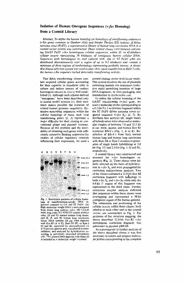
Isolation of Human Oncogene Sequences (v-fes Homolog)
from a Cosmid Library
Abstract. To define the human homolog (or homologs) of transforming sequences (v-fes gene) common to Gardner (GA) and Snyder Theilen (ST) isolates of feline sarcoma virus (FeSV), a representative library of human lung carcinoma DNA in a cosmid vector system was constructed. Three cosmid clones v.:ere·isolated containing GAIST FeSV v-fes homologous cellular sequences, within 32- to 42-kilobase cellular inserts representing 56 kifobases of contiguous human cellular DNA. Sequences both homologous to, and colir;ear with, GA or ST FeSV 1{-fes are distributed discontinuously over a region of up to 9.5 kilobases and contain a minimum of three regions of nonhomology representing probable introns. A thymidine kinase sele.ction system was used to show that, upon transfection to RAT-2 cells, the human c-fes sequence lacked detectable transforming activity.
That RNA transforming viruses contain acquired cellular ge_nes accounting for their capacity to transform cells in culture and induce tumors of various histological classes in vivo is well established(/). Although such ceiiular derived "oncogenes" have been described only in animal model systems (!), their existence makes possible the isolation of related human genomic sequences. Extensive noncoding sequences within the cellular homologs of many such viral transforming genes (2, J) represent a major difficulty for their cloning in conventional phage and plasmid systems. Because of this problem and the desirability of obtaining such genes with sufficiently extensive flanking sequences for studies of cellular regulatory controls influencing their expression, we used a
ABCDEFGH
Fig. 1. Restriction patterns of cellular homologs of transformation-specfic DNA sequences common toGA and ST FeSV. (A) High molecular weight DNA "s were prepared from kitten lung embryo cc:lls; (B) CCL64 mink lung cells; (C) NlH/3T3 mouse embryo cells; (D and G) nonnal human lung tissue; and (E, F, and H) human lung carcinoma tissue. DNA samples (20 IJ-g) were digest<:d with Eco Rl (A to E), Bam HI {F), or Kpn I (G and H), separat<:d by electrophoresis on 0.75 percent agarose gels, transferred to nitrocellulose, and analyzed by hybridization according to previously described procedurc:s (19). nP-Labe!ed DNA digested with Hind Ill is included as a molecular weight standard.
cosmid cloning vector (4-6) in our study. This system involves the use of plasmids containing lambda cos sequences (cohesive ends) permitting insertion of large DNA fragments, in vitro packaging, and transduction to Escherichia coli.
To define the cellular homolog of the GAIST transforming (v-fes) gene, we used a molecular probe corresponding to a 0.5-kb Pst I restriction fragment within the ST FeSV (feline sarcoma virus) acquired sequence (v-fes Sd (2, 7). By Southern blot analysis (8), single bands of hybridization were observed at molecular. weights of between 7.0 and 12.5 kb in cat, mink, mouse, and human Eco RI restricted DNA's (Fig. I, A to E). Restriction of DNA's from both normal human lung and human lung carcinoma with Bam HI or Kpn I resulted in generation of single bands hybridizing at 3.8 kb (Fig. I F) and 2.4 kb (Fig. 1, G and H), respectively.
A cosmid library was constructed and screened for v-fes homologous sequences (Fig. 2). Three clones were initially selected on the basis of hybridization to v-fes SL and were propagated for restriction endonuclease analysis. Two of the clones contained a 12.0-kb Eco RI restriction fragment with hofnology to both v:fes SL and v-jes SR. while only the 9.8-kb 5' region of this fragment was represented in the third clone. Further restriction enzyme analysis indicated that sequences within these clones were overlapping and represented a 58-kb contiguous region of the human genome. The orientation and positioning of the cellular inserts within these clones, both relative to each other and to the cosmid vector, are summarized in Fig. 3. For purposes of fine structure mapping, the above described 12.0-kb Eco RI v-fes homologous restriction fragment was subcloned in plasmid pBR328.
As a prerequisite to further analysis of the above described clones it was first necessary to isolate and prepare molecular probes corresponding to the complete
99
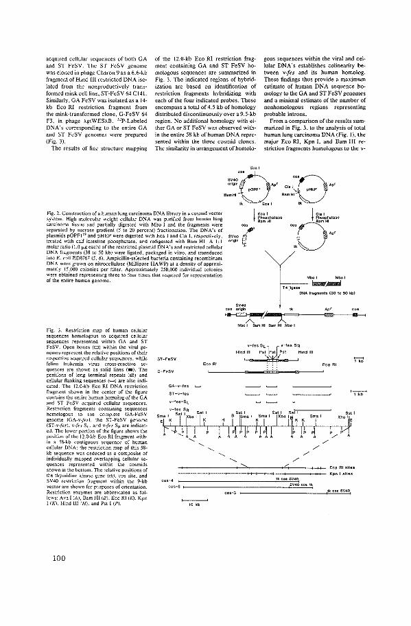
acquired cellular sequences of both GA and ST FeSV. The ST FeSV genome was cloned in phage Charon 9 as a 6.6-kb fragment of Hind Ill restricted DNA isolated from the nonproductively transformed mink cell line, ST-FeSV 64 C!4l. Similarly, GA FeSV was isolated as a 14-kb Eco RI restriction fragment from the mink-transformed clone, G-FeSV 64 F3, in phage ;l..gtWES;~..B. 32P-Labeled DNA's corresponding to the entire GA and ST FeSV genomes were prepared (Fig. 3).
The results of fine structure mapping
of the 12.0-kb Eco Rl restriction fragment containing GA and ST FeSV homologous sequences are summarized in Fig. 3. The indicated regions of hybridization are based on identification of restriction fragments hybridizing with each of the four indicated probes. These encompass a total of 4.5 kb of homology distributed discontinuously over a 9.5-kb region. No additional homology with either GA or ST FeSV was observed within the entire 58 kb of human DNA represented within the three cosmid clones. The similarity in arrangement ofhomolo-
Eca I
gous sequences within the viral and cellular DNA's establishes colinearity between v-fes and its human homolog. These findings thus provide a maximum estimate of human DNA sequence homology to the GA and ST FeSV genomes and a minimal estimate of the number of nonhomologous regions representing probable introns.
From a comparison of the results summarized in Fig. 3, to the analysis of total human lung carcinoma DNA (Fig. 1), the major Eco RI, Kpn I, and Bam HI restriction fragments homologous to the v-
Bo
SV40 origin Apr
' Bam HI
lk Eoa I B.
"' Cia I pHEP
Bam HI
" Fig. 2. Construction of a human lung carcinoma DNA library in a cosmid vector system. High molecular weight cellular DNA was purified from human lung carcinoma ti~sue and partially digested with Mbo I and the fragments were separated by sucrose gradient (5 to 20 percent) fractionation. The DNA's of plasmids pOPFI 19 and pHEP were digested with Eca I and Cia I. respectively, treated with calf intestine phosphatase, and redigested with Bam HI. A I: t molar ratio (1.0 ,_..g each) of the restricted plasmid DNA's and restricted cellular DNA fragments (30 to 50 kb) were ligated. packaged in vitro, and transduced into E. coli ED8767 (5. 6). Ampicillin-selected bacteria containing recombinant DNA were grown on nitrocellulose (Millipore HA WP) at a density of approximately 15.000 colonies per filter. Approximately 250,000 individual colonies were obtained representing three to four times that required for representation of the entire human genome,
sr-F.sv
GA-v-fes
ST-v-fes
v-fes-Sl
Eco Rl
"" '(" origin ·'
j "" Phosphatase Bam HI
j Cia I
0:::~~~""
"' "'
"
T4llgue ONA fragmenl> (SO 10 50 kb)
SV40 cos or111in 1~ Apr
~ .. ·)(~==~---~r-~-~ Mbo I Sam HI Sam HI Mb<> I
Eco Rl
Fig. 3. Restriction map of human cellular sequences homologous to acquired cellular oequences represented within GA and ST FeSV. Open boxes {D) within the viral genomes represent the relative positions of their respective acquired cellular sequences, while feline leukemia virus cross-reactive sequences are shown as solid lines (•). The positions of long terminal repeats (fll!l) and cellular flanking sequences(.....,) are also indicated. The 12.0-kb Eco RJ DNA restriction fragment shown in the center of the figure contains the entire human homolog of the GA and ST FeSV acquired cellular sequences. Restriction fragments containing sequences homologous to the complete GA-FeSV genome (GA-v-fe•). the ST-FeSV genome (ST-v-fes). v-Jes S~. and v-fes s,. are indicated. The lower portion of the figure shows the position of the 12.0-kb Eco Rl fragment with· in a 58-kb contiguous sequence of human cellular DNA: the restriction map of this 58-kb sequence was deduced as a composite of individually mapped overlapping cellular sequences represented within the cosmids shown at the bollom. The relative positions of the thymidine kinase gene (lk). cos site, and SV40 restriction fragment within the 9-kb vector are shown for purposes of orientation. Restriction enzymes are abbreviated as follows: Ava I (A). Bam HI (B). Eco Rl (EJ. Kpn I (KJ. Hind Jll {H}. and Pst I (PJ
~::::::;::::;::::;::::;::::;:::;::::::=::;:::;::;::;:::::;::::::/::::;::::;::::;:::;:= Eco Rl sites - Kpn I sites
100
cos·4 ~-======;;;:;::==:===~'"~•~··~'~'~"'~==--"'-'"'-"'~ cos•5 SV40 coo~~
cos•3 tk coo SV40
~
10 kb
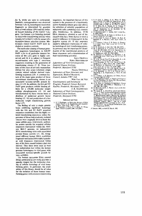
fes S1.. probe are seen to correspond. ~imilarly, correspondence was observed between v-fes S~t homologous restriction fragments as determined by genomic blots of total cellular DNA and the isolated human homolog of the GAIST v-fes gene. The human v-fes homolog showed no detectable transforming activity when transfected to RAT-2 cells by means of a thymidine kinase selection system. Molecularly cloned GAIST FeSV were included as positive controls.
The molecular cloning of human genomic sequences hQmologous to GAIST FeSV v-fes is of particular interest because (i) the c-fes gene is highly conserved and {ii) It is subject to frequent recombination with type C retrovirus sequences resulting in the generation of transforming viruses (7, 9). Virus isolates of this class of not only mammalian (feline) but also avian (chicken) origin contain related cellular derived transforming sequences (10). A common "feature of the major gene product of these recombinant transforming viruses is an associated tyrosine-specific protein kinase (11-13). In addition, the GA and ST FeSV gene products exhibit binding affinity for a 150,000 molecular weight cellular phosphoprotein {12, 14) and transformation by these viruses leads to abolition of epidermal growth factor binding (15, 16) and production of a low molecular weight transforming growth factor (I 7).
The finding of only a single genetic locus exhibiting significant homology with the GA and ST FeSV acquired sequences establishes that the highly related transforming sequences within the genomes of these independently isolated viruses were originally derived from the same cellular gene. Conversely, molecular probes specific for acquired cellular sequences represented within the Abelson MuL V genome, an independent RNA transforming virus.with associated protein kinase activity (I J, 18), recognized different human DNA restriction fragments and lacked detectable homology with sequences represented within any of the three cosmid isolates (data not shown). Thus there must exist at least two independent loci within the human genome homologous to viral genes with associated tyrosine-specific protein ki-nase activities.
The human carcinoma DNA cosmid library generated in our study provides a specific reagent for the molecular cloning of cellular homologs of viral transforming genes. In particular, the availability of this library should be of value for the isolation of those human transforming genes with extensive intervening
sequences. An important feature of this system is the presence of a functionally active thymidine kinase gene that allows for selection of minority populations of eukaryotic cells containing such cosmids after transfection. In addition, SV40 DNA sequences situated in one of the cosmid arms have been shown to exert a positive influence on transcription in the ~-globin system (6). If this sequence similarly influences expression of cellular homologs of viral transforming genes, its presence may be important for identification of the translational products of these sequences and a determination of their transforming potential.
JOHN GROFFEN
NoRA HEJSTEitKAMP
Laboratory of Viral Carcinogenesis. National Cancer Institute, Frederick. Maryland 21701
Fli.ANK G~tOSVELD
Laboratory of Gene Structure and &pression, Medical Research, Council, London NW7 fAA
WIM VAN DE YEN
Carcinogenesis and Intramural Program, Frederick Cancer Research Faci/iry, Frederick, Maryland 21701
J. R. STEPHENSON
Laboratory of Viral Carcinogenesis, National Cancer Jnsritute, Frederick, Maryland 21701
ReFerences and Notes
I P. J. Fisching"'. in Moluular Biology of RNA Tumor Viru<es .J. R. Stephenson. Ed. (Academic ~ross. New York. 1980). p. 162.
2. G. Franchini. J. Even. C. J. Sherr. F. WongStaal. NaTure !London) 290. 154 (1981).
3. S. P. Golf. E. Gllboa. 0. N. w,ne. D. Bal!imorc. Ce/i 22.777 (1980); R. Dalla·Favera. E. P. Gelmann. R. C. Gallo. F. Wong-Staal. Narure (London) 291.31 (19811: R. W. Ellis. D. DeFeo. T. Y. Shih. M.A. Gonda. H. A. Young. N Tsuchida. D. R. Lowy. E. M. Scolnick. ibid .. p. 506; D. Shalloway. A. D. Zelenetz. G. M Cooper, Ce/124. 53! {1981)
4 D. Horowkz and J. F. Burke. Nuc/.1\dds Res. 9. 2989 (1981)_
5. F. G. Grosveld. H. M. Dahl, E. de Boer. R_ A Flavell. Gene !3. 227 ( 1981)
6 F. Grosveld. T. Lund. A. Mellor. H. Bud. H Bullman. R. A. Flavell. J. Mol. IIppi. Genet.. in pre«
7. L.A. Fedele,). Even. C. F. Garon. L. Donner. C. 1. Sherr. Proc. Nwl. A cad. Sci. U.S.A. 78. 4036 0981).
8 E. M. Southern. J. Mol. Bioi. 98. 503 119751. 9. M.A. Barbaoid. A. V. Lauver. S. G. Devare.J
Virol. 33. 196 (1980); W. J. M. Van de Yen. A. S. Khan, F. H. Reynolds. Jr., K. T. Mason. J. R. Stephenson. ibid. 33, 1034 (1980).
10. M_ Shibuya. T. Hanafusa. H. Hanafusa. J. R. Stephenson. Proc. Nar/. Acod. Sci. U.S.A. 77. 6536 (1980).
II. W. J. M. Van de Yen. F. H. Reynolds. Jr..J. R Stephenson. Virology lUI. 185 (1980).
12 F. H. Reynolds. Jr .• W. J. M. Van de Vcn. J. R. Stephenson. J. Bioi. Chern. 25$. I 1040 (1980)
JJ. M. Barbadd. K. Beemon. S. G. Dcvare. Proc Nat/. Acad. Sci. U.S.A. 77,5158 11980).
14 F. H. Reynolds. Jr., W. J. M. Van de Yen. J. R Stephenson, Nalure /London) 286, 409 11980).
15. F. H. Reynold•. Jr .. W. J. M. Van de Von. 1 Blomberg. J. R. Stephenson. J. Viral. 37. 643 (1981).
16 G. l. Todaro,]. E. Del..arco, S. Cohen. Narure !London) 264. 26 (1976).
17. J. lt. Stephenson and G. J. Todaro. in Ad1•ances in Viral Oncology. G. Klein. Ed. (~leaven Pre«, New York, in press1. vol. I
18 0. N. Witte. A. Dasgupta. D. Baltimore. Narure !London) 283, 826 (1980); J. Blomberg era! .. ibid. 286,504 (1980).
19. R. Bernard$ and R. A. Flavell. Nuci.Acids Res 8. 1521 (1980)
20. We thank R. A. Flavell for advice and discussion, C. J. Sherr for providing plasmid< contain· ing the SR v-fes and Sc v-fes suholones. and. G. T. Blennerhassett and P. Hansen for technlc:.al assistance. Supported under Public Health Service Contract No. NOl·C0-75380 from the Na· tiona! Cancer Institute. by the British Medical Research Council IMRC), and by a grant (NUKC-BIOCH 80-1) awarded to J.G .. N.H .. and W.V.D.V. by the Royal Netherlands Cancor foundation (KWF).
18 January 1982; revised 22 March 1982
101

APPENDIX PAPER IV

Transforming Genes of Avian (v-fps) and Mammalian (v-fes) Retroviruses
Correspond to a Common Cellular Locus
JOHN GROFFEN,* NORA HEISTERKAMP,* MASABUMI SHIBUYA,t HIDESABURO HANAFUSA,t AND JOHN R. STEPHENSON*·1
*Laboratory of Viral Carcinogenesis, Natir:mal Cancer Institute, Frederick, Maryland 21701, and tThe Roclrefelkr University, 1230 York Avenue, New York, New York 10021
Received August 26, 1982; accepted November 29, 1982
The Gardner (GA) and Snyder-Theilen (ST) isolates of feline sarcoma virus (FeSV) represent genetic recombinants between feline leukemia virus (FeLV) and transformation-specific sequences {v-fes gene) of cat cellular origin. A related transforming gene (v-fps), common to the Fujinami, PRC II, and UR 1 strains of avian sarcoma virus has also been described. Translational products of each of these recombinant virus isolates are expressed in the form of polyproteins exhibiting protein kinase activities with specificity for tyrosine residues. In the present study, v-fes and v-fps homologous sequences of GA-FeSV, ST-FeSV, and Fujinami sarcoma virus (FSV) are defined and these independently derived transforming genes are shown to correspond to a common cellular genetic locus which has remained highly conserved throughout vertebrate evolution.
Several independent acute transforming retrovirus isolates have been shown to encode as their major translation products tyrosine-specific protein kinases which are closely associated with their transforming function (12). Such viruses can be separated into six classes on the basis of their cellular "oncogenic" sequences. Acquired cellular "oncogenes" and representative virus isolates of each group include: v-src (RSV) (6, 20, 22), v-fes (STand GA FeSV) (3, 12-14,27-29, 33-35), v-fps (FSV, PRCII, URI) (10, 18,23-25,30, 36), v-yes (Y73, Esh) (15, 21, 30, 39), v-ros (UR2) (11, 30), and vabl (A MuLV) (5, 16, 26, 38). Two of these genes (v-fes and v-abl) are of mammalian cellular origin while the remaining four (v-src, v-fps, v-yes, and v-ros) were derived from naturally occurring avian tumors. Earlier studies indicating low levels of sequence homology between the v-fes and vfps genes (31), and both immunologic (2) and structural relatedness (.4) between their translational products, raised the possibility that these either represent re~ lated genes or alternatively that they correspond to a common cellular locus which
1 To whom reprint requests should be addressed.
has remained highly conserved throughout vertebrate evolution. Precedence for the first model was derived from the relatedness of Harvey and Kirsten strains of rat sarcoma virus (8). The present study was undertaken to resolve these alternative possibilities.
To determine the extent of relatedness of FSV v-fps to the GA and ST FeSV vfes genes, molecular probes corresponding to the complete GA-FeSV and ST FeSV genomes, designated GA-v-fes and ST-vfes, respectively, and to 0.5-kb Psti subgenomic fragments of ST-FeSV, designated v-fes SL and v-fes SR (Fig.l), were utilized (13, 17). Each was tested for hybridization by Southern blot analysis to a series of restriction fragments of a previously described (30) molecular clone of FSV in Agt WES-AB. As shown in Fig. lA, the extent of v-fps homology with the individual v-fes probes varies in a manner colinear with their arrangement in the STM and GA-FeSV genomes. For instance, homology to v-fes SR was restricted to the Psti-Smai fragment at the 3' region ofFSV v-fps, while v-fes-SL lacked homology with the 3'-terminal BamHI-Smai fragment and v-fes SL homology extended much fur-
105
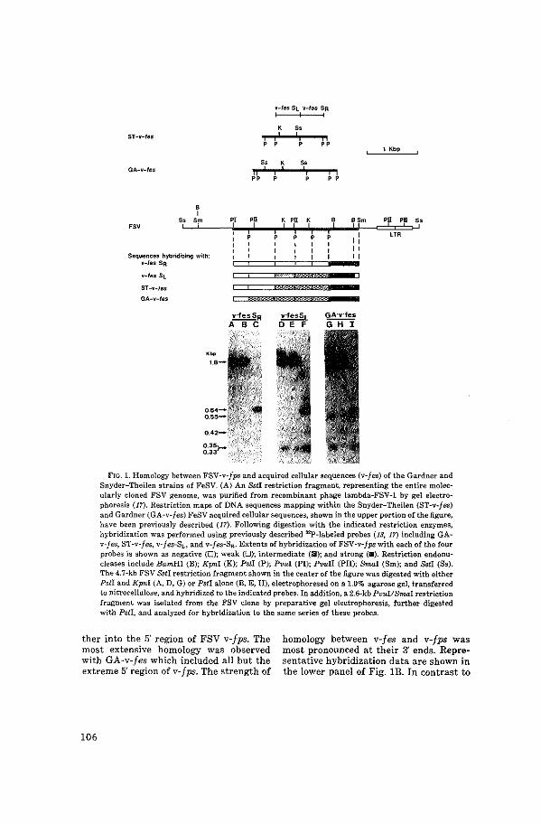
ST -v-fes
GA-v-fes
' I Ss Sm
Sequences' hybridizing with: v-fes SR
ST-v-les
GA-v-fes
0.42-
0.35>-0.33
"
PI PII
v-Ies St v-fes SR
' " ' ' '
Ss K Ss
K Pll K
v·fesSL 0 E F
' I I I
"
"
' I ,, " "
1 Kbp
PII Pll Ss
FIG. 1. Homology between FSV-v-fps and acquired cellular sequences (v-fes) of the Gardner and Snyder-Theilen strains of FeSV. (A) An Sstl restriction fragment, representing the entire molecularly cloned FSV genome, was purified from recombinant phage lambda-FSV-1 by gel electrophoresis (17). Restriction maps of DNA sequences mapping within the Snyder-Theilen (ST-v-fes) and Gardner (GA-v-fes) FeSV acquired cellular sequences, shown in the upper portion of the figure, have been previously described (17). Following digestion with the indicated restriction enzymes, hybridization was performed using previously described 32P-labeled probes (13, 17) including GAv-fes, ST-v-fes, v-fes-~, and v-fes-SR. Extents of hybridization of FSV-v-fps with each of the four probes is shown as negative (0); weak (0); intermediate (D); and strong (•). Restriction endonucleases include BamHI (B); Kpni (K); Psti (P); Pvul (PI); Pvuii (PII); Smai (Sm); and &tl (Ss). The 4.7-kb FSV Sst! restriction fragment shown in the center of the figure was digested with either Psti and Kpnl (A, D, G) or Pstl alone (B, E, H), electrophoresed on a 1.0% agarose gel, transferred to nitrocellulose, and hybridized to the indicated probes. In addition, a 2.6-kbPvui/Smal restriction fragment was isolated from the FSV clone by preparative gel electrophoresis, further digested with Pstl, and analyzed for hybridization to the same series of these probes.
ther into the 5' region of FSV v-fps. The most extensive homology was observed with GA-v-fes which included all but the extreme 5' region of v-fps. The strength of
homology between v-fes and v-fps was most pronounced at their 3' ends. Representative hybridization data are shown in the lower panel of Fig. lB. In contrast to
106
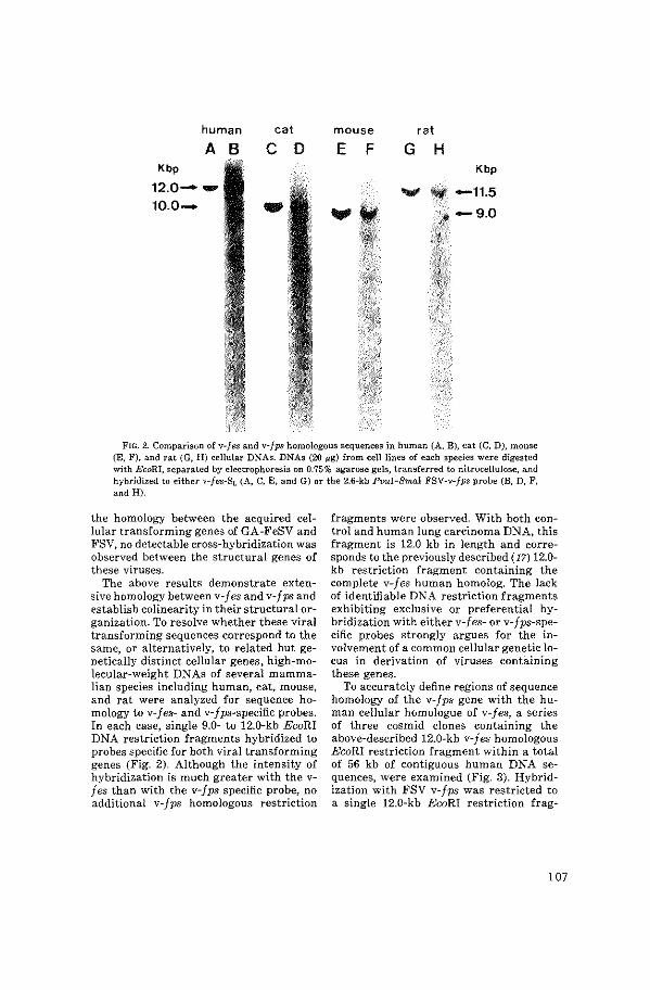
human cat mouse rat
A B c 0 E F G H Kbp Kbp
12.o-- -• -11.5 10.0- - w " -9.0
' ' . ;;<
FIG. 2. Comparison of v-fes and v-fps homologous sequences in human (A, B), cat (C, D), mouse (E, F), and rat (G, H) cellular DNAs. DNAs (20 ~g) from cell lines of each species were digested with EcoRI, separated by electrophoresis on 0.75% agarose gels, transferred to nitrocellulose, and hybridized to either v-fes-SL (A, C, E, and G) or the 2.6-kb Pvui-Smai FSV-v-Jps probe (B, D, F, and H).
the homology between the acquired cellular transforming genes of GA -FeSV and FSV, no detectable cross-hybridization was observed between the structural genes of these viruses.
The above results demonstrate extensive homology between v-fes and v-fps and establish co linearity in their structural organization. To resolve whether these viral transforming sequences correspond to the same, or alternatively, to related but genetically distinct cellular genes, high-molecular-weight DNAs of several mammalian species including human, cat, mouse, and rat were analyzed for sequence homology to v-fes- and v-fps-specific probes. In each case, single 9.0- to 12.0-kb EcoRI DNA restriction fragments hybridized to probes specific for both viral transforming genes (Fig. 2). Although the intensity of hybridization is much greater with the vfes than with the v-fps specific probe, no additional v-fps homologous restriction
fragments were observed. With both control and human lung carcinoma DNA, this fragment is 12.0 kb in length and corresponds to the previously described (17) 12.0-kb restriction fragment containing the complete v-fes human homolog. The lack of identifiable DNA restriction fragments exhibiting exclusive or preferential hybridization with either v-fes- or v-fps-specific probes strongly argues for the involvement of a common cellular genetic locus in derivation of viruses containing these genes.
To accurately define regions of sequence homology of the v-fps gene with the human cellular homologue of v-fes, a series of three cosmid clones containing the above-described 12.0-kb v-fes homologous EcoRI restriction fragment within a total of 56 kb of contiguous human DNA sequences, were examined (Fig. 3). Hybridization with FSV v-fps was restricted to a single 12.0-kb EcoRI restriction frag-
107
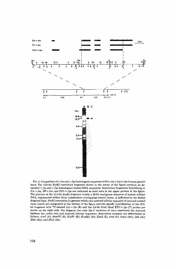
GA-v-les - ~ - ~ ' ST -v-Ies - -- -FSV-v-lps - ' '
'm 'm I 71 ' ss Xb "' ' ~ s~l : ' 'm " ' ' II II '
I I ' ' ' ~I ' ~ ' ' K
' ~ pn ' ' --.E. ' ' ' ' ' '
' ' " )(h ISsK K 'm ;f 'I I ~I I\ ,I ' ' ' ' '
'- / '-. /
'-. '-. /
'-. / '-. /
'--- /
' ' ' ' ' ' " cos-5
'' 19.8 12.0 2.5 C.3
"' iBC 19.8-
12.0- --0R<%
4.s-*'"··
0.3-
FIG. 3. Comparison of v-fes and v-fps homologous sequences within the c-fes/c-fps human genetic locus. The 12.0-kb EcoRl restriction fragment shown in the center of the figure contains all detectable v-fes and v-fps homologous human DNA sequences. Restriction fragments hybridizing to GA-v-fes, ST-v-jes, and FSV-v-fps are indicated as solid bars in the upper portion of the figure. The position of the 12.0-kb EcoRI fragment within a 58-kb contiguous sequence of human cellular DNA, represented within three independent overlapping cosmid clones, is indicated by the dotted diagonal lines. EcoRI restriction fragments within the inserted cellular sequence of one such cosmid clone (cos-5) are designated at the bottom of the figure and the spedfic hybridization of the 12.0-kb fragment with '9>-labeled GA-v-fes (B) and the 2.6-kb Pvui-Smal FSV-v-fps (C) probes are shown on the right side. The diagonal line near the 5' terminus of cos-5 represents the junction between the vector arm and inserted cellular sequences. Restriction enzymes are abbreviated as follows: A val (A); BamHI (B); EcoRI (E); Hindiii (H); Kpnl (K); Pstl (P); Smai (Sm); Sst! (Ss); Xba.l (Xb); and XMI (Xh).
108

ment containing v-fes homologous sequences. Following subcloning, the 12.0-kb EcoRI fragment was digested with the indicated combinations of restriction endonucleases and analyzed for hybridization to v-fes- and v-fps-specific probes (Fig. 4). FSV v-fps homology to human DNA most closely resembled that of GA FeSV v-fes. However, the 3'-terminal v-fes homologous region of human DNA common to STand GA-v-fes did not hybridize to a detectable extent with the FSV v-fps probe. ST v-fes homologous sequences differ from those of both GA v-fes and FSV v-fps in that they are not represented within the 5'-terminal region hybridizing to the other two. Finally, two regions of human DNA homology unique to FSV-v-fps were identified in the 5' half of the 12.0-kb human EcoRI DNA fragment. Thus, although all three viral "oncogenes" appear to be entirely represented by homologous sequences within a single c-feslc-fps human genetic locus, the exact positions at which regions of homology map differ among the individual viral transforming genes. The lack of detectable hybridization of v-fps with the 3' region of the c-fes/c-fps locus could reflect evolutionary divergence. Alternatively, there may exist within the vfeslv-fps locus, sequences not necessary for transformation which were lost, either in the generation of FSV and the two FeSV isolates, or by subsequent deletions. Thus a comparison of the 5'-terminal position of ST-v-fes, and 3' position of FSV-v-fps homologous sequences within the human cfes!c-fps locus, indicated by the dotted lines, may provide a minimal estimate of those sequences required for transformation.
The larger size of the FSV v-fps gene (2.6 kb) (30) as compared to that of GA-vfes (1.8 kb) (9) may be accounted for by regions of the human c-feslc-fps locus which are uniquely hom<>logous with FSV v-fps. These FSV v-fps unique sequences can be explained by one of several alternative possibilities. For instance such sequences may have been initially represented within GA-v-fes but deleted during passage of GA-FeSV subsequent to its ini-
D E F
-1.60
-0.98 -0.84
-0.93
-0.74
-0.64 -0.61
-0.45
-0.30
FIG. 4. Restriction endonuclease analysis of human cellular sequences hybridizing to 32p-labeled GA-vfes and FSV-v-/ps probes. DNA from a variant of pBR328, containing the 12.0-kb human cellular EcoRI fragment from cos-5 (pBR328 cos-5), was digested either with Avo:I (A-C); or Psti (E-F), separated by electrophoresis on 0.75% agarose gels, and stained with ethidium bromide (A, D). Following transfer to nitrocellulose, hybridization was performed according to previously described (17) procedures. 32p-labeled probes include the 2.6-kb Pvui-Smai FSV-vfps fragment (B, E) and GA-v-fes (C, F). Restriction fragments which hybridized to either the FSV-v-fps or GA-v-fes probes and correspond to the 12.0-kb EcoRI insert of pBR328 cos-5, are indicated by arrows; additional hybridizing restriction fragments in lanes C and F correspond to pBR328 plasmid sequences. The 0.3-kb Avai restriction fragment, although not visible in this figure, was found upon longer exposure to hybridize to both probes.
tial derivation. Alternatively, the differences in sequences in these viruses may reflect differences in the positions of coding and noncoding regions of the c-feslcfps RNA transcripts of the divergent host species from which they were isolated. Finally, mechanisms of incorporation of cellular oncogenic sequences in retroviral ge-
109
names may be imprecise and the resulting insert within the viral genomic RNA not necessarily correspond exactly to the cellular mRNA from which it was derived. Either the first or third possibilities appear most probable in that one of these must be envoked to explain the differences between the acquired cellular sequences of GA- and ST-v-fes (9, 17). Resolution of these alternative models may provide insight into mechanisms by which cellular oncogenes become stabily associated with the genomes of retroviruses.
The present findings establish that the v-fes and v-fps sequences correspond to a common cellular genetic locus which has been highly conserved throughout vertebrate evolution. Thus, tyrosine-specific protein kinase activities associated with the translational product of such sequences, have probable functional significance to their host. Although the mammalian homologues of other avian retrovirus encoding tyrosine-specific protein kinases have not been characterized, the human homolog of the mouse v-abl gene has been shown to be independent of the c-feslc-fps locus (19). By analysis of appropriate somatic cell hybrids, c-fes/c-fps has been localized on human chromosome 15 (7, 19).
ACKNOWLEDGMENTS
We thank G. T. Blennerhassett and P. Hansen for excellent technical assistance. This work was supported under Public Health Service Contract NOIC0-75380 and by Grant CA-14935 from the National Cancer Institute.
REFERENCES
1. BARBACIO, M., BEEMON, K., and DEVARE, 8. G., Proc. Nat. Acad. Sci USA 77, 5158-5162 (1980).
2. BARBACID, M., BREITMAN, M. L., LAUVER, A. V., LONG, L. K., and VOGT, P. K., Virology liO, 411-419 (1981).
3. BARBACID, M. A., LAUVER, A. V., and DEVARE, S. G., J. ViroL 33, 196-207 (1980).
4. BEEMON, K., Cel/24, 145-153 (1981). 5. BLOMBERG, J., REYNOLDS, F. H. JR., VAN DE VEN,
W. J. M., and STEPHENSON, J. R., Nature (Urndon) 286, 504-507 (1980).
6. COLLE'IT, M. S., and ERIKSON, R. L., Proc. Nat. Acad. Sci USA 75, 2021-2024 (1978).
110
7. DALLA-FA VERA, R., FRANCHINI, G., MARTINO'ITI, S., WONG-STAAL, F., GALLO, R. C., and CROCE, C. M., Proc. Nat. Acad. Sci USA 79, 4714-4717 (1982).
8. ELLIS, R. W., DEFEO, D., SHU'!, T. Y., GONDA, M. A., YOUNG, H. A., TSUCHIDA, N., LOWY, D. R., and ScoLNICK, E. M., Nature (London) 292, 506-511 (1981).
9. FEDELE, L. A., EVEN, J., GARON, C. F., DONNER, L., and SHERR, C. J., Proc. Nat. Acad. Sci USA 78, 4036--4040 (1981).
10. FELDMAN, R. A., HANAFUSA, T., and HANAFUSA, H., CeU 22, 757-765 (1980).
11. FELDMAN, R. A., WANG, L.-H., HANAFUSA, H., and BALDUZZI, P. C., J. ViroL 42, 228-236 (1982).
12. FISCHINGER, P. J., In "Molecular Biology of RNA Tumor Viruses" (J. R. Stephenson, ed.), pp. 162-198. Academic Press, New York, 1980.
13. FRANCHINI, G., EVEN, J., SHERR, C. J., and WONGSTAAL, F., Nature (London) 290, 154-157 (1981).
14. FRANKEL, A. E., GILBERT, J. H., PORZIG, K. J., SCOI.NICK, E. M., and AARONSON, 8. A., J. ViroL 30, 821-827 (1979).
15. GHYSDAEL, J., NEIL, J. C., and VOGT, P. K., Proc. Nat. Acad. Sci USA 768,2611-2615 (1981).
16. GOFF, S. P., GILBOA, E., WI'ITE, 0. N., and BALTIMORE, D., Cell22, 777-785 (1980).
17. GROFFEN, J., HEISTERKAMP, N., GROSVELD, F., VAN DE VEN, W. J. M., and STEPHENSON, J. R., Science 216, 1136-1138 (1982).
18. HANAFUSA, T., WANG, L.-H., ANDERSON, S. M., KARESS, R. E., HAYWARD, W. S., and HANAFUSA, H., Proc. Nat. A cad. Sci USA 77, 3009-3013 (1980).
19. HEISTERKAMP, N., GROFFEN, J., STEPHENSON, J. R., SPURR, N. K., GOODFELLOW, P. N., SoLOMON, E., CARRI'I'T, B., and BODMER, W. F., Nature (London) 299, 747-749 (1982).
20. HUNTER, T., and SEFTON, B. M., Proc. Nat. Acad. Sci USA 77, 1311-1315 (1980).
21. KAWAI, S., YOSHIDA, M., SEGAWA, K., SUGIYAMA, H., ISHIZAKI, R., and TOYOSHIMA, K., Proc. Nat. Acad. Sci USA 71, 6199-6203 (1980).
22. LEVINSON, A. D., OPPERMANN, H., LEVINTOW, L., VARMUS, H. E., and BISHOP, J. M., CeU 15, 561-572 (1978).
23. NEIL, J. C., BREITMAN, M. L., and VOGT, P. K., Virology 108,98-110 (1981).
24. NEIL, J. C., GHYSDAEL, J., and VOGT, P. K., Virology 109, 223-228 (1981).
25. PAWSON, T., GUYDEN, J., KUNG, T.-H., RADKE, K., GILMORE, T., and MARTIN, G. S., Cell 22, 767-775 (1980).
26. REYNOLDS, F. H., JR., SACKS, T. L., DEOBAGKAR, D. N., and STEPHENSON, J. R., Proc. Nat. Acad. Sci USA 15, 3974-3978 (1978).
27. REYNOLDS, F. H., JR., VANDEVEN, W.J. M.,BLOM-
BERG, J., and STEPHENSON, J. R., J. ViroL 37, 643-653 (1981).
£8. REYNOLDS, F. H., JR., VAN DE VEN, W. J. M., and STEPHENSON, J. R., J. BioL Chem.. 255, 11040-11047 (1980).
29. SHERR, C. J., SEN, A., TODARO, G. J., SLIKSI, A. H., and ESSEX, M., Proc. Nat. Acad. Sci. USA 75, 1505-1509 (1978).
30. SHIBUYA, M., HANAFUSA, H., and BLADUZZI, P. C., J. ViroL 42, 143-152 (1982).
31. SHIBUYA, M., HANAFUSA, T., HANAFUSA, H., and STEPHENSON, J. R., Proc. Nat. Acad. Sci USA 77, 6536-6540 (1980).
82. SHIBIJ.YA, M., WANG, L.-W., and HANAFUSA, H.,J. ViroL, in press.
33. STEPHENSON, J. R., KHAN, A. S., SLISKI, A. H., and
ESSEX, M., Proc. Nat. Acad. Sci. USA 74, 5608-5612 (1977).
34. VAN DE VEN, W. J. M., KHAN, A. S., REYNOLDS, F. H., JR., MASON, K. T., and STEPHENSON, J. R., J. ViroL 33, 1034-1045 (1980).
35. VAN DE VEN, W. J. M., REYNOLDS, F. H., JR., and STEPHENSON, J. R., Virology 101, 185-197 (1980).
36. WANG, L.-H., FELDMAN, R. A., SHIBUYA, M., HANAFUSA, H., NOTTER, M. F. D., and BALDUZZI, P. C., J. Virol 40, 258-267 (1981).
37. WITTE, 0. N., DASGUPTA, A., and BALTIMORE, D., Nature (London) 283, 826-831 (1980).
38. WITTE, 0. N., ROSENBERG, N., PASKIND, M., SHIELDS, A., and BALTIMORE, D., Proc. Nat. Acad. Sci. USA 75, 2488-2492 (1978).
89. YoSHIDA, M., KAWAI, S., and TOYOSHIMA, K., Nature (London) 287, 653-654 (1980).
111


APPENDIX PAPER V

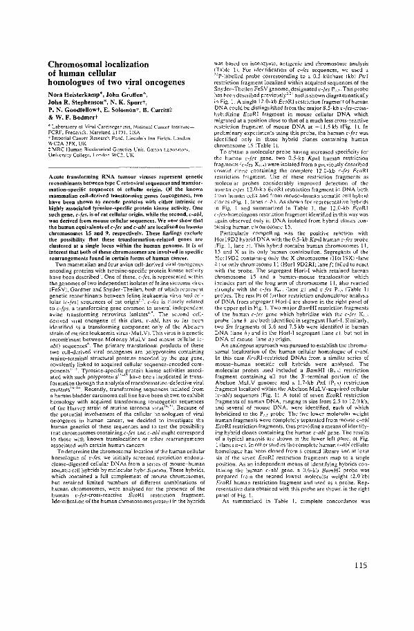
Chromosomal localization of human cellular homologues of two v.iral oncogenes Nora Heisterkamp*, John Groffen*, John R. Stephenson*, N. K. Spurrt, P. N. Goodfellowt, E. Solomont, B. Carritt:J: & W. F. Bodmert
• Laborawry of Viral Card~ogenesis, N;otional Cancer lnstit\lkFCRF, Frederick, Maryland 217()1. USA -;- Tmperial Cancer Research Fund, Lincoln·, Inn Field<. l.om.lon WC2A 3PX. UK :> MRC Human Biochemical Genetics Unit. Galton Lahornton. University College. LGndon WC2. UK
Acute transforming RNA tumour viruses represent genetic recombinants between type C retroviral sequences and transformation-spedlic sequences of cellular origin. Of the known mammalian cell-derived transforminj! genes (oncogenes), two have been shown to encode proteins with either intrinsic or highly associated tyfosine-specilic protein kinase activity. One such gene, c-fes, is of ~at cellular origin, while the second, c-ab/, was derived from mouse cellular sequences. We now show that the human equivalents of c-fes and c-ab/ are localized on human chromosomes 15 and 9, respectively. These lindings e:>eclude the possibility that these transformation-related genes are clustered at a single locus within the human genome. It is of interest that both of these chromosomes are involved in specific rearrangements found in certain forms of human cancer.
Two mammalian and four avian cell-derived viral oncogene" encoding proteins with tyrosine-specific protein kinase activity have been described'. One of these, c-fes. is represented within the genomes of two independent isolates of feline sarcoma '"iru~ (FeSV), Gardner and Snyder-Theilen. both of which represent genetic recombinants between feline leukaemia virus and cellular lc-fes) sequences of cat origin''. c-[<"s is closely related to c-fps, a transforming gene common to several independent avian transforming retrovirus isolates-'·'. The second cellderived viral oncogene of this class. t·-abl. has so far been identified as a transforming component only of the Abelson strain of murine leukaemia virus I MuL V). This virus is a genetic recombinant between Moloney MulV and mouse cellular !cab/) sequences". The primary translational products of these two cell-derived viral oncogenes are polyproteins containing amino-terminal structural proteins encoded by the gag gene, covalently linked to acquired cellular sequence-encoded com· ponents7
·'". Tyrosine-specific protein kinase activities associated with such polyproteins 1
,_., have been implicated in transformation through the analysis of transformation-defective viral mutants"·-'". Recently, transforming sequences isolated from a human bladder carcinoma cell line have been shown to exhibit homology with acquired transforming !oncogenic) sequences of the Harvey strain of murine sarcoma virus'"'". Because of the potential involvement of the cellular homologues of viral oncogenes in human cancer, we decided to investigate the human genetics of these sequences and to test the possibility that chromosomes containing c-fes and c-ab/ might correspond to those with known translocations or other rearrangements associated with certain human cancers
To determine the chromosomallocation of the human celluiH homologue of ~·-fes, we initially screened restriction endonuclease-digested cellular DNAs from a serie> of mouse-human somatic cell hybrids by molecular hybridization. These hybrids. which contained a full complement of mouse chromosomes. but retained limited numbers of different combinations of human chromosomes, were analysed for the presence of the human ~·-fes-cross-reactive EcoRI restriction fra!!ment. Identification of the human chromosomes present in the hybrids
was based on isoenzyme, antigenic and chromosome analysis !Table 11. For identification of c-fes sequences, we used a "P-labelled probe corresponding to a 0.5-kilobase ikbi Psrl restriction fragment localized within acquired sequences of the Snyder-Theilen FeSV genome, designated v-fes P"·'· This probe has he en described previously'' 1 and is shown diagrammatically in Fig. I. A single 12.0-kb EcoRI restriction fragment of human Dl" A could he distinguished from the major 8.5·kb ~·-/es-crosshybidizing EcoRI fragment in mouse cellular DNA which migrated at a position close to that of a much less cross-reactive restriction fragment of mouse DNA at -11.5 kb !Fig. 11. In preliminary experiments using this probe, the human c-fes was identified only in those hybrid clones containing human chromosome 15 !Table 1).
To obtain a molecular probe having increased specificity for the human c-[fs gene, two 0.5-kb Kpnl human restriction fragments I c-[fs K" .) were isolated from a previously described wsmid clone containing the complete 12.0-kb c-fes EcoRI restriction fragment. Use of these restriction fragments as molecular probes considerably improved detection of the human c-fn 12.0-kb EcoRI restriction fragment in DNA both from human cells and from mouse-human somatic cell hybrid clones I Fig. Llanes c-111. As shown for representative hybrids in Fig. 1 and summarized in Table 1, the 12.0-kb EcoRI c-J<'s-homologous restriction fragment identified in this way was ngain observed only in DNA isolated from hybrid clones .:ontaining human chromosome 15.
Particularly compelling was the positive reaction with Horl9D2 hybrid DNA with the 0.5-kb Kpnl human c-fcs probe 1Fig. L lane:·). This hybrid contains human chromosomes 11, 15 and X as its only human conTribution. Segregants of the l--!orl9D2 containing only the X chromosome 1Hor19Xillane hI or only chromosome 11 (Horl 9D2RI; lane [J failed to react "With the probe. The segregant Horl-1 which retained hu[11an chromosome 15 and a human-mouse translocation which includes part of the long arm of chromosome 11, also reacted strongly with the c-fcs K,, {lane g) and ~--{es p,, !Table 11 probes. The results of further restriction endonuclease analysis of DNA from segregant Horl-1 are shown in the right panel of the upp~r gel in Fig. 1. Two major BamHl restriction fragments of the human c-fn gene which hybridize with the c-fn K", probe Iiane h 1 are both identified in segregant Hori-1. Similarly, two Sst fragments of 3.6 and 7.5 kb were identified in human DNA Iiane hJ and in the Horl-1 segregant Iiane cl, but not in DNA of mouse IIane a J origin.
An analogous approach was pursued to establish the chromosomal localization of the human cellular homologue of ~--abl In this case £caRl-restricted DNAs from a similar series of mouse-human somatic cell hybrids were analysed. The molecular probes used included a Bam HI fB, ,J restriction fragment containing all but the 3'-terminal portion of the Abelson M'.!LV genome and a 1.7-kb Pst! (p, 7 ) restriction fragment localized within the Abelson MuL V -acquired cellular lr-abl) sequences IFig. 1). A total of seven £coR! restriction fragments of human DNA, ranging in size from 2.5 to 12.0 kb, and several of mouse DNA, were identified, each of which hybridized to the P, _, probe. The five lower molecular weight human fragments were completely separated from mouse c-ab! EcoRl restriction fragments, thus providing a means of identifying hybrid clones containing the human c-ab/ gene. The results of a typical analysis are shown in the lower left panel of Fig. 1 IIanes a-cr. In other studies the complete human l·-abl cellular homologue has been cloned from a cosmid library and at least six of the seven EcoRI restriction fragments map to a single position. As an independent means of identifying hybrids containing the human c-ab/ gene, a 0.6-kb BamHI probe was prepared from the second lowest molecular weight 12.9 kbi EcoRI human restriction fragment and used as a probe. Representative data obtained with this probe are shown in the right panel of Fig. 1.
As summarized in Table 1, complete concordance was
115
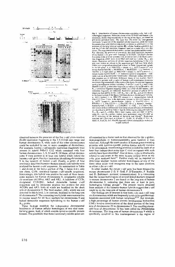
$1-FeSV
t~-1--
A-MuLV '
""'"'" l·l~<oom•"'
~-~-----~ .,
observed between the presence of the five v-abl cross-reactive EcoRl restriction fragments in the 2.5-5.0-kb size range and human chromosome 9, while each of the other chromosomes could be excluded by one or more examples of discordance. For example, human c-abl-specific restriction fragments were present in hybrid F4Sc13 Cl12 which contained only four human chromosomes: 1, 9, 14 and X. Of these, all but chromosome 9 were present in at least one hybrid which lacked the human c-ab/ gene, thus by elimination identifying chromosome 9 as the location of human c-ab/. Finally, a series of four previously described hamster-human somatic cell hybrids were analysed for human c-ab/ sequences. As summarized in Table 1 and shown in the lower portion of Fig. 1 (lanes h-k ), only one done, ClOb, contained human c-abl-specific sequences. Interestingly, this hybrid was positive for each of three isoenzyme markers for human chromosome 9, cytoplasmic soluble cis-aconitase {A CONs), AK3 and AKl. A subclone of ClOb, designated Cl0b2BU, lacked detectable human c-ab/ sequences and by isoenzyme analysis was positive for only ACONs and AK3, both of which are localized on the short arm of chromosome 9. The third marker, AKl, which was not detected in this hybrid, is in contrast, localized to the long arm of chromosome 9. The remaining two hybrids were nonreactive in enzyme assays for all three markers for chromosome 9 and lacked detectable sequences hybridizing to the human c-ab! B,,_,; probe.
These findings establish the independent chromosomal localization of human cellular homologues of two viral transforming genes, both of which encode tyrosine-specific protein kinases. The possibility that these functionally similar genes are
116
Fig. I !dentificanon of human chromosomes containing v-[es- and v-ablhomologous sequences. Molecular clones of the ST-FeSV and human c-{e.< sequences. shown diagrammatically at the top of the figure. are based on pr"'·iously published data'. The Open box IDJ within the vnal genome represonts the position of the acquired cellular sequences. while the feline leukaemia virus cross-reactive sequences are shown as solid lines 1-)_ The positions of tile long terminal repeats 111.'ll). cellular flanking sequences iU I and the 0.5-kb Psi I restriction fragment used as a probe IP, ,) are also shown. The organi<ation of the human c-fes llas been pubHsiled previously' For rderence, the posllions of previously identified restriction fragments of t'le humon :-f.,s g~n~ with hnmology to ST-FeSV. GA-FeSV and the Fuiianami strain of avian sarcoma virus. as well as two 0.5-kb Kpnl restriction fragments which wore co-purified and used as a probe IK.,,J. arc shown_ Restriction patterns of cellular sequences hybddizin~ to tile ''-fes and c-fes probes are shown in the uppermost gel. Cell lines from which r>NAs were prepared arc described in detail in Table I legend and include in the left panel: b. d. N!H/3T3(-I mouse; a. c. A673 human(+!'" and mouse-human hybrids;<. Hori9D2i+l; {. Hori9D2Rli- I; g. Horlli+i; h. Horl9xt-)-. in right panel. a. NIH/3T31-I mouse; h, A67.1 human! t-1; c. mouse-human hybrid Horl[] + )_ 1+ Indicates a posillve assignment: - indicates absence of the particular chromosome.) Molecular probes and restriction enzymes are dS indicated. Molecular clones of the Abelson MuL V
lA-MuL Vi genome and a region of human c•-ab/-!10mologous <equences are shown diagrammatically in tile centre of the figure. RestrictiOn fragments used as hybridization probes. including 1.7-kb Pslli,•-abJ P, ,I and 3.5-kb BamHiic•-abl B, ,J restriction fragments »well as a 0.6 kb BamHI (c-ab! B,") restriction fragment mapping withm the 2.9-kb EcoRI human c-abi r<striction fragment, arc mdicated. RestTJction patterns of cellular DNA sequences hybndizing to the o-aM B, _, and c-ab! B.," probes are shown in the lower portion of the figure. Cell lines shown in the left panel include: a, A673 humani+l; b. :--IIH/3T3 mouse(-): c. mouse-human hybricl F4sci3Cl 121+ ). Those in !ile right panel include: a, NIH/JT3 mousc1-1; b. A673 llumani+l. mouse-human hybridso c. F4sci:IC1121~J: d, FIR5R3(-I: <, F4sd3C19i-l; f. F4sc1Kli21TJ: g. hamster: hamsterhuman hybrid<: 11. Dl$201-·1; i. C10b2BL"I-I; j, C4al-l: k. ClOb(+l_ The derivations of these cell lmes and the<r complements of human chromosomes are described in detail 1n Table I legend. Followin~ hybridization with the P, -'' B,. and P,, probes. filters were washed with 2.5 x SSC at 65 'C; ustng K .. , and B,," probes. was he< were performed using 0.3 x SSC ao 65T •coording to the method of Bernards and Flavell''. RestrictLM enzymes are abbrevtated as follows· E. EcoRI; H. Hindlll; P.- Ps<l. K. Kpnl; A. Ac•al; Ss. S"L Hindlll·digested A DNA is included as" molecular
weight >tandard
all organized in a cluster such as that observed for the y-globi n, immunoglobulin or histocompatibility gene families is thus ruled out. Although the exact number of human genes encoding proteins with tyrosine-specific protein kinase activity remains to be determined, transforming proteins encoded by each of at least four independent avian type C viral oncogenes with such activity have been identified t. One of these, v-{ps, is structurally related to and maps at the same human cellular locus as the c-fes gene analy~ed here4
·'. Further study will be required to
determine whether human cellular homologues of any of the three other avian viral oncogenes map to the same positions as either c-fes or c-ab!.
In other studies, the mouse c-ab/ gene has been assigned to mouse chromosome 2 (S." P. Goff, P. D'Eustachio, F. Ruddle and D. Baltimore, personal communication). It is interesting that the human homologues of several linked markers assigned 10 mouse chromosome 2 are found on the long arm of human chromosome 9, indicating that these are, at least in part, homologous linkage groups". The present results obtained from analysis of the hamster-human hybrids suggest that c-ab! could be on the long arm of human chromosome 9.
Our findings are of interest in that both c-fes and c-ab/ map on chromosomes which are frequently involved in specific translocations associated with human neoplasms23
•24
• For example, a high percentage of human chronic myelogenous leukaemias (CML) involve translocations of the distal portion of the long arm of chromosome 22 to chromosome 9. The resulting abnormal form of chromosome 22 has been called the Philadelphia chromosome. The long arm of human chromosome 9 which is specifically involved in this rearrangement is the region of
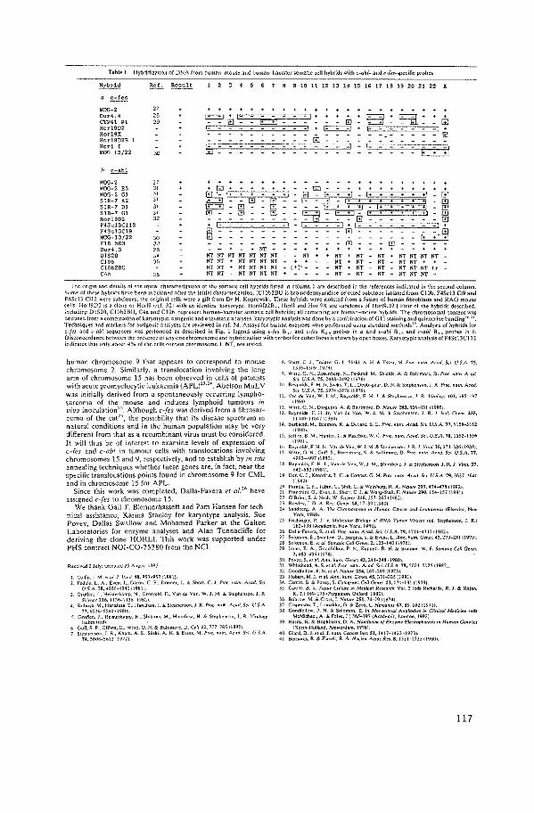
Table I Hybridization of DNA from human-mou<e and human-ham<tcr <omatic <ell hybrid; with c-aM- and c-fes-specific probes
Hvbrid ~ Result ' ,
' • ' ' ' " ' " " " " .. " " 17 18 19 20 21 " ' ' c-!es
~OG-2 " Our~. 4 " = = EJI: . . . B q·
"' ctP41 " " = "' Hc:r19D2 ~ Hcrl9X @ Hotl902R 1 OJ Horl ' liJ
_, . 1- C3l ·~ 13/22 ~
' c-ab!
lo!OG-2 " . . . . . r.!OG-2 E5 " " . G)
B lo!OG-2 Gl " f,S, ill ~ SIR-7 A2 " . "' "' ~ - . t1 R [!] SIR-7 Dl " E..:£! l'J G - - ~=:n B SIR-7 Gl " OJ m m ""GJ =- !!::I:~~ f!J Hotl9D2 " - - 0 8 F4Sd3Cll2
ffi 1-
F4Sd3Cl9 ill - - E:::d M{X;-13(22 " - - . FIR 5R3 " "' 'l' Dur4.3 " " 01820 " lfl" NT NT NT NT NT NT " " " " N1" NT NT NT ClOb " " " N1" NT N1" NT . " " " NT NT Cl0b2Bt; " " . NT NT NT NT (+]•- " " " NTN1"NTtr
"" "' " " - NT NT NT NT " " " NT NT NT -The origin and detail; of the initial characteril.ation of the somatic cell hybrids listed in column 1 are described in the references indicated in the second column.
Sumc of these hybrid; ha,·e been subcloned after the initial characterization. iC10b2BU is bromodcoxyuridine-selccted <ubclone isolated from C!Ob. F4Sc13 Cl9 and F4Scl3 Cll2 were subdones. the original cells were a gift from Dr H. Koprowski. These hybrids were isolated from a fusion of human fibroblasts and RAG mou.e cell<. Hori9D2 is a clone or Horl9 'fef 321 with an identical karyotype. Hori9D2Rl. Horll and Hori9X arc •ubcloncs of Hor19D2 J Four of the hybrids described. indudin~ DlS20. C10b2BU. C4a and C10b. represent human-hamster somatic cell hybrids; all remaining arc human-mouse hybrids. The chromosomal content was deduced I rom a combination of karyotypic. antigenic and enzymatic analy5es. karyotypic analysis was done by a combination of G II >taining and quinacrine banding·'"·". Techniqu., and morkers for antigenic analyses are reviewed in ref. 38. Assays for human en>ymes were performed usmg >tandard methods". Analysis of hybrids for c-fes and c-ob! sequences wa. performed a< described in Fig. I legend using u-fes B,, and c-fes K,., probes in a and u-abl B, and c-ab! B,.,. probes in b D isconcordance between the presence of any one chromosome and hybridization with pr~bc; for either locus i.< shown by open boxes. Karyotypic analysis of F4Scl3C I 1 2 indicates that only about 4% of the cells <Ontain chromosome I NT. not tc•ted
human chromosome 9 that appears to correspond to mouse chromosome 2. Similarly, a translocation involving the long arm of chromosome 15 has been observed in cells of patients with acute promyelocyticleukaemia (APL)'·'·'·. Abelson MuL V was initially derived from a spontaneously occurring lymphosarcoma of the mouse and induces lymphoid tumours in l'ivo inoculation25
• Although c-fes was derived from a fibrosarcoma of the cat25
, the possibility that its disease spectrum in natural conditions and in the human population may be very different from that as a recombinant virus must be considered. It will thus be of interest to examine levels of expression of c-fes and c-ab/ in tumour cells with translocations involving chromosomes 15 and 9, respectively. and to establish by in situ annealing techniques whether these genes are, in fact, near the specific translocations points found in chromosome 9 for CML and in chromosome 15 for APL.
Since this work was completed, Dalla-Favera et al."" have assigned c-fes to chromosome 15.
We thank Gail T. Blennerhassett and Pam Hansen for technical assistance, Karina· Stanley for karyotype analysis, Sue Povey, Dallas Swallow and Mohamed Parker at the Galton Laboratories for enzyme analyses and Alan Tunnacliffe for deriving the clone HORLI. This work was supported under PHS contract NOI-C0-75380 from the NCI.
Collin,!. M. "ol J v,,.,/ 40. 95l-9l1 <19~11 Fodol<. L. A. £,on.! .• Garon. C F .. Donne<. L & Shorr. C. L p.,,_ Ham. Aco<i. S<1
US A. 78. 4036-4U40 119811 Oro«on. J. Hei"e<kamp. N .• G<o,.cld. F .• Von de Von. W !. ;.\ & S"phon><>n. J R
s'""" 216. ttJO-ttJs lt9B21 sn,buyo. M .• Honal""· T .. Hanafu<>. J. & Steoh<nson.!. R. Pmr. """' A<o<i. s,._ U S.A
77.65l6-6>4Uil98IIJ l. Oroffen. J .. H01•torkamp. N. Sh;buya. M .. H.oafu<a. H. & S«phon,.,n. J. R. Vim!o"'
«ubmittcdl 6 Goff. S P .. G1tbo•. E .. WiHe. 0. N. & Balnmoro. D. C<il 2Z, 777-7R5 119301 7 Stcpilcn<Gn. J. R .• Kilom. A. S. 5l"k'. A. H & E'"''· M. Pm,. ""'"· A< ad S<i. l.i SA
74. 560$-56t2 lt977 I
"· Sh<r<. C). ToUMo. G.!. Sl.skl. A H. & E«". M Pmc. naln Mod. Sd. U.S.A 15, l50l-l5119<l97AI
Q W""· 0 N .. Ru•enh"'~- N .. P><kmd. M. Shield,. A & Boltimo;c. D. Pm<. nom. Ac~d. Sci U.>A.75.24AA-N9:11l97il
Hl Reynold<. F H. Jr. Sock,. T. L.. Deobe~kor. D. N. & Stophcnwn. J. R PrO<. nom. A<od s,,. U.S.A. 75.J974-J91811978J
ll Von do Von. W. J. M .. Reynold;. F H !r & S!ephcn•on.! R Vimlo.,· 101. IAl-197 •19801
t1 WiHc. 0 N. O"~"D"• A & BoUimo<e. D. No'"" ~8J, ~16-BJl I t980J 1) R"nold<. F. H. J<. Von d< Vco. W. J. M. & Sto?honson. J. R. J. b.ol. C"h<m. !55,
lltJ40-lltJ471l980J 14 Barhacod. M .. Becmon. K & De""· S. G. Pm<. """'Mod._;,._ U.S.A. 17, 51.18-5>61
<19801 10. Selin<~. B. ;.t .• Huntor. T. & Ra>ChKo. W. C. p,,,_ naln. A<od. 5". U.S.A. 78. l552·ll56
tl9ill 10. Reynold;. F H Jr.. Von de Von. W 1. M. & Stopheno;On.! R 1. Vimi.J6.374-)861t980J 17 Wiuc. 0 N .. GoR. S. Ro•;cnhur". !>; & Bollimmo. I) Pm<. naln. A< ad. Sci U.S.A. 77,
4993-4997 ll9BOI 18 Reynold<. F. H. Jr. Von de Yen. W. J. M .. Btom~e<~. J. & Stophono.on. !. R. J Viml. 37,
64J-MJ,,n,l t9 f.l<r. (. J. Krootiri>. T. C. & Coopor. G. M. !'roc. noln. A<od Sd. U S.A. 79.)6)7->1\40
tl9B21 2fi. raroda. L. F .• Tab;n. C.. Sh•h. C. & Wdnh<'l\. R A. -"a'"" 2n. 474-478 t\982> :!I. Fran<h•o•. G .. E'en.!. Shw. C !. & Wong-Sioll. F . .'lalu" !911, 154-l571l98l) 21 O"Bncn. S. & No;h. W. Smne< 216.257-205 ri9$2J 23 Rowley.] D.A.R"·.G<ne~.l4.17-Wil980J 24. Sondbcrg. A. A. n,, Clo'<)O>O<Om<S •n Humon Conw and L<uka""i" rEI>evior. Nc,.
York.\9801 25 '"'h;ngor. P. 1 in Mo!cculo< l!rnlop· o{ RNA T""'"' Vi'""' led. Stephenson. J. R.>
t62-193 IA<adcmio. New Yo1k. 1980). 26 Dallo-F"'"• R. e1 al. Pmc. nom. A cod. &i. U.S.A. 79, 47l4-47t7 tl9B21 27. Solomon.£ .• Swollow. D .• Bur~e<>. S. & han<. L. Ann. !,urn. G<n<l. 42.27J-231it979J 28. Solomon. E." a/ Somalic Cell Gcn<l. 2, t25-UO lt9761. 29. Jono<. E. A .. Goodfdtow. P. N .• Kennell. R. H. & BO<lmo<. W. F. Somo'l< Cdl Gcn<r.
2. 46)-490 119761 JO. Po"Y· S. "a/. A•n. ""'"· G<n<r. 43,241-248119801 ll. Whitehead. A. S. <rol. PrO<. nO ln. Acad. S<i. U S.A. 79.l02l-l02l tl98L) 32. Good/olio,... P. N. <1 ol. Nolu« 2~4. 267-269 (t975] JJ Hoba<t.M l.c~ai.Ann.hwn Gcn<I.4S.Jll-J35(l98lJ )4. Cmilt. B. & Povcy. S. Cyro"n<r. C,ll G<n<l. ZJ, t7t-lBlil979) 35. Carrilt. B. in TI""' C.lru« ;n M<dkal Rcuorck Vol. 2 (od• R"h"'~'• R. !. & R..jao.
K. T.J l69-l7l iPOf~amon. 0<IO<d.t9801 36. Bohcow. M. & c,o ... J. 1-Ja'"" 25l, 74-79 119741 )7. C"'P'"""· T .. Lomakka. G. & Zech. L. Hmd.ras 67.89-102 11971). JS. Gondlellow. P. N. &. Solomon. E. in Monoclonal Annl>od•« in Clinical M<didn< (od>
McMichael. A. & foi'C. J.) J6S-J9] IA<ademie. London. 1982] J9. Ha"''· H. & HopKinson. D. A. Handbook o{ E"'''"' Elcciropha<<>i> in lluman Gtn<lkl
li>;ollh-Holland. Am•oetdam. t976) 40. Giord. D !. "al J. naln. Cone« InS!. 51, \4\7-1423 lt973). 41. Bcm"d>. R. & Fl>vell. R. A. Nude" Actds R". 8. lS2l-lSJJ ll980>.
117


APPENDIX PAPER VI

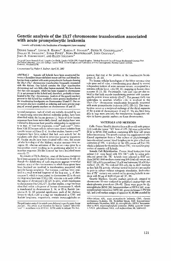
Genetic analysis of the 15;17 chromosome translocation associated with acute promyelocytic leukemia
[somalk cell hybrids/c:fes/localization of breakpoints/gene mapping)
DENISE SHEER*, LYI\:-IE R. HIORI\S*, KARINA F. STANLEY*, PETER N. GOODFELLOw*, DALLAS ~I. SWALLOWt, SUSAI\ POvEYt, NORA HEISTERKA:>IP:f:, JOH).! GROFFEN:j:, }OH:"J R. STEPHEI'<SQN:!:, AND ELLEN SOLOMON*
•Imperial Conoer Research fund, Lincoln's Inn Fields, London WC2.A 3PX, United Kingdom, t),tedical Research Council. Human Bioche~ical Genetios Unit, The Galton Laboratory, Cnivorsity College, London WC2, United Kmgdom; and H.aboratory of Vuil CarconogeneslS, Natwnal Cancer Ins!Jtute, Frederick, \faryland 21701
Communicated by Walter F. Bodmer, Apri/20, 1983
ABSTRACT Somatic cell hybrids have been constructed between a thymidine kinase-deficient mouse ce!l line and blood leukocytes from a patient with acute promyelocytic leukemia showing the 15q+;l7q- chromosome translocation frequently assodated with this disease. One hybrid contains the 15q+ translocation chromosome and very little other human materia!. We have shown that the cjffs oncogene, which has been mapped to chromosome 15, is not present in this hybrid and, therefore, probably is translocated to the l7q- chromosome. Analysis of the genetic markers present in this hybrid has enabled a more precise localization of the translocation breakpoints on chromosomes 15 and 17. Our experiments also have enabled an ordering and more precise mapping of several genetic markers on chromosomes 15 and 17.
Several oncogenes. each of which was initially detected by use of transfonning retrovirus-derived molecular probes, have been identified within the human genome (1). :O.fany of these human sequences have been molecularly cloned, and studies have been initiated to determine their possible relationship to oncogenicity in man. At least tv:o sequences, c-rasKi and c-rasHa, transform :'-IIH 3T3 mouse cells in culture upon direct isolation from speci fie tumor cell lints (2-4). In other studies, human c-rasH• sequences have been isolated that lack such activity but do transform cells when linked to retrovirus promoter sequences l51. Similar results have been obtained with c-rrws, the mouse cellular homologue of the :O.Ioloney murine sarcoma virus oncogene (6), whereas activation of the mouse c-rrws gene by a transposition event resulting in its positioning adjacent to an insertion sequence (ISHike element has been described more recently (7)
By somatic cell hybridization, many of the known oncogenes have been assigned to specific human chromosomes (8-15). Although the distribution of such sequences appears somewhat random. many of the chromosomes to which these genes have been localized are involved in translocations associated with specific human neoplasms. For instance, c-abl has been localized in a small terminal fragment of the long arm, q, of chromosome 9, which is translocated to chromosome 22 in chronic myel"Jgenous leukemia (CYIL) (16), whereas c-sis maps within the region of chromosome 22 (qll to qter), which translocates to chromosome 9 in CY!L (17). In other studies, c-myc has been identified within a fragment of human chromosome 8, which is translocated to chromosomes 2, 14, or 22 in Burkitt lymphoma (8. 9). Of potential significance. each of the chromosomes to which c-ab/ or c-myc are translocated in CML and Burkitt lymphoma, respectiv~ly, contain immunoglobulin se-
The publication ~011> of this article were defrayed in part by ~.age cha;ge payment. This article must therefore be hereby marked ad~ert•semen{' m accordance w1th 18 U. S.C. §1734 solely to mdicate th1s fact.
quences that map at the position of the translocation breakpoints (9, 18, 19).
The human cellular homologues of the feline sarcoma virus v-fes gene and of v-fps, a transforming gene shared by several independent isolates of avian sarcoma viruses. correspond to a common cellular locus, c-fr:s (20, 21), mapping on human chromosome 15 (13, 22). Functionally, v-fes and vfps are also related in that both encode transforming proteins with tyrosinespecific protein kinase activity (23-27). The present study was undertaken to ascertain whether c-fes is involved in the 15q+;l7q- chromosome translocation frequently associated with acute promyelocytic leukemia (APL) (28-31). This translocation occurs as a reciprocal exchange of the distal segments of the q arms of chromosomes 15 and 17. We also have been able to map more precisely the translocation breakpoints relative to known genetic markers on these chromosomes.
MATERIALS AND METHODS
Cells. Frozen blood leukocytes from a 26-yr-old male patient (J.D.) l'.'ith the typical"":0.-13'" form of APL (32) were cultured for 48 hr in RF:-.H 1640 medium containing 20% fetal rcalf serum before karyotyping. The growth medium was supplemented with filtered supernatant from a 7-day culture of phytohemagglutinin-stimulated normal blood lymphocytes to give a final concentration of 10%. A subclone of the 3T3 mouse cell line (33), which is deficient for thymidine kinase (TK), was used for preparation of somatic cell hybrids.
Somatic Ce!l Hybridization. Frozen blood leukocytes from patient J.D. were fused with 3T3 (TK-) cells by using polyethylene glycol (34). TK+ hybrids were selected in HAT medium [RPYII 1640 medium containing 20% fetal calf serum and HAT (100 ,u.:-.1 hypoxanthine/10 ,u.YI methotrexate/16 ,u.YI thymidine)] (35, 36). The unfused 3T3 cells die in HAT medium as they lack TK activity, whereas normal leukocytes are unable to grow in culture without mitogenic stimulation. Back-selection of TK- variants was carried out by growing hybrids in medium with 60 p.g of BrdUrd per ml (37, 38).
Genetic :\farkers. Genetic markers previously mapped to chromosome 15 were analyzed by published immunologic and electrophoretic procedures (39-42). These were genes for /32-microglobulin (B2M) (39), hexosaminidase a (HEXA) (41), mannosephosphate isomerase (:-.tPI) (42), pyruvate kinase-3 (PKYI2) (42), and a cell surface antigen of apparent M, 95,000 encoded by
Abbreviatiom: APL, acute promyelocytic leukemia; CML. chronic myelogenous leukemia; TK, thymidine kinase; HAT. hypoxanthine/ methotrexate/thymidine; B2M, {3~ microglobulin; HEXA, hexosaminidase tr; MPI, mannosephosphate isomerase; PKM2, pyruvate kinase-3, CALK. galactokinase; CAA, acid a-glucosidase, kb. ki!obase(s).
121
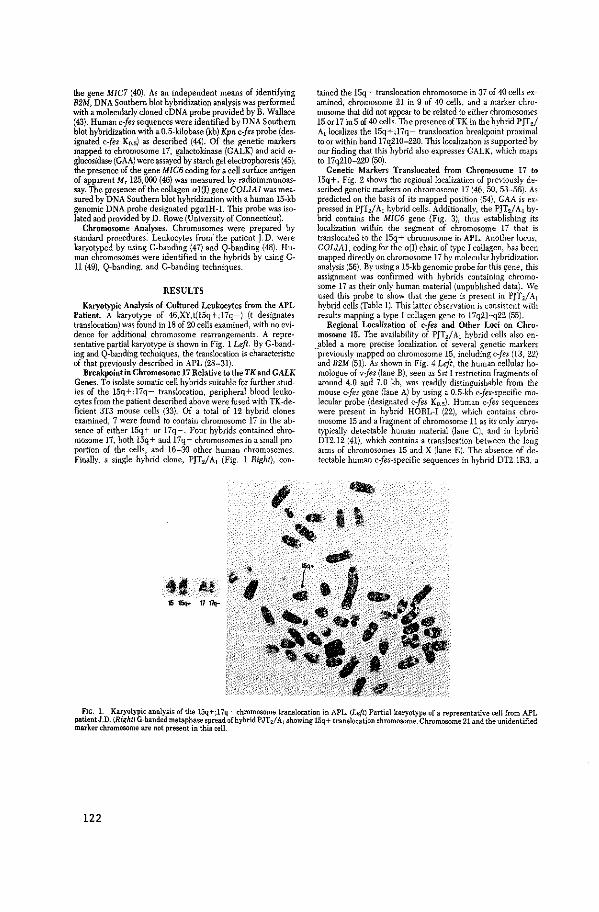
the gene MIC7 (40). As an independent means of identifying B2M, DNA Southern blot hybridization analysis was performed with a molecularly cloned eDNA probe provided by B. Wallace (43). Human c-fes sequences were identified by DNA Southern blot hybridization with a 0.5-kilobase (kb) Kpn c-fes probe (designated c-fes Ko.s) as described (44). Of the genetic markers mapped to chromosome 17, galactokinase (CALK) and acid aglucosidase (GAA) were assayed by starch gel electrophoresis (45); the presence of the gene MIC6 coding for a cell surface antigen of apparent M, 125,000 (46) was measured by radioimmunoassay. The presence of the collagen al(I) gene COLlAl was measured by DNA Southern blot hybridization with a human 15-kb genomic DNA probe designated pga1H-l. This probe was isolated and provided by D. Rowe (University of Connecticut).
Chromosome Analyses. Chromosomes were prepared by standard procedures. Leukocytes from. the patient J.D. were karyotyped by using G-banding (47) and Q-banding (48). Human chromosomes were identified in the hybrids by using G-11 (49}, Q-banding, and G-banding techniques.
RESULTS
Karyotypic Analysis of Cultured Leukocytes from the APL Patient. A karyotype of 46,XY,t(l5q+;l7q-) (t designates translocation) was found in 18 of 20 cells examined, with no evidence for additional chromosome rearrangements. A representative partial karyotype is shown in Fig. 1 Left. By G-banding and Q-banding techniques, the translocation is characteristic of that previously described in APL (28-31}.
Breakpoint in Chromosome 17 Relative to the TK and CALK Genes. To isolate somatic cell hybrids suitable for further studies of the 15q+;l7q- translocation, peripheral blood leukocytes from the patient described above were fused with TK-deficient 3T3 mouse ce!!s (33). Of a total of 12 hybrid clones examined, 7 were found to contain chromosome 17 in the absence of either ISq+ or 17q-. Four hybrids contained chromosome 17, both l5q+ and 17q- chromosomes in a small proportion of the cells, and 16-30 other human chromosomes. Finally, a single hybrid clone, PJT2/A1 (Fig. l Right), con-
tt A~ 1S 15qo- 11 17q-
tained the 15q+ translocation chromosome in 37 of 40 cells examined, chromosome 21 in 9 of 40 cells, and a marker chromosome that did not appear to be related to either chromosomes 15 or 17 inS of 40 cells. The presence ofTK in the hybrid PJf zl A1 localizes the lSq+ ;17q- translocation breakpoint proximal to or within band 17q210-220. This localization is supported by our finding that this hybrid also expresses CALK, which maps to 17q210-220 (SO).
Genetic Markers Translocated from Chromosome 17 to I5q+. Fig. 2 shows the regional localization of previously described genetic markers on chromosome 17 (46, 50, 53-56). As predicted on the basis of its mapped position (54}, GAA is expressed in PJTziAI hybrid cells. Additionally, the PJTJ A1 hybrid contains the MIC6 gene (Fig. 3), thus establishing its localization within the segment of chromosome 17 that is translocated to the 15q+ chromosome in APL. Another locus, COLIAl, coding for the a( I) chain of type I collagen, has been mapped dire<:tly on chromosome 17 by molecular hybridization analysis (56). By using a 15-kb genomic probe for this gene, this assignment was confirmed with hybrids containing chromosome 17 as their only human material (unpublished data). We used this probe to show that the gene is present in PJT2/A1 hybrid cells (Table 1). This latter observation is consistent with results mapping a type I collagen gene to 17q2I-q22 (55).
Regional Localization of cjes and Other Loci on Chromosome 15. The availability of PJT~/A 1 hybrid cells also en
__ abled a more precise localization of several genetic markers previously mapped on chromosome 15, including c-fes (13, 22) and B2M (51). As shown in Fig. 4 Left, the human cellular homologue of vjes (lane B), seen as Sst I restriction fragments of around 4.0 and 7.0 kb, was readily distinguishable from the mouse c-fes gene (lane A) by using a 0.5-kb cjes-specific molecular probe (designated c-fes Ko.s). Human c-fes sequences were present in hybrid HORL-I (22), which contains chromosome 15 and a fragment of chromosome 11 as its only karyotypically detectable human material (lane C), and in hybrid DT2.12 (41), which contains a translocation between the long arms of chromosomes IS and X (lane E). The absence of detectable human cjes-specific sequences in hybrid DT2.1R3, a
FIG. 1. Karyotypic analysis of the 15q+;l7q- chromosome translocation in APL. (Left) Partial karyotype of a representative cell from APL patientJ.D. alight) G-banded metaphase spread of hybrid PJT z/ Al showing 15q + translocation chromosome. Chromosome 21 and the unidentified mLU"ker chromosome are not present in this cell.
122
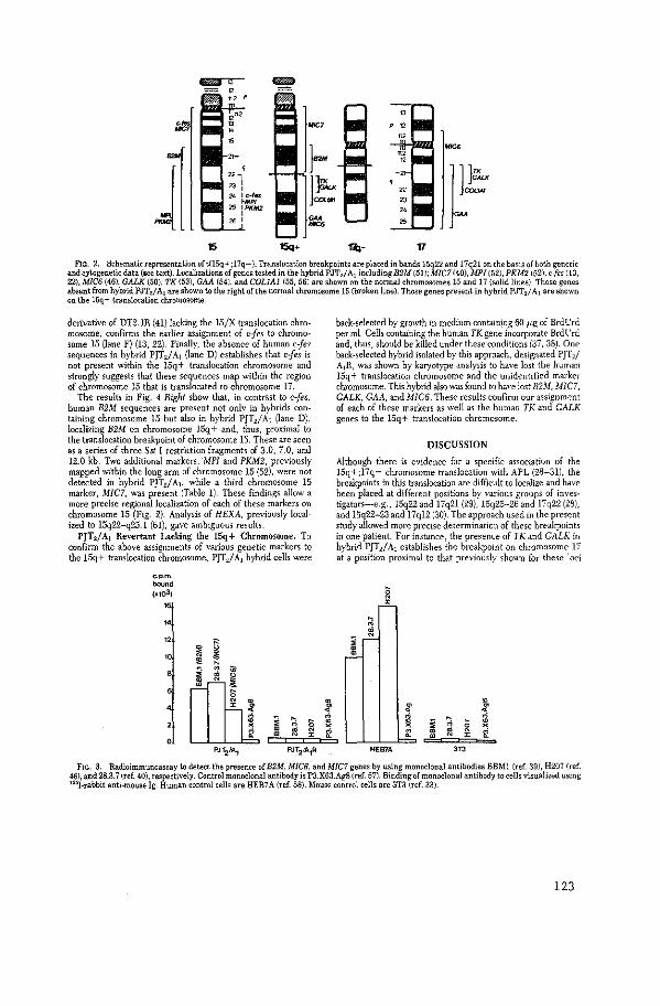
15 15q+ r.t,- 17
FIG. 2. Schematic representation oft(l5q+;17q-). Translocation breakpoints are placed in bands l5q22 and 17q21 on the basis of both genetic and cytogenetic data (see text). Localizations of genes Wsted in the hybrid PJT.; A1 including B2M (51); MIC7 (40), MPI (52), PKM2 (52), c·fes (13, 22), MIC6 (46), GALK {50}, TK (53), GAA (54), and COLlAl {55, 56) are shown on the normal chromosomes 15 and 17 (solid lines). Those genes absent from hybrid PJT2/ A1 are shown to the right of the nonnal chromosome 15 (broken line). Those genes present in hybrid PJTzf A1 are shown on the l5q+ translocation chromosome.
derivative of DT2.1R (41} lacking the 15/X translocation chromosome, confirms the earlier assignment of c-fes to chromosome 15 (lane F) (13, 22). Finally, the absence of human c-fes sequences in hybrid PJT2/A! (lane D) establishes that c{es is not present within the ISq+ translocation chromosome and strongly suggests that these sequences map within the region of chromosome 15 that is translocated to chromosome 17.
The results in Fig. 4 Right show that, in contrast to c-fes, human B2M sequences are present not only in hybrids containing chromosome 15 but also in hybrid PJT2/A1 (lane D), localizing B2M on chromosome ISq+ and, thus, proximal to the translocation breakpoint of chromosome 15. These are seen as a series of three Sst I restriction fragments of 3.0, 7 .0, and 12.0 kb. Two additional markers, "MPI and PKM2, previously mapped within the long ann of chromosome IS {52), were not detected in hybrid PJT2/A1, while a third chromosome 15 marker, MIC7, was present {Table 1). These findings allow a more precise regional localization of each of these markers on chromosome 15 (Fig. 2). Analysis of HEXA, previously localized to 15q22-q25.1 {61), gave ambiguous results. PJT~/A 1 Revertant Lacking the 15q+ Chromosome. To
confirm the above assignments of various genetic markers to the 15q+ translocation chromosome, PJT2/A1 hybrid cells were
c. p.m.
""""' (~103)
"
"
' 0
* $ " " " ' ~
PJT2tA1
l ;; ~ ' • ~ ~ • "
PJT2J.A.1R
~ ,.; ~ ~
back-selected by growth in medium containing 60 }Lg of BrdL'rd per mi. Cells containing the human TK gene iilcorporate BrdL'rd and, thus, should be killed under these conditions (37, 38}. One back-selected hybrid isolated by this approach, designated PJT2/
A1R, was shown by karyotype analysis to have lost the human 15q + translocation chromosome and the unidentified marker chromosome. This hvbrid also was found to have lost B2M, MIC7, CALK, GAA, and MIC6. These results confirm our assignment of each of these markers as well as the human TK and CALK genes to the 15q+ translocation chromosome.
DISCUSSION
Although there is evidence for a specific association of the 15q+;l7q- chromosome translocation with APL (28-31}, the breakpoints in this translocation are difficult to localize and have been placed at different positions by various groups of investigators-e.g., 15q22 and 17q21 (29), 15q25-26 and 17q22 (28), and 15q22-23 and l7ql2 (30). The approach used in the present study allowed more precise determination of these breakpoints in one patient. For instance, the presence of TK and CALK in hybrid PJT2/A1 establishes the breakpoint on chromosome 17 at a position proximal to that previously shown for these loci
. ~ :t ~ ;; ,.; • ~ ~ ~ • re ~ •
HEB7A "' F1c;... 3. Radioimmunoassay to detect the presence of B2M, MIC6, and MIC7 genes by using monoclonal antibodies BBMl (ref. 39l, H207 (ref.
461, and 28.3.7 (ref. 40), respectively. Control monoclonal antibody is P3.X63.Ag8 (ref. 157). Binding of monoclonal antibody to cells visualized using ' 25!-rabbit anti-mouse !g. Human control cells are HEB7A (ref. 58). Mouse control cells are 3T3 (ref. 33}.
123
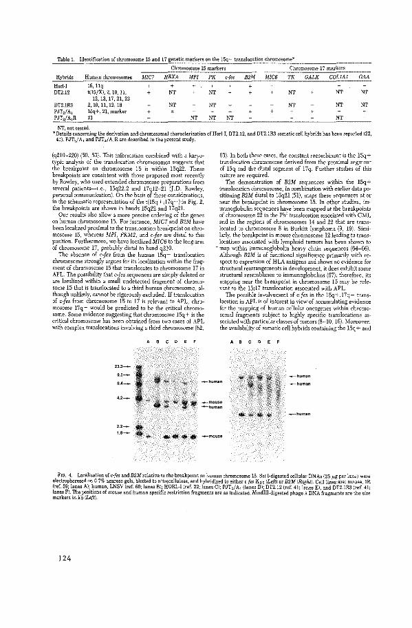
Table L Identification of chromosome 15 and 17 genetic markers on the 15q+ translocation chromosome*
Chromosome 15 markers Chromosome 17 markers
Hybrids Human chromosomes MICJ HEXA MPI PK c-fes B2M MIC6 TK GALK COLlAl GAA
Horl-I 15, llq + + + + DT2.12 t(15/Xl, 2, 10, 11, + NT NT NT NT NT
12, 13, 17' 21, 22 DT2.1R3 2, 10, 11, 12, 18 NT NT NT NT NT PJT2fA, 15q+, 21, marker + ± + PJT2/A1R 21 NT NT NT NT
NT, not tested. *Details concerning the derivation and chromosomal characterization of Horl-I, DT2.12, and DT2.1R3 somatic cell hybrids has been reported (22,
41). PJTzf A, and PJT2f A1R are described in the present study.
(q210-220) (50, 53). This information combined with a karyotypic analysis of the translocation chromosomes suggests that the breakpoint on chromosome 15 is within 15q22. These breakpoints are consistent with those proposed most recently by Rowley, who used extended chromosome preparations from several patients-i.e., l5q22.2 and l7ql2-2l (J.D. Rowley, personal communication). On the basis of these considerations, in the schematic representation of the t(l5q + ;l7q-) in Fig. 2, the breakpoints are shown in bands l5q22 and 17q21.
Our results also allow a more precise ordering of the genes on human chromosome 15. For instance, MIC7 and B2M have been localized proximal to the translocation breakpoint on chromosome 15, whereas l\-JPI, PKM2, and c-fes are distal to this position. Furthermore, we have localizedMIC6 to the long arm of chromosome 17, probably distal to band q2IO.
The absence of cjes from the human l5q+ translocation chromosome strongly argues for its localization within the frag~ ment of chromosome 15 that translocates to chromosome 17 in APL. The possibility that cjes sequences are simply deleted or are localized within a small undetected fragment of chromosome 15 that is translocated to a third human chromosome, although unlikely, cannot be rigorously excluded. If translocation of cjes from chromosome 15 to 17 is relevant to APL, chromosome 17q- would be predicted to be the critical chromosome. Some evidence suggesting that chromosome l5q+ is the critical chromosome has been obtained from two cases of APL with complex translocations involving a third chromosome (62,
A B C 0 E
63). In both these cases, the constant recombinant is the 15q+ translocation chromosome derived from the proximal segment of 15q and the distal segment of 17q. Further studies of this nature are required.
The demonstration of 82M sequences within the 15q+ translocation chromosome, in combination with earlier data po· sitioning B2M distal to 15q2l (51), maps these sequences at or near the breakpoint in chromosome 15. In other studies, im" munoglobulin sequences have been mapped at the breakpoints of chromosome 22 in the Ph1 translocation associated with C:O.'iL and in the regions of chromosomes 14 and 22 that are translocated to chromosome 8 in Burkitt lymphoma (9, 19). Similarly, the breakpoint in mouse chromosome 12leading to translocations associated with lymphoid tumors has been shown to
~ map within immunoglobulin heavy chain sequences (64-66). Although B2M is of functional significance primarily with respect to expression of HLA antigens and shows no evidence for structural rearrangements in development, it does exhibit some structural resemblance to immunoglobulins (67); therefore, its mapping near the breakpoint in chromosome 15 may be relevant to the Ei;l7 translocation associated with APL.
The possible involvement of c-fes in the 15q+;l7q- translocation in APL is of interest in view of accumulating evidence for the mapping of human cellular oncogenes within chr(lmo~ somal fragments subject to highly specific translocations associated with particular classes of tumors (8-10, 16). :\loreover, the availability of somatic cell hybrids containing the l-5q + and
' ' ' ' '
-human ---human -human
.__mouse -human
-o-human
2.2-
1.8--.... $ ---mouse
FIG. 4. Localization of c-fes andB2M relative to the breakpoint on human chromosome 15. Sst !-digested cellular DNA~ (25 p.g per lane) were electrophoresed on 0.7% agarose gels, blotted to nitrocellulose, and hybridized to either c-fes Ko~ (Left) or B2M (Right). Cell lines are: mouse, 1R (ref. 59; lanes A); human, LNSV (ref. 60; lanes BJ; HORL-1 (ref. 22; lanes CJ; PJTzfA1 Oanes DJ; DT2.12 (ref. 41: lanes E), and DTZ.lR3 (ref. 41: lanes F). The positions of mouse and human specific restriction fragments are as indicated. Hindiii-digested phage A DNA fragments are the size markers in kb (Left).
124
17q- translocation chromosomes should facilitate studies of the breakpoints in this translocation and a determination of the proximity of c{es to the translocation breakpoint.
We thank Mohamed Parkar, Barbara Pym, G. T. Blennerhassett, and P. Hanson fortheir excellent technical assistance; Dr. Janet Rowley for stimulatmg discussions; and Christine Furse for typing. We are also grateful to the Imperial Cancer Research Fund Medical Oncology Unit, St. Bartholomew s Hoop1tal, London, for generously providing us with cells from APL patients. This work was partially supported under Contract N01-C0-76380 from the National Cancer Instill•te
l. Varmus, H. E. (1982) Science 216, 812-820 2 Santos, E., Tronick, S. R., Aaronson, S. A., Pulciani, S. & Bar
bacid, XL (1982) Nature (London) 298, 343-347 3. Der, C. J., Krontiris, T. G. & Cooper, G. M. (1982) Proc. NatL
Acad. Sci. USA 79, 3637-3640. 4. Parada, L. F., Tabin, C. J., Shih. C. & Weinberg, R. A. (!982)
Nat11re (London) 297, 474-478. 5, Chang, E. H., Furth, M. E., Smlnick, E. M. & Lowy, D. R. (1982)
Nature {London) 297, 479-483. 6. .Blair, D. G., Oskarsson, ).1., Wood, T. G., McClements, ·w, L ..
Fischinger, P. J, & Vande Woude, G. F. (1981) Science 212, 941-943.
7. Rechavi, G., Givol. D. & Canaani, E. (1982) Nature (London) 300, 607-611 Dalla-Favera, R, Bregni, M., Erik5on. J., Patterson, D., Gallo, ~Bz~: & Croce, C. M. (1982) Proc. Nati.Acad. Sci. USA 79, 7824~
9. Taub, R, Kirsch, L, Morton, C., Lenoir, G., Swan, D., Tronick S., Aaronson. S. & Leder, P. {1982) Proc. Nat/. Acad. Sci. l'SA 79, 7837-7841
10. Nee!, B. G., Jhanwar, S, C., Chaganti, R. S. K. & Hayward, W S. (1982) Proc. Nat/, Acad. Sci. USA 79, 7842-7846.
ll Swan, D. C., ).fcBride, 0. \V.. Robbins, K. C., Keithl<'y, D. A .. Reddy, E. P. & Aaronson, S. A. (1982) Proc. Nat/. Acad. Sci. US.'\ 79, 4691-4695.
12. Dalla-Favera, R., Gallo. R C., Giallongo, A. & Croce, C. )d. (19821 Science 218. 686-688
13 Dalla-fa,·era, R., Franchini, G., Martionotti, S., Wong-Staal, F., Gallo, R. C. & Croce, C. !vi. (19521 Proc. Nat/. Acad. sCi. USA 79, 4714-4717
14 Prakash, K., McBride. Q_ W .. S\\'an, D. C., Devare, S. G., Iron· ick, S. R, & Aaronson, S. A. (19821 Proc. Nat/. Acad_ Sci. l!SA 79, 5210-5214.
15. ~lcBride, 0. \\' .. Swan. D. C., Santos, E., Barbacid, ~!.. Tronick, S. R. & Aaronson. S. A_ (1982) ?\'ai!Jre (London) 300, 773-774
16 de Klein, A., van Kessel, A. G., Grosveld, G., Bartra:n, C. R.. Hagemeijer, A., Bootsma, D .. Spurr, N. K., Heisterkamp, ~ .. Groffen. J. & Stephenson. J. R. (1982) Nature (Londari) 300, 765-767
l7 Groffen, J., Heisterkamp, :\ .. Stephenson, J. R.. van KesseL A ~.,de Klein, A., Gros\'eld, C. & Bootsma, D. (1983)]. Exp. :\fed., 1n preso
18. Erik>on, J., ~!artinis, J- & Croce. C. M. (19811 Sature (LOildan! 294,173-115
19. Kinch, L R., !.lorton, C. C., Nakahara, K. & Leder, P. (W821 Science 216, 301-303.
20. Shibuya, ),-1., Hanafusa, H., Hanafusa, T & Stephenson, J. R (1980) Proc. Sat/. Acad. Sci. USA 77, 6536-6540.
21 Graffeo, J, Heisterkamp. ~ .. Shibuya, !.! .. Hanafusa, H. & Stephenson.]. R. (1953) Virology 125, 480-486
22. Heisterkamp, \' .. Groffen. J-, Stephenson, J. R., Spurr,:\_ K .. Goodfellow, p_ N., Solomon, E .. Carritt. B. & Bodmer. \\'. F (1982) Nature (London) 299, 747-749
23 Van de Ven. W. J. ~1.. Reynolds, F. H .. Jr., & Stephenson, J. R (1980) Virology 101, 185-197
24 Feldman, R. A .. Hanafusa, T. & Hanafusa. H. (19801 Cell 22, 7'57-765.
25 Pawson. T., Guyden, J .. Kung, T·H., Radke, K .. Gilmore, T. & :"1-lartin, C. S. (1980) Ce/122, 767-775
26 Reynold>, F. H., Jr .. Van de Ven, W. ("M, & Stephenson, J. R. (1980)]. Bioi. C/,em. 255, ll040-ll047.
27 Barbacid, M., Beeman, K. & Devare, S. G. {1980) Proc- ?\'atLAcad Sci. USA 77, 5158-'5162
28 Second International Workshop on Chromosomes in Leukemia, 1979: Chromosomes in Acute Promyelocytic Leukemia (1980: Cancrr Genet. Cytogenet. 2, 103-107
29 Rowley, J.D .. Golomb,-H. M., Vardiman, J., Fukuhara, 5., Dougherty, C. & Potter, D. (1977) lnt.]. Cancer20, 869-8i2
30. ~~ndo, K. & Sasaki, M. (1982) Cancer Genet. Cytogenet. 6, 39-
31. Van Den Berghe, H., Louwagie, A., Broeckaert-Van Orshoven, A., David, G., Verwilghen, R.. Michaux, J. L. & Sakai, G. (1979) Cancer 43, 558-562. 1
32. Bennet, J. M., Catovsky, D., Daniel, M. T., Flandrin, G., Galton, D. A. G., Gralnick, H. R. & Sultan, C. (1976) Br.). Hacmalo/. 33, 451-458.
33, Todaro, G. J. & Green, H. (1963)]. Cell Bioi. 17, 299~316. 34. Galfre, G., Howe, S. C., Milstein, C .. Butcher, G. W. & How·
ard, J. C. (1977) Nature (London) 266. 550-552. 35. Szybalski, W., Szybalski, E. & Rayni, C. (1962) .'>'at/. Cancer lnst
Monogr. 7, 75-89. 36. Littlefield,]. (1964) Science 145, 709-710. 37. Kit, S., Dubbs, D. R., Piekarski. J, R & Hsu, T. C. (19631 E:rp.
Cell Res. 31, 297-312. 38 1,\'eis.>. M. C. & Green, H. {1967) Proc. Nat/. A cad. Sci. USA 58,
1104~1111
39. Brodsky, F. M .. Bodmer, W. F. & Parham, p_ {19791 Eur.].lmmuno/. 9, 536-545
40 Blaineau, C .. Avner, P., Tunnacliffe, A. & Goodfellow, P. K (19831 EMBO ]., in press.
41. Swallow, D. \-1., Solomon, E. & Pajunen. L. (1977) Cytogenet. Cell Genet. 18, 136-148
42, van Heyningen, V., Bobrow. ~1., Bodmer, W. F., Gardiner, S. E., Pavey, S. & Hopkinson, D. A. (1975} Arw. Hwn_ Genet. 38, 295-302.
43. Suggs, S. V., Wallace_ R. B., Hirose, T., Kawaohima, E. H & lkatura. K. (1981) Proc. Nat/. Acad. Sci. USA 78, 6613-6611
44. Croffen, J., Heisterkamp, N., Gros\'eld. F .. \'an de Ven, \\". & Stephenson, J- R (1982) Science 216, 1136-1138
45 Solomon, E .. Swallow, D., Burgess, S. & Evans, L. (19791 Ann Hum, Genet. 42, 273-281
46. Bai, Y., Sheer, D .. Hiorns, L., Knowles, R. \\'. & Tunnadiffe. A. (1982) Ann_ Hu.m. Genet. 46, .337-347.
47 Seabright. M. (1972) Chromowma {Berlin) 36, 204-210. 48. Caspersson. T., Lomakka, C. & Zech. L. {1971) Hereditas 67, 89-
102, 49. Bobro\\', !.l. & Cross, J. (!9741 \'ature (Londonl25!, 17-79 50. Elsev.ier, S. M .. Kucherlapati. R. S .. \"icho!,, E A .. Creagan. R
P., G1les, R. P .. Ruddle, F_ H .. \\'1llecke, K. & ~/cDougalL J. K {1974) ,\'at11re {London) 25L 633-635
51 Pajunen, L., Solomon. E., Burgess, S., Bobrow,~!., Pmc\·, S & Swallow, D. (19781 Cytogenet. Cell Cer1et. 22. 5ll-512. .
52. Chern, C.]., Kennett, R., Engel. E., ~!ellman, \\'_ J- & Croce, C. ~!. (19771 Somatic Cell Genet. 3, 553-560
53 Human Gene ~lapping 5 (1919! Cytof!,enet. Cell Genet. 25, 59-/3 54 Nitkel, B_ E., Chudlev, A. E .. Pabello, P. D. & ~!cAlpine, P. J
(1982) Cytogenet. Cell Gcnd 32, 303 55. Church. R. L.. Sundar Raj.\'. & ).!cDougall, J- K (1950) Cyto·
genet. Cell Genet, 27, 24-30. 56 Huerre. C., Junien, C., \Yeil. D., Chu. ~L·L, ).!nrahito, \!.. nn
Cong. N., ~!yers, J. C., Foubert, C., Cross, ),!.-S., Prockop. D J .. Boue, A .. Kaplan.]. C., de Ia Chapdlc, /1,_ & Ramirez, F. ·:19821 Proc. ,\'~11. A cad. Sci. L'SA 79, 6627-66.31
57. Kohler, G. & !.lilstein. C. (1975: .\'11ture (l"ondonl 256, -195-497 58 Wallace, D.C. Bunn. C. L. & Eisenst:<dt, J- !\!. (1975)]. Cell BioL
67, 114-188 59. J\abholz. \!., ~!iggiano. \'. & Bodmer,\\'. f_ (1969) .\'ature !Lon
don) 223, 358-363 60. Croce, C.\!., Knmdes. B. B. & Kopmwsh H. (19731 E.\'J!- Cell
Res. 82, 457~461 61 ).1age'lis, R. E., \'idgoff, J .. Chamberlin, J- & Brown.~!. C. 11g791
Cytogenet. Cell Genet. 25, 181 62. Bernstein, R., !.·kndelow, B., Pinto, ~!. R., ~Iarcom, G & Bez·
woda, W. {1980) Br.]. Haemato/. 46. 3ll-314 63. Rowley, J. D. (19821 Science 216, 749-751 64. Crew.>, S., Barth, R., Hood, L.. Prehn, J- & Calame K. {1982:
Science 218, 1319-!321 65. Shen·Ong, G. L. C .. Keath, E. J., Picc(lli, S. P. & Cole, \1. D.
(1982) Ce/131, 443-4.52 66. Calame, K., Kim, S .. Lalley, P., HilL R.. Da"i', M. & Hood, L
(1982) Proc . .\'Gtl. Acad. Sci. USA 79, 6994-6998. 67. Ploegh. H. L., Orr, H. T. & Strominger, J. L. {19811 Ce/124. 287-
299
125


APPENDIX PAPER VII

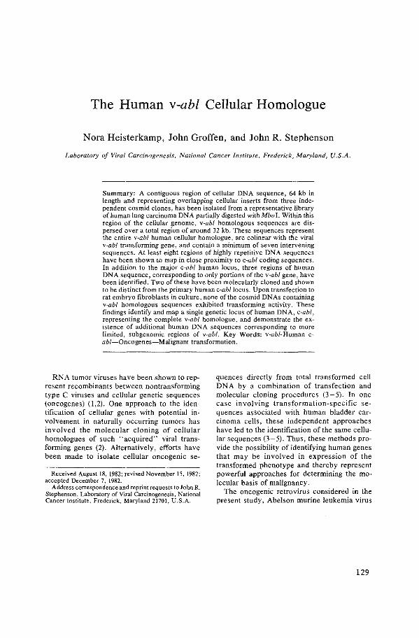
The Human v-abl Cellular Homologue
Nora Heisterkamp, John Groffen, and John R. Stephenson
Laboratory of Viral Carcinogenesis, National Cancer Institute, Frederick, Maryland, U.S.A.
Summary: A contiguous region of cellular DNA sequence, 64 kb in length and representing overlapping cellular inserts from three independent cos mid clones, has been isolated from a representative library of human lung carcinoma DNA partially digested with Mba I. Within this region of the cellular genome, v-abl homologous sequences are dispersed over a total region of around 32 kb. These sequences represent the entire v-abl human cellular homologue, are colinear with the viral v-abl transforming gene, and contain a minimum of seven intervening sequences. At least eight regions of highly repetitive DNA sequences have been shown to map in close proximity to c-abf coding sequences. In addition to the major c-ab! human locus, three regions of human DNA sequence, corresponding to only portions of the v-abl gene, have been identified. Two of these have been molecularly cloned and shown to be distinct from the primary human c--ab/locus. Upon transfection to rat embryo fibroblasts in culture, none of the cosmid DNAs containing v-abl homologous sequences exhibited transforming activity. These findings identify and map a single genetic locus of human DNA, c-ab!, representing the complete v-abl homologue, and demonstrate the existence of additional human DNA sequences corresponding to more limited, subgenomic regions of v-abl. Key Words: v-ab/-Human cab/-Oncogenes-Malignant transformation.
RNA tumor viruses have been shown to represent recombinants between nontransforming type C viruses and cellular genetic sequences (oncogenes) (1,2). One approach to the identification of cellular genes with potential involvement in naturally occurring tumors has involved the molecular cloning of cellular homologues of such "acquired" viral transforming genes (2). Alternatively, efforts have been made to isolate cellular oncogenic se-
Received August 18, 1982; revised November 15, 1982; accepted December 7, 1982.
Address correspondence and reprint requests to John R.
quences directly from total transformed cell DNA by a combination of transfection and molecular cloning procedures (3-5). In one case involving transformation-specific sequences associated with human bladder carcinoma cells, these independent approaches have led to the identification of the same cellular sequences (3-5). Thus, these methods provide the possibility of identifying human genes that may be involved in expression of the transformed phenotype and thereby represent powerful approaches for determining the molecular basis of malignancy.
Stephenson, Laboratory of Viral Carcinogenesis, National Cancer Institute, Frederick, Maryland 21701, U.S.A.
The oncogenic retrovirus considered in the present study, Abelson murine leukemia virus
129
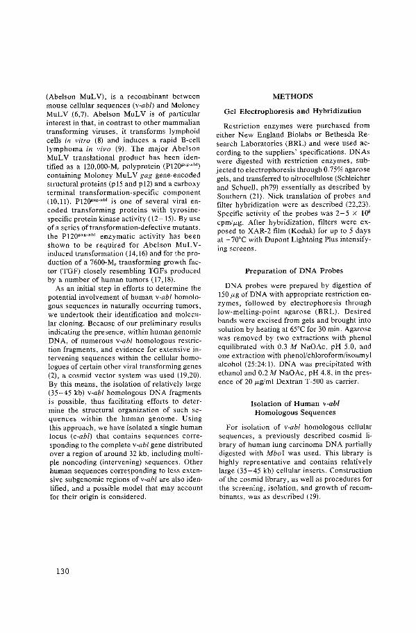
(Abelson MuLV), is a recombinant between mouse cellular sequences (v-abl) and Moloney MuLV .(6,7). Abelson MuLV is of particular interest in that, in contrast to other mammalian transforming viruses, it transforms lymphoid cells in vitro (8)' and induces a rapid B-ee!! lymphoma in vivo (9). The major Abelson MuL V translational product has been identified as a 120,000-Mr polyprotein (P12~a~:·abl) containing Moloney MuL V gag gene-encoded structural proteins (p 15 and p 12) and a carboxy terminal transformation-specific component (10,11). P12~ag.abJ is one of several viral encoded transforming proteins with tyrosinespecific protein kinase activity (12-15). By use of a series of transformation-defective mutants, the P120gag.abl enzymatic activity has been shown to be required for Abelson MoLYinduced transformation ( 14, 16) and for the production of a 7600-Mr transforming growth factor (TGF) closely resembling TGFs produced by a number of human tumors (17,18).
As an initial step in efforts to determine the potential involvement of human v-abl homologous sequences in naturally occurring tumors, we undertook their identification and molecular cloning. Because of our preliminary results indicating the presence, within human genomic DNA, of numerous v-abl homologous restriction fragments, and evidence for extensive intervening sequences within the cellular homologues of certain other viral transforming genes (2), a cosmid vector system was used (19,20). By this means, the isolation of relatively large (35-45 kb) v-abl homologous DNA fragments is possible, thus facilitating efforts to determine the structural organization of such sequences within the human genome. Using this approach, we have isolated a single human locus (c-abl) that contains sequences corresponding to the complete v-abl gene distributed over a region of around 32 kb, including multiple noncoding (intervening) sequences. Other human sequences corresponding to less extensive subgenomic regions of v-abl are also identified, and a possible model that may account for their origin is considered.
130
METHODS
Gel Electrophoresis and Hybridization
Restriction enzymes were purchased from either New England Biolabs or Bethesda Research Laboratories (BRL) and were used according to the suppliers' specifications. DNAs were digested with restriction enzymes, subjected to electrophoresis through 0.75% agarose gels, and transferred to nitrocellulose (Schleicher and Schuell, ph79) essentially as described by Southern (21). Nick translation of probes and filter hybridization were as described (22,23). Specific activity of the probes was 2-5 x lOS cpm/ fLg. After hybridization, filters were exposed to XAR-2 film (Kodak) for up to 5 days at -70°C with Dupont Lightning Plus intensifying screens.
Preparation of DNA Probes
DNA probes were prepared by digestion of 150 fLg of DNA with appropriate restriction enzymes, followed by electrophoresis through low-melting-point agarose (BRL). Desired bands were excised from gels and brought into solution by heating at 65°C for 30 min. Agarose was removed by two extractions with phenol equilibrated with 0.3 M NaOAc, pH 5.0, and one extraction with phenoUchloroform/isoamyl alcohol (25:24: l). DNA was precipitated with ethanol and 0.2 M NaOAc, pH 4.8, in the presence of 20 fLg/ml Dextran T-500 as carrier.
Isolation of Human v-abl Homologous Sequences
For isolation of v-abl homologous cellular sequences, a previously described cosmid library of human lung carcinoma DNA partially digested with Mbol was used. This library is highly representative and contains relatively large (35-45 kb) cellular inserts. Construction of the cosmid library, as well as procedures for the screening, isolation, and growth of recombinants, was as described (19).
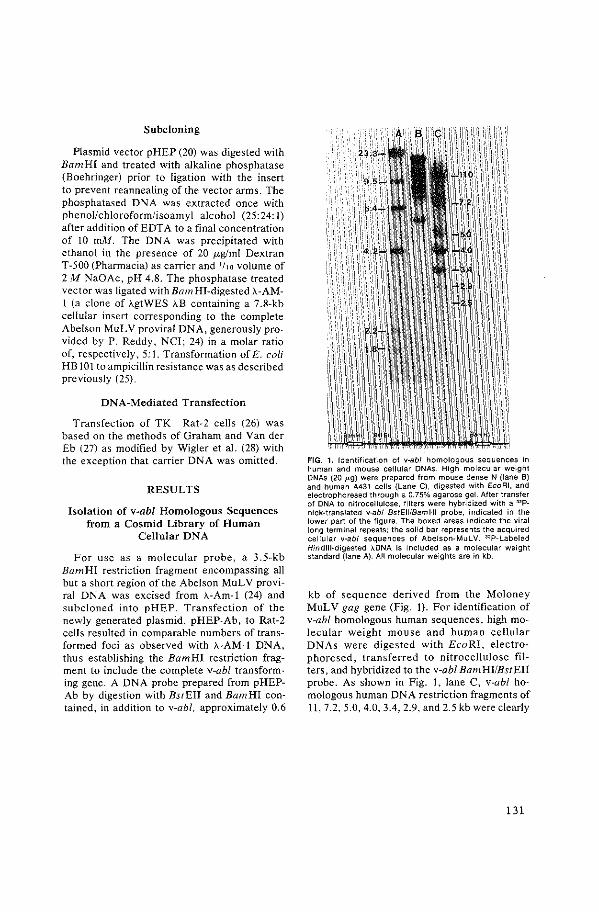
Subcloning
Plasmid vector pHEP (20) was digested with Bam HI and treated with alkaline phosphatase (Boehringer) prior to ligation with the insert to prevent reannealing of the vector arms. The phosphatased DNA was extracted once with phenol/chloroform/isoamyl alcohol (25:24: I) after addition of EDTA to a final concentration of 10 rnM. The DNA was precipitated with ethanol in the presence of 20 ,ug/ml Dextran T-500 (Pharmacia) as carrier and 1/1o volume of 2M NaOAc, pH 4.8. The phosphatase treated vector was ligated with Bam HI-digested A-AMI (a clone of AgtWES AB containing a 7.8-kb cellular insert corresponding to the complete Abelson MuL V proviral DNA, generously provided by P. Reddy, NCI; 24) in a molar ratio of, respectively, 5: I. Transformation of E. coli HB 101 to ampicillin resistance was as described previously (25).
DNA-Mediated Transfection
Transfection of TK- Rat-2 cells (26) was based on the methods of Graham and Vander Eb (27) as modified by Wigler et al. (28) with the exception that carrier DNA was omitted.
RESULTS
Isolation of v-abl Homologous Sequences from a Cosmid Library of Human
Cellular DNA
For use as a molecular probe, a 3.5-kb BamHI restriction fragment encompassing all but a short region of the Abelson MoL V proviral DNA was excised from A-Am-1 (24) and subcloned into pHEP. Transfection of the newly generated plasmid, pHEP-Ab, to Rat-2 cells resulted in comparable numbers of transformed foci as observed with A-AM-I DNA, thus establishing the Bam HI restriction fragment to include the complete v-abl transforming gene. A DNA probe prepared from pHEPAb by digestion with BstEII and Bam HI contained, in addition to v-ahl, approximately 0.6
FIG. 1. Identification of v-abl homologous sequences in human and mouse cellular DNAs. High molecular weight DNAs (20 !'g) were prepared from mouse dense N (lane B) and human A431 cells (Lane C), digested with EcoRI, and electrophoresed through a 0.75% agarose gel. After transfer of DNA to nitrocellulose. fllters were hybridized with a "P· nick·translated v-ab/ 8stEII/8amHI probe, indicated in the lower part of the figure. The boxed areas indicate the viral long terminal repeats; the solid bar represents the acquired cellular v-abl sequences of Abelson-MuLV. "P-Labeled Hindlll-digested !..DNA is included as a molecular weight standard (lane A). All molecular weights are in kb.
kb of sequence derived from the Moloney MuLV gag gene (Fig. 1). For identification of v-abl homologous human sequences, high molecular weight mouse and human cellular DNAs were digested with EcoRI, electrophoresed, transferred to nitrocellulose filters, and hybridized to the v-abl BamHI!BstEII probe. As shown in Fig. 1, lane C, v-abl homologous human DNA restriction fragments of ll, 7.2, 5.0, 4.0, 3.4, 2.9, and 2.5 kb were clearly
131
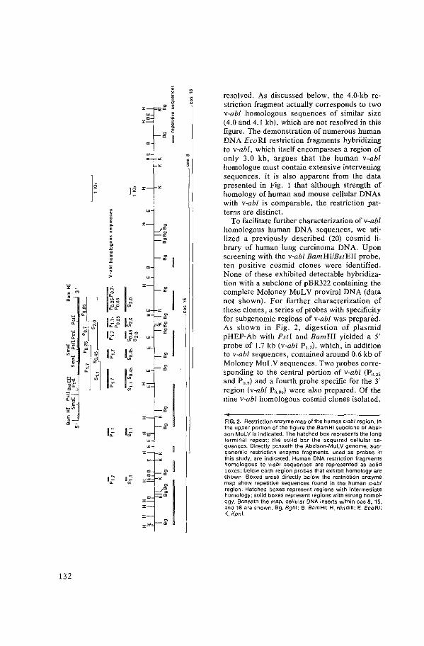
I~
12 F •
132
•
resolved. As discussed below, the 4.0-kb restriction fragment actually corresponds to two v-abl homologous sequences of similar size (4.0 and 4.1 kb), which are not resolved in this figure. The demonstration of numerous human DNA EcoRI restriction fragments hybridizing to v-abl, which itself encompasses a region of only 3.0 kb, argues that the human v-abl homologue must contain extensive intervening sequences. It is also apparent from the data presented in Fig. 1 that although strength of homology of human and mouse cellular DNAs with v-abl is comparable, the restriction patterns are distinct.
To facilitate further characterization of v-abl homologous human DNA sequences, we utilized a previously described (20) cosmid library of human lung carcinoma DNA. Upon screening with the v-abl BamHI/BstEII probe, ten positive cosmid clones were identified. None of these exhibited detectable hybridization with a subclone of pBR322 containing the complete Moloney MuLV proviral DNA (data not shown). For further characterization of these clones, a series of probes with specificity for subgenomic regions of v-abl was prepared. As shown in Fig. 2, digestion of plasmid pHEP-Ab with Psti and Bam HI yielded a 5' probe of 1.7 kb (v-abl P1.7), which, in addition to v-abl sequences, contained around 0.6 kb of Moloney MuL V sequences. Two probes corresponding to the central portion of v-abl (P0 _25
and P 0 _7) and a fourth probe specific for the 3' region (v-abl P0.85) were also prepared. Of the nine v-abl homologous cosmid clones isolated,
FIG. 2. Restriction enzyme map of the human c-ab/ region. In the upper portion of the figure the Bam HI subclone of Abel· son MuLV is indicated. The hatched box represents the long terminal repeat; the solid bar the acquired cellular sequences. Directly beneath the Abelson-MuLV genome, subgenomic restriction enzyme fragments, used as probes in this study, are indicated. Human DNA restriction fragments homologous to v-abt sequences are represented as solid boxes; below each region probes that exhibit homology are shown. Boxed areas directly below the restriction enzyme map show repetitive sequences found in the human c-ab/ region. Hatched boxes represent regions with intermediate homology; solid boxes represent regions with strong homology. Beneath the map, cellular DNA inserts within cos 6. 15, and 16 are shown. Bg, Bg/!1; B, Bam HI; H, Hindllt; E. EcoRI; K, Kpnl.
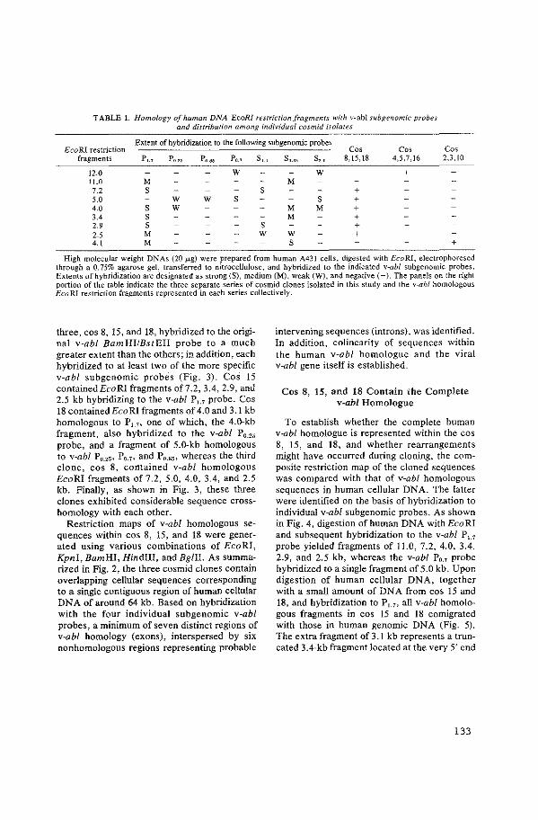
TABLE 1. Homology of human DNA EcoR! restriction fragments with v-ab! subgenomic probes and distribution among individual cosmid isolates
Extent of hybridization to the following subgenomic probes Co' co, co, EcoRI restriction
fragments Pu Po.2o Po.ao Po.7 sl.l S0.45 S2_o 8, 15,18 4,5,7,16 2,3, 10
12.0 w w + 11.0 M M 7.2 s s + 5.0 w w s s + 4.0 s w M M + 3.4 s M + 2.9 s s + 2.5 M w w + 4.1 M s +
High molecular weight DNAs (20 p.g) were prepared from human A43l cells, digested with EcoRI, electrophoresed through a 0.75% agarose gel, transferred to nitrocellulose, and hybridized to the indicated v-abl subgenomic probes. Extents of hybridization are designated as strong (S), medium (M), weak (W), and negative ( -). The panels on the right portion of the table indicate the three separate series of cosmid clones isolated in this study and the v-abl homologous Eco RI restriction fragments represented in each series collectively.
three, cos 8, 15, and 18, hybridized to the original v-abl BamHI/BstEII probe to a much greater extent than the others; in addition, each hybridized to at least two of the more specific v-abl subgenomic probes (Fig. 3). Cos 15 containedEcoRI fragments of7.2, 3.4, 2.9, and 2.5 kb hybridizing to the v-abl Pu probe. Cos 18 contained EcoRI fragments of 4.0 and 3.1 kb homologous to P1.1, one of which, the 4.0-kb fragment, also hybridized to the v-abl P0 _25
probe, and a fragment of 5.0-kb homologous to v-abl P0 _25 , P0 _7 , and P0 _85 , whereas the third clone, cos 8, contained v-abl homologous EcoRI fragments of 7.2, 5.0, 4.0, 3.4, and 2.5 kb. Finally, as shown in Fig. 3, these three clones exhibited considerable sequence crosshomology with each other.
Restriction maps of v-abl homologous sequences within cos 8, 15, and 18 were generated using various combinations of EcoRI, Kpni, Bam HI, Hindiii, and Bg!II. As summarized in Fig. 2, the three cosmid clones contain overlapping cellular sequences corresponding to a single contiguous region of human cellular DNA of around 64 kb. Based on hybridization with the four individual subgenomic v-abl probes, a minimum of seven distinct regions of v-abl homology (exons), interspersed by six nonhomologous regions representing probable
intervening sequences (introns), was identified. In addition, colinearity of sequences within the human v-abl homologue and the viral v-abl gene itself is established,
Cos 8, 15, and 18 Contain i:he Complete v-abl Homologue
To establish whether the complete human v~abl homologue is represented within the cos 8, 15, and 18, and whether rearrangements might have occurred during cloning, the com~ posite restriction map of the cloned sequences was compared with that of v-abl homologous sequences in human cellular DNA. The latter were identified on the basis of hybridization to individual v-abl subgenomic probes. As shown in Fig. 4, digestion of human DNA withEcoRI and subsequent hybridization to the v-abl Pu probe yielded fragments of 11.0, 7.2, 4.0, 3.4, 2.9, and 2.5 kb, whereas the v-abl P0.7 probe hybridized to a single fragment of 5.0 kb. Upon digestion of human cellular DNA, together with a small amount of DNA from cos 15 and 18, and hybridization to Pu, all v-abl homologous fragments in cos 15 and 18 comigrated with those in human genomic DNA (Fig. 5). The extra fragment of 3.1 kb represents a truncated 3.4-kb fragment located at the very 5' end
133
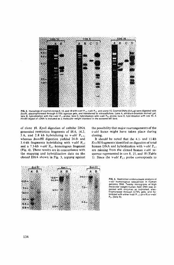
FIG. 3. Homology of cosmid clones 8, 15, and 18with v-abl P,_,, v-abl P0_7, and clone 15. Cosmid DNAs (0.4p.g} were digested with EcoRI, electrophoresed through 0.75% agarose gels, and transferred to nitrocellulose. Lane A, ethidium-bromide stained gel; lane B, hybridization with the v-abl P,_, probe; lane C, hybridization with v-abl P0•7 probe; lane 0, hybridization with cos 15. A Hindlll digest of IlONA is included as a molecular weight standard in the extreme left lane.
of clone 18. Kpnl digestion of cellular DNA generated restriction fragments of 18.6, 14.2, 5.6, and 2.8 kb hybridizing to v-abl P~, 7 ,
whereas BamHI digestion yielded 24.0- and 3.4-kb fragments hybridizing with v-abl P1.1 and a 7.3-kb v-abl P0 _7 homologous fragment (Fig. 4). These results are in concordance with the mapping and hybridization data on the cloned DNA shown in Fig. 3, arguing against
134
the possibility that major rearrangements of the c-ab! locus might have taken place during cloning.
It should be noted that the 4.1- and 11-kb Eco RI fragments identified on digestion of total human DNA and hybridization with v-abl Pu are missing from the cloned human c-abl sequence represented in cos 8, 15, and 18 (Table 1). Since the v-abl P1.1 probe corresponds to
FIG. 4. Restriction endonuclease analysis of v-ab/ homologous sequences in human genomic DNA. Twenty micrograms of high molecular weight human A431 DNA was digested with enzymes as indicated, electrophoresed through 0.75% gels, and hybridized with either v-abl P,,, (lane A) or v-abl P,_, (lane B).
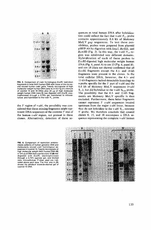
FIG. 5. Comparison of v-abl homologous EcoRI restriction fragments in total A431 human DNA with those of the molecular cloned human c-ab/ gene. Twenty micrograms of high molecular weight human DNA (lane A) or 0.3 ng of a mixture of cosmid 15 and 18 DNAs plus 20 J.>g of high molecular weight human DNA (lane B) was digested with EcoRI, electrophoresed through a 0.75% gel, transferred to nitrocellulose, and hybridized to the v-abl P,_1 probe.
the 5' region of v-abl, the possibility was considered that these missing fragments might represent DNA sequences at the extreme 5' end of the human c-abl region, not present in these clones. Alternatively, detection of these se-
FIG. 6. Comparison of restriction endonuclease patterns of human genomic DNA and molecularly cloned v-abl homologous sequences in cosmid 15. Twenty micrograms of high molecular weight A431 human DNA {A) or 0.41-'g of DNA from cosmid clone 15 {B) was digested with EcoRI, electrophoresed through a 0.75% agarose gel, and blotted onto nitrocellulose. Probes used are indicated above each lane. The first lane in {B) shows the ethidium bromide-stained gel of cosmid 15 DNA.
quences in total human DNA after hybridization could reflect the fact that v-abl P1_7 probe contains approximately 0.6 kb of Moloney MuLV gag sequences. To test these possibilities, probes were prepared from plasmid pHEP-Ab by digestion with Smal, BstEII, and Bam HI (Fig. 2). In this way, the v-abl P1.1 region was subdivided into different domains. Hybridization of each of these probes to EcoRI-digested high molecular weight human DNA (Fig. 6, panel A) to cos 15 (Fig. 6, panel B), and cos 18 (data not shown) confirmed that all EcoRI fragments except the 4.1- and 11-kb fragments were present in the clones. In the total cellular DNA, however, the 4.1- and 11-kb fragments lacked detectable homology to a probe specific for the 5' part of v-abl and the 0.6 kb of Moloney MuLV sequences (v-abl Su), but did hybridize to the v-abl S0 _45 probe. The possibility that the 4.1- and 11-kb fragments are Moloney MuLV specific is thus excluded. Furthermore, these latter fragments cannot represent 5' c-ab! sequences located upstream from the major c-abl locus, because they do not hybridize to the v-abl S~.~ extrem_e 5' probe. We therefore conclude that cosmid clones 8, 15, and 18 encompass a DNA sequence representing the complete v-abl human
135
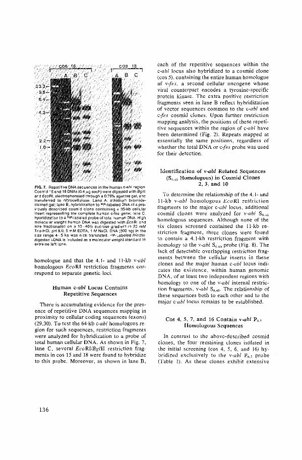
FIG. 7. Repetitive DNA sequences in the human c-ab/ region Cosmid 15 and 18 DNAs (0.4 J.<.g each) were digested with Bg/11 and EcoRI, electrophoresed through a 0.75% agarose gel, and transferred to nitrocellulose. Lane A, ethidium bromidestained gel: lane B, hybridization to "P-Iabeled DNA of a previously described cosmid clone containing a 35-kb cellular insert representing the complete human c-fes gene: lane C, hybridization to a "P-Iabeled probe of total human DNA. High molecular weight human DNA was digested with EcoRI and size fractionated on a 10-40% sucrose gradient in 20 mM Tris-HCI, pH 8.0, 5 mM EDTA, i M NaCI. DNA (300 ng) in the size range 4-5 kb was nick translated. "P-Labeled Hind Illdigested ADNA is included as a molecular weight standard in extreme left lane.
homologue and that the 4.1- and 11-kb v-abl homologous EcoRI restriction fragments correspond to separate genetic loci.
Human c-abl Locus Contains Repetitive Sequences
There is accumulating evidence for the presence of repetitive DNA sequences mapping in proximity to cellular coding sequences {exons) (29,30). To test the 64-kb c-ab! homologous region for such sequences, restriction fragments were analyzed for hybridization to a probe of total human cellular DNA. As shown in Fig. 7, lane C, several EcoRI/Bglll restriction fragments in cos 15 and 18 were found to hybridize to this probe. Moreover, as shown in lane B,
136
each of the repetitive sequences within the c-ab! locus also hybridized to a cosmid clone (cos 5), containing the entire human homologue of v-fes, a second cellular oncogene whose viral counterpart encodes a tyrosine-specific protein kinase. The extra positive restriction fragments seen in lane B reflect hybridization of vector sequences common to the c-ab! and c-fes cosmid clones. Upon further restriction mapping analysis, the positions of these repetitive sequences within the region of c-abf have been determined (Fig. 2). Repeats mapped at essentially the same positions, regardless of whether the total DNA or c-fes probe was used for their detection.
Identification of v-abl Related Sequences (Sn.4~ Homologous) in Cosmid Clones
2, 3, and 10
To determine the relationship of the 4.1- and 11-kb v-abf homologous EcoRI restriction fragments to the major c-ab! locus, additional cosmid clones were analyzed for v-abl Srui; homologous sequences. Although none of the six clones screened contained the 11-kb restriction fragment, three clones were found to contain a 4.1-kb restriction fragment with homology to the v-abl S0 .4 5 probe (Fig. 8). The lack of detectable overlapping restriction fragments between the cellular inserts in these clones and the major human c-abf locus indicates the existence, within human genomic DNA, of at least two independent regions with homology to one of the v-abl internal restriction fragments, v-abl Su.~5 . The relationship of these sequences both to each other and to the major c-abf locus remains to be established.
Cos 4, 5, 7, and 16 Contain v-abl P0 _7
Homologous Sequences
In contrast to the above-described cosmid clones, the four remaining clones isolated in the initial screening (cos 4, 5, 6, and 16) hybridized exclusively to the v-abl P11 •7 probe (Table l). As these clones exhibit extensive

FIG. 8. Identification of v--abl homologous sequences in cos 3 and cos 7. Cosmid DNAs (0.41-'g) were digested with £coAl, electrophoresed through a 0.75% agarose gel, and trans· ferred to nitrocellulose. Lane A, ethidium bromide-stained gels: lane 8 (left), hybridization to v-abl $ 0 • .,; lane B (right), hybridization to v-abl Po.>· ' 2P-Labeled Hindlll-digested ADNA is included as a molecular weight standard in extreme left lane.
homology to one another, they appear to represent a single contiguous DNA region. All contain, in part or in whole, a 12.0-kb EcoRI restriction fragment with homology to v-abl P0. 7 • The fact that this particular fragment was not detected in £caRl-digested total cellular DNA using the same probe argues that the extent of v-abl homology within the sequence represented in these four cosmid clones is much less than that described above for cos 5, 15, and 18. To exclude the possibility that the 12.0-kb EcoRI fragment might represent an artifact generated during cloning, a probe of 0. 7 kb representing an internal Ava I restriction fragment of the 12.0-kb cloned v-abl homologous sequence was prepared. Using this latter probe, a 12.0-kb £caRl fragment could be demonstrated in total human cellular DNA (data not shown), establishing that the v-abl Po. 7 homologous sequences in cosmid clones 4, 5, 7, and 16 are representative of nonrearranged cellular sequences.
Although cos 4, 5, 7, and 16 showed some
cross-homology to clones 8, 15, and 18, even when posthybridization washings were performed under stringent conditions, no overlapping restriction fragments were detected. Since the human c-abl locus contains numerous repetitive sequences, this hybridization is probably due to repeats common to these clones. Thus the v-ab/-related sequences represented in cos 4, 5, 7, and 16 appear to represent a different region of human genome from the primary c-ab! locus. Moreover, if the sequences in cos 4, 5, 7, and 16 are contiguous with sequences at the 3' end of clone 18, they must be separated by at least 26 kb.
Transfection Analysis of Molecularly Cloned Human c-abl Sequences
Since the cos mid vector used for the cloning of v-abl homologous sequences in humans contains the TK gene, transfection of these clones to a recipient TK- Rat-2 cell line was possible, using a HAT selection system to ensure that the transfection was successful. Upon such analysis, using molecularly cloned v-abl (pHEP-Ab) as a positive control, none of the three cosmids containing inserts representing portions of the major c-abl locus exhibited detectable transforming activity. Similarly, cos mid clones containing either v-abl SoAs (2, 3, and 10) or P0.7 (4, 5, 7, and 16) homologous sequences were analyzed and found to lack transforming activity.
DISCUSSION
A cos mid library constructed of human lung carcinoma DNA has been applied to the molecular cloning and restriction endonuclease analySis of human cellular sequences (c-ab!) homologous to the Abelson MuL V transforming gene v-abl. A contiguous human DNA sequence, 64 kb in length, represented by overlapping cellular inserts of three independent cos mid clones was identified and shown to contain v-abl homologous sequences distributed discontinuously over a region of 32 kb, as compared with
137
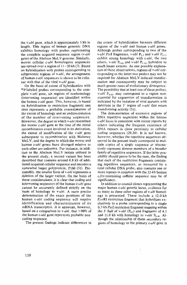
the v-abl gene, which is approximately 3 kb in length. This region of human genomic DNA exhibits homology with probes representing the complete acquired sequence region (v-abl gene) of the Abelson MuLV genome. Similarly, mouse cellular c-abl homologous sequences are spread over a region of 11-20 kb (7). Based on hybridization with probes corresponding to subgenomic regions of v-abl, the arrangement of human c-abl sequences is shown to be colinear with that of the viral v-abi gene.
On the basis of extent of hybridization with 32P-labeled probes corresponding to the complete v-abl gene, six regions of nonhomology (intervening sequences) are identified within the human c-abl gene. This, however, is based on hybridization to restriction fragments and thus represents a probable overestimation of the extent of homology and an underestimation of the number of intervening sequences. Moreover, the degree to which v-abl resembled the mouse c-ab! gene at the time of the initial recombination event invo.lved in its derivation, the extent of modification of the v-abl gene subsequent to recombination with Moloney MuL V, and the degree to which the mouse and human c-abl genes have diverged relative to each other are unknown. For instance, in addition to the Abelson MuL V isolate utilized in the present study, a second variant has been described that contains around 0.8 kb of additional acquired cellular sequence and encodes a somewhat larger polyprotein, P160 (3l). Presumably, the smaller form of v-abl represents a deletion of the larger variant. On the basis of these considerations, it is clear that coding and intervening sequences of the human c-abl gene cannot be accurately defined strictly on the basis of homology to v-abl. A more precise determination of the exact positions of the human c-abl coding sequence will require identification and characterization of its mRNA transcript(s). It is apparent, however, based on a comparison to v-abl, that >90% of the human c-abl gene represents probable noncoding sequence.
The present findings indicate differences in
138
the extent of hybridization between different regions of the v-abl and human c-ab! genes. Although probes corresponding to two of the v-abf Pstl fragments, v-abl P1.1 and v-abl P0 .7 ,
exhibit strong homology with c-ab!, the two others, v-abl P0.25 and v-abl P0.85 , hybridize to much lesser extents. As one possible explanation of these observations, regions ofv-abl corresponding to the latter two probes may not be required for Abelson MuL V -induced transformation and consequently may be subject to much greater rates of evolutionary divergence. The possibility that at least one of these probes, v-abf P0.85 , may correspond to a region nonessential for expression of transformation is indicated by the isolation of viral mutants with deletions in the 3' region of v-abl that retain transforming activity (3l).
The demonstration of extensive regions of DNA repetitive sequences within the human c-ab! locus is consistent with recent reports by others indicating the frequent existence of DNA repeats in close proximity to celluiar coding sequences (29,30). It is not known, however, whether the repetitive sequences observed in the present study correspond to multiple copies of a single sequence or alternatively represent diverse members of a broader family of repetitive sequences. If the latter possibility should prove to be the case, the finding that each of the restriction fragments containing repetitive sequences, as measured by a total cellular DNA probe, also contains one or more repeats in common with the 35-kb human c:fes-containing cellular sequence may be of significance.
In addition to cosmid clones representing the major human c-ab! genetic locus, evidence for as many as three other regions of v-abl homology is presented. These include a 12.0-kb EcoRI restriction fragment that hybridizes exclusively to a probe corresponding to a single 0. 7-kb Psti restriction fragment mapping within the 3' half of v-abf (P0 .7 ) and fragments of 4.1 and 11.0 kb with homology to v-abl S0 .45 . Although the relationship of these secondary regions of homology to the primary c-abl gene is
unclear, a number of possibilities can be considered. For instance, _the secondary regions may correspond to highly divergent independent loci that arose because of transposition of portions of, or even the complete, c-ab/locus, and as such may represent pseudogenes. Alternatively, the homology of these secondary sequences to v-abl may reflect relatedness of distinct but functionally analogous cellular genes. As an example of the latter possibility, one or more of the secondary v-abl homologous sequences could correspond to other cellular oncogenes with tyrosine-specific protein kinase activity·. Finally, it is not resolved as to whether these secondary v-abl homologous sequences map in close proximity to each other or are randomly distributed throughout the human genome.
None of the above-described c-abl containing cos mid clones exhibit transforming activity on transfection to Rat-2 cells. This does not rule out the possibility that the human c-abl gene has such activity, since two of the clones, 8 and 18, are missing 5' regions that are likely to contain promoter sequences. Clone 15 is truncated and lacks sequences homologous to the 3' terminal of the v-abl gene. Each of the remaining cosmid clones contains sequences corresponding to only limited regions of v-abl, and thus their lack of transforming activity is not unexpected. Although on molecular cloning from normal tissues the Harvey murine sarcoma virus e-ras gene lacks transforming activity (32), an active form of the gene is found in certain bladder carcinomas cell lines (3~5); even by appropriate in vitro modification, the inactive molecularly cloned e-ras gene can be converted to an active form (32). Similarly, the human c-abl gene may have oncogenic potential that becomes realized only subsequent to specific modification.
Acknowledgments: The authors thank F. Grosveld for helpful discussions and G. T. Blennerhassett and P. Hanson for their excellent technical assistance. This work was supported under Contract No. NOl-C0-76380 from the National Cancer Institute, Bethesda, Maryland.
REFERENCES
J. Fischinger PJ. Type C RNA transforming viruses. In: Stephenson JR, ed. Molecular biology of RNA 111mor viruses. New York: Academic Press, 1980:162-94.
2. Klein G. Advances in viral oncology. New York: Raven Press, 1982.
3. Parada LF, Tabin CJ, Shih C, Weinberg RA. Human EJ bladder carcinoma oncogene is homologue of Harvey sarcoma virus ras gene. Nature 1982;297: 474-8.
4. Der CJ, Krontiris TG, Cooper GM. Transforming genes of human bladder and lung carcinoma cell lines are homologous to the ras genes of Harvey and Kirsten sarcoma viruses. Proc Nat! Acad Sci USA 1982;79:3637-40.
5. Santos E, Tronick SR, Aaronson SA, Pulciani S, Barbacid M. T24 human bladder carcinoma oncogene is an activated form of the normal human homologue of BALB- and Harvey-MSV transforming genes. Narure 1982;298:343-7.
6. Scolnick EM. Howk RS, Anisowicz A, Peebles P, Scher CD, Parks WP. Separation of sarcoma virusspecific and leukemia virus-specific genetic sequences of Moloney sarcoma virus. Proc Nat/ A cad Sci USA 1975;72:4650-4.
7. GoffSP, Gilboa E, Witte ON, Baltimore D. Structure of the Abelson murine leukemia virus genome and the homologous cellular gene: Studies with cloned viral DNA. Cell 1980:22:777-85.
8. Rosenberg N, Baltimore D, Scher C. In vitro transformation of lymphoid cells by Abelson murine leukemia virus. Proc Nat/ Acad Sci USA 1975;72: 1932-6.
9. Abelson HT, Rabstein LS. Lymphosarcoma virusinduced thymic independent disease in mice. Cancer Res 1970;30:2213-22.
10. Reynolds FH Jr. Sacks TL, Deobagkar DN, Stephenson JR. Cells nonproductively transformed by Abelson murine leukemia virus express a high molecular weight polyprotein containing structural and nonstructural components. Proc Nat/ Acad Sci USA 1980;75:3974-8.
II. Witte ON, Rosenberg N, Paskind M, Shields A, Baltimore D. Identification of an Abelson murine leukemia virus-encoded protein present in transformed fibroblasts and lymphoid cells. Proc Nat! Acad Sci USA 1978;75:2488-92.
12. Van de Yen WJM, Reynolds FH Jr, Stephenson JR. The nonstructural components of polyproteins encoded by replication-defective mammalian transforming retroviruses are phosphorylated and have associated protein kinase activity. Virology 1980: 101:185-97.
13. Witte ON. Dasgupta A, Baltimore D. Abelson murine leukemia virus protein is phosphorylated in vitro to form phosphotyrosine. Nature 1980;283:826-31.
14. Reynolds FH Jr, Van de Yen WJ, Stephenson JR. Abelson murine leukemia virus transformation· defective mutants with impaired P120-associated
139

protein kinase activity. J Viral 1980;36:374-86. 15- Sefton BM, Hunter T, Raschke WC. Evidence that
the Abelson virus protein functions in vivo as a protein kinase that phosphorylates tyrosine. Proc Nat/ Acad Sci USA 1981;78:1552-6.
16- Witte ON, Goff S, Rosenberg N, Baltimore D. A transformation defective mutant of Abelson murine leukemia virus lacks protein kinase activity. Proc Nat/ Acad Sci USA 1980;77:4993-7.
17. Blomberg J, Reynolds FH Jr, Van de Yen WJM, Stephenson JR. Abelson murine leukemia virus transformation involves loss of epidermal growth factor-binding sites. Nature 1980;286:504-7.
18. Twardzik DR, Todaro OJ, Marquardt H, Reynolds FH Jr, Stephenson JR. Abelson MuLV induced transformation involves production of a polypeptide growth factor. Science 1982;216:894-7.
19. Grosveld FG, Dahl H-HM, de Boer E, Aavell RA. Isolation of .B-globin-related genes from a human cosmid library. Gene 1981;13:227-37.
20. Groffen J, Heisterkamp N, Grosveld F, Van de Ven WJM, Stephenson JR. Isolation of human oncogene sequences (v-fes homolog) from a cosmid library. Science 1982;216: 1136-8.
21. Southern EM. Detection of specific sequences among DNA fragments separated by gel electrophoresis. J Mol Bioi 1975;98:503-17.
22. Bernards R, Flavell RA. Physical mapping of the globin gene deletion in hereditary persistence of foetal haemoglobin (HPFH). Nucleic Acids Res 1980;8: 152!-34.
23. Flave!l RA, Kooter JM, de Boer E, Little PFR, Williamson R. Analysis of the .B-S-globin gene loci in normal and Hb Lepore DNA: Direct determination
140
of gene linkage and intergene distance. Cell 1978; 15:25-41.
24. Srinivasan A, Reddy EP, Aaronson SA. Abelson murine leukemia virus: Molecular cloning of infectious integrated proviral DNA. Biochemistry 1981;78:2077 -81.
25. Morrison DA. Transformation and preservation of competent bacterial cells by freezing. In: Wu R, ed. Methods in enzymology, vol 68. New York: Academic Press, !980:326-31.
26- Topp WC. Normal rat cell lines deficient in nuclear thymidine kinase. Virology 1981;113:408-11.
27. Graham FL, Vander Eb AJ. Transformation of rat cells by DNA of human adenovirus 2. Virology 1973;54:536-9.
28. Wigler M, Pellicer A, Silverstein S, Axel R. Bio-chemical transfer of single-copy genes using total cellular DNA as donor. Ce/l1978;!4:725-3L
29. Shen C-KJ, Maniatis T. The organization of repetitive sequences in a cluster of rabbit .B-Iike globin genes. Cell 1980;19:379-91.
30. Jelinek WR, Toomey TP, Leinwand L, et al. Ubiquitous, interspersed repeated sequences in mammalian genomes. Proc Nat/ Acad Sci USA 1981;77: 1398-402.
31. Ziegler SF, Whitlock CA, Goff SP, Gifford A, Witte ON. Lethal effect of the Abelson murine leukemia virus transforming gene product. Ceil 1981 ;27: 477-86.
32. Chang EH, Furth ME, Scolnick EM, Lowy DR. Tumorigenic transformation of mammalian cells induced by a normal human gene homologous to the oncogene of Harvey murine sarcoma virus. NaJure 1982;297:479-83.

APPENDIX PAPER VIII

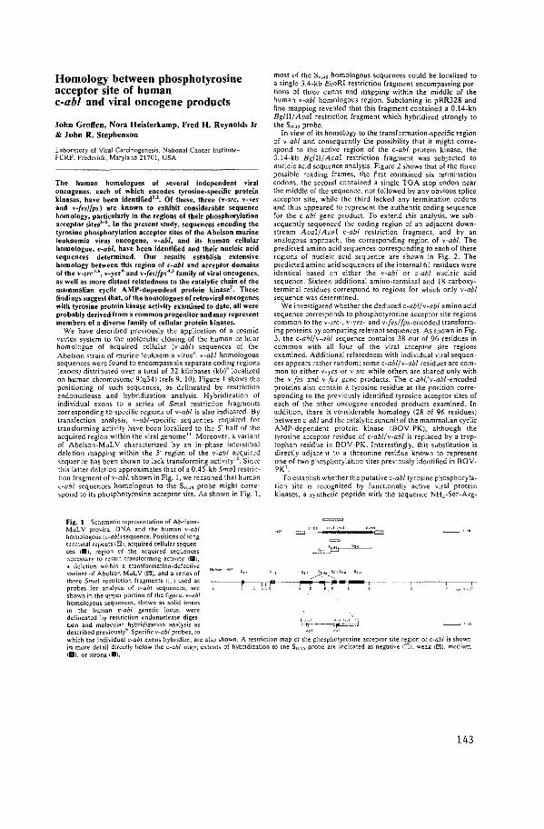
Homology between phosphotyrosine acceptor site of human c·abl and viral oncogene products
John Grotfen, Nora Heisterkamp, Fred H. Reynolds Jr & John R. Stephenson
Labo-ratory of Viral Carcinogenesis. National Cancer !nstituteFCRF. Frederick, Maryland 11701, USA
The human homologues of several independent viral oncogenes, each of which encodes tyrosine-specific protein kinases, have been identified'·'. Of -these, three (v-src, v-~es and v·fes/fps) are known to exhibit considerable seque~ce homology, particularly in the regions of their phosphorylation arceptor sites"-'. In the present study, sequences encoding the tyrosine phosphorylation acceptor sites of the Abelson murine leuk:aemia virus oncogene, v-abl, and its human cellular homologue, c-ab/, have been identified and their nucleic acid seq11ences determined. Our results establish extensive homology between this region of c-ab/ and acceptor domains of the v-src'·", v-yes• and v·fes{fps 4
·' family of viral oncogenes, as w-ell as more distant relatedness to the catalytic chain of the mammalian cyclic AMP-dependent protein kinase7
• These findings suggest that, of the homologues of retroviral oncogenes with tyrosine protein kinase activity examined to date, all were probably derived from a common progenitor and may represent members of a diverse family of cellular protein kinases.
We have described previously the application of a cosmid vector system to the molecular cloning of the human cellular homologue of acquired cellular (v-abl) sequences of the Abelson strain of murine leukaemia virus". v-abl homologous sequences were found to encompass six separate coding regions (exons) distributed over a total of 32 kilo bases (kb)" localized on human chromosome 9(q34) (refs 9, 10). Figure I shows the positioning of such sequences, as delineated by restriction endonuclease and hybridization analysis. HybridizatiorJ of individual exor1s to a series of Smal restriction fragments corresponding to specific regions of v-abl is also indicated. By transfection analysis, v-abl-specific sequences required for transforming activity have been localized to the 5' half of the acquired region within the viral genome". Moreover. a variant of Abelson-MuL V characterized by an in-phase interstitial deletion mapping within the 5' region of the v-abl acquired sequence has been shown to lack transforming activity 12
• Since this latter deletion approximates that of a 0.45-kb Sma! restriction fragment of v-abl, shown in Fig. I, we reasoned that human (-abl sequences homologous to the S".-'' probe might correspond to its phosphotyrosine acceptor site. As shown in Fig. 1,
Fig. 1 Schematic representation of AbelsonMuLV proviral DNA and the human v-abl homologous lc-ab/1 sequence. Positions of long terminal repeats I [II), acquired cellular sequences tBI, region of the acquired sequences ncce~'ary to retain transforming activity IIIII I, a deletion within a transformation-defective variant of Abelson MuLV (E!;l), and a series of three Smal restriction fragments li.J) used as probes for analysis of c-ab/ sequences, are sbown in the upper portion of the figure. v-abl homologous sequences, shown as solid boxes in the human c-ab/ genetic locus, were
,,
most (>[ the S".4 ·' homologous sequences could be locali:zed to a single 3.4-kb EcaRI restriction fragment encompassing portion> of three exons and mapping within the middle of the human v-abl homologous region. Subdoning in pBR328 and fine mapping revealed that this fragment contained a 0.14-kb Bg!II/At•al restriction fragment which hybridized strongly to the$,_.~ probe.
In view of its homology to the transformation-specific region of v-ab/ and consequently the possibility that it might correspond to the active region of the c-ab/ prutein kinase, the 0.14-kb Bgi!I/AL•al restriction fragment was subjected to nucleic add sequence analysis. Figure 2 shows that of the three possible reading frames, the first contained six termination codons, the second contained a single TGA stop codon near the middle of the sequence, not followed by any obvious splice acceptor site, while the third lacked any termination codons and thus appeared to represent the authentic coding sequence for the c-ab/ gene product. To extend this analysis, we subsequently sequenced the coding region of an adjacent downstream A(.•al/A~:al c-ab/ restriction fragment, and by an analogous approach, the corresponding region of v-abl. The predicted amino acid sequences corresponding to each of these regions of nucleic acid sequence are shown in Fig. 2. The predicted amino acid sequences of the internal 61 residues were identical based on either the v-abl or c-ab/ nucleic acid sequence. Sixteen additional amino-terminal and 18 carboxyterminal residues correspond to regions for which only v-abl sequence was determined.
We investigated whether the deduced c-ab!/ v-abl amino acid sequence correspor1ds to phosphotyrosine acceptor site regions common to the v-src-. v-res- and v-[es/fps-encoded transforming proteins by comparing relevant sequences. As shown in Fig. 3. the c-ablfv-abl sequence contains 38 out of 96 residues in common with all four of the viral acceptor site regions examined. Additional relatedness with individual viral sequences appears rather random; some c-abl/v-ab/ residues are common to either v-res or v-src while others are shared only with the v-fes and v-fps gene products. The c-abl/v-abl-encoded proteins also contain a tyrosine residue at the position corresponding to the previously identified tyrosine acceptor sites of each of the other oncogene-encoded products examined. In addition, there is considerable homology (28 of 96 residues) between c-ab/ and the catalytic sun unit of the mammalian cyclic AMP-dependent protein kinase IBOY-PK), although the tyrosine acceptor residue of c-abl/v-abl is replaced by a tryptophan residue in BOV-PK. Interestingly, this substitution is directly adjacent t0 a threonine residue known to represent one of two phosphorylation sites previously identified in BOVPK'.
To establish whether the putative c-ab/ tyrosine phosphorylation site is recognized by functionally active viral protein kinases, a synthetic peptide with the sequence NH,-Ser-Arg-
' ~ ···'
~~l~n:~~dm~e~~~~;c~~~ri~~~aot~:~l:~•:ly~:~e~ i--tF- ----~~ described previously" Specific v-abl probes, to "' 1
"'1
which the individual c-ab/ exons hybridize, are also shown A restriction map of the phosphotyrosine acceptor site region of c-ab/ is shown in more detail directly below the c-ab/ map; extents of hybridization to the$,_., probe are indicated as negative I'JJ. weak lrn), medium Iilii. or strong iBJ,
143
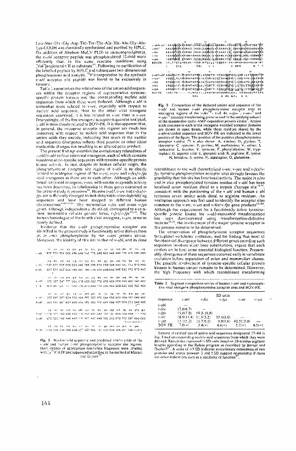
Lcu-\1et-Thr-Giy-Asp-Thr-Tyr-Thr-Aia-His-Aia-Giy-AiaLys-COOH was chemically synthesized and purified by HPLC. On addition of Abelson-MuLV P120 to immunoprecipitates, the c-ab/ acceptor peptide was phosphorylated 10-fold more dficiently than in the same reaction conditions using [Val']angiotensin I! as substrate". Following re-purification of the labelled peptide by HPLC and subsequent two-dimensional phosphoamino acid analysis, "P incorporation by the synthetic c-ab/ acceptor site peptide was found to be exclusively in tyro•;~ne
Table I summarizes the relatedness of the amino acid sequences within the acceptor regions of representative tyrosine'Pe<:ific protein kinases and the corresponding nucleic acid sequences from which these were deduced. Although c-ab/ is 'mmcwhat more related to v-src especially with respect to nucleic acid sequence. than to the other viral oncogene sequences ~xamined. it is less related to v-src than is v-yes. Interestingly. of the five oncogene acceptor sequences analysed, <:-11hi is mo>t closely related to BOY -PK. It is also apparent that, in general, the oncogene acceptor site regions are much Jess conserved with respect to nucleic acid sequence than in the amino ~cids they encode, indicating that much of the nucleic acid >equencc divergence reflects third position or other silent nuclcutide <.:hanges not resulting in an altered gene product.
The present findings establish the evolutionary relatedness of c-abl/v-abl to four retroviral oncogenes. each of which contains transformation-specific sequences with tyrosine-specific protein kinas<' ;~<:tivit). In fact. despite its human cellular origin, the phosphotyro~m<' acct'ptor site region of c-ab! is as closely related to analogous regions of the v-src, v-yes and v-fesjv-fps vital oncogenes as these are to each other. Although an additional rdro-viral oncogene, v-ros. with similar enzymatic activity has heen described, its relationship to those genes examined in the present study is unknown 14
• Human c-ob!. c-src and c-fes/c!P-' arc sufficiently diverged to lack detectable cross-hybridizing sequences and have been mapped to different human chromosomes"·'"·"'" The mammalian v-fes and avian v-fps genes. although indt'pendently identified. correspond to a common mammalian cellular genetic locus, c-fesjc-fps' 7
·'". The human homologue of I he fourth viral oncogene, v-yes, remains poorly ddlned
Evid~nce that the c-ab/ phosphotyrosine acceptor site identified in the present study is functionally active derives from it> in n'rr:~ phosphorylation by the v-abl protein kinase. \-lorcovcr, the iGcntity of this sit:: to that of v-abl. and its close
mol glo ''' leo Oh• '" ''' ·"" OM '" hi> ••O
~-~-~-~~----~---o'>P IC<I ·"0 .F.O 010 loo ool gl'/ glu "" h" "" "' '''
-··==---==----~=--··=----==----~=-
"'"" gl1 •,eo "'9 '"" mel lh• gly O>P lhl 1y1 lh•
••••=n•=••=-~~----~= ~••-n•===-~~----~-
ql\ OM 010 ''' lVI otp 1hl '~ P~ 91" •~
=~=-•cr-~=~--===-= ,;CC CAT ~C\ CC" Oct r,<G r fC CCL AYC AAA fC~ ACT GCA CCC GAG AGC
CTG CCC lAC AAC AAG ttl: <tC AlC AAG 'CC GAC 01C fOG GOT AAG GGC
lop O'o l1• ~~~ moo '" p•o ''' P•O
Fig. 2 Nucl"ic aci<.l s~qucncc and prcdtctcd amino acids of the ,_a!JI and human ·-·-abl phmphotyrosine acceptor site regrons. Both <trands of appropriate restriction fragme11ts were labelled with [ y ''P]A TP and <cquenced according to the method of Maxam
and Gilhen"
144
Fig. 3 Comparison of the deduced amino acid sequem:e uf th" v-abl and human c-ab/ phosphotyrosine acce~tor sites to analogous regions of the v-fes''- {ref. 4), v-fps- , v-src-' and v-yes-' encoded transforming proteins and to the catalytic subunit of the mammalian cyclic AMP-dependent protein kinase'. Amino acids common to each of the oncogene-encoded acceptor domains are shown in open boxes, while those residues shared by the c-abl-encoded sequence and BOV-PK are indicated in the lower portion of the figure. The position of the putative phosphotyrosine acceptor residue (*) is also shown. A, alanine; G, glycine; T. threonine: C. cysteine; P, proline; M, methionine: V. valine; I. isoleucine: L. leucine: Y, tyrosine; F, phenylalanine; W, tryp· tophan: D. aspartic acid: E. glutamic acid; R, arginine; K, lysine:
H. histidine:· S. '~rine: N, asparagine; Q, glutamine.
resemblance tu th~ well characterized v-src, v-yes and v-fes/vfps tyrosine phosphorylation acceptor sites strongly favour~ the possibility that this site has functional activity. The major in ~itro and in vivo phosphorylated tyrosine residue of v-abl has been localized seven residues distal to a trypsin cleavage site'"·"", consistent with the positioning of the v-abl and human c-ab/ tyrosines seven amino acids distal to arginine residues. An analogous approach was first used to identify the acceptor sites common to the v-src, v-yes and v-fes/v-fps gene products"-24
•
Although the requirement for a functionally active tyrosinespecific protein kinase for v-ab/-associated transformation has been demonstrated using transformation-defective mutants"-'". the involvement of the major tyrosine acceptor in this process remains to be determined.
The conservation of phosphotyrosine acceptor sequences throughout vertebrate evolution, and the finding that most of the observed divergence between different genes encoding such sequences involves silent base substitutions, argues that each confers on its host some essential biological function. Presumably, divergence of these sequences occurred early in vertebrate evolution before separation of avian and mammalian classes. The possible involvement of tyrosine-specific cellular protein kinases in human cancer remains to be determined. However, the high frequency with which recombinant transforming
Table 1 Segment comparison scores of human c-ab/ and representati,·e viral "neogene phosphotyrosine acceptor sites and BOY -PK
SD units Sequence c-ab! v-/es v-fps V•\'eS
c-ab/ v-(es 17.616.71 v-fps t5.6{7.8) 19.51 16.9)
18.911 1.4) I 1.9{3.2) 13.6{3.0) "-yes 17.1[5.2) 15.7(0.21 9.8{1.6) 42.5(15.8) BOV-PK 7.61-t 5.4(-) 4.8(-) 5.7{-) 4.5{-)
Extents of relatedness of amino acid sequences designated 17-64 in Fig. 3 and corresponding nucleic acid sequences from which they w~re derived. Resul!s are expressed in SO units based on 20-residue segm~nt lengths according to the Relate program as described by Barker and Dayhoff" A score of >5 SO indicates evolutionary relatedness of two proteins and scores between 3 and 5 SD support relationship if there are other indicattons such as a similarity of function".
viruses containing such sequences have been isolated and the recent localization of c-ab/ within a small fragment of human chromosome 9 which is translocated to chromosome 22 in chronic myelogenous leukaemia"·'", would seem to favour such a modeL
We thank G. T. Blennerhassett and P. Hansen for technical assistance and K. L McNitt (Computer and Statistical Services, IMS) for help with calculation of the sequence homology. This work was supported under NCI contract NOI-CO 76380.
R.c"i'c~ l Mo"h: "<'<P'<~ 16 May 19SJ
1. Varmu<. H. E.~'""" Il6, 81l-~l<J <IO~ll 2. Bi•hop, 1. M. Ado. Conm R". 37, 1-32 119821 J (.emdol•ky, A P, <1al NoM< 187,198-203119801 4. H~mpo, A., Loprevottc, I., Galil><ro, F., Fedele, L.A. & Shoe<, C.J, C.tl JO, 775-785
119821 l, Shibuya. M. & Hanolu", H. C•ll JO, 7K7-795i1982! 6. Ki"muro, N., Kitomuro, A., Toy"'himo. K., Hiroyama. Y & Y.,_.h,da. M. No""' lin,
20l-ZU8 119821 7 ShoJi, 5 "a/. Proc, nam. Amd. Sci. U.S A. 78, 848-851 119811 8 H~i,.<rkomp, N .. Groff en, J. & Slephcn<nn. J R.) mol"- appl Gen<l. 2, 57-68 119831 9. HciSicrl<amp. N <10/. No'"'' 2\19, 747-749!1982)
Ill. de Klein, A. ;ro/. No'"" )00, 765-767 tl9821 II. St<niva;an. A." a/. Pro< noro. Arad. Sd. U.S.A. 79,5108-55 11tl982i 12. Goff. S. P .. G<lhoo, E .. Wit\0, 0. N. & Boltimore. D. Cdl U. 777-78.111981" ll. Won~, T. W. & Goldberg. A. R. J />iol, Ch<rn. 25H, l!l22-lfi25 119Sll 14. Feldman, R. A .. Wan~. L·H., H,,nafuso, H & Bolduw, P.C J V<rol 42, l28-2l6tl98ll II. Dalla-Favero, R, <r al Pror. na!n Acad. S<L U.S.A 79,4714--4717 119811 16. Sakaguchi. A. Y .. Najlor, S. L. & Show•, T. B Proo N"d'" And R.,_ maf". Bioi. (10 !h<
pro.< I 17. Shihuyo, M., Honofu,., T. Haoalu"'. H & St<phon,on.J. R. Proc. nnm. A<nd 5" U.S A
77.6536-6540 \1980J IS GroiTen.J .. Hctstorkamp. N., Shihuya. M., Hooolu<a, H & Slephoo•on.J. R. v,ro/O")' 125,
4HD--486tl98ll 19. Reynolds, F. H. Jr. Oro><lan. S. & Stophen<on. 1 R. J Vim/. 44, ID9l-llHI <!98lJ 20 Pat«lliosl<y, T,. Hun"'· T., E«:h. F. S. Cooper. J. A.&. Sdwn. E. M. Proc. narn. A<M.
S<i.US.A 75,97J-977<19~ll )I Smart.l.E.<raf.Pmr.na!n Mad. Sci U.S.A.78,MllJ-MI7i19Sll 2l Elamhc<g.l .. Van do Yeo. W 1 M. Reynolds, F. H. Jr, N>low'lk. R P. & Stophoo<on.J
R.J. Virol. 38,886-894119811 23. NOli,J. E. Ghysdaol. J .. Vo~t, P. K & Sm,l.) E No'"" !91, 675-677 tl9811 2~- Roynolds, F H.lr, O""'lan, S. Blon11><r~, J. & Stephenson.! R Vim I""' 122, 114-146
119821 25. Reynold,, F. H Jr. Van de Von, W,], M. & Stop~e"'oo,J R.J Vim/.36, J74-J86 il9RIII 26. Witte, 0. N .. Goff, S .. R<,.enbor". N & B>llimorc, D, p,ar. ""'"· A<nd S" US A, 77,
4993--4997(l9SOI 27 Batl<or. W C. & Dayhoff.~- 0. p,,, ""'" Acod. Sn U S.A 79,l8.16-2R19 ii9Rll 1.8. M .. am, A.M. & G,Jbe.-t. W. Mcrl,, En'''"· 6S, 499-5Ml19RDI
145


APPENDIX PAPER IX

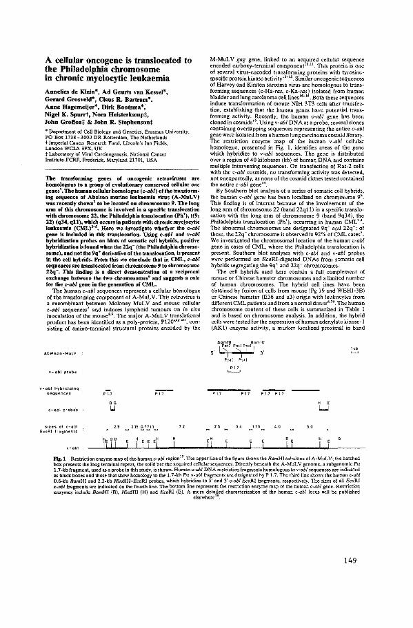
A cellular oncogene is translocated to the Philadelphia chromosome in chronic myelocytic leukaemia
Annelies de Klein*, Ad Geurts van Kessel*, Gerard Grosveld*, Claus R. Bartram*, Anne Hagemeijer*, Dirk Bootsma*, Nigel K. Spurrt, Nora Helslerkamp:l:, John Grollent & John R. Stephenson:!:
• Department of Cell Biology and Genetics, Erasmus University, PO Bo~ 1738-3000 DR Rotterdam, The Netherlands t Imperial Cancer Research Fund, Lincoln's Inn Fields, London WC2A 3PX, UK t Laboratory of Viral Carcinogenesis, National Cancer Institute~FCRF, Frederick, Maryland 21701, USA
The transforming genes of oncogenic retroviruses are homologous to a group of evolutionary conserved cellular one genes'. The human cellularbomologne (c-ab!) of the transform· iog sequence of Abelson murine leukaemia virus (A-MoL V) was recentJy shown2 to be located on chromosome 9. The long IHJD of this chromosome is involved in a specific translocation with chromosome 22, the Philadelphia translocation (Ph'), t(9; 22) (q34, qll), which occurs in patients with chronic myelocytic leukaemia (CML)~. Here we investigate whether the~ c-abl gene is included in this translocation. Using c-abl and ..,.abf hybridization probes on blots of somatic ceU hybrids, positive hybridization is found when the 22cC (the Philadelphia chromosome), and not the 9q.,. deri11ttive of the translocation, is present in the ceU hybrids. From this we conclude that in CML, c-abl sequences are translocated from chromosome 9 to chromosome 22q~. This finding is a direct demonstration of a reciprocal exchange between the two chromosomel and suggests a role for the _c-ab/ gene in the generation of CML.
The human c-ab/ sequences represent a cellular homologue of the transforming component of A-MuLV. This retrovirus is a recombinant between Moloney MuL V and mouse cellular c-abl sequences' and induces lymphoid tumours on in vivo inoculation of the mouse~·". The major A-MuLV translational product has been identified as a poly-protein, P120 .. """"b1
, consisting of amino-terminal structural proteins encoded by the
Ab~lson-MulV
•· o.bl probe
v·abl hybr•dizong sequenou
c-o.bl probes
sozes ol c-abl EcoRI frognHMS
" I.J
" "
M-MuLV gag gene, linked to an acquired cellular sequence encoded carbox:y-terminal component'"·". This protein is one of several virus-encoded transforming proteins with tyrosinespecific protein kinase activityn-ls. Similar oncogenic sequences of Harvey and Kirsten sarcoma virus are homologous to transforming sequences (c-Ha-ras, c-Ka-ras) isolated from human bladder and lung carcinoma cell lines'.,._'". Both these sequences induce transformation of mouse NIH 3T3 cells after transfection, establishing that the human genes have potential transforming activity. Recently, the human c-ab/ gene has been cloned in cosmids19
• Using v-abl DNA as a probe, several clones containing overlapping sequences representing the entire c-ab£ gene were isolated from a human lung carcinoma cosmid library. The restriction enzyme map of the human v-abl cellular homologue, presented in Fig. 1, identifies areas of the gene which hybridize to v-abl sequences. The gene is distributed over a region of 40 kilobases (kb) of human DNA and contains multiple intervening sequences. On transfection of Rat-2 cells with the c-abl cosmids, no transforming activity was detected, not unexpectedly, as none of the cosmid clOnes tested contained the entire c-abl gene19
•
By Southern blot analysis of a series of somatic cell hybrids, the human c-ab/ gene has been localized on chromosome 9'. This finding is of interest because of the involvement of the long arm of chromosome 22 (band 22qll) in a specific translocation with the long arm of chromosome 9 (band 9q34), the Philadelphia translocation (Ph'), occurring in human CML'·•. The abnormal chromosomes are designated 9q• and 22q-; of these, the 22q· chromosome is observed in 92% of CML cases'. We investigated the chromosomal location of the human c-abl gene in cases of CML, where the Philadelphia translocation is present. Southern blot analyses with c-ab/ and v-abl probes were performed on EcoRI-digested DNAs from somatic cell hybrids segregating the 9q• and 22q- chromosomes.
The cell hybrids used here contain a full complement of mouse or Chinese hamster chromosomes and a limited number of human chromosomes. The hybrid cell lines have been obtained by fusion of cells from mouse (Pg 19 and 'WEHI-3B) or Chinese hamster (E36 and a3) origin with leukocytes from different CML patients and from a normal donor"·'". The human chromosome content of these cells is summarized in Table I and is based on chromosome analysis. In addition, the hybrid cells were tested for the ex:pression of human adenylate kinase-1 (AKl) enzyme activity, a marker localized proximal in band
8omHf BamHl I :;II P~l Pill I
s· . ~I -1 . , J"
"'
Psll Psll
P1.7 ~
30 .. 175
" ' w
F'ti.l Restriction enzyme map of the human c-ab/ region 10• The upper line of the figure shows the BamHI subdone of A-MuL V; the hatched
box presents the long terminal repeat, the solid bar the acquired cellular sequences. Directly beneath the A-MuLV genome, a subgenomic Psr 1.7-kb fragment, used as a probe in this study, is shown. Human c-ab/ DNA restriction fragments homologous to v·abl sequences are indicated as black boxes and those that show homology to the 1.7-kb Psi v-ab/ fragments are designated by P 1.7. The third line shows the human c-ab/ 0.6-l::b BamHJ and 2.2-kb Hi11di11-EcoRJ probes, which hybridize to 5' and 3' c-ab/ EcaRI fragments, respectively. The sizes of all EcoRI c-ab/ fragments are indicated on the fourth line. The bottom line represents the restriction enzyme map of the human c-ab/ gene. Restriction enzymes include BamHI (B), Hi11diii (H) and EcoRI (E). A ~~;:~:rt:,it~d characterization of the human c-ab/ locus will be published
!49
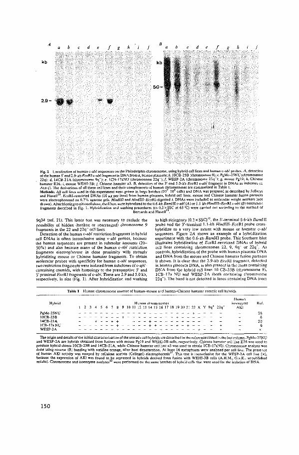
A d ' b f g h a j
B b f g h j a
kb kb
5.0-
2.9- , ••
,i
Fog. 2 Localization of human c-ab/ sequences on the Philadelphia chromosome, using hybrid cell lines and human c-ab/ probes. A, detection of the human 5' end 2.9~kbEcoRI c-ab/ fragment in DNA from a, human placenta; b, IOCB-238 (chromosome 9); c, PgMe-2SNU (chromosome 22q); d, 14CB-21A (chromosome 9q +); e. 1CN-11aNU (chromosome 22q-); f, WESP-2A (chromosome 22q -); g, mouse Pg19: h. Chine~e hamster E36; i, mouse WEHI-38: j, Chinese hamster a3. 8, detection of the 3'-end 5.0-kb EcoR! c-ab/ fragment in DNAs as indicate<.< m A{a-j). The derivations of all these cell lines and their complements of human chromesomes are summarized in Table 1. Methods: All cell lines used in this experiment were grown in large batches (107 -10" cells) and DNA was prepared as described by Jeffreys and Flavell'". EcoRI-restricted DNAs (10 fLg per lane) from human placenta, hybrid cell lines, mouse and Chinese hamster fusion partners were electrophoresed on 0.7% agarose gels. HindiU and HindliJ-EcoRI-digested A DNAs were included as molecular weight markers (not shown). After blotting to nitrocellulose, the filters were hybridized to the 0.6-kb BamHI c-ab/ {A) or 1.1-kb Hindiii-EcoRI c-abl (B) restriction fragments described in Fig. L Hybridization and washing :;~~;~~r:~J~~~~~l7'~.SC at 65 'C) were carried out according to the method of
9q34 (ref. 21). This latter test was necessary to exclude the possibility of hidden (broken or rearranged) chromosome 9 fragments in the 22 and 22q- eel! lines,
Detection of the human c-ab/ restriction fragments in hybrid cell DNAs is often inconclusive using v-abl probes, because the human sequences are present in submolar amounts (20-50%) and also because many of the human c-ab/ restriction fragments electrophorese in dose proximity with strongly hybridizing mouse or Chinese hamster fragments. To obtain molecular probes with specificity for human c-abl sequences, two restriction fragments were isolated from Subdones of c-qblcontaining cosmids, with homology to the presumptive 5' and 3' proximal EcoRI fragments of c-abl. These are 2.9 and 5.0 kb, respectively, in size (Fig. 1). After hybridization and washing
to high stringency (0.1 xSSC)", the 5'-terminal 0.6-kb BamH! probe and the 3'-terminatl.l-kb Hindiii-EcoRl probe crosshybridize to a very low extent with mouse or hamster c-ab/ sequences. Figure 2A shows an example of a hybridization experiment with the 0.6-kb BamHI probe. This Southern blot illustrates hybridization of EcoRI-restricted DNAs of hybrid cell Jines containing chromosomes 22, 9, 9q+ or 22q-. As controls, hybridization of the probe with human placenta DNA and DNA from the mouse and Chinese hamster fusion partners is shown. It is clear that the 2.9-kb EcoRI fragment, detected in human placenta DNA, is also present in the lanes containing DNA from the hybrid cell lines 10 CB-23B (chromosome 9), 1CB-17a NU and WESP-2A (both containing chromosome 22q-). The band is not detected in Janes containing DNA from
Table 1 Human chromosome content of human-mouse and human-chinese hamster somatic cell hybrids
Hybrid
PgMe-25NU 10CB-23B 14CB-21A lCB-17aNU WESP-2A
' 2 4
+ - - + - + + -
- + + +
Human Human chromosomes isoenzyme
10 11 12 13 14 15 16 17 18 19 20 21 22 X Y 9q• 22q- AKI
+ - - - + -- - - +
- - + + + - + + -
Ref.
26 6
20 6
The origin and details of the initial characterization oft he somatic cell hybrids are described in the references llsted in the last column. PgMe-25NU and WESP-2A are hybrids obtained from fusions with mouse Pgl9 and WEHI-3B cells, respectively. Chinese hamster cell line E36 was used to produce hybrid clones 10CB-23B and 14CB-21A, while Chinese hamster cell line a3 was used to obtain 1CB-17aNU. Chromosome analysis was done using reverse (R) banding with acridine orange, after heat denaturation. At least 16 metaphases were analysed per cell line. The presence of human AKl activity was assayed by cellulose acetate (Cellogel) electrophoresis27
. This test is inconclusive for the WESP-2A cell line (x), because the expression of AKl was found to be repressed in hybrids derived from fusion with WEHI-3B cells (A.H.M .. G.v.K., unpublished results). Chromosome and isoenzyme analyses22 were performed on the same batches of hybrid cells that were used for the isolation of DNA
150
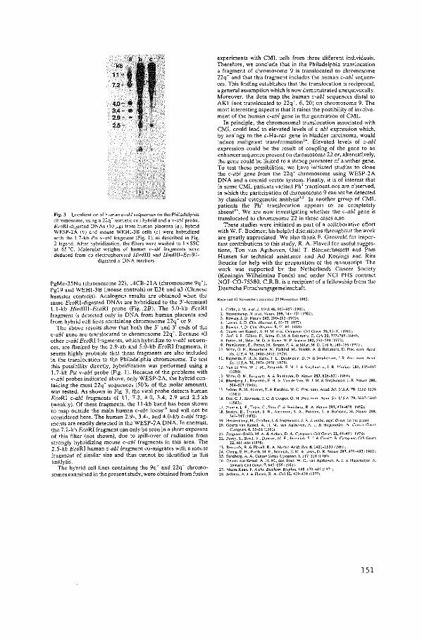
11
4.0--· 3A- e ,_,_ . 2.5-
FiR. 3 Locali>.ation of human c-ab/ sequences on the Philadelphia chromosome, using a 22q- somatic cell hybrid and a v-a&/ probe. EcoRJ-digested DNAs ilOJ.i.g) from human placenta (a), hybrid WESP-2A (b) and mouse WEHl-38 cells (c) were hybridized with the L7-kb Psi v-abl fragment (Fig. 1), as described in Fig 2 legend. After hybridization, the filters were washed to 1 X $$C at 65 'C. Molecular weights of human c-ab! fragments were deduced from co-electrophoresed Hindlll and Hindlli-EcoRl-
digested A DNA markers
f'gMe-25Nu (chromosome 22}, 14CB-21A (chromosome 9q•), Pgl9 and WEHI-38 (mouse controls) or E36 and a3 (Chinese hamster controls). Analogous results are obtained when the same EcoRI-digested DNAs are hybridized to the 3'-terminal 1.1-kb Hindii!-EcoRJ probe (Fig .. 2B}. The 5.0-kb EcoRI fragment is detected only in DNA from human placenta and frGm hybrid cell lines containing chromosome 22q- or 9.
The above results show that both the 5' and 3' ends of the c-ab/ gene are translocated to chromosome 22q-. Because all other c-ab/ EcoRI fragments, which hybridize to v-abl sequences, are flanked by the 2.9-kb and 5.0-kb EcoRJ fragments, it seems highly probable that these fragments are also included in the translocation to the Philadelphia chromosome. To test this possibility directly, hybridization was performed using a 1.7-kb Psi v-abl probe (Fig. 1). Because of the problems with v-abl probes indicated above, only WESP-2A, the hybrid containing the most 22q- sequences (50% of the molar amount), was tested. As shown in Fig. 3, the viral probe detects human . EcoRI c-ab/ fragments of 11, 7.2, 4.0, 3.4, 2.9 and 2.5 kb (weakly). Of these fragments, the 11-kb band has been shown to map outside the main human c-ab/ locus10 and will not be considered here. The human 2.9-, 3.4-, and 4.0-kb c-ab/ fragments are readily detected in the WESP-2A DNA. In contrast, the 7.2-kb EcoRJ fragment can only be seen in a short exposure of this filter (not shown), due to spill-over of radiation from strongly hybridizing mouse c-ab/ fragments in this area. The 2.5-kb EcoRJ human c-ab£ fragment co-migrates with a mouse fragment of similar size and thus cannot be identified in this analysis.
The hybrid cell lines containing the 9q+ and 22q' chromosomes examined in the present study, were obtained from fusion
experiments with CML cells from three different individuals. Therefore, we conclude that in the Philadelphia translocation a fragment of chromosome '} is translocated to chromosome 22q- and that this fragment includes the human c-ab! sequences. This finding establishes that the translocation is reciprocal, a general assumption which is now demonstrated unequivocally. Moreover, the data map the human c-ab/ sequences distal to AK1 (not translocated to 22q~, 6, 20) on chromosome 9. The most interesting aspect is that it raises the possibility of involvement of the human c-ab! gene in the generation of CML.
In principle, the chromosomal translocation associated with CML could lead to elevated levels of c-ab/ expression which, by analogy to the c-Ha-ras gene in bladder carcinoma, would induce malignant transformation". Elevated levels of c-ab! expression could be the result of coupling of the gene to an enhancer sequence present on chromosome 22 or, alternatively, the gene could be linked to a strong promoter of another gene. To test these possibilities, we have initiated studies to done the c-ab/ gene from the 22q- chromosome using WESP-2A DNA and a cosmid vector system. Finally, it is of interest that in some CML patients variant Ph' translocations are observed, in which the participation of chromosome 9 cannot be detected by classical cytogenetic analysis'·' In another group of CML patients the Ph' translocation appears to be completely absent". We are now investigating whether the c-ab/ gene is translocated to chromosome 22 in these cases also.
These studies were initiated as part of a collaborative effort with W. F. Bodmer; his helpfl'l discussions throughout the work are greatly appreciated. We also thank F. Grosveld for important contributions to this study, R. A. Flavell for useful sugges· tions, Ton van Agthoven, Gail T. Blennerhassett and Pam Hansen for technical assistance and Ad Konings and Rita Boucke for help with the preparation of the manuscript. The work was supported by the Netherlands Cancer Society (Koningin Wilhelmina Fonds) and under NCI PHS contract NOI-C0-75380. C.R.B. is a recipient of a fellowship from the Deutsche Forschungsgemeinschaft.
I. Coff<n. J. M. rt ol./. Vim/. 4G, 95)-957 i19B1i 2 Ho,.<orkamp. ).[. <fO!. "'"'""' 299,747-150 f198ll 3 Rowlo,.J D NoM<24~.290-293i19731 4 Lawloc.S D Clin.Hn<mu!.6,55-7511977! S. Rowl"· !. 0 Cl'". H""""'- 9, 55-ff6 il980J 6. Geurts'''" Kc5>oll. A H ~l "a/. C'"''"" Cdl G<n« 30.83-91 119811 7 Goff. S. P .. o;JO<Ja. E .. WHt<, 0. ).1. & BoiCimore. 0. CHI 2l. 777-nl i1980> B. Po""·M .. Sktac. M.D. & Row<. W. P -'"'"" ISZ. 592-594119731 9. Proml<uma<, E .. Pottcc, M .. S'"•"· P. A & Sklo;.M 0 Cd/6, 1H-159 fl97ll
10. Wmo. 0. K .• Ro>enbcr~. N .• Pou;kind, M .. ';h><id<. A & H;l«mo". D. Pmc. nom A<aJ -'"· U S.A 75, 2488·2492 tl97~1
11. Roynold•. F fl l<. So,b. T. L., Doob,okor, D.~ N. S"ohoo,nn,J R Pmc ""'"·A cod Sci. U.S.A. 15. )974-)976 d97Ai
12 Von de v,n, W i M .• R"nnlds. F. H. Jc & Stephenson, i. R V"ol"" 101. 1~5-197 (19801
)J Wi"c. 0 N .. D<>;•uri>. A. & Eol01moro. 0. Nar"" 28~. 8l6-8JI i19BOJ 14. Elomb«g, J. Reynold•. F H lc. Von de Ven, W. J. M. & Stophenson. J. R ·""'"" 286,
504-507rl9801 15. Schon. B. ).1.,Hunl<r, T,& Ro>ehk W C Pmc. naln A'od.&i US.A. "7S, ll52-ll56
11981) 16 Do.. C J. K<M«<". T. C & Coop<:!, 0. M. Pm,. """· A'on" ScL U S.A. 79, J6J7-J640
11982) 17. Porod>. L. F. Tobin. C. Shrh. C. & We'"b""· R. A."'"'""' 29"7. 474-418 f\982J lB. Sonoos. E .• Tronic<. S R. Ao;nn.on, S. A. Pub>m. S & Barbo.,d, M .• ~·"'"" 298.
343-347!1982> 19. He"<eckomp. N .• Groffen,J. & S"phoowo. !. R I mol,-,. arpl. G'"" i'" tho P""l ll>. Gcurt< von K".el. A. H. M .. "" A,thoven, A. J. & H'oome<ior. A Canw C,·,t<L
c''"'"" 6.ss-sso9s1, ll. Fcr~u>an-Sm;th. M A. & A" ken. D. A. CJ""'~'""· Cell G<ntr. n. 49-411 11978J l2. Povo1. s .• Boyd. Y. D"""'"· M E.!"'"'"'"· S 1 a C>mn. B. c,·w''""· C<il Gcn<'
22,461-464(1978) 23. B<m>cd<. R. & Fl>•ell. R. A. .~'u<i"' A<1dJ f<«. 8. 14l1-15lJ 11980J )4. Chon~. E. H .. Fucth. M E. s,orn;,k, E. M. & Lowy, D. R -""'"'' 297, 479-<.83 11982J 15. S>ndbccg. A. A. Canw Ccn<l. C'"''""· I. 217-228 119~0! 26 Goum "" K"'><l. A. H. M .• don B<:><•. W C..,.,, A,th<>'<n. A.J. & Ho~em6J<r. A
Somatic Cdl G<n<t. 7. &45-656 ll981l 27. Mco.a Khan. P. A ceO< B"'"""· Biop0y>. 141. 4711-48) :1971J 28. JoiTroy<. A.!. & flavcEl. R A Cdlll. 429-439 11977J
151


APPENDIX PAPER

c-sis IS TRANSLOCATED FROM CHROMOSOME 22 TO
CHROMOSOME 9 IN CHRONIC MYELOCYTIC LEUKEMIA*
BY JOHN GROFFEN, NORA HEISTERKAMP, JOHN R. STEPHENSON, AD GEURTS VAN KESSEL, ANNELIES DE KLEIN, GERARD GROSVELD,
AND DIRK BOOTSMA
From the Laboratory of Viral Carcinogenesis, National Cancer Institute, Frederick, Maryland 21701; and the Departmmt of Cell BioiOg)' and Gmetics, Erasmus Universil)',
Rotterdam, The Netherlands
Several relatively specific chromosomal translocations are known to be associated with particular human cancers (1-3). One of these, the Philadelphia translocation, t(9;22) (q34;qll) is observed in over 90% of chronic myelocytic leukemias (CML)1 (3). Translocation of the qll to qter segment of chromosome 22 to chromosome 9 results in a deleted form of chromosome 22, referred to as the Philadelphia (Ph') chromosome (4). Recently, we have localized a human oncogene, c-ab!, on chromosome 9 (q34 to qter) (5) and demonstrated its translocation to chromosome 22q- (the Ph' chromosome) in CML (6). Because of the small size of the segment of chromosome 9 that translocates to chromosome 22 (6) and the localization of immunoglobulin A light chain sequences on chromosome 22 (7), c-ab! appears to map in close proximity to A sequences in the Ph' chromosome.
Another acute transforming retrovirus, the Simian sarcoma virus (SSV), is a genetic recombinant between a nontransforming retrovirus and cellular sequences of woolly monkey origin (8, 9). The SSV transforming gene, v-sis, and its human cellular homologue, c-sis, have been molecularly cloned (8-IO), and csis has been localized on the q arm of chromosome 22 (II, I2). In the present study, we report the localization of c-sis on the q 11 to qter segment of chromosome 22 and its translocation from chromosome 22 to chromosome 9 in CML.
Materials and Methods Cells. Ce!l lines, propagated in Dulbecco's modified Eagle's medium supplemented
with 10% calf serum, included NIH/3T3 mouse cells and a human cell line, A673 (5). Somatic cell hybrids containing full complements of either mouse or Chinese hamster chromosomes and a limited number of human chromosomes were derived by fusion of either mouse or Chinese hamster cells with leukocytes from different CML patients or from normal donors (Table I); details concerning their origin and initial characterization have been previously reported (13, 14).
Preparation of a Human c-sis Probe (c-sis Bu). A cosmid clone with a cellular insert of -30 kb containing v-sis-homologous sequences was isolated from a library of human lung
*Supported by contract NOI-C0-75380 from the National Cancer Institute and by the Netherlands Cancer Society (Koningin Wilhelmina Funds).
1 Abbrroialions used h1this paper: CML, chronic myelocytic leukemia; Ph', Philadelphia chromosome; SSV, Simian sarcoma virus.
155
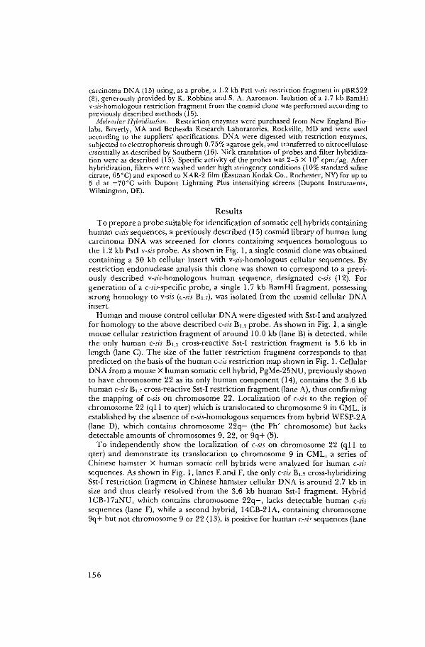
carcinoma DNA (15) using, as a probe, a 1.2 kb Psti v-sis restriction fragment in pBR322 (8), generously provided by K. Robbins and S. A. Aaronson. Isolation of a 1. 7 kb BamHl v-sis-homologous restriction fragment from the cosmid clone was performed according to previously described methods (15).
Molecular Hybridization. Restriction_ enzymes were purchased from New England Biolabs, Beverly, MA and Bethesda Research Laboratories, Rockville, MD and were used according to the suppliers' specifications. DNA were digested with restriction enzymes, subjected to electrophoresis through 0. 75% agarose gels, and transferred to nitrocellulose essentially as described by Southern (16). Nick translation of probes and filter hybridization were as described (15). Specific activity of the probes was 2-5 X I 08 cpm/ fig. After hybridization, filters were washed under high stringency conditions (1 0% standard saline citrate, 65°C) and exposed to XAR-2 film (Eastman Kodak Co., Rochester, NY) for up to 5 d at -70"C with Dupont Lightning Plus intensifying screens (Dupont Instruments, Wilmington, DE).
Results To prepare a probe suitable for identification of somatic cell hybrids containing
human c-sis sequences, a previously described (15) cosmid library of human lung carcinoma DNA was screened for clones containing sequences homologous to the 1.2 kb Pstl v-sis probe. As shown in Fig. 1, a single cosmid clone was obtained containing a 30 kb cellular insert with v-sis-homologous cellular sequences. By restriction endonuclease analysis this clone was shown to correspond to a previously described v-sis-homologous human sequence, designated c-sis (12). For generation of a c-sis-specific probe, a single 1. 7 kb BamHl fragment, possessing strong homology to v-sis (c-sis Bu), was isolated from the cosmid cellular DNA insert.
Human and mouse control cellular DNA were digested with Sst-I and analyzed for homology to the above described c-sis Bu probe. As shown in Fig. 1, a single mouse cellular restriction fragment of around 10.0 kb (lane B) is detected, while the only human c-si.s B1.7 cross-reactive Sst-1 restriction fragment is 3.6 kb in length (lane C). The size of the latter restriction fragment corresponds to that predicted on the basis of the human c-sis restriction map shown in Fig. 1. Cellular DNA from a mouse X human somatic cell hybrid, PgMe-25NU, previously shown to have chromosome 22 as its only human component (14), contains the 3.6 kb human c-sis B1.7 cross-reactive Sst-I restriction fragment (lane A), thus confirming the mapping of c-sis on chromosome 22. Localization of c-sis to the region of chromosome 22 (qll to qter) which is translocated to chromosome 9 in CML, is established by the absence of c-sis-homologous sequences from hybrid WESP-2A (lane D), which contains chromosome 22q- (the Ph' chromosome) but lacks detectable amounts of chromosomes 9, 22, or 9q+ (5).
To independently show the localization of c-sis on chromosome 22 (q11 to qter) and demonstrate its translocation to chromosome 9 in CML, a series of Chinese hamster X human somatic cell hybrids were analyzed for human cHsi.s sequences. As shown in Fig. I, lanes E and F, the only c-sis Bu cross-hybridizing Sst-1 restriction fragment in Chinese hamster cellular DNA is around 2.7 kb in size and thus clearly resolved from the 3.6 kb human Sst-1 fragment. Hybrid lCB-17aNU, which contains chromosome 22q-, lacks detectable human c-sis sequences (lane F), while a second hybrid, 14CB-21 A, containing chromosome 9q+ but not chromosome 9 or 22 (13), is positive for human c~sis sequences (lane
156
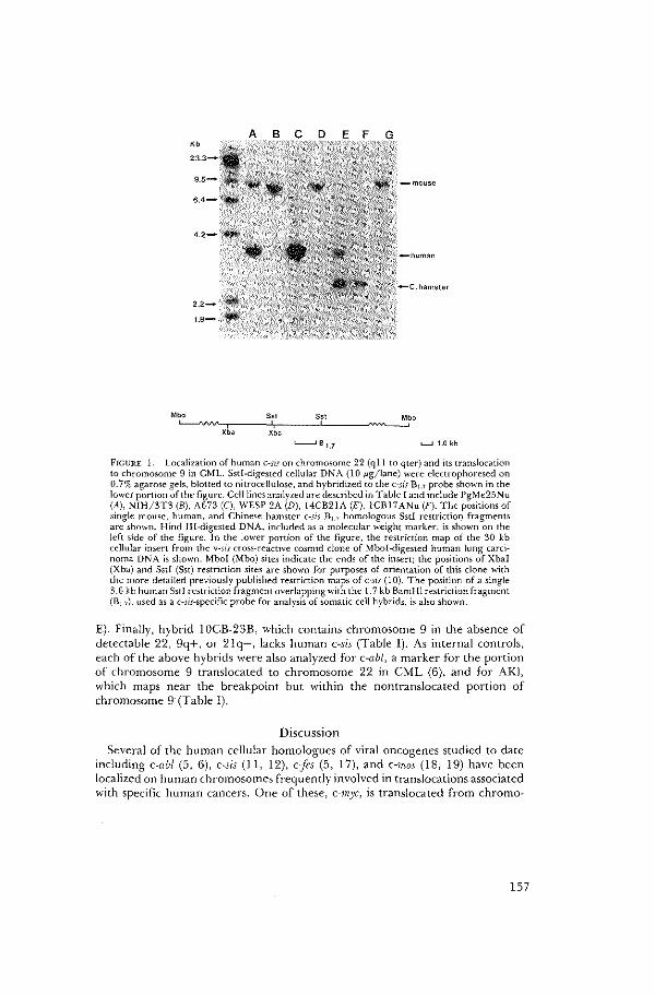
Mbo Sst Sst Mbo
"--"vv'c,r,c,-----c,',-.------JC-------"'"--_:; .......__, B 1.7 L......l 1.0 kb
FlGL'RE l. Localization of human c~sis on chromosome 22 (qll to qter) and its translocation to chromosome 9 in CML. Sstl-digested cellular DNA (10 J.<g/lane) were electrophoresed on 0. 7% agarose gels, blotted to nitrocellulose, and hybridized to the c~sis Bu probe shown in the lower portion of the figure. Cell lines analyzed are described in Table I and include PgMe25Nu (A), NIH/3T3 (B), A673 (C), WESP 2A (D), 14CB21A (E), lCBI7ANu (F). The positions of single mouse, human, and Chinese hamster c-sis 81.7 homologous Sstl restriction fragments are shown. Hind III-digested DNA, included as a molecular weight marker, is shown on the left side of the figure. In the lower portion of the figure, the restriction map of the 30 kb cellular insert from the v-sis cross-reactive cosmid done of Mbol-digested human lung carcinoma Dl\A is shown. Mbol (Mbo) sites indicate the ends of the insert; the positions of Xbal (Xba) and Sstl (Sst) restriction sites are shown for purposes of orientation of this clone with the more detailed previously published restriction maps of c~sis (l 0). The position of a single 3.6 kb human Sstl restriction fragment overlapping with the 1.7 kb BamHl restriction fragment (8 1_7), used as a c-sis-specific probe for analysis of somatic cell hybrids, is also shown.
E). Finally, hybrid I OCB-23B, which contains chromosome 9 in the absence of detectable 22, 9q+, or 21q-, lacks human c-sis (Table I). As internal controls, each of the above hybrids were also analyzed for c-abl, a marker for the portion of chromosome 9 translocated to chromosome 22 in CML (6), and for AKJ, which maps near the breakpoint but within the nontranslocated portion of chromosome 9· (Table 1).
Discussion
Several of the human cellular homologues of viral oncogenes studied to date including c-abl (5, 6), c-sis ( 11, 12), c{es (5, l 7), and c-mos (18, 19) have been localized on human chromosomes frequently involved in translocations associated with specific human cancers. One of these, c-myc, is translocated from chromo~
157
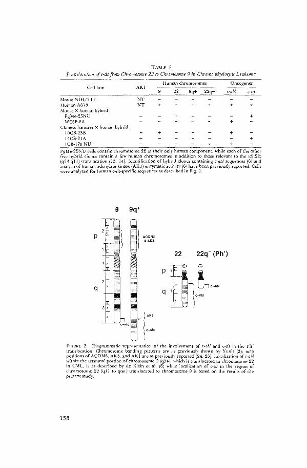
TABLE I
Translocation of c~sis from Chromosome 22 to Chromosome 9 in Chronic Myelocytic Leukemia
Human chromosomes Oncogenes Cell line AKI
9 22 9q+ 22q c-ab/ c-sis
Mouse NIH/3T3 NT Human A673 NT + + + + + + Mouse X human hybrid
PgMe-25NU + + WESP-2A + +
Chinese hamster X human hybrid IOCB-23B + + + 14CB-21A + + + ICB-17a NU + +
PgMe-25~U cells contain chromosome 22 as their only human component, while each of the other five hybrid clones contain a few human chromosomes in addition to those relevant to the t(9;22) (q34;qll) translocation (13, 14). Identification of hybrid clones containing c-ab! sequences (6) and analysis of human adenylate kinase (AKI) enzymatic activity (6) have been previously reported. Cells were analyzed for human c-sis-specific sequences as described in Fig. 1.
9 9q+
w;
p t, ACONS &AK3
22 22q- (Ph')
0 0
q t~~~· C-SIS
)
- : AK1 ,,
' ;_t: { c-sis
I
FIGL'RE 2. Diagrammatic representation of the involvement of c-ab! and c-sis in the Ph' translocation. Chromosome banding pauerns are as previously shown by Yunis (3); map positions of A CONS, AK3, and AK I are as previously reponed (24, 25). Localization of c-ab/ within the terminal portion of chromosome 9 (q34), which is translocated to chromosome 22 in CML, is as described by de Klein et al. (6) while localization of c-sis in the region of chromosome 22 (q II to qter) translocated to chromosome 9 is based on the results of the present study.
!58
some 8 to chromosomes 14, 2, or 22, each of which contain immunoglobulin sequences, in Burkitt's lymphoma (1, 19-21). Similarly, c-abl maps on the region of chromosome 9 which translocates to chromosome 22 in CML (5, 6). Other trans!ocations, such as the t( 15; 17) reciprocal translocation associated with acute promyelocytic leukemia (3), involve regions to which human cellular oncogenes (in this case cfes) have been mapped (22), but appear to be independent of immunoglobulin sequences. The present demonstration that c-sis is translocated from chromosome 22 to chromosome 9 in CML raises the possibility that c-sis rather than c-ab! may be involved in CML. Resolution of these alternatives will require a determination of the proximity of these genes to the breakpoints in chromosomes 22 and 9, respectively, and analysis of the expression of their transcriptional and translational products in CML cells.
In addition to possible implications regarding the cause of CML and the significance of the associated t(9;22) (q34;qll) translocation, the localization of c-sis within the translocated region of chromosome 22 (Fig. 2) provides a unique molecular marker for studies of the more complex translocations associated with minority populations of CML patients. These can involve translocation of the q II to qter region of chromosome 22 to chromosomes other than chromosome 9, or can in some instances involve more complex rearrangements including three or occasionally even four or five chromosomes (23). Analysis of these translocations using c-ab! and c-sis probes should allow a determination of the critical translocation event resulting in the generation of CML.
Summary By analysis of a series of somatic cell hybrids derived by fusion of either mouse
or Chinese hamster cells with leukocytes from different chronic myelocytic leukemia (C;'viL) patients or from normal donors, we have localized the human oncogene, c-sis, on the q 11 to qter segment of chromosome 22 and demonstrated its translocation from chromosome 22 to chromosome 9 (q34) in CML.
We thank P. Hansen and G. T. Blennerhassett for excellent technical assistance.
Rmi~'ed for publimtion 10 Janual)' 1983 and in revised form 23 March 1983.
References 1. Rowley, J. D. 1982. Identification of the constant chromosome regions involved in
human hematologic malignant disease. Science (Wash. DC). 216:749. 2. Yunis,J.J., C. Bloomfield, and K. Ensrud. 1981. All patients with acute nonlympho
cytic leukemia may have a chromosomal defect. N. Engl. j. Med. 305:135. 3. Yunis, J. J. 1982. Most cancers may have a chromosomal defect. In Gene Amplifica
tion. R. T. Schimke, editor. Cold Spring Harbor Laboratory, Cold Spring Harbor, New York. 297pp.
4. Nowell, P. C., D. A. Hungerford. 1960. A minute chromosome in human chronic granulocytic leukemia. Srin1re (Wash. DC). 132:1497.
5. Heisterkamp, N., J. Groffen, J R. Stephenson, N. K. Spurr, P. N. Goodfellow, E. Solomon, B. Carritt, and W. F. Bodmer. 1982. Chromosomal localization of human cellular homologues of two viral oncogenes. Nature (Lon d.), 299:747.
6. de Klein, A., A. Geurts van Kessel, G. Grosveld, C. R. Bartram, A. Hagemeijer, D.
159
Bootsma, N. K. Spurr, N. Heisterkamp,J. Graffeo, and]. R. Stephenson. 1982. A cellular oncogene (c-ab!) is translocated to the Philadelphia chromosome in chronic myelocytic leukemia. Nature (Lond.). 300:765.
7. Erikson, J., J. Martinis, and C. M. Croce. 1981. Assignment of the genes for human immunoglobulin chains to chromosome 22. Nature (Lond.). 294:173.
8. Robbins, K. C., S. G. Devare, and S. A. Aaronson. 1981. Molecular cloning of integrated simian sarcoma virus: genome organization of infectious DNA clones. Proc. Nat!. Acad. Sci. USA. 78:2918.
9. Gelmann, E. P., F. Wong-Staal, R. A. Kramer, and R. C. Gallo. 1981. Molecular cloning and comparative analyses of the genomes of simian sarcoma virus and its associated helper virus. Proc. Nail. Acad. Sci. USA. 78:3373.
10. Dalla-Favera, R., E. P. Gelmann, R. C. Gallo, and F. Wong-Staal. 1981. A human one gene homologous to the transforming gene (v-sis) of simian sarcoma virus. Nature (Lond.). 292:31.
II. Swan, D. C., 0. W. McBride, K. C. Robbins, D. A. Keithley, E. P. Reddy, and S. A. Aaronson. 1982. Chromosomal mapping of the simian sarcoma virus one gene analogue in human cells. Proc. Nat!. Acad. Sci. USA. 79:4691.
12. Dalla-Favera, R., R. C. Gallo, A. Giallongo, and C. M. Croce. 1982. Chromosomal localization of the human homolog (c-sis) ofthe Simian sarcoma virus one gene. Science (Wash. DC). 218:686.
13. Geurts van Kessel, A. H. M., H. den Brinke, W. A.M. Boere, W. C. den Boer, P. G. de Groot, A. Hagemeijer, P. Meera Khan, and P. L. Pearson. 1981. Characterization of the Philadelphia chromosome by gene mapping. Cytogenet. Cell Genet. 30:83.
14. Geurts van Kessel, A. H. M., W. C. den Boer, A.J. van Agthoven, and A. Hagemeijer. 1981. Decreased tumorigenicity of rodent cells after fusion with leukocytes from normal and leukemic donors. Somatic Cell Genet. 7:645.
15. Groffen, J., N. Heisterkamp, F. Grosveld, W. Van de Ven, and]. R. Stephenson. 1982. Isolation of human oncogene sequences (vjes homolog) from a cosmid library. Science (Wash. DC) 216:1136.
16. Southern, E. M. 1975. Detection of specific sequences among DNA fragments separated by gel electrophoresis.]. Mol. Biol. 98:503.
17. Dalla-Favera, R., G. Franchini, S. Martinotti, F. Wong-Staal, R. C. Gallo, and C. M. Croce. 1982. Chromosomal assignment of the human homologues of feline sarcoma virus and avian myeloblastosis virus one genes. Proc. J.Vatl. Acad. Sci. USA. 79:4714.
18. Prakash, K., 0. W. McBride, D. C. Swan, S. G. Devare, S. R. Tronick, and S. A. Aaronson. 1982. Molecular cloning and chromosomal mapping of a human locus related to the transforming gene of Moloney murine sarcoma virus. Proc. Nat!. Acad. Sci. USA. 79:5210.
19. Nee!, B. G., S.C. Jhanwar, R. S. K. Chagami, and W. S. Hayward. 1982. Two human c-one genes are located on the long arm of chromosome 8. Proc. Natl. Acad. Sci. USA. 79,7842.
20. Dalla-Favera, R., M. Bregnni,J. Erikson, D. Patterson, R. C. Gallo, and C. M. Croce. 1982. Human c-myc one gene is located on the region of chromosome 8 that is translocated in Burkitt lymphoma cells. Proc. Natl. Acad. Sci. USA. 79:7824.
21. Taub, R., I. Kirsch, C. Morton, G. Lenoir, D. Swan, S. Tronick, S. Aaronson, and P. Leder. 1982. Translocation of the c-m;•c gene into the immunoglobulin heavy chain locus in human Burkitt lymphoma and murine plasmacytoma cells. Proc. Natl. Acad. Sci. USA. 79:7837.
22. Sheer, D., L. R. Hiorns, K. F. Stanley, P. N. Goodfellow, D. M. Swallow, S. Pavey, N. Heisterkamp,J. Groffen,J. R. Stephenson, and E. Solomon. 1983. Genetic analysis of the 15; J 7 chromosome translocation associated withacutepromyelocytic leukemia.
160
Proc. Natl. Acad. Sci. USA. In press. 23. Koeffler, H. P., and D. W. Golde. 1981. Chronic myelogenous leukemia-new
concepts. New Engl. J. Med. 304:120 I. 24. Carritt, B., and S. Pavey. 1979. Regional assignments of the loci AK~, A CONS and
ASS on human chromosome 9. Cytogenet. Cell Genet. 23:171. 25. Shows, T. B., A. Y. Sakaguchi, and S. L. Naylor. 1982. Mapping the human genome,
cloned genes, DNA polymorphisms, and inherited disease. Adv. Hum. Genet. 12:341.
161

APPENDIX PAPER XI

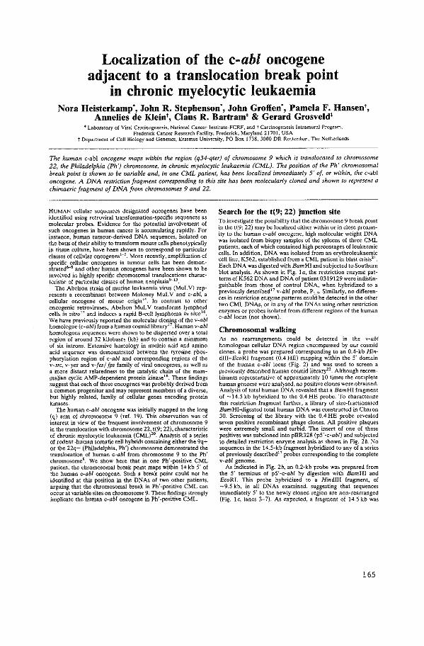
Localization of the c-abl oncogene adjacent to a translocation break point
in chronic myelocytic leukaemia Nora Heisterkamp", John R. Stephenson", John Groffen·, Pamela F. Hansen',
Annelies de Klein', Claus R. Bartram' & Gerard Grosveld' • Laboratory of Viral Carcinogenesis, National Cancer Institute-FCRF, and t Carcinogenesis Intramural Program,
Frederick Cancer Research Facility, Frederick, Maryland 21701, USA t Department of Cell Biology and Genetics, Erasmus Univen;ity, PO Box 1738, 3000 DR Rotterdam, The Netherlands
The human c~abl oncogene maps within the region (q34·qter) of chromosome 9 which is translocated to chromosome 22, the Philadelphia (Ph') chromosome, in chronic myelocytic leukaemia (CML). The position of the Ph' chromosomal break point is shown to be variable and, in one CML patient, has been localized immediately 5' of, or within, the c-abl oncogene. A DNA restriction fragment corresponding to this site has been molecularly cloned and shown to represent a chimaeric fragment of DNA from chromosomes 9 and 22.
HUMAN cellular sequences designated oncogenes have been identified using retroviral transformation-specific sequences as molecular probes. Evidence for the potential involvement of such oncogenes in human cancer is accumulating rapidly. For instance, human tumour-derived DNA sequences, isolated on the basis of their ability to transform mouse cells phenotypically in tissue culture, have been shown to correspond to particular classes of cellular oncogenes1
-5
. More recently, amplification of specific cellular oncogenes in tumour cells has been demonstrated6-8 and other human oncogenes have been shown to be involved in highly specific chromosomal translocations characteristk of particular classes of human neoplasia"""13
.
The Abelson strain of murine leukaemia virus (MuL V) represents a recombinant between Moloney MuLV and c-ab/, a cellular oncogene of mouse origin14
. In contrast to other oncogenic retroviruses, Abelson MuL V transforms lymphoid cells in virro 1
' and induces a rapid 8-ce\llymphoma in vivo16,
We have previously reported the molecular cloning of the v-abl homologue (c-ab/) from a human cosmid library17
. Human v-abl homologous sequences were shown to be dispersed over a total region of around 32 kilobases (kb) and to contain a minimum of six introns. Extensive homology in nucleic acid and amino acid sequence was demonstrated between the tyrosine phosphorylation region of c-ab/ and corresponding regions of the v-.src, v-yes and v-fes/ fps family of viral oncogenes, as well as a more distant relatedness to the catalytic chain of the mammalian cyclic AMP-dependent protein kinase18
• These findings suggest that each of these oncogenes was probably derived from a common progenitor and may represent members of a diverse, but highly related, family of cellular genes encoding protein kinases.
The human c-ab! oncogene was initially mapped to the long (q) arm of chromosome 9 (ref. 19). This observation was of interest in view of the frequent involvement of chromosome 9 in the translocation with chromosome 22, t(9; 22), characteristic of chronic myelocytic leukaemia (CML)~0• Analysis of a series of rodent-human somatic cell hybrids containing either the 9q+ or the 22q- (Philadelphia, Ph') chromosome demonstrated the translocation of human c-ab/ from chromosome 9 to the Ph' chromosome9• We show here that in one Ph'-positive CML patient, the chromosomal break point maps within 14 kb 5' of the human c-ab/ oncogene. Such a break point could not be identified at this position in the DNAs of two other patients, arguing that the chromosomal break in Ph' -positive CML can occur at variable sites on chromosome 9. These findings strongly implicate the human c-ab/ oncogene in Ph'-positive CML.
Search for the t(9; 22) junction site To investigate the possibility that the chromosome 9 break point in the t(9; 22) may be localized either within or in close proximity to the human c-ab/ oncogene, high molecular weight DNA was isolated from biopsy samples of the spleens of three CML patients, each of which contained high percentages of leukaemic cells. In addition, DNA was isolated from an erythroleukaemic cell line, K562, established from a CML patient in blast crisis21
.
Each DNA was digested wi.th Bam HI and subjected to Southern blot analysis. As shown in Fig. I a, the restriction enzyme patterns of K562 DNA and DNA of patient 0319129 were indistinguishable from those of control DNA, when hybridized to a previously described' 7 v-ilbl probe, P1 7. Similarly, no differences in restriction enzyme patterns could be detected in the other two CML DNAs, or in any of the DNAs using other restriction enzymes or probes isolated from different regions of the human c-ab/ locus (not shown).
Chromosomal walking As no rearrangements could be detected in the v-abl homologous cellular DNA region encompassed by our cosmid clones, a probe was prepared corresponding to an 0.4-kb Hindiii-EcoRI fragment (0.4 HE) mapping within the 5' domain of the human c-ab/ locus (Fig. 2) and was used to screen a previously described human cosmid library22
• Although recombinants representative of approximately 10 times the complete human genome were analysed, no positive clones were obtained. Analysis of total human DNA revealed that a Bam HI fragment of -14.5 kb hybridized to the 0.4 HE probe. To characterize this restriction fragment further, a library of size-fractionated Bam HI-digested total human DNA was constructed in Charon 30. Screi:ning of the library with the 0.4 HE probe revealed seven positive recombinant phage clones. All positive plaques were extremely small and turbid. The insert of one of these positives was subcloned into pBR328 (p5' -c-ab!) and subjected to detailed restriction enzyme analysis as shown in Fig. 2b. No sequences in the 14.5-kb fragment hybridized to any of a series of previously described17 probes corresponding to the complete v-abl genome.
As indicated in Fig. 2b, an 0.2-kb probe was prepared from the 5' terminus of p5' -c-ab/ by digestion with Bam HI and EcoRI. This probe hybridized to a Hindiii fragment, of -9.5 kb, in all DNAs examined, suggesting that sequences immediately 5' to the newly cloned region are non-rearranged (Fig. lc, lanes 3-7). As expected, a fragment of 14.5 kb was
165
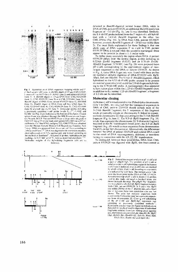
' ' ' ,.,_,~
3.4- •
'----' v-sbl
' ' ' 1234567
-9.5
Fig. l lckntoltc~tiun c>f DNA 'e<JUl"ncc' mapping within and 5" "lth~ hum"n c-uhll"cu,_ u. HumHJ Jige't of Ill )J.g of DNA from human cdllmc A On I lane I 1. K%2 Iiane 21 an <.I patient 031 ~ 12~ Iiane .1). h. BamHl digc't of \0 f<); DNA from C:>.1L02120185 Iiane II. CML1131'il29 IIane 21 ami CML03JJ(Ihl\ IIane 31. c, BamHI <ligc;t of DNA from patient 031'1129 (lane 1), 0311061\ IIane 21. Hmdlll <ligc't of DNA from cdl line K51':>2 IIane 3). CML02120IKS {lane 41. CML0319129ilane 51, DIJL0311116g Iiane I'>J anJ cdllonc A21l4 Iiane 71. Molecular prohc' rnclu<ling a prcviomly Jc,ni~ed v-ab/ pmb~ [o-abl P, 7 )
17, {1.4 HE ami
0.2 BE l'ee Fig. 2) are imlicatcd at the t>ottom of the ligurc. Frozen 'plccn ti>'uc w"' ohtarnetl through the :-.J!H Rc;ource' and Logi;ti~' Branch, DCCP. Pati~ntll31 'il2'i "'a' a 20-yr·old male, patient 1121~1{)) wa' a 57-yr·ol<.l male and patient 031 l06H wa' a 1:>7-yr· olJ female. Cc>lllmc K%2, pedigree 722, (GM )372) wa' uhtaineJ from the Human Genetic Mutant Cell Repo,itory. Camden. ~ew Je"ey_ DNA"'"' prepare<.llrom t;,_,ue and cell line' accor<.lmg to Jeffrey' an<.l Flavell'"- DNA wa' <.lige,ted with rc,triction ~nzymes, dectrophore,e<.l on 0.75"/o agaro>e gel> an<.l hloned according to the method of Southern·"_ l'olation of prnbe,, hybri<.lizmion and o.a,htng,, to 11.3 x SSC" at t\5 'C were "' dc,crihed d'cwhcre 17
''
!1-lnlecular v.,cight> of the hyhri<.lizing fragments (khl arc a>
0 ~ = 0 2 BE ~ 52E 0 70HE
166
~ 1 2 HBg
incilcated
0 OOHE
detected in BumHI-digestcd normal human DNA. while in DNA ofCML patient 031 \l129, an additional Bum HI restriction fragment of -3.1 kb (Fig. !c. lane II was id~ntified. Similarly. the 0.4-kb HE probe isolated from the 5' region of c-ub/ hybridized with a 14.5-kb BumHl fragment in the other two CMLDNAs {Fig. I b). In DNA from CML patient 0319129. however. an extra Bum HI fragment of -30 kb was visible (lane 2!. The most likely explanation for these findings is that one allelic copy of DNA sequences 5' to c-ab/ in CML patient 0319129 DNA is normal while the second is rearranged: these appear to be present in about a I; I molar ratio.
To define more accurately the region where DNA of patient 0319129 differs from the normal region, probes including an 0.52-kb £coR! fragment (0.52 E) and an 0.76-kb EcoR!-1-find!l! fragment {0.76 HE) (>e~ Fig. 2b) were prepared from pS'-c-ubL corresponding to the approximate region of rearranged sequences in the DNA of patient 0319129. As shown in Fig. 3, extra DNA fragments were found with these probes on restriction enzyme digestion of DNA0319129 with Bglll. X bal. Sst! and Hindi!!. The 9.5-kb 5' Hindlll fragment, which hybridized to the 0.52-kb £coR! probe, seemed to be present in a normal quantative ltvel, as did the :!.0-kb frdgment hybridizing to the 0.76-kb HE probe. A rearrangement thus appeared to have taken place within the 1.25-kb Hind Ill fragment since, in addition to the normal 1.:!5-kb Hindll! fragment, a fragment of 2.2-kb can be identified
Molecular cloning As human c-ub/ is translocated to the Philadelphia chromosome (22q-) in CML, we rea~0ned that the transposed sequences in DNA of CML patient 031Yl29 should include most of the 14.5-kb BumH! fragment; the 5' sequences of this fragment would presumably remain on chromosome 9, linked to scquenc~'> fromchromn>ome 22, thus accounting for the 3.1-kb Bum HI fragment (Fig. lc, lane 11. The Y.5-kb Bglll fragment {Fig. 3b) would thu> repre:.ent the chromm;ome 22/9 chimaeric fragment localized at the Ph' translocation break point; the 6.0-kb Bglll fragment (Fig. 3b) would contain sequences from chromosomes 9 and 22, on the 9q+ chromosom~. Alternatively, the differences' between the DNA of patient 0319129 and control DNA could bt the result of DNA rearrangements, deletions or insertions having no connection with the t(9; 22) Ph' translocation.
To distinguish between these possibilities, DNA from CML pati~nt 0319129 was digested with Bg/IJ, size fractionated in
L--' , kb
'----.J 'kb
'------' 1 kb
fi~. 2 R~striction enzym~ analysi' of p.'i' ·c-ab/ and "part oJ' pl3glll ~q+. Th~ p<Ntirm of p5'-c·-abl in relatiun to th~ v-abl hybridizing r~gion in the human c-ahllo~u' ;, indicated in a_ EwRI site' are marked hy 'mall ,·cnical line<; v-abl homologous sequences ~'"indicated by 'olid bars. The vertical arrow indi<:at~s the hrcak point m the D~A ot CML (JJ 1912'1. A '"'triction map of p5' -c-ab/ is shown in b; probes U>Cd in thi' 'tudy, mdkated a> hatched boxe>. are :h<>wn bcn~ath the map. The pBgll! 9q+ fmgment Ln ,. "a 'ubclone of a 6.0-kb Bglll fragment isolated lrom C:v!L patient 0319129: in band c the Bglrr 'ite within 200 hp of the 5' Bam HI site an<.l a Hpal 'ire rn hetll'cen these two restriction ;tte> are not ,nown. The soli<.l bar indicate< sequence; from c·h"'mmome 9 whil~ the <>peo bar rcpresenb~~4uen· c·c, f"'m chromo>ome 22. Cloning and subdoning nf th~ p5'-c-ab/ and Bg/Jl-9q+ sequences "'"' ~ccording to pr~'iously described methoJs-''. Re>tricuon enzyme map> are based nn hybri<.lization <lata and nn re~ults of double <.ligest•ons of individual lrugments ."olated from low meltmg pomt agarose :;"b. Re,tnctwn enzymes include: Bam HI tBl. Bgll! IBgl. BstEI! iSs). Hin<lll! !HI. Sst! IS). XbaliXbJ,
\'loa! iXhl. EwRI tEl and Kpn! IKJ.
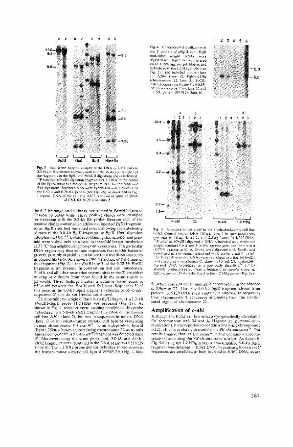
11.0-q .. · ...•. ·.·· 9.5- i
\~- ' '
6.0- .'
.• Iii.·. ~' ~-¥ ~-
" b
-9.5
-2.2
-2.0
-1.25
L-..J l.__j L-...J l__j Bglll Xbal Ssll Hindlll
Fig. 3 ReMriction enzyme analysis of the DNA of CML patient 0319129. Restriction enzymes used and the molecular weights of the fragments in the Bgll1 and Hindlll digestion> are as indicated. -"P-Iabelled Hind!ll digestion frdgments of A DNA 'm the centre of the figure serve as a molecular weight marker for the Xbal and S<tl digestions. Southern blots were hybridized with a mixture of the 0.52 E and 0.76 HE probe~ (;ee Fig. 2b), as described in Fig 1 legend DNA of the cell line A673 is >hown in lanes a: DNA
of CML0319!29 is in lanes b.
the 6-7-kb range, and a library constructed in Bam HI-digested Charon 30 phage arms. Three positive clones were identified on screening with the 0.2-kb BE probe. Because each of the po5itive clones contained an additional inserted Bglll fragment. some Bgiii sites had remained intact. allowing the subcloning of most of the 6.0-kb Bgiii fragment by Bglii~Hpa! digestion into plasmid ORFn. Colonies containing this recombinant plasmid were visible only on a two- to threefold longer incubation at 37 'C than neighbouring non-positive colonies. This particular DNA re£.ion may thus contain wquences that inhibit bacterial growth. Possibly explaining our failure to detect these sequences in cosmid libraries. As shown in the restriction enzyme map of this fragment (Fig. 2c). the EcoRI site 3' of the 0.52-kb EcoR! fragment is still present. In contrast. an Sst! site immediately 3' of it and all other restriction enzyme sites to the 3' arc either missing or different from those found in the same region in pY-c-abl. These findings localize a putative break point in pY-c-abl between the EcoR! and Sst! sites. S~quence; 5' of this point in the 6.0-kb Bg/JI fragment hybridize to p5' -c-ab/: sequences 3' of it do not (results not shown).
To determine the origin of the 6.0-kb Bg/11 fragment, a 1.2-kb Hindlll-Bg/Jl probe (1.2 HBg) was prepared (Fig_ 2c). As shown in Fig. 4. using ;tringent washing conditions. this probe hybridized to a 5.0-kb Bglil fragment in DNA of the human cell line A204 (lane 2). but not to sequences in mouse DNA (lane 1) or in rodent-human somatic cell hybrids containing human chromosome 9 (lane 4)" 1n an independent hybrid (PgMe-25Nu), ho~ever. containing chromo>ome 22 as it> only human component , a 5.0-kb Bglll fragment was detected (lane 3). Moreover, using the same probe both 5.0-kb and 6.0-kb BgliJ fragments were identified in the DNA of patient 0319129 (lane 6). The 1.2 HBg probe did not hybridize to sequences in the human-mouse somatic cell hybrid WESP-2A (Fig. 4, lane
fig. 4 Chrommomalloc·ul!zation of the 3' domain of pBglll·9q+. High molecular weight DNA' were digested with Bg/1!. electrophore,ed on an 0. 75% agMo'c gel, blottd and hybridized to the I .2 HBg pro he (,cc Fig. 2cl and included mome ll~ne I), A204 (lane 21. Pg.Mc-25:-Ju lchromo">me 22. bne JJ. IIICB-23B lchromo\nmc 9.lane 4), WESP-2A ithromooome 22q-, lane 51 and
CML p~tient 031912'1 (lane fil
2.2-""
1.8-
L-,-_,-,c,~ 'c;-'
2 3 4 5 6
' 2
' t -#··~
t ~
d
Fig. 5 Amplification of c-ab/ in the crythroleukacmic cell line K.'i62. Control human DNA 110 f!.g. lane> I in each panel) v.-a> run next to Ill fl.g. Iiane~ 21 and 2.5tJ-~ (lanes JJ K502 DNA "P·Iat>clled Hmdlll-digcsted A DNA ;,-included"' a molecular weight standard for a a~d b (0.6% agarme gels I and for c and d 10.75% agarme gels). a, DNA, were dige,!ed with EwRI and hybridized to a previou,]y described v-abl probe. v-abl P, 7 I ref 17)_ b, EcoRl-dtge,1cd DNA\ were hybridized to a Bglll-Hindlll probe. isola!ed from a human C, done HuA .'i (rd. 251. c. BarnHIdige\red DNA hybridized to a previOusly de;cribed'" 1.7-kb BarnHl probe prep-ored from a human <>sis cu;mid clone. d, Bglll-Jige;tcd D:--JA, hybridized 10 the 1.2 HBg probe (FJg. 2cJ.
5). which contain; the Philadelphia chromooome in the absence of 9,9q+ or 22. Thus. the 6.0-kb Bgll! fragment cloned from patient 0319129 D:-JA must contain, in addition to >eqHence> from chromosome lJ, sequences originating from the translocated region of chromosome 2:?..
Amplification of c~abl Although the K562 cell line l~cks a cytogenetically identifiable Ph' chromosome (ref. 24 and A. Hagemeijer, personal communication). it was reponed to contain a small ring chromosome rl22), which 't> probably derived from a Ph' chromosome'~. Our results sugge;t that, at a minimum. K51i2 contains a chromosomc(s) resembling the Ph' chromosome marker. As shown in Fig. 5d. using the 1.2 HBg probe. a non-amplified 5.0-kb Bg/ll fragment was detected in K562 DNA. In contrast. human c-ab/ sequences are amplified at least fourfold in K562 DNA, as are
167
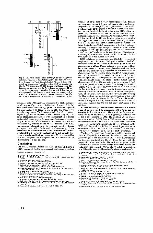
22
,, 5 Kb
,, I
'· 9.5 Kb
I ,, J c-abl
5' Bg-
1 11 Kb
I
~· '" ~g-3' Bg J ,_,.,
fig. 6 Schematic representation of the 1(9; 22} in CML patient 0319129. The sizes of the Bglll fragments detected with probes from chromosome 9 and 22 are indicated. Regions in black originate from chromosome 9; hatched regions are from chromosome 22. An X indicates the 1(9; 22) chromosomal break point The human c-sis oncogene and the C, region on chromosome 22 are shown for purposes of orientation; human c-sis is localized on chromosome 22 (ref. 34) and is translocated to chromosome 9 in CML'6. C, is localized in band q 11 of ctuomosome 22 (ref. 10), and is not involved in the Ph' translocation (unpublished results).
sequences up to 17 kb upstream of the most 5' v-abl homologous EcoRI region (Fig. lc). A ll.O-kb EcoRI fragment (Fig. Sa) that hybridizes to the v-ab/ P~. 7 probe but has no linkage with the main human c-ab/ locus17 is non-amplified and thus serves as an internal control. The immunoglobulin light-chain constant region (C,)25 is also amplified at least fourfold (Fig. Sb). This latter observation is consistent with the localization of human c-ab/ and C, sequences on the same amplification 1.1nit, presumably a part of the Ph' chromosome. In concordance with this conclusion, C, remains on the Ph' chromosome in the t9: 22 (unpublished observations). In contrast, another human oncogene, c-sis, which is located on chromosome 22 and transferred to chromosome 9 in the Ph' translocation26
, is nonamplified (Fig. 5c). Fina!ly, the fact that the 5.0-kb Bglll fragment normally localized on chromosome 22, is non-amplified in K562, supports the assumption that it is translocated to chromosome 9 in this cell line.
Conclusions The present findings establish that in one of three CML patient DNAs examined, the Ph' chromosomal break point is localized Roc01"od ~July, •c.:epted 2 S<ptomi><r 1983
I. P"kiooi. S. tf 0/. NGIU>< JO(I, 5]9-541. (19821 2. Oer. C. J .. Kron<id,. T. G. & C"oop<r. G. M. f',x. "0"1. Mod. Scr. U.S.A. 19. 3617-364()
(19~11 3. Tailin. C. J. tf oi. NGIUr< JOO. 14:!-149 (19821. 4. Tapatow<l<y. E. " ol. No'"'' JOO. 761-765 ( 19821 5. Shimi<U. K. tr at 1'>-o<. """' A<od. Scl U.S.A. 80, 2112-2116 (19~Jl. 6. Collin,. S. & Groodine. M. NoM< 298, 67H81 {198:!). 7. Dall>·fovero. R .• Wong-Sraal. F. & Gallo. R. NaM< 199. 61-!;l {19821 8. Alitolo. K .. Schwab. M .• Lin. C.. V>tmll>. H. E. & lli>hop. J. M. Pnx:. "0"1. Mad. Sc1.
U.£A. 80, 1707-1711 (19831 9. do Klein. A. "'ol. No'"" JOO, 165-167 (19821.
10. T•"b. R. "a( Pro<."""' A<od. Sc( U.S.A.19. 7837-7841 {1981.1 11. Adams. J. M .. Oerondo<i>. S .• Webb. E..'"""'""· l ~- & Cary. S. P,cc. no<n. AroO.
Sd U.S.A. 8-0. 1981.-1986 (19811 12. Dallo-F>vor>. R. <IGI. Smnu lU, 96]-967 (198]1. 13. Shoot. O. "'oL 1'>-o<. """'" A<od. 5<( U.S.A. 80, 5007~5011 {1981) 14. G<>ff. S. P •. Gilboo. E .. Witte. 0. N. & Baltimo«. D. Cdl 22. 777-78j {19801. 15. Ro><ni><rg. N .. Boltimoro. D. & Sehor. C. Ptoc. nom. Acod. Sci. U.S.A. 1!. 1932-1930
{1~75).
16. Abel>On. H. T. & Rot><tdn.l.. '· ,.,.,.,.,.,J<,., .ln, ::1.'--llll {19Jnl
l68
within 14 kb of the most 5' v-ab/ homologous region. Becau~e the position of the most 5' exon in human c-ab/ is not known, the possibility that the t(9; 22) break point may even map within a coding region of the human c~ab/locus cannot be excluded. We have not localized the break points in the DNAs of the two other CML patients or in DNA of the cell line WESP-2A, containing the 22q- chromosome. Our findings, however, establish that the site of the Ph' translocation break point is variable and suggest that break points in the latter DNAs may be located 5' of the -80 kb of cloned DNA encompassing the human c-ab/ locus. Similarly, the t(8; 14) translocation in Burkitt lymphoma, involving the human c-myc oncogene, does not appear to involve .l. single siteHI-lz.n. The orientation of human c-ab/ on chromosome 9, with its 5' region towards the centromere of the chromosome (Fig. 6), is established by the fact that the break point has been found 5' to the human c-ab/ locus.
K562 cells lack a cytogenetica!ly identifiable Ph' chromosome despite their derivation from a CML patient in blast cell crisis2
'.
However, genes on either side of the break point in the Ph' chromosome, c-ab/ and C,_, are not only present in this cell line, but are amplified. In contrast, no amplification was found in the case of c-sis, normally on chromosome 22 but translocated to chromosome 9 in Ph' -positive CML, or a DNA region translocated to chromosome 9 corresponding to a restriction fragment containing the t(9; 22) break point isolated from a CML patient. The amplification seems to be specific; neither human c-fes, on chromosome 15 (ref. 19), nor c-fms, on chromosome 5 {ref. 28), is amplified (data not shown). The finding that c-ab/ is amplified in K562 can be explained in two ways: it can reflect the fact that these cells were grown in tissue culture, possibly resulting in specific chromosomal aberrations and/ or amplifications; alternatively, the c-ab! may have been amplified during the blast crisis of the patient, the overproduction of the c-ab/ gene product being the cause of the disease. The relative amplification of a region of DNA, which includes both c-ab/ and C, sequences, suggests that the two are nearly contiguous in this cell line.
We have previously demonstrated the translocation of a small piece of chromosome 9 to chromosome 22 in CML patients carrying the Ph' chromosome9
• The size of this fragment was calculated to be less than 5,000 kb, suggesting a potential role of the c-ab/ oncogene in CML. The isolation, in the present study, of a region of DNA from a CML patient that contains a chromosomal break point which is localized within 14 kb of the c-ab/ locus, the specific amplification of c-ab! sequence in the K562 cell line, and the finding that c-abl is translocated to chromosome 22 in complex Ph' translocations29 further implicate the c-ab! oncogene in chronic myelocytic leukaemia.
We thank A. Geurts van Kessel for providing somatic cell lines, A. Hagemeijer for valuable discussion, P. Leder for the generous gift of the recombinant HuA5, Gail Blennerhassett for technical assistance and Beverly Bales for help with the preparation of the manuscript. The work was supported by the Netherlands Cancer Society (Koningin Wilhelmina Fonds) and under NCI PHS contract NOI-CP-75380. C.R.B. is a recipient of a fellowship from the Deutsche Forschungsgemeinschaft. 17. Hel!to<kamp. N .• Gtoffen. J. & Stephenson. J. R. J. mo!<e. opp[ G<n<l 2, 57-!;9 !198)1 18. Groff en. J .. Ho~tork•mp. :. .• Royoo1d>. F h. & S«phen>On. J. R. """'"" 304. 167-169
119831 19 .. Heimrkamp. N. "ol Na<uro 2~, 747-150 {19!21 20. Rowley. J. D. NaM< 24J. :!W-293 {1973) 21. Lottio. C. !l. & loZ<io. B. B. Blood 45, 321-334 (!9751 22. Groff en. J .. Heisterl<amp. N .. Gro,eld. G .• von de Ven. W. & Soephensoo. J. R. s'"""
116,1136-1136119821 23. Wetn<toc<. G. M. "o!. Ptoc. "'""· Mod. 5" U.S.A an. 44ll.-.. J6 (198)) 24. Kloin. E. "olin•. 1. Cane<> 18, 411-4)1 119761 25. Hieo<r. P. A .. Holl~. G. F. Kor>meyer. S. J .• W•1dmann. T. A. & Ledet. P. No'"" 294,
536-540 119811 26. G1oHon.J. tfol. J. oxp. Mod. 1511.9-15 (19831 27. S"""'"· l. W .. Wott. R & M>m>. K. B. N""" lOJ, 401....006 1198lJ ZS. Groffen. J. "ol. N•<l<i< A<•ds R•~ (Ln •he pte><) :29. ll>ruom. C. R. "oJ. NOiur< 306, l77-Z•O ( ~~~31 lD. Jeff«)'. A.]. & Fl.,ell. R A. C•/112., 429--439 11977). )I. Southern. E. M. J. mol". Bioi. 98, SOJ.-517 {1975t. J2. llotn.,d<. R. & Fl>V<ll. R. A. N•d<" M1ds R<1 a. 1411-1533 I t980J ]]. Gto~'"·J .. Hei,orkamp. N .. Ble"n"h"""· G. & S«phenwn.J. R Vi,o/ogy 126, llJ-~27
{198]1 34. s~>n. D. C, "ol P'"' ""'" '''"' ''' (;.'A. 79, 4691-•696 119811
APPENDIX PAPER XII

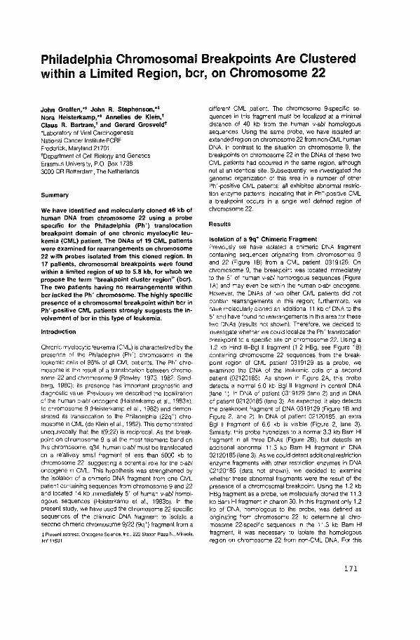
Philadelphia Chromosomal Breakpoints Are Clustered within a limited Region, bcr, on Chromosome 22
John Groffen, ** John R. Stephenson,** Nora Heisterkamp, ** Annelies de Klein, t Claus R. Bartram,t and Gerard Grosveldt *Laboratory of Viral Carcinogenesis National Cancer lnstitute-FCAF Frederick, Maryland 21701 toepartment of Cell Biology and Genetics Erasmus University, P.O. Box 1738 3000 DR Rotterdam, The Netherlands
Summary
We have identified and molecularly cloned 46 kb of human DNA from chromosome 22 using a probe specific for the Philadelphia (Ph') translocation breakpoint domain of one chronic myelocytic leukemia (CML) patient. The DNAs of 19 CML patients were examined for rearrangements on chromosome 22 with probes isolated from this cloned region. In 17 patients, chromosomal breakpoints were found within a limited region of up to 5.8 kb, for which we propose the term "breakpoint cluster region" (bcr). The two patients having no rearrangements within bcr lacked the Ph' chromosome. The highly specific presence of a chromosomal breakpoint within bcr in Ph' -positive CML patients strongly suggests the involvement of bcr in this type of leukemia.
Introduction
Chronic myelocyt1c leukemia (CML) is characterized by the presence of the Ph1ladelph1a (Ph') chromosome 1n the leukemic cells of 96% of all CML patients. The Ph' chro· mosome is the result of a translocation between chromosome 22 and chromosome 9 (Rowley, 1973, 1982; Sandberg, 1980); its presence has Important prognostic and diagnostic value. Previously we described the localization of the human c-ab/ oncogene (Heisterkamp et al .. 1983a). to chromosome 9 (Heisterkamp et al., 1982) and demonstrated its translocation to the Philadelphia (22q-) chromosome in CML (de Klein et al., 1982}. This demonstrated unequivocally that the t(9:22) is recprocal. As the breakpoint on chromosome 9 is at the most telomenc band on this chromosome, q34, human c-ab/ must be translocated on a relatively small fragment of less than 5000 kb to chromosome 22, suggesting a potential role lor the c-ab/ oncogene in CML. This hypothesis was strengthened by the isolation of a chimeric DNA fragment from one CML pat1ent containing sequences from chromosome 9 and 22 and located 14 kb immediately 5' of human v-abl homologous sequences (Heisterkamp et al .. 1983b). In the present study, we have used the chromosome 22-specific sequences of the chimeric DNA fragment to isolate a second chimeric chromosome 9/22 (9q+) fragment from a
:j: Present address: Oncogene Sc1ence. !nc 222 Statoon Plaza N., Mineola, NY 11501
different CML patient. The chromosome 9-specific sequences in this fragment must be localized at a minimal distance of 40 kb from the human v-abl homologous sequences. Using the same probe, we have isolated an extended region on chromosome 22 from non-CML human DNA. In contrast to the situation on chromosome 9, the breakpoints on chromosome 22 in the DNAs of these two CML patients had occurred in the same region, although not at an identical site. Subsequently, we investigated the genomic organization of this area in a number of other Ph' -positive CML patients: all exhibited abnormal restriction enzyme patterns, indicating that in Ph+ -positive CML a breakpoint occurs in a single well defined region of chromosome 22.
Results
Isolation of a 9q ... Chimeric Fragment Previously we have ISOlated a chimeric DNA fragment containing sequences originating from chromosomes 9 and 22 !Figure 18} from a CML patient, 0319129. On chromosome 9, the breakpoint was located immediately to the 5' of human v-abl homologous sequences {Figure 1A) and may even be w'lth'n the human c-ab/ oncogene. However, the DNAs of two other CML patients did not conta1n rearrangements 1n this region: furthermore, we have molecularly cloned an additional 11 kb of DNA to the 5' and have found no rearrangements in th1s area lor these two DNAs (results not shown}. Therefore, we decided to Investigate whether we could localize the Ph' translocation breakpoint to a specific s1te on chromosome 22. Using a 1.2 kb H1nd 111-Bgtll fragment (1.2 HBg, see Figure 18} containing chromosome 22 sequences from the breakpoint reg1on of CML patient 0319129 as a probe, we examined the DNA of the leukemic cells of a second patient (02120185}. As shown in Figure 2A, this probe detects a normal 5.0 kb Bgl II fragment in control DNA (lane 1), in DNA of patient 0319129 (lane 2) and 1n DNA of patient 02120185 (lane 3}. As expected, it also detects the breakpoint fragment of DNA 0319129 (Figure 1 B and F1gure 2, lane 2). In DNA of patient 02120185, an extra Bgt II fragment of 6.6 kb is VIsible (Figure 2, lane 3). Similarly, this probe hybridizes to a normal 3.3 kb Bam HI fragment in all three DNAs (Figure 2B}, but detects an additional abnormal 11.3 kb Bam HI fragment in DNA 02120185 {lane 3}. As we could detect additional restriction enzyme fragments with other restnction enzymes in DNA 02120185 (data not shown}, we decided to examine whether these abnormal fragments were the result of the presence of a chromosomal breakpoint. Using the 1.2 kb HBg fragment as a probe, we molecularly cloned the 11.3 kb Bam HI fragment in charon 30. In this fragment only 1.2 kb of DNA, homologous to the probe, was defined as or'1g.1nating from chromosome 22; to determine all chromosome 22-specific sequences in the 11.3 kb Bam HI fragment, it was necessary to isolate the homologous region on chromosome 22 from non-CML DNA For this
!71
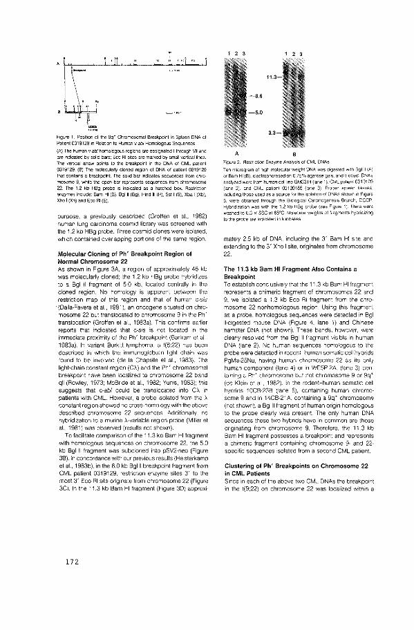
F1gure 1. Position of the 9q~ Chromosomal Breakpoint in Spleen DNA of Pat1ent 0319129 in Relat1on to Human v-abl Homologous Sequences (A) The human v-abl homologous regions are designated I through VII and are Indicated by sol1d bars; Eco Rl $lies are marked by small vertical lines. The vertical arrow points to the breakpoint in the DNA of CML patient 0319129. (8) The molecularly cloned reg•on of DNA of patient 0319129 that contains a breakpoint. The solid bar indicates sequences from chro· mosome 9, while the open bar represents sequences from chromosome 22. The 1.2 kb HBg probe 1S indicated as a hatched box. Restriction enzymes incJude: Bam HI (8), Bglll (Bg), Htnd Ill (H), Sst I ($), Xba I (Xb), Xho I (Xh) and Eco AI (E)
purpose, a previously described (Groffen et al., 1982) human lung carcinoma cosmid library was screened w1th the 1.2 kb HBg probe. Three cosmid clones were isolated. which contained overlapping portions of the same region.
Molecular Cloning of Ph' Breakpoint Region of Normal Chromosome 22 As shown in Figure 3A, a regton of approxtmately 46 kb was molecularly cloned: the 1.2 kb HBg probe hybridizes to a Bgl II fragment of 5.0 kb, located centrally in the cloned region. No homology is apparent between the restriction map of this region and that of human c-sis {Dalla-Favera et al., 1981), an oncogene sttuated on chromosome 22 but translocated to chromosome 9 in the Ph' translocation (Grotfen et al., 1983a). Th1s conftrms earlier reports that indicated that c-sis is not located tn the immediate proximity of the Ph' breakpoint (Bartram et al., 1983a). In variant Burkitt lymphoma. a t{8:22) has been described in whtch the immunoglobulin light chain was found to be involved {de Ia Chapelle et al., 1983). The light-chain-constant regton (CA) and the Ph' chromosomal breakpoint have been localized to chromosome 22 band qll (Rowley, 1973: McBnde et al., 1982: Yums, 1983): this suggests that c-ab/ could be translocated into CA in patients with CML However, a probe isolated from the A constant regton showed no cross-homology with the above described chromosome 22 sequences_ Additionally. no hybridizatton to a murine A-variable region probe (Miller et al., 1981) was observed (results not shown).
To facilitate comparison of the 11.3 kb Bam HI fragment wtth homologous sequences on chromosome 22, the 5.0 kb Bgl II fragment was subcloned into pSV2-neo (Figure 38). In concordance wtth our previous results (Hetsterkamp et al., 1983b), in the 6.0 kb Bglll breakpotnt fragment from CML patient 0319129. restriction enzyme sites 3' to the most 3' Eco Rl stte originate from chromosome 22 (Figure 3Cl. In the 11.3 kb Bam HI fraqment (Figure 3D) approxi-
172
1 2 3 1 2 3
A B F•gure 2 Restnct10n Enzyme Analysis of CML ONAs Ten m1crogram of h•gh molecular weight DNA was digested wtlh 8giii(A) or Bam HI (8). electrophoresed on 0 75% agarose gels_ and blotted ON As analyzed were from human celll1ne GM3314 (lane 1 ). CML pat1ent 0319129 (lane 2). and CML pa11en1 02120185 (lane 3). Frozen spleen t1ssues 1nclud1ng those used as a source for the 1solatron of DNAs shown •n -F•gure 5, were obtained through tM B10IOQ1Cal Carc1nogenes1s Branch, DCCP Hybndlzat,on was w1th the 1.2 kb Heg probe (see F1gure 1 ): l1llers were washed to 0_3 x SSC at es•c. Molecular we1gh1s ot tragments hybrid1Z1ng to the probe are 1nd1cated tn k1iobases
mately 2.5 kb of DNA, including the 3' Bam HI stte and extending to the 3' Xho I site, originates from chromosome
22.
The 11.3 kb Bam HI Fragment Also Contains a Breakpoint To establish conclusively that the 11.3 kb Bam HI fragment represents a chimeric fragment of chromosomes 22 and 9. we isolated a 1.3 kb Eco Rl fragment from the chro· mosome 22 nonhomologous region. Using thts fragment as a probe, homologous sequences were detected tn Bgl 11-dtgested mouse DNA (Ftgure 4. lane 1) and Chinese hamster DNA (not shown). These bands. however, were clearly resolved from the Bgl II fragment vistble tn human DNA (lane 2). No human sequences homologous to the probe were detected in rodent-human somatic cell hybrtds PgMe-25Nu, havtng human chromosome 22 as tts only human component (lane 4) or in WESP-2A. (lane 3) containing a Ph' chromosome but not chromosome 9 or 9q+ (de Klein et al .. 1982). In the rodent-human somattc cell hybrids lOCB-238 (lane 5), containtng human chromosome 9 and tn 14CB-21A. containing a 9q+ chromosome (not shown), a Bglll fragment of human origin homologous to the probe clearly was present. The only human DNA sequences these two hybrtds have in common are those ongtnating from chromosome 9. Therefore. the 11.3 kb Bam HI fragment possesses a breakpoint and represents a chimeric fragment containing chromosome 9- and 22-specilic sequences isolated from a second CML patient
Clustering of Ph' Breakpoints on Chromosome 22 in CML Patients Since in each of the above two CML DNAs the breakpoint in the t(9:22) on chromosome 22 was localized within a
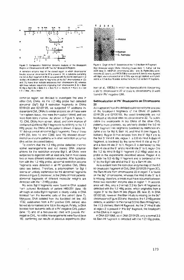
.... ji' ·wr i
\l'·· ',i ~ r "L
i ' llq,LI, i I ' F ~· •i' il "'I ' ' ! j, '"~ I
F•gure 3 ComparaHve Restnction En;>;yme Analysis of the Breakpoint Region on Chromosome 22 w~h Two Sq• Breakpoint Reg1ons
A restrict<on enzyme map of the cloned reg1on 1n which chromosomal breaks occur on chromosome 22 is shown in (A): a subclone con:aining the 5.0 kb Bglll fragment In (B) is compared w.th the 6.0 kb Bgtll and 11.3 kb Bam HI restriction en;>;yme fragments of the 9q• chromosomes in (C) and (D): heavy Iones ind1cate sequences from chromosome 22. whereas llghllines Indicate sequences originating from chromosome 9. B = Bam HI: Bg = Bglll, Bs = BstE II; E = Eco AI: H = Hind Ill: K = Kpn 1: S = Sst t:X=Xbai:Xh=Xhol
common region, we decided to investigate this area in other CML ONAs As the 1.2 HBg probe had detected abnormal (9q+) Bgl II restriction fragments in DNAs 0319129 and 02120185, we subJected 17 additional independent CML DNAs to similar analysts: six of these were from spleen tissue, nine were from patient blood. and two were from bone marrow_ As shown in Figure 5, lanes 1-13. CML DNAs from spleen, blood, and bone marrow all contatned idditionai Bgl II fragments hybridtzing to the 1.2 HBg probe; the DNAs of the patients shown in lanes 14-17 did not exhibit abnormal Bgi II fragments. Two of these (H81-258. lane 14 and COSO. lane 15) showed deviant restriction enzyme patterns with other restriction enzymes (this will be discussed below)
To confirm that the 1.2 HBg probe detected chromo· somal rearrangements and not merely DNA polymorphisms for the restriction enzyme Bgl II, all DNAs were subjected to digestion with at least one. but in most cases two or more different restriction enzymes. After hybridization with tt>e 1.2 HBg probe, abnormal restriction enzyme fragments were detected in ail Ph' -positive CML DNAs (also see below). Therefore, a polymorphism for Bgl II seems an unlikely explanation for the abnormal fragments shown in Figure 5; moreover, in the DNAs of most patients, abnormal fragments of different molecular weights are detected with the 1.2 HBg probe.
No extra Bgi II fragments were found in DNA isolated from cultured fibroblasts of patient H80-251 (lane 18) although an extra Bgl II fragment is clearly visible in DNA isolated from the leukemic cells of this patient (lane 7). Moreover, DNA isolated from the fibroblast cell line, AG 1732. established from a Ph'-positive CML patient, also lacked abnormalities (lane 1g) in this regton. Finally, in DNA isolated from leukemic cells ol a Ph' -negative CML patient (lane 17) and of a two-year-old child with juvenile Ph'negative CML, no visible rearrangements were found (lane 16), confirming our results of previous experiments (Bar·
2 3 4 5
Fogure 4_ Oflgin of the 5' Sequences of the 11.3 kb Bam HI Fragment
High molecular weoght DNAs, including mouse (lane 1 ). human cell l1ne A204 (tane 2). WESP-2A (chromosome 22q-, lane 3). PgMe·25Nu (chro mosome 22, lane 4), and tOCB-236 (chromosome 9, lane 5) were d1gested with Bglll. electrophoresed on an 0.75% agarose gel, blotted, and hybnd· 1zed to a 1.3 kb Eco Rl probe ISOlated from the 1 1_3 kb Bam HI fragment
tram et ai., 1983b) in which no transiocations concerning c-ab! to chromosome 22 or c·sis to chromosome g were found in Ph' -negative CML.
Sublocalization of Ph' Breakpoints on Chromosome 22 As is apparent from the detatled restriction enzyme analysis of the breakpoirt lragments of the DNAs of patients 0319129 and 02120185, the exact breakpoints are not localtzed at identical sites on chromosome 22. To subio· caltze the breakpoints in the DNAs ol the other CML patients more precisely, we arbitrarily divided the 5.0 kb Bgl II fragment into segments bordered by restriction enzyme sites for Bgi II, Bam HI, and Hind Ill (see F-igure 6, bottom)_ Region 0 thus extends from the 5' Bgi II stte to the first 5' Hind ill site, region 1, a 0.6 kb Hind ill-Bam HI fragment, is bordered by the same Hind ill site at the 5' and a Bam HI site 3' to it Region 2 is delineated by this Bam HI stte at the 5' and a Hind 1111 site 3' to it: region 3 is the 1.2 kb Hind 111-Bgl II fragment (1.2 HBg) used as a probe in the experiments described above. Region 4 is outside the 5.0 kb Bgi II fragment and is bordered at the 5' by the Bglll site and at the 3' by a Bam HI site.
As is evident from the restriction enzyme map of the 6.0 kb breakpoint fragment of CML DNA 0319129 (Figure 3C), the Bam HI site from chromosome 22 tn region 1 is found on the 9q+ chromosome, whereas the Hind Ill site 5' to it is missing: therefore, a break must have occurred between these two restriction enzyme sites in region 1_ In accord· ance with this, only a (normal) 3.3 kb Bam HI fragment is detected with the 1.2 HBg probe, which originates from a region 3' to the Bam HI site (Figure 28, lane 2). In DNA 02120185, however, this Bam HI site is missing on the 9q+ chromosome (Figure 30) and, therefore, the 1.2 HBg probe detects, in addition to the normal 3.3 kb Bam Hi fragment, the 11.3 chimeric Bam HI fragment As the 3' Hind Ill site at region 2 is present in this 9q+ fragment, the breakpoint in this DNA is in region 2.
In DNA 0311068, as in DNA 0319129, only a normal3.3 kb Bam HI fragment is detected with the 1.2 HBg probe,
!73
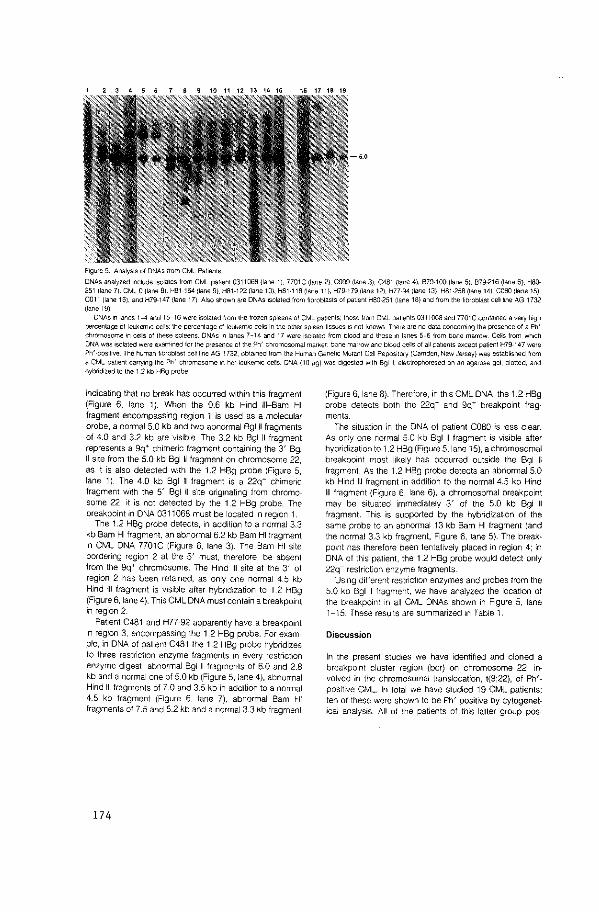
2 3 4 5 6 ~ 10 11 12 13 14 15 16 17 18 19
F1gure 5. AnalySIS of DNAs from CML Pallents
ONAs analyzed 1ncluda 1solates from CML pat<ent 0311068 {lane 1), 7701C (lane 2). C999 (lane 3), C481 {lane 4), 679·100 (lane 5), 879-216 (lane 6}. HSQ-251 {lane 7). CML 0 (lane 8). H81-154 (lane 9). H81-122 (lane 10). H81 ·1 18 (lane 11 ), H79-179 (lane 12). H77-94 (lane 13), H81·258 (lane 14). COSO (lane 15).
COl 1 (lane 16). and H79-147 (lane 1 7) Also shown are DNAs iSOlated from fibroblasts of pat•ent HS0-251 (lane 18) and from the fibroblast celll1ne AG 1732 (lane 19)
DNAs 1n lanes 1-4 and 15-16 were •solated from the frozen spleens of CML pa11ents: those from CML pat1ents 0311068 and 7701C contained a very h1gh
percenlage of leiJ<erniC cells~ the percentage of leukemiC cells 1n the other spleen tissues IS not known There are no data concermng the presence of a Ph'
chromosome if\ cells of these spleens. DNAs 1n lanes 7-14 and 17 were 1solaled lrom blood and those'" lanes 5-Sirom bone marrow. Cells from wh•ch DNA was •solated were examned lor lhe presence ol the Ph' chromosomal marker. bone marrow and blood cells of all pahenls except patient H7g·147 were
Ph' -pos•l1ve. The human f•broblast cell line AG 1732. obtained from the Human Genet1c Mutant Cell RepOSIIDry (Camden, New Jersey) was establ•shed from
a CML pat1ent carry1ng the Ph' chromosome 1n her leukemiC cells. DNA (tO 11g) was d1gested With Bglll, electrophoresed on an agarose gel, blotted. and hybr;d,zed to the 1.2 kb HBg probe
indicating that no break has occurred within this fragment (Figure 6, lane 1 ). When the 0.6 kb Hind Ill-Bam HI fragment encompassing region 1 is used as a molecular probe, a normal 5.0 kb and two abnormal Bgtll fragments of 4.0 and 3.2 kb are VISible. The 3.2 kb Bgt II fragment represents a 9q+ chimeric fragment containing the 3' Bgl II site from the 5.0 kb Bglll fragment on chromosome 22, as it is also detected with the 1 .2 HBg probe (Figure 5, lane 1). The 4.0 kb Bgl II fragment is a 22q- chimeric fragment with the 5' Bgl II s'1te or'1g·1nating from chromosome 22; it is not detected by the 1.2 HBg probe. The breakpoint in DNA 0311068 must be located in region 1.
The 1.2 HBg probe detects, in addition to a normal 3.3 kb Bam HI fragment. an abnormal 6.2 kb Bam HI fragment 1n CML DNA 7701C (Figure 6, lane 3). The Bam HI site bordering region 2 at the 5' must, therefore, be absent from the 9q+ chromosome. The Hind Ill site at the 3' of region 2 has been retained, as only one normal 4.5 kb Hind Ill fragment is visible alter hybridization to 1.2 HBg (Figure 6, lane 4). This CML DNA must contain a breakpoint in region 2.
Patient C481 and H77 ·92 apparently have a breakpoint in region 3, encompassing the 1.2 HBg probe. For example, in DNA of patient C481 the 1.2 HBg probe hybridizes to three restr'1ct1on enzyme fragments ·In every restr'tction enzyme digest: abnormal Bglll fragments of 6.0 and 2.8 kb and a normal one of 5.0 kb (Figure 5, lane 4), abnormal Hind Ill fragments of 7.0 and 3.5 kb in addition to a normal 4.5 kb fragment (Figure 6, lane 7), abnormal Bam HI fragments o17.5 and 5.2 kb and a normal3.3 kb fragment
174
(Figure 6, lane 8). Therefore, in this CML DNA, the 1.2 HBg probe detects both the 22q- and 9q+ breakpoint fragments.
The situation in the DNA of patient COSO is less clear. As only one normal 5.0 kb Bgl II fragment is visible after hybridization to 1.2 HBg (Figure 5, lane 15), a chromosomal breakpoint most likely has occurred outside the Bgl II fragment. As the 1.2 HBg probe detects an abnormal 5.0 kb Hind Ill fragment in addition to the normal 4.5 kb Hind Ill fragment (Figure 6, lane 6), a chromosomal breakpoint may be situated immediately 3' of the 5.0 kb Bgl II fragment. This is supported by the hybridization of the same probe tc an abnormal13 kb Bam HI fragment (and the normal 3.3 kb fragment, Figure 6, lane 5). The breakpotnt has therefore been tentatively placed in region 4; in DNA of this patient, the 1.2 HBg probe would detect only 22q- restnction enzyme fragnents
Using different restriction enzymes and probes from the 5.0 kb Bgl 11 fragment, we have analyzed the location of the breakpoint in all CML DNAs shown in Figure 5, lane 1-15. These results are summarized in Table 1.
Discussion
In the present studies we have identified and clon-ed a breakpoint cluster region {bcr) on chromosome 22, involved in the chromosomal translocation, t(9;22), of Ph'· positive CML. In total we have studied 19 CML patients; ten of these were shown to be Ph' -positive by cytogenetical analysis. All ot the patients of this latter group pOS·
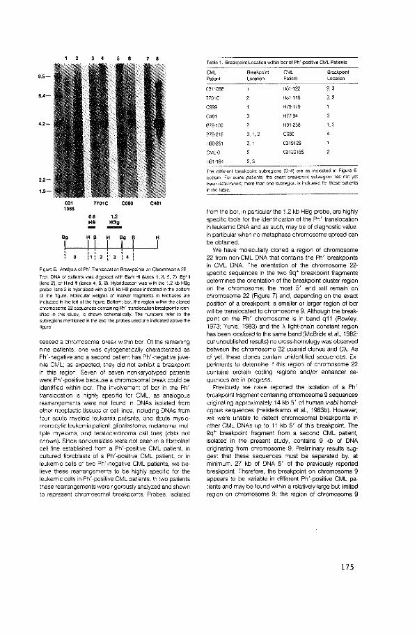
3 • 7 • 9.5-
6.4-
4.2-
2.2-
1.8-
031 note coso C<81 1068
0.6 1.2 HB "'' -,, H B H " B
! ! ! ! ! I ' ' ' ! • 1 Pl '
H
I
F1gure 6 Analysis of Ph' TransJocat1on Breaxpninls on Chromosome 22
Top. DNA of patients was digested with Bam HI (lanes 1. 3, 5, 7). Bgt It
{lane 2), or Hind Ill (lanes 4. 6, 8). Hybridization was with lhe 1.2 kb HBg
probe; lane 2 tS hybnd;zed with a 0.6 kb HB probe indtcated in the botlom of the figure. Molecular weights of marker fragments in kilobases are tndi)':ated in the left of the figure. Bottom: bCf, the region w1lh1n the cloned
chromosome 22 sequences contaJn1ng Ph' translocation breakpoints 1den·
t1fied 1n this study, 1s shown schematically. The numbers refer to the subre{Jions mentioned in the text: the probes used are indicated above the
figure
sessed a chromosomal break Within bcr Of the remaining nine patients, one was cytogenetically charactenzed as Ph' -negative and a second patient has Ph' -negative juvenile CML: as expected, they did not exhibit a breakpoint in this region. Seven of seven nonkaryotyped patients were Ph' ·positive because a chromosomal break could be identified within bcr. The involvement of bcr in the Ph' translocation is highly specific for CML, as analogous rearrangements were not found in DNAs isolated from other neoplastic tissues or cell lines, Including DNAs from four acute myeloid leukemia patients, one olcute myelomonocytic leukemia patient, glioblastoma, melanoma, multiple myeloma, and teratocarcinoma cell lines (data not shown). Since abnormalities were not seen in a fibroblast cell line established from a Ph' -positive CML patient, 1n cultured fibroblasts of a Ph' -positive CML patient. or in leukemic cells of two Ph' -negative CML patients, we believe these rearrangements to be highly specific for the leukemic cells in Ph' -positive CML patients. In two patients these rearrangements were ngorously analyzed and shown to represent chromosomal breakpoints. Probes, isolated
Table 1. Breakpoint location WJ\hin bcr of Ph' -positive CML Patients
CML Breakpoint CML 8reakpo1nt Patient Location Patient Location
0311068 HBt-122 2. 3
7701C H81-t18 2. 3
C9<l9 H79-179
C481 H77-94
879-100 HSt-258 u 878-216 o. 1, coso HS0-251 0.' 0319129
CML-0 02120185
HBt-164 '· 3
The dillerenl breakpoint subregions (0 4) are as lndocated 1n F1gure 6,
bottom For some pal1ents, the exact breakpoint subreg1on has not yet been determoned: more than one subregion IS Indicated for these pat1ents
1n the table
from the bcr, in particular the 1.2 kb HBg probe, are highly specific tools for the identification of the Ph' translocation in leukemic DNA and as such, may be of diagnostic value, in particular when no metaphase chromosome spread can be obtained.
We have molecularly cloned a region of chromosome 22 from non·CML DNA that contains the Ph' breakpoints in CML DNA The orientation of the chromosome 22· specific sequences in the two 9q+ breakpoint fragments determines the orientation of the breakpoint cluster region on the chromosome: the most 5' end will remain on chromosome 22 (Figure 7) and, depending on the exact position of a breakpoint, a smaller or larger region of bcr will be translocated to chromosome 9. Although the breakpoint on the Ph' chromosome is in band q11 (Rowley, 1973; Yunis, 1983) and the A light-chain constant region has been localized to the same band (McBride et al., 1982; our unpublished results) no cross-homology was observed between the chromosome 22 cosmid clones and CA. As of yet, these clones contain unidentified sequences. Ex· periments to determine if this region of chromosome 22 contains protein coding regions andjor enhancer sequences are in progress.
Previously we have reported the isolation of a Ph' breakpoint fragment containing chromosome 9 sequences originating approximately 14 kb 5' of human v-ab/ homol· ogous sequences (Heisterkamp et al., 1983b). However, we were unable to detect chromosomal breakpoints in other CML DNAs up to 11 kb 5' of this breakpoint. The 9q+ breakpoint fragment from a second CML patient, isolated in the present study, contains 9 kb of DNA originating from chromosome 9. Preliminary results suggest that these sequences must be separated by, at minimum, 27 kb of DNA 5' of the previously reported breakpoint. Therefore, the breakpoint on chromosome 9 appears to be variable in different Ph'-positive CML patients and may be found within a relatively large but limited region on chromosome 9; the region of chromosome 9
175
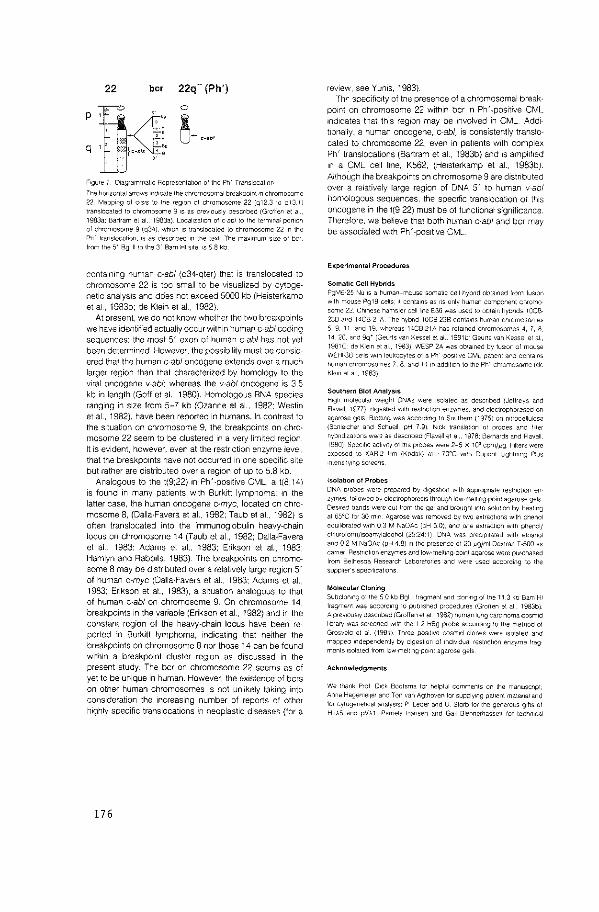
22 be< 22q- (Ph')
Ftgure 7 Dtagrammaltc Representalton o! the Ph' Translocatto~
The honzontal arrows tnd•cate the chromosomal breakpotnt rn chromosome
22 Mapptng of c-s1s to the reQIOn of chromosome 22(o123 to qt3 li \ra~slocated to chromosome 9 IS as prevtously descnbed (Grolfen et at 19il3a: Bartram et al.. 1983a) LocaltLalton of c-ab! to the termt~ai portton of chromosome 9 (q34), whrcn 15 translocated to chromosom~ 22 tn the
Ph' transtocotton_ 15 as descnbed '" the lex: The max,rr.um stze ol bcr from the 5' Bgllllo the 3' Bam HI stte IS 58 kb
contatning human c-ab! (q34-qter) that is translocated to chromosome 22 is too small to be visualized by cytogenetic analysis and dcies not exceed 5000 kb (Heisterkamp et al., 1983b; de Klein et al., 1982}.
At present, we do not know whether the two breakpoints we have Identified actualiy occur within human c-ab/ coding sequences: the most 5' exon of human c-ab! has not yet been determined. However. the possibility must be considered that the human c-ab/ oncogene extends over a much larger region than that characterized by homology to the viral oncogene v-abl: whereas the v·abl oncogene is 3.5 kb in length (Goff et at., 1980). Homologous RNA species ranging in s1ze from 5-7 kb (Ozanne et al., 1982: Westin et al., 1982}, have been reported 1n humans. In contrast to the Situation on chromosome 9, the breakpoints on chromosome 22 seem to be clustered in a very limited region It is evident. however, even at the restriction enzyme level, that the breakpoints have not occurred in one specific site but rather are distributed over a reg1on of up to 5.8 kb.
Analogous to the t(9;22} in Ph'-positive CML, a t(8;14) is found in many patients with Burkitt lymphoma: in the latter case, the human oncogene c-myc, located on chromosome 8, (Dalla-Favera et al., 1982; Taub et aL, 1982} is often translocated 1nto the immunoglobulin heavy-chain locus on chromosome 14 (Taub et al., 1982; Dalla-Favera et aL, 1983: Adams et al., 1983; Erikson et al., 1983; Hamlyn and Rabbits, 1983). The breakpoints on chromosome 8 may be distnbuted over a relatively large reg1on 5' of human c-myc (Oalla-Favera et al., 1983: Adams et al., 1983: Erikson et al., 1983), a situation analogous to that of human c-ab/ on chromosome 9. On chromosome 14. breakpoints 1n the variable (Erikson et al., 1982) and in the constant region of the heavy-chain locus have been reported in Burkitt lymphoma, 1ndicat1ng that neither the breakpoints on chromosome 8 nor those 14 can be found w1th1n a breakpoint cluster region as discussed in the present study. The bcr on chromosome 22 seems as of yet to be unique in human. However, the existence of bcrs on other human chromosomes is not unlikely taking into consideration the Increasing number of reports of other highly specific translocations in neoplastic diseases (for a
176
review, see Yunis, 1983). Thr specifiCity of tne presence of a chromosomal break·
point on chromosome 22 within bcr in Ph' -positive CML indicates that this region may be involved in CML. Additionally, a human oncogene, c-ab/, is consistently translocated to chromosome 22, even in patients with complex Ph' translocations (Bartram et al., 1983b} and is amplified 1n a CML cell l1ne, K562, (Heisterkamp et al., 1983b). Although the breakpomts on chromosome 9 are d1stnbuted over a relatively large region of DNA 5' to human v-abl homologous sequences, the specific translocation of this oncogene in the t(9;22) must be of functional significance Therefore. we believe that both human c-ab/ and bcr may be associated with Ph' -positive CML
EKperimental Procedures
Somatic Cell Hybrids PgMt:-25 Nu IS a human-mouse somat1c cell hybnd obta,ned from fus,on w1lh mouse Pg19 cells. 11 con1a1ns as 1ts only human component chromo some 22 Ch1nese hamsler cell''ne E36 was used 10 oota1n hybnds 10C8 238 a~c HC8-21A The hybnd 10CB-23B conta.ns human chromoso-1les 5. 9. 11 and 19. whereas 14CB-21A has rela1ned chromosomes 4. 7. 8 14 20. and 9q• (Geurts van Kessel et a• _ t98tb Geuns van Kessel el al.. 1981C de Kle1n el al. 1983) WESP 2A was obta1ned by tusK•n ol mouse WEHI-38 ce'IS w•lh leui<ocytes of a Ph' -pos111ve CML pallent and conta1ns r,uman chromosomes 7. 8. and 14 1n add1l1on to lhe Ph' chromosome (de Kle1rt el al 1983)
Southern Blot Analysis H1gt- molecular we1ght ONAs were ISOlated as descnbed {Jeffreys and Flavell. 1977). digESted w1lh restncl10n enzymes. a~d electrophoreseo on agarose gets Blolttng was accordrng to Southern (1975) on nrtrocellulose (Scnlelcher and Schuell. pH 7 9) N1ck transla•.1on ol probes and (,Iter hybndiZJI,ons were as descnbeo (Flavell et al.. 1978. Bernards and Flavell 1980) Spec,f,c activity ol the probes were 2-5 x 10" cpm/1'9 Fillers were Bxposed to XAR-2 111m (Kodak) al ~?O'C w1th Ouponl Llghtn,ng Prus nlens,lylf\g screens
!solation of Probes
DNA D!obes ·Nere prepared by d1gest'on w1lh appropnale restnct,on en lymes I oil ow eo by electrophoresiS lhrough low-rr8'11ng po1n1 agarose gels Oewed bands were cut trom the gel and brought Into solut10n by heat1ng al 65'C lor 30 mtn Agarose was removed by two exlract,ons w1lh phenol equllbraled wrth 0 3 M NaOAc (pH 5.0). ano one extracl1on w1th phenol/ chlorolormflsOamylalcohol (25·24"1) DNA was preCIPitated w1lh elhanol a~o 0 2 M NaOAc {pH 4 8) 1n I he presance ol 20 ~gfml Dextran T -500 as earner Resrnct,on enzymes and 1ow-melt1ng po1nl agarose were pur-chased lrorn Bethesda Research Laboratones and were used according to the suppl1er"s spec,flcal,ons
Molecular Cloning Subclon,ng olthe 50 kb Bgllllragrne~t and con,ng of tne 11 3 ko Barr HI !ragmen: was according to publ•shed procedures (Grotlen et al. 1983bi A pre-<IOUS'Y oescnbed (Grollen el al 1982) human lung carc,noma cosm1d l•brary was screened w1tn the 1 2 H8g probe according to the rretnod ol Grosvelo el a! {198!) Three POS1t1ve cosm1d clones were ISO!ated and mappeo 1ndependenlly by digestion ol 1nd1v1dual restnct,on enzyme lrag menls Isolated from low-mell,ng-pornt agarose gels
Acknowledgments
We :hank Prot D1ck Boolsma lor helpful comments on the rnanuscnpt. Anne Hagemeqer and Ton van Agthoven lor supplying pa11ent maleml and tor cytogenetical analySIS, P Leder and U Storb lor the generous g1f\S ot HuA5 and pVA1. Pamela hansen and Ga•l Btennerhasse:t tor lechn,cal
ass,slance: Beverly Bales lor help wtlh the preparation ol the manuscript rne work was supported by the Netherlands Cancer Society (Komngtn Wtlhelr>11na Fonds) and under NCI PHS contract NOI-CP-75380. C. R. B tS a rec1p1ent of a feltowsh1p from me Deutsche Forschungsgeme<nschaft
Tne costs ol publtcabon of thts arttcle were defrayed in part by the payment of page charges. Th•s arttcle must therelore be llereby marked 'advert,sement" 1n accordance wtth 18 U.S.C Sechon 1734 solely to
tnd•cal-e th•s fact
Recetved November 14 1983
References
Adams. J M Gerondakts. S, Webb, E Corcoran, L. M .. and Cory, S (1983). Cellular myc oncogene IS altered by chromosome lranstocatton to an tmmuno9lobul1n locus 1n munne plasmacytomas and IS rearranged Slf1"-tlarly 1n human Burkitt lymphomas. Proc Nat. Acad. Sc1 USA BO, 1982-
1986
Sartram. C. R. Ce Kletn. A .. Hageme11er, A., Grosveld, G. Hetslerkamp, N and Groffe~. J. (1983a) Localtzatton af the huma~ C·SIS oncogene rn p~· postltve and Ph' -negattve chran•c myelocytic leukemia by m $/lu hybndr·
zalton Blood, <n press
Bartram, C. A_, C!e Klein, A., Hagemeijer. A .. van Agthoven, T., Geurts van Kessel. A .. Bootsma, D., Grosveld, G .. Ferguson-Sm<th, M. A, DaVleS, T, Stone, M., Heisterkamp, N .. Stephenson, J. R., and Groften. J (1983b) Transtucalion of the human c-ab! oncogene occurs in variant Ph' -posi\!ve but not Ph' -negaltve chronic myelocytiC leukaem•a. Nature, 306, 277-280
Bemards, R., and Flavell. A. A (1980). Phystcal mapping of the globin gene detellon rn hered•tary persistence of foetal haemoglobin {HPFH). Nuct. Actds Res_ a_ 1521-1534
Oalla-Favera, A .. Gelmann, E P, Gallo. A C., and Wong-Staal. F (1981). A human one gene homologous to the transformrng gene (v-sis) of simian
sarcoma vtrus. Nature 292, 31-35
Dalla-Favera. R., Bregn•, M. Enkson, J., Patterson_ D .. Gallo, R C, and Croce. C. M. (1982). Human c-myc one gene is located on the region of chromosome 8 thai 1S translocated 1n Burkttt lymphoma cells. Proc. Nat
Acad Sc1. USA 79, 7824-7827
Dalla-Favera, R, Mart•nottl. S., Gallo, R. C., Erikson. J .. and Croce, C. M (1983). Transtocatton and rearrangements of tile c-myc oncogene locus 1n human und•tferentiated 8-celllymphomas. Science 219, 963-967
de Kletn. A .. Geurts van Kessel. A , Grosveld. G., Bartram, C. R .. Hagemetter A, Bootsma, D .. Spurr, N. K., Herslerkamp, N., Grotten, J., and Stephenson. J. R. (1982). A cellular oncogene rs trnnslocated to the Phtladelphia chromosome 1n chronic myelocytiC leukaemta. Nature 300,
765-767
oe Ia Chapelle, A, Lenotr G, Boue, J .. Boue, A, Galiano, P .. Huerre, C .. Sza]ner. M -F, Jeanpterre, M .. Laloue, J. M .. and Kaplan, J.-C. (1983) Lambda lg constant regton genes are translocate<:i to chromosome 8 1n Burkttl's lymphoma w•th ti8:22). Nucl. ActdS Res. 11, 1133-1142
Enksan_ J., Finann, J., Nowell. P. C., and Croce. C. M. (1982) Translocation of tmmunoglobulin V" genes •n Burkitt lymphoma. Proc. Nat. Acad. Sci USA 79,5611-5615
Errkson, J., ar-Ruslldl, A, Drwtnga, H. L., Nowell, P. C .. and Croce, C. M (198.3) Transcnptionat activation of the translocated c-myc oncogene on Burk>tllymphoma. Proc. Nat. Acad. Set USA 80. 820-824
Flavell, R. A, Kooter, J M., de Boer, E .. Little, P. F. R, and Wilhamson, R {1978). Analysts ol the !i'-0-globtn gene lOCI Ill normal and Hb Lepore DNA direct determination of gene ltnkage and tntergene dtstance. Cell 15, 25-
" Geurts van Kessel. A. H. M , den Boer, W. C., van Agthoven, A. J .. and Hagemet]er, A (1981a). Decreased tumorir;entctty of rodent cells after fus1on wllh leukocytes from normal and leukefTlic donors Somatic Cell Genet. 7, 645-656
Geurts van Kessel. A. H. M .. ten 8rinke, H .. Boere, W. A.M .. den Boer. W c , de Groot, P. G., Hagemetjer, A., Meera Kahn, P., and Pearson, P L (1981 b). Charactenzation of the Philadelphta chromosome by gene mapo•ng. Cytogenet. Cell Genet. 30, 83-91
Geurts van Kessel. A H. M .. van Agthoven, A. J., and Hagemeijer. A
(1981c) Clonal origin of the Phtladelphoa translocation in chronic myelotd leukemia demonstrated in somatic cell hybrids using an adenytate kinase-1 polymorphism. Cancer Genet. Cytogenet. 6, 55-58.
Goft, S. P., Gilboa, E .. Witte, 0. N., and Balhmore, D. (1980). Structure of
the Abelson murine leukemia wus genome and the homologous cellular gene· studies with cloned viral DNA Cell22. 777-785
Grotten, J., Hetsterkamp, N .. Grosveld, F., Van de Ven, w_ J. M .. and Stephenson, J_ R_ (1982). Isolation of human oncogene sequences (v-Ies
homolog) from a cosmd library. Sc•ence 216, 1136-1138
Groften. J., Heisterkamp, N .. Stephenson, J_ R., Geurts van Kessel, A .. de Klein, A, Grosveld, G., and Bootsma, D. (1983a). c·sis is translocated from chromosome 22 to chromosome 9 1n chronic myelocytic leukem•a- J_ Exp Med. 158,9-15
Groffen, J., Heisterkamp, N., Blennerhassett, G., and Stephenson, J_ R (1983b). Regulation of wal and cellular oncogene expresston by cytostne methylation. Virology 126, 213-227
Grosveld. F. G .. Dahl, H.-H M, de Boer, E., and Flavell, R. A (1981) Isolation of {i'-globon-related genes !rom a human cosmtd library. Gene 13, 227-237
Hamlyn, P. H .. and Rabbits, T H (1983). Translocation Joins c-myc and tmmunoglobul•n 1 genes rna Burkitt lymphoma revealing a th<rd ex on 1n the c-myc oncogene. Nature 304, 135-139
Heisterkamp, N, Groffen_ J .. Stephenson. J. R, Spurr, N. K., Goodfellow, P. N, Solomon. E .. Carrit, 8., and Bodmer, W. F. (1982). Chromosomal localization of human cellular homologues of two vtral oncogenes Nature 299, 747-749
Heisterkamp, N, Grotten, J .. and Stephenson, J_ R. (1983a). The human v-abl cellular homologue. J. Mol. App. Genet 2, 57-68
Heisterkamp, N .. Stephenson, J. R., Groften, J., Hansen, P. F., de Kletn, A., Bartram, C. R , and Grosveld, G. (1983b). Localization of the C·abt oncogene adJacent to a translocation breakpotnl tn cllronic myelocyt•c leukemia. Nature, 306, 239-242
Jeffreys, A. J., and Flavell, R. A. (1977). A physical map of the DNA regions flanktng the rabbit j'l-gtobtn gene. Cell12, 429-439.
Mc8nde, 0. W., H<eter, P_ A, Hollts, G. F .. Swan, D .. Otey, M_ C., and Leder, P. (1982). Chromosomal location of human kappa and lambda <mmunoglobultn light chain constant region genes. J. Exp. Med. 155, 1480-
1490
Miller, J .. Bolhwell, A. and Storb, U. (1981). Physicalltnkage oi the constant reg•on genes for Immunoglobulins A 1 and ~Ill. Proc. Nat Acad. Sc1. USA
78, 3829-3833
Ozanne, B .. Wheeler. T_, Zack, J., Sm1th, G., and Dale, B. (1982). Transformtng gene ol a human leukaemia cell IS unrelated to the expressed tumour wus related gene of the cell. Nature 299, 744-747
Rowley, J.D. (1973). A new constslenlchromosomal abnormal tty 1n chrontc myelogenous leukaemta tdenllfted by quinacnne fluorescence and Gtemsa statning. Nature 243. 290-293
Rowley. J_ D. (1982). tdentilication of the constant chromosome reg•ons involved 1n human hematologiC maltgnant diseases. Sc•ence 216, 749-
"' Sandberg, A. A. (1980). The Chromosomes in Human Cancer and Leuke mta. {New York: Elsevter)
Southern, E. M_ (1975). Detection of spectitC sequences among DNA fragments separated by gel electrophorests. J Mol. Bioi. 98, 503-517
Taub, R .. Krrsch, 1., Morton, C .. Lenotr. G .. Swan, D .. Tromck, S., Aaronson, S., and Leder, p_ (1982). Translocation of the c-myc gene tnlD the Immunoglobulin heavy chain locus 1n human Burkitt lymphoma and munne plasmacytoma cells Proc_ Nat. Acad. Sci. USA 79, 7837-7841
Westin, E. H., Wong-Staal, F., Gelmann, E. P., Datla-Favera, R., Papas. T s, Lautenberger. J. A., Eva, A., Reddy, E. P, TroniCk, S. R., Aaronson, S A .. and Gallo, A. C. (1982). ExpreSSIOn or cellular homologues of retroviral one genes in human hematopotettc cells. Proc. Nat. Acad. Set. USA 79, 2490-2494
Yunts. J. J. 11983) The chrcmosomal basts of hvman neoplasta_ Sctence
221.227-236
177


![Scarica il PDF [4MB]](https://img.pdfslide.net/doc/110x75/586ec5401a28abf6308b4add/scarica-il-pdf-4mb.jpg)