Embed Size (px)
Citation preview
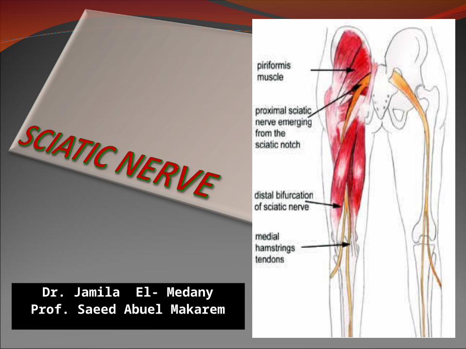
Dr. Jamila El- MedanyProf. Saeed Abuel Makarem
OBJECTIVESOBJECTIVES
By the end of the lecture, the students should be able to:
Describe the anatomy (origin, course & distribution) of the sciatic nerve.
List the branches of the sciatic nerve.Describe briefly the main motor and
sensory manifestations in case of injury of the sciatic nerve or its main branches.
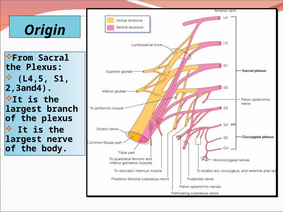
Origin
From Sacral the Plexus: (L4,5, S1, 2,3and4).It is the largest branch of the plexus It is the largest nerve of the body.
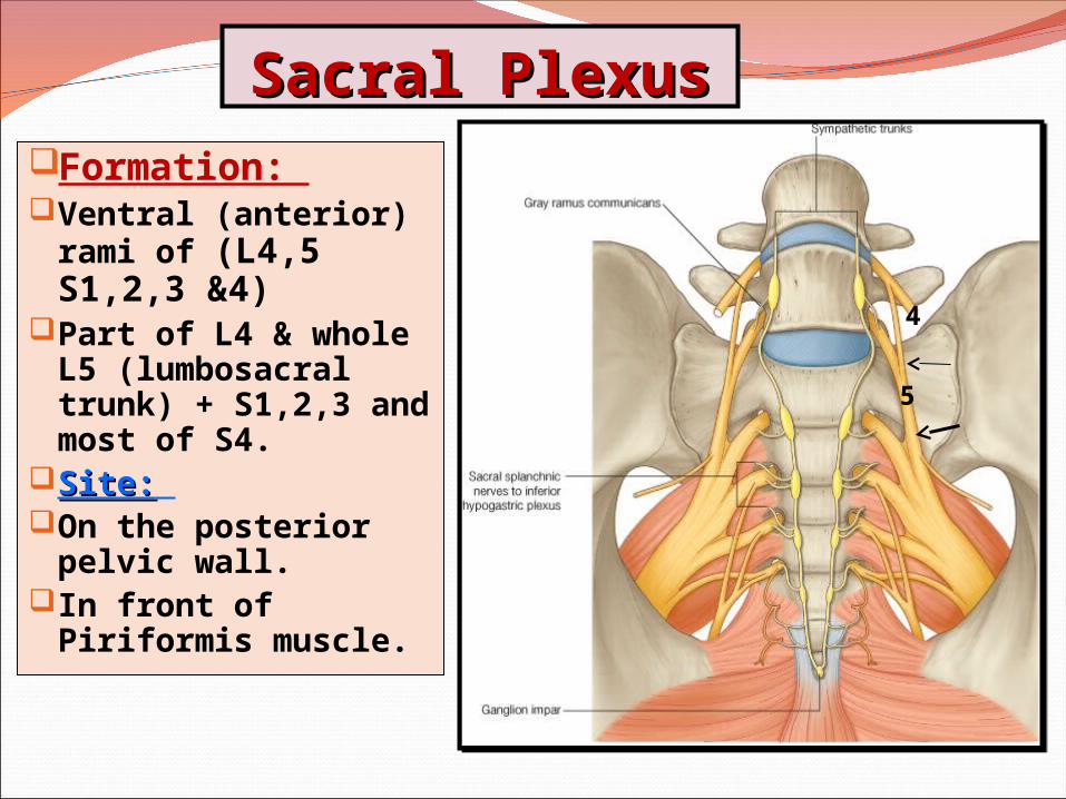
Sacral PlexusSacral PlexusFormation: Ventral (anterior)
rami of (L4,5 S1,2,3 &4)
Part of L4 & whole L5 (lumbosacral trunk) + S1,2,3 and most of S4.
Site:Site: On the posterior
pelvic wall.In front of
Piriformis muscle.
4
5
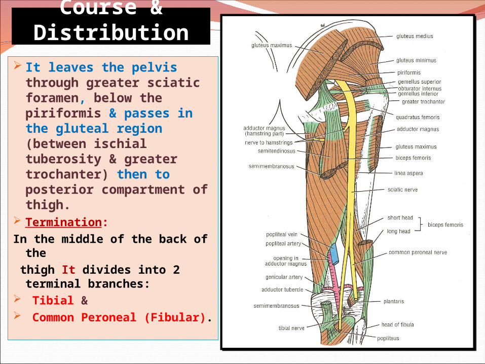
Course & Distribution It leaves the pelvis
through greater sciatic foramen, below the piriformis & passes in the gluteal region (between ischial tuberosity & greater trochanter) then to posterior compartment of thigh.
Termination: In the middle of the back of
the thigh It divides into 2
terminal branches: Tibial & Common Peroneal
(Fibular).
Branches of Sciatic NerveBranches of Sciatic Nerve
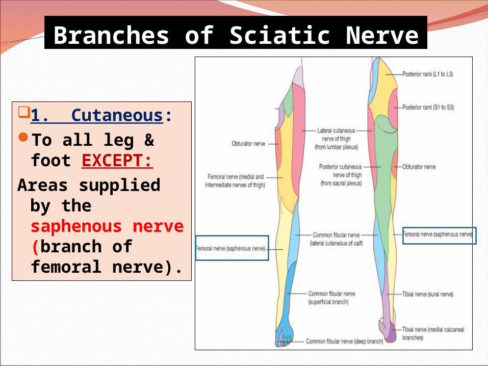
1. Cutaneous:To all leg & foot
EXCEPT: Areas supplied by
the saphenous nerve (branch of femoral nerve).
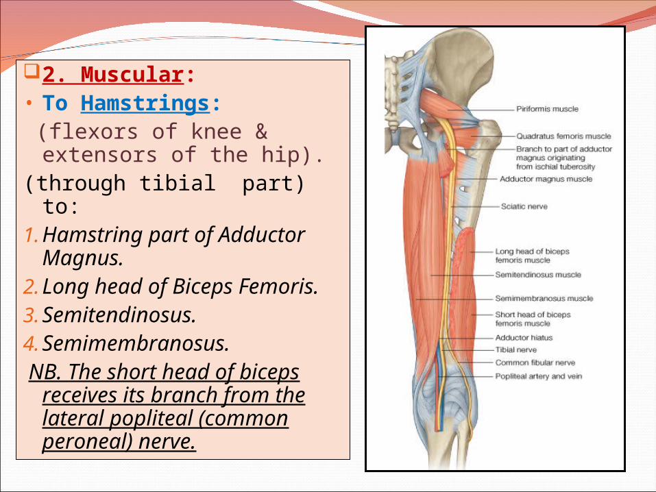
2. Muscular:• To Hamstrings: (flexors of knee & extensors
of the hip).(through tibial part) to:1.Hamstring part of
Adductor Magnus.2.Long head of Biceps
Femoris.3.Semitendinosus.4.Semimembranosus. NB. The short head of
biceps receives its branch from the lateral popliteal (common peroneal) nerve.
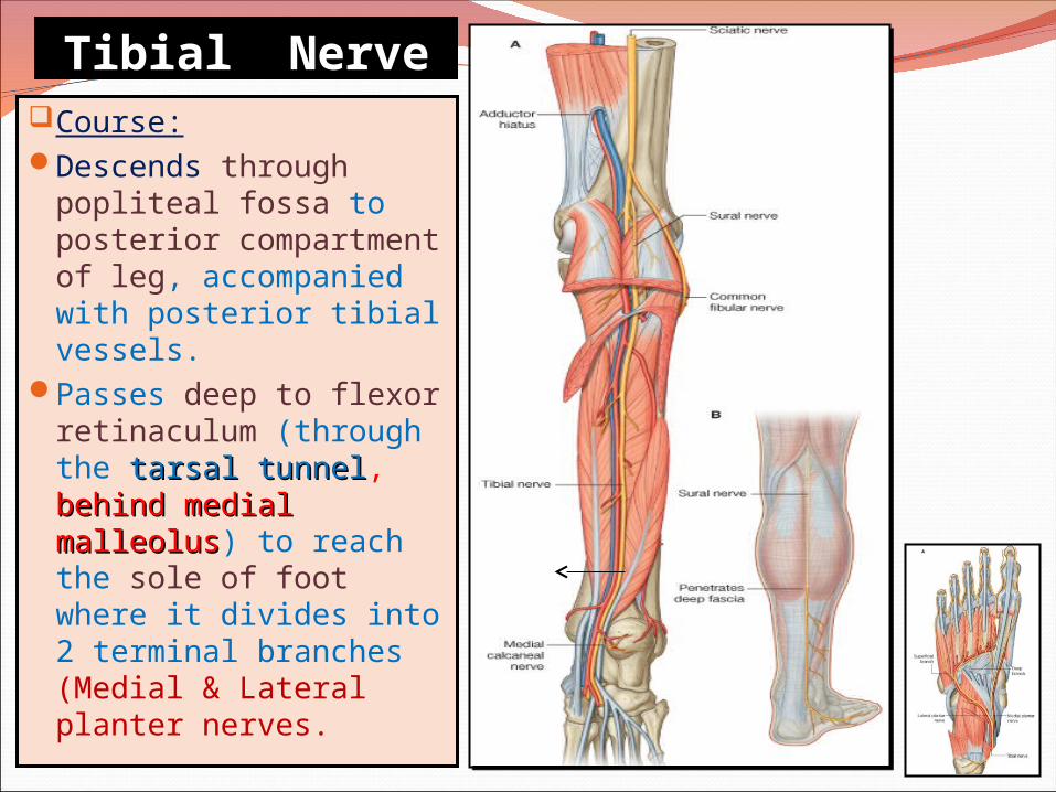
Tibial NerveTibial NerveCourse:Descends through
popliteal fossa to posterior compartment of leg, accompanied with posterior tibial vessels.
Passes deep to flexor retinaculum (through the tarsal tunneltarsal tunnel, behind medial behind medial malleolusmalleolus) to reach the sole of foot where it divides into 2 terminal branches (Medial & Lateral planter nerves.
Muscular Branches
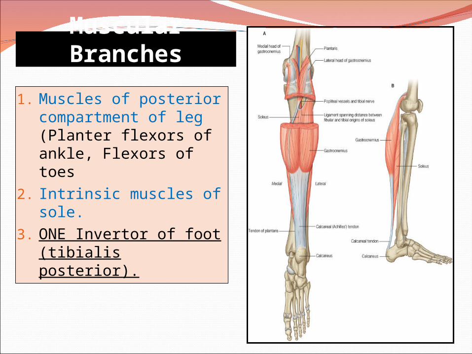
1. Muscles of posterior compartment of leg (Planter flexors of ankle, Flexors of toes
2. Intrinsic muscles of sole.
3. ONE Invertor of foot (tibialis posterior).
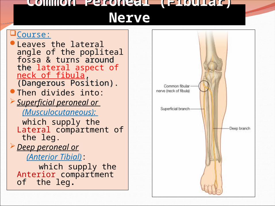
Common Peroneal (Fibular) NerveCommon Peroneal (Fibular) NerveCourse:Leaves the lateral angle
of the popliteal fossa & turns around the around the lateral aspect of neck of fibula, , (Dangerous Position).(Dangerous Position).
Then divides into:Superficial peroneal or (Musculocutaneous): which supply the
Lateral compartment of the leg.
Deep peroneal or (Anterior Tibial): which supply the
Anterior compartment of the leg.
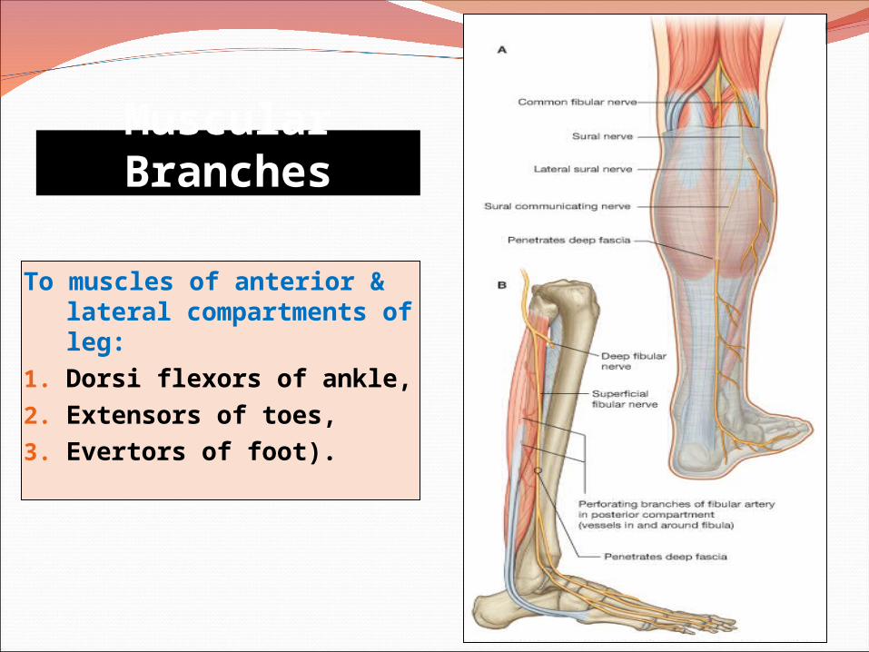
Muscular Branches
To muscles of anterior & lateral compartments of leg:
1. Dorsi flexors of ankle,2. Extensors of toes,3. Evertors of foot).
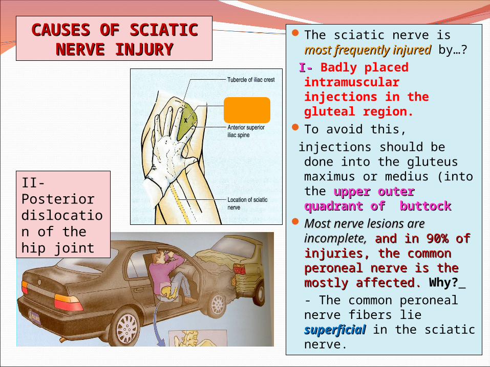
The sciatic nerve is most most frequently injuredfrequently injured by…?
I- I- Badly placed intramuscular injections in the gluteal region.
To avoid this, injections should be done
into the gluteus maximus or medius (into the upper upper outer quadrant of outer quadrant of buttock buttock
Most nerve lesions are Most nerve lesions are incomplete,incomplete, and in 90% of and in 90% of injuries, the common injuries, the common peroneal nerve is the peroneal nerve is the mostly affected. mostly affected. Why? - The common peroneal nerve fibers lie superficialsuperficial in the sciatic nerve.
CAUSES OF SCIATIC CAUSES OF SCIATIC NERVE INJURYNERVE INJURY
II-Posterior dislocation of the hip joint
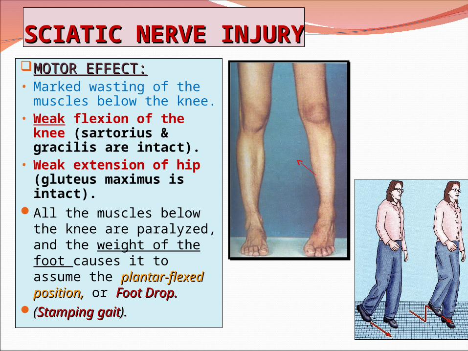
SCIATIC NERVE INJURYSCIATIC NERVE INJURYMOTOR EFFECT:MOTOR EFFECT:• Marked wasting of the
muscles below the knee.• Weak flexion of the
knee (sartorius & gracilis are intact).
• Weak extension of hip (gluteus maximus is intact).
All the muscles below the knee are paralyzed, and the weight of the foot causes it to assume the plantar-flexed position,plantar-flexed position, or Foot Drop.Foot Drop.
((Stamping gaitStamping gait).).
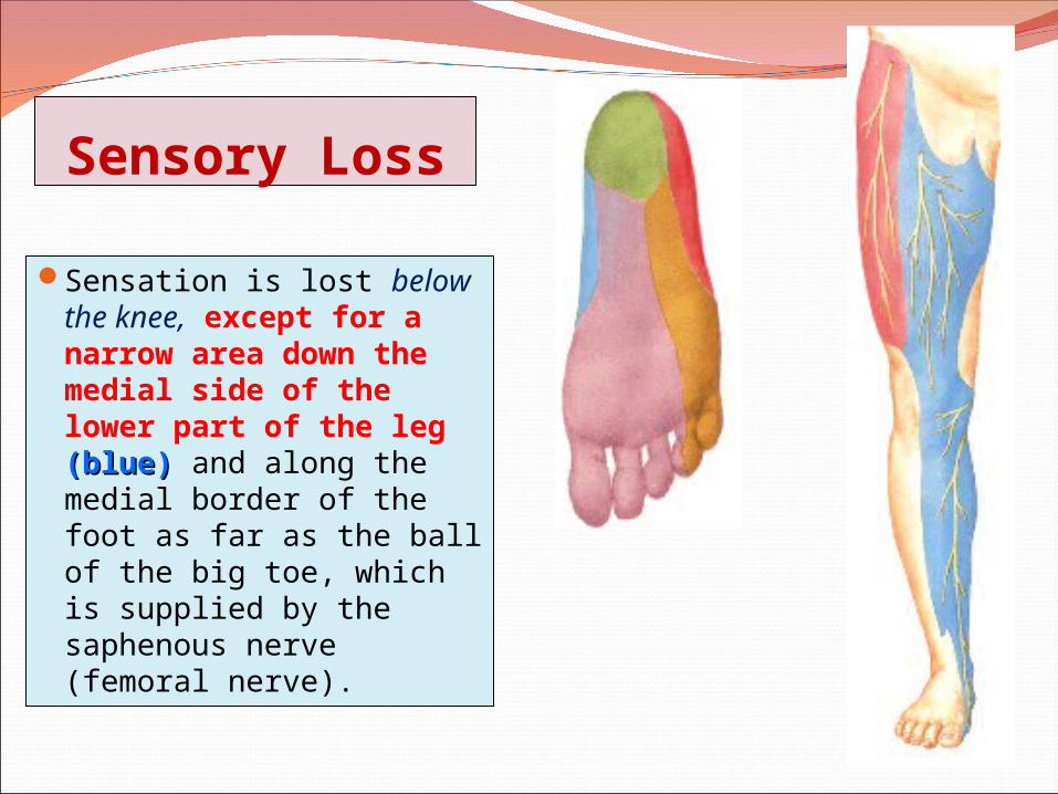
Sensory Loss
Sensation is lost below the knee, except for a narrow area down the medial side of the lower part of the leg (blue)(blue) and along the medial border of the foot as far as the ball of the big toe, which is supplied by the saphenous nerve (femoral nerve).
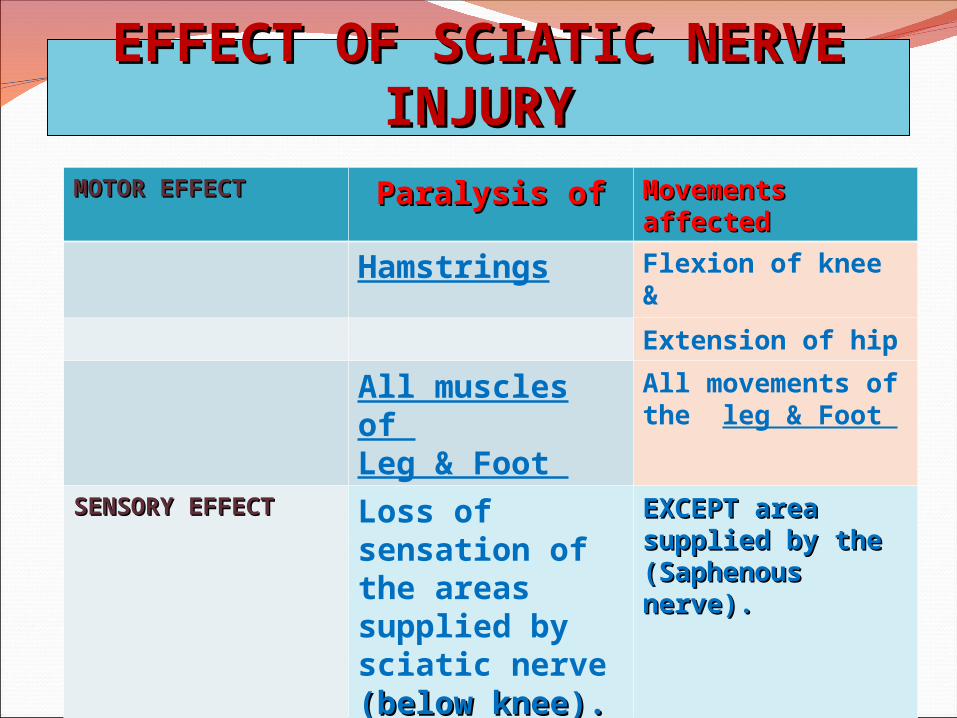
EFFECT OF SCIATIC NERVE INJURYEFFECT OF SCIATIC NERVE INJURYMOTOR EFFECTMOTOR EFFECT Paralysis ofParalysis of Movements Movements
affectedaffected
Hamstrings Flexion of knee &
Extension of hip
All muscles of Leg & Foot
All movements of the leg & Foot
SENSORY EFFECTSENSORY EFFECT Loss of sensation of the areas supplied by sciatic nerve (below knee).(below knee).
EXCEPT area EXCEPT area supplied by the supplied by the (Saphenous (Saphenous nerve).nerve).
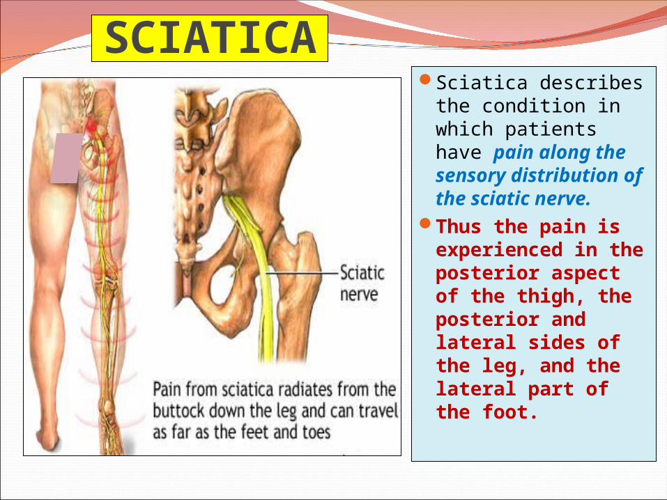
SCIATICASciatica describes
the condition in which patients have pain along the sensory distribution of the sciatic nerve.
Thus the pain is experienced in the posterior aspect of the thigh, the posterior and lateral sides of the leg, and the lateral part of the foot.
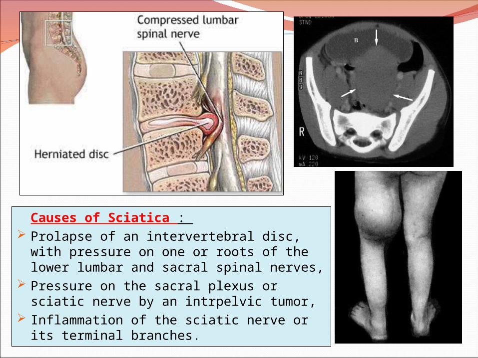
Causes of Sciatica : Prolapse of an intervertebral disc, with
pressure on one or roots of the lower lumbar and sacral spinal nerves,
Pressure on the sacral plexus or sciatic nerve by an intrpelvic tumor,
Inflammation of the sciatic nerve or its terminal branches.
Common Peroneal Nerve Injury
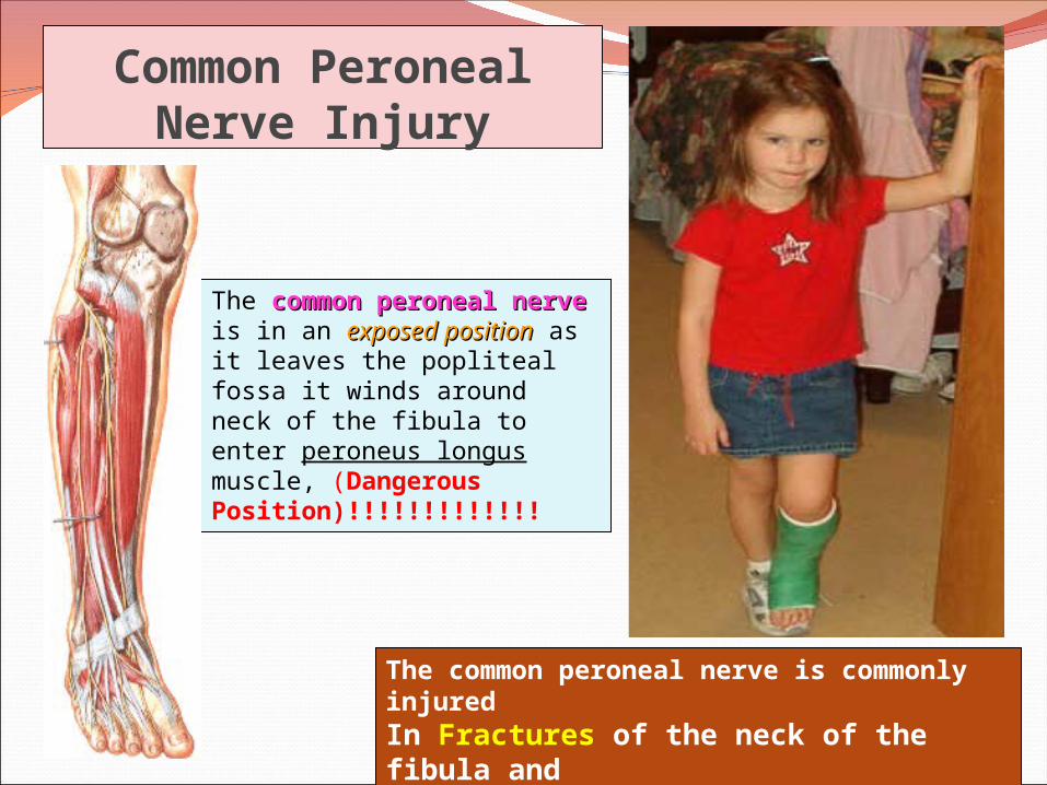
The common peroneal common peroneal nervenerve is in an exposed exposed positionposition as it leaves the popliteal fossa it winds around neck of the fibula to enter peroneus longus muscle, (Dangerous Position)!!!!!!!!!!!!!
The common peroneal nerve is commonly injuredIn Fractures of the neck of the fibula and By pressure from casts or splints.
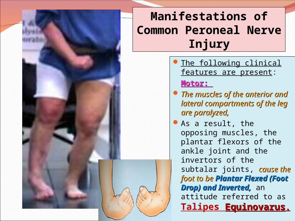
The following clinical features are present:
Motor: Motor: The muscles of the anterior The muscles of the anterior
and lateral compartments and lateral compartments of the leg are paralyzed,of the leg are paralyzed,
As a result, the opposing muscles, the plantar flexors of the ankle joint and the invertors of the subtalar joints, cause the foot to be cause the foot to be Plantar Flexed (Foot Plantar Flexed (Foot Drop) and Inverted,Drop) and Inverted, an attitude referred to as Talipes EquinovarusEquinovarus..
Manifestations of Common Peroneal Nerve
Injury
Common Peroneal Nerve Injury
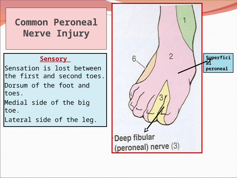
Sensory Sensation is lost between the first and second toes.
Dorsum of the foot and toes.
Medial side of the big toe.
Lateral side of the leg.
Superficial peroneal
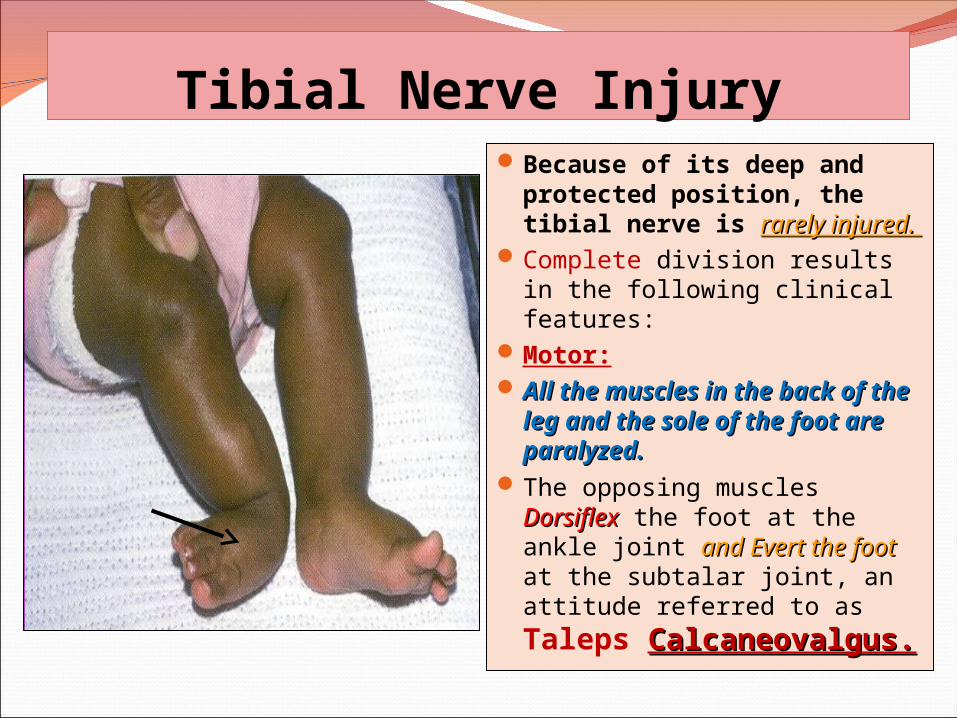
Tibial Nerve InjuryBecause of its deep and
protected position, the tibial nerve is rarely injured. rarely injured.
Complete division results in the following clinical features:
Motor: All the muscles in the back All the muscles in the back
of the leg and the sole of of the leg and the sole of the foot are paralyzed. the foot are paralyzed.
The opposing muscles DorsiflexDorsiflex the foot at the ankle joint and Evert the footand Evert the foot at the subtalar joint, an attitude referred to as Taleps Calcaneovalgus.Calcaneovalgus.
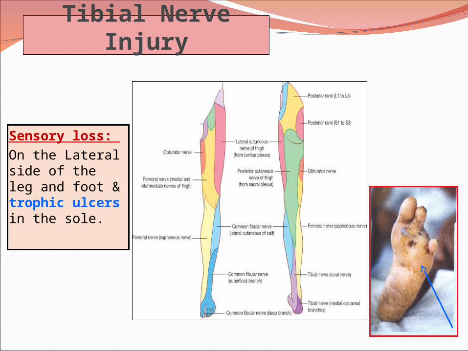
Tibial Nerve Injury
Sensory loss: On the Lateral side of the leg and foot & trophic ulcers in the sole.