Embed Size (px)
Citation preview
White Paper
Drug Target Interference in Immunogenicity Assays: Recommendationsand Mitigation Strategies
Zhandong Don Zhong,1,9 Adrienne Clements-Egan,2 Boris Gorovits,3 Mauricio Maia,4 Giane Sumner,5
Valerie Theobald,6 Yuling Wu,7 and Manoj Rajadhyaksha8
Received 23 March 2017; accepted 11 July 2017; published online 23 October, 2017
Abstract. Sensitive and specific methodology is required for the detection andcharacterization of anti-drug antibodies (ADAs). High-quality ADA data enables theevaluation of potential impact of ADAs on the drug pharmacokinetic profile, patient safety,and efficacious response to the drug. Immunogenicity assessments are typically initiated atearly stages in preclinical studies and continue throughout the drug development program.One of the potential bioanalytical challenges encountered with ADA testing is the need toidentify and mitigate the interference mediated by the presence of soluble drug target. Adrug target, when present at sufficiently high circulating concentrations, can potentiallyinterfere with the performance of ADA and neutralizing antibody (NAb) assays, leading toeither false-positive or, in some cases, false-negative ADA and NAb assay results. Thispublication describes various mechanisms of assay interference by soluble drug target, as wellas strategies to recognize and mitigate such target interference. Pertinent examples arepresented to illustrate the impact of target interference on ADA and NAb assays as well asseveral mitigation strategies, including the use of anti-target antibodies, soluble versions ofthe receptors, target-binding proteins, lectins, and solid-phase removal of targets. Further-more, recommendations for detection and mitigation of such interference in different formatsof ADA and NAb assays are provided.
KEY WORDS: anti-drug antibody; immunogenicity; mitigation; neutralizing antibody; targetinterference.
INTRODUCTION
The ADA and NAb responses have the potential toimpact the pharmacokinetic profile, patient safety, andefficacy of biotherapeutics [1–5]. Therefore, it is essentialthat bioanalytical methods be sufficiently sensitive and highlyspecific to accurately detect and characterize ADA and NAbresponses to these drugs. Numerous publications and industrywhite papers have been published detailing recommendationsfor the design, development and validation of ADA
screening, confirmatory, titration, and NAb assays to supportthe tiered approach generally used for immunogenicityassessments [6–9]. Guidance documents providing regulatoryexpectations for immunogenicity assessments have also beenpublished by global regulatory agencies [1–3].
The performance of ligand binding assays in complexmatrices, such as serum, can be impacted by specificendogenous components interfering with the assay. In ADAand NAb assays, interference may arise from the presence ofcirculating drug, an endogenous counterpart of the drug, thedrug target, or other serum factors such as rheumatoid factor[7, 10–18]. Just as high levels of drug interfere with ADAdetection, the drug target, when present at sufficiently highconcentrations in a sample, may also impact the ADA signalor readout and potentially interfere with the detection ofclinically relevant ADAs [12–18].
Biotherapeutics exert their biological activity by bindingto specific targets, which can then either stimulate or inhibitthe pathways related to those targets. Depending on thespecific biology of the disease, drug-targeted soluble ligands,shed receptors or receptors that originate from cellularbreakdown, may be found in circulation. The presence ofthese soluble drug targets can lead to erroneous results in the
1Amgen Inc., One Amgen Center Dr., Thousand Oaks, California91320, USA.
2 Janssen Research and Development LLC, Janssen Biotherapeutics,Spring House, Pennsylvania, USA.
3 BioMedicines Design, Pfizer Inc, Andover, Massachusetts, USA.4 Genentech, 1 DNA Way, South San Francisco, California, USA.5 Regeneron Pharmaceuticals Inc, Tarrytown, New York, USA.6 Shire Pharmaceuticals, Lexington, Massachusetts, USA.7MedImmune, Gaithersburg, Maryland, USA.8 Regeneron Pharmaceuticals Inc, Tarrytown, New York, USA.9 To whom correspondence should be addressed. (e-mail:[email protected])
The AAPS Journal, Vol. 19, No. 6, November 2017 (# 2017)DOI: 10.1208/s12248-017-0148-7
15641550-7416/17/0600-1564/0 # 2017 The Author(s). This article is published with open access at Springerlink.com
ADA measurement, with the amount of targeted ligand orshed receptor reported to correlate with the severity ofinterference [12–18]. Target interference can present as eitherfalse-positive or theoretically false-negative ADA results,depending on the assay format, the concentration of thedrug, the level of ADAs, and the affinity and avidity of theoverall ADA response. The physico-chemical properties of asoluble drug target and its interactions with the drug undervarious assay conditions determine the outcome of interfer-ence observed. The levels of soluble target ligand or receptormay also fluctuate among individuals and at various samplingtime points from the same individual, even within a singledisease indication, introducing an additional layer of com-plexity and variability in ADA assays. Additionally, in somesituations, the levels of circulating drug target in a subjectmay be low and non-interfering at baseline but may increasesubstantially upon treatment due to the accumulation of drug-target complexes, either by enhanced shedding of drug-engaged cell receptors, increased release of receptors fromcellular breakdown, decreased clearance of the drug-targetcomplexes, or perhaps due to feedback mechanisms inherentto the biological pathway. For example, a therapeuticmonoclonal antibody (mAb) may sequester a soluble targetfrom its normal degradation pathway, thereby extending thehalf-life of the inactive target in systemic circulation. Whensuch serum samples are tested in an ADA assay, the targetmay potentially dissociate from these complexes and achievesufficient concentrations to cause assay interference. There-fore, it is important to identify appropriate strategies andreagents to mitigate target interference to ensure an accurateimmunogenicity assessment [13, 14, 18, 19].
This manuscript provides diverse and relevant examplesencompassing target interference observed in both ADA andNAb assays. These examples provide a framework for therecommendations proposed to predict, assess and mitigatetarget interference. The recommendations include a decisiontree that can be consulted throughout the different phases ofthe drug development program, as an immunogenicity risk-assessment is implemented, and the ADA assay developmentis undertaken.
OVERVIEW OF ADA ASSAYS
Several ligand binding assay formats may be used todetect ADAs. The sandwich immunoassay, in which ADAsare captured by a drug-derived reagent and detected with aspecies-specific detector antibody, could be more tolerant tointerference from both circulating drug and drug target [17,20]. However, this format requires a species-specific positivecontrol ADA, and the detection reagent needs to becharacterized to ensure detection of all potential ADAisotypes. Additionally, this assay format is not suitable fordetection of human ADAs against a human therapeutic mAb,as the anti-human detector antibody would bind to both theADAs and the therapeutic. Due to these challenges, thebridging ligand binding assay has emerged as a commonformat for ADA detection. In a bridging immunoassay, drug-derived reagents are typically used to capture and detectADAs. For example, a biotin-labeled drug can be used as acapture reagent (in conjunction with a streptavidin or avidin-coated plate), and a ruthenium-labeled drug used as a
detection reagent in an electrochemiluminescence assayplatform (Fig. 1a). The drug-derived capture and detectionreagents are mixed with the sample. ADAs in the sample canbe detected by the formation of a molecular Bbridge^between the drug-derived capture reagent and detectionreagent. Bridging immunoassays are particularly useful asthey can detect the majority of antibody isotypes with theexception of IgG4 in the same assay. Additionally, the sameassay format can be adapted across species, and a positivecontrol can be obtained from any species without the need forseparate species-specific detection reagents.
The bridging immunoassay format, however, is susceptibleto interference primarily due to (1) excess drug that maycompete with the drug-derived reagents and reduce the signalof the assay and (2) other non-ADA molecular interactions ofserummatrix components that may potentially form a bridge andcontribute to the signal in an ADA assay. As this assay format isdesigned to detect multivalent drug binding, a false-positiveresult can be generated due to bridging formation mediated byan alternative multi-valent molecule with specific binding withthe drug, such as soluble multimeric targets (Fig. 1b). Whilemultimeric soluble drug targets have greater propensity forinterference in a bridging assay, it is also theoretically possible forfalse-negative results to occur if a soluble monomeric target ispresent in a sufficient quantity to compete out ADAs binding ator near the target-binding epitope (Fig. 1c).
Many biotherapeutics are chronically administered, andthe drug serum concentrations may be greater than drugtolerance levels of a standard bridging assay [7]. There arevarious approaches that have been reported to improve thedrug tolerance [20–27]. Although drug interference is outsidethe scope of this publication, it is worth noting that acidtreatment has been frequently exploited to dissociate ADA-drug complexes in a sample to enhance drug tolerance of theassay [24, 26]. Unfortunately, in some cases, these acidicconditions may exacerbate the drug target interference bydisrupting drug-target complexes and releasing the accumu-lated target [14]. Acid treatment has also been reported tomultimerize a monomeric target in a sample [28]. The targetcan then potentially bridge with the capture and detectionreagents upon neutralization of the sample pH, leading tofalse-positive results in the ADA screening assay.
In the tiered approach to ADA analysis, a sample that isscreened and classified as potentially positive for ADA isevaluated in a confirmatory assay. In confirmatory assayformats where excess unlabeled drug is added to the sampleto inhibit the drug-specific assay signal, the unlabeled drugcan also bind to the interfering target. This may result in theinhibition of a Bfalse positive^ signal due to multimericsoluble targets, incorrectly Bconfirming^ the Bfalse positive^signal obtained due to target interference. As this type ofdrug-specific confirmation does not verify the presence ofimmunoglobulins directed to the drug molecule, the bridgingassay format is prone to interference by the drug target. If thesoluble drug target does not contain an immunoglobulindomain, an immunodepletion procedure with protein A/G/Lcan be used to differentiate the binding of a soluble drugtarget from that of the ADAs [29]; however, this procedurerequires significant manipulation of the sample and is proneto causing additional artifacts. Therefore, it may be more
1565Drug Target Interference
efficient to assess the potential for target interference forADA assays in each biotherapeutic program and, if possible,proactively adopt an alternative mitigation strategy.
To aid in this process, the following sections describe themost commonly used strategies for identifying, overcoming orpreventing target interference in ADA assays (Table I).
Competition with Anti-target Antibody
If an alternative anti-target antibody is available tocompete with the drug for binding to target, it may be addedto the assay to Bscavenge^ the drug target during theexecution of the ADA bridging assay. It is worth noting that,with the use of anti-target antibodies, it may be necessary toassess whether or not non-neutralizing ADAs are inadver-tently removed by the anti-target antibodies. More impor-tantly, if the biotherapeutic is a mAb, the drug and thescavenging antibody should not be conformationally similar,have a similar framework, or share close sequence homology,as any of these attributes may result in cross-reactivity ofADAs to the scavenging anti-target antibody. Therefore,prior to employing an anti-target antibody, it is necessary toassess if non-neutralizing ADAs binding to the non-CDRportions of the drug are inadvertently being masked by theanti-target antibody.
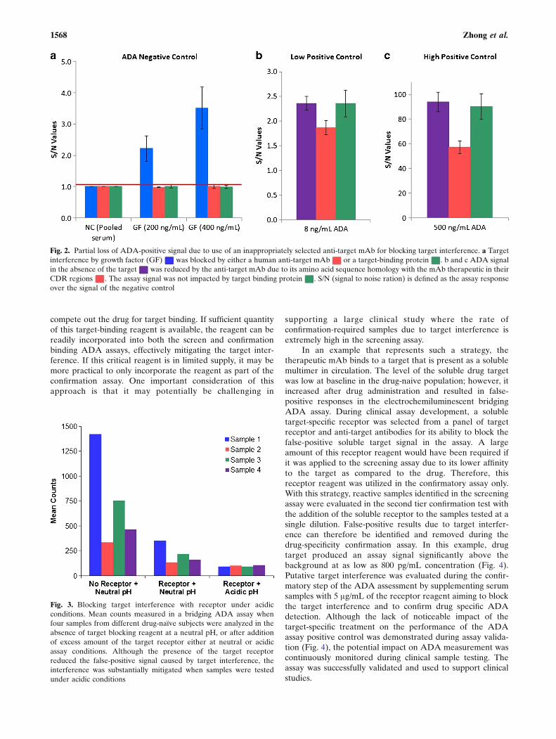
A recently published drug target interference mitigationstrategy highlights the importance of selecting competinganti-target antibodies with the appropriate framework [30]. Inthis example, a bridging assay was developed to detectbinding ADAs against a fully human mAb therapeutic, whichachieved its biological activity by binding to a growth factorand thus inhibiting its interaction with the receptor. As highlevels of the therapeutic were anticipated in circulationamong treated subjects, acid dissociation was employed toincrease the drug tolerance of the ADA bridging method. Inaddition, it was also observed that the levels of the target,known to exist in various multimeric forms, increased in
circulation in subjects treated with another biotherapeuticwith a similar mechanism of action, resulting in false-positiveADA results in the bridging assay [18]. A human anti-targetmAb that competed with the drug for binding to the targetwas readily available and tested as a possible reagent toeliminate or mitigate the target interference in the assay.Although this human anti-target mAb effectively blocked theinterference of the drug target, the assay signal of true ADAswas also reduced, due to partial overlapping of the CDRsbetween the human anti-target mAb and the therapeuticmAb (Fig. 2). Thus, it is critical to ensure that competing anti-target mAbs do not have overlapping sequences that maycompete with the drug for ADA binding. Additional humanand mouse anti-target mAbs, as well as other target-bindingproteins were also evaluated for their ability to block theinterference in the presence and absence of the ADA positivecontrol in this study. In the final validated assay design, arecombinant fusion protein was utilized in the neutralizationstep to inhibit the target interference without affectingdetection of ADAs.
Competition with Soluble Receptors for Interfering Ligand
For biotherapeutics that neutralize ligand-receptor inter-actions by binding to the ligand (the drug target), the solubleversions of the receptor can be utilized to effectively mitigatetarget interference, since as the natural binding partner of theligand, the receptor should have high affinity towards thetargeted ligand. As such, the conditions under which thereceptor-derived reagents out-compete the drug for targetbinding are often achievable. In an example where the targetof the mAb therapeutic is a multimeric ligand at variableconcentrations in drug-naïve samples, the disparity of thetarget concentration contributed to high variability in theassay response observed in pre-dose samples. Additionally,after drug administration, the level of the target increased dueto accumulation of target-drug complexes in circulation. The
Fig. 1. Interference of drug target in a bridging ADA assay. (a) A Representative example of a truepositive signal in the ADA bridging assay resulting from bivalent binding of the ADA to two labeled-drugmolecules. (b) A false-positive assay signal arising from multimeric target bridging two labeled-drugmolecules. (c) The presence of a molar excess of drug target in a sample, relative to drug, results in targetoccupying both drug binding sites thus preventing binding of any ADAwhose specify is to the drug’s target-binding region, resulting in a false-negative result
1566 Zhong et al.
target was released under the optimized assay conditions,resulting in high assay signals and variability (Fig. 3). A highreported rate of false-positive measurements was thus ob-served in the ADA assay. No competing anti-target antibod-ies could be identified to minimize this false-positive targetsignal. The use of the target receptor alone, at highconcentrations, reduced the target signal in a dose-dependent manner; however, the receptor alone did notcompletely abrogate the false-positive signal. With carefulconsideration of the biochemical properties of the targetedligand, a mildly acidic pH was implemented, in conjunctionwith the receptor at a lower concentration, to further reducethe false-positive signal contributed by the target. With thismethod the variability observed in both pre- and post-dosesamples was effectively managed. Further testing of ADA-positive samples from preclinical studies confirmed that themild acidic conditions had minimal impact on detection oftrue ADAs.
There can be some limitations in using soluble receptorto minimize drug target interference, however, due to thebiophysical and biochemical properties of the soluble versionof the receptor. First, sometimes the receptor is a cell surfacemembrane-bound receptor, and an adequate recombinantsoluble extracellular domain may not be readily produced in
sufficient quantities to support the bioanalytical work. Sec-ondly, the recombinant version of the target receptor may beconformationally different from its native counterpart. Solu-ble receptor can also form high molecular weight multimericcomplexes that could potentially lead to false-positive ADAsignals. Furthermore, depending on the source of production,the costs of purifying the target receptor in requiredquantities may be prohibitive. Finally, the stability of therecombinant version of the receptor may not be suitable forefficient and long-term use as a critical assay reagent.Nonetheless, if a receptor is available in soluble form withoutthese limitations, it can certainly serve as a good alternativeto anti-target antibodies to mitigate target interference.
Use of Target-Binding Reagent with Lower Affinity in theConfirmatory Assay
It is desired for a competing specific target-bindingreagent to have higher affinity to the target than the drug’saffinity to the target; however, such high affinity reagents maynot be always readily available. Thus, high concentrations ofthe target-binding reagent may be added to the samplesolution, within the assay constraints, regardless of its bindingaffinity to the target, to ensure that the reagent can effectively
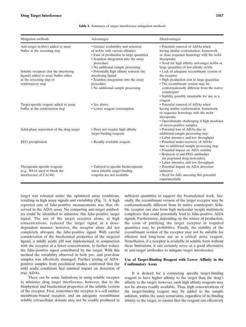
Table I. Summary of target interference mitigation methods
Mitigation methods Advantages Disadvantages
Anti-target mAb(s) added to assaybuffer at the screening step
▪ Greater availability and selectionof mAbs with various affinities▪ Ease of production in large quantities▪ Seamless integration into the assayprocedure
▪ No additional sample processing
▪ Potential removal of ADAs whenhaving similar conformation, frameworkor close sequence homology with the mAbtherapeutic▪ Need for high affinity anti-target mAbs orlarge quantities of low-affinity mAbs
Soluble receptors (for the interferingligand) added to assay buffer eitherat the screening step orconfirmatory step
▪ Potentially high affinity towards theinterfering ligand▪ Seamless integration into the assayprocedure▪ No additional sample processing
▪ Lack of adequate recombinant version ofthe receptor▪ High production cost in large quantities▪ The recombinant version may beconformationally different from the nativecounterpart
▪ Stability possibly unsuitable for use as areagent
Target-specific reagent added to assaybuffer at the confirmation step
▪ See above▪ Lower reagent consumption
▪ Potential removal of ADAs whenhaving similar conformation, framework,or sequence homology with the mAbtherapeutic▪ Operationally challenging if high incidenceof screen-positive samples.
Solid-phase separation of the drug target ▪ Does not require high affinitytarget-binding reagents
▪ Potential loss of ADAs due toadditional sample processing step▪ Labor intensive and low throughput
PEG precipitation ▪ Readily available reagent ▪ Potential under-recovery of ADAsdue to additional sample processing step▪ Potential impact on ADA stability▪ Removal of anti-PEG antibodies(in pegylated drug molecules)
▪ Labor intensive and low throughputTherapeutic-specific reagents(e.g., WGA used to block theinterference of CA148)
▪ Tailored to specific biotherapeuticwhen suitable target-bindingreagents are not available
▪ Potential impact on ADA detectionunknown▪ Need for fully assessing this potentialexperimentally
1567Drug Target Interference
compete out the drug for target binding. If sufficient quantityof this target-binding reagent is available, the reagent can bereadily incorporated into both the screen and confirmationbinding ADA assays, effectively mitigating the target inter-ference. If this critical reagent is in limited supply, it may bemore practical to only incorporate the reagent as part of theconfirmation assay. One important consideration of thisapproach is that it may potentially be challenging in
supporting a large clinical study where the rate ofconfirmation-required samples due to target interference isextremely high in the screening assay.
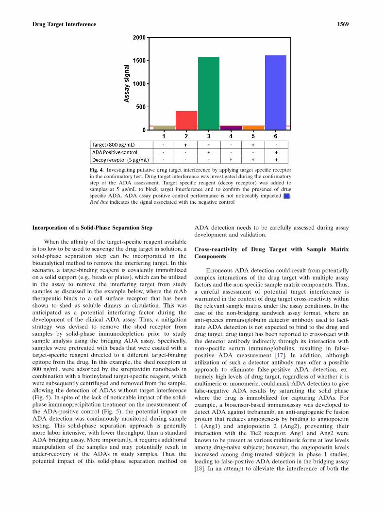
In an example that represents such a strategy, thetherapeutic mAb binds to a target that is present as a solublemultimer in circulation. The level of the soluble drug targetwas low at baseline in the drug-naive population; however, itincreased after drug administration and resulted in false-positive responses in the electrochemiluminescent bridgingADA assay. During clinical assay development, a solubletarget-specific receptor was selected from a panel of targetreceptor and anti-target antibodies for its ability to block thefalse-positive soluble target signal in the assay. A largeamount of this receptor reagent would have been required ifit was applied to the screening assay due to its lower affinityto the target as compared to the drug. Therefore, thisreceptor reagent was utilized in the confirmatory assay only.With this strategy, reactive samples identified in the screeningassay were evaluated in the second tier confirmation test withthe addition of the soluble receptor to the samples tested at asingle dilution. False-positive results due to target interfer-ence can therefore be identified and removed during thedrug-specificity confirmation assay. In this example, drugtarget produced an assay signal significantly above thebackground at as low as 800 pg/mL concentration (Fig. 4).Putative target interference was evaluated during the confir-matory step of the ADA assessment by supplementing serumsamples with 5 μg/mL of the receptor reagent aiming to blockthe target interference and to confirm drug specific ADAdetection. Although the lack of noticeable impact of thetarget-specific treatment on the performance of the ADAassay positive control was demonstrated during assay valida-tion (Fig. 4), the potential impact on ADA measurement wascontinuously monitored during clinical sample testing. Theassay was successfully validated and used to support clinicalstudies.
Fig. 2. Partial loss of ADA-positive signal due to use of an inappropriately selected anti-target mAb for blocking target interference. a Targetinterference by growth factor (GF) was blocked by either a human anti-target mAb or a target-binding protein . b and c ADA signalin the absence of the target was reduced by the anti-target mAb due to its amino acid sequence homology with the mAb therapeutic in theirCDR regions . The assay signal was not impacted by target binding protein . S/N (signal to noise ration) is defined as the assay responseover the signal of the negative control
Fig. 3. Blocking target interference with receptor under acidicconditions. Mean counts measured in a bridging ADA assay whenfour samples from different drug-naïve subjects were analyzed in theabsence of target blocking reagent at a neutral pH, or after additionof excess amount of the target receptor either at neutral or acidicassay conditions. Although the presence of the target receptorreduced the false-positive signal caused by target interference, theinterference was substantially mitigated when samples were testedunder acidic conditions
1568 Zhong et al.
Incorporation of a Solid-Phase Separation Step
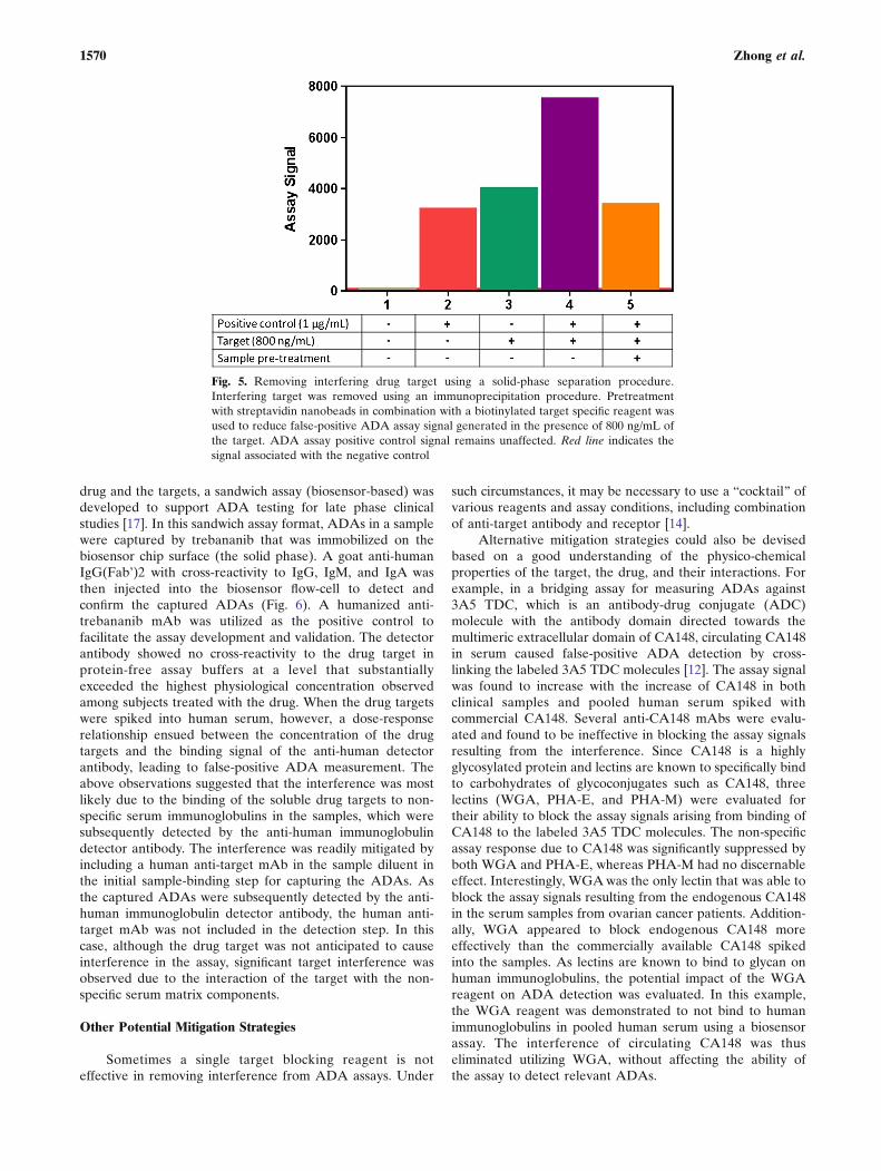
When the affinity of the target-specific reagent availableis too low to be used to scavenge the drug target in solution, asolid-phase separation step can be incorporated in thebioanalytical method to remove the interfering target. In thisscenario, a target-binding reagent is covalently immobilizedon a solid support (e.g., beads or plates), which can be utilizedin the assay to remove the interfering target from studysamples as discussed in the example below, where the mAbtherapeutic binds to a cell surface receptor that has beenshown to shed as soluble dimers in circulation. This wasanticipated as a potential interfering factor during thedevelopment of the clinical ADA assay. Thus, a mitigationstrategy was devised to remove the shed receptor fromsamples by solid-phase immunodepletion prior to studysample analysis using the bridging ADA assay. Specifically,samples were pretreated with beads that were coated with atarget-specific reagent directed to a different target-bindingepitope from the drug. In this example, the shed receptors at800 ng/mL were adsorbed by the streptavidin nanobeads incombination with a biotinylated target-specific reagent, whichwere subsequently centrifuged and removed from the sample,allowing the detection of ADAs without target interference(Fig. 5). In spite of the lack of noticeable impact of the solid-phase immunoprecipitation treatment on the measurement ofthe ADA-positive control (Fig. 5), the potential impact onADA detection was continuously monitored during sampletesting. This solid-phase separation approach is generallymore labor intensive, with lower throughput than a standardADA bridging assay. More importantly, it requires additionalmanipulation of the samples and may potentially result inunder-recovery of the ADAs in study samples. Thus, thepotential impact of this solid-phase separation method on
ADA detection needs to be carefully assessed during assaydevelopment and validation.
Cross-reactivity of Drug Target with Sample MatrixComponents
Erroneous ADA detection could result from potentiallycomplex interactions of the drug target with multiple assayfactors and the non-specific sample matrix components. Thus,a careful assessment of potential target interference iswarranted in the context of drug target cross-reactivity withinthe relevant sample matrix under the assay conditions. In thecase of the non-bridging sandwich assay format, where ananti-species immunoglobulin detector antibody used to facil-itate ADA detection is not expected to bind to the drug anddrug target, drug target has been reported to cross-react withthe detector antibody indirectly through its interaction withnon-specific serum immunoglobulins, resulting in false-positive ADA measurement [17]. In addition, althoughutilization of such a detector antibody may offer a possibleapproach to eliminate false-positive ADA detection, ex-tremely high levels of drug target, regardless of whether it ismultimeric or monomeric, could mask ADA detection to givefalse-negative ADA results by saturating the solid phasewhere the drug is immobilized for capturing ADAs. Forexample, a biosensor-based immunoassay was developed todetect ADA against trebananib, an anti-angiogenic Fc fusionprotein that reduces angiogenesis by binding to angiopoietin1 (Ang1) and angiopoietin 2 (Ang2), preventing theirinteraction with the Tie2 receptor. Ang1 and Ang2 wereknown to be present as various multimeric forms at low levelsamong drug-naive subjects; however, the angiopoietin levelsincreased among drug-treated subjects in phase 1 studies,leading to false-positive ADA detection in the bridging assay[18]. In an attempt to alleviate the interference of both the
Fig. 4. Investigating putative drug target interference by applying target specific receptorin the confirmatory test. Drug target interference was investigated during the confirmatorystep of the ADA assessment. Target specific reagent (decoy receptor) was added tosamples at 5 μg/mL to block target interference and to confirm the presence of drugspecific ADA. ADA assay positive control performance is not noticeably impacted .Red line indicates the signal associated with the negative control
1569Drug Target Interference
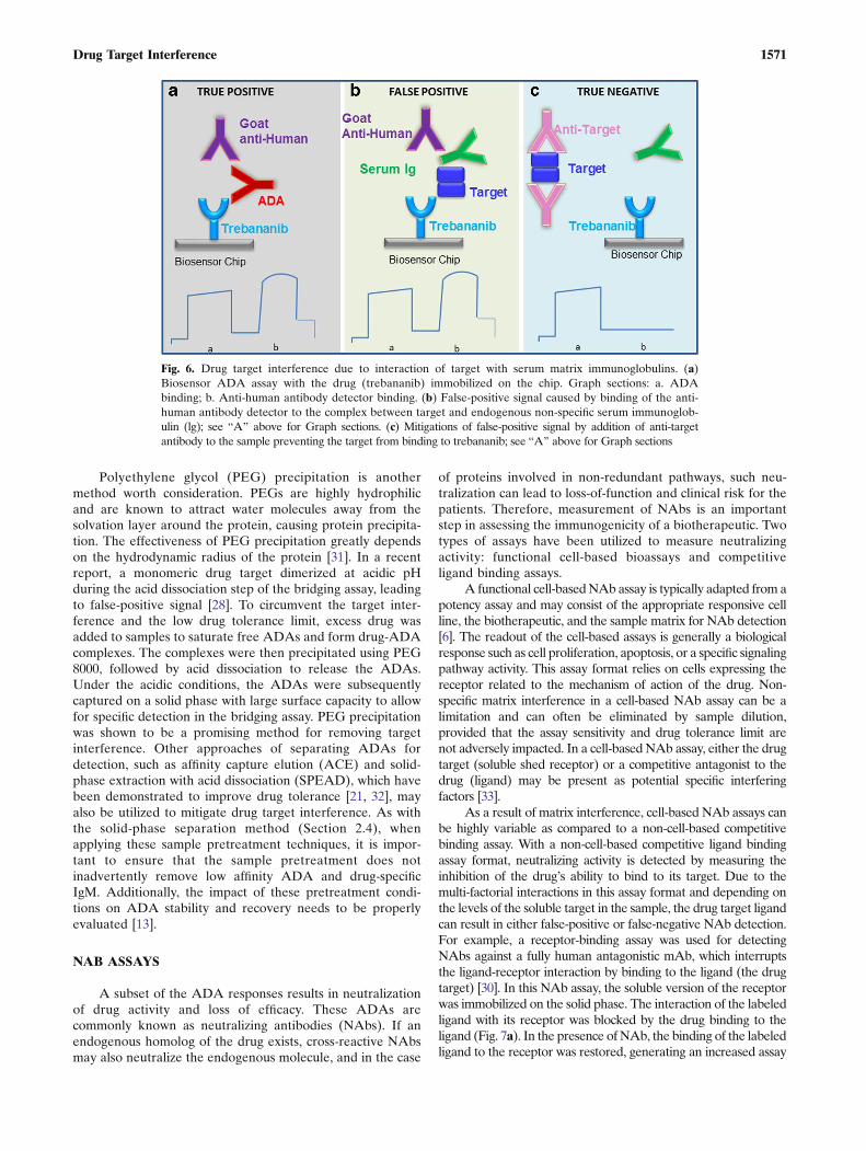
drug and the targets, a sandwich assay (biosensor-based) wasdeveloped to support ADA testing for late phase clinicalstudies [17]. In this sandwich assay format, ADAs in a samplewere captured by trebananib that was immobilized on thebiosensor chip surface (the solid phase). A goat anti-humanIgG(Fab’)2 with cross-reactivity to IgG, IgM, and IgA wasthen injected into the biosensor flow-cell to detect andconfirm the captured ADAs (Fig. 6). A humanized anti-trebananib mAb was utilized as the positive control tofacilitate the assay development and validation. The detectorantibody showed no cross-reactivity to the drug target inprotein-free assay buffers at a level that substantiallyexceeded the highest physiological concentration observedamong subjects treated with the drug. When the drug targetswere spiked into human serum, however, a dose-responserelationship ensued between the concentration of the drugtargets and the binding signal of the anti-human detectorantibody, leading to false-positive ADA measurement. Theabove observations suggested that the interference was mostlikely due to the binding of the soluble drug targets to non-specific serum immunoglobulins in the samples, which weresubsequently detected by the anti-human immunoglobulindetector antibody. The interference was readily mitigated byincluding a human anti-target mAb in the sample diluent inthe initial sample-binding step for capturing the ADAs. Asthe captured ADAs were subsequently detected by the anti-human immunoglobulin detector antibody, the human anti-target mAb was not included in the detection step. In thiscase, although the drug target was not anticipated to causeinterference in the assay, significant target interference wasobserved due to the interaction of the target with the non-specific serum matrix components.
Other Potential Mitigation Strategies
Sometimes a single target blocking reagent is noteffective in removing interference from ADA assays. Under
such circumstances, it may be necessary to use a Bcocktail^ ofvarious reagents and assay conditions, including combinationof anti-target antibody and receptor [14].
Alternative mitigation strategies could also be devisedbased on a good understanding of the physico-chemicalproperties of the target, the drug, and their interactions. Forexample, in a bridging assay for measuring ADAs against3A5 TDC, which is an antibody-drug conjugate (ADC)molecule with the antibody domain directed towards themultimeric extracellular domain of CA148, circulating CA148in serum caused false-positive ADA detection by cross-linking the labeled 3A5 TDC molecules [12]. The assay signalwas found to increase with the increase of CA148 in bothclinical samples and pooled human serum spiked withcommercial CA148. Several anti-CA148 mAbs were evalu-ated and found to be ineffective in blocking the assay signalsresulting from the interference. Since CA148 is a highlyglycosylated protein and lectins are known to specifically bindto carbohydrates of glycoconjugates such as CA148, threelectins (WGA, PHA-E, and PHA-M) were evaluated fortheir ability to block the assay signals arising from binding ofCA148 to the labeled 3A5 TDC molecules. The non-specificassay response due to CA148 was significantly suppressed byboth WGA and PHA-E, whereas PHA-M had no discernableeffect. Interestingly, WGAwas the only lectin that was able toblock the assay signals resulting from the endogenous CA148in the serum samples from ovarian cancer patients. Addition-ally, WGA appeared to block endogenous CA148 moreeffectively than the commercially available CA148 spikedinto the samples. As lectins are known to bind to glycan onhuman immunoglobulins, the potential impact of the WGAreagent on ADA detection was evaluated. In this example,the WGA reagent was demonstrated to not bind to humanimmunoglobulins in pooled human serum using a biosensorassay. The interference of circulating CA148 was thuseliminated utilizing WGA, without affecting the ability ofthe assay to detect relevant ADAs.
Fig. 5. Removing interfering drug target using a solid-phase separation procedure.Interfering target was removed using an immunoprecipitation procedure. Pretreatmentwith streptavidin nanobeads in combination with a biotinylated target specific reagent wasused to reduce false-positive ADA assay signal generated in the presence of 800 ng/mL ofthe target. ADA assay positive control signal remains unaffected. Red line indicates thesignal associated with the negative control
1570 Zhong et al.
Polyethylene glycol (PEG) precipitation is anothermethod worth consideration. PEGs are highly hydrophilicand are known to attract water molecules away from thesolvation layer around the protein, causing protein precipita-tion. The effectiveness of PEG precipitation greatly dependson the hydrodynamic radius of the protein [31]. In a recentreport, a monomeric drug target dimerized at acidic pHduring the acid dissociation step of the bridging assay, leadingto false-positive signal [28]. To circumvent the target inter-ference and the low drug tolerance limit, excess drug wasadded to samples to saturate free ADAs and form drug-ADAcomplexes. The complexes were then precipitated using PEG8000, followed by acid dissociation to release the ADAs.Under the acidic conditions, the ADAs were subsequentlycaptured on a solid phase with large surface capacity to allowfor specific detection in the bridging assay. PEG precipitationwas shown to be a promising method for removing targetinterference. Other approaches of separating ADAs fordetection, such as affinity capture elution (ACE) and solid-phase extraction with acid dissociation (SPEAD), which havebeen demonstrated to improve drug tolerance [21, 32], mayalso be utilized to mitigate drug target interference. As withthe solid-phase separation method (Section 2.4), whenapplying these sample pretreatment techniques, it is impor-tant to ensure that the sample pretreatment does notinadvertently remove low affinity ADA and drug-specificIgM. Additionally, the impact of these pretreatment condi-tions on ADA stability and recovery needs to be properlyevaluated [13].
NAB ASSAYS
A subset of the ADA responses results in neutralizationof drug activity and loss of efficacy. These ADAs arecommonly known as neutralizing antibodies (NAbs). If anendogenous homolog of the drug exists, cross-reactive NAbsmay also neutralize the endogenous molecule, and in the case
of proteins involved in non-redundant pathways, such neu-tralization can lead to loss-of-function and clinical risk for thepatients. Therefore, measurement of NAbs is an importantstep in assessing the immunogenicity of a biotherapeutic. Twotypes of assays have been utilized to measure neutralizingactivity: functional cell-based bioassays and competitiveligand binding assays.
A functional cell-basedNAb assay is typically adapted from apotency assay and may consist of the appropriate responsive cellline, the biotherapeutic, and the sample matrix for NAb detection[6]. The readout of the cell-based assays is generally a biologicalresponse such as cell proliferation, apoptosis, or a specific signalingpathway activity. This assay format relies on cells expressing thereceptor related to the mechanism of action of the drug. Non-specific matrix interference in a cell-based NAb assay can be alimitation and can often be eliminated by sample dilution,provided that the assay sensitivity and drug tolerance limit arenot adversely impacted. In a cell-based NAb assay, either the drugtarget (soluble shed receptor) or a competitive antagonist to thedrug (ligand) may be present as potential specific interferingfactors [33].
As a result of matrix interference, cell-based NAb assays canbe highly variable as compared to a non-cell-based competitivebinding assay. With a non-cell-based competitive ligand bindingassay format, neutralizing activity is detected by measuring theinhibition of the drug’s ability to bind to its target. Due to themulti-factorial interactions in this assay format and depending onthe levels of the soluble target in the sample, the drug target ligandcan result in either false-positive or false-negative NAb detection.For example, a receptor-binding assay was used for detectingNAbs against a fully human antagonistic mAb, which interruptsthe ligand-receptor interaction by binding to the ligand (the drugtarget) [30]. In this NAb assay, the soluble version of the receptorwas immobilized on the solid phase. The interaction of the labeledligand with its receptor was blocked by the drug binding to theligand (Fig. 7a). In the presence ofNAb, the binding of the labeledligand to the receptor was restored, generating an increased assay
Fig. 6. Drug target interference due to interaction of target with serum matrix immunoglobulins. (a)Biosensor ADA assay with the drug (trebananib) immobilized on the chip. Graph sections: a. ADAbinding; b. Anti-human antibody detector binding. (b) False-positive signal caused by binding of the anti-human antibody detector to the complex between target and endogenous non-specific serum immunoglob-ulin (lg); see BA^ above for Graph sections. (c) Mitigations of false-positive signal by addition of anti-targetantibody to the sample preventing the target from binding to trebananib; see BA^ above for Graph sections
1571Drug Target Interference
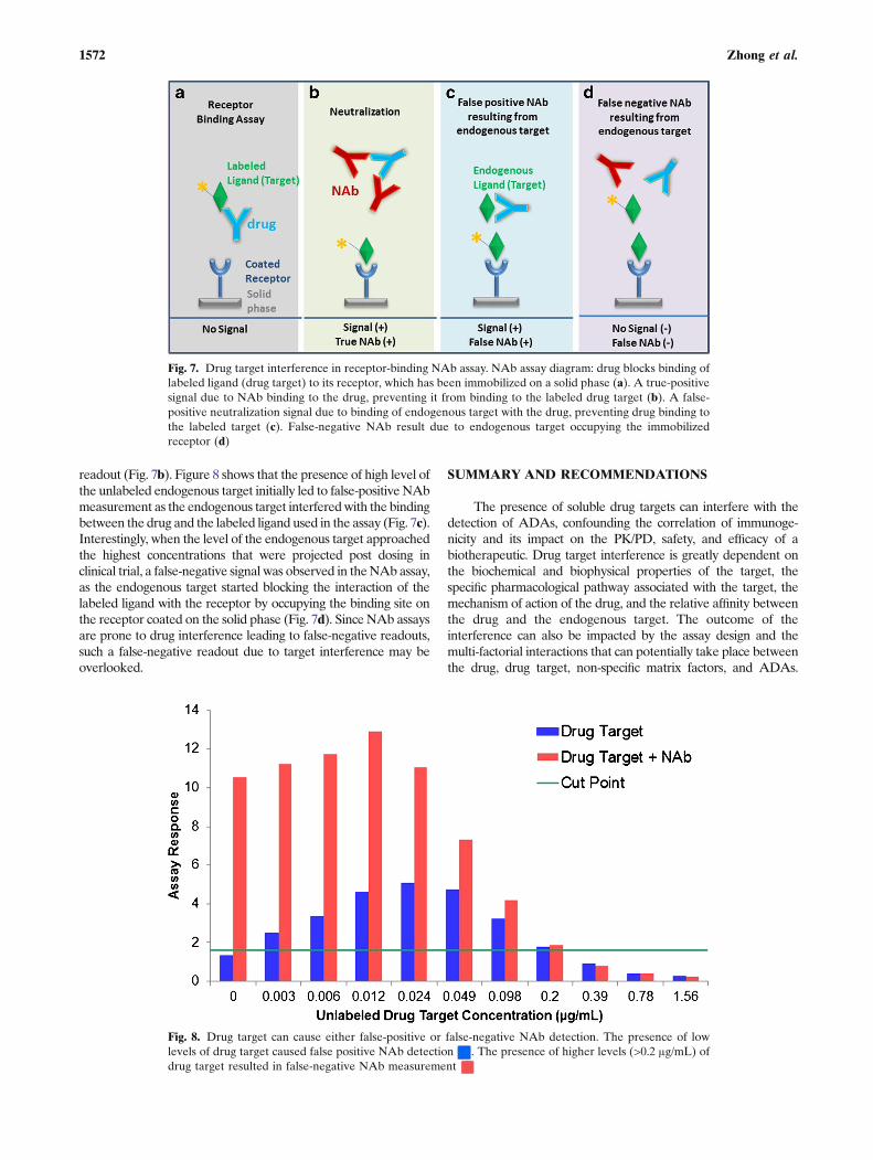
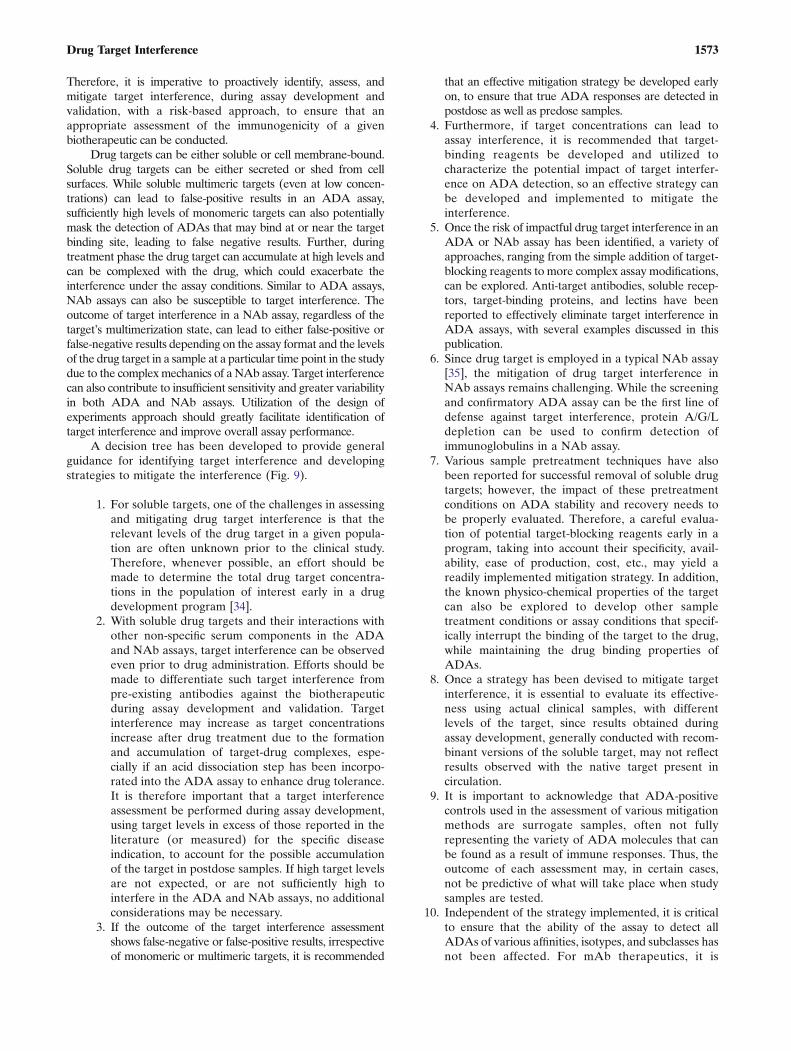
readout (Fig. 7b). Figure 8 shows that the presence of high level ofthe unlabeled endogenous target initially led to false-positive NAbmeasurement as the endogenous target interferedwith the bindingbetween the drug and the labeled ligand used in the assay (Fig. 7c).Interestingly, when the level of the endogenous target approachedthe highest concentrations that were projected post dosing inclinical trial, a false-negative signal was observed in theNAb assay,as the endogenous target started blocking the interaction of thelabeled ligand with the receptor by occupying the binding site onthe receptor coated on the solid phase (Fig. 7d). Since NAb assaysare prone to drug interference leading to false-negative readouts,such a false-negative readout due to target interference may beoverlooked.
SUMMARY AND RECOMMENDATIONS
The presence of soluble drug targets can interfere with thedetection of ADAs, confounding the correlation of immunoge-nicity and its impact on the PK/PD, safety, and efficacy of abiotherapeutic. Drug target interference is greatly dependent onthe biochemical and biophysical properties of the target, thespecific pharmacological pathway associated with the target, themechanism of action of the drug, and the relative affinity betweenthe drug and the endogenous target. The outcome of theinterference can also be impacted by the assay design and themulti-factorial interactions that can potentially take place betweenthe drug, drug target, non-specific matrix factors, and ADAs.
Fig. 7. Drug target interference in receptor-binding NAb assay. NAb assay diagram: drug blocks binding oflabeled ligand (drug target) to its receptor, which has been immobilized on a solid phase (a). A true-positivesignal due to NAb binding to the drug, preventing it from binding to the labeled drug target (b). A false-positive neutralization signal due to binding of endogenous target with the drug, preventing drug binding tothe labeled target (c). False-negative NAb result due to endogenous target occupying the immobilizedreceptor (d)
Fig. 8. Drug target can cause either false-positive or false-negative NAb detection. The presence of lowlevels of drug target caused false positive NAb detection . The presence of higher levels (>0.2 μg/mL) ofdrug target resulted in false-negative NAb measurement
1572 Zhong et al.
Therefore, it is imperative to proactively identify, assess, andmitigate target interference, during assay development andvalidation, with a risk-based approach, to ensure that anappropriate assessment of the immunogenicity of a givenbiotherapeutic can be conducted.
Drug targets can be either soluble or cell membrane-bound.Soluble drug targets can be either secreted or shed from cellsurfaces. While soluble multimeric targets (even at low concen-trations) can lead to false-positive results in an ADA assay,sufficiently high levels of monomeric targets can also potentiallymask the detection of ADAs that may bind at or near the targetbinding site, leading to false negative results. Further, duringtreatment phase the drug target can accumulate at high levels andcan be complexed with the drug, which could exacerbate theinterference under the assay conditions. Similar to ADA assays,NAb assays can also be susceptible to target interference. Theoutcome of target interference in a NAb assay, regardless of thetarget’s multimerization state, can lead to either false-positive orfalse-negative results depending on the assay format and the levelsof the drug target in a sample at a particular time point in the studydue to the complexmechanics of a NAb assay. Target interferencecan also contribute to insufficient sensitivity and greater variabilityin both ADA and NAb assays. Utilization of the design ofexperiments approach should greatly facilitate identification oftarget interference and improve overall assay performance.
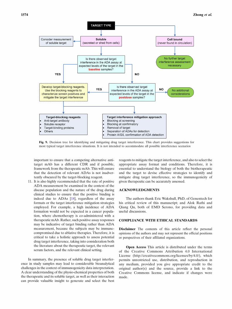
A decision tree has been developed to provide generalguidance for identifying target interference and developingstrategies to mitigate the interference (Fig. 9).
1. For soluble targets, one of the challenges in assessingand mitigating drug target interference is that therelevant levels of the drug target in a given popula-tion are often unknown prior to the clinical study.Therefore, whenever possible, an effort should bemade to determine the total drug target concentra-tions in the population of interest early in a drugdevelopment program [34].
2. With soluble drug targets and their interactions withother non-specific serum components in the ADAand NAb assays, target interference can be observedeven prior to drug administration. Efforts should bemade to differentiate such target interference frompre-existing antibodies against the biotherapeuticduring assay development and validation. Targetinterference may increase as target concentrationsincrease after drug treatment due to the formationand accumulation of target-drug complexes, espe-cially if an acid dissociation step has been incorpo-rated into the ADA assay to enhance drug tolerance.It is therefore important that a target interferenceassessment be performed during assay development,using target levels in excess of those reported in theliterature (or measured) for the specific diseaseindication, to account for the possible accumulationof the target in postdose samples. If high target levelsare not expected, or are not sufficiently high tointerfere in the ADA and NAb assays, no additionalconsiderations may be necessary.
3. If the outcome of the target interference assessmentshows false-negative or false-positive results, irrespectiveof monomeric or multimeric targets, it is recommended
that an effective mitigation strategy be developed earlyon, to ensure that true ADA responses are detected inpostdose as well as predose samples.
4. Furthermore, if target concentrations can lead toassay interference, it is recommended that target-binding reagents be developed and utilized tocharacterize the potential impact of target interfer-ence on ADA detection, so an effective strategy canbe developed and implemented to mitigate theinterference.
5. Once the risk of impactful drug target interference in anADA or NAb assay has been identified, a variety ofapproaches, ranging from the simple addition of target-blocking reagents to more complex assay modifications,can be explored. Anti-target antibodies, soluble recep-tors, target-binding proteins, and lectins have beenreported to effectively eliminate target interference inADA assays, with several examples discussed in thispublication.
6. Since drug target is employed in a typical NAb assay[35], the mitigation of drug target interference inNAb assays remains challenging. While the screeningand confirmatory ADA assay can be the first line ofdefense against target interference, protein A/G/Ldepletion can be used to confirm detection ofimmunoglobulins in a NAb assay.
7. Various sample pretreatment techniques have alsobeen reported for successful removal of soluble drugtargets; however, the impact of these pretreatmentconditions on ADA stability and recovery needs tobe properly evaluated. Therefore, a careful evalua-tion of potential target-blocking reagents early in aprogram, taking into account their specificity, avail-ability, ease of production, cost, etc., may yield areadily implemented mitigation strategy. In addition,the known physico-chemical properties of the targetcan also be explored to develop other sampletreatment conditions or assay conditions that specif-ically interrupt the binding of the target to the drug,while maintaining the drug binding properties ofADAs.
8. Once a strategy has been devised to mitigate targetinterference, it is essential to evaluate its effective-ness using actual clinical samples, with differentlevels of the target, since results obtained duringassay development, generally conducted with recom-binant versions of the soluble target, may not reflectresults observed with the native target present incirculation.
9. It is important to acknowledge that ADA-positivecontrols used in the assessment of various mitigationmethods are surrogate samples, often not fullyrepresenting the variety of ADA molecules that canbe found as a result of immune responses. Thus, theoutcome of each assessment may, in certain cases,not be predictive of what will take place when studysamples are tested.
10. Independent of the strategy implemented, it is criticalto ensure that the ability of the assay to detect allADAs of various affinities, isotypes, and subclasses hasnot been affected. For mAb therapeutics, it is
1573Drug Target Interference
important to ensure that a competing alternative anti-target mAb has a different CDR and if possible,framework from the therapeutic mAb. This will ensurethat the detection of relevant ADAs is not inadver-tently obscured by the target-blocking reagent.
11. It is also highly recommended that the rate of positiveADA measurement be examined in the context of thedisease population and the nature of the drug duringclinical studies to ensure that the positive binding isindeed due to ADAs [18], regardless of the assayformats or the target interference mitigation strategiesemployed. For example, a high incidence of ADAformation would not be expected in a cancer popula-tion, where chemotherapy is co-administered with atherapeutic mAb. Rather, such positive assay responsesmay be indicative of target binding rather than ADAmeasurement, because the subjects may be immune-compromised due to ablative therapies. Therefore, it iscritical to take a holistic approach to assess potentialdrug target interference, taking into consideration boththe literature about the therapeutic target, the relevantserum factors, and the relevant clinical setting.
In summary, the presence of soluble drug target interfer-ence in study samples may lead to considerable bioanalyticalchallenges in the context of immunogenicity data interpretation.A clear understanding of the physio-chemical properties of boththe therapeutic and its soluble target, as well as their interactioncan provide valuable insight to generate and select the best
reagents tomitigate the target interference, and also to select theappropriate assay format and conditions. Therefore, it isessential to understand the biology of both the biotherapeuticand the target to devise effective strategies to identify andmitigate drug target interference, so the immunogenicity ofgiven therapeutic can be accurately assessed.
ACKNOWLEDGMENTS
The authors thank Eric Wakshull, PhD, of Genentech forhis critical review of this manuscript; and Alok Rathi andQiang Qu, both of EMD Serono, for providing data anduseful discussions.
COMPLIANCE WITH ETHICAL STANDARDS
Disclaimer The contents of this article reflect the personalopinions of the authors and may not represent the official positionsor perspectives of their affiliated organizations.
Open Access This article is distributed under the termsof the Creative Commons Attribution 4.0 InternationalLicense (http://creativecommons.org/licenses/by/4.0/), whichpermits unrestricted use, distribution, and reproduction inany medium, provided you give appropriate credit to theoriginal author(s) and the source, provide a link to theCreative Commons license, and indicate if changes weremade.
Fig. 9. Decision tree for identifying and mitigating drug target interference. This chart provides suggestions formost typical target interference situations. It is not intended to accommodate all possible interference scenarios
1574 Zhong et al.
REFERENCES
1. US Food and Drug Administration (FDA) (2014) Guidance forIndustry. Immunogenicity assessment for therapeutic proteinproducts. Published as a notice in the Federal Register: 79 Fed.Reg. 47649, August 14, 2014. Docket No. FDA-2013-D-0092.
2. Committe for Medicinal Product for Human Use (CHMP) ( draft)Guideline on immunogenicity assessment of biotechnology-derived therapeutic proteins (draft guidance). Doc. Ref.EMEA/CHMP/BMWP/14327/2006, London, 2016.
3. US Food and Drug Administration (FDA) (2016) Guidance forindustry: assay development forimmunogenicity testing oftherapeutic proteins (draft guidance). Published as a notice inthe Federal Register: 81 FR 24106, April 2016. Docket No.FDA-2009-D-0539.
4. Buttel IC, Chamberlain P, Chowers Y, Ehmann F, GreinacherA, Jefferis R, et al. Taking immunogenicity assessment oftherapeutic proteins to the next level. Biologicals: J Int AssocBiol Stand. 2011;39(2):100–9.
5. Koren E, Smith HW, Shores E, Shankar G, Finco-Kent D, RupB, et al. Recommendations on risk-based strategies for detec-tion and characterization of antibodies against biotechnologyproducts. J Immunol Methods. 2008;333(1–2):1–9.
6. Gupta S, Devanarayan V, Finco D, Gunn GR 3rd, Kirshner S,Richards S, et al. Recommendations for the validation of cell-based assays used for the detection of neutralizing antibodyimmune responses elicited against biological therapeutics. JPharm Biomed Anal. 2011;55(5):878–88.
7. Wang YM, Fang L, Zhou L, Wang J, Ahn HY. A survey ofapplications of biological products for drug interference ofimmunogenicity assays. Pharm Res. 2012;29(12):3384–92.
8. Mire-Sluis AR, Barrett YC, Devanarayan V, Koren E, Liu H,Maia M, et al. Recommendations for the design and optimiza-tion of immunoassays used in the detection of host antibodiesagainst biotechnology products. J Immunol Methods.2004;289(1–2):1–16.
9. Shankar G, Devanarayan V, Amaravadi L, Barrett YC,Bowsher R, Finco-Kent D, et al. Recommendations for thevalidation of immunoassays used for detection of host antibod-ies against biotechnology products. J Pharm Biomed Anal.2008;48(5):1267–81.
10. Araujo J, Zocher M, Wallace K, Peng K, Fischer SK. Increasedrheumatoid factor interference observed during immunogenicityassessment of an Fc-engineered therapeutic antibody. J PharmBiomed Anal. 2011;55(5):1041–9.
11. Tatarewicz S, Miller JM, Swanson SJ, Moxness MS. Rheumatoidfactor interference in immunogenicity assays for humanmonoclonalantibody therapeutics. J Immunol Methods. 2010;357(1–2):10–6.
12. Carrasco-Triguero M, Mahood C, Milojic-Blair M, Amaya C,Ruppel J, Hong K, et al. Overcoming soluble target interferencein an anti-therapeutic antibody screening assay for an antibody–drug conjugate therapeutic. Bioanalysis. 2012;4(16):2013–26.
13. Chen K, Page JG, Schwartz AM, Lee TN, DeWall SL,Sikkema DJ, et al. False-positive immunogenicity responsesare caused by CD20+ B cell membrane fragments in an anti-ofatumumab antibody bridging assay. J Immunol Methods.2013;394(1–2):22–31.
14. Dai S, Schantz A, Clements-Egan A, Cannon M, Shankar G.Development of a method that eliminates false-positive resultsdue to nerve growth factor interference in the assessment offulranumab immunogenicity. AAPS J. 2014;16(3):464–77.
15. Mikulskis A, Yeung D, Subramanyam M, Amaravadi L.Solution ELISA as a platform of choice for development ofrobust, drug tolerant immunogenicity assays in support of drugdevelopment. J Immunol Methods. 2011;365(1–2):38–49.
16. Schwickart M, Mehrzai F, Pearson J, Shaghasi N, Chavez C,Schneider A, et al. Identification and elimination of target-related matrix interference in a neutralizing anti-drug antibodyassay. J Immunol Methods. 2014;403(1–2):52–61.
17. Weeraratne DK, Lofgren J, Dinnogen S, Swanson SJ, ZhongZD. Development of a biosensor-based immunogenicity assaycapable of blocking soluble drug target interference. J ImmunolMethods. 2013;396(1–2):44–55.
18. Zhong ZD, Dinnogen S, Hokom M, Ray C, Weinreich D,Swanson SJ, et al. Identification and inhibition of drug targetinterference in immunogenicity assays. J Immunol Methods.2010;355:21–8.
19. Liao K, Meyer E, Lee TN, Loercher A, Sikkema D. Inhibitionof interleukin-5 induced false positive anti-drug antibodyresponses against mepolizumab through the use of a competitiveblocking antibody. J Immunol Methods. 2017;441:15–23.
20. Li J, Schantza A, Schwegler M, Shankar G. Detection of low-affinity anti-drug antibodies and improved drug tolerance inimmunogenicity testing by Octet® biolayer interferometry. JPharm Biomed Anal. 2011;54:286–94.
21. Bourdage JS, Cook CA, Farrington DL, Chain JS, Konrad RJ.An affinity capture elution (ACE) assay for detection of anti-drug antibody to monoclonal antibody therapeutics in thepresence of high levels of drug. J Immunol Methods.2007;327(1–2):10–7.
22. Lofgren JA, Wala I, Koren E, Swanson SJ, Jing S. Detection ofneutralizing anti-therapeutic protein antibodies in serum orplasma samples containing high levels of the therapeuticprotein. J Immunol Methods. 2006;308(1–2):101–8.
23. Neubert H, Grace C, Rumpel K, James I. Assessing immuno-genicity in the presence of excess protein therapeutic usingimmunoprecipitation and quantitative mass spectrometry. AnalChem. 2008;80(18):6907–14.
24. Patton A, Mullenix MC, Swanson SJ, Koren E. An aciddissociation bridging ELISA for detection of antibodies directedagainst therapeutic proteins in the presence of antigen. JImmunol Methods. 2005;304(1–2):189–95.
25. Rispens T, Hart MH, Ooijevaar-de Heer P, van Leeuwen A,Vennegoor A, Killestein J, et al. Drug interference in immuno-genicity assays depends on valency. J Pharm Biomed Anal.2013;85:179–85.
26. Sickert D, Kroeger K, Zickler C, Chokote E, Winkler B, GrenetJM, et al. Improvement of drug tolerance in immunogenicitytesting by acid treatment on Biacore. J Immunol Methods.2008;334(1–2):29–36.
27. Smith HW, Butterfield A, Sun D. Detection of antibodiesagainst therapeutic proteins in the presence of residual thera-peutic protein using a solid-phase extraction with acid dissoci-ation (SPEAD) sample treatment prior to ELISA. RegulToxicol Pharmacol: RTP. 2007;49(3):230–7.
28. Zoghbi J, Xu Y, Grabert R, Theobald V, Richards S. Abreakthrough novel method to resolve the drug and targetinterference problem in immunogenicity assays. J ImmunolMethods. 2015;426:62–9.
29. Sanchez S, Barger T, Zhou L, Hale M, Mytych D, Gupta S, et al.Strategy to confirm the presence of anti-erythropoietin neutral-izing antibodies in human serum. J Pharm Biomed Anal.2011;55(5):1265–74.
30. Jacques S, Lee S, Shalini G, Zhong ZD. Considerations inMitigating Drug Target Interference in Immunogenicity Testing.AAPS Annual Meeting. Orlando, FL. 2015. Abstract R6218.
31. Sim SL, He T, Tscheliessnig A, Mueller M, Tan RB, JungbauerA. Protein precipitation by polyethylene glycol: a generalizedmodel based on hydrodynamic radius. J Biotechnol.2012;157(2):315–9.
32. Butterfield AM, Chain JS, Ackermann BL, Konrad RJ.Comparison of assay formats for drug-tolerant immunogenicitytesting. Bioanalysis. 2010;2:1961–9.
33. Wu Y, Li JJ, Kim HJ, Liu X, Liu W, Akhgar A, et al. Aneutralizing antibody assay based on a reporter of antibody-dependent cell-mediated cytotoxicity. AAPS J. 2015;17(6):1417–26.
34. Lee JW, Kelley M, King LE, Yang J, Salimi-Moosavi H, TangMT, et al. Bioanalytical approaches to quantify "total" and"free" therapeutic antibodies and their targets: technical chal-lenges and PK/PD applications over the course of drugdevelopment. AAPS J. 2011;13(1):99–110.
35. Wu B, Chung S, Jiang XR, McNally J, Pedras-Vasconcelos J,Pillutla R, et al. Strategies to determine assay format for theassessment of neutralizing antibody responses to biotherapeutics.AAPS J. 2016.
1575Drug Target Interference