Embed Size (px)
Citation preview
Research Collection
Doctoral Thesis
Refined candidate region for arthrogryposis multiplex congenita(AMC) and inheritance of humpy back, splay leg and AMC-likephenotypes in pigs
Author(s): Haubitz, Monika
Publication Date: 2012
Permanent Link: https://doi.org/10.3929/ethz-a-007313540
Rights / License: In Copyright - Non-Commercial Use Permitted
This page was generated automatically upon download from the ETH Zurich Research Collection. For moreinformation please consult the Terms of use.
ETH Library

Diss. ETH N° 20159
Refined candidate region for Arthrogryposis multiplex congenita (AMC)
and inheritance of Humpy Back, Splay Leg and AMC-like phenotypes in pigs
A dissertation submitted to
ETH Zurich
for the degree of
Doctor of Sciences
presented by
Monika Haubitz
Dipl. Natw. ETH Zurich
born 4th of October 1979
citizen of Würenlos, Aargau
accepted on the recommendation of
Prof. Dr. Peter Vögeli, examiner
Prof. Dr. Michael Kreuzer, co-examiner
PD Dr. Stefan Neuenschwander, co-examiner
Dr. med. vet. Xaver Sidler, co-examiner
2012

2
3
Contents
Summary 5
Zusammenfassung 7
Introduction 9
Part I: Arthrogryposis multiplex congenita in Swiss Large White pigs 11
I.1 Introduction 14
I.2 Material and methods 22
I.3 Results 36
I.4 Discussion of part I 56
I.5 References 60
Part II: Breeding of Humpy Back pigs - an observational study 67
II.1 Introduction 69
II.2 Material and methods 77
II.3 Results 78
II.4 Discussion of part II 80
II.5 References 82
Part III: Breeding of Splay Leg pigs - an observational study 87
III.1 Introduction 89
III.2 Material and methods 99
III.3 Results 100
III.4 Discussion of part III 102
III.5 References 104
Part IV: Breeding of AMC-like piglets - an observational study 111
IV.1 Introduction 113
IV.2 Material and methods 114
IV.3 Results 115
IV.4 Discussion of part IV 117
IV.5 References 118
Discussion 121
Notation 123
Appendix 125
Acknowledgements 127

4
5
Summary In pigs (Sus scrofa), arthrogrypotic symptoms are caused by environmental and genetic factors. This study presents a monogenetic autosomal recessively inherited Arthrogryposis multiplex congenita (AMC) disease. The symptoms of AMC are persistent flexion of the limbs evident at birth, spinal curvatures and shortening of the lower jaw. The diseased piglets die during the birth process. Apart from AMC, they are full-term piglets of normal size. AMC spread through the Swiss Large White population because of a boar that was widely used for artificial insemination. An experimental herd that had AMC carriers was initiated for research purposes. Sem Genini started the project and mapped amc to a region on porcine chromosome 5 (SSC5), between the microsatellite markers SW152 and SW904, a region of 5.15 Mb (Genini 2006). Genini also established the first AMC genotyping test. Several genes in the AMC candidate region have been partially sequenced, in order to identify informative markers and also, eventually, to find the causative mutation. As soon as the porcine SNP chip (with 60'000 SNPs) was available, 16 pigs were analysed. The SNP chip resulted in supplemental informative markers in the AMC candidate region. Pigs from the experimental herd were genotyped with the markers in and around the region of interest. Then, to further refine the AMC candidate region, the haplotypes of informative, possibly recombinant, pigs were determined. By combining the recombined haplotypes found in two pedigrees with recombined pigs, the candidate region could be refined to a 2.32 Mb region between the two SNPs ALGA0032767 and DRGA0006010. The first DNA-based test for AMC included the microsatellite markers bE77C1SP6 and SW904. The improved AMC test used bE77C1SP6 and AMC-SNP-1 for commercial breeds and only AMC-SNP-2 for samples from the experimental herd. After excluding AMC carriers, identified with the test, from commercial breeding in Switzerland, AMC cases decreased significantly. The narrowed AMC candidate region contained only genes mapped on the syntenic region of human chromosome 12 (HSA12q12). Six positional candidate genes could be identified in the narrowed candidate region: KIF21A, ABCD2, SLC2A13, LRRK2, MUC19 and C12orf40. Of these candidate genes, gene ABCD2 has been partially sequenced and sequences variations were identified. However, the causative AMC mutation, amc, has not yet been identified.
Humpy back (HB) is an abnormal posture observed in weaned pigs. Affected pigs show thoracic lordosis and lumbar kyphosis that appears at three weeks of age and lasts until four months of age. Diverse cases of kyphotic and lordotic pigs have been described and many differential diagnoses are possible. Our theory was that if HB has a genetic cause, the number of HB pigs should increase or/and the phenotype should be more severe in an inbred herd. To test this theory, we bred a small experimental herd with former HB pigs. The physical condition of the grown HB pigs was characterised by a stiffened back. In general, our pigs were commercially acceptable as far as growth performance and body size. Even with matings of former HB pigs, HB remained rare and about three quarters of the litters were completely healthy. We observed that the HB phenotype was most obvious in the week after weaning, when physical stress was increased. In the last generation, we reached an inbreeding coefficient of 0.5. We analysed the pedigrees of 37 HB piglets born in 65 litters; a total
6
of 688 piglets were born in the experimental herd. No differences were noted in the occurrence of HB between the more inbred or less inbred litters.
Congenital splay leg (SL) is a common malfunction in the limbs of newborn piglets. Litters of all pig breeds are affected more or less frequently. Physiologically, SL has been described as congenital paresis of the hind limbs, and rarely, also the fore limbs. Several theories about the condition have been confirmed or disproved, depending on breed and test setup. Our theory was that if SL is caused genetically, the number of SL piglets in an inbred herd should increase and/or the phenotype should be more severe. To test the theory, we bred a small experimental herd with former SL piglets. The physical condition of the grown SL piglets was characterised by weak hind limbs, particularly at higher bodyweight, making it difficult to breed with these pigs. In our experimental herd, we produced fully healthy litters, even in matings of former SL piglets. We reached a maximal inbreeding coefficient of 0.46875 in the experimental herd. Altogether, 293 piglets were born in 28 litters. Of these piglets, 39 were SL diseased. There were no significant differences in the occurrence of SL between the more or less inbred litters. Therefore, we excluded a monogenetic inheritance of SL.
In pigs (Sus scrofa), arthrogrypotic symptoms are caused by environmental and genetic factors. A probably inherited congenital malformation that resembles AMC was examined. The first cases of this AMC-like malformation were identified in a litter analysed with the AMC genotyping test. None of the alleles associated with AMC were found in either the parents or in the litter. This mating was repeated and again a piglet showing the same malformations was born. In addition to the symptoms of AMC (which include persistent flexion of the limbs present at birth, spinal curvatures, shortening of the lower jaw and stillbirth), these piglets presented underdeveloped hips and a malformed skull. Apart from these symptoms, the piglets were full-term and of normal size. For research purposes an experimental herd with littermates of the AMC-like piglets was established. However, due to the physical state and fertility problems, breeding was difficult. Until now, no such piglets have been born at the Chamau experimental research station.
7
Zusammenfassung Im Schwein (Sus scrofa) können arthrogryposis-artige Symptome durch umweltbedingte oder durch genetische Faktoren ausgelöst werden. In dieser Arbeit wird eine monogenetisch, autosomal rezessiv vererbte Arthrogryposis multiplex congenita (AMC) vorgestellt. Die Symptome von AMC sind angeborene versteifte Gelenke, Rückgratverkrümmung und ein verkürzter Unterkiefer. Die betroffenen Ferkel sterben während ihrer Geburt. Abgesehen von den Missbildungen sind die Ferkel voll entwickelt und weisen ein normales Gewicht auf. Im Bestand des Schweizer Edelschweins breitete sich AMC durch einen Eber aus, der in der künstlichen Besamung eingesetzt wurde. Zu Forschungszwecken wurde eine Zuchtgruppe für AMC etabliert. Das Projekt wurde von Sem Genini begonnen. Er konnte amc einer 5.15 Mb Region auf Schweinechromosom 5 (SSC5), zwischen den Mikrosatellitenmarkern SW152 und SW904, zuordnen. Ausserdem entwickelte er den ersten AMC Genotypisierungstest. Mehrere Gene in der AMC-Kandidatenregion wurden teilweise sequenziert, zum einen, um informative Marker zu finden und zum anderen, um die ursächliche Mutation zu identifizieren. Nachdem ein SNP Chip (mit 60'000 SNPs) für Schweine erhältlich war, analysierten wir 16 Schweine aus unserer Versuchsherde. Der SNP Chip lieferte uns zusätzliche informative Marker in der AMC Kandidatenregion. Die Schweine der experimentellen Herde wurden nun mit den Markern in und um die Kandidatenregion genotypisiert. Zur Verkleinerung der Region wurden die Haplotypen von informativen und eventuell rekombinanten Schweinen bestimmt. Für den ersten AMC-Genotypisierungstest verwendeten wir die Mikrosatellitenmarker bE77C1SP6 und SW904. Der verbesserte AMC Genotypisierungstest bestand aus bE77C1SP6 und AMC-SNP-1, welche für Tiere aus der kommerziellen Zucht verwendet werden und AMC-SNP-2 wird als einziger Marker für die experimentelle Herde verwendet. Durch konsequentes Ausschliessen von AMC-Trägern aus der kommerziellen Zucht konnte die Anzahl der AMC-Fälle in der Schweiz stark reduziert werden. Durch den Vergleich der Haplotypen zweier Stammbäume mit rekombinanten Tieren konnte die AMC Kandidatenregion auf eine Region von 2.32 Mb zwischen den SNPs ALGA0032767 und DRGA0006010 eingegrenzt werden. In der AMC-Kandidatenregion befinden sich somit nur noch wenige Gene. Die Region zeigt Syntenie mit dem humanen Chromosom 12 (HSA12q12). Die sechs positionellen Kandidatengene KIF21A, ABCD2, SLC2A13, LRRK2, MUC19 und C12orf40 wurden in der Region identifiziert. Von diesen Kandidatengenen wurde das Gen ABCD2 bereits teilweise Sequenziert und so wurden Sequenzvarianten identifiziert. Die AMC-auslösende Mutation, amc, wurde bis jetzt nicht identifiziert.
Senkrücken (HB) ist eine Körperfehlhaltung, welche bei Absetzferkeln beobachtet werden kann. Zwischen der dritten Woche und dem vierten Monat zeigen betroffene Ferkel thorakale Lordosis und lumbale Kyphosis. Verschiedenste Fälle von Kyphosis und von Lordosis in Schweinen wurden beschrieben mit genau so vielen verschiedenen Diagnosen. Unsere Annahme war, wenn ein HB genetischen Ursprungs ist, sollte die Anzahl Ferkel mit HB in einer Inzuchtherde ansteigen und/oder der Phänotyp sollte sich verstärken. Um diese Frage zu klären, wurde eine kleine Zuchtgruppe mit ehemals von HB Symptomen betroffenen Schweinen
8
gegründet. Bei ausgewachsenen Tieren zeigte sich noch ein versteifter Rücken aber im Allgemeinen waren unsere Schweine gut in Wachstum und Körpergrösse. Jedoch blieben die HB Fälle selten, auch wenn ehemals Betroffene miteinander verpaart wurden. So gab es auch dann noch viele Würfe ohne jegliche HB Ferkel darin. Der Phänotyp war am deutlichsten in der Woche nach dem Absetzen sichtbar, dann wenn der physische Stress für die Ferkel am grössten war. Am Ende war der Inzuchtkoeffizient in unserer Zuchtgruppe 0.5. Wir werteten die Abstammung von 37 HB Ferkeln aus 65 Würfen aus, im Ganzen wurden 688 Ferkel in der experimentellen Herde erzeugt. Es wurden keine Unterschiede im Auftreten von HB Fällen zwischen den mehr oder weniger ingezüchteten Würfen gefunden.
Kongenitales Ausgrätschen (SL), ist eine häufig Fehlfunktion der Beine bei neugeborenen Ferkeln. Würfe von allen Schweinerassen sind betroffen, manche Rassen mehr, manche weniger. Physiologisch wurde SL als kongenitale Parese der Hinterbeine und seltener auch der Vorderbeine beschrieben. Verschiedenste Annahmen über den Zustand wurden bestätigt und wiederlegt, je nach der Rasse und dem Testaufbau. Unsere Annahme war, wenn SL genetischen Ursprungs ist, sollte die Anzahl Ferkel mit SL in einer Inzuchtherde ansteigen und/oder der Phänotyp sollte sich verstärken. Um diese Frage zu klären, wurde eine kleine Zuchtgruppe mit ehemals von SL Symptomen betroffenen Schweinen gegründet. Die ausgewachsenen Tiere zeigten auch weiterhin eine Schwäche der Hinterbeine, ins Besondere bei höherem Körpergewicht, was die Zucht mit diesen Tieren schwierig gestaltete. In der Zuchtgruppe konnten wir vollkommen gesunde Würfe beobachten, obwohl sie aus Paarungen ehemals Betroffener stammten. Schlussendlich erreichten wir einen Inzuchtkoeffizienten von 0.46875 in unserer Zuchtgruppe. Insgesamt wurden 293 Ferkel in 28 Würfen geboren. Von diesen Ferkeln litten 39 an kongenitalem Ausgrätschen. Es wurden keine signifikanten Unterschiede im Auftreten von SL Fällen zwischen den mehr oder weniger ingezüchteten Würfen gefunden. Daher schlossen wir für das kongenitale Ausgrätschen einen monogenetischen Erbgang aus.
Bei Schweinen (Sus scrofa) können Arthrogrypose-ähnliche Symptome durch umweltbedingte oder genetische Faktoren induziert werden. Eine womöglich vererbte kongenitale Fehlbildung wurde untersucht, welche grosse Ähnlichkeiten zu AMC aufweist. Die ersten Fälle dieser AMC-ähnlichen Fehlbildung wurden in einem Wurf identifiziert, der zuvor mittels AMC-Genotypisierungstest überprüft wurde. Keines der mit AMC assoziierten Allele wurde gefunden weder in den Eltern noch im Wurf. Die Paarung wurde wiederholt und auch in der Wiederholung wurde ein Ferkel mit den gleichen Missbildungen geboren. Zusätzlich zu den Symptomen von AMC (angeborene versteifte Gelenke, Rückgradverkrümmung, ein verkürzter Unterkiefer und Totgeburt) zeigten diese Ferkel eine unterentwickelte Hüfte und einen fehlgebildeten Schädel. Abgesehen von den Missbildungen sind die Ferkel voll entwickelt und von normalem Gewicht. Zu Forschungszwecken wurde eine Zuchtgruppe mit Wurfgeschwistern dieser AMC-ähnlichen Ferkel aufgebaut. Auf Grund physischer und Fertilitäts-bedingter Gründe, zeigte sich die Zucht dieser Tiere als schwierig. Bis heute gab es keinen Wurf mit diesen missgebildeten Ferkeln auf der Forschungsstation Chamau.
9
Introduction In animal breeding and husbandry, animals were bred for useful, interesting or physically attractive attributes, such as tamability, coat colour, growth or meat quality.
Today, breeding lines with genetic traits causing diseases are of great use in investigating inherited diseases. In such breeding lines, not only can the phenotype of diseased animals be observed and described in detail over generations, but their pedigree can be analysed using genetic markers. These data provide information about the mode of inheritance, for example with regard to whether a single gene or several genes are involved or environmental factors are influencing the development of the phenotype. In the case of inherited diseases, it is of great scientific, animal welfare and economic importance to determine the causative mutation or to develop diagnostic markers, which are closely linked to the unknown mutation. This allows to identify unaffected carriers and to limit the distribution of the disease allele in the population.
The Chamau research station offers the opportunity to breed pigs for special genetic traits in individual herds, under conditions that are conducive to observation and genetic investigations. Four herds, each with a naturally occurring trait, have been bred and investigated, which is described in one of the four parts of this thesis:
Part I: Arthrogryposis multiplex congenita (AMC). It is an autosomal mono-genetical inherited disease. AMC causes malformations and stillbirth in piglets. One out of four piglets in affected litters is diseased.
Part II: Humpy Back (HB). It is a temporary spinal malformation in adolescent pigs.
Part III: Splay Leg (SL). It is a temporary leg weakness in newborn piglets.
Part IV: AMC-like phenotype. It is very similar to AMC, but in addition to AMC symptoms, piglets suffering also from an underdeveloped pelvic bone.
In the herd described in part I, the inheritance of AMC was investigated in family material through genetic analyses. The other three herds, with HB, SL and AMC-like phenotype diseased pigs, were bred to establish the herd, to observe the phenotypes and to determine the mode of inheritance.

10

11
Part I: Arthrogryposis multiplex congenita in Swiss Large White pigs
12
Table of Contents I.1 Introduction 14
I.1.1 The AMC phenotype 16
I.1.2 Comparative genetics, BAC and Sscrofa10.2 genome assembly 17
I.1.3 Marker and genes in the AMC region 17
Candidate genes in the AMC region 19 I.1.3.1
I.1.3.1.1 ABCD2 - ATP-binding cassette, sub-family D (ALD), member 2 19
I.1.3.1.2 CNTN1 - contactin 1 19
I.1.3.1.3 PDZRN4 - PDZ domain containing RING finger 4 19
I.1.3.1.4 PEX26 - peroxin 26 20
I.1.3.1.5 RNF217 - ring finger protein 217 20
I.1.3.1.6 TUBA8 - tubulin, alpha 8 20
I.1.3.1.7 USP18 - ubiquitin specific protease 18 20
I.1.4 Objectives of the study 21
I.2 Materials and methods 22
I.2.1 Pigs of the experimental herd 22
Parentage control 22 I.2.1.1
I.2.2 Extraction of genomic DNA 22
DNA from whole-blood 22 I.2.2.1
DNA from tail tissue or hair roots 23 I.2.2.2
I.2.3 Nucleic acid concentration measurement 23
I.2.4 Primer design 23
I.2.5 Polymerase chain reaction (PCR) 23
I.2.6 Agarose gel electrophoresis 24
I.2.7 PCR-RFLP assay 24
Primers for RFLP 27 I.2.7.1
I.2.8 Microsatellite analysis 29
Gene scan analysis software 29 I.2.8.1
Primers for gene scan assay 29 I.2.8.2
I.2.9 DNA purification 31
Ethanol or isopropanol precipitation 31 I.2.9.1
Millipore Montage Microcon centrifugal filter devices 31 I.2.9.2
I.2.10 Sequencing 31
Sequencing at Microsynth AG 31 I.2.10.1
Sequence analysis software 31 I.2.10.2
Primers for sequencing 31 I.2.10.3
13
I.2.11 Pedigree drawing 35
I.2.12 Haplotype determination 35
I.2.13 Porcine bead chip 35
I.3 Results 36
I.3.1 Breeding 36
I.3.2 Sequence variations (SV) detected in the AMC candidate region 38
ABCD2 38 I.3.2.1
CNTN1 and identification of AMC-SNP-1 39 I.3.2.2
PDZRN4 43 I.3.2.3
PEX26 43 I.3.2.4
RNF217 - a sequence of 416 bp at the 5' side of RNF217 44 I.3.2.5
TUBA8 45 I.3.2.6
USP18 47 I.3.2.7
I.3.3 SNPs in the AMC region 49
SNPs from the porcine SNP chip and identification of AMC-SNP-2 49 I.3.3.1
SNPs provided by M. Groenen 51 I.3.3.2
I.3.4 Haplotype determination 52
Family A of recombinant AMC-piglet 555t 52 I.3.4.1
Family B of recombinant carrier pig 5876 52 I.3.4.2
I.3.5 The improved AMC genotyping test 55
Boar ´Genium´ 55 I.3.5.1
I.4 Discussion of part I 56
I.4.1 The six genes in the AMC candidate region 56
I.4.2 Candidate genes on SSC5 excluded by position 57
I.4.3 The AMC genotyping test in commercial breeds 58
I.4.4 Haplotype determination 58
I.4.5 Perspectives 59
I.5 References 60
I.6 List of figures 66
I.7 List of tables 66
14
I.1 Introduction In mammalian species, sporadic and inherited forms of multiple congenital contractures are known, manifesting as joint malformations fully present at birth. Arthrogryposis multiplex congenita is one of the multiple congenital contracture syndromes in which these inherited and sporadic forms are known. In animal husbandry, such syndromes are found in pigs (Genini et al. 2004; Philbey et al. 2007), cattle (Nawrot et al. 1980; Duchesne & Eggen 2005), horses (Nes et al. 1982), sheep (MacHugh et al. 2007; Tejedor et al. 2010), and goats (Dantas et al. 2010). Arthrogryposis in pigs is of interest not only because of the associated losses in pig production, but also as an animal model for human inherited malformations (Selby et al. 1971). Records of human arthrogryposis go back to the Middle Ages. The malformations described in a boy, born in Kent in 1568, are interpreted today as Arthrogryposis multiplex congenita (Anderson 1997): ´the left leg growing upward toward the head, and the ryght leg bending toward the left leg, the foote thereof growing into the buttocke of the sayd left leg´ and ´the left arme lying upon the brest, fast therto joyned, having as it were stumpes on the handes´. The cases reported by Gordon (1935), described as the ´frog child´, could also be diagnosed as Arthrogryposis multiplex congenita. At present Arthrogryposis multiplex congenita is only a clinical definition of what is probably a heterogeneous group of congenital disorders, which are similar in causing extreme stiffness and contracture of joints with absence of muscle development around them (Wynne-Davies & Lloyd-Roberts 1976). True Arthrogryposis multiplex congenita is present at birth and neither improves nor worsens with age, though untreated deformities will progress with the growth of the child; it is likely that most cases of arthrogryposis are non-genetic and result from a defective intrauterine environment, based on hormonal, vascular, mechanical, or possibly infective alterations (Wynne-Davies & Lloyd-Roberts 1976). Different multiple congenital contractures occur with varying degrees of severity in different body parts, and symptoms can be of myogenic or neurogenic origin, with the neurogenic origin occurring more frequently (Parsch & Pietrzak 2007; Bamshad et al. 2009). Arthrogrypotic symptoms start with a malformed limb and aggravate to diverse dysmorphic features and additional malformations in organs, especially the central nervous system (Chang et al. 2010). In the worst cases the agglomerated symptoms are lethal. Examples of extrinsic factors causing arthrogryposis in humans are instancing maternal factors such as antibodies against foetal structures (Dalton et al. 2006; Hoffmann et al. 2007; Nascimento et al. 2007; Reimann et al. 2007), febrile infections (Graham et al. 1998), foetal immobilization and structural abnormalities of the uterus (Gordon 1998). Examples of genetic factors causing arthrogryposis symptoms in humans are instancing deletion of the SMN gene (Burglen et al. 1996), aberrant splicing of ERBB3 (Narkis et al. 2007b), mutations in genes encoding proteins of the contractile apparatus specific to fast-twitch myofibres (TNNI and TPM2) (Sung et al. 2003a; Sung et al. 2003b), mutations in the VPS33B gene (Gissen et al. 2009), and deletion in the FLVCR2 gene (Attie-Bitach et al. 2010). Phenotypes and causes of arthrogryposis in pigs are as diverse as in humans. In piggery the generation succession in breeding pigs is quite fast, and if a pig stands out with a deformed sire, the replacement is even faster. Examples of extrinsic factors
15
causing arthrogryposis in pigs are mainly instancing teratogenic toxins such as those found in hemlock (Dyson 1977; Markham 1985) or in tobacco (Crowe & Pike 1973; Crowe & Swerczek 1974; Green et al. 2010), and maternal infections and fevers (Philbey et al. 2007). Many infectious diseases in sows cause foetal failure, such as SMEDI, parvovirus, and hog cholera virus (Straw et al. 2006). Generally, fever during gestation is known to disrupt development in the embryo (Cawdellsmith et al. 1992; Smith et al. 1992). Menangle virus-induced malformations are an example of the outcome of a viral infection influencing foetal development and so causing malformed piglets. The clinical signs of Menangle virus-affected litters are stillborn piglets, malformed piglets, and mummified foetuses (Straw et al. 2006; Philbey et al. 2007). These malformed and stillborn piglets resemble AMC-piglets. In an intensive piggery in Australia, piglets dying in the first week were examined (Mulley & Edwards 1984). Congenital malformations were detected in 2.3% of the piglets, of which 47.4% were in the group of limb defects, primarily amongst myofibrillar hypoplasia-diseased piglets (39.4%; see Part III: Splay Leg). Only one tenth as many as the limb defect group were arthrogrypotic piglets (3.9%). Similar ratios for congenital malformations were calculated for farms in Ontario (Partlow et al. 1993). In Switzerland less than 0.8% of the piglets suffered from congenital abnormalities (Gugelmann 2010). In two studies, an autosomal recessively inherited arthrogryposis was investigated. In the first study, they described arthrogryposis in Yorkshire piglets resulting from homozygosity of a simple autosomal recessive gene, and they suggested that death of a diseased newborn piglet resulted from respiratory arrest (Ely & Leipold 1979). The second study is a case report of a herd where two litters occurred with piglets showing congenital deformities (Lomo 1985). The abnormal piglets all survived birth but were infirm. They showed arthrogryposis and additional malformations. The dams of the litters were mated with their grandsire so it was supposed that a recessive gene was inherited from this boar (Lomo 1985). This study presents monogenetic autosomal recessively inherited porcine Arthrogryposis multiplex congenita (AMC) in Switzerland. The AMC cases in the Large White population in Switzerland may be traced back to boar 2401NJ, born in 1990 and called Hift, which was the first known AMC carrier (AMC/amc) and was widely used for artificial insemination. In the year 2000 an experimental herd was initiated. Subsequently AMC was mapped to a region on porcine chromosome 5 (SSC5), between the microsatellite markers SW152 and SW904 (Genini 2006). In addition, the first AMC genotyping test was established using markers bE77C1SP6 and SW904.
16
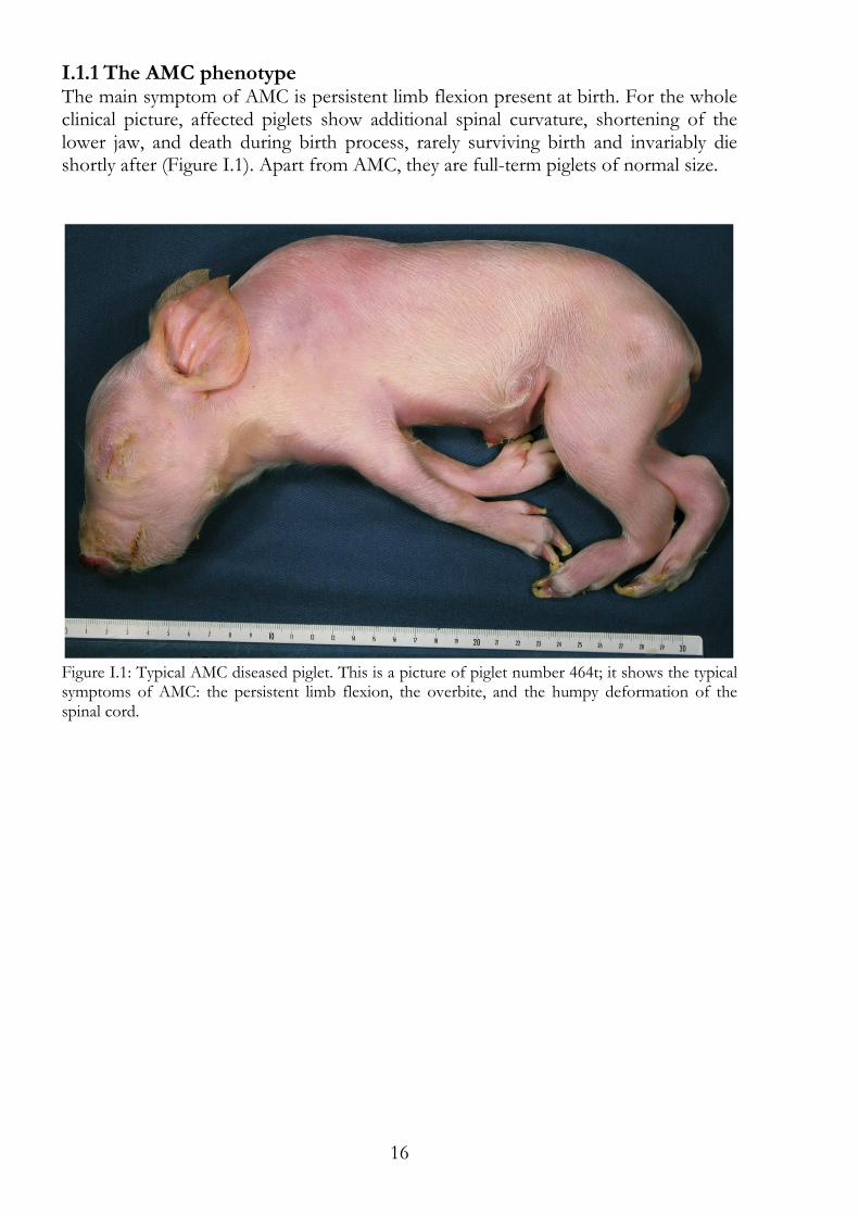
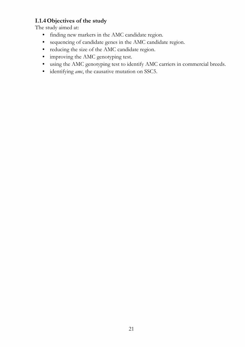
I.1.1 The AMC phenotype The main symptom of AMC is persistent limb flexion present at birth. For the whole clinical picture, affected piglets show additional spinal curvature, shortening of the lower jaw, and death during birth process, rarely surviving birth and invariably die shortly after (Figure I.1). Apart from AMC, they are full-term piglets of normal size.
Figure I.1: Typical AMC diseased piglet. This is a picture of piglet number 464t; it shows the typical symptoms of AMC: the persistent limb flexion, the overbite, and the humpy deformation of the spinal cord.
17
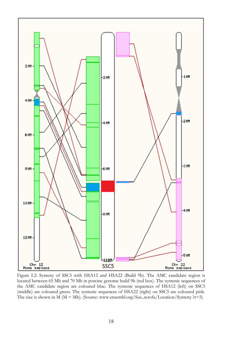
I.1.2 Comparative genetics, BAC and Sscrofa10.2 genome assembly The Sscrofa10.2 genome assembly, out in August 2011, is the most recent genome build. It is a merging of the project's Bacterial Artificial Chromosome (BAC), which are also available separately. The region spans 33 accessioned and partially overlapping BAC sequences between BACs CU424457 and CU915415. If no gene sequence or genomic sequence data were available, the synteny between the human and pig genomes was used to find candidate genes. Syntenic sequences of pig chromosome 5q21, the AMC candidate region, are on human chromosomes 12q12 and 22q11 (Figure I.2). Before the genomic sequence was available, homologous gene sequences of several species had to be compared to find conserved sequences to design primers (mostly based on exon sequences, which were located close to each other). Gene sequences of humans, cattle, sheep, macaques, chickens, rats, and mice were used for this purpose. For analysis, multi species alignments were performed and mapped to the sequences of the BAC library (Green et al. 1990) (www.sanger.ac.uk/Projects/S_scrofa and www.sanger.ac.uk/resources/downloads/ othervertebrates/pig.html) in order to find homologous porcine gene sequences. The BAC sequences provided partial genomic sequences, which made it possible to sequence whole exons. This was because the primers could be designed on the basis of the pig genome, which made sequencing more successful.
I.1.3 Marker and genes in the AMC region A collection of microsatellite markers was available at www.animalgenome.org/pig/ resources/fprimerintr.html and are distributed by M. F. Rothschild. A collection of microsatellites for parentage control was presented by Nechtelberger et al. (Nechtelberger et al. 2001). SNP data form the porcine SNP chip were used (provided by Illumina) and additional SNP data were kindly provided by M. Groenen described in Ramos et al. (2009).
18
Figure I.2: Synteny of SSC5 with HSA12 and HSA22 (Build 9b). The AMC candidate region is located between 65 Mb and 70 Mb in porcine genome build 9b (red box). The syntenic sequences of the AMC candidate region are coloured blue. The syntenic sequences of HSA12 (left) on SSC5 (middle) are coloured green. The syntenic sequences of HSA22 (right) on SSC5 are coloured pink. The size is shown in M (M = Mb). (Source: www.ensembl.org/Sus_scrofa/Location/Synteny ?r=5)
19
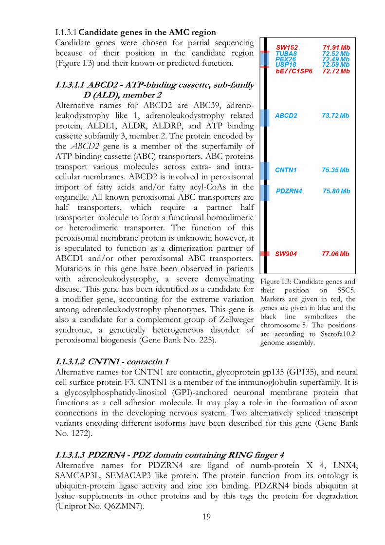
Candidate genes in the AMC region I.1.3.1Candidate genes were chosen for partial sequencing because of their position in the candidate region (Figure I.3) and their known or predicted function.
I.1.3.1.1 ABCD2 - ATP-binding cassette, sub-family D (ALD), member 2
Alternative names for ABCD2 are ABC39, adreno-leukodystrophy like 1, adrenoleukodystrophy related protein, ALDL1, ALDR, ALDRP, and ATP binding cassette subfamily 3, member 2. The protein encoded by the ABCD2 gene is a member of the superfamily of ATP-binding cassette (ABC) transporters. ABC proteins transport various molecules across extra- and intra-cellular membranes. ABCD2 is involved in peroxisomal import of fatty acids and/or fatty acyl-CoAs in the organelle. All known peroxisomal ABC transporters are half transporters, which require a partner half transporter molecule to form a functional homodimeric or heterodimeric transporter. The function of this peroxisomal membrane protein is unknown; however, it is speculated to function as a dimerization partner of ABCD1 and/or other peroxisomal ABC transporters. Mutations in this gene have been observed in patients with adrenoleukodystrophy, a severe demyelinating disease. This gene has been identified as a candidate for a modifier gene, accounting for the extreme variation among adrenoleukodystrophy phenotypes. This gene is also a candidate for a complement group of Zellweger syndrome, a genetically heterogeneous disorder of peroxisomal biogenesis (Gene Bank No. 225).
I.1.3.1.2 CNTN1 - contactin 1 Alternative names for CNTN1 are contactin, glycoprotein gp135 (GP135), and neural cell surface protein F3. CNTN1 is a member of the immunoglobulin superfamily. It is a glycosylphosphatidy-linositol (GPI)-anchored neuronal membrane protein that functions as a cell adhesion molecule. It may play a role in the formation of axon connections in the developing nervous system. Two alternatively spliced transcript variants encoding different isoforms have been described for this gene (Gene Bank No. 1272).
I.1.3.1.3 PDZRN4 - PDZ domain containing RING finger 4 Alternative names for PDZRN4 are ligand of numb-protein X 4, LNX4, SAMCAP3L, SEMACAP3 like protein. The protein function from its ontology is ubiquitin-protein ligase activity and zinc ion binding. PDZRN4 binds ubiquitin at lysine supplements in other proteins and by this tags the protein for degradation (Uniprot No. Q6ZMN7).
Figure I.3: Candidate genes and their position on SSC5. Markers are given in red, the genes are given in blue and the black line symbolizes the chromosome 5. The positions are according to Sscrofa10.2 genome assembly.
20
I.1.3.1.4 PEX26 - peroxin 26 Alternative names for PEX26 are FLJ20695, peroxisome biogenesis factor 26, and PEX26M1T. The PEX26 gene belongs to the peroxin-26 gene family. It anchors PEX1 and PEX6 to peroxisome membranes, possibly to form heteromeric AAA ATPase complexes required for the import of proteins into peroxisomes. Defects in this gene are the cause of peroxisome biogenesis disorder complementation group 8 (PBD-CG8). PBD refers to a group of peroxisomal disorders arising from a failure of protein import into the peroxisomal membrane or matrix. The PBD group is comprised of four disorders: Zellweger syndrome (ZWS), neonatal adreno-leukodystrophy (NALD), infantile Refsum disease (IRD), and classical rhizomelic chondrodysplasia punctata (RCDP). Alternatively spliced transcript variants have been identified for this (Gene Bank No. 55670).
I.1.3.1.5 RNF217 - ring finger protein 217 Alternative names RNF217 are C6orf172, chromosome 6 open reading frame 172, dJ84N20.1, MGC26996, OTTHUMP00000017130, and OTTHUMP00000017132. By similarity, RNF217 is a possible E3 ubiquitin-protein ligase, which accepts ubiquitin from E2 ubiquitin-conjugating enzymes in the form of a thioester and then directly transfers the ubiquitin to targeted substrates (Uniprot No. Q8TC41).
I.1.3.1.6 TUBA8 - tubulin, alpha 8 An alternative name for TUBA8 is TUBAL2. The TUBA8 gene encodes a member of the alpha tubulin protein family. Alpha tubulins are one of two core protein families (alpha and beta tubulins) that heterodimerize and assemble to form microtubules. Mutations in this gene are associated with polymicrogyria and optic nerve hypoplasia. Alternate splicing results in multiple transcripts (Gene Bank No. 51807).
I.1.3.1.7 USP18 - ubiquitin specific protease 18 Alternative names for USP18 are 43 kDa ISG15 specific protease, hUBP43, interferon stimulated gene, 43 KD, ISG15 specific processing protease, ISG43, ubiquitin specific protease, 43 KD, Ubl carboxyl terminal hydrolase 18, Ubl thiolesterase 18, and UBP43. USP18 is a member of the deubiquitinating protease family of enzymes and removes ubiquitin adducts from a broad range of protein substrates (Gene Bank No. 11274). The USP18 is a cytosol or nucleus based and is needed for protein degradation in the ubiquitin conjugation pathway (Uniprot No. Q9UMW8). In this pathway ubiquitin conjugates to proteins to mark them for degradation.
21
I.1.4 Objectives of the study The study aimed at:
• finding new markers in the AMC candidate region.
• sequencing of candidate genes in the AMC candidate region.
• reducing the size of the AMC candidate region.
• improving the AMC genotyping test.
• using the AMC genotyping test to identify AMC carriers in commercial breeds.
• identifying amc, the causative mutation on SSC5.
22
I.2 Materials and methods
I.2.1 Pigs of the experimental herd This material comprised the pigs used in the earlier published studies of Genini (Genini et al. 2004; Genini 2006; Genini et al. 2006) and the pigs produced since then. Genini analysed 39 litters in which 358 piglets were born of which 84 were AMC-diseased. Figure I.4 in section I.3.1 shows the breeding pigs and their relationship and the herd is described in more detail in section I.3.1. Piglets were classified as diseased by optical examination; piglets showing the diseased phenotype were assigned to the genotype amc/amc. Since AMC is recessively inherited all living pigs carry at least one healthy allele (AMC). Healthy (AMC/AMC) and AMC carriers (AMC/amc) were classified after genotyping with the AMC-Test but the only reliable carriers were pigs with litters containing diseased piglets. AMC carriers (amc/AMC) were kept for breeding. As early as possible DNA samples were taken and examined with the AMC genotyping test. Only AMC carriers with good physical appearance were chosen for breeding. Mostly, male and female siblings were chosen. By this two to three boars of different age were present at the research station together with at least six sows of different age. The DNA of four piglets was used as standard DNA. For this, two pairs of littermates from two litters were selected. From one litter pig no. 205t and 208t and from the other litter pig no. 229t and 230t were selected. Pigs 205t and 229t were AMC diseased (amc/amc) and pigs 208t and 230t were stillborn but not AMC diseased (AMC/AMC). In the case of differential sequencing or typing results additional pigs were examined.
Parentage control I.2.1.1If the parentage in the experimental herd was questionable, blood groups (Voegeli 1990) were analysed and/or microsatellite markers of other chromosomes than SSC5 were genotyped (Nechtelberger et al. 2001). In this study, the microsatellite markers S0101 on SSC7, S0335 on SSC15 und S0228 on SSC6 were used (Table I.4) because they were informative in the experimental herd.
I.2.2 Extraction of genomic DNA
DNA from whole-blood I.2.2.1Blood samples were taken of pigs older than four weeks. Blood was collected in EDTA containing tubes and stored at 4°C or -20°C till DNA extraction. Mainly the DNA was extracted from blood cells by the lysis protocol described by Vogeli et al. (1994). Briefly, to 600 µl whole-blood, 500 µl lysis buffer for blood (appendix) were added and mixed. After 15 - 30 min at room temperature the mixture was centrifuged at 13000x g for 30 s. The pellet was resuspended in 1 ml lysis buffer for blood and vortexed, incubated for 15 min at room temperature and centrifuged for 25 s at 13000x g. Without the incubation, these steps were repeated two times. Then, the pellet was resuspended in 200 µl PCR turbo buffer and 20 µl proteinase K (20 mg/ ml) were added; the mixture was incubated at 54°C for 2 h. Finally, the mixture was incubated for 10 min at 95°C to deactivate the proteinase. After this final step the DNA was ready to use or to store at -20°C.
23
Besides this lysis protocol, kits were also used for DNA extraction from blood cells, as there were the GenElute™ Mammalian Genomic DNA Miniprep Kit provided by SIGMA (Sigma-Aldrich Chemie GmbH, Buchs, Switzerland) and the DNeasy Blood & Tissue Kit provided by Qiagen (Qiagen AG, Basel, Switzerland). The kits were used according to the supplier's protocol.
DNA from tail tissue or hair roots I.2.2.2Tissue samples were taken of dead pigs (e. g. still born piglets). Hair samples were taken from piglets too young for blood sampling. Tissue parts of the tail were collected in tubes and stored at 4°C or -20°C till DNA extraction. Hair, hair with roots, were collected in bags and stored at 4°C or -20°C till DNA extraction. The extraction of DNA was done using the lysis protocol by Laird et al. (1991), the Extract-N-Amp™ Tissue PCR Kit provided by Sigma or the DNeasy Blood & Tissue Kit provided by QIAGEN. The kits were used according to the supplier's protocol. The protocol by Laird at al. (1991) in brief: a 1 mm slice of the tail or at least 10 hairs with roots were mixed with 500 µl lysis buffer for tissue (appendix) and 2.5 µl proteinase K (20 mg/ ml). The mixture was incubated overnight at 55°C on a rotating shaker. After incubation, the mixture was centrifuged at 11000x g for 10 min. The supernatant was transferred to a new tube containing 500 µl isopropanol and the content of the tube was mixed by a tilting movement. Depending on the amount of precipitate, it was either transferred into a new tube with a pipette tip or centrifuged at 11000x g for 30 min. The DNA was washed in 70% ethanol and dried for 10 min. Finally, the DNA was resuspended in 10 to 600 µl pure water or TE buffer.
I.2.3 Nucleic acid concentration measurement Spectrophotometers for nucleic acid quantitation were used as well as Qubit. Qubit is a fluorometer to measure quantities of DNA, RNA and protein depending on the fluorescent labelling used in the Quant-iT assay. The fluorometer was used according to the supplier's protocol (Invitrogen, Life Technologies Europe BV).
I.2.4 Primer design The software Primer3 (v. 0.4.0) was used for primer design (Rozen & Skaletsky 2000) using the following restrictions: average length of 20 to 25 bp, average GC content of 50%, melting temperature of 62°C, difference of the melting temperatures was supposed to be smaller than 1°C and eventually, at 3' the last two bases were a G or a C (CG-clamp). Primers matching the demands were ordered at Microsynth AG (9436 Balgach, Switzerland).
I.2.5 Polymerase chain reaction (PCR) PCR was carried out in a reaction volume of 25 µl using 20 - 50 ng of genomic DNA, 0.75 units antibody inactivated Taq polymerase, 0.4 µM of each primer, 0.2 mM of each deoxynucleotide and PCR buffer (Sigma-Aldrich Chemie GmbH). PCR consisted of 30 to 35 cycles. The initial denaturation step was at 95°C for 5 min. Cycling conditions were denaturation at 95°C for 30 s, annealing at 58 - 62°C for 30 s and elongation at 72°C for 30 s. The final extension was carried out at 72°C for 5 min. The PCR machines were FTGene5D (Techgene), Progene FPROG5D (Techne), Robocycler Gradient 96 (Stratagen), Gene Amp PCR System 9700 (Applied Biosystems), T1 Thermocycler (Biometra), PTC-100 (MJ Research Inc.) and PCR
24
Express (Hybaid). For special conditions 0.8x to 1.2x PCR buffer and/or additional MgCl2 and/or BSA were added to the reaction solution.
I.2.6 Agarose gel electrophoresis PCR fragments were analysed on ethidium bromide stained agarose gels composed of 1% - 2% agarose (Sigma-Aldrich Chemie GmbH) in 0.5x TBE buffer (appendix). The agarose gels were run in electrophoresis buffer (0.5x TBE buffer) at 120 Volt and analysed under ultraviolet light.
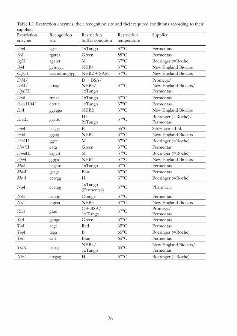
I.2.7 PCR-RFLP assay Restriction enzymes were selected according to the recognition site and the following fragmentation pattern (Table I.1). Key fragments had to be clearly distinguishable by agarose gel electrophoresis. PCR products were digested over night by adding 15 µl of digestion mix, including pure water, enzyme buffer and 2 units endonuclease (Table I.2) and the following incubation at the suggested temperature (Table I.2). If required, BSA or S-adenosylmethionine (SAM) was added. The restriction fragments were analysed using agarose gel electrophoresis (Section I.2.6). Table I.1: The names of primer pairs used for RFLP with their corresponding restriction enzyme and the resulting restriction fragments of both alleles. Primer pair names marked with * are primers designed for sequencing, which have been used also for RFLP (reference sequence = rs, deletion = del).
Primer pair name SNP Restriction
enzyme Amplicon length (bp)
Fragments of allele 1 (bp)
Fragments of allele 2 (bp)
A2800 GA G>A RsaI 194 A: 194 G: 95, 99
A6254 GA G>A MboII 174 A: 174 G: 72, 102
A6257 GC G>C TaiI 129 C: 129 G: 47, 82
A6258 TC T>C SatI 150 T: 150 C: 55, 95
A754 GT G>T NsiI 139 G: 139 T: 56, 83
A767 AG A>G RsaI 151 G: 151 A: 55, 96
A777 GA Seq * G>A AluI 405 A: 405 G: 174, 231
A903 GA G>A HaeIII 120 A: 120 G: 40, 80
A907 GA G>A NdeI 196 A: 196 G: 96, 100
A922 TC T>C FokI 187 T: 187 C: 73, 114
D356 GC G>C BclI 632 G: 632 C: 293, 339
D5996 AC A>C TasI 155 C: 155 A: 59, 96
D6006 TC T>C EcoRI 459 C: 459 T: 139, 320
D6010 AG A>G TaqI 182 A: 182 G: 67, 115
D6025 GA G>A TasI 196 G: 196 A: 39, 157
D6032 GA G>A TaiI 136 A: 136 G: 53, 83
H6817 GA G>A NcoI 555 G: 555 A: 188, 367
H6818 AT A>T HphI 639 A: 189, 450 T: 52, 189, 398
H723 AC A>C FauI 161 A: 161 C: 79, 82
H783 GA G>A Hin1II 153 G: 153 A: 69, 84
H790 TC T>C TspRI 145 T: 145 C: 61, 84
H804 AG A>G SatI 415 A: 9, 37, 369 G: 9, 37, 147, 221
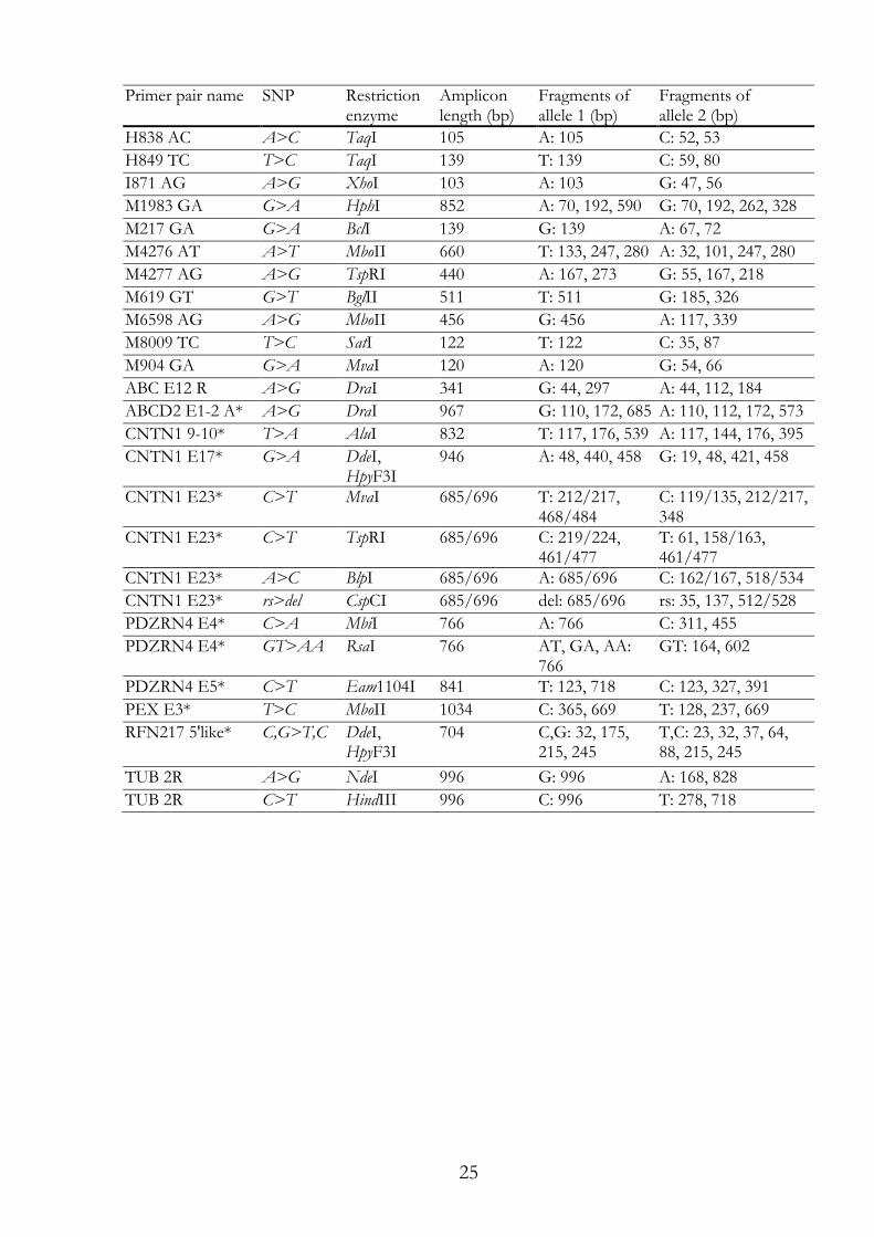
25
Primer pair name SNP Restriction enzyme
Amplicon length (bp)
Fragments of allele 1 (bp)
Fragments of allele 2 (bp)
H838 AC A>C TaqI 105 A: 105 C: 52, 53
H849 TC T>C TaqI 139 T: 139 C: 59, 80
I871 AG A>G XhoI 103 A: 103 G: 47, 56
M1983 GA G>A HphI 852 A: 70, 192, 590 G: 70, 192, 262, 328
M217 GA G>A BclI 139 G: 139 A: 67, 72
M4276 AT A>T MboII 660 T: 133, 247, 280 A: 32, 101, 247, 280
M4277 AG A>G TspRI 440 A: 167, 273 G: 55, 167, 218
M619 GT G>T BglII 511 T: 511 G: 185, 326
M6598 AG A>G MboII 456 G: 456 A: 117, 339
M8009 TC T>C SatI 122 T: 122 C: 35, 87
M904 GA G>A MvaI 120 A: 120 G: 54, 66
ABC E12 R A>G DraI 341 G: 44, 297 A: 44, 112, 184
ABCD2 E1-2 A* A>G DraI 967 G: 110, 172, 685 A: 110, 112, 172, 573
CNTN1 9-10* T>A AluI 832 T: 117, 176, 539 A: 117, 144, 176, 395
CNTN1 E17* G>A DdeI, HpyF3I
946 A: 48, 440, 458 G: 19, 48, 421, 458
CNTN1 E23* C>T MvaI 685/696 T: 212/217, 468/484
C: 119/135, 212/217, 348
CNTN1 E23* C>T TspRI 685/696 C: 219/224, 461/477
T: 61, 158/163, 461/477
CNTN1 E23* A>C BlpI 685/696 A: 685/696 C: 162/167, 518/534
CNTN1 E23* rs>del CspCI 685/696 del: 685/696 rs: 35, 137, 512/528
PDZRN4 E4* C>A MbiI 766 A: 766 C: 311, 455
PDZRN4 E4* GT>AA RsaI 766 AT, GA, AA: 766
GT: 164, 602
PDZRN4 E5* C>T Eam1104I 841 T: 123, 718 C: 123, 327, 391
PEX E3* T>C MboII 1034 C: 365, 669 T: 128, 237, 669
RFN217 5'like* C,G>T,C DdeI, HpyF3I
704 C,G: 32, 175, 215, 245
T,C: 23, 32, 37, 64, 88, 215, 245
TUB 2R A>G NdeI 996 G: 996 A: 168, 828
TUB 2R C>T HindIII 996 C: 996 T: 278, 718
26
Table I.2: Restriction enzymes, their recognition site and their required conditions according to their supplier. Restriction enzyme
Recognition site
Restriction buffer condition
Restriction temperature
Supplier
AluI agct 1xTango 37°C Fermentas
BclI tgatca Green 55°C Fermentas
BglII agatct M 37°C Boeringer (=Roche)
BlpI gctnagc NEB4 37°C New England Biolabs
CspCI caannnnngtgg NEB2 + SAM 37°C New England Biolabs
DdeI/ DdeI/ HpyF3I
ctnag D + BSA/ NEB3/ 1xTango
37°C Promega/ New England Biolabs/ Fermentas
DraI tttaaa 1xTango 37°C Fermentas
Eam1104I ctcttc 1xTango 37°C Fermentas
EciI ggcgga NEB2 37°C New England Biolabs
EcoRI gaattc H/ 2xTango
37°C Boeringer (=Roche)/ Fermentas
FauI cccgc B 55°C SibEnzyme Ltd.
FokI ggatg NEB4 37°C New England Biolabs
HaeIII ggcc M 37°C Boeringer (=Roche)
Hin1II catg Green 37°C Fermentas
HindIII aagctt M 37°C Boeringer (=Roche)
HphI ggtga NEB4 37°C New England Biolabs
MbiI ccgctc 1xTango 37°C Fermentas
MboII gaaga Blue 37°C Fermentas
MvaI ccwgg H 37°C Boeringer (=Roche)
NcoI ccatgg 1xTango (Fermentas)
37°C Pharmacia
NdeI catatg Orange 37°C Fermentas
NsiI atgcat NEB3 37°C New England Biolabs
RsaI gtac C + BSA/ 1x Tango
37°C Promega/ Fermentas
SatI gcngc Green 37°C Fermentas
TaiI acgt Red 65°C Fermentas
TaqI tcga B 65°C Boeringer (=Roche)
TasI aatt Blue 65°C Fermentas
TspRI castg NEB4/ 1xTango
65°C New England Biolabs/ Fermentas
XhoI ctcgag H 37°C Boeringer (=Roche)
27
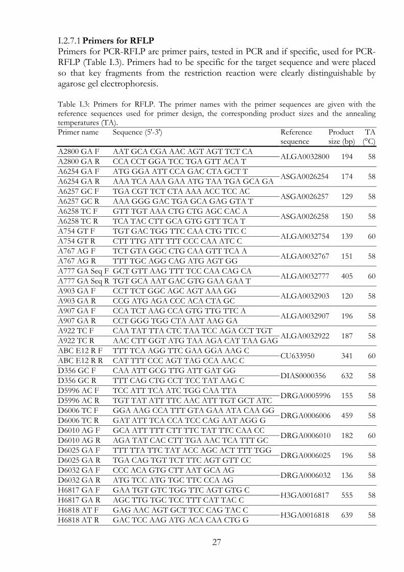
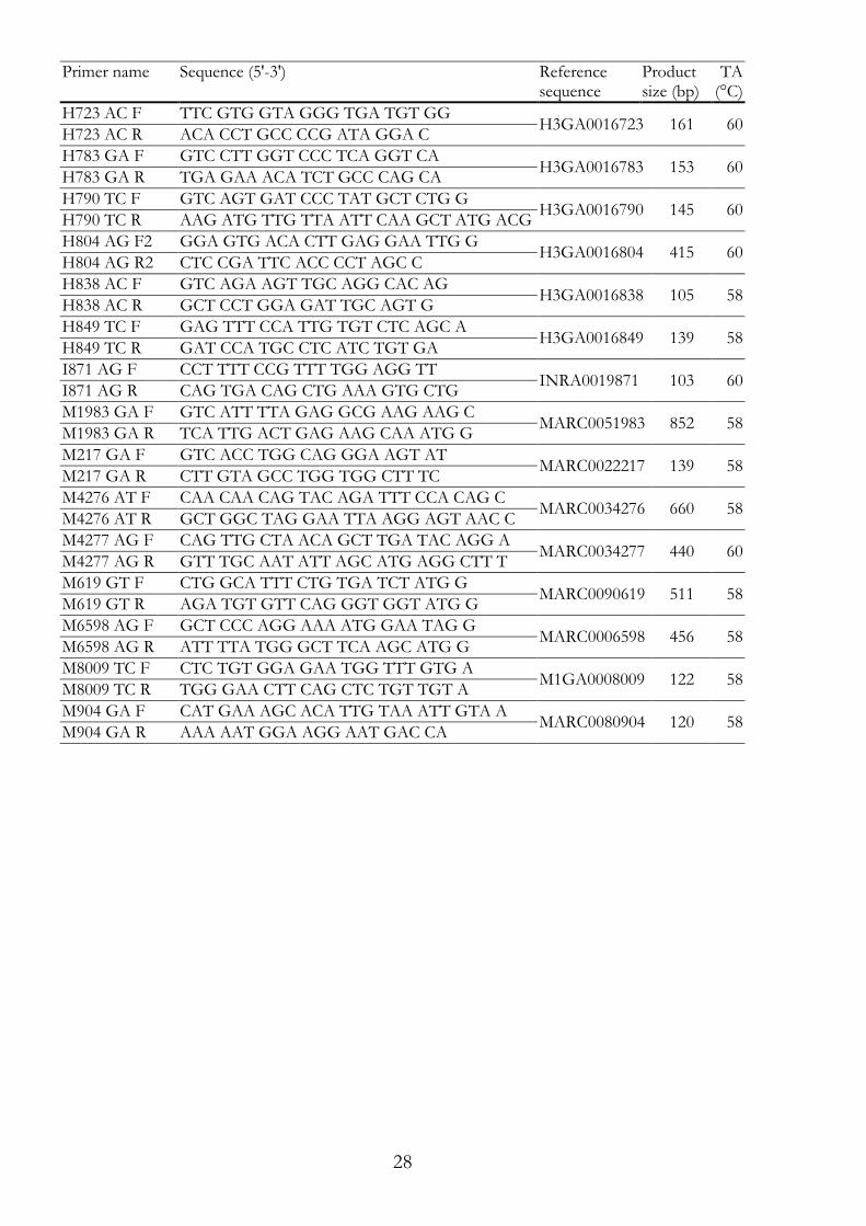
Primers for RFLP I.2.7.1Primers for PCR-RFLP are primer pairs, tested in PCR and if specific, used for PCR-RFLP (Table I.3). Primers had to be specific for the target sequence and were placed so that key fragments from the restriction reaction were clearly distinguishable by agarose gel electrophoresis. Table I.3: Primers for RFLP. The primer names with the primer sequences are given with the reference sequences used for primer design, the corresponding product sizes and the annealing temperatures (TA). Primer name Sequence (5'-3') Reference
sequence Product size (bp)
TA (°C)
A2800 GA F AAT GCA CGA AAC AGT AGT TCT CA ALGA0032800 194 58
A2800 GA R CCA CCT GGA TCC TGA GTT ACA T
A6254 GA F ATG GGA ATT CCA GAC CTA GCT T ASGA0026254 174 58
A6254 GA R AAA TCA AAA GAA ATG TAA TGA GCA GA
A6257 GC F TGA CGT TCT CTA AAA ACC TCC AC ASGA0026257 129 58
A6257 GC R AAA GGG GAC TGA GCA GAG GTA T
A6258 TC F GTT TGT AAA CTG CTG AGC CAC A ASGA0026258 150 58
A6258 TC R TCA TAC CTT GCA GTG GTT TCA T
A754 GT F TGT GAC TGG TTC CAA CTG TTC C ALGA0032754 139 60
A754 GT R CTT TTG ATT TTT CCC CAA ATC C
A767 AG F TCT GTA GGC CTG CAA GTT TCA A ALGA0032767 151 58
A767 AG R TTT TGC AGG CAG ATG AGT GG
A777 GA Seq F GCT GTT AAG TTT TCC CAA CAG CA ALGA0032777 405 60
A777 GA Seq R TGT GCA AAT GAC GTG GAA GAA T
A903 GA F CCT TCT GGC AGC AGT AAA GG ALGA0032903 120 58
A903 GA R CCG ATG AGA CCC ACA CTA GC
A907 GA F CCA TCT AAG CCA GTG TTG TTC A ALGA0032907 196 58
A907 GA R CCT GGG TGG CTA AAT AAG GA
A922 TC F CAA TAT TTA CTC TAA TCC AGA CCT TGT ALGA0032922 187 58
A922 TC R AAC CTT GGT ATG TAA AGA CAT TAA GAG
ABC E12 R F TTT TCA AGG TTC GAA GGA AAG C CU633950 341 60
ABC E12 R R CAT TTT CCC AGT TAG CCA AAC C
D356 GC F CAA ATT GCG TTG ATT GAT GG DIAS0000356 632 58
D356 GC R TTT CAG CTG CCT TCC TAT AAG C
D5996 AC F TCC ATT TCA ATC TGG CAA TTA DRGA0005996 155 58
D5996 AC R TGT TAT ATT TTC AAC ATT TGT GCT ATC
D6006 TC F GGA AAG CCA TTT GTA GAA ATA CAA GG DRGA0006006 459 58
D6006 TC R GAT ATT TCA CCA TCC CAG AAT AGG G
D6010 AG F GCA ATT TTT CTT TTC TAT TTC CAA CC DRGA0006010 182 60
D6010 AG R AGA TAT CAC CTT TGA AAC TCA TTT GC
D6025 GA F TTT TTA TTC TAT ACC AGC ACT TTT TGG DRGA0006025 196 58
D6025 GA R TGA CAG TGT TCT TTC AGT GTT CC
D6032 GA F CCC ACA GTG CTT AAT GCA AG DRGA0006032 136 58
D6032 GA R ATG TCC ATG TGC TTC CCA AG
H6817 GA F GAA TGT GTC TGG TTC AGT GTG C H3GA0016817 555 58
H6817 GA R AGC TTG TGC TCC TTT CAT TAC C
H6818 AT F GAG AAC AGT GCT TCC CAG TAC C H3GA0016818 639 58
H6818 AT R GAC TCC AAG ATG ACA CAA CTG G
28
Primer name Sequence (5'-3') Reference sequence
Product size (bp)
TA (°C)
H723 AC F TTC GTG GTA GGG TGA TGT GG H3GA0016723 161 60
H723 AC R ACA CCT GCC CCG ATA GGA C
H783 GA F GTC CTT GGT CCC TCA GGT CA H3GA0016783 153 60
H783 GA R TGA GAA ACA TCT GCC CAG CA
H790 TC F GTC AGT GAT CCC TAT GCT CTG G H3GA0016790 145 60
H790 TC R AAG ATG TTG TTA ATT CAA GCT ATG ACG
H804 AG F2 GGA GTG ACA CTT GAG GAA TTG G H3GA0016804 415 60
H804 AG R2 CTC CGA TTC ACC CCT AGC C
H838 AC F GTC AGA AGT TGC AGG CAC AG H3GA0016838 105 58
H838 AC R GCT CCT GGA GAT TGC AGT G
H849 TC F GAG TTT CCA TTG TGT CTC AGC A H3GA0016849 139 58
H849 TC R GAT CCA TGC CTC ATC TGT GA
I871 AG F CCT TTT CCG TTT TGG AGG TT INRA0019871 103 60
I871 AG R CAG TGA CAG CTG AAA GTG CTG
M1983 GA F GTC ATT TTA GAG GCG AAG AAG C MARC0051983 852 58
M1983 GA R TCA TTG ACT GAG AAG CAA ATG G
M217 GA F GTC ACC TGG CAG GGA AGT AT MARC0022217 139 58
M217 GA R CTT GTA GCC TGG TGG CTT TC
M4276 AT F CAA CAA CAG TAC AGA TTT CCA CAG C MARC0034276 660 58
M4276 AT R GCT GGC TAG GAA TTA AGG AGT AAC C
M4277 AG F CAG TTG CTA ACA GCT TGA TAC AGG A MARC0034277 440 60
M4277 AG R GTT TGC AAT ATT AGC ATG AGG CTT T
M619 GT F CTG GCA TTT CTG TGA TCT ATG G MARC0090619 511 58
M619 GT R AGA TGT GTT CAG GGT GGT ATG G
M6598 AG F GCT CCC AGG AAA ATG GAA TAG G MARC0006598 456 58
M6598 AG R ATT TTA TGG GCT TCA AGC ATG G
M8009 TC F CTC TGT GGA GAA TGG TTT GTG A M1GA0008009 122 58
M8009 TC R TGG GAA CTT CAG CTC TGT TGT A
M904 GA F CAT GAA AGC ACA TTG TAA ATT GTA A MARC0080904 120 58
M904 GA R AAA AAT GGA AGG AAT GAC CA
29
I.2.8 Microsatellite analysis For microsatellite analysis (gene scan analysis) the ABI PRISM® 377 Genetic Analyzer (Applied Biosystems) was used. In order to assess the fragments of the microsatellite markers, size standards spanning up to 350 bp or up to 500 bp were chosen. Depending on the fluorophores used to mark the PCR fragments (Table I.4) and the filter set, the fluorophore of the size standard (TAMRA or ROX) was chosen. Per sample, up to 0.5 µl PCR were mixed with 2.5 µl of formamide, 0.5 µl dye-labelled size standard and 0.5 µl gel loading buffer. If several PCR products were separated on the same line, the whole PCRs were mixed and from this mixture 0.5 µl were then taken for loading preparations. After a heat shock at 95°C, the samples were put back on ice and were ready to load. For the assay, a 4.5% polyacrylamide gel containing 6 M urea was prepared. For this, 18 g urea were dissolved in 23 ml pure water, 5 ml 10x TBE buffer and 7.5 ml 30% Acrylamide/Bis Solution, 29:1 (Bio-Rad Laboratories, Inc., Reinach, Switzerland) were added. After degasing, 350 µl of 10% ammonium persulfate and 15 µl TEMED were added shortly before pouring the gel. Of the prepared samples 1.5 µl were loaded in one lane for analysis. Alternatively, a 48 capillary device was used for fragment analysis (ABI 3730xl DNA Analyzer).
Gene scan analysis software I.2.8.1To assess the lengths of the fragments, the gene scan results were analysed with software provided by Applied Biosystems (GeneScan 2.1 and Genotyper 2.1 or the Peak Scanner Software v1.0 from Applied Biosystems, respectively).
Primers for gene scan assay I.2.8.2Primers for Gene Scan assay are primer pairs, tested in PCR and if specific ordered one primer of the pair modified (Table I.4). Modification was added at the 5' site of the primer. Fluorophores such as HEX, FAM, TET were used as modification (Table I.4).
30
Table I.4: Primers for microsatellite analysis. The primer names with the primer sequences are given with the reference sequences used for primer design, the corresponding product sizes and the annealing temperatures (TA). Primer name Sequence (5'-3') 5' Modification Reference
sequence Product size (bp)
TA (°C)
ABCD2 GS F HEX
TTT TAC TTG TCC CTA GAA ATA CAT AGC
HEX CU928451
284 - 287
60 ABCD2 GS R ATT TGG TTC CTG GAT CTA CTC C
bE77C1SP6 1F TGC TAT ACA GCA AAT TGA CCC ACT FAM NM_213826
281 - 306
58 bE77C1SP6 3R GCA GAC ACA GCT CAG ATC CA
CNTN1E23V1F FAM
GTT TTT CTT TTC AGG GCT ACG C FAM
CU457707 184 - 200
64 CNTN1E23 V12 R
TGA GGA GGT GGG TTT GAT CC
RM_10 F hex GGA CAG AGC CTT CTA GGA CTG G HEX CU407335
224 - 284
62 RM_10 R2 CTA GGA CAA GTA TAT GTG TTT GGG
S0101 F GAA TGC AAA GAG TTC AGT GTA GG HEX UniSTS:252630
196 - 224
58 S0101 R GTC TCC CTC ACA CTT ACC GCA G
S0228 F GGC ATA GGC TGG CAG CAA CA TET UniSTS:252792
221 - 245
62 S0228 R AGC CCA CCT CAT CTT ATC TAC ACT
S0355 F TCT GGC TCC TAC ACT CCT TCT TGA TG
FAM
UniSTS:253051 244 - 271
55 S0355 R TTG GGT GGG TGC TGA AAA ATA
GGA
SW1071 F AGT GCT GAT ATC AAG CAC AAG C TET AF235185
126 - 156
62 SW1071 R TCA CTT CCC ACC CCT TAC AC
SW1094 F GAT CAT GGT GTA CCA TCC TTT ATA TET AF235193
142 - 172
58 SW1094 R ATT CTT GAT GTT GGT ACA TGG TG
SW152 F GGA TTT TAG GGC TGA ATC TGC FAM AF235220
166 - 182
62 SW152 R GAT GAC CTT GCA ATG CCC
SW1987 F TGA GCA GAT AGG CAG ACT TCT G FAM AF253763
152 - 168
58 SW1987 R TTT AAG GGG CAT GTT TGA GG
SW904 F CCC CTT TCA GAA GAA TGA AAA FAM AF235398
163 - 182
60 SW904 R CCT AGT GGC CAA CAC CAA GT
SW963 F TCT GTT GTT TCC CAC CAG C FAM AF253858
143 - 173
55 SW963 R TGT GCA CCT GAC ACA TAG ACT C
SWR1526 F CGG TGG CTA CAG ATA ACA ATA C TET AF253886
114 - 147
62 SWR1526 R ATC CGA TTC AAC CCC TAG C
UMNp1275 F TAT GTG AGA AGG TGA GGG TGG FAM AY285380
198 - 206
62 UMNp1275 R TGG AGA CAA CAG ATG CAA GG
USP18 2F fam TGG TTT ACA TGA TGG CTG AGT C FAM
ss472055141 and ss472055142
395 - 416
60 USP18 2R ATG CCA GTA CAG TCA CAT GAG C
USP18 F short FAM
GAC CTT GTG CAG GTC TGG AT FAM 208 - 229
62 USP18 R short TGC CAG TAC AGT CAC ATG AGC
31
I.2.9 DNA purification
Ethanol or isopropanol precipitation I.2.9.1The sample was mixed with 0.1x volumes of 3 M sodium acetate (pH 5.5) and either 1x volume of isopropanol or 3x volumes of 100% ethanol. The mixture was placed at -20°C for 0.5 h to overnight and centrifuged at 4°C for 30 min at 13000x g. The pellet was washed with 70% ethanol, centrifuged again at 4°C for 30 min at 13000x g and after drying, the pellet was resuspended in the favoured buffer (pure water or TE buffer (appendix)) in the needed volume.
Millipore Montage Microcon centrifugal filter devices I.2.9.2PCR fragments for sequencing at Microsynth or to improve restriction performance were purified and concentrated using the filter devices as recommended by the supplier's protocol (Millipore AG, Billerica, MA 01821).
I.2.10 Sequencing
Sequencing at Microsynth AG I.2.10.1In total volume of 10 µl, purified PCR fragments in pure water (Section I.2.9.2) were mixed with 20 pmol of the sequencing primer (Table I.5). For the ´Economy Run Service´ or the ´Barcode Economy Run Service´, the amount of DNA fragments had to be 15 ng/ 100 bases (Section I.2.3). Samples were sent in for sequencing to Microsynth (Microsynth AG, Balgach, Switzerland). Microsynth provides Sanger sequencing using a 3730xl DNA Analyzer from Applied Biosystems.
Sequence analysis software I.2.10.2To analyse the chromatograms Chromas software (Technelysium Pty Ltd.; www.technelysium.com.au) and CLC Sequence Viewer software (Version 6.4 by CLC bio A/S; www.clcbio.com) were used. The sequences of the genomic reference sequence, healthy controls and diseased pigs were aligned and analysed using these software.
Primers for sequencing I.2.10.3Primers for sequencing were designed so that they are spanning a region including an exon, the primer pair was named after the exon (Table I.5). If no genomic sequence was available, the primers were designed on conserved sequences within exon sequences. Exon sequences were identified by gene alignments of several species (human, cattle, mouse, rat, macaque, chicken and/or sheep) and if available, porcine sequences.
32
Table I.5: Primers for sequencing. The primer names with the primer sequences are given with the reference sequences used for primer design, the corresponding product sizes and the annealing temperatures (TA). Primer name Sequence (5'-3') Reference
sequence Product size (bp)
TA (°C)
A777 GA Seq F GCT GTT AAG TTT TCC CAA CAG CA ALGA0032777 405 60
A777 GA Seq R TGT GCA AAT GAC GTG GAA GAA T
ABCD2 E1-2 A F GGG AGA GAA TGG AAA ACA GAT CC CU633950 967 60
ABCD2 E1-2 A R CAT TCA CTC CAG GCG AAG G
ABCD2 E1-2 B F CTT ACC CGG CAG CAG AGA AC CU633950 879 60
ABCD2 E1-2 B R ACG CTA AAG GAG TGG GTC TGT C
ABCD2 E1-2 C F CCC ACT CCT TTA GCG TTT GA CU633950 952 60
ABCD2 E1-2 C R TCT GCC ATT GTT TCT GAC CA
ABCD2 E3 F AAG CAG GGA ATT GAA TTT GG CU928451 856 60
ABCD2 E3 R TTG GCT AAC ATT TAT CGA GTG C
ABCD2 E4-5 A F GCC ATG CTG CTT TCT TGG CU928451 840 60
ABCD2 E4-5 A R AAT GTC AAT ATC ATG AGA GCA AAG C
ABCD2 E4-5 B F CAC ACC TGG CAC ATA TTT TTG G CU928451 886 60
ABCD2 E4-5 B R CCA GAT TTC TGA GGA CGT TTC C
ABCD2 E6 F GTG GAA ATG AAT CCG ACT GG CU928451 938 62
ABCD2 E6 R GAA ATT TGT ATG CTG CTT GAT GG
ABCD2 E7-3 F ACT GAG TTG TCT TTT GAA CTT CTG C CU928451 700 60
ABCD2 E7-3 R GTG TGC TTA TGT TCC CAA TAA TAG C
ABCD2 E8 F TGC GCT TCG AAA ACT CAG ATA CU928451 643 60
ABCD2 E8 R ACT TGG AGA TTT CTG ACA CAT CAA
ABCD2 E9-3 F CAT GCA GTA ACT ACC TCA TTT TTC C CU928451 620 60
ABCD2 E9-3 R AAA CAC ATG AAC TTC AAC TTT CAG C
ABCD2 E10 A F TTT TAG TAT TTG TGT TGC CGA AGC CU928451 972 60
ABCD2 E10 A R ATG TGC CCA CAT TGA GAT GC
ABCD2 E10 B F CCC GAA ATT CTT CAC AAA TAT GG CU928451 963 60
ABCD2 E10 B R TGT GGC AAA TGG AGG TTC C
CNTN1 I1 F TGA ACA CAA GAT GAA AAT GTG GT FP089683 1333 60
CNTN1 I1 R CCA TGA CCA TAT CTT CTG TGC
CNTN1 E1 F GCT GCT GCA TAC CTT TTC CAC CU914199 756 60
CNTN1 E1 R ACT GGG AGG CAA ATG AAA CC
CNTN1 E3 F GGA TAA TTG CAT ATG GCA CTG G CU914199 903 58
CNTN1 E3 R TAT GGT CAC CTG TCC CCA AG
CNTN1 E5.2 F AGC CCT AGC AAG TGA TGC AG CU914199 814 60
CNTN1 E5.2 R GGC AAG GTT GAA GCA GAA TG
CNTN1 E5 F TGA TGG TTT CTC CCC CAA AG CU633524 781 60
CNTN1 E5 R AAG GGA AAT TTT TCA TGC ACT TG
CNTN1 E7 F CTG AAC TAC AGG CAA CCA GTC C CU633524 938 60
CNTN1 E7 R TTT ATA GAT TGC AGT AGG GAA AAT GG
CNTN1 E8-9 F CGA TCC TCA GTC CAG CAC AG CU633524 820 60
CNTN1 E8-9 R GTT GGA TCA CAT TGG TCA CAC A
CNTN1 E9 F GCA AGG GAC TTG AGG AAT GC CU914199 948 60
CNTN1 E9 R GAC CCC CAC AGA AAC AGA GG
CNTN1 9-10 R2 CGG CAT TTT CAA AAG TCA CA FP089683 832 53
CNTN1 9-10F AGC ATT CCC TGA GTG GGT AG
33
Primer name Sequence (5'-3') Reference sequence
Product size (bp)
TA (°C)
CNTN1 E10 F TTG TCC CCA GGG CTA AAG GT CU914199 951 58
CNTN1 E10 R TTG AAG GTT CCA GCA AAA GGA
CNTN1 E10-11 F ATG CCG GAA TGT ACC AGT GC FP089683 1876 51
CNTN1 E10-11 R GAC CTG CTG CTA TTG ACA AGC
CNTN1 E11 F CCC AAA GCC ACC ATA ACA TTG CU914199 963 60
CNTN1 E11 R ATG CAT AGC GCA TTT GGT TG
CNTN1 12-13F TTT GGG AAG ATG GTA GCT TGG FP089683 530 58
CNTN1 12-13R CGA TCA CAT AGC CAT TGA AGG
CNTN1 E12-13 F CCT CAG GGC TAG CCA GTT TTA G CU914199 990 60
CNTN1 E12-13 R TTT CCA AAA CAA CTT GTC AAT GC
CNTN1 E14 F GTC CCG AGC TGG CCT TAT AG CU914199 906 60
CNTN1 E14 R GGG GCT AAA AAG CAC CTT ACG
CNTN1 E15 F GAA GGC CTC TCT TTG CTT TCC CU914199 776 60
CNTN1 E15 R CTG TCA GCT CTG ACC CAT GC
CNTN1 E16 F GTC TCG GCA GGT GTC AAG TG CU914199 931 60
CNTN1 E16 R AGG ACC TCC CCT CTC ACT CC
CNTN1-3 E16 F CAC ACC TCA ACC GAA ACC TGT CU633524 1047 60
CNTN1-3 E16 R TGC TCC GAA ATG GGC ATC
CNTN1 E17 F CAT GAG TGC AGC TTG TCT TTC C CU457707 946 58
CNTN1 E17R TGC CGT TTT AAG ACT CTC TCT GC
CNTN1 E18 F AGG CCT TGT TCT TGG TTC TAG G CU457707 834 58
CNTN1 E18-M R TTC TTC CTC CCT CCT CAT GC
CNTN1 E18-M F TTC CCC CTG TTA ACT CTT TTG C CU457707 630 58
CNTN1 E18 R TGG ATG CAC AAA CAT ACT GAG G
CNTN1 E19 F TGA CTT GAT GCC CAG TTG GA CU914199 936 58
CNTN1 E19 R GCC AGT TAG GAA CCC CTG CT
CNTN1 E20 F GAT CTG CAT TGT GTG CCT TG CU914199 836 58
CNTN1 E20 R ATG TGA GCA GGG CAG GTA AC
CNTN1 E21 F TGA TGA CCT TGG GAA CAG CA CU457707 819 58
CNTN1 E21 R TCC AGT GAA AAC TGT GAT GGT G
CNTN1 E22 F TGT TGC CAA GCA CAC AGT GA CU457707 832 58
CNTN1 E22 R CAT GTT GCC AGT GTC CTT TTG
CNTN1 E23 F TGC CCC AAG TAA TGT TGT TAG C CU457707
685/696
58 CNTN1 E23-M R CAA GAA GAC ACG GGG AGA GG
CNTN1 E23-M F TAT GGC AAC ACC CAC TGA GC CU457707 706 58
CNTN1 E23 R CAC GTA CGA TCA ACA TTT CAA CC
I88-90 seq F2 TCC TTG CTT TAT TGT TGC ATG T INRA0019888 - INRA0019890
773 56 I88-90 seq R ACC ACT TTG AAT TCC CAA CG
M81-90 seq F3 TCC TCA GCT TGG GGT ATA AAA A MARC0108180 - MARC0108190
753 56 M81-90 seq R TAA AGG CTG AAC ACA CTT CAC C
PDZRN4 E1 F GTC ATC TGA AGG GGG AGT GG CU457707 866 60
PDZRN4 E1 R TGT GGC AAA ACA TGG AGA GG
PDZRN4 E4 F AAA TCT GAT GGC GTG CTT CC CU407335 766 58
PDZRN4 E4 R AGT GCC CAT GAC TTG TGT GG
PDZRN4 I4 F GGG CAC TGG TCA GGG AAG TA CU407335 334 58
PDZRN4 I4 R CAC AGA TGG GGC TAC GTG AA
34
Primer name Sequence (5'-3') Reference sequence
Product size (bp)
TA (°C)
PDZRN4 E5 F GGA GGC CAG AGG ATG GAT AA CU407335 841 55
PDZRN4 E5 R CTA AAT CAG CGA GGG GGA ATA PDZRN4 E6 F GCA TTT GGA ATG GAC AAG CA
CU407335 914 56 PDZRN4 E6 R GGC TTT CAT GGG GCT GAA TA
PDZRN4 E7-2 F GGT GGA GCT GGA AGG TGA AG CU407335 746 64
PDZRN4 E7 R TTG AAC CTG CAA CCT CAT GG
PDZRN4 E8 F GGG AAC CAC ACC ATG CTA CC CU407335 712 60
PDZRN4 E8 R GAT GAG CCC ATT GTG TGA GC PDZRN4 E9 F AAC ACG GGA CTG TTG GAG GT
CU407335 802 56 PDZRN4 E9 R TGG GGT TGC CAC ATG AGA TA PDZRN4 E10-A F TGC AGC ATA GAT TGC AAC TGG
CU407335 838 58 PDZRN4 E10-AR ACC TTC TGG AGC CTG TGA GC PDZRN4 E10-B F ACT CAA GCC GCA CAG TAG GC
CU407335 861 58 PDZRN4 E10-B R CGC TCA TGG TGT CAT CAT CC PDZRN4 E10-C F GCG GTA CAT CAC CAA GAG ACC
CU407335 813 58 PDZRN4 E10-C R ACG TTT TGG CAT TGA GTT GC
PDZRN4 E10-DF CAG AGG GTG CTA CCA GTT TGG CU407335 875 58
PDZRN4 E10-DR GAA CTC ACG AAC CCT GAA TGC
PEX E0 F AGA ATG ACC CAC TAA GAA AGA CTC C CU855719 911 60
PEX E0 R ATC TGA AGA CAT GAA TAC CAA CAC C PEX E3 F CAG TAT TAC CAG ATT CCT GAG AAG C
CU855719 1034 60 PEX E3 R AAG ATC TAA CGA TAG GAC CTC AAG C RFN217 5'like F CAG ATG TTT AAA GGG CTT GAT GC
CU855719 704 56 RFN217 5'like R GTG CCA ACT TCT GCT ACA CAG C TUB E1 F TGA TGA TAA AGA TGA AAG AGA GTT GG
CU855719 922 56 TUB E1 R CAC TCT ATG ATA AAG GGC ATT GG TUB E2 R F TGG TTT CTG CTG TTC TCT CTG G
CU914454 996 60 TUB E2 R R CCT ACT TGA GCC TCC TCC TTC C
TUB E2-1 F AGT AAC ATG GAG ATA AGG ACA TTC G CU914454 800 56
TUB E2-1 R ATC AAG ACT CAG TTC CAA AGA AGC
TUB E2-2 F GAT CCC TAA CCT ACT GAG CAA GG CU914454 903 58
TUB E2-2 R TAC TTT GGG AAA CTG ACT ATT GAG G
TUB E3 F AAC ACT TTG ATG CAA GTA AAA TTG G CU914454 767 56
TUB E3 R CAA AAT ACA AAG AGC TTC ACC TTC C
TUB E4 F ATT AAC CAC GCA AGT GTG TCC CU914454 914 56
TUB E4 R GGA ATA AAA TCC ATG TTA CAT TTG G
TUB E5 F CTT TTA CCC TCT CTT CCA CAC G CU914454 882 56
TUB E5 R CTC ATT TGT TTG TGT CTT TAC AAG G
USP18 F GGG AAA CTG CAT ATC TTC TGG T NM_213826 552 60
USP18 R CTG GAG AGT CGG TGA CCA ATA C
USP18 E4 F ACT CAC ATG GTG CTT CTC AAC C NM_213826 473 55
USP18 E4 R TTG CTT CTT GTT GCT TCA CTG C
USP18 E5-E6_2 F GTG AGG AGC TTC TGT CTC TTG G NM_213826 489 58
USP18 E5-E6_2 R AGA TTT GAG CTG GCA CAT TAG C
USP18 E7-E8_2 F GCT AGT ATC TGG GAC GCT GTG G NM_213826 555 58
USP18 E7-E8_2 R AGA CTC TCG CTT CAT TGG AAG G
35
I.2.11 Pedigree drawing Pedigraph 2.3b (Garbe JR & Y. 2008) software was used for drawing pedigrees and calculating inbreeding coefficients.
I.2.12 Haplotype determination Haplotypes were calculated by the software Merlin 1.1.2 (Abecasis et al. 2002) and plotted by software HaploPainter V.1.043 (Thiele & Nurnberg 2005). The number of pedigree members was limited to 17 pigs by the software. For calculations three text files had to be assembled. The data file is a list of all marker names including the disease locus. The pedigree file is composed of the family label, animal identity, father, mother, gender, phenotype, and marker genotypes. The map file consists of the markers and their position. We used the “best” haplotype estimation mode to statistically analyse the genotypes with Merlin. To display the analysis results, HaploPainter creates a family tree using the pedigree file. The calculated haplotypes from merlin.chr were incorporated into the figure and finally, the data of the map file were added to the family tree.
I.2.13 Porcine bead chip To analyse the AMC region, 16 samples were analysed with the Illumina Porcine 60K iSelect TM Beadchip (PorcineSNP60) which provides genotypes of 60'000 SNPs. For the assay, genomic DNA in good quality solved in diluted TE buffer, 25 µl of 50 ng/ µl concentrated DNA, was sent cooled by express mail to the NCCR "Frontiers in Genetics" laboratory (Université de Genève/CMU, 1, rue Michel-Servet, CH-1211 Geneva 4, Switzerland). Further information about the bead chip are available at: www.illumina.com/products/porcine_snp60_whole_genome_genotyping_kits.ilmn or www.animalgenome.org/pig/projects/SNPchip.php .
36
I.3 Results
I.3.1 Breeding Boar 596 was mated to the sows 597 and 598; the sows were litter sisters. From these two litters a boar (589) and five sows (590, 591, 592, 593 and 594) were used for breeding the experimental AMC herd (Figure I.4). All of them were Large White pigs. Boar 589 was mated 24 times within the AMC herd. It was mated two times with 590, two times with 592 and once with 594. With sow 951, boar 589 was mated six times and with sow 593, boar 589 was mated five times. In addition, boar 589 was mated eight times with six of its daughters. Two of the daughters were not AMC carriers (1891 and 2539). Four daughters were AMC carriers (2537, 2540, 3051 and 3800). In September 2004 a boar (5264) at the Chamau research station was found to be AMC carrier and was therefore included in the experimental herd (Figure I.4). Boar 5264 was mated 13 times within the AMC herd. In April 2008 two litters were born, each with one parent from the AMC herd and one parent from another herd. From these litters two boars and five sows were kept for further breeding. From the first litter the brothers 1004 and 1006 and the sisters 1009 and 1010 were kept. Here, the father was the Humpy Back boar 3857CH (Figure I.4). From the second litter the three sows 1023, 1024 and 1025 were kept. Here, the mother was the breeding sow 4799CH from the Chamau research station. Through these external parents we intended to attain more genetic diversity in the herd, so that sequence variants identified only in AMC diseased pigs are linked to amc with a higher probability. Till the end of 2011, six generations have been bred in the AMC herd using 15 boars and 37 sows (Figure I.4). In this time 812 samples were taken from 97 litters, including 195 samples of AMC diseased piglets. These 195 diseased piglets equate 24% of the samples taken of the AMC herd. The expected 25% of AMC diseased piglets was almost achieved. However, in some litters mummies were born, of which no usable samples could be taken and the phenotype was not clearly classifiable.
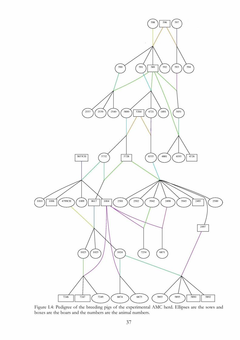
37
Figure I.4: Pedigree of the breeding pigs of the experimental AMC herd. Ellipses are the sows and boxes are the boars and the numbers are the animal numbers.
38
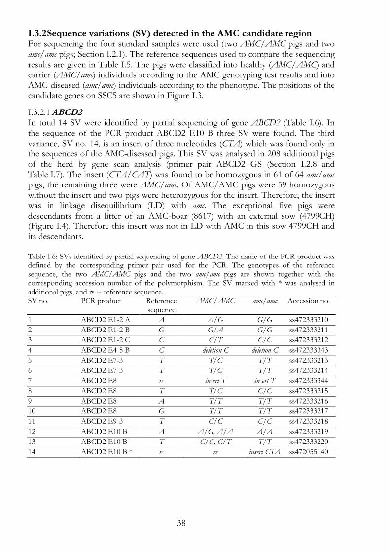
I.3.2 Sequence variations (SV) detected in the AMC candidate region For sequencing the four standard samples were used (two AMC/AMC pigs and two amc/amc pigs; Section I.2.1). The reference sequences used to compare the sequencing results are given in Table I.5. The pigs were classified into healthy (AMC/AMC) and carrier (AMC/amc) individuals according to the AMC genotyping test results and into AMC-diseased (amc/amc) individuals according to the phenotype. The positions of the candidate genes on SSC5 are shown in Figure I.3.
ABCD2 I.3.2.1In total 14 SV were identified by partial sequencing of gene ABCD2 (Table I.6). In the sequence of the PCR product ABCD2 E10 B three SV were found. The third variance, SV no. 14, is an insert of three nucleotides (CTA) which was found only in the sequences of the AMC-diseased pigs. This SV was analysed in 208 additional pigs of the herd by gene scan analysis (primer pair ABCD2 GS (Section I.2.8 and Table I.7). The insert (CTA/CAT) was found to be homozygous in 61 of 64 amc/amc pigs, the remaining three were AMC/amc. Of AMC/AMC pigs were 59 homozygous without the insert and two pigs were heterozygous for the insert. Therefore, the insert was in linkage disequilibrium (LD) with amc. The exceptional five pigs were descendants from a litter of an AMC-boar (8617) with an external sow (4799CH) (Figure I.4). Therefore this insert was not in LD with AMC in this sow 4799CH and its descendants. Table I.6: SVs identified by partial sequencing of gene ABCD2. The name of the PCR product was defined by the corresponding primer pair used for the PCR. The genotypes of the reference sequence, the two AMC/AMC pigs and the two amc/amc pigs are shown together with the corresponding accession number of the polymorphism. The SV marked with * was analysed in additional pigs, and rs = reference sequence. SV no. PCR product Reference
sequence AMC/AMC amc/amc Accession no.
1 ABCD2 E1-2 A A A/G G/G ss472333210
2 ABCD2 E1-2 B G G/A G/G ss472333211
3 ABCD2 E1-2 C C C/T C/C ss472333212
4 ABCD2 E4-5 B C deletion C deletion C ss472333343
5 ABCD2 E7-3 T T/C T/T ss472333213
6 ABCD2 E7-3 T T/C T/T ss472333214
7 ABCD2 E8 rs insert T insert T ss472333344
8 ABCD2 E8 T T/C C/C ss472333215
9 ABCD2 E8 A T/T T/T ss472333216
10 ABCD2 E8 G T/T T/T ss472333217
11 ABCD2 E9-3 T C/C C/C ss472333218
12 ABCD2 E10 B A A/G, A/A A/A ss472333219
13 ABCD2 E10 B T C/C, C/T T/T ss472333220
14 ABCD2 E10 B * rs rs insert CTA ss472055140
39
Table I.7: The potentially interesting SV in gene ABCD2 was used for analysis in additional pigs. The SV position (bp) was mapped according to Sscrofa10.2 genome assembly, and rs = reference sequence. PCR Product Position Polymorphism AMC/AMC AMC/amc amc/amc
ABCD2 GS (SV no. 14)
73'681'279 rs/rs 59 0 0 rs/CTA 2 83 0
CTA/CTA 0 3 61
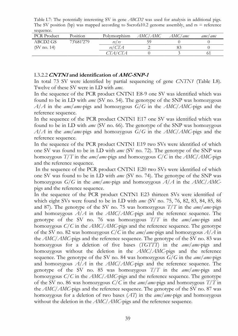
CNTN1 and identification of AMC-SNP-1 I.3.2.2In total 73 SV were identified by partial sequencing of gene CNTN1 (Table I.8). Twelve of these SV were in LD with amc. In the sequence of the PCR product CNTN1 E8-9 one SV was identified which was found to be in LD with amc (SV no. 54). The genotype of the SNP was homozygous A/A in the amc/amc-pigs and homozygous G/G in the AMC/AMC-pigs and the reference sequence. In the sequence of the PCR product CNTN1 E17 one SV was identified which was found to be in LD with amc (SV no. 66). The genotype of the SNP was homozygous A/A in the amc/amc-pigs and homozygous G/G in the AMC/AMC-pigs and the reference sequence. In the sequence of the PCR product CNTN1 E19 two SVs were identified of which one SV was found to be in LD with amc (SV no. 72). The genotype of the SNP was homozygous T/T in the amc/amc-pigs and homozygous C/C in the AMC/AMC-pigs and the reference sequence. In the sequence of the PCR product CNTN1 E20 two SVs were identified of which one SV was found to be in LD with amc (SV no. 74). The genotype of the SNP was homozygous G/G in the amc/amc-pigs and homozygous A/A in the AMC/AMC-pigs and the reference sequence. In the sequence of the PCR product CNTN1 E23 thirteen SVs were identified of which eight SVs were found to be in LD with amc (SV no. 75, 76, 82, 83, 84, 85, 86 and 87). The genotype of the SV no. 75 was homozygous T/T in the amc/amc-pigs and homozygous A/A in the AMC/AMC-pigs and the reference sequence. The genotype of the SV no. 76 was homozygous T/T in the amc/amc-pigs and homozygous C/C in the AMC/AMC-pigs and the reference sequence. The genotype of the SV no. 82 was homozygous C/C in the amc/amc-pigs and homozygous A/A in the AMC/AMC-pigs and the reference sequence. The genotype of the SV no. 83 was homozygous for a deletion of five bases (TGTTT) in the amc/amc-pigs and homozygous without the deletion in the AMC/AMC-pigs and the reference sequence. The genotype of the SV no. 84 was homozygous G/G in the amc/amc-pigs and homozygous A/A in the AMC/AMC-pigs and the reference sequence. The genotype of the SV no. 85 was homozygous T/T in the amc/amc-pigs and homozygous C/C in the AMC/AMC-pigs and the reference sequence. The genotype of the SV no. 86 was homozygous C/C in the amc/amc-pigs and homozygous T/T in the AMC/AMC-pigs and the reference sequence. The genotype of the SV no. 87 was homozygous for a deletion of two bases (AT) in the amc/amc-pigs and homozygous without the deletion in the AMC/AMC-pigs and the reference sequence.
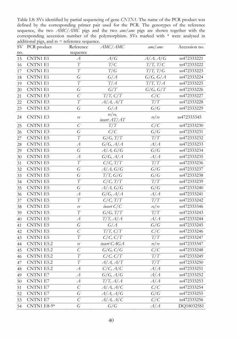
40
Table I.8: SVs identified by partial sequencing of gene CNTN1. The name of the PCR product was defined by the corresponding primer pair used for the PCR. The genotypes of the reference sequence, the two AMC/AMC pigs and the two amc/amc pigs are shown together with the corresponding accession number of the polymorphism. SVs marked with * were analysed in additional pigs, and rs = reference sequence. SV no.
PCR product Reference sequence
AMC/AMC amc/amc Accession no.
15 CNTN1 E1 A A/G A/A, A/G ss472333221
16 CNTN1 E1 T T/C T/T, T/C ss472333222
17 CNTN1 E1 T T/G T/T, T/G ss472333223
18 CNTN1 E1 G G/A G/G, G/A ss472333224
19 CNTN1 E1 T T/A T/T, T/A ss472333225
20 CNTN1 E1 G G/T G/G, G/T ss472333226
21 CNTN1 E3 C T/T, C/T C/C ss472333227
22 CNTN1 E3 T A/A, A/T T/T ss472333228
23 CNTN1 E3 G G/A G/G ss472333229
24 CNTN1 E3 rs rs/rs,
insert AT/AT rs/rs ss472333345
25 CNTN1 E3 C T/T C/C ss472333230
26 CNTN1 E3 G C/C G/G ss472333231
27 CNTN1 E5 T G/G, T/T T/T ss472333232
28 CNTN1 E5 A G/G, A/A A/A ss472333233
29 CNTN1 E5 G A/A, G/G G/G ss472333234
30 CNTN1 E5 A G/G, A/A A/A ss472333235
31 CNTN1 E5 T C/C, T/T T/T ss472333236
32 CNTN1 E5 G A/A, G/G G/G ss472333237
33 CNTN1 E5 G T/T, G/G G/G ss472333238
34 CNTN1 E5 T C/C, T/T T/T ss472333239
35 CNTN1 E5 G A/A, G/G G/G ss472333240
36 CNTN1 E5 A G/G, A/A A/A ss472333241
37 CNTN1 E5 T C/C, T/T T/T ss472333242
38 CNTN1 E5 rs insert C/C rs/rs ss472333346
39 CNTN1 E5 T G/G, T/T T/T ss472333243
40 CNTN1 E5 A T/T, A/A A/A ss472333244
41 CNTN1 E5 G G/A G/G ss472333245
42 CNTN1 E5 C T/T, C/T C/C ss472333246
43 CNTN1 E5 T C/C, C/T T/T ss472333247
44 CNTN1 E5.2 rs insert CAGA rs/rs ss472333347
45 CNTN1 E5.2 C G/G, C/G C/C ss472333248
46 CNTN1 E5.2 T C/C, C/T T/T ss472333249
47 CNTN1 E5.2 T A/A, A/T T/T ss472333250
48 CNTN1 E5.2 A C/C, A/C A/A ss472333251
49 CNTN1 E7 A G/G, A/G A/A ss472333252
50 CNTN1 E7 A T/T, A/A A/A ss472333253
51 CNTN1 E7 C A/A, A/C C/C ss472333254
52 CNTN1 E7 G A/A, A/G G/G ss472333255
53 CNTN1 E7 C A/A, A/C C/C ss472333256
54 CNTN1 E8-9* G G/G A/A DQ180325S1
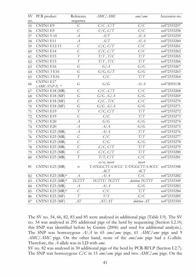
41
SV no.
PCR product Reference sequence
AMC/AMC amc/amc Accession no.
55 CNTN1 E9 C C/C , C/T C/C ss472333257
56 CNTN1 E9 C C/C, C/T C/C ss472333258
57 CNTN1 9-10 A A/T A/A ss472333259
58 CNTN1 E11 A A/T A/A ss472333260
59 CNTN1 E12-13 C C/C, C/T C/C ss472333261
60 CNTN1 E14 C C/C, C/T C/C ss472333262
61 CNTN1 E15 T T/T , T/C T/T ss472333265
62 CNTN1 E15 T T/T , T/C T/T ss472333266
63 CNTN1 E16 G G/A G/G ss472333267
64 CNTN1-3 E16 G G/G, G/T G/G ss472333263
65 CNTN1-3 E16 T C/C T/T ss472333264
66 CNTN1 E17 (AMC-SNP-1) *
G G/G A/A ss472055138
67 CNTN1 E18 (MR) C C/C , C/T C/C ss472333268
68 CNTN1 E18 (MF) G G/G , G/A G/G ss472333269
69 CNTN1 E18 (MF) C C/C , T/C C/C ss472333270
70 CNTN1 E18 (MF) G G/G , G/A G/G ss472333271
71 CNTN1 E19 T C/C, C/T T/T ss472333272
72 CNTN1 E19 C C/C T/T ss472333273
73 CNTN1 E20 A G/G G/G ss472333274
74 CNTN1 E20 A A/A G/G ss472333275
75 CNTN1 E23 (MR) A A/A T/T ss472333276
76 CNTN1 E23 (MR) C C/C T/T ss472333277
77 CNTN1 E23 (MR) C C/G G/G ss472333278
78 CNTN1 E23 (MR) C C/C, C/T T/T ss472333279
79 CNTN1 E23 (MR) C C/C, C/T T/T ss472333280
80 CNTN1 E23 (MR) T T/T, C/T C/C ss472333281
81 CNTN1 E23 (MR) rs rs/rs, insert
TATGGCTTAACCCACT
insert TATGGCTTAACCC
ACT ss472333348
82 CNTN1 E23 (MR)* A A/A C/C ss472333282
83 CNTN1 E23 (MR)* TGTTT TGTTT/ TGTTT deletion TGTTT ss472333349
84 CNTN1 E23 (MR) A A/A G/G ss472333283
85 CNTN1 E23 (MR)* C C/C T/T ss472333284
86 CNTN1 E23 (MF) T T/T C/C ss472333285
87 CNTN1 E23 (MF) AT AT/AT deletion AT ss472333350
The SV no. 54, 66, 82, 83 and 85 were analysed in additional pigs (Table I.9). The SV no. 54 was analysed in 295 additional pigs of the herd by sequencing (Section I.2.10, this SNP was identified before by Genini (2006) and used for additional analysis.). The SNP was homozygous A/A in 65 amc/amc pigs, 61 AMC/amc pigs and 9 AMC/AMC pigs. On the other hand, none of the amc/amc pigs had a G-allele. Therefore, the A-allele was in LD with amc. SV no. 82 was analysed in 30 additional pigs of the herd by PCR-RFLP (Section I.2.7). The SNP was homozygous C/C in 15 amc/amc pigs and two AMC/amc pigs. On the
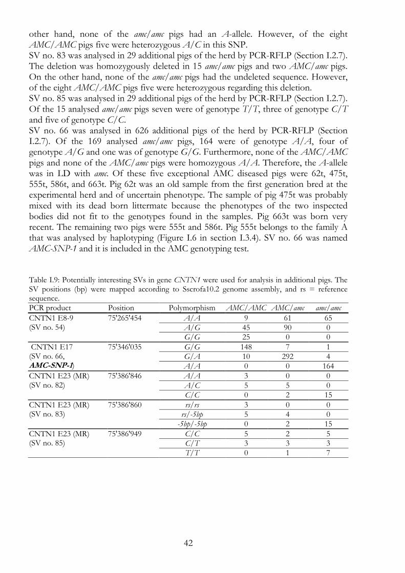
42
other hand, none of the amc/amc pigs had an A-allele. However, of the eight AMC/AMC pigs five were heterozygous A/C in this SNP. SV no. 83 was analysed in 29 additional pigs of the herd by PCR-RFLP (Section I.2.7). The deletion was homozygously deleted in 15 amc/amc pigs and two AMC/amc pigs. On the other hand, none of the amc/amc pigs had the undeleted sequence. However, of the eight AMC/AMC pigs five were heterozygous regarding this deletion. SV no. 85 was analysed in 29 additional pigs of the herd by PCR-RFLP (Section I.2.7). Of the 15 analysed amc/amc pigs seven were of genotype T/T, three of genotype C/T and five of genotype C/C. SV no. 66 was analysed in 626 additional pigs of the herd by PCR-RFLP (Section I.2.7). Of the 169 analysed amc/amc pigs, 164 were of genotype A/A, four of genotype A/G and one was of genotype G/G. Furthermore, none of the AMC/AMC pigs and none of the AMC/amc pigs were homozygous A/A. Therefore, the A-allele was in LD with amc. Of these five exceptional AMC diseased pigs were 62t, 475t, 555t, 586t, and 663t. Pig 62t was an old sample from the first generation bred at the experimental herd and of uncertain phenotype. The sample of pig 475t was probably mixed with its dead born littermate because the phenotypes of the two inspected bodies did not fit to the genotypes found in the samples. Pig 663t was born very recent. The remaining two pigs were 555t and 586t. Pig 555t belongs to the family A that was analysed by haplotyping (Figure I.6 in section I.3.4). SV no. 66 was named AMC-SNP-1 and it is included in the AMC genotyping test. Table I.9: Potentially interesting SVs in gene CNTN1 were used for analysis in additional pigs. The SV positions (bp) were mapped according to Sscrofa10.2 genome assembly, and rs = reference sequence. PCR product Position Polymorphism AMC/AMC AMC/amc amc/amc
CNTN1 E8-9 (SV no. 54)
75'265'454
A/A 9 61 65 A/G 45 90 0 G/G 25 0 0
CNTN1 E17 (SV no. 66, AMC-SNP-1)
75'346'035 G/G 148 7 1 G/A 10 292 4 A/A 0 0 164
CNTN1 E23 (MR) (SV no. 82)
75'386'846 A/A 3 0 0 A/C 5 5 0 C/C 0 2 15
CNTN1 E23 (MR) (SV no. 83)
75'386'860 rs/rs 3 0 0 rs/-5bp 5 4 0 -5bp/-5bp 0 2 15
CNTN1 E23 (MR) (SV no. 85)
75'386'949 C/C 5 2 5 C/T 3 3 3 T/T 0 1 7
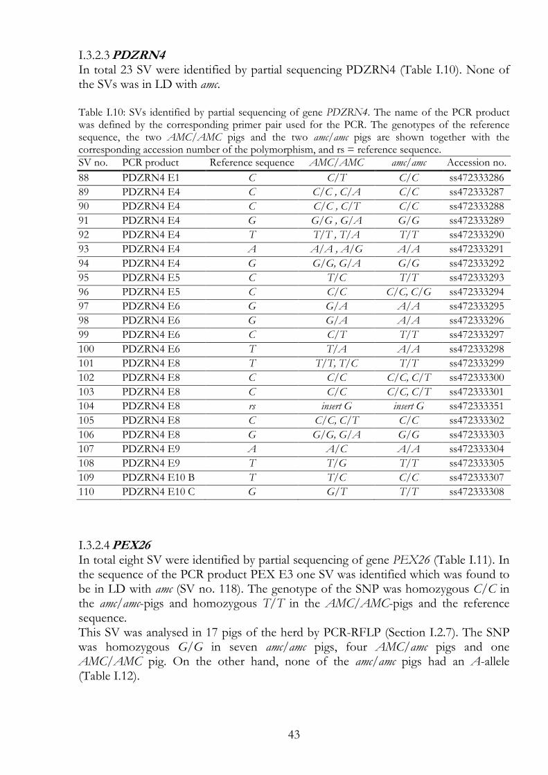
43
PDZRN4 I.3.2.3In total 23 SV were identified by partial sequencing PDZRN4 (Table I.10). None of the SVs was in LD with amc. Table I.10: SVs identified by partial sequencing of gene PDZRN4. The name of the PCR product was defined by the corresponding primer pair used for the PCR. The genotypes of the reference sequence, the two AMC/AMC pigs and the two amc/amc pigs are shown together with the corresponding accession number of the polymorphism, and rs = reference sequence. SV no. PCR product Reference sequence AMC/AMC amc/amc Accession no.
88 PDZRN4 E1 C C/T C/C ss472333286
89 PDZRN4 E4 C C/C , C/A C/C ss472333287
90 PDZRN4 E4 C C/C , C/T C/C ss472333288
91 PDZRN4 E4 G G/G , G/A G/G ss472333289
92 PDZRN4 E4 T T/T , T/A T/T ss472333290
93 PDZRN4 E4 A A/A , A/G A/A ss472333291
94 PDZRN4 E4 G G/G, G/A G/G ss472333292
95 PDZRN4 E5 C T/C T/T ss472333293
96 PDZRN4 E5 C C/C C/C, C/G ss472333294
97 PDZRN4 E6 G G/A A/A ss472333295
98 PDZRN4 E6 G G/A A/A ss472333296
99 PDZRN4 E6 C C/T T/T ss472333297
100 PDZRN4 E6 T T/A A/A ss472333298
101 PDZRN4 E8 T T/T, T/C T/T ss472333299
102 PDZRN4 E8 C C/C C/C, C/T ss472333300
103 PDZRN4 E8 C C/C C/C, C/T ss472333301
104 PDZRN4 E8 rs insert G insert G ss472333351
105 PDZRN4 E8 C C/C, C/T C/C ss472333302
106 PDZRN4 E8 G G/G, G/A G/G ss472333303
107 PDZRN4 E9 A A/C A/A ss472333304
108 PDZRN4 E9 T T/G T/T ss472333305
109 PDZRN4 E10 B T T/C C/C ss472333307
110 PDZRN4 E10 C G G/T T/T ss472333308
PEX26 I.3.2.4In total eight SV were identified by partial sequencing of gene PEX26 (Table I.11). In the sequence of the PCR product PEX E3 one SV was identified which was found to be in LD with amc (SV no. 118). The genotype of the SNP was homozygous C/C in the amc/amc-pigs and homozygous T/T in the AMC/AMC-pigs and the reference sequence. This SV was analysed in 17 pigs of the herd by PCR-RFLP (Section I.2.7). The SNP was homozygous G/G in seven amc/amc pigs, four AMC/amc pigs and one AMC/AMC pig. On the other hand, none of the amc/amc pigs had an A-allele (Table I.12).
44
Table I.11: SVs identified by partial sequencing of gene PEX26. The name of the PCR product was defined by the corresponding primer pair used for the PCR. The genotypes of the reference sequence, the two AMC/AMC pigs and the two amc/amc pigs are shown together with the corresponding accession number of the polymorphism. The SV marked with * was analysed in additional pigs. SV no. PCR product Reference sequence AMC/AMC amc/amc Accession no.
111 PEX E0 T T/A T/A ss472333309
112 PEX E0 G G/A G/A ss472333310
113 PEX E0 T T/A T/A ss472333311
114 PEX E0 C C/A C/A ss472333312
115 PEX E0 G G/A G/A ss472333313
116 PEX E0 T T/C T/C ss472333314
117 PEX E0 C C/A C/C ss472333315
118 PEX E3 * T T/T C/C ss472333316
Table I.12 The potentially interesting SV in gene PEX26 was used for analysis in additional pigs. The SV position (bp) was mapped according to Sscrofa10.2 genome assembly. Primer pair Position Polymorphism AMC/AMC AMC/amc amc/amc
PEX E3 (SV no. 118) 72'489'157 A/A 1 0 0 A/G 3 1 0 G/G 1 4 7
RNF217 - a sequence of 416 bp at the 5' side of RNF217 I.3.2.5In total five SV were identified by partial sequencing of the 416 bp at the 5' side of RNF217 (Table I.13). In the sequence of the PCR product RFN217 5'like all five SVs were identified of which four SVs were found to be in LD with amc (SV no. 119, 120, 122 and 123). The genotype of SV no. 119 was homozygous T/T in the amc/amc-pigs and homozygous C/C in the AMC/AMC-pigs and the reference sequence. The genotype of SV no. 120 was homozygous T/T in the amc/amc-pigs and homozygous C/C in the AMC/AMC-pigs and the reference sequence. The genotype of SV no. 122 was homozygous C/C in the amc/amc-pigs and homozygous G/G in the AMC/AMC-pigs and the reference sequence. The genotype of SV no. 122 was homozygous A/A in the amc/amc-pigs and homozygous G/G in the AMC/AMC-pigs and the reference sequence. The SVs no. 120 and 122 were analysed together in 18 additional pigs of the herd by PCR-RFLP (Section I.2.7). The SNPs were homozygous T/T and C/C in eight amc/amc pigs. Furthermore, none of the amc/amc pig had one of the other alleles. However, of the four AMC/AMC pigs two were heterozygous in these SNPs (Table I.14).
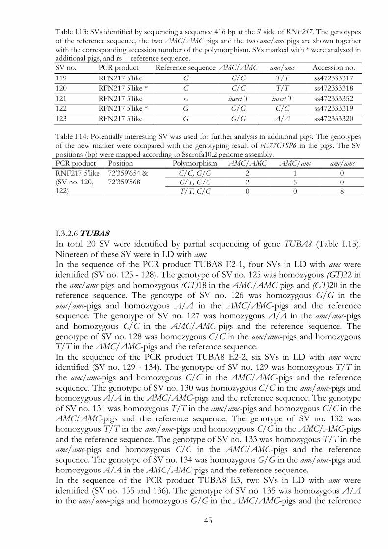
45
Table I.13: SVs identified by sequencing a sequence 416 bp at the 5' side of RNF217. The genotypes of the reference sequence, the two AMC/AMC pigs and the two amc/amc pigs are shown together with the corresponding accession number of the polymorphism. SVs marked with * were analysed in additional pigs, and rs = reference sequence. SV no. PCR product Reference sequence AMC/AMC amc/amc Accession no.
119 RFN217 5'like C C/C T/T ss472333317
120 RFN217 5'like * C C/C T/T ss472333318
121 RFN217 5'like rs insert T insert T ss472333352
122 RFN217 5'like * G G/G C/C ss472333319
123 RFN217 5'like G G/G A/A ss472333320
Table I.14: Potentially interesting SV was used for further analysis in additional pigs. The genotypes of the new marker were compared with the genotyping result of bE77C1SP6 in the pigs. The SV positions (bp) were mapped according to Sscrofa10.2 genome assembly. PCR product Position Polymorphism AMC/AMC AMC/amc amc/amc
RNF217 5'like (SV no. 120, 122)
72'359'654 & 72'359'568
C/C, G/G 2 1 0 C/T, G/C 2 5 0 T/T, C/C 0 0 8
TUBA8 I.3.2.6In total 20 SV were identified by partial sequencing of gene TUBA8 (Table I.15). Nineteen of these SV were in LD with amc. In the sequence of the PCR product TUBA8 E2-1, four SVs in LD with amc were identified (SV no. 125 - 128). The genotype of SV no. 125 was homozygous (GT)22 in the amc/amc-pigs and homozygous (GT)18 in the AMC/AMC-pigs and (GT)20 in the reference sequence. The genotype of SV no. 126 was homozygous G/G in the amc/amc-pigs and homozygous A/A in the AMC/AMC-pigs and the reference sequence. The genotype of SV no. 127 was homozygous A/A in the amc/amc-pigs and homozygous C/C in the AMC/AMC-pigs and the reference sequence. The genotype of SV no. 128 was homozygous C/C in the amc/amc-pigs and homozygous T/T in the AMC/AMC-pigs and the reference sequence. In the sequence of the PCR product TUBA8 E2-2, six SVs in LD with amc were identified (SV no. 129 - 134). The genotype of SV no. 129 was homozygous T/T in the amc/amc-pigs and homozygous C/C in the AMC/AMC-pigs and the reference sequence. The genotype of SV no. 130 was homozygous C/C in the amc/amc-pigs and homozygous A/A in the AMC/AMC-pigs and the reference sequence. The genotype of SV no. 131 was homozygous T/T in the amc/amc-pigs and homozygous C/C in the AMC/AMC-pigs and the reference sequence. The genotype of SV no. 132 was homozygous T/T in the amc/amc-pigs and homozygous C/C in the AMC/AMC-pigs and the reference sequence. The genotype of SV no. 133 was homozygous T/T in the amc/amc-pigs and homozygous C/C in the AMC/AMC-pigs and the reference sequence. The genotype of SV no. 134 was homozygous G/G in the amc/amc-pigs and homozygous A/A in the AMC/AMC-pigs and the reference sequence. In the sequence of the PCR product TUBA8 E3, two SVs in LD with amc were identified (SV no. 135 and 136). The genotype of SV no. 135 was homozygous A/A in the amc/amc-pigs and homozygous G/G in the AMC/AMC-pigs and the reference
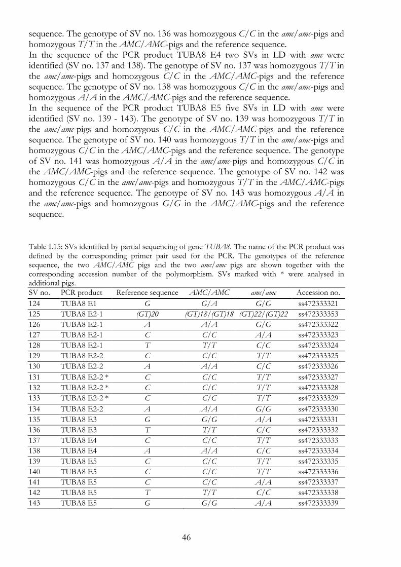
46
sequence. The genotype of SV no. 136 was homozygous C/C in the amc/amc-pigs and homozygous T/T in the AMC/AMC-pigs and the reference sequence. In the sequence of the PCR product TUBA8 E4 two SVs in LD with amc were identified (SV no. 137 and 138). The genotype of SV no. 137 was homozygous T/T in the amc/amc-pigs and homozygous C/C in the AMC/AMC-pigs and the reference sequence. The genotype of SV no. 138 was homozygous C/C in the amc/amc-pigs and homozygous A/A in the AMC/AMC-pigs and the reference sequence. In the sequence of the PCR product TUBA8 E5 five SVs in LD with amc were identified (SV no. 139 - 143). The genotype of SV no. 139 was homozygous T/T in the amc/amc-pigs and homozygous C/C in the AMC/AMC-pigs and the reference sequence. The genotype of SV no. 140 was homozygous T/T in the amc/amc-pigs and homozygous C/C in the AMC/AMC-pigs and the reference sequence. The genotype of SV no. 141 was homozygous A/A in the amc/amc-pigs and homozygous C/C in the AMC/AMC-pigs and the reference sequence. The genotype of SV no. 142 was homozygous C/C in the amc/amc-pigs and homozygous T/T in the AMC/AMC-pigs and the reference sequence. The genotype of SV no. 143 was homozygous A/A in the amc/amc-pigs and homozygous G/G in the AMC/AMC-pigs and the reference sequence.
Table I.15: SVs identified by partial sequencing of gene TUBA8. The name of the PCR product was defined by the corresponding primer pair used for the PCR. The genotypes of the reference sequence, the two AMC/AMC pigs and the two amc/amc pigs are shown together with the corresponding accession number of the polymorphism. SVs marked with * were analysed in additional pigs. SV no. PCR product Reference sequence AMC/AMC amc/amc Accession no.
124 TUBA8 E1 G G/A G/G ss472333321
125 TUBA8 E2-1 (GT)20 (GT)18/(GT)18 (GT)22/(GT)22 ss472333353
126 TUBA8 E2-1 A A/A G/G ss472333322
127 TUBA8 E2-1 C C/C A/A ss472333323
128 TUBA8 E2-1 T T/T C/C ss472333324
129 TUBA8 E2-2 C C/C T/T ss472333325
130 TUBA8 E2-2 A A/A C/C ss472333326
131 TUBA8 E2-2 * C C/C T/T ss472333327
132 TUBA8 E2-2 * C C/C T/T ss472333328
133 TUBA8 E2-2 * C C/C T/T ss472333329
134 TUBA8 E2-2 A A/A G/G ss472333330
135 TUBA8 E3 G G/G A/A ss472333331
136 TUBA8 E3 T T/T C/C ss472333332
137 TUBA8 E4 C C/C T/T ss472333333
138 TUBA8 E4 A A/A C/C ss472333334
139 TUBA8 E5 C C/C T/T ss472333335
140 TUBA8 E5 C C/C T/T ss472333336
141 TUBA8 E5 C C/C A/A ss472333337
142 TUBA8 E5 T T/T C/C ss472333338
143 TUBA8 E5 G G/G A/A ss472333339
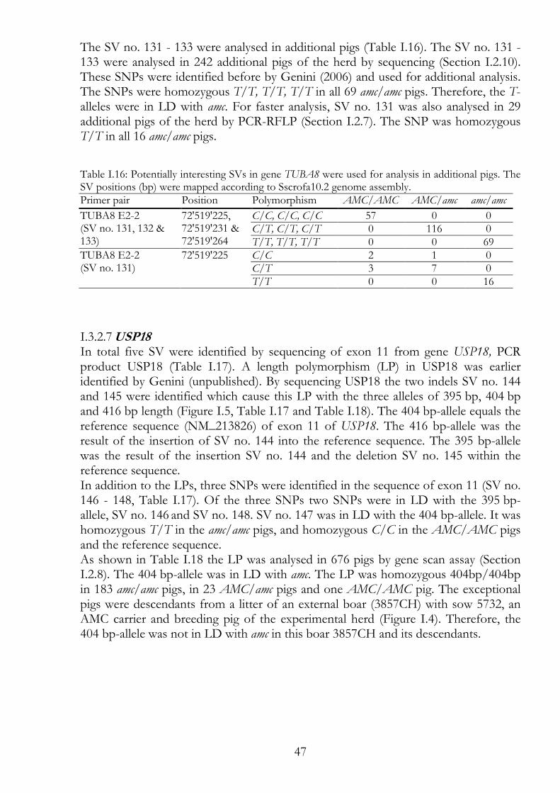
47
The SV no. 131 - 133 were analysed in additional pigs (Table I.16). The SV no. 131 - 133 were analysed in 242 additional pigs of the herd by sequencing (Section I.2.10). These SNPs were identified before by Genini (2006) and used for additional analysis. The SNPs were homozygous T/T, T/T, T/T in all 69 amc/amc pigs. Therefore, the T-alleles were in LD with amc. For faster analysis, SV no. 131 was also analysed in 29 additional pigs of the herd by PCR-RFLP (Section I.2.7). The SNP was homozygous T/T in all 16 amc/amc pigs.
Table I.16: Potentially interesting SVs in gene TUBA8 were used for analysis in additional pigs. The SV positions (bp) were mapped according to Sscrofa10.2 genome assembly. Primer pair Position Polymorphism AMC/AMC AMC/amc amc/amc
TUBA8 E2-2 (SV no. 131, 132 & 133)
72'519'225, 72'519'231 & 72'519'264
C/C, C/C, C/C 57 0 0 C/T, C/T, C/T 0 116 0 T/T, T/T, T/T 0 0 69
TUBA8 E2-2 (SV no. 131)
72'519'225 C/C 2 1 0 C/T 3 7 0 T/T 0 0 16
USP18 I.3.2.7In total five SV were identified by sequencing of exon 11 from gene USP18, PCR product USP18 (Table I.17). A length polymorphism (LP) in USP18 was earlier identified by Genini (unpublished). By sequencing USP18 the two indels SV no. 144 and 145 were identified which cause this LP with the three alleles of 395 bp, 404 bp and 416 bp length (Figure I.5, Table I.17 and Table I.18). The 404 bp-allele equals the reference sequence (NM_213826) of exon 11 of USP18. The 416 bp-allele was the result of the insertion of SV no. 144 into the reference sequence. The 395 bp-allele was the result of the insertion SV no. 144 and the deletion SV no. 145 within the reference sequence. In addition to the LPs, three SNPs were identified in the sequence of exon 11 (SV no. 146 - 148, Table I.17). Of the three SNPs two SNPs were in LD with the 395 bp-allele, SV no. 146 and SV no. 148. SV no. 147 was in LD with the 404 bp-allele. It was homozygous T/T in the amc/amc pigs, and homozygous C/C in the AMC/AMC pigs and the reference sequence. As shown in Table I.18 the LP was analysed in 676 pigs by gene scan assay (Section I.2.8). The 404 bp-allele was in LD with amc. The LP was homozygous 404bp/404bp in 183 amc/amc pigs, in 23 AMC/amc pigs and one AMC/AMC pig. The exceptional pigs were descendants from a litter of an external boar (3857CH) with sow 5732, an AMC carrier and breeding pig of the experimental herd (Figure I.4). Therefore, the 404 bp-allele was not in LD with amc in this boar 3857CH and its descendants.
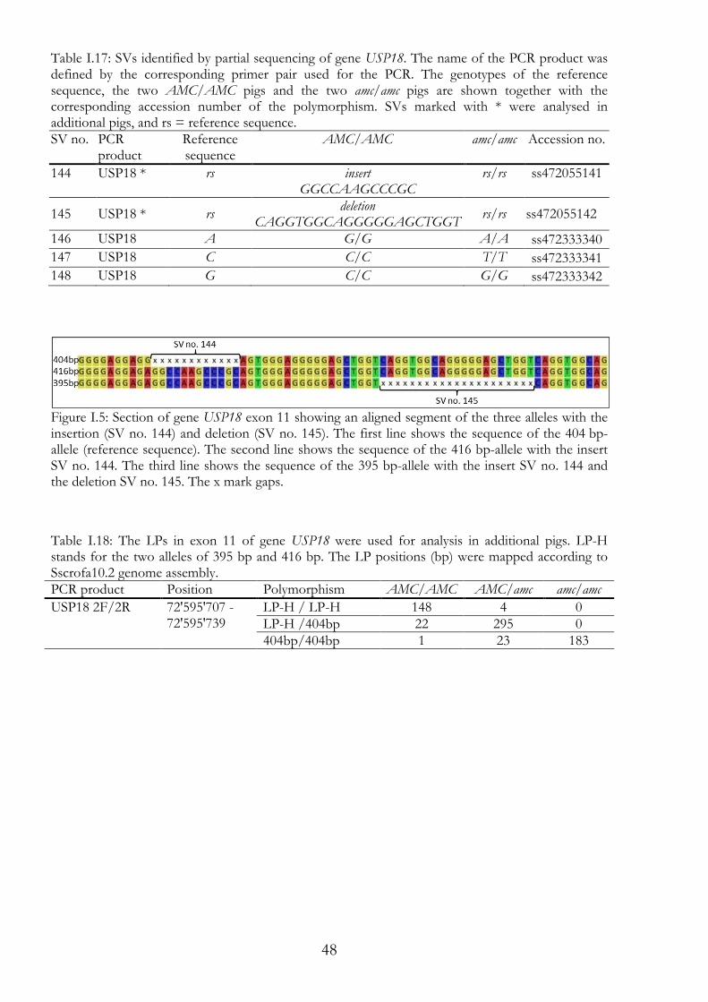
48
Table I.17: SVs identified by partial sequencing of gene USP18. The name of the PCR product was defined by the corresponding primer pair used for the PCR. The genotypes of the reference sequence, the two AMC/AMC pigs and the two amc/amc pigs are shown together with the corresponding accession number of the polymorphism. SVs marked with * were analysed in additional pigs, and rs = reference sequence. SV no. PCR
product Reference sequence
AMC/AMC amc/amc Accession no.
144 USP18 * rs insert GGCCAAGCCCGC
rs/rs ss472055141
145 USP18 * rs deletion
CAGGTGGCAGGGGGAGCTGGT rs/rs ss472055142
146 USP18 A G/G A/A ss472333340
147 USP18 C C/C T/T ss472333341
148 USP18 G C/C G/G ss472333342
Figure I.5: Section of gene USP18 exon 11 showing an aligned segment of the three alleles with the insertion (SV no. 144) and deletion (SV no. 145). The first line shows the sequence of the 404 bp-allele (reference sequence). The second line shows the sequence of the 416 bp-allele with the insert SV no. 144. The third line shows the sequence of the 395 bp-allele with the insert SV no. 144 and the deletion SV no. 145. The x mark gaps.
Table I.18: The LPs in exon 11 of gene USP18 were used for analysis in additional pigs. LP-H stands for the two alleles of 395 bp and 416 bp. The LP positions (bp) were mapped according to Sscrofa10.2 genome assembly. PCR product Position Polymorphism AMC/AMC AMC/amc amc/amc
USP18 2F/2R 72'595'707 -72'595'739
LP-H / LP-H 148 4 0 LP-H /404bp 22 295 0 404bp/404bp 1 23 183
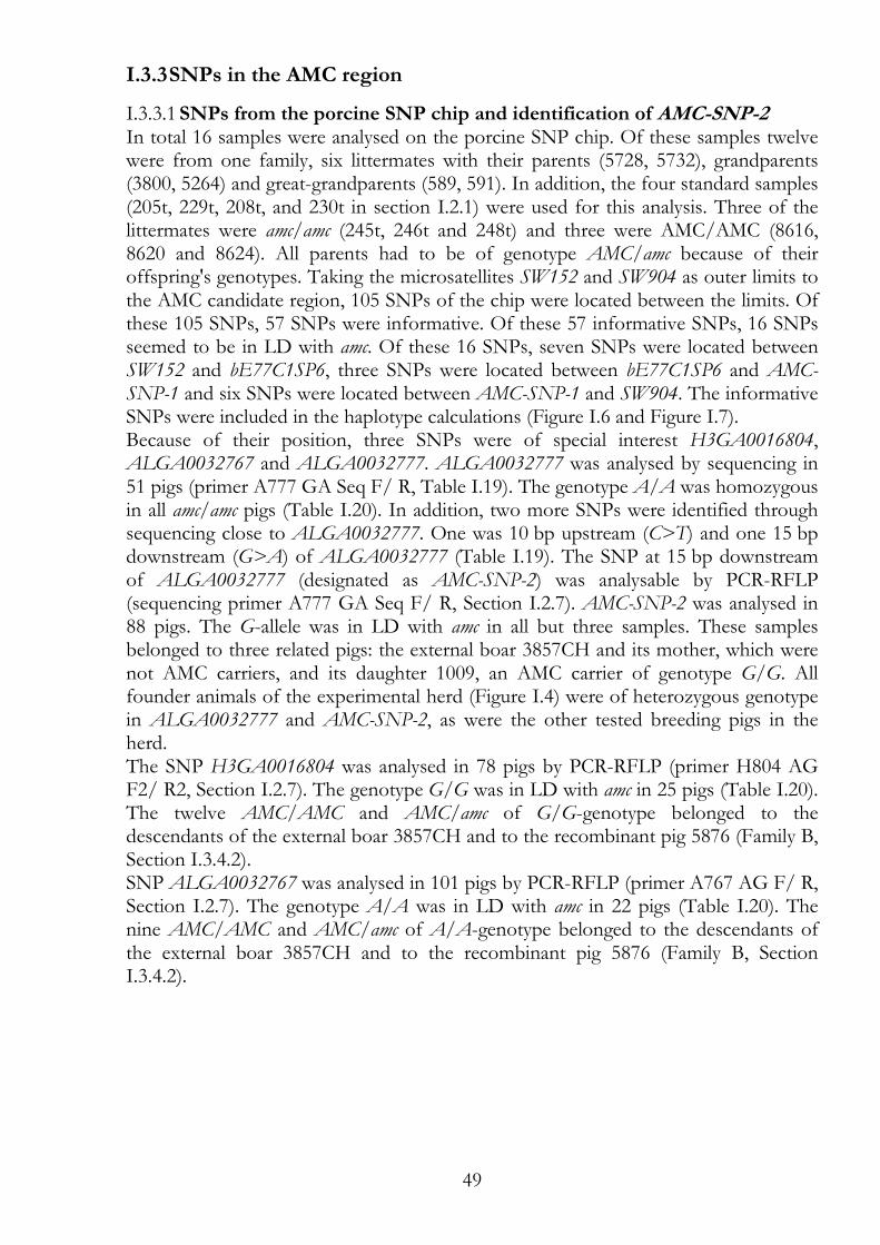
49
I.3.3 SNPs in the AMC region
SNPs from the porcine SNP chip and identification of AMC-SNP-2 I.3.3.1In total 16 samples were analysed on the porcine SNP chip. Of these samples twelve were from one family, six littermates with their parents (5728, 5732), grandparents (3800, 5264) and great-grandparents (589, 591). In addition, the four standard samples (205t, 229t, 208t, and 230t in section I.2.1) were used for this analysis. Three of the littermates were amc/amc (245t, 246t and 248t) and three were AMC/AMC (8616, 8620 and 8624). All parents had to be of genotype AMC/amc because of their offspring's genotypes. Taking the microsatellites SW152 and SW904 as outer limits to the AMC candidate region, 105 SNPs of the chip were located between the limits. Of these 105 SNPs, 57 SNPs were informative. Of these 57 informative SNPs, 16 SNPs seemed to be in LD with amc. Of these 16 SNPs, seven SNPs were located between SW152 and bE77C1SP6, three SNPs were located between bE77C1SP6 and AMC-SNP-1 and six SNPs were located between AMC-SNP-1 and SW904. The informative SNPs were included in the haplotype calculations (Figure I.6 and Figure I.7). Because of their position, three SNPs were of special interest H3GA0016804, ALGA0032767 and ALGA0032777. ALGA0032777 was analysed by sequencing in 51 pigs (primer A777 GA Seq F/ R, Table I.19). The genotype A/A was homozygous in all amc/amc pigs (Table I.20). In addition, two more SNPs were identified through sequencing close to ALGA0032777. One was 10 bp upstream (C>T) and one 15 bp downstream (G>A) of ALGA0032777 (Table I.19). The SNP at 15 bp downstream of ALGA0032777 (designated as AMC-SNP-2) was analysable by PCR-RFLP (sequencing primer A777 GA Seq F/ R, Section I.2.7). AMC-SNP-2 was analysed in 88 pigs. The G-allele was in LD with amc in all but three samples. These samples belonged to three related pigs: the external boar 3857CH and its mother, which were not AMC carriers, and its daughter 1009, an AMC carrier of genotype G/G. All founder animals of the experimental herd (Figure I.4) were of heterozygous genotype in ALGA0032777 and AMC-SNP-2, as were the other tested breeding pigs in the herd. The SNP H3GA0016804 was analysed in 78 pigs by PCR-RFLP (primer H804 AG F2/ R2, Section I.2.7). The genotype G/G was in LD with amc in 25 pigs (Table I.20). The twelve AMC/AMC and AMC/amc of G/G-genotype belonged to the descendants of the external boar 3857CH and to the recombinant pig 5876 (Family B, Section I.3.4.2). SNP ALGA0032767 was analysed in 101 pigs by PCR-RFLP (primer A767 AG F/ R, Section I.2.7). The genotype A/A was in LD with amc in 22 pigs (Table I.20). The nine AMC/AMC and AMC/amc of A/A-genotype belonged to the descendants of the external boar 3857CH and to the recombinant pig 5876 (Family B, Section I.3.4.2).
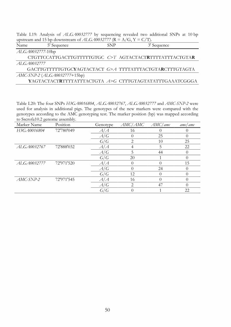
50
Table I.19: Analysis of ALGA0032777 by sequencing revealed two additional SNPs at 10 bp upstream and 15 bp downstream of ALGA0032777 (R = A/G, Y = C/T).
Table I.20: The four SNPs H3GA0016804, ALGA0032767, ALGA0032777 and AMC-SNP-2 were used for analysis in additional pigs. The genotypes of the new markers were compared with the genotypes according to the AMC genotyping test. The marker position (bp) was mapped according to Sscrofa10.2 genome assembly. Marker Name Position Genotype AMC/AMC AMC/amc amc/amc
H3GA0016804 72'780'049 A/A 16 0 0 A/G 0 25 0 G/G 2 10 25
ALGA0032767 72'888'032 A/A 4 5 22 A/G 5 44 0 G/G 20 1 0
ALGA0032777 72'971'520 A/A 0 0 15 A/G 0 24 0 G/G 12 0 0
AMC-SNP-2 72'971'545 A/A 16 0 0 A/G 2 47 0 G/G 0 1 22
Name 5' Sequence SNP 3' Sequence
ALGA0032777-10bp
CTGTTCCATTGACTTGTTTTTGTGC C>T AGTACTACTRTTTTATTTACTGTAR
ALGA0032777
GACTTGTTTTTGTGCYAGTACTACT G>A TTTTATTTACTGTARCTTTGTAGTA
AMC-SNP-2 (ALGA0032777+15bp)
YAGTACTACTRTTTTATTTACTGTA A>G CTTTGTAGTATATTTGAAATCGGGA
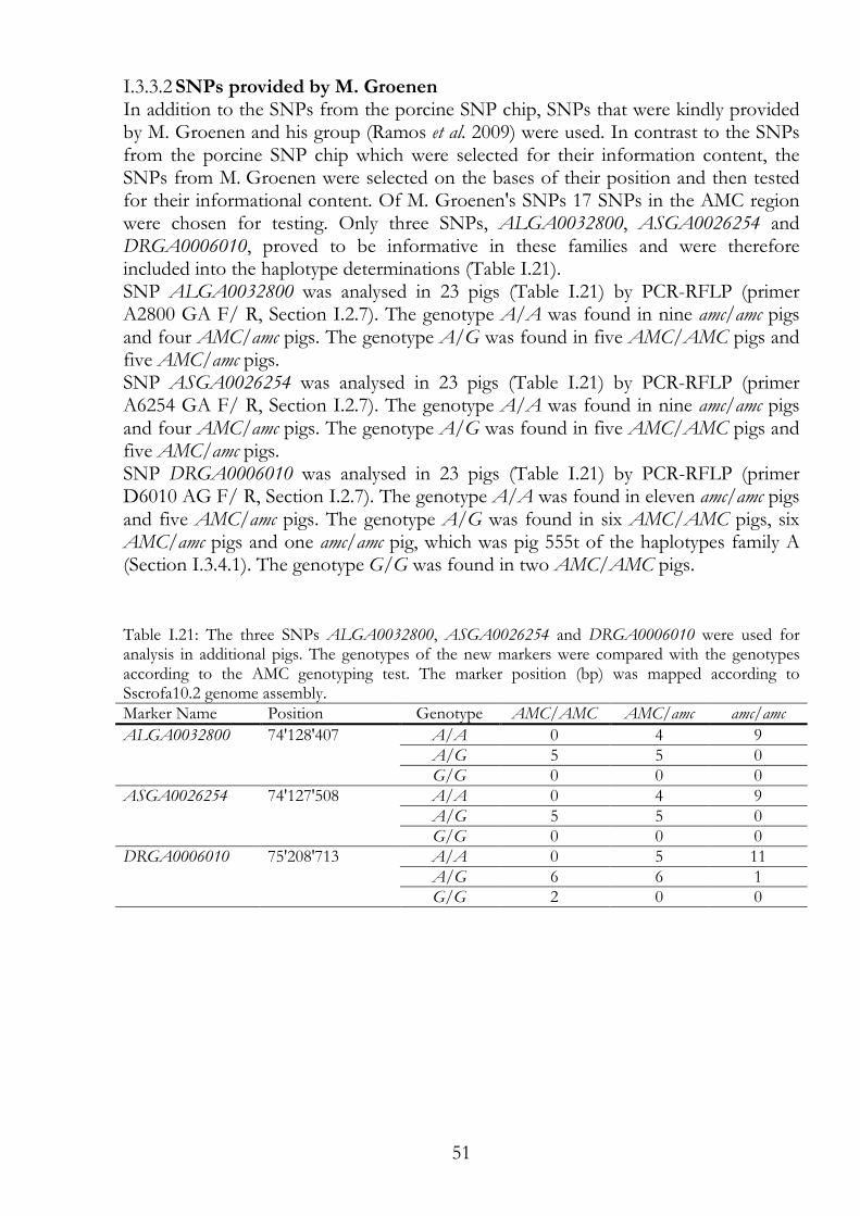
51
SNPs provided by M. Groenen I.3.3.2In addition to the SNPs from the porcine SNP chip, SNPs that were kindly provided by M. Groenen and his group (Ramos et al. 2009) were used. In contrast to the SNPs from the porcine SNP chip which were selected for their information content, the SNPs from M. Groenen were selected on the bases of their position and then tested for their informational content. Of M. Groenen's SNPs 17 SNPs in the AMC region were chosen for testing. Only three SNPs, ALGA0032800, ASGA0026254 and DRGA0006010, proved to be informative in these families and were therefore included into the haplotype determinations (Table I.21). SNP ALGA0032800 was analysed in 23 pigs (Table I.21) by PCR-RFLP (primer A2800 GA F/ R, Section I.2.7). The genotype A/A was found in nine amc/amc pigs and four AMC/amc pigs. The genotype A/G was found in five AMC/AMC pigs and five AMC/amc pigs. SNP ASGA0026254 was analysed in 23 pigs (Table I.21) by PCR-RFLP (primer A6254 GA F/ R, Section I.2.7). The genotype A/A was found in nine amc/amc pigs and four AMC/amc pigs. The genotype A/G was found in five AMC/AMC pigs and five AMC/amc pigs. SNP DRGA0006010 was analysed in 23 pigs (Table I.21) by PCR-RFLP (primer D6010 AG F/ R, Section I.2.7). The genotype A/A was found in eleven amc/amc pigs and five AMC/amc pigs. The genotype A/G was found in six AMC/AMC pigs, six AMC/amc pigs and one amc/amc pig, which was pig 555t of the haplotypes family A (Section I.3.4.1). The genotype G/G was found in two AMC/AMC pigs.
Table I.21: The three SNPs ALGA0032800, ASGA0026254 and DRGA0006010 were used for analysis in additional pigs. The genotypes of the new markers were compared with the genotypes according to the AMC genotyping test. The marker position (bp) was mapped according to Sscrofa10.2 genome assembly. Marker Name Position Genotype AMC/AMC AMC/amc amc/amc
ALGA0032800 74'128'407 A/A 0 4 9 A/G 5 5 0 G/G 0 0 0
ASGA0026254 74'127'508 A/A 0 4 9 A/G 5 5 0 G/G 0 0 0
DRGA0006010 75'208'713 A/A 0 5 11 A/G 6 6 1 G/G 2 0 0
52
I.3.4 Haplotype determination Several pedigrees were analysed but in two, families A and B, recombinations were identified in the AMC candidate region. We selected 25 consecutive markers (3 microsatellites, 19 SNPs and 2 indels) in the candidate region to determine the haplotypes in these two families, thereby refining the AMC region.
Family A of recombinant AMC-piglet 555t I.3.4.1Family A (Figure I.6) consisted of two healthy breeding pigs and their offspring. Boar 1004 and sow 1023 were both proven AMC carriers (amc/AMC). Five offspring are shown of which three were AMC diseased (amc/amc), 553t, 554t and 555t, and two pigs, 5182 and 5184, were healthy. Piglet 555t showed the typical phenotype of an AMC-diseased piglet and therefore, its genotype is considered homozygous amc/amc. However, the genotype of AMC-SNP-1 was heterozygous A/G. AMC-SNP-1 was one of the AMC test markers and its G-allele was usually associated with a healthy phenotype, whereas the A/A genotype was usually associated with amc/amc. The recombination occurred between the SNPs ALGA0032800 and DRGA0006010. Piglet 553t also had the typical phenotype of an AMC diseased piglet. Here, the recombination occurred between the SNPs H3GA0016838 and ALGA0032903. This result further supports our findings that the distal border of the AMC region is upstream of SW904. The diseased haplotypes in family A were inherited from only one ancestor 5732, therefore the amc genotype was identical-by-decent (autozygously inherited) in this family. Namely, sow 5732 is the mother of boar 1004 and the grand-mother of sow 1023 (Figure I.4).
Family B of recombinant carrier pig 5876 I.3.4.2Family B (Figure I.7) consisted of two siblings, boar 2497 and sow 2498, mated to each other and their offspring. Boar and sow were both proven AMC carriers (amc/AMC). Five offspring are shown. Pig 588t was AMC diseased and the littermates 5875, 5876, 5877 and 5879 were healthy. The genotype of pig 5876 was 6/6 (306bp/306bp) in the microsatellite bE77C1SP6. This genotype was usually associated with amc/amc. The recombination occurred between the SNPs ALGA0032767 and ALGA0032777. In the region upstream of ALGA0032777 the genotype resembles that of the diseased littermate 588t. In the region downstream of ALGA0032767 the genotype resembles that of the carrier littermate 5877.
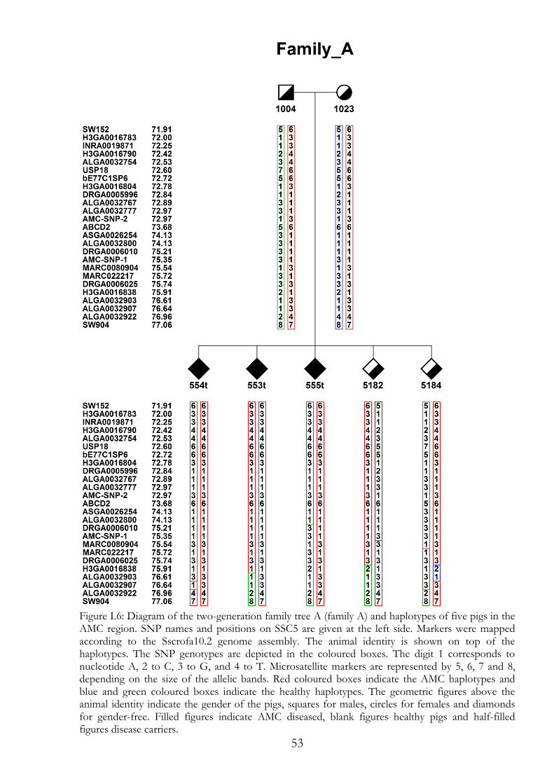
53
Figure I.6: Diagram of the two-generation family tree A (family A) and haplotypes of five pigs in the AMC region. SNP names and positions on SSC5 are given at the left side. Markers were mapped according to the Sscrofa10.2 genome assembly. The animal identity is shown on top of the haplotypes. The SNP genotypes are depicted in the coloured boxes. The digit 1 corresponds to nucleotide A, 2 to C, 3 to G, and 4 to T. Microsatellite markers are represented by 5, 6, 7 and 8, depending on the size of the allelic bands. Red coloured boxes indicate the AMC haplotypes and blue and green coloured boxes indicate the healthy haplotypes. The geometric figures above the animal identity indicate the gender of the pigs, squares for males, circles for females and diamonds for gender-free. Filled figures indicate AMC diseased, blank figures healthy pigs and half-filled figures disease carriers.
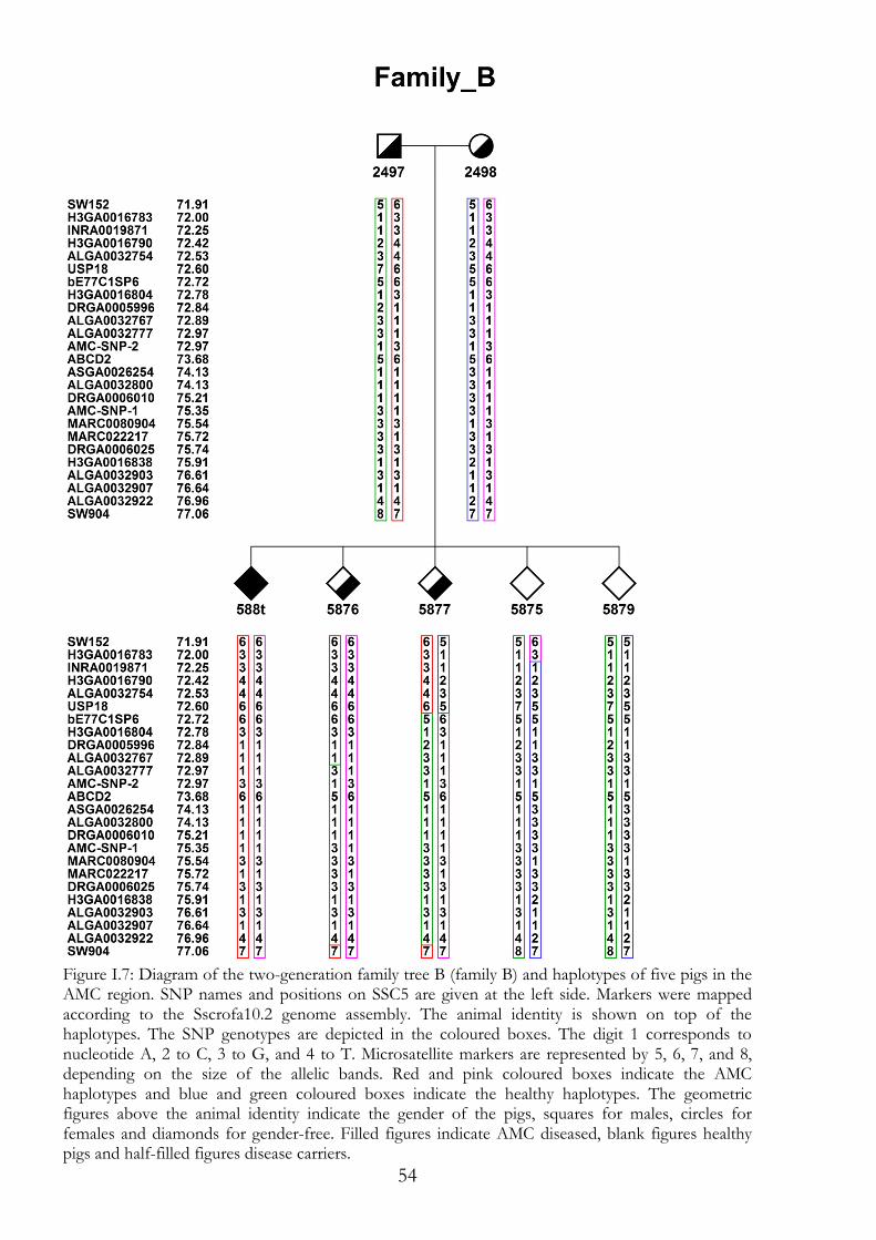
54
Figure I.7: Diagram of the two-generation family tree B (family B) and haplotypes of five pigs in the AMC region. SNP names and positions on SSC5 are given at the left side. Markers were mapped according to the Sscrofa10.2 genome assembly. The animal identity is shown on top of the haplotypes. The SNP genotypes are depicted in the coloured boxes. The digit 1 corresponds to nucleotide A, 2 to C, 3 to G, and 4 to T. Microsatellite markers are represented by 5, 6, 7, and 8, depending on the size of the allelic bands. Red and pink coloured boxes indicate the AMC haplotypes and blue and green coloured boxes indicate the healthy haplotypes. The geometric figures above the animal identity indicate the gender of the pigs, squares for males, circles for females and diamonds for gender-free. Filled figures indicate AMC diseased, blank figures healthy pigs and half-filled figures disease carriers.
55
I.3.5 The improved AMC genotyping test For the standard AMC genotyping test, bE77C1SP6 and SW904 were the default markers (Genini 2006). The 306bp-allele of bE77C1SP6 and the 172bp-allele and 180bp-allele of SW904 were in LD with amc in the experimental herd and also in pigs of commercial breeds. The LPs in the sequence of USP18 (Section 0) were analysed in addition to the two microsatellites. Since several recombinations between SW904 and amc have been observed, SW904 is least reliable of the markers. The A-allele of SNP AMC-SNP-1 was in LD with amc in the experimental herd (Section 0) and also in samples of commercial breeds. Therefore, AMC-SNP-1 was included into the improved AMC genotyping test used for both the experimental herd and commercial breeds. The SNPs ALGA0032777 and AMC-SNP-2 are closely located (Table I.19). The A-allele of SNP ALGA0032777 and the G-allele of AMC-SNP-2 are in LD with amc in the experimental herd and there, they are part of the improved AMC genotyping test (Table I.20). Since ALGA0032777 was selected as the SNP for the porcine SNP chip, its alleles are commonly distributed in commercial breeds. Therefore, the informational content of these two SNPs was limited to the experimental herd and certain pedigrees of commercial breeds.
Boar ´Genium´ I.3.5.1The boar ´Genium´ was from a commercial breeding line of boars. These boars were heavily used for artificial insemination in Switzerland and, up to that date, unsuspicious as far as AMC was concerned. Except that some members of this family had genotypes as if AMC diseased or at least AMC carriers. In spite of their genotypes, they were fully healthy, as were their recorded offspring. Five tested boars were great-grandsons of the founder. Of these, two boars had the genotypes of an AMC carrier and one of the two was ´Genium´. The genotypes of ´Genium´ were homozygous 306bp/306bp in bE77C1SP6, homozygous 404bp/404bp in the LPs of USP18, heterozygous A/G in AMC-SNP-1, and heterozygous A/G in AMC-SNP-2. To examine its true genotype in AMC, ´Genium´ was mated with three sows and known AMC carriers of the experimental herd (sow 1010, 1023 and 1025). In the resulting three litters, 30 piglets were born and not one of the piglets was AMC-diseased. The calculated χ2 (chi square) value for no AMC in these litters was 7.5, which is greater than 6.635. The value 6.635 is the limiting critical value of χ2 for a distribution with one degree of freedom and a threshold of tolerance for error of 1%. Despite the AMC test results, this breeding experiment demonstrated that the genotype of ´Genium´ was AMC/AMC, with 1% error. Thus, the AMC test is not conclusive in this commercial breeding line of boars.
56
I.4 Discussion of part I Stating with a candidate region between the markers SW152 and SW904, the haplotypes in family A and family B showed that AMC is located between the two SNPs ALGA0032767 and DRGA0006010 (Figure I.6 and Figure I.7). This result redefined the AMC candidate region, now spanning 2.32 Mb on SSC5. The three SNPs around ALGA0032777 marked the upstream border of the candidate region. The downstream border was marked by SNP ALGA0032800, which was not fully informative in the experimental herd, as there were other markers left in the refined AMC region. This region also maps to a region where no fully informative markers were found using the porcine SNP chip, the SNPs provided by M. Groenen or the ones identified by partial sequencing of genes; this left ALGA0032777 and AMC-SNP-2 as the markers with alleles in the highest LD with amc. However, only genes that share homology with HSA12q12 map in the refined AMC candidate region (Figure I.2), which significantly restricts the search of candidate genes involved in AMC (Lahbib-Mansais et al. 2003; Robic et al. 2003; Hamasima et al. 2008). AMC is autosomal recessively inherited, following Mendelian laws. Consequently, amc should be a single mutation or a group of mutations so close together that there are almost no recombination events in between them. Neither a specific gene family nor a pathway is an obvious candidate to cause AMC because of the complexity of the phenotype. Since many data are available from human pathways, screening human pathways to see what kinds of genes are candidates worth further investigation will create a good possibility of identifying candidate genes in the AMC region. Annotated genes in the candidate region include KIF21A, ABCD2, C12orf40, SLC2A13, LRRK2 and MUC19. These six genes are not known to cause arthrogryposis in any species. However, some of them are associated with muscle development and function. Since amc alters the function of a gene, genes and regulatory elements are candidates to screen for the causative mutation (van Heyningen & Kleinjan 1998). Regulatory elements such as micro RNA and small interfering RNAs are of interest in this regard (Wu & Belasco 2008). However, the mutation does not significantly alter gene expression at birth, as Genini (2006) did not identify any candidate gene within the differentially expressed genes found by microarray analysis of RNA from the muscle, brain and spinal cord. On the other hand, alternative splicing and differential transcription are also possibilities when it comes to searching for the causative mutation. To identify SVs between healthy and diseased pigs, sequencing of the whole candidate region is considered to be the next step. Hopefully, the causative mutation can be identified in this way.
I.4.1 The six genes in the AMC candidate region The characteristics of the six genes in the AMC candidate region are as follows. The MUC19 gene codes for a gel-forming mucin protein (Wu et al. 2004). Mutations in the kinesin KIF21A are associated with congenital fibrosis of the extraocular muscles type 1 (CFEOM1) (Bouayed-Tiab et al. 2004; Kakinuma & Kiyama 2009) and cause an autosomal dominant strabismus disorder (Yamada et al. 2003). ABCD2 and SLC2A13 code for transporter proteins. ABCD2 is associated with late-onset cerebellar and sensory ataxia (Pujol et al. 2005), while SLC2A13 has been shown to be involved in the control of the myo-inositol brain metabolism (Thorens et al.
57
2001). ABCD2 was partially sequenced in this study and one SV (3 bp insert) with an allele in LD with amc was identified (Section I.3.2.1). However, the insert was not in LD with amc in the descendants of sow 4799CH in the AMC herd. SLC2A13 was partially sequenced by S. Keel (data shown in his bachelor thesis). In these sequences, neither SVs in LD with amc nor useful markers were found. Interaction of LRRK2 with the family of phosphoproteins DVL1, DVL2 and DVL3 was described. These are key regulators of Wingless (WNT) signalling pathways (Sancho et al. 2009). WNT signalling seems to be involved in the organogenesis of synovial joints (Guo et al. 2004). A dysfunction in the involved genes may lead to malformations in limbs. Furthermore, LRRK2 is associated with Parkinson's disease (Perez-Tur et al. 2004; Thomas & Beal 2007; Sancho et al. 2009). In humans, C12orf40 shows an acetylation and a phosphorylation posttranslational modification residue. Homologous sequences of the gene can be found in 27 species. C12orf40 is described as one of the low-abundance transcripts specific for human neural crest cells (hNCC) (Thomas et al. 2008). Errors in hNCC seem to influence diverse developmental pathways and induce birth defects (Etchevers et al. 2006). However, little is known about the function C12orf40. Thus, C12orf40 is a good candidate gene.
I.4.2 Candidate genes on SSC5 excluded by position CNTN1 was partially sequenced in this study and five SVs were analysed in additional pig. One of the five SVs had an allele in LD with amc and was selected for the AMC test that was SNP AMC-SNP-1. Nevertheless, CNTN1 was just excluded by position from the candidate region as the haplotypes in family A showed (Figure I.6) and so was AMC-SNP-1. The gene CNTN1 is associated with embryotic neural differentiation and congenital myopathy (Rougon et al. 1999; North et al. 2008; Faissner et al. 2010), and acts as a functional ligand of the Notch signalling cascade (Hu et al. 2003). A CNTN1-null mutant mouse presents with ataxia, progressive muscle weakness and postnatal lethality (North et al. 2008). USP18, PDZRN4 and PEX26 are associated with protein metabolism. Defects in the ubiquitin-proteasome pathway have been shown to be associated with neurodegenerative, for example defects such as mutations in the ubiquitin-activating enzyme E1 (Ramser et al. 2008). Nevertheless, USP18, PDZRN4 and PEX26 were excluded by position from the candidate region as the haplotypes in families A and B showed (Figure I.6 and I.7) The causative mutation of lethal congenital contracture syndrome type 2 (LCCS2, OMIM ID: 607598) was described as a splice mutation in ERBB3 (Narkis et al. 2007a). This gene is in located on HSA12q13. The porcine homolog maps on SSC5 at 46 Mb upstream of the AMC region. Members of the WNT signalling pathway, other than LRRK2, map on SSC5 and are in involved in signalling in limb organogenesis and bone morphogenesis. The Genes LRP6, WNT5b, ZNF384, BMP11 and GDF3 map upstream of the AMC region. The Genes COL2A1 and TMPRSS6 map downstream of the AMC region (Johnson & Tabin 1997; Yang 2003; Geetha-Loganathan et al. 2008). For example, Lrp6 is a co-receptor in the WNT pathway that induces FGF8, which promotes the proximal-distal limb bud outgrowth. Wnt5b is part of chondrogenesis signalling during organogenesis (Geetha-Loganathan et al. 2008). ZNF384 is a transcription factor
58
regulating COL1A1 transcription (Thunyakitpisal et al. 2001). COL2A1 is associated with lethal chondrodysplasia syndrome. Furthermore, it is a member of group II collagen and is specific for chondral tissue. However, it also participates in the core gene network for skeletogenesis in chordates (Bonaventure et al. 1994; Bonaventure et al. 1995; Hecht et al. 2008). In addition to the genes listed above, several HOX genes (HOXC4, HOXC5, HOXC10 and HOXC12) map on HSA12q13. In general, HOX genes are associated with structural changes in the foetus (Pacifici et al. 2005). The homologous region of the human DiGeorge syndrome maps upstream of the AMC region in pigs (Meechan et al. 2009; Meechan et al. 2010). This syndrome is also called 22q11 deletion syndrome, and is a collection of several symptoms that include contracted joints.
I.4.3 The AMC genotyping test in commercial breeds The AMC genotyping test is informative enough that in the year 2011, due to consequent exclusion of AMC carriers from breeding in commercial breeds and the artificial insemination market, only two breeding pigs, a boar and a sow, were confirmed as AMC carriers. The mating of these two pigs resulted in a litter with five AMC-diseased piglets. The AMC genotyping test, together with the offspring data of the involved boars increased the certainty of the AMC test in general in the examined cases. The remaining allele frequency of amc in the commercial breeds is unknown. AMC cases have been detected less and less often than at the beginning of the AMC genotyping test. After an article was published in the magazine ´Suisseporcs´ (Haubitz & Vögeli 2008) the number of samples sent in for the AMC genotyping test (Section I.3.5) increased. Therefore, reminding farmers to be sensitive to this phenotype increased the number of cases examined, and therefore the possibility of detecting AMC cases. Currently, in the case of a suspicious piglet, the pedigree data of the parents are reviewed before arrangements are made for the AMC test, because most of the suspect pedigrees are known and most of the boars used for artificial insemination have already been tested for the AMC associated genotypes (Section I.3.5.1).
I.4.4 Haplotype determination Recombinant loci can change according to pedigree composition. By including littermates of parents, the results and probably the significance of the results can be improved. Since Merlin is unable to handle more than 17 relatives in a pedigree, individuals have to be carefully selected for analysis. One problem is the positioning and succession of the markers in the candidate region. In particular, the succession of the markers is crucial for haplotype determination. The calculations here were done on the assumption that the marker succession was true in the Sscrofa10.2 genome assembly. This was because the porcine genomic sequences were updated several times and some of the marker positions changed relative to each other from one Sscrofa genome assembly to the next assembly.
59
I.4.5 Perspectives The refined AMC candidate region of 2.32 Mb is small enough for targeted re-sequencing. Crucial for this next step would be the quality of the reference sequence for which the probes are designed. Unfortunately, the Sscrofa10.2 genome assembly does not yet meet the required quality in the AMC candidate region.
60
I.5 References Anderson T. (1997) Earliest evidence for arthrogryposis multiplex congenita or larsen
syndrome? American Journal of Medical Genetics 71, 127-9. Attie-Bitach T., Thomas S., Encha-Razavi F., Devisme L., Etchevers H., Bessieres-
Grattagliano B., Goudefroye G., Elkhartoufi N., Pateau E., Ichkou A., Bonniere M., Marcorelle P., Parent P., Manouvrier S., Holder M., Laquerriere A., Loeuillet L., Roume J., Martinovic J., Mougou-Zerelli S., Gonzales M., Meyer V., Wessner M., Feysot C.B., Nitschke P., Leticee N., Munnich A., Lyonnet S., Wookey P., Gyapay G., Foliguet B. & Vekemans M. (2010) High-Throughput Sequencing of a 4.1 Mb Linkage Interval Reveals FLVCR2 Deletions and Mutations in Lethal Cerebral Vasculopathy. Human Mutation 31, 1134-41.
Bamshad M., Van Heest A.E. & Pleasure D. (2009) Arthrogryposis: A Review and Update. J Bone Joint Surg Am 91A, 40-6.
Bonaventure J., Cohen-Solal L., Ritvaniemi P., Van Maldergem L., Kadhom N., Delezoide A.L., Maroteaux P., Prockop D.J. & Ala-Kokko L. (1995) Substitution of aspartic acid for glycine at position 310 in type II collagen produces achondrogenesis II, and substitution of serine at position 805 produces hypochondrogenesis: analysis of genotype-phenotype relationships. Biochem J 307 ( Pt 3), 823-30.
Bonaventure J., Kadhom N., Cohen-Solal L., Ng K.H., Bourguignon J., Lasselin C. & Freisinger P. (1994) Reexpression of cartilage-specific genes by dedifferentiated human articular chondrocytes cultured in alginate beads. Exp Cell Res 212, 97-104.
Bouayed-Tiab L., Agosti C., Favez T., Favre I., Munier F.L. & Schorderet D.F. (2004) Mutations analysis of KIF21A congenital fibrosis of the extraocular muscles (CFEOM) patients. Investigative Ophthalmology & Visual Science 45, U942-U.
Burglen L., Amiel J., Viollet L., Lefebvre S., Burlet P., Clermont O., Raclin V., Landrieu P., Verloes A., Munnich A. & Melki J. (1996) Survival motor neuron gene deletion in the arthrogryposis multiplex congenita-spinal muscular atrophy association. Journal of Clinical Investigation 98, 1130-2.
Cawdellsmith J., Upfold J., Edwards M. & Smith M. (1992) Neural-Tube and Other Developmental Anomalies in the Guinea-Pig Following Maternal Hyperthermia during Early Neural-Tube Development. Teratogenesis Carcinogenesis and Mutagenesis 12, 1-9.
Chang B.S., Poduri A., Chitsazzadeh V., D'Arrigo S., Fedrizzi E., Pantaleoni C., Riva D., Busse C., Kuster H., Duplessis A., Gaitanis J., Sahin M., Garganta C., Topcu M., Dies K.A., Barry B.J., Partlow J., Barkovich A.J. & Walsh C.A. (2010) The syndrome of perisylvian polymicrogyria with congenital arthrogryposis. Brain & Development 32, 550-5.
Crowe M.W. & Pike H.T. (1973) Congenital Arthrogryposis Associated with Ingestion of Tobacco Stalks by Pregnant Sows. Journal of the American Veterinary Medical Association 162, 453-5.
Crowe M.W. & Swerczek T.W. (1974) Congenital Arthrogryposis in Offspring of Sows Fed Tobacco (Nicotiana-Tabacum). Am J Vet Res 35, 1071-3.
Dalton P., Clover L., Wallerstein R., Stewart H., Genzel-Boroviczeny O., Dean A. & Vincent A. (2006) Fetal arthrogryposis and maternal serum antibodies. Neuromuscul Disord 16, 481-91.
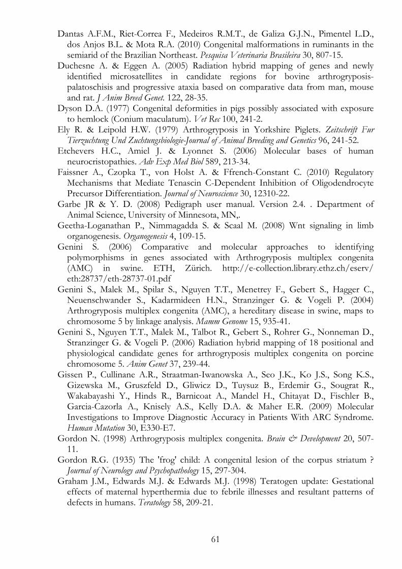
61
Dantas A.F.M., Riet-Correa F., Medeiros R.M.T., de Galiza G.J.N., Pimentel L.D., dos Anjos B.L. & Mota R.A. (2010) Congenital malformations in ruminants in the semiarid of the Brazilian Northeast. Pesquisa Veterinaria Brasileira 30, 807-15.
Duchesne A. & Eggen A. (2005) Radiation hybrid mapping of genes and newly identified microsatellites in candidate regions for bovine arthrogryposis-palatoschisis and progressive ataxia based on comparative data from man, mouse and rat. J Anim Breed Genet. 122, 28-35.
Dyson D.A. (1977) Congenital deformities in pigs possibly associated with exposure to hemlock (Conium maculatum). Vet Rec 100, 241-2.
Ely R. & Leipold H.W. (1979) Arthrogryposis in Yorkshire Piglets. Zeitschrift Fur Tierzuchtung Und Zuchtungsbiologie-Journal of Animal Breeding and Genetics 96, 241-52.
Etchevers H.C., Amiel J. & Lyonnet S. (2006) Molecular bases of human neurocristopathies. Adv Exp Med Biol 589, 213-34.
Faissner A., Czopka T., von Holst A. & Ffrench-Constant C. (2010) Regulatory Mechanisms that Mediate Tenascin C-Dependent Inhibition of Oligodendrocyte Precursor Differentiation. Journal of Neuroscience 30, 12310-22.
Garbe JR & Y. D. (2008) Pedigraph user manual. Version 2.4. . Department of Animal Science, University of Minnesota, MN,.
Geetha-Loganathan P., Nimmagadda S. & Scaal M. (2008) Wnt signaling in limb organogenesis. Organogenesis 4, 109-15.
Genini S. (2006) Comparative and molecular approaches to identifying polymorphisms in genes associated with Arthrogryposis multiplex congenita (AMC) in swine. ETH, Zürich. http://e-collection.library.ethz.ch/eserv/ eth:28737/eth-28737-01.pdf
Genini S., Malek M., Spilar S., Nguyen T.T., Menetrey F., Gebert S., Hagger C., Neuenschwander S., Kadarmideen H.N., Stranzinger G. & Vogeli P. (2004) Arthrogryposis multiplex congenita (AMC), a hereditary disease in swine, maps to chromosome 5 by linkage analysis. Mamm Genome 15, 935-41.
Genini S., Nguyen T.T., Malek M., Talbot R., Gebert S., Rohrer G., Nonneman D., Stranzinger G. & Vogeli P. (2006) Radiation hybrid mapping of 18 positional and physiological candidate genes for arthrogryposis multiplex congenita on porcine chromosome 5. Anim Genet 37, 239-44.
Gissen P., Cullinane A.R., Straatman-Iwanowska A., Seo J.K., Ko J.S., Song K.S., Gizewska M., Gruszfeld D., Gliwicz D., Tuysuz B., Erdemir G., Sougrat R., Wakabayashi Y., Hinds R., Barnicoat A., Mandel H., Chitayat D., Fischler B., Garcia-Cazorla A., Knisely A.S., Kelly D.A. & Maher E.R. (2009) Molecular Investigations to Improve Diagnostic Accuracy in Patients With ARC Syndrome. Human Mutation 30, E330-E7.
Gordon N. (1998) Arthrogryposis multiplex congenita. Brain & Development 20, 507-11.
Gordon R.G. (1935) The 'frog' child: A congenital lesion of the corpus striatum ? Journal of Neurology and Psychopathology 15, 297-304.
Graham J.M., Edwards M.J. & Edwards M.J. (1998) Teratogen update: Gestational effects of maternal hyperthermia due to febrile illnesses and resultant patterns of defects in humans. Teratology 58, 209-21.
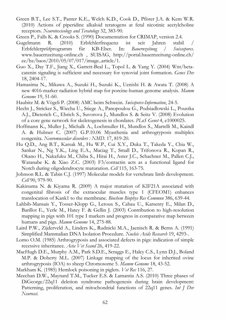
62
Green B.T., Lee S.T., Panter K.E., Welch K.D., Cook D., Pfister J.A. & Kem W.R. (2010) Actions of piperidine alkaloid teratogens at fetal nicotinic acetylcholine receptors. Neurotoxicology and Teratology 32, 383-90.
Green P., Falls K. & Crooks S. (1990) Documentation for CRIMAP, version 2.4. Gugelmann R. (2010) Erbfehlerfrequenz ist seit Jahren stabil /
Erbfehlerprüfprogramm für KB-Eber. In: Bauernzeitung / Suisseporcs. www.bauernzeitung-online.ch , SUISAG, http://portal.bauernzeitung-online.ch/ ee/bz/baos/2010/05/07/017/image_article/1.
Guo X., Day T.F., Jiang X., Garrett-Beal L., Topol L. & Yang Y. (2004) Wnt/beta-catenin signaling is sufficient and necessary for synovial joint formation. Genes Dev 18, 2404-17.
Hamasima N., Mikawa A., Suzuki H., Suzuki K., Uenishi H. & Awata T. (2008) A new 4016-marker radiation hybrid map for porcine-human genome analysis. Mamm Genome 19, 51-60.
Haubitz M. & Vögeli P. (2008) AMC beim Schwein. Swissporcs-Information, 24-5. Hecht J., Stricker S., Wiecha U., Stiege A., Panopoulou G., Podsiadlowski L., Poustka
A.J., Dieterich C., Ehrich S., Suvorova J., Mundlos S. & Seitz V. (2008) Evolution of a core gene network for skeletogenesis in chordates. PLoS Genet 4, e1000025.
Hoffmann K., Muller J., Michalk A., Lochmuller H., Mundlos S., Martelli M., Kaindl A. & Hubner C. (2007) G.P.10.06 Myasthenia and arthrogryposis multiplex congenita. Neuromuscular disorders : NMD. 17, 819-20.
Hu Q.D., Ang B.T., Karsak M., Hu W.P., Cui X.Y., Duka T., Takeda Y., Chia W., Sankar N., Ng Y.K., Ling E.A., Maciag T., Small D., Trifonova R., Kopan R., Okano H., Nakafuku M., Chiba S., Hirai H., Aster J.C., Schachner M., Pallen C.J., Watanabe K. & Xiao Z.C. (2003) F3/contactin acts as a functional ligand for Notch during oligodendrocyte maturation. Cell 115, 163-75.
Johnson R.L. & Tabin C.J. (1997) Molecular models for vertebrate limb development. Cell 90, 979-90.
Kakinuma N. & Kiyama R. (2009) A major mutation of KIF21A associated with congenital fibrosis of the extraocular muscles type 1 (CFEOM1) enhances translocation of Kank1 to the membrane. Biochem Biophys Res Commun 386, 639-44.
Lahbib-Mansais Y., Tosser-Klopp G., Leroux S., Cabau U., Karsenty E., Milan D., Barillot E., Yerle M., Hatey F. & Gellin J. (2003) Contribution to high-resolution mapping in pigs with 101 type I markers and progress in comparative map between humans and pigs. Mamm Genome 14, 275-88.
Laird P.W., Zijderveld A., Linders K., Rudnicki M.A., Jaenisch R. & Berns A. (1991) Simplified Mammalian DNA Isolation Procedure. Nucleic Acids Research 19, 4293-.
Lomo O.M. (1985) Arthrogryposis and associated defects in pigs: indication of simple recessive inheritance. Acta Vet Scand 26, 419-22.
MacHugh D.E., Murphy A.M., Park S.D.E., Scraggs E., Haley C.S., Lynn D.J., Boland M.P. & Doherty M.L. (2007) Linkage mapping of the locus for inherited ovine arthrogryposis (IOA) to sheep Chromosome 5. Mamm Genome 18, 43-52.
Markham K. (1985) Hemlock poisoning in piglets. Vet Rec 116, 27. Meechan D.W., Maynard T.M., Tucker E.S. & Lamantia A.S. (2010) Three phases of
DiGeorge/22q11 deletion syndrome pathogenesis during brain development: Patterning, proliferation, and mitochondrial functions of 22q11 genes. Int J Dev Neurosci.
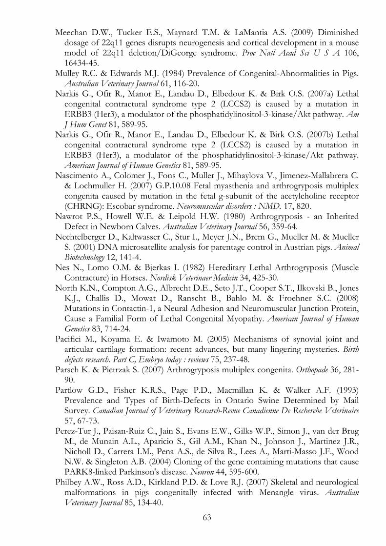
63
Meechan D.W., Tucker E.S., Maynard T.M. & LaMantia A.S. (2009) Diminished dosage of 22q11 genes disrupts neurogenesis and cortical development in a mouse model of 22q11 deletion/DiGeorge syndrome. Proc Natl Acad Sci U S A 106, 16434-45.
Mulley R.C. & Edwards M.J. (1984) Prevalence of Congenital-Abnormalities in Pigs. Australian Veterinary Journal 61, 116-20.
Narkis G., Ofir R., Manor E., Landau D., Elbedour K. & Birk O.S. (2007a) Lethal congenital contractural syndrome type 2 (LCCS2) is caused by a mutation in ERBB3 (Her3), a modulator of the phosphatidylinositol-3-kinase/Akt pathway. Am J Hum Genet 81, 589-95.
Narkis G., Ofir R., Manor E., Landau D., Elbedour K. & Birk O.S. (2007b) Lethal congenital contractural syndrome type 2 (LCCS2) is caused by a mutation in ERBB3 (Her3), a modulator of the phosphatidylinositol-3-kinase/Akt pathway. American Journal of Human Genetics 81, 589-95.
Nascimento A., Colomer J., Fons C., Muller J., Mihaylova V., Jimenez-Mallabrera C. & Lochmuller H. (2007) G.P.10.08 Fetal myasthenia and arthrogryposis multiplex congenita caused by mutation in the fetal g-subunit of the acetylcholine receptor (CHRNG): Escobar syndrome. Neuromuscular disorders : NMD. 17, 820.
Nawrot P.S., Howell W.E. & Leipold H.W. (1980) Arthrogryposis - an Inherited Defect in Newborn Calves. Australian Veterinary Journal 56, 359-64.
Nechtelberger D., Kaltwasser C., Stur I., Meyer J.N., Brem G., Mueller M. & Mueller S. (2001) DNA microsatellite analysis for parentage control in Austrian pigs. Animal Biotechnology 12, 141-4.
Nes N., Lomo O.M. & Bjerkas I. (1982) Hereditary Lethal Arthrogryposis (Muscle Contracture) in Horses. Nordisk Veterinaer Medicin 34, 425-30.
North K.N., Compton A.G., Albrecht D.E., Seto J.T., Cooper S.T., Ilkovski B., Jones K.J., Challis D., Mowat D., Ranscht B., Bahlo M. & Froehner S.C. (2008) Mutations in Contactin-1, a Neural Adhesion and Neuromuscular Junction Protein, Cause a Familial Form of Lethal Congenital Myopathy. American Journal of Human Genetics 83, 714-24.
Pacifici M., Koyama E. & Iwamoto M. (2005) Mechanisms of synovial joint and articular cartilage formation: recent advances, but many lingering mysteries. Birth defects research. Part C, Embryo today : reviews 75, 237-48.
Parsch K. & Pietrzak S. (2007) Arthrogryposis multiplex congenita. Orthopade 36, 281-90.
Partlow G.D., Fisher K.R.S., Page P.D., Macmillan K. & Walker A.F. (1993) Prevalence and Types of Birth-Defects in Ontario Swine Determined by Mail Survey. Canadian Journal of Veterinary Research-Revue Canadienne De Recherche Veterinaire 57, 67-73.
Perez-Tur J., Paisan-Ruiz C., Jain S., Evans E.W., Gilks W.P., Simon J., van der Brug M., de Munain A.L., Aparicio S., Gil A.M., Khan N., Johnson J., Martinez J.R., Nicholl D., Carrera I.M., Pena A.S., de Silva R., Lees A., Marti-Masso J.F., Wood N.W. & Singleton A.B. (2004) Cloning of the gene containing mutations that cause PARK8-linked Parkinson's disease. Neuron 44, 595-600.
Philbey A.W., Ross A.D., Kirkland P.D. & Love R.J. (2007) Skeletal and neurological malformations in pigs congenitally infected with Menangle virus. Australian Veterinary Journal 85, 134-40.
64
Pujol A., Ferrer I., Kapfhammer J.P., Hindelang C., Kemp S., Troffer-Charlier N., Broccoli V., Callyzot N., Mooyer P., Selhorst J., Vreken P., Wanders R.J.A. & Mandel J.L. (2005) Inactivation of the peroxisomal ABCD2 transporter in the mouse leads to late-onset ataxia involving mitochondria, Golgi and endoplasmic reticulum damage. Hum Mol Genet 14, 3565-77.
Ramos A.M., Crooijmans R.P., Affara N.A., Amaral A.J., Archibald A.L., Beever J.E., Bendixen C., Churcher C., Clark R., Dehais P., Hansen M.S., Hedegaard J., Hu Z.L., Kerstens H.H., Law A.S., Megens H.J., Milan D., Nonneman D.J., Rohrer G.A., Rothschild M.F., Smith T.P., Schnabel R.D., Van Tassell C.P., Taylor J.F., Wiedmann R.T., Schook L.B. & Groenen M.A. (2009) Design of a high density SNP genotyping assay in the pig using SNPs identified and characterized by next generation sequencing technology. PLoS One 4, e6524.
Ramser J., Ahearn M.E., Lenski C., Yariz K.O., Hellebrand H., von Rhein M., Clark R.D., Schmutzler R.K., Lichtner P., Hoffman E.P., Meindl A. & Baumbach-Reardon L. (2008) Rare missense and synonymous variants in UBE1 are associated with X-linked infantile spinal muscular atrophy. American Journal of Human Genetics 82, 188-93.
Reimann J., Kau N., Vincent A. & Kornblum C. (2007) G.P.10.07 Arthrogryposis multiplex congenita caused by maternal antibodies against fetal acetylcholine receptor subunits. Neuromuscular disorders : NMD. 17, 820.
Robic A., Faraut T., Iannuccelli N., Lahbib-Mansais Y., Cantegrel V., Alexander L. & Milan D. (2003) A new contribution to the integration of human and porcine genome maps: 623 new points of homology. Cytogenet Genome Res 102, 100-8.
Rougon G., Revest J.M., Faivre-Sarrailh C., Maeda N., Noda M. & Schachner M. (1999) The interaction between F3 immunoglobulin domains and protein tyrosine phosphatases zeta/beta triggers bidirectional signalling between neurons and glial cells. European Journal of Neuroscience 11, 1134-47.
Rozen S. & Skaletsky H. (2000) Primer3 on the WWW for general users and for biologist programmers. Methods Mol Biol 132, 365-86.
Sancho R.M., Law B.M. & Harvey K. (2009) Mutations in the LRRK2 Roc-COR tandem domain link Parkinson's disease to Wnt signalling pathways. Hum Mol Genet 18, 3955-68.
Selby L.A., Hopps H.C. & Edmonds L.D. (1971) Comparative Aspects of Congenital Malformations in Man and Swine. Journal of the American Veterinary Medical Association 159, 1485-&.
Smith M.S.R., Upfold J.B., Edwards M.J., Shiota K. & Cawdellsmith J. (1992) The Induction of Neural-Tube Defects by Maternal Hyperthermia - a Comparison of the Guinea-Pig and Human. Neuropathology and Applied Neurobiology 18, 71-80.
Straw B.E., Zimmerman J.J., D'Allaire S. & Taylor D.J. (2006) Diseases of swine. Blackwell, Oxford.
Sung S.S., Brassington A.M.E., Grannatt K., Rutherford A., Whitby F.G., Krakowiak P.A., Jorde L.B., Carey J.C. & Bamshad M. (2003a) Mutations in genes encoding fast-twitch contractile proteins cause distal arthrogryposis syndromes. American Journal of Human Genetics 72, 681-90.
Sung S.S., Brassington A.M.E., Krakowiak P.A., Carey J.C., Jorde L.B. & Bamshad M. (2003b) Mutations in TNNT3 cause multiple congenital contractures: A second locus for distal arthrogryposis type 2B. American Journal of Human Genetics 73, 212-4.
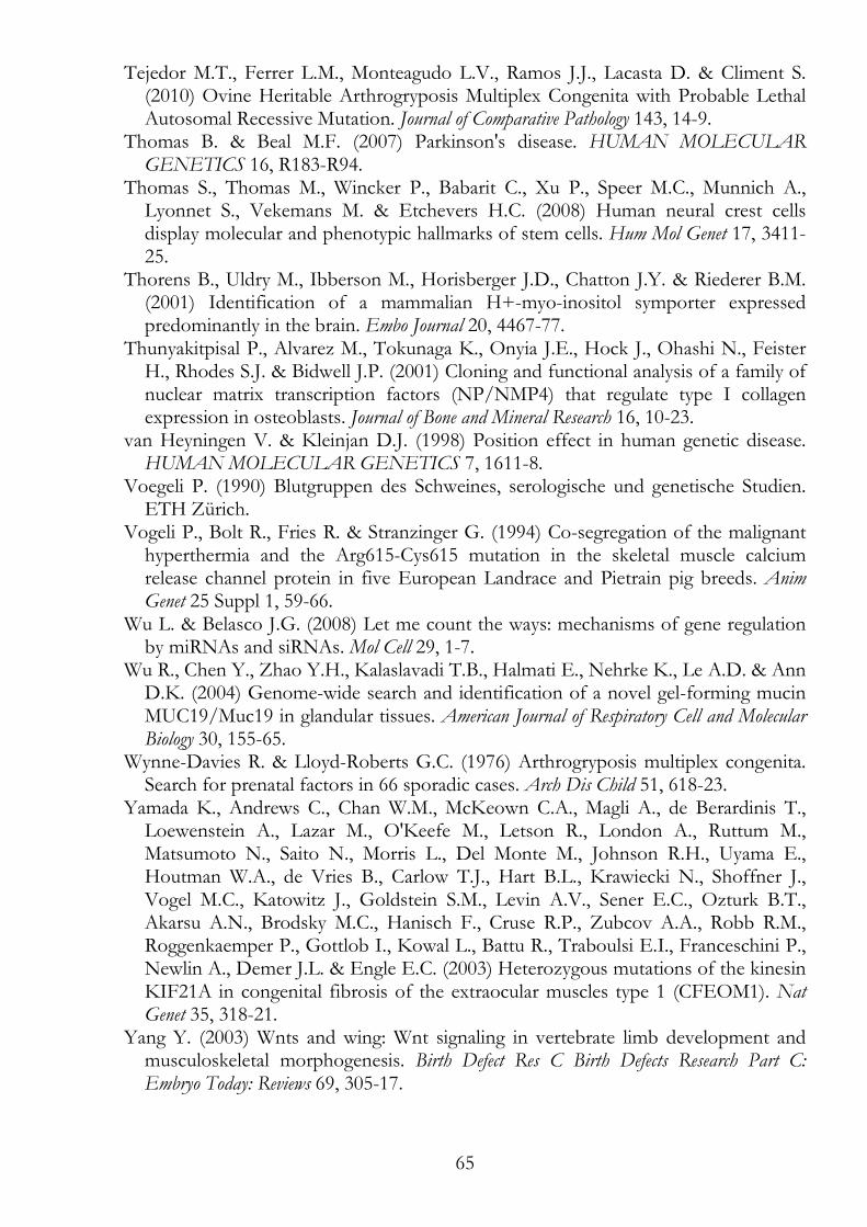
65
Tejedor M.T., Ferrer L.M., Monteagudo L.V., Ramos J.J., Lacasta D. & Climent S. (2010) Ovine Heritable Arthrogryposis Multiplex Congenita with Probable Lethal Autosomal Recessive Mutation. Journal of Comparative Pathology 143, 14-9.
Thomas B. & Beal M.F. (2007) Parkinson's disease. HUMAN MOLECULAR GENETICS 16, R183-R94.
Thomas S., Thomas M., Wincker P., Babarit C., Xu P., Speer M.C., Munnich A., Lyonnet S., Vekemans M. & Etchevers H.C. (2008) Human neural crest cells display molecular and phenotypic hallmarks of stem cells. Hum Mol Genet 17, 3411-25.
Thorens B., Uldry M., Ibberson M., Horisberger J.D., Chatton J.Y. & Riederer B.M. (2001) Identification of a mammalian H+-myo-inositol symporter expressed predominantly in the brain. Embo Journal 20, 4467-77.
Thunyakitpisal P., Alvarez M., Tokunaga K., Onyia J.E., Hock J., Ohashi N., Feister H., Rhodes S.J. & Bidwell J.P. (2001) Cloning and functional analysis of a family of nuclear matrix transcription factors (NP/NMP4) that regulate type I collagen expression in osteoblasts. Journal of Bone and Mineral Research 16, 10-23.
van Heyningen V. & Kleinjan D.J. (1998) Position effect in human genetic disease. HUMAN MOLECULAR GENETICS 7, 1611-8.
Voegeli P. (1990) Blutgruppen des Schweines, serologische und genetische Studien. ETH Zürich.
Vogeli P., Bolt R., Fries R. & Stranzinger G. (1994) Co-segregation of the malignant hyperthermia and the Arg615-Cys615 mutation in the skeletal muscle calcium release channel protein in five European Landrace and Pietrain pig breeds. Anim Genet 25 Suppl 1, 59-66.
Wu L. & Belasco J.G. (2008) Let me count the ways: mechanisms of gene regulation by miRNAs and siRNAs. Mol Cell 29, 1-7.
Wu R., Chen Y., Zhao Y.H., Kalaslavadi T.B., Halmati E., Nehrke K., Le A.D. & Ann D.K. (2004) Genome-wide search and identification of a novel gel-forming mucin MUC19/Muc19 in glandular tissues. American Journal of Respiratory Cell and Molecular Biology 30, 155-65.
Wynne-Davies R. & Lloyd-Roberts G.C. (1976) Arthrogryposis multiplex congenita. Search for prenatal factors in 66 sporadic cases. Arch Dis Child 51, 618-23.
Yamada K., Andrews C., Chan W.M., McKeown C.A., Magli A., de Berardinis T., Loewenstein A., Lazar M., O'Keefe M., Letson R., London A., Ruttum M., Matsumoto N., Saito N., Morris L., Del Monte M., Johnson R.H., Uyama E., Houtman W.A., de Vries B., Carlow T.J., Hart B.L., Krawiecki N., Shoffner J., Vogel M.C., Katowitz J., Goldstein S.M., Levin A.V., Sener E.C., Ozturk B.T., Akarsu A.N., Brodsky M.C., Hanisch F., Cruse R.P., Zubcov A.A., Robb R.M., Roggenkaemper P., Gottlob I., Kowal L., Battu R., Traboulsi E.I., Franceschini P., Newlin A., Demer J.L. & Engle E.C. (2003) Heterozygous mutations of the kinesin KIF21A in congenital fibrosis of the extraocular muscles type 1 (CFEOM1). Nat Genet 35, 318-21.
Yang Y. (2003) Wnts and wing: Wnt signaling in vertebrate limb development and musculoskeletal morphogenesis. Birth Defect Res C Birth Defects Research Part C: Embryo Today: Reviews 69, 305-17.
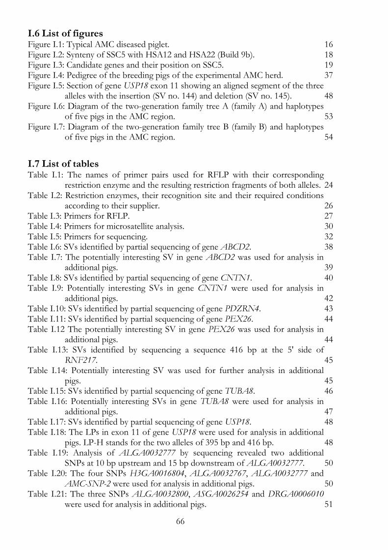
66
I.6 List of figures Figure I.1: Typical AMC diseased piglet. 16
Figure I.2: Synteny of SSC5 with HSA12 and HSA22 (Build 9b). 18
Figure I.3: Candidate genes and their position on SSC5. 19
Figure I.4: Pedigree of the breeding pigs of the experimental AMC herd. 37
Figure I.5: Section of gene USP18 exon 11 showing an aligned segment of the three alleles with the insertion (SV no. 144) and deletion (SV no. 145). 48
Figure I.6: Diagram of the two-generation family tree A (family A) and haplotypes of five pigs in the AMC region. 53
Figure I.7: Diagram of the two-generation family tree B (family B) and haplotypes of five pigs in the AMC region. 54
I.7 List of tables Table I.1: The names of primer pairs used for RFLP with their corresponding
restriction enzyme and the resulting restriction fragments of both alleles. 24
Table I.2: Restriction enzymes, their recognition site and their required conditions according to their supplier. 26
Table I.3: Primers for RFLP. 27
Table I.4: Primers for microsatellite analysis. 30
Table I.5: Primers for sequencing. 32
Table I.6: SVs identified by partial sequencing of gene ABCD2. 38
Table I.7: The potentially interesting SV in gene ABCD2 was used for analysis in additional pigs. 39
Table I.8: SVs identified by partial sequencing of gene CNTN1. 40
Table I.9: Potentially interesting SVs in gene CNTN1 were used for analysis in additional pigs. 42
Table I.10: SVs identified by partial sequencing of gene PDZRN4. 43
Table I.11: SVs identified by partial sequencing of gene PEX26. 44
Table I.12 The potentially interesting SV in gene PEX26 was used for analysis in additional pigs. 44
Table I.13: SVs identified by sequencing a sequence 416 bp at the 5' side of RNF217. 45
Table I.14: Potentially interesting SV was used for further analysis in additional pigs. 45
Table I.15: SVs identified by partial sequencing of gene TUBA8. 46
Table I.16: Potentially interesting SVs in gene TUBA8 were used for analysis in additional pigs. 47
Table I.17: SVs identified by partial sequencing of gene USP18. 48
Table I.18: The LPs in exon 11 of gene USP18 were used for analysis in additional pigs. LP-H stands for the two alleles of 395 bp and 416 bp. 48
Table I.19: Analysis of ALGA0032777 by sequencing revealed two additional SNPs at 10 bp upstream and 15 bp downstream of ALGA0032777. 50
Table I.20: The four SNPs H3GA0016804, ALGA0032767, ALGA0032777 and AMC-SNP-2 were used for analysis in additional pigs. 50
Table I.21: The three SNPs ALGA0032800, ASGA0026254 and DRGA0006010 were used for analysis in additional pigs. 51
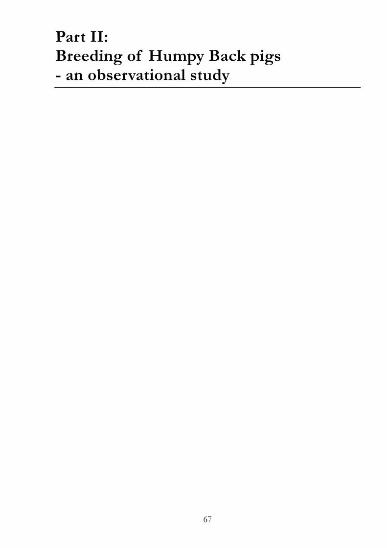
67
Part II: Breeding of Humpy Back pigs - an observational study
68
Table of Contents II.1 Introduction 69
II.1.1 Aim of the inbreeding experiment 70
II.1.2 Review 70
II.1.2.1 Case reports and pathophysiology 70
II.1.2.2 Differential diagnoses 71
II.1.2.3 Spinal deformations in humans as a model for HB 72
II.1.2.4 Induction of spinal deformities 73
II.1.2.5 Genetics of HB 74
II.1.2.5.1 Extreme vertebra number variations 76
II.1.2.6 Humpy Back in sheep 76
II.2 Materials and methods 77
II.2.1 Breeding of HB pigs 77
II.2.2 Genomic DNA 77
II.2.3 Pedigree drawing 77
II.3 Results 78
II.3.1 Additional observations 78
II.4 Discussion of part II 80
II.4.1 Further investigations 80
II.5 References 82
II.6 List of figures 86
II.7 List of tables 86
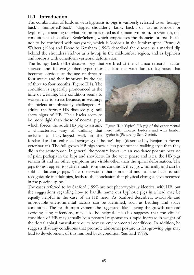
69
Figure II.1: Typical HB pig of the experimental herd with thoracic lordosis and with lumbar kyphosis (Picture by Sem Genini).
II.1 Introduction The combination of lordosis with kyphosis in pigs is variously referred to as ´humpy-back´, ´hump(-ed)-back´, ´dipped shoulder´, ´kinky back´, or just as lordosis or kyphosis, depending on what symptom is rated as the main symptom. In German, this condition is also called ´Senkrücken´, which emphasises the thoracic lordosis but is not to be confused with swayback, which is lordosis in the lumbar spine. Penny & Walters (1986) and Done & Gresham (1998) described the disease as a marked dip behind the shoulders and/or as a hump in the mid-lumbar region, and as kyphosis and lordosis with cuneiform vertebral deformation. The humpy back (HB) diseased pigs that we bred at the Chamau research station showed the following phenotype: thoracic lordosis with lumbar kyphosis that becomes obvious at the age of three to four weeks and then improves by the age of three to four months (Figure II.1). The condition is especially pronounced at the time of weaning. The condition seems to worsen due to stress because, at weaning, the piglets are physically challenged. As adults, the former HB diseased pigs still show signs of HB. Their backs seem to be more rigid than those of normal pigs, which forces the adult HB pig to assume a characteristic way of walking that includes a shaky-legged walk in the forehand and an enhanced swinging of the pig's hips (described by Benjamin Furter, veterinarian). The full grown HB pigs show a less pronounced walking style than they did in the acute phase. In general, the posture looks like an avoidance posture because of pain, perhaps in the hips and shoulders. In the acute phase and later, the HB pigs remain fit and no other symptoms are visible other than the spinal deformation. The pigs do not appear to suffer much from this condition; they grow normally and can be sold as fattening pigs. The observation that some stiffness of the back is still recognizable in adult pigs, leads to the conclusion that physical changes have occurred in the porcine spine. The cases referred to by Sanford (1999) are not phenotypically identical with HB, but the suggestions regarding how to handle numerous kyphotic pigs in a herd may be equally helpful in the case of an HB herd. As Sanford described, avoidable and improvable environmental factors can be identified, such as bedding and space conditions. The health improvements he suggested, like slowing the growth rate and avoiding lung infections, may also be helpful. He also suggests that the clinical condition of HB may actually be a postural response to a rapid increase in weight of the dorsal spinal musculature or to adverse environmental conditions. In addition, he suggests that any conditions that promote abnormal posture in fast-growing pigs may lead to development of this humped back condition (Sanford 1999).
70
II.1.1 Aim of the inbreeding experiment If there is a genetic cause for HB, we should see an increasing number of affected piglets or an intensified phenotype through generations. In other words, if former HB diseased pigs transmit the HB genes to their progeny, we should observe increasing numbers of HB piglets per litter with progressing generations of breeding if HB is indeed heritable, as Laitat et al. (2006) suggested. In order to understand the genetics of the HB disease, we bred a small herd using former HB diseased pigs as parents and we observed the development/evolution of HB for several successive generations. We also collected blood and tissue samples and the corresponding pedigrees for further studies on this subject.
II.1.2 Review
II.1.2.1 Case reports and pathophysiology Penny & Walters (1986) investigated 24 HB cases and observed no seasonal frequency; no particular breeds or combinations of breeds that were affected; equal occurrence in both sexes; symptoms that were mostly observable at 8-16 weeks of age; occurrence mostly in pigs housed intensively or semi-intensively indoors; a more frequent appearance in thriving pigs; and no influence of HB on growth rate. They calculated a maximum herd frequency of 4%, although an outbreak with up to 30% was reported. Heritability of HB was questioned because outbreaks in different breeds and combinations of breeds were reported. Instead, management and environmental factors seemed more important. HB cases were identified in particular in genetically improved pigs that were intensively housed and managed and that were growing rapidly. Ultimately, these researchers concluded that no single etiological factor was responsible and that HB seems to be a multifactorial disease. As advice for breeders, they suggested slowing down the growth rate of the HB pigs and allowing the pigs more exercise. They also recommended reducing stress factors to improve the condition of the HB diseased pigs. Done & Gresham (1998) clinically examined two of 49 cases (about 4.5% of the group) that were of different age and severity. All of the affected pigs were aged between 3 and 15 weeks. The researchers found that levels of bone magnesium were increased and calcium and phosphorus were decreased, but nothing else was noticeably different other than the spinal curvature. Five concepts were subsequently discussed: Muscle structure, rapid gain of muscle weight, bone formation, riding behaviour and Erysipelas rhusiopathiae infection. In the following case report by Done et al. (1999), the HB cases were different: the symptoms were congenital, seasonally distributed, spinal deformation was severe, the diseased pig did not recover, and pathological examination suggested that only one of the two ossification centres worked in the vertebrae resulting in formation of hemivertebrae. The authors interpreted these observations as a genetically caused HB. Connatal formation of block vertebrae and kyphosis in the lumbar region was found in two weaning pigs (Lahrmann & Staudt 1991). The pigs showed no locomotor disorders. In both pigs, the 2nd and the 3rd lumbar vertebrae were merged, with ventral shortening, producing a phenotype comparable to the one seen in our HB herd. In a subsequent case report, Lahrmann & Hartung (1993) described pathological findings in kyphotic spines that included acquired wedge shaped vertebrae and
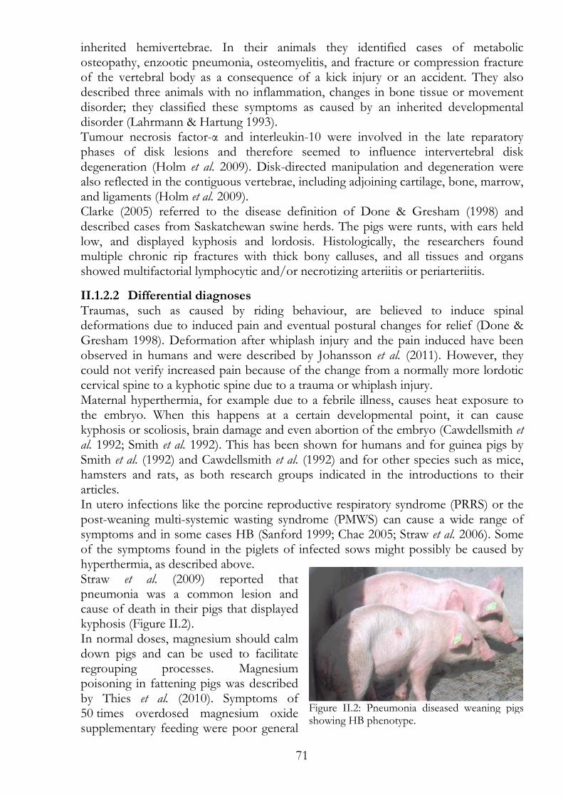
71
inherited hemivertebrae. In their animals they identified cases of metabolic osteopathy, enzootic pneumonia, osteomyelitis, and fracture or compression fracture of the vertebral body as a consequence of a kick injury or an accident. They also described three animals with no inflammation, changes in bone tissue or movement disorder; they classified these symptoms as caused by an inherited developmental disorder (Lahrmann & Hartung 1993). Tumour necrosis factor-α and interleukin-10 were involved in the late reparatory phases of disk lesions and therefore seemed to influence intervertebral disk degeneration (Holm et al. 2009). Disk-directed manipulation and degeneration were also reflected in the contiguous vertebrae, including adjoining cartilage, bone, marrow, and ligaments (Holm et al. 2009). Clarke (2005) referred to the disease definition of Done & Gresham (1998) and described cases from Saskatchewan swine herds. The pigs were runts, with ears held low, and displayed kyphosis and lordosis. Histologically, the researchers found multiple chronic rip fractures with thick bony calluses, and all tissues and organs showed multifactorial lymphocytic and/or necrotizing arteriitis or periarteriitis.
II.1.2.2 Differential diagnoses Traumas, such as caused by riding behaviour, are believed to induce spinal deformations due to induced pain and eventual postural changes for relief (Done & Gresham 1998). Deformation after whiplash injury and the pain induced have been observed in humans and were described by Johansson et al. (2011). However, they could not verify increased pain because of the change from a normally more lordotic cervical spine to a kyphotic spine due to a trauma or whiplash injury. Maternal hyperthermia, for example due to a febrile illness, causes heat exposure to the embryo. When this happens at a certain developmental point, it can cause kyphosis or scoliosis, brain damage and even abortion of the embryo (Cawdellsmith et al. 1992; Smith et al. 1992). This has been shown for humans and for guinea pigs by Smith et al. (1992) and Cawdellsmith et al. (1992) and for other species such as mice, hamsters and rats, as both research groups indicated in the introductions to their articles. In utero infections like the porcine reproductive respiratory syndrome (PRRS) or the post-weaning multi-systemic wasting syndrome (PMWS) can cause a wide range of symptoms and in some cases HB (Sanford 1999; Chae 2005; Straw et al. 2006). Some of the symptoms found in the piglets of infected sows might possibly be caused by hyperthermia, as described above. Straw et al. (2009) reported that pneumonia was a common lesion and cause of death in their pigs that displayed kyphosis (Figure II.2). In normal doses, magnesium should calm down pigs and can be used to facilitate regrouping processes. Magnesium poisoning in fattening pigs was described by Thies et al. (2010). Symptoms of 50 times overdosed magnesium oxide supplementary feeding were poor general
Figure II.2: Pneumonia diseased weaning pigs showing HB phenotype.
72
condition, apathy, kyphosis, diarrhoea and dermatitis of the hind limbs. Some pig even died in the course of magnesium poisoning. Teratogenic piperidine alkaloids, like nicotine, are plant toxins that can induce malformations in foetuses by depolarizing cells that express foetal-muscle type nicotinic acetylcholine receptors, at least in human cell lines. The mechanism behind the teratogenic potential of these compounds seems to be the stimulation of muscle-type nicotinic acetylcholine receptors, followed by desensitization and finally inhibition of foetal movement (Green et al. 2010). Nicotine can delay foetal skeletal growth by acting on growth plate chondrocytes (Kawakita et al. 2008). The toxic effect of these alkaloids, acting through nicotinic acetylcholine receptors, produces foetal musculoskeletal defects, including arthrogryposis, kyphosis, lordosis, scoliosis and more. Teratogenic effects of tobacco have also been shown to induce lordosis and additional symptoms in sheep (Keeler & Crowe 1984).
II.1.2.3 Spinal deformations in humans as a model for HB Humans are probably not an ideal model for porcine HB disease because they are two-legged and stand upright. This posture means that humans only need gravity and a fragile spine or weak back muscles to develop kyphosis and/or lordosis. However, because pigs are four-legged, if their spine or back muscles are weak, they become swaybacked. In contrast to the human that forms a kyphotic back, in pig the issue appears to be a matter of a too strong muscle tension. Humans also develop kyphosis primarily in the thoracic spine and lordosis in the lumbar spine, whereas HB pigs develop lordosis in the thoracic spine and kyphosis in the lumbar spine. Interestingly, the diseased spines have strong resemblance in both species (human kyphosis and HB in pig); namely, a spine with a normal spinal curvature in the extreme. Apart from all of the differences Busscher et al. (2010) concluded in their study that the spines of humans and pigs are comparable from a surgical point of view, with reservations as the intervertebral discs in humans are higher than in pigs and pigs have more vertebrae within the same total spine length. Although, differences exist between the two species, similarities can be noted between HB and certain spinal malformations seen in adolescent humans, such as Scheuermann's disease or idiopathic scoliosis. Phenotypically, the closest human spinal malformation to porcine HB is adolescent kyphosis or Scheuermann's disease. The review by Lowe (1990) about Scheuermann's disease reports that patients suffer from structural kyphosis at the thoracic or thoracolumbar spine, with an onset usually appearing at puberty. In 90 families with members with Scheuermann's disease, the segregation analysis revealed a dominant inheritance with complete penetrance in boys and incomplete penetrance in girls (Axenovich et al. 2001). Suggested aetiologies of Scheuermann's disease are: a form of avascular necrosis of the ring apophysis, enchondral ossification inhibition or alteration, persistence of the anterior vascular groove in the adolescent, upright posture and tightness of the anterior longitudinal ligament of the spine, osteoporosis, low-calcium diet, and abnormal cartilaginous end-plates. Working at heavy labour was also implicated suggesting that mechanical factors play a role, by allowing for uneven growth of the vertebral bodies, with wedging as a response to Wolff’s law (Lowe 1990). Scheuermann's disease has also been associated with endocrine diseases, hypovitaminosis, inflammatory diseases, neuromuscular disorders, dural cysts, and
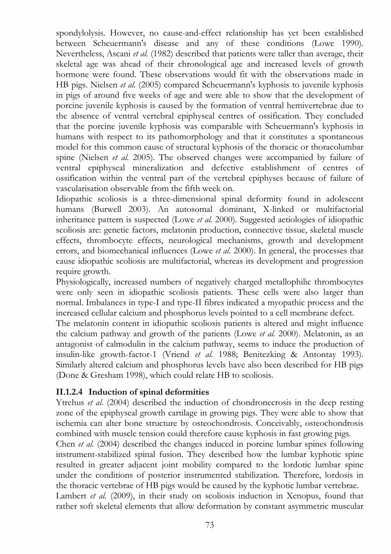
73
spondylolysis. However, no cause-and-effect relationship has yet been established between Scheuermann's disease and any of these conditions (Lowe 1990). Nevertheless, Ascani et al. (1982) described that patients were taller than average, their skeletal age was ahead of their chronological age and increased levels of growth hormone were found. These observations would fit with the observations made in HB pigs. Nielsen et al. (2005) compared Scheuermann's kyphosis to juvenile kyphosis in pigs of around five weeks of age and were able to show that the development of porcine juvenile kyphosis is caused by the formation of ventral hemivertebrae due to the absence of ventral vertebral epiphyseal centres of ossification. They concluded that the porcine juvenile kyphosis was comparable with Scheuermann's kyphosis in humans with respect to its pathomorphology and that it constitutes a spontaneous model for this common cause of structural kyphosis of the thoracic or thoracolumbar spine (Nielsen et al. 2005). The observed changes were accompanied by failure of ventral epiphyseal mineralization and defective establishment of centres of ossification within the ventral part of the vertebral epiphyses because of failure of vascularisation observable from the fifth week on. Idiopathic scoliosis is a three-dimensional spinal deformity found in adolescent humans (Burwell 2003). An autosomal dominant, X-linked or multifactorial inheritance pattern is suspected (Lowe et al. 2000). Suggested aetiologies of idiopathic scoliosis are: genetic factors, melatonin production, connective tissue, skeletal muscle effects, thrombocyte effects, neurological mechanisms, growth and development errors, and biomechanical influences (Lowe et al. 2000). In general, the processes that cause idiopathic scoliosis are multifactorial, whereas its development and progression require growth. Physiologically, increased numbers of negatively charged metallophilic thrombocytes were only seen in idiopathic scoliosis patients. These cells were also larger than normal. Imbalances in type-I and type-II fibres indicated a myopathic process and the increased cellular calcium and phosphorus levels pointed to a cell membrane defect. The melatonin content in idiopathic scoliosis patients is altered and might influence the calcium pathway and growth of the patients (Lowe et al. 2000). Melatonin, as an antagonist of calmodulin in the calcium pathway, seems to induce the production of insulin-like growth-factor-1 (Vriend et al. 1988; Benitezking & Antontay 1993). Similarly altered calcium and phosphorus levels have also been described for HB pigs (Done & Gresham 1998), which could relate HB to scoliosis.
II.1.2.4 Induction of spinal deformities Ytrehus et al. (2004) described the induction of chondronecrosis in the deep resting zone of the epiphyseal growth cartilage in growing pigs. They were able to show that ischemia can alter bone structure by osteochondrosis. Conceivably, osteochondrosis combined with muscle tension could therefore cause kyphosis in fast growing pigs. Chen et al. (2004) described the changes induced in porcine lumbar spines following instrument-stabilized spinal fusion. They described how the lumbar kyphotic spine resulted in greater adjacent joint mobility compared to the lordotic lumbar spine under the conditions of posterior instrumented stabilization. Therefore, lordosis in the thoracic vertebrae of HB pigs would be caused by the kyphotic lumbar vertebrae. Lambert et al. (2009), in their study on scoliosis induction in Xenopus, found that rather soft skeletal elements that allow deformation by constant asymmetric muscular
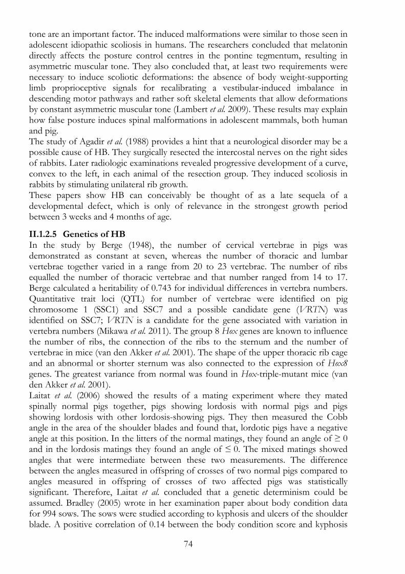
74
tone are an important factor. The induced malformations were similar to those seen in adolescent idiopathic scoliosis in humans. The researchers concluded that melatonin directly affects the posture control centres in the pontine tegmentum, resulting in asymmetric muscular tone. They also concluded that, at least two requirements were necessary to induce scoliotic deformations: the absence of body weight-supporting limb proprioceptive signals for recalibrating a vestibular-induced imbalance in descending motor pathways and rather soft skeletal elements that allow deformations by constant asymmetric muscular tone (Lambert et al. 2009). These results may explain how false posture induces spinal malformations in adolescent mammals, both human and pig. The study of Agadir et al. (1988) provides a hint that a neurological disorder may be a possible cause of HB. They surgically resected the intercostal nerves on the right sides of rabbits. Later radiologic examinations revealed progressive development of a curve, convex to the left, in each animal of the resection group. They induced scoliosis in rabbits by stimulating unilateral rib growth. These papers show HB can conceivably be thought of as a late sequela of a developmental defect, which is only of relevance in the strongest growth period between 3 weeks and 4 months of age.
II.1.2.5 Genetics of HB In the study by Berge (1948), the number of cervical vertebrae in pigs was demonstrated as constant at seven, whereas the number of thoracic and lumbar vertebrae together varied in a range from 20 to 23 vertebrae. The number of ribs equalled the number of thoracic vertebrae and that number ranged from 14 to 17. Berge calculated a heritability of 0.743 for individual differences in vertebra numbers. Quantitative trait loci (QTL) for number of vertebrae were identified on pig chromosome 1 (SSC1) and SSC7 and a possible candidate gene (VRTN) was identified on SSC7; VRTN is a candidate for the gene associated with variation in vertebra numbers (Mikawa et al. 2011). The group 8 Hox genes are known to influence the number of ribs, the connection of the ribs to the sternum and the number of vertebrae in mice (van den Akker et al. 2001). The shape of the upper thoracic rib cage and an abnormal or shorter sternum was also connected to the expression of Hox8 genes. The greatest variance from normal was found in Hox-triple-mutant mice (van den Akker et al. 2001). Laitat et al. (2006) showed the results of a mating experiment where they mated spinally normal pigs together, pigs showing lordosis with normal pigs and pigs showing lordosis with other lordosis-showing pigs. They then measured the Cobb angle in the area of the shoulder blades and found that, lordotic pigs have a negative angle at this position. In the litters of the normal matings, they found an angle of ≥ 0 and in the lordosis matings they found an angle of ≤ 0. The mixed matings showed angles that were intermediate between these two measurements. The difference between the angles measured in offspring of crosses of two normal pigs compared to angles measured in offspring of crosses of two affected pigs was statistically significant. Therefore, Laitat et al. concluded that a genetic determinism could be assumed. Bradley (2005) wrote in her examination paper about body condition data for 994 sows. The sows were studied according to kyphosis and ulcers of the shoulder blade. A positive correlation of 0.14 between the body condition score and kyphosis
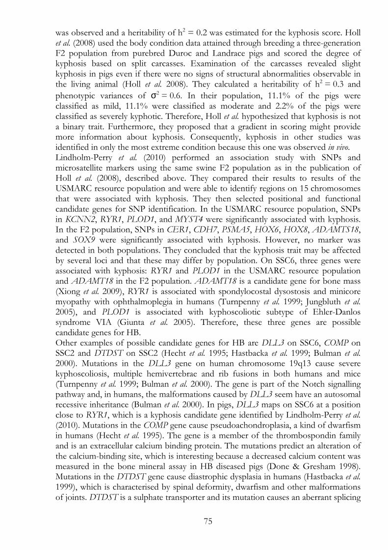
75
was observed and a heritability of h2 = 0.2 was estimated for the kyphosis score. Holl et al. (2008) used the body condition data attained through breeding a three-generation F2 population from purebred Duroc and Landrace pigs and scored the degree of kyphosis based on split carcasses. Examination of the carcasses revealed slight kyphosis in pigs even if there were no signs of structural abnormalities observable in the living animal (Holl et al. 2008). They calculated a heritability of h2 = 0.3 and
phenotypic variances of σ2 = 0.6. In their population, 11.1% of the pigs were classified as mild, 11.1% were classified as moderate and 2.2% of the pigs were classified as severely kyphotic. Therefore, Holl et al. hypothesized that kyphosis is not a binary trait. Furthermore, they proposed that a gradient in scoring might provide more information about kyphosis. Consequently, kyphosis in other studies was identified in only the most extreme condition because this one was observed in vivo. Lindholm-Perry et al. (2010) performed an association study with SNPs and microsatellite markers using the same swine F2 population as in the publication of Holl et al. (2008), described above. They compared their results to results of the USMARC resource population and were able to identify regions on 15 chromosomes that were associated with kyphosis. They then selected positional and functional candidate genes for SNP identification. In the USMARC resource population, SNPs in KCNN2, RYR1, PLOD1, and MYST4 were significantly associated with kyphosis. In the F2 population, SNPs in CER1, CDH7, PSMA5, HOX6, HOX8, ADAMTS18, and SOX9 were significantly associated with kyphosis. However, no marker was detected in both populations. They concluded that the kyphosis trait may be affected by several loci and that these may differ by population. On SSC6, three genes were associated with kyphosis: RYR1 and PLOD1 in the USMARC resource population and ADAMT18 in the F2 population. ADAMT18 is a candidate gene for bone mass (Xiong et al. 2009), RYR1 is associated with spondylocostal dysostosis and minicore myopathy with ophthalmoplegia in humans (Turnpenny et al. 1999; Jungbluth et al. 2005), and PLOD1 is associated with kyphoscoliotic subtype of Ehler-Danlos syndrome VIA (Giunta et al. 2005). Therefore, these three genes are possible candidate genes for HB. Other examples of possible candidate genes for HB are DLL3 on SSC6, COMP on SSC2 and DTDST on SSC2 (Hecht et al. 1995; Hastbacka et al. 1999; Bulman et al. 2000). Mutations in the DLL3 gene on human chromosome 19q13 cause severe kyphoscoliosis, multiple hemivertebrae and rib fusions in both humans and mice (Turnpenny et al. 1999; Bulman et al. 2000). The gene is part of the Notch signalling pathway and, in humans, the malformations caused by DLL3 seem have an autosomal recessive inheritance (Bulman et al. 2000). In pigs, DLL3 maps on SSC6 at a position close to RYR1, which is a kyphosis candidate gene identified by Lindholm-Perry et al. (2010). Mutations in the COMP gene cause pseudoachondroplasia, a kind of dwarfism in humans (Hecht et al. 1995). The gene is a member of the thrombospondin family and is an extracellular calcium binding protein. The mutations predict an alteration of the calcium-binding site, which is interesting because a decreased calcium content was measured in the bone mineral assay in HB diseased pigs (Done & Gresham 1998). Mutations in the DTDST gene cause diastrophic dysplasia in humans (Hastbacka et al. 1999), which is characterised by spinal deformity, dwarfism and other malformations of joints. DTDST is a sulphate transporter and its mutation causes an aberrant splicing
76
site with unknown consequences. However, measured mutant mRNA levels are often low.
II.1.2.5.1 Extreme vertebra number variations Extreme forms of variable vertebra numbers are seen in the tail of the Manx cat (shorter tail than normal) and in the tail of humans (longer tail than normal). Manx cats ´suffer´ from taillessness caused by a single autosomal dominant gene with incomplete penetrance, so different manifestations of the mutation are possible ranging from rumpy cats with no visible tail at all to stumpy cats with a very short tail (James et al. 1969; Deforest & Basrur 1979). In tails of stumpy cats, cartilage or bone is present. The homozygous condition for the tailless gene is lethal. Deforst & Basrur (1979) and James et al. (1969) describe lack of coordination of the hind limbs that leads to inability to climb and a characteristic way of walking. In extreme cases, cats are incontinent and suffer from sensory loss and abnormal hind limb action (Deforest & Basrur 1979). The human tail is a rare malformation; it results from developmental defects in foetal development. However, the condition seems not to be inherited. Several case reports of newborns with tails or pseudotails are available. Examples were published by Noack et al. (2003) and Amirjamshidi et al. (2006). Classification of the human tail as a true tail or as a pseudotail depends on the structures identified within the tail; i.e., if bone and/or cartilage or neurological structures are present.
II.1.2.6 Humpy Back in sheep The term humpy back as it applies to sheep must be differentiated from HB in pigs. In sheep, the name humpy back is used to describe a locomotory disorder that is a seasonal phenomenon. In the summer, after rains, when summer annuals and grasses grow rapidly, the condition called humpy back can be observed in sheep. Acuteness is triggered by heat (summer heat together with a thick wool layer) combined with physical activity. Typically, the clinical symptoms are characterised by a short-stepping, stilted gait of the hind limbs, followed by lowering of the head, arching of the back and inability to continue walking (O'Sullivan 1976). Suspected caused include myopathies, neuropathies and especially intoxication due to ingestion of Solanum plants (Mcmeniman 1976; Bourke 1995). Humpy back in sheep can be lethal in extreme cases when not enough rest is given to the affected animal in between phases of physical activity.
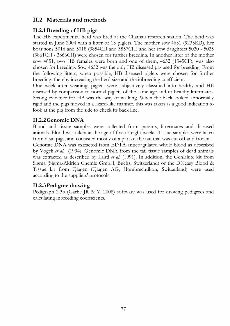
77
II.2 Materials and methods
II.2.1 Breeding of HB pigs The HB experimental herd was bred at the Chamau research station. The herd was started in June 2004 with a litter of 15 piglets. The mother sow 4651 (9235RD), her boar sons 5016 and 5018 (3854CH and 3857CH) and her sow daughters 5020 - 5025 (3861CH - 3866CH) were chosen for further breeding. In another litter of the mother sow 4651, two HB females were born and one of them, 4652 (1345CF), was also chosen for breeding. Sow 4652 was the only HB diseased pig used for breeding. From the following litters, when possible, HB diseased piglets were chosen for further breeding, thereby increasing the herd size and the inbreeding coefficient. One week after weaning, piglets were subjectively classified into healthy and HB diseased by comparison to normal piglets of the same age and to healthy littermates. Strong evidence for HB was the way of walking. When the back looked abnormally rigid and the pigs moved in a lizard-like manner, this was taken as a good indication to look at the pig from the side to check its back line.
II.2.2 Genomic DNA Blood and tissue samples were collected from parents, littermates and diseased animals. Blood was taken at the age of five to eight weeks. Tissue samples were taken from dead pigs, and consisted mostly of a part of the tail that was cut off and frozen. Genomic DNA was extracted from EDTA-anticoagulated whole blood as described by Vogeli et al. (1994). Genomic DNA from the tail tissue samples of dead animals was extracted as described by Laird et al. (1991). In addition, the GenElute kit from Sigma (Sigma-Aldrich Chemie GmbH, Buchs, Switzerland) or the DNeasy Blood & Tissue kit from Qiagen (Qiagen AG, Hombrechtikon, Switzerland) were used according to the suppliers' protocols.
II.2.3 Pedigree drawing Pedigraph 2.3b (Garbe JR & Y. 2008) software was used for drawing pedigrees and calculating inbreeding coefficients.
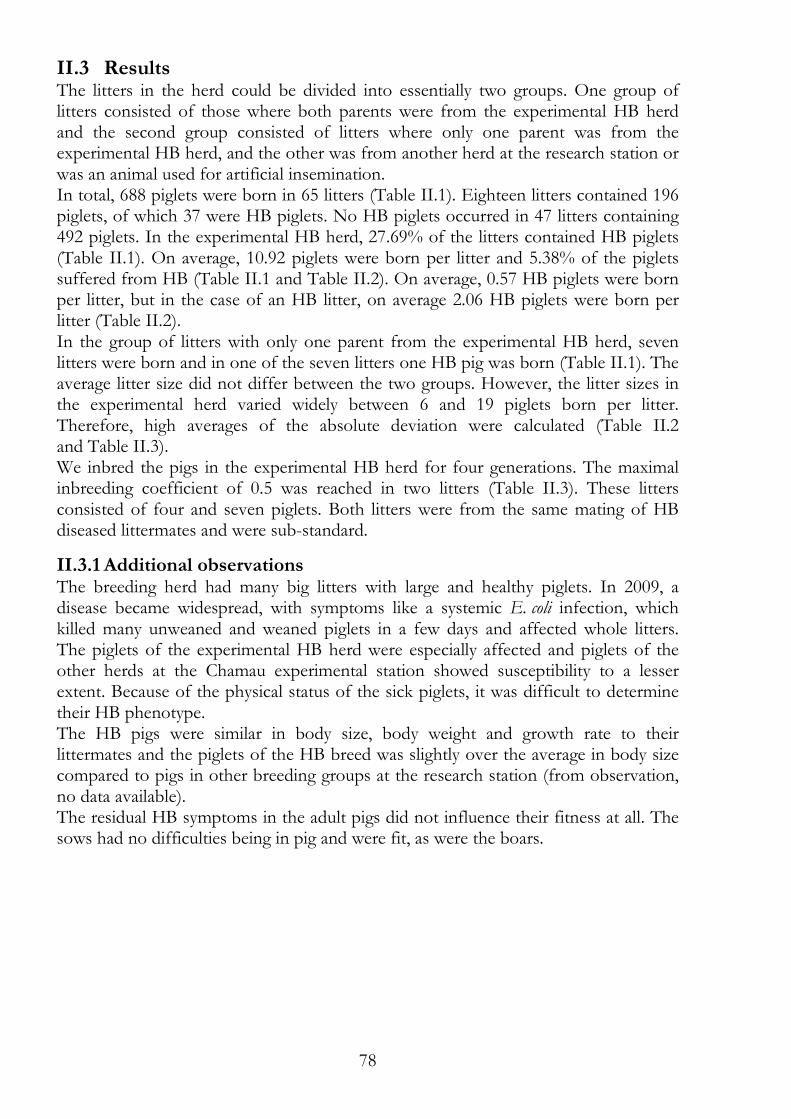
78
II.3 Results The litters in the herd could be divided into essentially two groups. One group of litters consisted of those where both parents were from the experimental HB herd and the second group consisted of litters where only one parent was from the experimental HB herd, and the other was from another herd at the research station or was an animal used for artificial insemination. In total, 688 piglets were born in 65 litters (Table II.1). Eighteen litters contained 196 piglets, of which 37 were HB piglets. No HB piglets occurred in 47 litters containing 492 piglets. In the experimental HB herd, 27.69% of the litters contained HB piglets (Table II.1). On average, 10.92 piglets were born per litter and 5.38% of the piglets suffered from HB (Table II.1 and Table II.2). On average, 0.57 HB piglets were born per litter, but in the case of an HB litter, on average 2.06 HB piglets were born per litter (Table II.2). In the group of litters with only one parent from the experimental HB herd, seven litters were born and in one of the seven litters one HB pig was born (Table II.1). The average litter size did not differ between the two groups. However, the litter sizes in the experimental herd varied widely between 6 and 19 piglets born per litter. Therefore, high averages of the absolute deviation were calculated (Table II.2 and Table II.3). We inbred the pigs in the experimental HB herd for four generations. The maximal inbreeding coefficient of 0.5 was reached in two litters (Table II.3). These litters consisted of four and seven piglets. Both litters were from the same mating of HB diseased littermates and were sub-standard.
II.3.1 Additional observations The breeding herd had many big litters with large and healthy piglets. In 2009, a disease became widespread, with symptoms like a systemic E. coli infection, which killed many unweaned and weaned piglets in a few days and affected whole litters. The piglets of the experimental HB herd were especially affected and piglets of the other herds at the Chamau experimental station showed susceptibility to a lesser extent. Because of the physical status of the sick piglets, it was difficult to determine their HB phenotype. The HB pigs were similar in body size, body weight and growth rate to their littermates and the piglets of the HB breed was slightly over the average in body size compared to pigs in other breeding groups at the research station (from observation, no data available). The residual HB symptoms in the adult pigs did not influence their fitness at all. The sows had no difficulties being in pig and were fit, as were the boars.
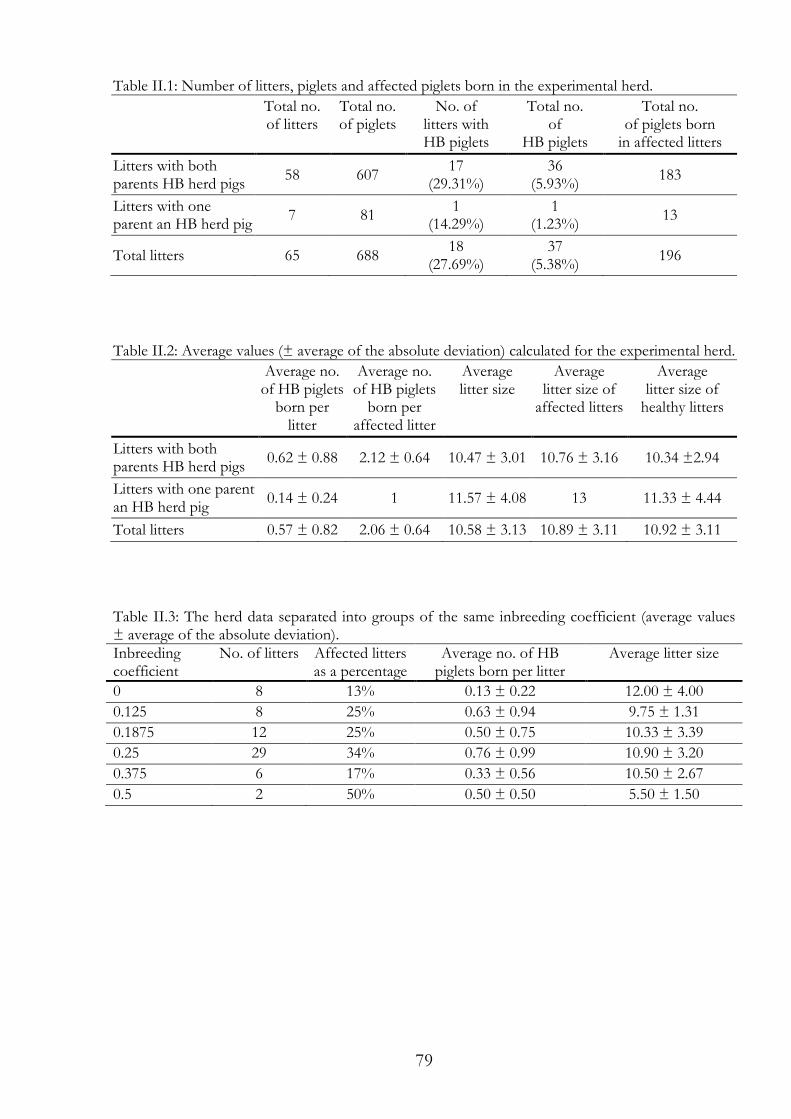
79
Table II.1: Number of litters, piglets and affected piglets born in the experimental herd.
Total no. of litters
Total no. of piglets
No. of litters with HB piglets
Total no. of
HB piglets
Total no. of piglets born
in affected litters
Litters with both parents HB herd pigs
58 607 17
(29.31%) 36
(5.93%) 183
Litters with one parent an HB herd pig
7 81 1
(14.29%) 1
(1.23%) 13
Total litters 65 688 18
(27.69%) 37
(5.38%) 196
Table II.2: Average values (± average of the absolute deviation) calculated for the experimental herd.
Average no. of HB piglets
born per litter
Average no. of HB piglets
born per affected litter
Average litter size
Average litter size of
affected litters
Average litter size of
healthy litters
Litters with both parents HB herd pigs
0.62 ± 0.88 2.12 ± 0.64 10.47 ± 3.01 10.76 ± 3.16 10.34 ±2.94
Litters with one parent an HB herd pig
0.14 ± 0.24 1 11.57 ± 4.08 13 11.33 ± 4.44
Total litters 0.57 ± 0.82 2.06 ± 0.64 10.58 ± 3.13 10.89 ± 3.11 10.92 ± 3.11
Table II.3: The herd data separated into groups of the same inbreeding coefficient (average values ± average of the absolute deviation). Inbreeding coefficient
No. of litters Affected litters as a percentage
Average no. of HB piglets born per litter
Average litter size
0 8 13% 0.13 ± 0.22 12.00 ± 4.00
0.125 8 25% 0.63 ± 0.94 9.75 ± 1.31
0.1875 12 25% 0.50 ± 0.75 10.33 ± 3.39
0.25 29 34% 0.76 ± 0.99 10.90 ± 3.20
0.375 6 17% 0.33 ± 0.56 10.50 ± 2.67
0.5 2 50% 0.50 ± 0.50 5.50 ± 1.50
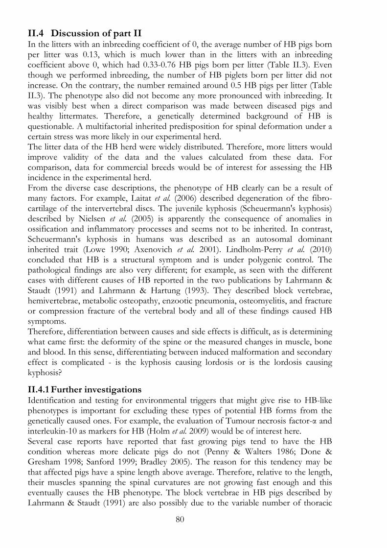
80
II.4 Discussion of part II In the litters with an inbreeding coefficient of 0, the average number of HB pigs born per litter was 0.13, which is much lower than in the litters with an inbreeding coefficient above 0, which had 0.33-0.76 HB pigs born per litter (Table II.3). Even though we performed inbreeding, the number of HB piglets born per litter did not increase. On the contrary, the number remained around 0.5 HB pigs per litter (Table II.3). The phenotype also did not become any more pronounced with inbreeding. It was visibly best when a direct comparison was made between diseased pigs and healthy littermates. Therefore, a genetically determined background of HB is questionable. A multifactorial inherited predisposition for spinal deformation under a certain stress was more likely in our experimental herd. The litter data of the HB herd were widely distributed. Therefore, more litters would improve validity of the data and the values calculated from these data. For comparison, data for commercial breeds would be of interest for assessing the HB incidence in the experimental herd. From the diverse case descriptions, the phenotype of HB clearly can be a result of many factors. For example, Laitat et al. (2006) described degeneration of the fibro-cartilage of the intervertebral discs. The juvenile kyphosis (Scheuermann's kyphosis) described by Nielsen et al. (2005) is apparently the consequence of anomalies in ossification and inflammatory processes and seems not to be inherited. In contrast, Scheuermann's kyphosis in humans was described as an autosomal dominant inherited trait (Lowe 1990; Axenovich et al. 2001). Lindholm-Perry et al. (2010) concluded that HB is a structural symptom and is under polygenic control. The pathological findings are also very different; for example, as seen with the different cases with different causes of HB reported in the two publications by Lahrmann & Staudt (1991) and Lahrmann & Hartung (1993). They described block vertebrae, hemivertebrae, metabolic osteopathy, enzootic pneumonia, osteomyelitis, and fracture or compression fracture of the vertebral body and all of these findings caused HB symptoms. Therefore, differentiation between causes and side effects is difficult, as is determining what came first: the deformity of the spine or the measured changes in muscle, bone and blood. In this sense, differentiating between induced malformation and secondary effect is complicated - is the kyphosis causing lordosis or is the lordosis causing kyphosis?
II.4.1 Further investigations Identification and testing for environmental triggers that might give rise to HB-like phenotypes is important for excluding these types of potential HB forms from the genetically caused ones. For example, the evaluation of Tumour necrosis factor-α and interleukin-10 as markers for HB (Holm et al. 2009) would be of interest here. Several case reports have reported that fast growing pigs tend to have the HB condition whereas more delicate pigs do not (Penny & Walters 1986; Done & Gresham 1998; Sanford 1999; Bradley 2005). The reason for this tendency may be that affected pigs have a spine length above average. Therefore, relative to the length, their muscles spanning the spinal curvatures are not growing fast enough and this eventually causes the HB phenotype. The block vertebrae in HB pigs described by Lahrmann & Staudt (1991) are also possibly due to the variable number of thoracic
81
and lumbar vertebrae (Berge 1948; Mikawa et al. 2011) and are caused by an unfinished separating process of two vertebrae. Hence, genes controlling vertebra number and formation are good HB candidate genes. To initiate a search for specific candidate genes, the genes listed in section II.1.2.5 are good starting points. Even better would be a genome-wide association study comparing HB inbred pigs, such as those produced in our experimental herd, and healthy unrelated ones. The genetics of extreme body forms (Section II.1.2.5.1) may be a good starting point for future HB research.
82
II.5 References Agadir M., Sevastik B., Sevastik J.A., Persson A. & Isberg B. (1988) Induction of
Scoliosis in the Growing-Rabbit by Unilateral Rib-Growth Stimulation. Spine 13, 1065-9.
Amirjamshidi A., Abbassioun K. & Bidabadi M.S. (2006) Skin-covered midline spinal anomalies: a report of four rare cases with a discussion on their genesis and milestones in surgical management. Childs Nervous System 22, 460-5.
Ascani E., Borelli P., Larosa G., Montanato A. & Turturro F. (1982) Malattia di Scheuermann. I: Studio ormonale. Progressi in patologia vertebrale. I.e cifosi, vol. 5-A, p. 97. Bologna, A. Gaggi, 1982. 5-A, 97.
Axenovich T.I., Zaidman A.M., Zorkoltseva I.V., Kalashnikova E.V. & Borodin P.M. (2001) Segregation analysis of Scheuermann disease in ninety families from Siberia. American Journal of Medical Genetics 100, 275-9.
Benitezking G. & Antontay F. (1993) Calmodulin Mediates Melatonin Cytoskeletal Effects. Experientia 49, 635-41.
Berge S. (1948) Genetical Researches on the Number of Vertebrae in the Pig. Journal of Animal Science 7, 233-8.
Bourke C.A. (1995) The clinical differentiation of nervous and muscular locomotor disorders of sheep in Australia. Australian Veterinary Journal 72, 228-34.
Bradley H. (2005) Variation in back conformation and prevalence of ulcers on the shoulders : a cohort study of related Swedish Landrace and Landrace * Yorkshire sows. http://ex-epsilon.slu.se:8080/archive/00001276/.
Bulman M.P., Kusumi K., Frayling T.M., McKeown C., Garrett C., Lander E.S., Krumlauf R., Hattersley A.T., Ellard S. & Turnpenny P.D. (2000) Mutations in the human Delta homologue, DLL3, cause axial skeletal defects in spondylocostal dysostosis. Nature genetics 24, 438-41.
Burwell R.G. (2003) Aetiology of idiopathic scoliosis: current concepts. Pediatric rehabilitation 6.
Busscher I., Ploegmakers J.J.W., Verkerke G.J. & Veldhuizen A.G. (2010) Comparative anatomical dimensions of the complete human and porcine spine. European Spine Journal 19, 1104-14.
Cawdellsmith J., Upfold J., Edwards M. & Smith M. (1992) Neural-Tube and Other Developmental Anomalies in the Guinea-Pig Following Maternal Hyperthermia during Early Neural-Tube Development. Teratogenesis Carcinogenesis and Mutagenesis 12, 1-9.
Chae C. (2005) A review of porcine circovirus 2-associated syndromes and diseases. Veterinary Journal 169, 326-36.
Chen W.J., Lai P.L., Tai C.L., Chen L.H. & Niu C.C. (2004) The effect of sagittal alignment on adjacent joint mobility after lumbar instrumentation - a biomechanical study of lumbar vertebrae in a porcine model. Clinical Biomechanics 19, 763-8.
Clarke T. (2005) Hump-Back Pigs. In: Animal Health Expositor, p. 2. Prairie Diagnostic Services and Saskatchewan Agriculture, Food and Rural Revitalization, http://www.pdsinc.ca/Portals/0/AH200501.pdf.
Deforest M.E. & Basrur P.K. (1979) Malformations and the Manx syndrome in cats. The Canadian veterinary journal. La revue vétérinaire canadienne 20, 304-14.

83
Done S.H. & Gresham A.C.J. (1998) Lordosis and Kyphosis ("Humpy Back") in Pigs. PIG JOURNAL 41, 134-41.
Done S.H., Potter R.A., Courtenay A. & Peissel K. (1999) Lordosis and Kyphosis ("Humpy-Back") in Pigs: A Second Type of the Condition Associated with Hemivertebrae. PIG JOURNAL 43, 148-53.
Garbe JR & Y. D. (2008) Pedigraph user manual. Version 2.4. . Department of Animal Science, University of Minnesota, MN,.
Giunta C., Randolph A. & Steinmann B. (2005) Mutation analysis of the PLOD1 gene: An efficient multistep approach to the molecular diagnosis of the kyphoscoliotic type of Ehlers-Danlos syndrome (EDS VIA). Molecular Genetics and Metabolism 86, 269-76.
Green B.T., Lee S.T., Panter K.E., Welch K.D., Cook D., Pfister J.A. & Kem W.R. (2010) Actions of piperidine alkaloid teratogens at fetal nicotinic acetylcholine receptors. Neurotoxicology and Teratology 32, 383-90.
Hastbacka J., Kerrebrock A., Mokkala K., Clines G., Lovett M., Kaitila I., de la Chapelle A. & Lander E.S. (1999) Identification of the Finnish founder mutation for diastrophic dysplasia (DTD). European Journal of Human Genetics 7, 664-70.
Hecht J.T., Nelson L.D., Crowder E., Wang Y., Elder F.F.B., Harrison W.R., Francomano C.A., Prange C.K., Lennon G.G., Deere M. & Lawler J. (1995) Mutations in Exon 17b of Cartilage Oligomeric Matrix Protein (Comp) Cause Pseudoachondroplasia. Nature genetics 10, 325-9.
Holl J.W., Rohrer G.A., Shackelford S.D., Wheeler T.L. & Koohmaraie M. (2008) Estimates of genetic parameters for kyphosis in two crossbred swine populations. Journal of Animal Science 86, 1765-9.
Holm S., Mackiewicz Z., Holm A.K., Konttinen Y.T., Kouri V.P., Indahl A. & Salo J. (2009) Pro-inflammatory, Pleiotropic, and Anti-inflammatory TNF-alpha, IL-6, and IL-10 in Experimental Porcine Intervertebral Disk Degeneration. Veterinary Pathology 46, 1292-300.
James C.C.M., Lassman L.P. & Tomlinso.Be (1969) Congenital Anomalies of Lower Spine and Spinal Cord in Manx Cats. Journal of Pathology 97, 269-&.
Johansson M.P., Baann Liane M.S., Bendix T., Kasch H. & Kongsted A. (2011) Does cervical kyphosis relate to symptoms following whiplash injury? Manual Therapy 16, 378-83.
Jungbluth H., Zhou H., Hartley L., Halliger-Keller B., Messina S., Longman C., Brockington M., Robb S.A., Straub V., Voit T., Swash M., Ferreiro A., Bydder G., Sewry C.A., Muller C. & Muntoni F. (2005) Minicore myopathy with ophthalmoplegia caused by mutations in the ryanodine receptor type 1 gene. Neurology 65, 1930-5.
Kawakita A., Sato K., Makino H., Ikegami H., Takayama S., Toyama Y. & Umezawa A. (2008) Nicotine Acts on Growth Plate Chondrocytes to Delay Skeletal Growth through the alpha 7 Neuronal Nicotinic Acetylcholine Receptor. PLoS One 3.
Keeler R.F. & Crowe M.W. (1984) Teratogenicity and Toxicity of Wild Tree Tobacco, Nicotiana-Glauca in Sheep. Cornell Veterinarian 74, 50-9.
Lahrmann K.H. & Hartung K. (1993) Causes of Kyphosis and Lordosis with Cuneiform Vertebral Deformation in Swine. Berliner Und Munchener Tierarztliche Wochenschrift 106, 127-32.
84
Lahrmann K.H. & Staudt B. (1991) Formation of Block Vertebrae in Swine. Journal of Veterinary Medicine Series a-Zentralblatt Fur Veterinarmedizin Reihe a-Physiology Pathology Clinical Medicine 38, 691-5.
Laird P.W., Zijderveld A., Linders K., Rudnicki M.A., Jaenisch R. & Berns A. (1991) Simplified Mammalian DNA Isolation Procedure. Nucleic Acids Research 19, 4293-.
Laitat M., Harmegnies N., Van Cauwenberge H., Georis P., Buys N. & M. G. (2006) Lordosis in pigs: Evidence for inherited predisposition. In: Proceedings of the 19th Int. Pig Vet. Soc. (IPVS) Congress, p. 111, Copenhagen, Denmark.
Lambert F.M., Malinvaud D., Glaunes J., Bergot C., Straka H. & Vidal P.P. (2009) Vestibular Asymmetry as the Cause of Idiopathic Scoliosis: A Possible Answer from Xenopus. Journal of Neuroscience 29, 12477-83.
Lindholm-Perry A.K., Rohrer G.A., Kuehn L.A., Keele J.W., Holl J.W., Shackelford S.D., Wheeler T.L. & Nonneman D.J. (2010) Genomic regions associated with kyphosis in swine. Bmc Genetics 11, -.
Lowe T.G. (1990) Current Concepts Review - Scheuermann Disease. Journal of Bone and Joint Surgery-American Volume 72A, 940-5.
Lowe T.G., Edgar M., Chir M., Margulies J.Y., Miller N.H., Raso V.J., Reinker K.A. & Rivard C.H. (2000) Current concepts review - Etiology of idiopathic scoliosis: Current trends in research. Journal of Bone and Joint Surgery-American Volume 82A, 1157-68.
Mcmeniman N.P. (1976) Solanum-Esuriale, a Possible Cause of Humpy Back in Sheep. Australian Veterinary Journal 52, 432-3.
Mikawa S., Sato S., Nii M., Morozumi T., Yoshioka G., Imaeda N., Yamaguchi T., Hayashi T. & Awata T. (2011) Identification of a second gene associated with variation in vertebral number in domestic pigs. Bmc Genetics 12.
Nielsen L.W.D., Hogedal P., Arnbjerg J. & Jensen H.E. (2005) Juvenile kyphosis in pigs - A spontaneous model of Scheuermann's kyphosis. APMIS 113, 702-7.
Noack F., Reusche E. & Gembruch U. (2003) Prenatal diagnosis of 'true tail' with cartilage content? Fetal Diagnosis and Therapy 18, 226-9.
O'Sullivan B.M. (1976) Humpy back of sheep. Clinical and pathological observations. Australian Veterinary Journal 52, 414-8.
Penny R.H.C. & Walters J.R. (1986) A "humpy-backed" syndrome of pigs. Vet. Ann. 26, 128-35.
Sanford S.E. (1999) Helping your herds get over the hump. In: Farm and Country PORK. Agricultural Publishing Company Limited, http://www.agpub.on.ca/pork/ june99/prk1.htm.
Smith M.S.R., Upfold J.B., Edwards M.J., Shiota K. & Cawdellsmith J. (1992) The Induction of Neural-Tube Defects by Maternal Hyperthermia - a Comparison of the Guinea-Pig and Human. Neuropathology and Applied Neurobiology 18, 71-80.
Straw B., Bates R. & May G. (2009) Anatomical abnormalities in a group of finishing pigs: prevalence and pig performance. Journal of Swine Health and Production 17, 28-31.
Straw B.E., Zimmerman J.J., D'Allaire S. & Taylor D.J. (2006) Diseases of swine. Blackwell, Oxford.
Thies K., Kriegler W. & Waldmann K.H. (2010) Poisoning with magnesium in fattening pigs. Praktische Tierarzt 91, 144-7.
85
Turnpenny P.D., Bulman M.P., Frayling T.M., Abu-Nasra T.K., Garrett C., Hattersley A.T. & Ellard S. (1999) A gene for autosomal recessive spondylocostal dysostosis maps to 19q13.1-q13.3. American Journal of Human Genetics 65, 175-82.
van den Akker E., Fromental-Ramain C., de Graaff W., Le Mouellic H., Brulet P., Chambon P. & Deschamps J. (2001) Axial skeletal patterning in mice lacking all paralogous group 8 Hox genes. Development 128, 1911-21.
Vogeli P., Bolt R., Fries R. & Stranzinger G. (1994) Co-segregation of the malignant hyperthermia and the Arg615-Cys615 mutation in the skeletal muscle calcium release channel protein in five European Landrace and Pietrain pig breeds. Anim Genet 25 Suppl 1, 59-66.
Vriend J., Sheppard M.S. & Bala R.M. (1988) Melatonin Increases Serum Insulin-Like Growth Factor-I in Male Syrian-Hamsters. Endocrinology 122, 2558-61.
Xiong D.H., Liu X.G., Guo Y.F., Tan L.J., Wang L., Sha B.Y., Tang Z.H., Pan F., Yang T.L., Chen X.D., Lei S.F., Yerges L.M., Zhu X.Z., Wheeler V.W., Patrick A.L., Bunker C.H., Guo Y., Yan H., Pei Y.F., Zhang Y.P., Levy S., Papasian C.J., Xiao P., Lundberg Y.W., Recker R.R., Liu Y.Z., Liu Y.J., Zmuda J.M. & Deng H.W. (2009) Genome-wide Association and Follow-Up Replication Studies Identified ADAMTS18 and TGFBR3 as Bone Mass Candidate Genes in Different Ethnic Groups. American Journal of Human Genetics 84, 388-98.
Ytrehus B., Haga H.A., Mellum C.N., Mathisen L., Carlson C.S., Ekman S., Teige J. & Reinholt F.P. (2004) Experimental ischemia of porcine growth cartilage produces lesions of osteochondrosis. Journal of Orthopaedic Research 22, 1201-9.
86
II.6 List of figures Figure II.1: Typical HB pig of the experimental herd with thoracic lordosis and
with lumbar kyphosis (Picture Sem Genini). 69
Figure II.2: Pneumonia diseased weaning pigs showing HB phenotype. 71
II.7 List of tables Table II.1: Number of litters, piglets and affected piglets born in the experimental
herd. 79
Table II.2: Average values (± average of the absolute deviation) calculated for the experimental herd. 79
Table II.3: The herd data separated into groups of the same inbreeding coefficient (average values ± average of the absolute deviation). 79
87
Part III: Breeding of Splay Leg pigs - an observational study
88
Table of Contents III.1 Introduction 89
III.1.1 Aim of the SL breeding experiment 89
III.1.2 Review 89
III.1.2.1 SL in breeding experiments and commercial herds 89
III.1.2.2 Histological findings 91
III.1.2.3 SL candidate genes 92
III.2 Materials and methods 99
III.2.1 Breeding of SL pigs 99
III.2.2 Genomic DNA 99
III.2.3 Pedigree drawing 99
III.3 Results 100
III.3.1 Additional observations 100
III.4 Discussion of part III 102
III.4.1 Further investigations 103
III.5 References 104
III.6 List of tables 109
89
III.1 Introduction Piglets affected by congenital splay leg (SL) suffer from paresis of the hind limbs, and rarely of the front legs as well. The weakness in the hind limbs leads to the characteristic upright posture in the first week postnatal. Normally, the condition improves so that the piglets appear recovered after a week. However, grown up and heavier adult SL pigs in our breed again showed weakness in the hind limbs, particularly sows in pig. It is possibly that after an acute onset, SL is a latent disease. For SL piglets, the main problems of this condition are the danger of being squeezed to death by the mother and the difficulty of getting enough milk. Secondary problems are inflammations and ulceration of the over-stressed skin and joints. For SL piglets, as an aid to survive the critical days, ´taping the hind limbs together or the use of elastic bands´ has been promoted, for example, on information-providing pages on the internet like www.vetsweb.com. On pages like www.agrar-direct.de, it is possible to order such therapeutic appliances. Then, as soon as the SL piglets are able to walk normally, they catch up in growth in relation to their littermates. In the literature, the condition is also referred to as splay legged or spraddle leg, in French ´le syndrome de l’abduction des membres´ or in German ´kongenitales Ausgrätschen beim Saugferkel´ and ´Spreizer´.
III.1.1 Aim of the SL breeding experiment If there is a genetic background involved in SL, we should see an increasing number of affected piglets through generations or an intensified phenotype. Thus, if former SL-diseased pigs transmit the SL genes to their progeny, we should experience increasing proportions of SL piglets per litter with generations of breeding. In order to understand the genetics of the SL disease, we bred a small herd using former SL-diseased pigs as parents and observed the development/evolution of SL beyond several generations. This is similar to the breeding experiment done by T. Lax (1971); however, he observed only three generations of a small group of pigs. In addition to the observations, we should be able to collect genomic DNA samples with corresponding pedigrees for further studies on this subject.
III.1.2 Review In recent years, six dissertations about or touching on the subject of SL have been published, specifically those by S. Schwarz (2003), P. T. Ooi (2006), S. Jäsert (2003), B. Beißner (2003) and A. Bull (2008), not forgetting the habilitation treatise published by S. Maak (2001). The dissertations by S. Schwarz, S. Jäsert, B. Beisser and A. Bull as well as the habilitation treatise by S. Maak, are written in German.
III.1.2.1 SL in breeding experiments and commercial herds
III.1.2.1.1 Observational studies Compared to SL affecting the hind leg, SL affecting the front and hind legs is rare. Sellier and Ollivier (1982) reported up to 20% of SL cases affecting all four legs. Mostly, this four-legged variant is not mentioned separately. The mortality of this more severe SL is even higher than the variant affecting only the hind legs. In the breeding experiment described by T. Lax (1971), a boar and two sows were used as starting population and the herd was bred for three generations. The diseased piglets in this family were affected at the hind and front legs. All of the diseased
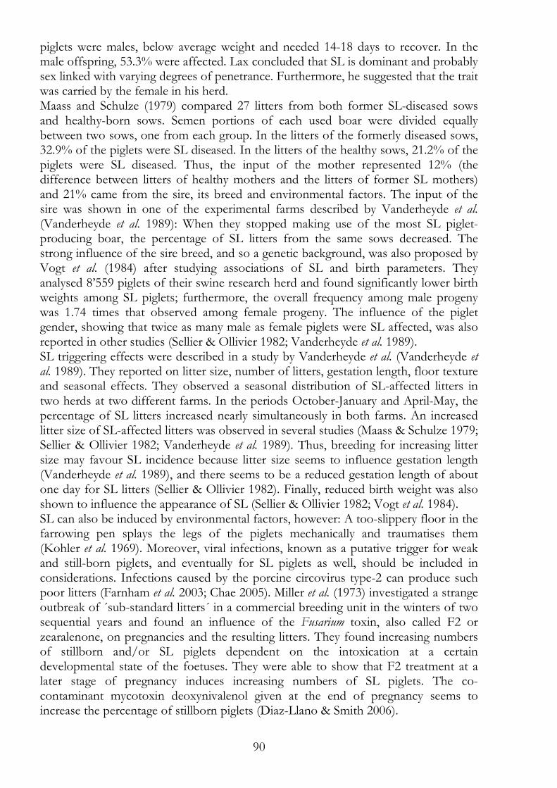
90
piglets were males, below average weight and needed 14-18 days to recover. In the male offspring, 53.3% were affected. Lax concluded that SL is dominant and probably sex linked with varying degrees of penetrance. Furthermore, he suggested that the trait was carried by the female in his herd. Maass and Schulze (1979) compared 27 litters from both former SL-diseased sows and healthy-born sows. Semen portions of each used boar were divided equally between two sows, one from each group. In the litters of the formerly diseased sows, 32.9% of the piglets were SL diseased. In the litters of the healthy sows, 21.2% of the piglets were SL diseased. Thus, the input of the mother represented 12% (the difference between litters of healthy mothers and the litters of former SL mothers) and 21% came from the sire, its breed and environmental factors. The input of the sire was shown in one of the experimental farms described by Vanderheyde et al. (Vanderheyde et al. 1989): When they stopped making use of the most SL piglet-producing boar, the percentage of SL litters from the same sows decreased. The strong influence of the sire breed, and so a genetic background, was also proposed by Vogt et al. (1984) after studying associations of SL and birth parameters. They analysed 8’559 piglets of their swine research herd and found significantly lower birth weights among SL piglets; furthermore, the overall frequency among male progeny was 1.74 times that observed among female progeny. The influence of the piglet gender, showing that twice as many male as female piglets were SL affected, was also reported in other studies (Sellier & Ollivier 1982; Vanderheyde et al. 1989). SL triggering effects were described in a study by Vanderheyde et al. (Vanderheyde et al. 1989). They reported on litter size, number of litters, gestation length, floor texture and seasonal effects. They observed a seasonal distribution of SL-affected litters in two herds at two different farms. In the periods October-January and April-May, the percentage of SL litters increased nearly simultaneously in both farms. An increased litter size of SL-affected litters was observed in several studies (Maass & Schulze 1979; Sellier & Ollivier 1982; Vanderheyde et al. 1989). Thus, breeding for increasing litter size may favour SL incidence because litter size seems to influence gestation length (Vanderheyde et al. 1989), and there seems to be a reduced gestation length of about one day for SL litters (Sellier & Ollivier 1982). Finally, reduced birth weight was also shown to influence the appearance of SL (Sellier & Ollivier 1982; Vogt et al. 1984). SL can also be induced by environmental factors, however: A too-slippery floor in the farrowing pen splays the legs of the piglets mechanically and traumatises them (Kohler et al. 1969). Moreover, viral infections, known as a putative trigger for weak and still-born piglets, and eventually for SL piglets as well, should be included in considerations. Infections caused by the porcine circovirus type-2 can produce such poor litters (Farnham et al. 2003; Chae 2005). Miller et al. (1973) investigated a strange outbreak of ´sub-standard litters´ in a commercial breeding unit in the winters of two sequential years and found an influence of the Fusarium toxin, also called F2 or zearalenone, on pregnancies and the resulting litters. They found increasing numbers of stillborn and/or SL piglets dependent on the intoxication at a certain developmental state of the foetuses. They were able to show that F2 treatment at a later stage of pregnancy induces increasing numbers of SL piglets. The co-contaminant mycotoxin deoxynivalenol given at the end of pregnancy seems to increase the percentage of stillborn piglets (Diaz-Llano & Smith 2006).
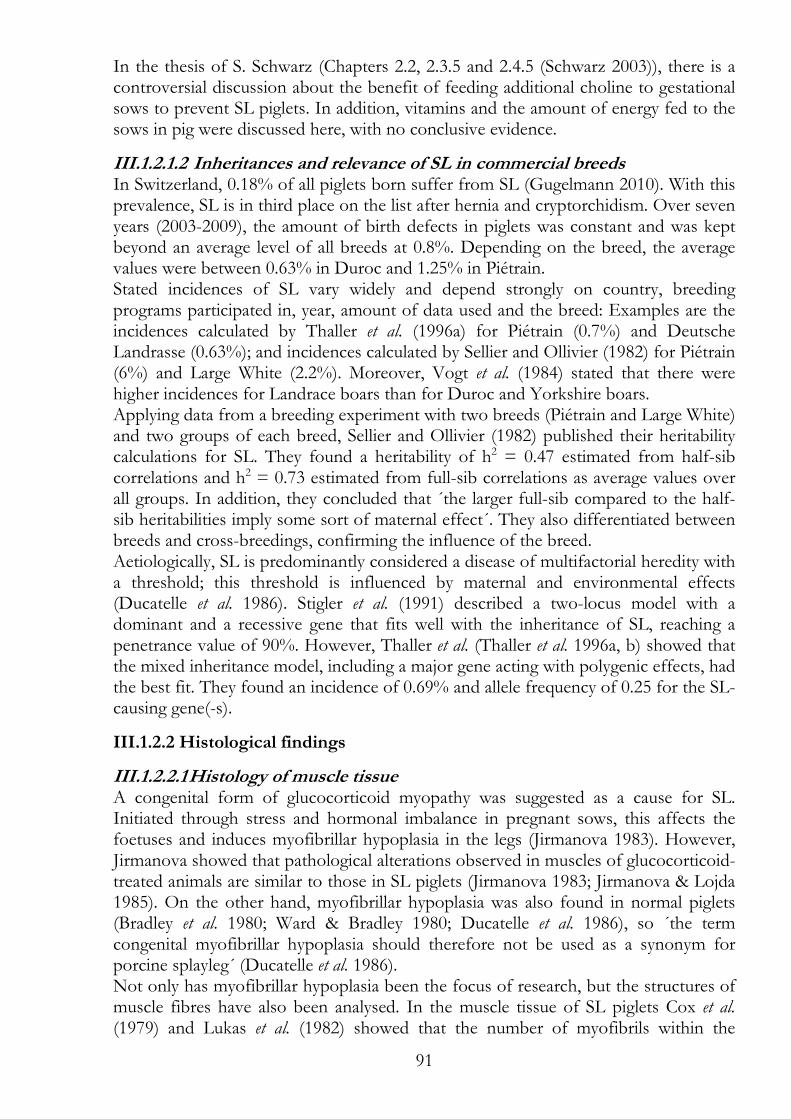
91
In the thesis of S. Schwarz (Chapters 2.2, 2.3.5 and 2.4.5 (Schwarz 2003)), there is a controversial discussion about the benefit of feeding additional choline to gestational sows to prevent SL piglets. In addition, vitamins and the amount of energy fed to the sows in pig were discussed here, with no conclusive evidence.
III.1.2.1.2 Inheritances and relevance of SL in commercial breeds In Switzerland, 0.18% of all piglets born suffer from SL (Gugelmann 2010). With this prevalence, SL is in third place on the list after hernia and cryptorchidism. Over seven years (2003-2009), the amount of birth defects in piglets was constant and was kept beyond an average level of all breeds at 0.8%. Depending on the breed, the average values were between 0.63% in Duroc and 1.25% in Piétrain. Stated incidences of SL vary widely and depend strongly on country, breeding programs participated in, year, amount of data used and the breed: Examples are the incidences calculated by Thaller et al. (1996a) for Piétrain (0.7%) and Deutsche Landrasse (0.63%); and incidences calculated by Sellier and Ollivier (1982) for Piétrain (6%) and Large White (2.2%). Moreover, Vogt et al. (1984) stated that there were higher incidences for Landrace boars than for Duroc and Yorkshire boars. Applying data from a breeding experiment with two breeds (Piétrain and Large White) and two groups of each breed, Sellier and Ollivier (1982) published their heritability calculations for SL. They found a heritability of h2 = 0.47 estimated from half-sib correlations and h2 = 0.73 estimated from full-sib correlations as average values over all groups. In addition, they concluded that ´the larger full-sib compared to the half-sib heritabilities imply some sort of maternal effect´. They also differentiated between breeds and cross-breedings, confirming the influence of the breed. Aetiologically, SL is predominantly considered a disease of multifactorial heredity with a threshold; this threshold is influenced by maternal and environmental effects (Ducatelle et al. 1986). Stigler et al. (1991) described a two-locus model with a dominant and a recessive gene that fits well with the inheritance of SL, reaching a penetrance value of 90%. However, Thaller et al. (Thaller et al. 1996a, b) showed that the mixed inheritance model, including a major gene acting with polygenic effects, had the best fit. They found an incidence of 0.69% and allele frequency of 0.25 for the SL-causing gene(-s).
III.1.2.2 Histological findings
III.1.2.2.1 Histology of muscle tissue A congenital form of glucocorticoid myopathy was suggested as a cause for SL. Initiated through stress and hormonal imbalance in pregnant sows, this affects the foetuses and induces myofibrillar hypoplasia in the legs (Jirmanova 1983). However, Jirmanova showed that pathological alterations observed in muscles of glucocorticoid-treated animals are similar to those in SL piglets (Jirmanova 1983; Jirmanova & Lojda 1985). On the other hand, myofibrillar hypoplasia was also found in normal piglets (Bradley et al. 1980; Ward & Bradley 1980; Ducatelle et al. 1986), so ´the term congenital myofibrillar hypoplasia should therefore not be used as a synonym for porcine splayleg´ (Ducatelle et al. 1986). Not only has myofibrillar hypoplasia been the focus of research, but the structures of muscle fibres have also been analysed. In the muscle tissue of SL piglets Cox et al. (1979) and Lukas et al. (1982) showed that the number of myofibrils within the
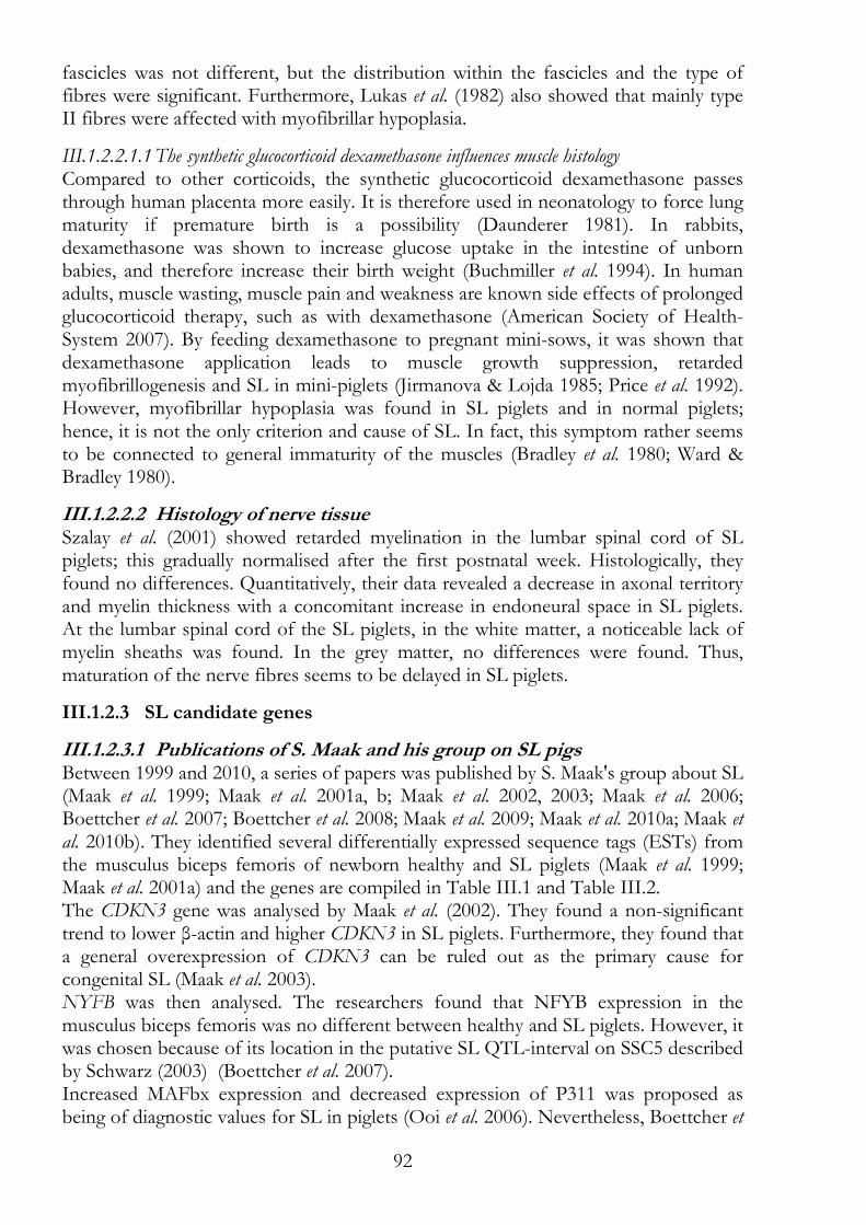
92
fascicles was not different, but the distribution within the fascicles and the type of fibres were significant. Furthermore, Lukas et al. (1982) also showed that mainly type II fibres were affected with myofibrillar hypoplasia.
III.1.2.2.1.1 The synthetic glucocorticoid dexamethasone influences muscle histology Compared to other corticoids, the synthetic glucocorticoid dexamethasone passes through human placenta more easily. It is therefore used in neonatology to force lung maturity if premature birth is a possibility (Daunderer 1981). In rabbits, dexamethasone was shown to increase glucose uptake in the intestine of unborn babies, and therefore increase their birth weight (Buchmiller et al. 1994). In human adults, muscle wasting, muscle pain and weakness are known side effects of prolonged glucocorticoid therapy, such as with dexamethasone (American Society of Health-System 2007). By feeding dexamethasone to pregnant mini-sows, it was shown that dexamethasone application leads to muscle growth suppression, retarded myofibrillogenesis and SL in mini-piglets (Jirmanova & Lojda 1985; Price et al. 1992). However, myofibrillar hypoplasia was found in SL piglets and in normal piglets; hence, it is not the only criterion and cause of SL. In fact, this symptom rather seems to be connected to general immaturity of the muscles (Bradley et al. 1980; Ward & Bradley 1980).
III.1.2.2.2 Histology of nerve tissue Szalay et al. (2001) showed retarded myelination in the lumbar spinal cord of SL piglets; this gradually normalised after the first postnatal week. Histologically, they found no differences. Quantitatively, their data revealed a decrease in axonal territory and myelin thickness with a concomitant increase in endoneural space in SL piglets. At the lumbar spinal cord of the SL piglets, in the white matter, a noticeable lack of myelin sheaths was found. In the grey matter, no differences were found. Thus, maturation of the nerve fibres seems to be delayed in SL piglets.
III.1.2.3 SL candidate genes
III.1.2.3.1 Publications of S. Maak and his group on SL pigs Between 1999 and 2010, a series of papers was published by S. Maak's group about SL (Maak et al. 1999; Maak et al. 2001a, b; Maak et al. 2002, 2003; Maak et al. 2006; Boettcher et al. 2007; Boettcher et al. 2008; Maak et al. 2009; Maak et al. 2010a; Maak et al. 2010b). They identified several differentially expressed sequence tags (ESTs) from the musculus biceps femoris of newborn healthy and SL piglets (Maak et al. 1999; Maak et al. 2001a) and the genes are compiled in Table III.1 and Table III.2. The CDKN3 gene was analysed by Maak et al. (2002). They found a non-significant trend to lower β-actin and higher CDKN3 in SL piglets. Furthermore, they found that a general overexpression of CDKN3 can be ruled out as the primary cause for congenital SL (Maak et al. 2003). NYFB was then analysed. The researchers found that NFYB expression in the musculus biceps femoris was no different between healthy and SL piglets. However, it was chosen because of its location in the putative SL QTL-interval on SSC5 described by Schwarz (2003) (Boettcher et al. 2007). Increased MAFbx expression and decreased expression of P311 was proposed as being of diagnostic values for SL in piglets (Ooi et al. 2006). Nevertheless, Boettcher et
93
al. (2008) observed no significant differences in relative MAFbx expression between SL piglets and healthy piglets. Thus, they declared the diagnostic value of the MAFbx expression pattern to be doubtful. In a GeneChip Porcine Genome Array from Affymetrix, Maak et al. (2009) found four genes to be differentially expressed (SQSTMI, SSRPI, DDIT4 and MAF). Moreover, the expression of the two genes ZDHH9 and ITGA5 stood out. Further investigations showed that the increased expression of ZDHH9 was not related to the expression of associated microRNAs. The gene ZDHHC9 maps to SSCX and participates in Ras-signalling (Maak et al. 2010b). Next to be analysed was the increased expression of ITGA5; Single-nucleotide polymorphisms (SNPs) identified in this gene could be excluded as causative (Maak et al. 2010a). The ITGA5 gene maps to SSC5.
III.1.2.3.2 Candidate genes for SL To identify possible SL candidate genes, four publications are considered in Table III.1. Maak et al. (2001a) identified differentially expressed ESTs from musculus biceps femoris of newborn healthy and SL piglets; Schwarz (2003) mapped SL to three regions on the porcine chromosomes 3, 5 and 11 by microsatellite analysis and created a linkage map; Davoli et al. (1999) identified genes expressed in porcine skeletal muscle; and Wimmers et al. (2007) examined candidate genes connected to meat quality. Comparison of meat quality genes to SL candidate genes seems to be of interest because muscle tissue has been shown to be histologically altered in SL piglets. The 17 ESTs on the three chromosomes pointed out by Schwarz (2003) are compiled in Table III.2. They are possible SL candidate genes. Table III.2 shows also that there are no joint candidate genes that were identified in SL piglets by Maak et al. (2001a) and the meat genes identified by Davoli et al. (1999) and Wimmers et al. (2007) (Table III.1). The four genes, SQSTMI (SSC2), SSRP1 (SSC2), DDIT4 (SSC14) and MAF (SSC6), identified by the Gene Chip experiment are good candidate genes, at least as markers for SL, with their proven altered expression (Maak et al. 2009). Sixteen other muscle-related genes (ESTs) also map to chromosomes SSC2, SSC6 and SSC14 (Table III.1). MAFbx maps to SSC4, and its expression was promoted as a marker for SL (Ooi et al. 2006); however, MAFbx was later excluded as a good candidate gene (Boettcher et al. 2008). EPLIN is a promising candidate gene because it was identified as EST AJ279591 on SSC5 (Table III.1 and Table III.2), and later its preferential expression was shown in the muscle tissue of SL piglets (Wang et al. 2007). In addition, ZDHHC9 on SSCX and ITGA5 on SSC5 are promising candidate genes because both showed increased expression in SL piglets (Maak et al. 2010a; Maak et al. 2010b). Furthermore, the ITGA5 gene maps at 21.2 Mb on SSC5, close to EPLIN, at 16.8 Mb (Table III.2), and is therefore an SL candidate gene along with EPLIN. On the other hand, genes for meat quality and carcass traits examined by Wimmers et al. (2007) are interesting concerning their influence on muscle tissue structure. CA3 (NM_001008688) on SSC4 is associated with muscle dystrophy (Heath et al. 1985; Ohta et al. 1991), and EPOR (AF274305) on SSC2 maps close to QTLs for muscle fibre size, number and proportion of intermediate fibres (Wimmers et al. 2006; Liu et al. 2007).
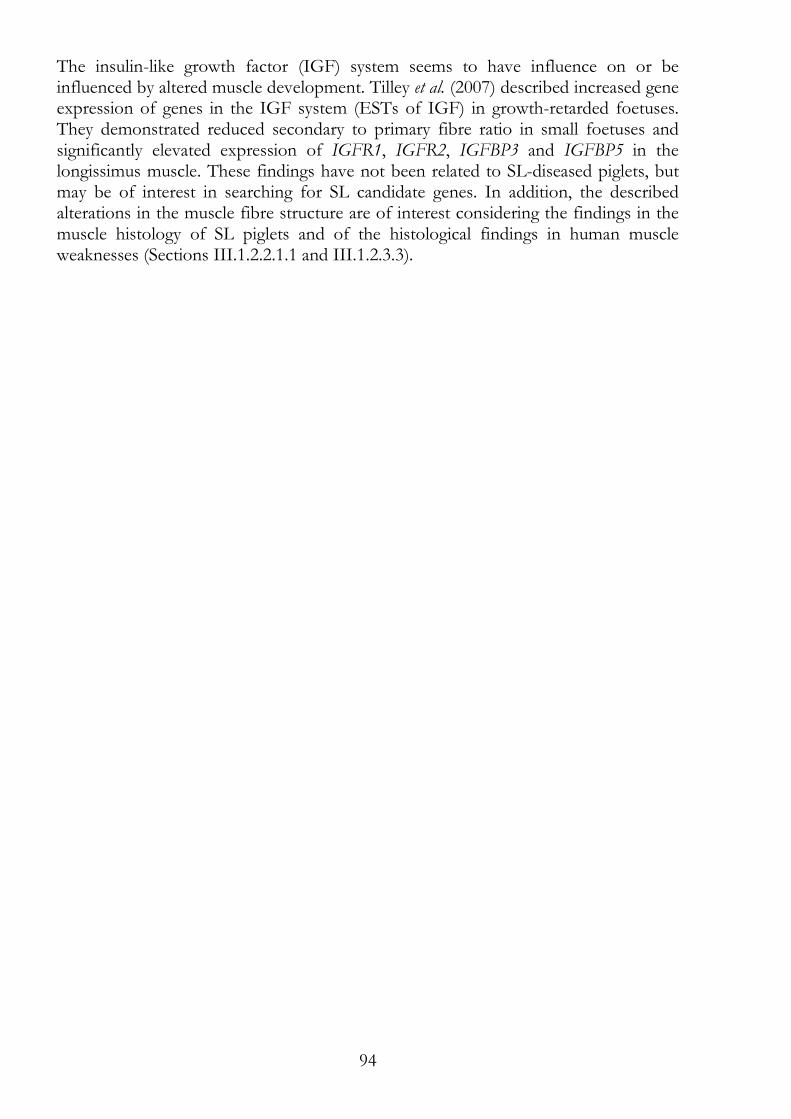
94
The insulin-like growth factor (IGF) system seems to have influence on or be influenced by altered muscle development. Tilley et al. (2007) described increased gene expression of genes in the IGF system (ESTs of IGF) in growth-retarded foetuses. They demonstrated reduced secondary to primary fibre ratio in small foetuses and significantly elevated expression of IGFR1, IGFR2, IGFBP3 and IGFBP5 in the longissimus muscle. These findings have not been related to SL-diseased piglets, but may be of interest in searching for SL candidate genes. In addition, the described alterations in the muscle fibre structure are of interest considering the findings in the muscle histology of SL piglets and of the histological findings in human muscle weaknesses (Sections III.1.2.2.1.1 and III.1.2.3.3).
95
Table III.1: Comparison of the ESTs (GeneBank No.) of the SL candidate regions, SL candidate genes and meat genes proposed by Schwarz (2003), Maak et al. (2001a), Davoli et al. (1999) and Wimmers et al. (2007). Chromosomal regions and identified EST are ordered by SSC. All ESTs which could be mapped to a single chromosomal region in pig genome build 10 are shown. SSC Schwarz 2003 Maak et al.2001a Davoli et al.1999 Wimmers et al.
2007
SSC1 --- AJ279583, AJ133889, AJ279584
Z98837, Z98798, Z98773, Z98799, Z98764, Z98765, Z98809, Z98810
---
SSC2 --- AJ271016 Z98825, Z98787, Z98786, Z98778, Z98797, Z98800, Z98801
AF274305
SSC3 evidence for linkage
AJ277993, AJ404881, AJ271018
Z98813, Z98780, X91849, X91850, Z98829
---
SSC4 --- AJ133890 Z98782, Z98783, Z98796, Z98844, Z98845
NM_001008688
SSC5 significant evidence for linkage
AJ279591, AJ271017
Z98807, Z98834, X94254, Z98766
DQ631866, DQ631862
SSC6 --- --- Z98841 --- SSC7 --- AJ279590 Z98805, Z98824, Y18405,
X91846, X91847 ---
SSC8 --- AJ279589 Z98818, Z98819 DQ631865 SSC9 --- AJ279587,
AJ133891, AJ271019
Z98816, Z98821, Z98822, Z98820, Z98804
---
SSC10 --- --- Z98838 --- SSC11 significant evidence
for linkage AJ133888 --- ---
SSC12 --- AJ271011 Z98790, Z98791, X91845, Z98835
DQ631864
SSC13 --- AJ279580 Z98770, Z98793, Z98836, Z98842
---
SSC14 --- --- Z98823, Z98774, Z98775, Z98781, Z98771
AJ560657, DQ631863
SSC15 --- --- Z98779, Z94252, Z98788, Y18404, X94689, Z98806, Z98827
AJ560658
SSC16 --- --- --- --- SSC17 --- AJ279581 Z98777, Z98839, Z98840 DV897570 SSC18 --- --- Z98802 --- SSCX --- AJ133887 Z98826, Z98832, Z98776 ---
96
Table III.2: ESTs identified in the SL candidate regions defined by Schwarz (2003) and their matching gene sequences and positions on the chromosome (Sscrofa10 genome assembly). SSC GeneBank
No Matches (Nucleotide blast NCBI) Position
SSC3 Z98813 similar to 14-3-3 protein gamma subtype tyrosine 3-monooxygenase/tryptophan 5-monooxygenase activation protein, gamma polypeptide (YWHAG)
9.3 Mb / 9.6 Mb
AJ404881 b-cell CLL/lymphoma 7 protein family member B-like (BCL7B) 10.7 Mb Z98780 similar to cytosolic malate dehydrogenase (MDH1) 53.0 Mb X91849, X91850
spectrin beta protein (pAZSP 3' end) spectrin, beta, non-erythrocytic 1 (SPTBN1)
90.5 Mb
Z98829 yjeF N-terminal domain-containing protein 3-like Canis familiaris similar to NADH-ubiquinone oxidoreductase B16.6 subunit (Complex I-B16.6) (CI-B16.6) Bos taurus NADH dehydrogenase (ubiquinone) 1 alpha subcomplex, 13
97.7 Mb
AJ271018 structural maintenance of chromosomes 6 (SMC6) 126.9 Mb AJ277993 partial TAF1B gene for TATA box binding protein (TBP)
associated factor 133.3 Mb
SSC5 AJ279591 EPLIN-a, EPLIN-b = LIMA1 Epithelial protein lost in neoplasm beta = LIM domain and actin binding 1
16.8 Mb
DQ631866 high mobility group AT-hook 2 (HMGA2), exon 11 and 3' UTR 32.6 Mb Z98807 Sus scrofa DNA-binding protein A-like
Canis lupus familiaris Y-box protein ZONAB-A (ZONAB) Homo sapiens cold shock domain protein A (CSDA)
62.3 Mb
Z98834 glyceraldehyde-3-phosphate dehydrogenase (GAPDH) 64.7 Mb DQ631862 Rab6-interacting protein 2 (RAB6IP2) gene, exons 9, 10 and partial
cds ELKS/RAB6-interacting/CAST family member 1, transcript variant 1 (ERC1)
69.3 Mb
X94254 phosphofructokinase, muscle (PFKM) 80.6 Mb AJ271017 nuclear transcription factor Y, beta (NFYB) 82.8 Mb Z98766 nudix (nucleoside diphosphate linked moiety X)-type motif 4
(NUDT4) Macaca mulatta diphosphoinositol polyphosphate phosphohydrolase 2-like
92.7 Mb
SSC11 AJ133888 PHD finger protein 11-like, transcript variant 1 or 2 (PHF11) 17.8 Mb
97
III.1.2.3.3 Comparable syndromes in the rabbit, turkey, dog and human The onset of SL in rabbits occurs at a later stage compared to SL in pigs and the symptoms stay; it has even been the observed that SL symptoms can occur during pregnancy in rabbits (Joosten et al. 1981). Joosten et al. showed that imbalances with respect to the speed of neural, muscular and skeletal development of the limb are the primary defects and that these genetic predispositions are the cause of torsion of the femur during early postnatal periods when the ability to walk, which is associated with frequent splaying in all animals, develops (Joosten et al. 1981). In addition, the mechanical induction of SL symptoms using a slippery floor was described for rabbits (Owiny et al. 2001). In birds ´spraddle leg´ is known at least for the chicken and turkey, for which several different origins were described. However, SL seems to be mainly the result of infections (Riddell 1980). SL comparable symptoms in dogs are mostly due hip dysplasia and muscular dystrophy (Conrad 2001). Both are chronic and/or progressive diseases also known in humans. In humans, many of the leg weaknesses described show either a progressive or persistent progress of the disease. Examples are the conditions connected to the generic term muscle dystrophy (North & Beggs 1996; Vainzof et al. 2008), scapuloperoneal myopathy (OMIM 181430, 181405 and more) and spinal muscular atrophy - of the lower extremity (OMIM 158600). This spinal muscular atrophy resembles SL phenotypically (Harms et al. 2010). However, there are some differences in the phenotypic description: Spinal muscular atrophy is static or slowly progressing with an onset in early childhood and the muscles in the leg are atrophic. In common are the solitary affection of the lower extremities or hind limbs, respectively and the non-affection of other muscles like the heart muscle. One reason, that we cannot compare humans directly with pigs maybe that humans do not walk in the first week after birth, as they are non-precocial individuals. Thus, if there were underdeveloped muscle or nerve tissue and following weakness in the lower extremities in the first week postnatal, it would not attract attention. As in humans, no exactly similar diseases are known, the closest symptoms seem to be those of congenital hip dysplasia. Nevertheless, the hips of the SL piglets seem normal; there is no skeletal contribution to the disease. In the literature about porcine SL concentrating on the physical characteristics of SL piglets, muscle and nerve tissue alterations have been described (Section III.1.2.2). Remarkable also is the very broad disease collection of (infantile) hypotonia syndromes, of which some resemble SL, but none fit closely to all the symptoms observed in piglets. Analogous candidate genes for SL are genes identified due to their association with human congenital muscle weaknesses. Some of these are genes associated with the rare human condition called ´congenital fibre-type disproportion´. With an onset at birth, congenital fibre-type disproportion might look like SL. The histological difference of the affected to normal muscle tissue lies within the relative hypotrophy of type 1 fibres, moreover, the type 1 muscle fibres are smaller than type 2 muscle fibres (Clarke et al. 2006). Similar tissue variances in pigs were described as myofibrillar hypoplasia. The variances in piglets were most pronounced at the periphery of the primary bundles, where there are type II.A fibres which have not undergone further differentiation (Lukas et al. 1982) and the different organisation in
98
the fascicles of the semitendinosus muscles (Cox et al. 1979). However, SL could not be linked directly to myofibrillar hypoplasia (Zelena & Jirmanova 1979; Ward & Bradley 1980). The biggest difference between human leg weakness diseases and SL in pigs is the progressive character of the human diseases. In pigs, SL piglets recover within a week postnatal, while in humans, the diseases described are progressive, all muscles in the body are affected and the diseases are accompanied by other symptoms.
99
III.2 Materials and methods
III.2.1 Breeding of SL pigs The SL experimental herd was bred at the Chamau research station. The herd was started in October 2005, with a litter of nine piglets. The mother sow 6733 (3970CH), its son boar 6737 (5000CH) and its daughter sow 6741 (5004CH) were chosen for breeding. Both, daughter and son suffered from SL. A second diseased female in this litter died before adulthood. Boar 6737 was mated to both sows 6733 and 6741. From these produced litters, SL-diseased piglets were chosen for further breeding, thereby increasing the herd size and the inbreeding coefficient. At birth or shortly after, the piglet's phenotype was assessed by the pig keeper. He marked SL-affected piglets for later blood or tissue sampling.
III.2.2 Genomic DNA Blood and tissue samples were collected from parents, littermates and diseased animals. Blood was taken at the age of five to eight weeks. Tissue samples were taken from dead pigs, and consisted mostly of a part of the tail that was cut off and frozen. Genomic DNA was extracted from EDTA-anticoagulated whole blood, as described by Vogeli et al. (1994). Genomic DNA from the tail tissue samples of dead animals was extracted as described by Laird et al. (1991). In addition, the GenElute kit from Sigma (Sigma-Aldrich Chemie GmbH, Buchs, Switzerland) or the DNeasy Blood & Tissue kit from Qiagen (Qiagen AG, Hombrechtikon, Switzerland) was used according to the suppliers' protocols.
III.2.3 Pedigree drawing For drawing pedigrees and calculating inbreeding coefficients, Pedigraph 2.3b software (Garbe JR & Y. 2008) was used.
100
III.3 Results Generally, the litters in the herd could be divided into two groups. One group consisted of litters with both parents from the experimental SL herd, and the second consisted of litters with only one parent from the experimental SL herd, while the other parent was from another herd at the research station or artificial insemination was used. In total, 293 piglets were born in 28 litters; in 13 of these litters, 142 piglets were born, of which there were 39 SL affected piglets (Table III.3). No SL occurred in 15 litters containing 151 piglets. In the experimental SL herd, 46.43% of the litters contained SL piglets; on average, 10.46 piglets were born per litter (Table III.4), and 13.31% of the piglets suffered from SL (Table III.3). In all litters, 1.39 SL affected piglets were born per litter; if only the SL-affected litters were counted, then 3.00 SL-affected piglets were born on average (Table III.4). The average values of litters with both parents from the experimental SL herd did not differ from the average values of litters with one of the parents from another herd (Table III.3 and Table III.4). However, the litter sizes in the experimental herd varied widely, with 4-17 piglets born per litter, and so high averages of the absolute deviation were calculated (Table III.4 and Table III.5). We inbred the pigs in the experimental SL herd for five generations. A maximal inbreeding coefficient of 0.46875 was reached in one litter (Table III.5). This litter consisted of eight piglets, seven SL piglets and one normal piglet. The parents were littermates and both suffered from SL. However, the general health condition of the piglets in this litter was poor; it was a small, sub-standard litter. This mating could not be repeated because the sow lost its dewclaw and therefore had to be slaughtered soon after. The highest portion of litters with SL piglets was born in the litters with an inbreeding coefficient of 0.3125. In these litters, the most piglets were born per litter and the most SL piglets were born per litter (Table III.5). Concerning the spread of the litter data and the low number of litters and piglets, only tendencies could be seen in the data.
III.3.1 Additional observations Adult former SL-affected pigs still had weak hind limbs, and so were difficult to mate. With age, sows sat all the time and boars were too weak to stay on the back of a sow while mating. Often, pregnant sows had to be separated from the herd because of the increasing leg weakness due to their weight. Only a few matings per sow were possible before their legs became too weak. A seasonal rise of cases was observed; in winter time 2010, at the piggery of the research station, noticeable amounts of SL-affected piglets were born in above-average numbers of litters.

101
Table III.3: Number of litters, piglets and affected piglets born in the experimental herd.
Total no. of litters
Total no. of piglets
No. of litters with SL piglets
Total no. of
SL piglets
Total no. of piglets born
in affected litters
Litters with both parents SL herd pigs
15 167 7
(46.67%) 23
(13.77%) 81
Litters with one parent an SL herd pig
13 126 6
(46.15%) 16
(12.70%) 61
Total litters 28 293 13
(46.43%) 39
(13.31%) 142
Table III.4: Average values (± average of the absolute deviation) calculated for the experimental herd.
Average no. of SL piglets
born per litter
Average no. of SL piglets
born per affected litter
Average litter size
Average litter size of
affected litters
Average litter size of
healthy litters
Litters with both parents SL herd pigs
1.53 ± 1.71 3.29 ± 1.55 11.13 ± 3.19 11.57 ± 2.20 10.75 ± 4.00
Litters with one parent an SL herd pig
1.23 ± 1.36 2.67 ± 1.00 9.69 ± 2.38 10.17 ± 2.11 9.29 ± 2.33
Total litters 1.39 ± 1.55 3.00 ± 1.23 10.46 ± 2.79 10.92 ± 2.09 10.07 ± 3.27
Table III.5: The herd data separated into groups of the same inbreeding coefficient (average values ± average of the absolute deviation). Inbreeding coefficient
No. of litters Affected litters as a percentage
Average no. of SL piglets born per litter
Average litter size
0 14 50% 1.36 ± 1.41 9.64 ± 2.27
0.25 5 40% 0.40 ± 0.64 7.40 ± 2.72
0.3125 5 60% 2.00 ± 1.60 14.60 ± 1.52 0.375 3 33% 0.33 ± 0.44 13.33 ± 0.44
0.46875 1 100% 7 8
102
III.4 Discussion of part III A high number of newborn piglets were squeezed to death by their previously SL-diseased mothers. This could either be due to the piglets' leg weakness or the mother's limb weakness and walking insecurities. These piglets were often killed before they were phenotypically assessed, and this affected the interpretation of the breeding results. On the other hand, if not killed by their mother, then piglets were classified as SL diseased even if they exhibited mechanically induced SL. The ratio of affected litters in the litters without inbreeding was similar to the litters with an inbreeding coefficient of 0.3125 (Table III.5), as was the number of SL piglets born per litter. Interestingly, the number of piglets born per litter in the inbred group (14.60 piglets) was higher than in the non-inbred group (9.64 piglets) (Table III.5). Here, the inbreeding coefficient of the mother seemed to influence the piglet number per litter. This is because, in the litters with no inbreeding, sows from the inbreeding herd were mostly mated to an unrelated boar; these sows definitely had a suboptimal condition for breeding with their weak hind limbs. There were two longer series of litters where no SL piglets were born in the experimental SL herd: the first series comprised the four litters born between April 2007 and February 2008, and the second series, the five litters born between August 2009 and May 2010 (data not shown). Thus, 9 of 15 litters with no SL piglets were born at different periods. This might be influenced by management changes, because while the herd was being bred, the pig keeper changed several times; this is stressful for the animals and may induce or suppress the occurrence of SL piglets. During winter 2010, when several SL-affected litters were born in all different breeding groups in the piggery at the research station, it looked like an infectious disease spreading all over the stable. Then, the phenomenon was over and SL piglets became rare again. As the sows live together in a herd and the feed is the same, there could have been an environmental trigger at this time. To extend the sample collection, we collected blood samples from the SL-diseased piglets, littermates and their parents, even if they belonged to other breeding herds. The occurrence of SL-affected piglets in the experimental herd of 13.31% (Table III.3) is above the average of commercial herds in Switzerland (0.18%, stable over the years 2003 - 2009 (Gugelmann 2010)). However, if the occurrence of SL increases with inbreeding, we should have had more than a single SL piglet in the three litters with an inbreeding coefficient of 0.375 (second largest in the herd, Table III.5). In the observed litters of Sellier and Ollivier (1982), the researchers reported up to 20% SL-piglets affected on all four legs (front and hind legs). In our herd, with 39 SL piglets (Table III.3) and a 20% incidence, we should have had at least 7 four-legged-SL piglets, but we had none. In one litter, we reached an inbreeding coefficient of 0.46875 (Table III.5). This litter marks the maximum of inbreeding in our experimental herd, and it was a ´substandard´ litter with seven SL piglets in a litter of eight. To explain the high number of SL piglets in this litter, it is not possible to distinguish between environmental triggers, other inbreeding effects and the probably inherited SL. This is because we had SL-free litters even when the parents were both former SL-diseased piglets, e.g. boar 6737 and his sister 6741 were mated twice with no SL piglets in the following litters.
103
We used boars which had produced a number of SL-affected litters in the commercial breed division of the Chamau experimental station. Mating these boars with the SL sows from our experimental herd did not produce significantly more or fewer SL-affected piglets than the inbred litters. This observation underlines the importance of the parents chosen for breeding. As Maass and Schulze (1979) described, when they used the same boar with formerly SL-diseased sows, 32.9% SL-affected piglets and 85.2% SL-affected litters were produced; healthy sows produced 21.2% SL-affected piglets and only 55.6% SL-affected litters. Thus, the input of the dam was observed in the difference between the two groups of dams; furthermore, the inputs of environmental factors and of the sire were observed in the group of healthy dams. The researchers concluded that individual genetics and the breed influence the number of SL piglets in a litter, and that breed disposition for SL may be explained by different aims in breeding, as some breeds are bred for larger litters and some are bred for more meat. Even though SL is such a disadvantage for newborn piglets, it is has somehow been preserved in the genome. This may be due to the location of the involved genes; if they are close to wanted QTLs, it is difficult to breed against SL. Furthermore, the proximity to QTLs explains the preferential incidence in certain breeds. Inheritance calculations propose two-locus models (Stigler et al. 1991) and mixed models, which are polygenic with major genes (Thaller et al. 1996a). We cannot exclude a genetic background of SL. Nevertheless, there is a strong environmental influence. These environmental factors may start with maternal factors; thus, they are determined through the genetics of the mother. In the end, the genetic endowments of the piglets could be secondary or just the last trigger. However, despite breeding against SL by excluding suspicious boars from artificial insemination and breeding, SL has been constantly occurring at 0.18% for years in Switzerland (Gugelmann 2010). The reason for this prevalence in Switzerland may be explained by the common practice of inducing birth, which may cause piglets to be born prematurely suffering from SL. This leads to the question: How many of the SL cases are caused genetically, and how many cases occur for other reasons? Reviewing the literature about the condition, it seems phenotypically impossible to differentiate between genetically inherited SL and environmentally induced SL. It is so easy to induce SL symptoms in piglets through early birth, mycotoxins, hormones, traumata, the wrong floor, and so on. Furthermore, in none of the genetic studies could really good candidate genes be identified. There is simply not enough known about genes and their regulation today to find the causative mutations. In particular, the regulation of genes in the embryotic and foetal development is still a black box.
III.4.1 Further investigations Interesting starting points for further investigations are some of the genes in Table III.2: the genes YWHAG and BCL7B, which map close together on SSC3; the genes PFKM, NFYB and NUDT4, which map close together on SSC5; and on SSC11, the PHF11 gene. Furthermore, the porcine 60K SNP Chips is available and may provide beneficial information if used with an informative pedigree. Together with further expression studies, it should be possible to find out more about this disease. Finally, imprinting should be taken into consideration and further investigated in this context.
104
III.5 References American Society of Health-System P. (2007) AHFS drug information. Beißner B. (2003) Genetische Analyse von angeborenen Anomalien bei den
Nachkommen von Besamungsebern. Tierärztliche Hochschule, Hannover. http://elib.tiho-hannover.de/dissertations/beissnerb_2003.pdf
Boettcher D., Paul S., Bennewitz J., Swalve H.H., Thaller G. & Maak S. (2007) Exclusion of NFYB as candidate gene for congenital splay leg in piglets and radiation hybrid mapping of further five homologous porcine genes from human chromosome 12 (HSA12). Cytogenet Genome Res 118, 67-71.
Boettcher D., Schmidt R., Rehfeldt C., Thaller G., Swalve H.H. & Maak S. (2008) Evaluation of MAFbx expression as a marker for congenital splay leg in piglets. Dev Biol (Basel) 132, 301-6.
Bradley R., Ward P.S. & Bailey J. (1980) The Ultrastructural Morphology of the Skeletal-Muscles of Normal Pigs and Pigs with Splayleg from Birth to One Week of Age. Journal of Comparative Pathology 90, 433-46.
Buchmiller T.L., Shaw K.S., Lam M.L., Stokes R., Diamond J.S. & Fonkalsrud E.W. (1994) Effect of Prenatal Dexamethasone Administration - Fetal Rabbit Intestinal Nutrient-Uptake and Disaccharidase Development. Journal of Surgical Research 57, 274-9.
Bull A. (2008) Vergleichende Untersuchungen zu ausgewählten Kriterien des Energiestoffwechsels in Skelettmuskulatur, Plasma und Lymphozyten beim Schwein in Abhängigkeit von Alter und Rassenzugehörigkeit, sowie bei Erkrankung an congenitaler myofibrillärer Hypoplasie. Justus-Liebig-Universität, Giessen. http://geb.uni-giessen.de/geb/volltexte/2008/5893/
Chae C. (2005) A review of porcine circovirus 2-associated syndromes and diseases. Veterinary Journal 169, 326-36.
Clarke N.F., Kidson W., Quijano-Roy S., Estournet B., Ferreiro A., Guicheney P., Manson J.I., Kornberg A.J., Shield L.K. & North K.N. (2006) SEPN1: Associated with congenital fiber-type disproportion and insulin resistance. Annals of Neurology 59, 546-52.
Conrad K. (2001) Analyse der Struktur des caninen [gamma]-Sarkoglykangens, einem Kandidatengen für erbliche Muskeldystrophie beim Hund.
Cox C.S., Ward P.S. & Baskerville A. (1979) Quantitative Image Analysis of Skeletal-Muscle from Newborn Pigs with Myofibrillar Hypoplasia and Splayleg. British Veterinary Journal 135, 370-&.
Daunderer M. (1981) Klinische Toxikologie. Ecomed, Landsberg/Lech. Davoli R., Zambonelli P., Bigi D., Fontanesi L. & Russo V. (1999) Analysis of
expressed sequence tags of porcine skeletal muscle. Gene 233, 181-8. Diaz-Llano G. & Smith T.K. (2006) Effects of feeding grains naturally contaminated
with Fusarium mycotoxins with and without a polymeric glucomannan mycotoxin adsorbent on reproductive performance and serum chemistry of pregnant gilts. Journal of Animal Science 84, 2361-6.
Ducatelle R., Maenhout D., Coussement W. & Hoorens J.K. (1986) Spontaneous and experimental myofibrillar hypoplasia and its relation to splayleg in newborn pigs. Journal of Comparative Pathology 96, 433-45.
105
Farnham M.W., Choi Y.K., Goyal S.M. & Joo H.S. (2003) Isolation and characterization of porcine circovirus type-2 from sera of stillborn fetuses. Canadian Journal of Veterinary Research-Revue Canadienne De Recherche Veterinaire 67, 108-13.
Garbe JR & Y. D. (2008) Pedigraph user manual. Version 2.4. . Department of Animal Science, University of Minnesota, MN.
Gugelmann R. (2010) Erbfehlerfrequenz ist seit Jahren stabil / Erbfehlerprüfprogramm für KB-Eber. In: Bauernzeitung / Suisseporcs. www.bauernzeitung-online.ch , SUISAG, http://portal.bauernzeitung-online.ch/ ee/bz/baos/2010/05/07/017/image_article/1.
Harms M.B., Allred P., Gardner R., Filho J.A.F., Florence J., Pestronk A., Al-Lozi M. & Baloh R.H. (2010) Dominant spinal muscular atrophy with lower extremity predominance Linkage to 14q32. Neurology 75, 539-46.
Heath R., Carter N.D., Jeffery S., Edwards R.J., Watts D.C. & Watts R.L. (1985) Evaluation of Carrier Detection of Duchenne Muscular-Dystrophy Using Carbonic Anhydrase-Iii and Creatine-Kinase. American Journal of Medical Genetics 21, 291-6.
Jäsert S. (2003) Molekulargenetische, biochemische und morphologische Untersuchungen zum Syndrom Congenitales Ausgrätschen beim Saugferkel. Martin-Luther-Universität Halle-Wittenberg, Halle/Saale. http://sundoc. bibliothek.uni-halle.de/diss-online/03/03H101/t1.pdf
Jirmanova I. (1983) The splayleg disease: a form of congenital glucocorticoid myopathy? Vet Res Commun 6, 91-101.
Jirmanova I. & Lojda L. (1985) Dexamethasone applied to pregnant minisows induces splayleg in minipiglets. Zentralbl Veterinarmed A 32, 445-58.
Joosten H.F.P., Wirtz P., Verbeek H.O.F. & Hoekstra A. (1981) Splayleg - a Spontaneous Limb Defect in Rabbits - Genetics, Gross-Anatomy, and Microscopy. Teratology 24, 87-104.
Kohler E.M., Cross R.F. & Ferguson L.C. (1969) Experimental Induction of Spraddled-Legs in Newborn Pigs. Journal of the American Veterinary Medical Association 155, 139-&.
Laird P.W., Zijderveld A., Linders K., Rudnicki M.A., Jaenisch R. & Berns A. (1991) Simplified Mammalian DNA Isolation Procedure. Nucleic Acids Research 19, 4293-.
Lax T. (1971) Hereditary splayleg in pigs. J Hered 62, 250-2. Liu G., Jennen D.G.J., Tholen E., Juengst H., Kleinwachter T., Holker M., Tesfaye
D., Un G., Schreinemachers H.J., Murani E., Ponsuksili S., Kim J.J., Schellander K. & Wimmers K. (2007) A genome scan reveals QTL for growth, fatness, leanness and meat quality in a Duroc-Pietrain resource population. Animal Genetics 38, 241-52.
Lukas Z., Pivnik L., Straka I., Rampochova J., Stepanova L., Kaman J. & Vitouch J. (1982) Early Postnatal Differentiation of Muscle-Fibers and the Occurrence of Myofibrillar Hypoplasia in Piglets. Acta Veterinaria Brno 51, 31-45.
Maak S. (2001) Untersuchungen zu Kandidatengenen für Leistungseigenschaften und Erbfehler beim Schwein. Martin-Luther-Universität Halle-Wittenberg, Halle (Saale). http://sundoc.bibliothek.uni-halle.de/habil-online/01/02H005/habil.pdf.
Maak S., Boettcher D., Komolka K., Tetens J., Wimmers K., Reinsch N., Swalve H.H. & Thaller G. (2010a) Exclusion of sequence polymorphisms in the porcine ITGA5 and MIR148B loci as causal variation for congenital splay leg in piglets. Anim Genet 41, 447-8.
106
Maak S., Boettcher D., Tetens J., Swalve H.H., Wimmers K. & Thaller G. (2010b) Expression of microRNAs is not related to increased expression of ZDHHC9 in hind leg muscles of splay leg piglets. Mol Cell Probes 24, 32-7.
Maak S., Boettcher D., Tetens J., Wensch-Dorendorf M., Nurnberg G., Wimmers K., Swalve H.H. & Thaller G. (2009) Identification of candidate genes for congenital splay leg in piglets by alternative analysis of DNA microarray data. Int J Biol Sci 5, 331-7.
Maak S., Jaesert S., Neumann K. & von Lengerken G. (2002) Rapid communication: nucleotide sequence and physical mapping of the porcine cyclin-dependent kinase inhibitor 3 (CDKN3) gene. J Anim Sci 80, 1698-9.
Maak S., Jaesert S., Neumann K. & von Lengerken G. (2003) Characterization of the porcine CDKN3 gene as a potential candidate for congenital splay leg in piglets. Genet Sel Evol 35 Suppl 1, S157-65.
Maak S., Jaesert S., Neumann K., Yerle M. & von Lengerken G. (2001a) Isolation of expressed sequence tags of skeletal muscle of neonatal healthy and splay leg piglets and mapping by somatic cell hybrid analysis. Anim Genet 32, 303-7.
Maak S., Jaesert S., Neumann K., Yerle M. & von Lengerken G. (2001b) Rapid communication: Chromosomal localization and partial cDNA sequence of the porcine ATP synthase, H+ transporting, mitochondrial F0 complex, subunit e (ATP5I) gene. J Anim Sci 79, 1352-3.
Maak S., Jasert S. & von Lengerken G. (1999) Identification of candidate genes for congenital splay leg in piglets - first results. Archiv Fur Tierzucht-Archives of Animal Breeding 42, 167-9.
Maak S., Neumann K. & Swalve H.H. (2006) Identification and analysis of putative regulatory sequences for the MYF5/MYF6 locus in different vertebrate species. Gene 379, 141-7.
Maass P. & Schulze J. (1979) Genetic Disposition of Nursed Piglet to Congenital Spreadleg. Monatshefte Fur Veterinarmedizin 34, 20-1.
Miller J.K., Hacking A., Harrison J. & Gross V.J. (1973) Stillbirths, Neonatal Mortality and Small Litters in Pigs Associated with Ingestion of Fusarium Toxin by Pregnant Sows. Veterinary Record 93, 555-9.
North K.N. & Beggs A.H. (1996) Deficiency of a skeletal muscle isoform of alpha-actinin (alpha-actinin-3) in merosin-positive congenital muscular dystrophy. Neuromuscular Disorders 6, 229-35.
Ohta M., Itagaki Y., Itoh N., Hayashi K., Nishitani H. & Ohta K. (1991) Carbonic Anhydrase-Iii in Serum in Muscular-Dystrophy and Other Neurological Disorders - Relationship with Creatine-Kinase. Clinical Chemistry 37, 36-9.
Ooi P.T., da Costa N., Edgar J. & Chang K.C. (2006) Porcine congenital splayleg is characterised by muscle fibre atrophy associated with relative rise in MAFbx and fall in P311 expression. BMC Vet Res 2, 23.
Owiny J.R., Vandewoude S., Painter J.T., Norrdin R.W. & Veeramachaneni D.N.R. (2001) Hip dysplasia in rabbits: Association with nest box flooring. Comparative Medicine 51, 85-8.
Price W.A., Stiles A.D., Moatsstaats B.M. & Dercole A.J. (1992) Gene-Expression of Insulin-Like Growth-Factors (Igfs), the Type-1 Igf Receptor, and Igf-Binding Proteins in Dexamethasone-Induced Fetal Growth-Retardation. Endocrinology 130, 1424-32.
107
Riddell C. (1980) A Survey of Skeletal Disorders in 5 Turkey Flocks in Saskatchewan. Canadian Journal of Comparative Medicine-Revue Canadienne De Medecine Comparee 44, 275-9.
Schwarz S. (2003) Kartierung von Loci für kongenitales Ausgrätschen beim neugeborenen Ferkel. Christian-Albrechts-Universität, Kiel. http://eldiss.uni-kiel.de/macau/receive/dissertation_diss_827
Sellier P. & Ollivier L. (1982) A Genetic-Study of Splayleg in the Newborn Piglet .1. Multifactorial Model with One Threshold. Annales De Genetique Et De Selection Animale 14, 77-92.
Stigler J., Distl O., Kruff B. & Krausslich H. (1991) Segregation Analysis of Hereditary Defects in Pigs. Zuchtungskunde 63, 294-305.
Szalay F., Zsarnovszky A., Fekete S., Hullar I., Jancsik V. & Hajos F. (2001) Retarded myelination in the lumbar spinal cord of piglets born with spread-leg syndrome. Anatomy and Embryology 203, 53-9.
Thaller G., Dempfle L. & Hoeschele I. (1996a) Investigation of the inheritance of birth defects in swine by complex segregation analysis. Journal of Animal Breeding and Genetics-Zeitschrift Fur Tierzuchtung Und Zuchtungsbiologie 113, 77-92.
Thaller G., Dempfle L. & Hoeschele I. (1996b) Maximum likelihood analysis of rare binary traits under different modes of inheritance. Genetics 143, 1819-29.
Tilley R.E., McNeil C.J., Ashworth C.J., Page K.R. & McArdle H.J. (2007) Altered muscle development and expression of the insulin-like growth factor system in growth retarded fetal pigs. Domestic Animal Endocrinology 32, 167-77.
Vainzof M., Ayub-Guerrieri D., Onofre P.C.G., Martins P.C.M., Lopes V.F., Zilberztajn D., Maia L.S., Sell K. & Yamamoto L.U. (2008) Animal models for genetic neuromuscular diseases. Journal of Molecular Neuroscience 34, 241-8.
Vanderheyde H., Demets J.P., Porreye L., Henderickx H., Calus A., Bekaert H. & Buysse F. (1989) Influence of Season, Litter Size, Parity, Gestation Length, Birth-Weight, Sex and Farrowing Pen on Frequency of Congenital Splayleg in Piglets. Livestock Production Science 21, 143-55.
Vogeli P., Bolt R., Fries R. & Stranzinger G. (1994) Co-segregation of the malignant hyperthermia and the Arg615-Cys615 mutation in the skeletal muscle calcium release channel protein in five European Landrace and Pietrain pig breeds. Anim Genet 25 Suppl 1, 59-66.
Vogt D.W., Gipson T.A., Akremi B., Dover S. & Ellersieck M.R. (1984) Associations of Sire, Breed, Birth-Weight, and Sex in Pigs with Congenital Splayleg. American Journal of Veterinary Research 45, 2408-9.
Wang H., Zhu Z., Yang S., Feng S. & Li K. (2007) Characterization of porcine EPLIN gene revealed distinct expression patterns for the two isoforms. Anim Biotechnol 18, 101-8.
Ward P.S. & Bradley R. (1980) The Light Microscopical Morphology of the Skeletal-Muscles of Normal Pigs and Pigs with Splayleg from Birth to One Week of Age. Journal of Comparative Pathology 90, 421-31.
Wimmers K., Fiedler I., Hardge T., Murani E., Schellander K. & Ponsuksili S. (2006) QTL for microstructural and biophysical muscle properties and body composition in pigs. Bmc Genetics 7.
Wimmers K., Murani E., Pas M.F.W.T., Chang K.C., Davoli R., Merks J.W.M., Henne H., Muraniova M., da Costa N., Harlizius B., Schellander K., Boll I., Braglia S., de
108
Wit A.A.C., Cagnazzo M., Fontanesi L., Prins D. & Ponsuksili S. (2007) Associations of functional candidate genes derived from gene-expression profiles of prenatal porcine muscle tissue with meat quality and muscle deposition. Animal Genetics 38, 474-84.
Zelena J. & Jirmanova I. (1979) Degenerative Changes in Skeletal-Muscles of Piglets with Congenital Myofibrillar Hypoplasia. Zentralblatt Fur Veterinarmedizin Reihe a-Journal of Veterinary Medicine Series a-Animal Physiology Pathology and Clinical Veterinary Medicine 26, 652-65.
109
III.6 List of tables Table III.1: Comparison of the ESTs (GeneBank No.) of the SL candidate regions,
SL candidate genes and meat genes proposed by Schwarz (2003), Maak et al. (2001a), Davoli et al. (1999) and Wimmers et al. (2007). 95
Table III.2: ESTs identified in the SL candidate regions defined by Schwarz (2003) and their matching gene sequences and positions on the chromosome (Sscrofa10 genome assembly). 96
Table III.3: Number of litters, piglets and affected piglets born in the experimental herd. 101
Table III.4: Average values (± average of the absolute deviation) calculated for the experimental herd. 101
Table III.5: The herd data separated into groups of the same inbreeding coefficient (average values ± average of the absolute deviation). 101

110
111
Part IV: Breeding of AMC-like piglets - an observational study
112
Table of Contents IV.1 Introduction 113
IV.2 Materials and methods 114
IV.2.1 Breeding of the pigs 114
IV.2.2 Genomic DNA 114
IV.3 Results 115
IV.3.1 First litter 115
IV.3.2 Second litter 116
IV.3.3 Breeding 116
IV.4 Discussion of part IV 117
IV.5 References 118
IV.6 List of figures 119
IV.7 List of tables 119
113
IV.1 Introduction The Large White breeder, O. Schwizer, identified three piglets with symptoms resembling arthrogryposis multiplex congenita (AMC) in a mating of the boar KEN and his own sow with the lab number 1284 (see Figure IV.1). He contacted the company (SUISAG) that was selling KEN's semen for artificial insemination. With a suspicion of KEN as a potential AMC carrier, samples of the litter, the sow, KEN and sons of KEN were selected for standard AMC testing. In the AMC genotyping test, none of the alleles associated with AMC were found in either the parents or the litter. Only one son of KEN showed an allele associated with AMC, allele 306 bp of bE77C1SP6. To clarify the inheritance of this phenotype, O. Schwizer repeated the mating. The repeat litter again showed one piglet with the same AMC-like phenotype (see Figure IV.2). In contrast to the true AMC diseased piglets which are full-term piglets presenting persistent flexion of the limbs at birth, together with spinal curvatures, shortening of the lower jaw and stillbirth (see part I), the symptoms presented in these piglets were stillbirth, an underdeveloped pelvis, flattened forehead and arthrogryposis involving the fore and hind limbs. In both conditions the diseased piglets were full-term piglets. Due to the fact that phenotypically identical piglets were born in two consecutive litters, we concluded that the phenotype had genetic background. For better analysis of the disease, we established an experimental breeding herd at the ETH research station at Chamau. Five pigs were selected from the second litter for breeding and were raised at Chamau; finally, four pigs retained for selective matings. Each of the two boars was then mated with each of the two sows. These matings resulted in no AMC-like piglets.
114
IV.2 Materials and methods
IV.2.1 Breeding of the pigs The experimental herd was started in June 2006 with five piglets from the second litter with a total of eleven piglets. The sons, boars 1273 and 1274 (7384WOZ and 7385WOZ) and the daughters, sows 1280 - 1282 (7391WOZ - 7393WOZ), were chosen for further breeding. Sow 1281 died before the first mating (Table IV.2).
IV.2.2 Genomic DNA Blood and tissue samples were collected from parents, littermates and diseased animals. Blood was taken at the age of five to eight weeks. Tissue samples were taken from dead pigs, and consisted mostly of a part of the tail that was cut off and frozen. Blood samples of the dam (1284), the father KEN (8779) and KEN's male progeny were available in our lab collection of boars from the artificial insemination station (SUISAG) (laboratory numbers: 349, 350, 351, 10086, 10093, and 10094). Genomic DNA was extracted from EDTA-anticoagulated whole blood as described by Vogeli et al. (1994). Genomic DNA from tail tissue samples of dead animals was extracted as described by Laird et al. (1991). In addition, the GenElute kit from Sigma (Sigma-Aldrich Chemie GmbH, Buchs, Switzerland) or the DNeasy Blood & Tissue kit from Qiagen (Qiagen AG, Hombrechtikon, Switzerland) were used according to the suppliers' protocols.
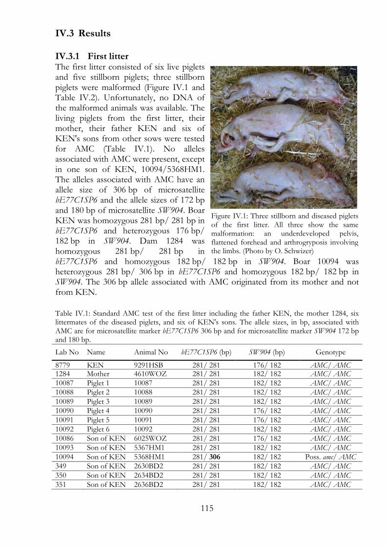
115
IV.3 Results
IV.3.1 First litter The first litter consisted of six live piglets and five stillborn piglets; three stillborn piglets were malformed (Figure IV.1 and Table IV.2). Unfortunately, no DNA of the malformed animals was available. The living piglets from the first litter, their mother, their father KEN and six of KEN's sons from other sows were tested for AMC (Table IV.1). No alleles associated with AMC were present, except in one son of KEN, 10094/5368HM1. The alleles associated with AMC have an allele size of 306 bp of microsatellite bE77C1SP6 and the allele sizes of 172 bp and 180 bp of microsatellite SW904. Boar KEN was homozygous 281 bp/ 281 bp in bE77C1SP6 and heterozygous 176 bp/ 182 bp in SW904. Dam 1284 was homozygous 281 bp/ 281 bp in bE77C1SP6 and homozygous 182 bp/ 182 bp in SW904. Boar 10094 was heterozygous 281 bp/ 306 bp in bE77C1SP6 and homozygous 182 bp/ 182 bp in SW904. The 306 bp allele associated with AMC originated from its mother and not from KEN.
Table IV.1: Standard AMC test of the first litter including the father KEN, the mother 1284, six littermates of the diseased piglets, and six of KEN's sons. The allele sizes, in bp, associated with AMC are for microsatellite marker bE77C1SP6 306 bp and for microsatellite marker SW904 172 bp and 180 bp.
Lab No Name Animal No bE77C1SP6 (bp) SW904 (bp) Genotype
8779 KEN 9291HSB 281/ 281 176/ 182 AMC/ AMC 1284 Mother 4610WOZ 281/ 281 182/ 182 AMC/ AMC 10087 Piglet 1 10087 281/ 281 182/ 182 AMC/ AMC 10088 Piglet 2 10088 281/ 281 182/ 182 AMC/ AMC 10089 Piglet 3 10089 281/ 281 182/ 182 AMC/ AMC 10090 Piglet 4 10090 281/ 281 176/ 182 AMC/ AMC 10091 Piglet 5 10091 281/ 281 176/ 182 AMC/ AMC 10092 Piglet 6 10092 281/ 281 182/ 182 AMC/ AMC 10086 Son of KEN 6025WOZ 281/ 281 176/ 182 AMC/ AMC 10093 Son of KEN 5367HM1 281/ 281 182/ 182 AMC/ AMC 10094 Son of KEN 5368HM1 281/ 306 182/ 182 Poss. amc/ AMC 349 Son of KEN 2630BD2 281/ 281 182/ 182 AMC/ AMC 350 Son of KEN 2634BD2 281/ 281 182/ 182 AMC/ AMC 351 Son of KEN 2636BD2 281/ 281 182/ 182 AMC/ AMC
Figure IV.1: Three stillborn and diseased piglets of the first litter. All three show the same malformation: an underdeveloped pelvis, flattened forehead and arthrogryposis involving the limbs. (Photo by O. Schwizer)
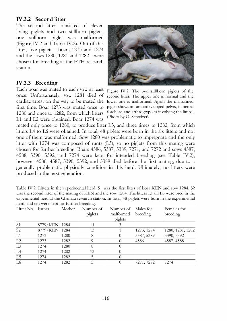
116
IV.3.2 Second litter The second litter consisted of eleven living piglets and two stillborn piglets; one stillborn piglet was malformed (Figure IV.2 and Table IV.2). Out of this litter, five piglets - boars 1273 and 1274 and the sows 1280, 1281 and 1282 - were chosen for breeding at the ETH research station.
IV.3.3 Breeding Each boar was mated to each sow at least once. Unfortunately, sow 1281 died of cardiac arrest on the way to be mated the first time. Boar 1273 was mated once to 1280 and once to 1282, from which litters L1 and L2 were obtained. Boar 1274 was mated only once to 1280, to produce litter L3, and three times to 1282, from which litters L4 to L6 were obtained. In total, 48 piglets were born in the six litters and not one of them was malformed. Sow 1280 was problematic to impregnate and the only litter with 1274 was composed of runts (L3), so no piglets from this mating were chosen for further breeding. Boars 4586, 5387, 5389, 7271, and 7272 and sows 4587, 4588, 5390, 5392, and 7274 were kept for intended breeding (see Table IV.2), however 4586, 4587, 5390, 5392, and 5389 died before the first mating, due to a generally problematic physically condition in this herd. Ultimately, no litters were produced in the next generation.
Table IV.2: Litters in the experimental herd. S1 was the first litter of boar KEN and sow 1284. S2 was the second litter of the mating of KEN and the sow 1284. The litters L1 till L6 were bred in the experimental herd at the Chamau research station. In total, 48 piglets were born in the experimental herd, and ten were kept for further breeding. Litter No Father Mother Number of
piglets Number of malformed piglets
Males for breeding
Females for breeding
S1 8779/KEN 1284 11 3 S2 8779/KEN 1284 13 1 1273, 1274 1280, 1281, 1282 L1 1273 1280 8 0 5387, 5389 5390, 5392 L2 1273 1282 9 0 4586 4587, 4588 L3 1274 1280 8 0
L4 1274 1282 13 0
L5 1274 1282 5 0
L6 1274 1282 5 0 7271, 7272 7274
Figure IV.2: The two stillborn piglets of the second litter. The upper one is normal and the lower one is malformed. Again the malformed piglet shows an underdeveloped pelvis, flattened forehead and arthrogryposis involving the limbs. (Photo by O. Schwizer)
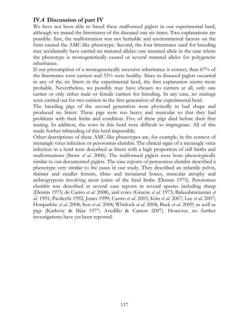
117
IV.4 Discussion of part IV We have not been able to breed these malformed piglets in our experimental herd, although we mated the littermates of the diseased one six times. Two explanations are possible: first, the malformation was not heritable and environmental factors on the farm caused the AMC-like phenotype. Second, the four littermates used for breeding may accidentally have carried no mutated alleles: one mutated allele in the case where the phenotype is monogenetically caused or several mutated alleles for polygenetic inheritance. If our presumption of a monogenetically recessive inheritance is correct, then 67% of the littermates were carriers and 33% were healthy. Since in diseased piglets occurred in any of the six litters in the experimental herd, the first explanation seems more probable. Nevertheless, we possibly may have chosen no carriers at all, only one carrier or only either male or female carriers for breeding. In any case, no matings were carried out for two carriers in the first generation of the experimental herd. The breeding pigs of the second generation were physically in bad shape and produced no litters. These pigs were too heavy and muscular so that they had problems with their limbs and condition. Five of these pigs died before their first mating. In addition, the sows in this herd were difficult to impregnate. All of this made further inbreeding of this herd impossible. Other descriptions of these AMC-like phenotypes are, for example, in the context of menangle virus infection or perosomus elumbis. The clinical signs of a menangle virus infection in a herd were described as litters with a high proportion of still births and malformations (Straw et al. 2006). The malformed piglets were born phenotypically similar to our documented piglets. The case reports of perosomus elumbis described a phenotype very similar to the cases in our study. They described an infantile pelvis, thinner and smaller femurs, tibias and metatarsal bones, muscular atrophy and arthrogryposis involving most joints of the hind limbs (Dennis 1975). Perosomus elumbis was described in several case reports in several species including sheep (Dennis 1975; de Castro et al. 2008), and cows (Greene et al. 1973; Balasubramanian et al. 1991; Paolicchi 1992; Jones 1999; Castro et al. 2003; Kim et al. 2007; Lee et al. 2007; Honparkhe et al. 2008; Son et al. 2008; Whitlock et al. 2008; Buck et al. 2009) as well as pigs (Karlovic & Ilijas 1977; Avedillo & Camon 2007). However, no further investigations have yet been reported.
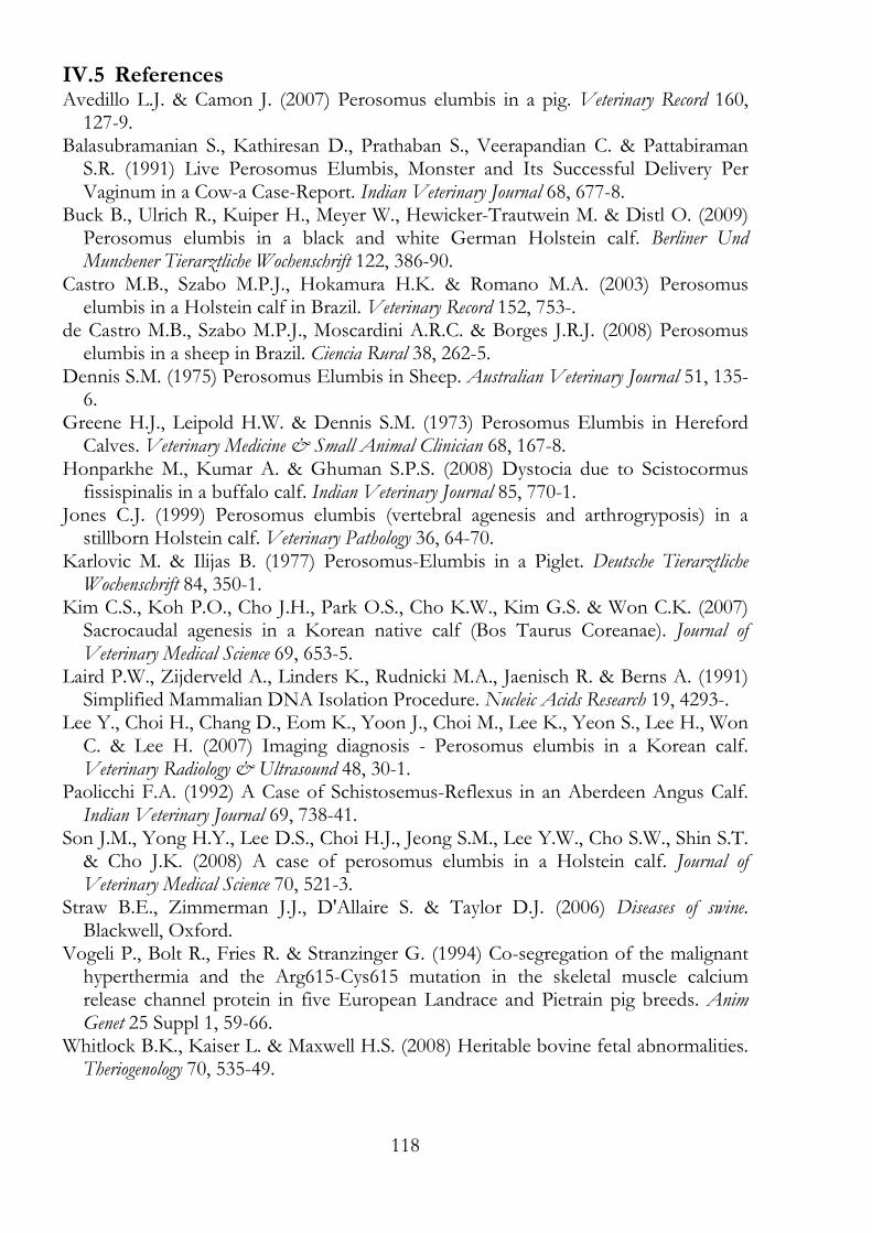
118
IV.5 References Avedillo L.J. & Camon J. (2007) Perosomus elumbis in a pig. Veterinary Record 160, 127-9.
Balasubramanian S., Kathiresan D., Prathaban S., Veerapandian C. & Pattabiraman S.R. (1991) Live Perosomus Elumbis, Monster and Its Successful Delivery Per Vaginum in a Cow-a Case-Report. Indian Veterinary Journal 68, 677-8.
Buck B., Ulrich R., Kuiper H., Meyer W., Hewicker-Trautwein M. & Distl O. (2009) Perosomus elumbis in a black and white German Holstein calf. Berliner Und Munchener Tierarztliche Wochenschrift 122, 386-90.
Castro M.B., Szabo M.P.J., Hokamura H.K. & Romano M.A. (2003) Perosomus elumbis in a Holstein calf in Brazil. Veterinary Record 152, 753-.
de Castro M.B., Szabo M.P.J., Moscardini A.R.C. & Borges J.R.J. (2008) Perosomus elumbis in a sheep in Brazil. Ciencia Rural 38, 262-5.
Dennis S.M. (1975) Perosomus Elumbis in Sheep. Australian Veterinary Journal 51, 135-6.
Greene H.J., Leipold H.W. & Dennis S.M. (1973) Perosomus Elumbis in Hereford Calves. Veterinary Medicine & Small Animal Clinician 68, 167-8.
Honparkhe M., Kumar A. & Ghuman S.P.S. (2008) Dystocia due to Scistocormus fissispinalis in a buffalo calf. Indian Veterinary Journal 85, 770-1.
Jones C.J. (1999) Perosomus elumbis (vertebral agenesis and arthrogryposis) in a stillborn Holstein calf. Veterinary Pathology 36, 64-70.
Karlovic M. & Ilijas B. (1977) Perosomus-Elumbis in a Piglet. Deutsche Tierarztliche Wochenschrift 84, 350-1.
Kim C.S., Koh P.O., Cho J.H., Park O.S., Cho K.W., Kim G.S. & Won C.K. (2007) Sacrocaudal agenesis in a Korean native calf (Bos Taurus Coreanae). Journal of Veterinary Medical Science 69, 653-5.
Laird P.W., Zijderveld A., Linders K., Rudnicki M.A., Jaenisch R. & Berns A. (1991) Simplified Mammalian DNA Isolation Procedure. Nucleic Acids Research 19, 4293-.
Lee Y., Choi H., Chang D., Eom K., Yoon J., Choi M., Lee K., Yeon S., Lee H., Won C. & Lee H. (2007) Imaging diagnosis - Perosomus elumbis in a Korean calf. Veterinary Radiology & Ultrasound 48, 30-1.
Paolicchi F.A. (1992) A Case of Schistosemus-Reflexus in an Aberdeen Angus Calf. Indian Veterinary Journal 69, 738-41.
Son J.M., Yong H.Y., Lee D.S., Choi H.J., Jeong S.M., Lee Y.W., Cho S.W., Shin S.T. & Cho J.K. (2008) A case of perosomus elumbis in a Holstein calf. Journal of Veterinary Medical Science 70, 521-3.
Straw B.E., Zimmerman J.J., D'Allaire S. & Taylor D.J. (2006) Diseases of swine. Blackwell, Oxford.
Vogeli P., Bolt R., Fries R. & Stranzinger G. (1994) Co-segregation of the malignant hyperthermia and the Arg615-Cys615 mutation in the skeletal muscle calcium release channel protein in five European Landrace and Pietrain pig breeds. Anim Genet 25 Suppl 1, 59-66.
Whitlock B.K., Kaiser L. & Maxwell H.S. (2008) Heritable bovine fetal abnormalities. Theriogenology 70, 535-49.
119
IV.6 List of figures Figure IV.1: Three stillborn and diseased piglets of the first litter. 115
Figure IV.2: The two stillborn piglets of the second litter. 116
IV.7 List of tables Table IV.1: Standard AMC test of the first litter including the father KEN, the
mother 1284, six littermates of the diseased piglets, and six of KEN's sons. 115
Table IV.2: Litters in the experimental herd. 116

120
121
Discussion Over the last few years we had the opportunity to breed four different disease models in pigs with potential genetic background. Arthrogryposis multiplex congenita (AMC) and AMC-like phenotypes causes malformations of the extremities and stillbirth, Splay Leg (SL) causes congenital leg weakness, and Humpy Back (HB) causes temporary spinal malformation in adolescent pigs. In the case of recessive monogenetically inherited disease, offspring typically separate into healthy and diseased at a ratio of 3:1, and this ratio was found in the AMC herd. The developed AMC test proved to be an adequate tool to determine AMC-carriers in the experimental herd and commercial breeds. Today, many of the boars used for artificial insemination are already tested for AMC, so most pedigrees with AMC carriers are known. Therefore, the ancestry of malformed piglets is reviewed before an AMC test is carried out in our laboratory. In the many lines of boars bred for artificial insemination, one breeding line attracted our attention as our markers were not informative enough. In order to determine the correct AMC status of the suspicious boar ´Genium´, it was mated with three sows of the experimental herd with proven AMC carrier status. In the following three litters, 30 healthy piglets were born, thus the absence of amc in the breeding line of these boars was proven. This experiment showed that the alleles in linkage disequilibrium with amc in the experimental herd are not uncommon in commercial breeds, and in some pedigrees are not necessarily linked with amc. The case of the AMC-like piglets shows another variant of AMC that is not detected with the AMC test. Here, genotypes resembled those of healthy piglets, but the phenotype resembled AMC. In the case of polygenic disease, the frequency of diseased individuals is less predictable when mating diseased animals with diseased/ healthy animals. However, the occurrence of diseased offspring tends to increase with inbreeding. The HB herd produced 65 litters and the SL herd produced 28 litters. Nevertheless, both herds were too small to predict an inheritance pattern for the disorders. However, healthy litters from matings of formerly diseased parents contradict a genetic background in the case of HB and SL. Nevertheless, breeding SL and HB pigs produced a DNA collection for future genetic studies. These DNA collections will be of interest for genotyping (genome wide association studies), and re-sequencing of candidate genes. Besides this, gene expression studies would be good starting point to examine these diseases further.

122
123
Notation SSC Sus scrofa chromosome HSA Homo sapiens chromosome SV Sequence variation LD Linkage disequilibrium AMC Dominant allele amc Recessive allele No. Number dNTPs Deoxyribonucleotides DNA Deoxyribonucleic acid A, C, G, T Nucleobases: adenine, cytosine, guanine, thymine SNP Single-nucleotide polymorphism indel Insertions and deletions PCR Polymerase chain reaction RFLP Restriction fragment length polymorphism PCR-RFLP Restriction fragment length polymorphism of PCR products H2O Pure Water EDTA Ethylenediaminetetraacetic acid TEMED Tetramethylethylenediamine SDS Sodium dodecyl sulfate NaCl Sodium chloride KCl Potassium chloride MgCl2 Magnesium chloride BSA Bovine serum albumin °C Degree Celsius mg, µg, ng Milligram (10−3 gram), microgram (10−6 gram), nanogram (10−9 gram) ml, µl Milliliter (10−3 liter), microliter (10−6 liter) h, min, s Hours, minutes, seconds g Gravity of Earth, 9.81 m/s2 M, mM, µM Molar, millimolar (10−3 molar), micromolar (10−6 molar) mol, pmol Mole, picomole (10−12 mole) bp, Mb Base pairs, mega base pairs (1'000'000 bp)

124
125
Appendix
Chemicals
Chemicals for media and solutions Sigma-Aldrich, Buchs, Switzerland λ-phage DNA GE Healthcare, Glattbrugg, Switzerland Taq DNA Polymerase Sigma-Aldrich, Buchs, Switzerland JumpStart™ Taq DNA Polymerase Sigma-Aldrich, Buchs, Switzerland PCR buffer Sigma-Aldrich, Buchs, Switzerland 100 bp ladder Sigma-Aldrich, Buchs, Switzerland 100 bp ladder directload Sigma-Aldrich, Buchs, Switzerland 50 bp ladder Sigma-Aldrich, Buchs, Switzerland 50 bp ladder directload Sigma-Aldrich, Buchs, Switzerland Agarose low EEO Sigma-Aldrich, Buchs, Switzerland Restriction enzymes Various supplieres listed in table Formamide Sigma-Aldrich, Buchs, Switzerland Ethanol Merck, Zug, Switzerland Isopropanol Merck, Zug, Switzerland
Media and solutions
DNA loading dye for agarose gel electrophoresis Xylene Cyanol FF 0.25 % (w/v) Bromphenol blue 0.25 % (w/v) Glycerol 70 %
TBE Buffer (10x) Trizma base 0.9 M (Tris(hydroxymethyl)aminomethane) Boric acid 0.88 M EDTA (pH 8) 20 mM pH 8.3
TE Buffer Tris-HCl (pH 7.5-8) 10 mM EDTA (pH 8) 1 mM pH 7.5-8
Lysis buffer for DNA isolation from blood D (+) - saccharose 0.32 M Tris-HCl (pH 7.5) 10 mM MgCl2 5 mM Triton X-100 0.94 % (w/v) Proteinase K 100 µg/ ml
126
Lysis buffer for DNA isolation from tissue Tris-HCl (pH 8.5) 100 mM EDTA (pH 8) 5 mM SDS 0.2 % NaCl 200 mM Proteinase K 100 µg/ ml
PCR buffer turbo for DNA isolation from blood KCl 50 mM Tris-HCl (pH 8.3) 10 mM Gelatin 0.1 mg/ mg Nonidet P40 0.45 % (v/v) Tween-20 0.45 % (v/v)
Tris-HCl pH 7.5 (Tris(hydroxymethyl)aminomethane hydrochloride) Tris-HCl 200 mM pH 7.5
127
Acknowledgements In erster Linie möchte ich Prof. Dr. Peter Vögeli danken. Er hat die Projekte betreut und gab mir die Möglichkeit, diese Doktorarbeit zu schreiben. Und ich danke PD Dr. Stefan Neuenschwander für die Betreuung meiner Arbeit. Dann möchte ich Prof. Dr. Michael Kreuzer und Dr. med. vet. Xaver Sidler dafür danken, dass sie sich als meine Korreferenten zur Verfügung gestellt haben. Ausserdem danke ich Dr. Sem Genini für die gute Vorarbeit, da er die Projekte angefangen hatte. Liebe TANnensträsslerInnen und Ex-TANnensträssler, Euch allen danke ich für Eure Unterstützung, das gute Arbeitsklima, die anregenden Kaffeepausen und die schönen Ausflüge und Feste, die wir zusammen erleben durften. Im Gedenken an meine Schweine – ein grosses Dankeschön an die Leitung und an die Schweinehirten der ETH Forschungsstation Chamau. Dr. Andreas Hofer und Dr. Henning Luther von der SUISAG, den Verbänden und den vielen Betrieben möchte ich für Ihre (auch finanzielle) Unterstützung und Ihr Interesse an meiner Arbeit danken. Dem GDC der ETH Zürich, dem FGCZ der Universität Zürich und dem NCCR "Frontiers in Genetics" Laboratorium der Universite de Geneve danke ich für die zur Verfügung gestellten Gerätschaften und ihre Unterstützung. Prof. Dr. Martien Groenen stellte freundlicherweise einige seiner SNP Daten zur Verfügung, herzlichen Dank. Die Arbeit war Teil der COST Action 861 (European Network for Pig Genomics „PigNet“) und wurde finanziell unterstützt durch die COST. Zum Abschluss noch einen herzlichen Dank an meine Familie und Freunde. Und ein Dankeschön für die süssen (im doppelten Sinn), plüschigen oder originellen Schweinchen-Geschenke.

128