Embed Size (px)
Citation preview
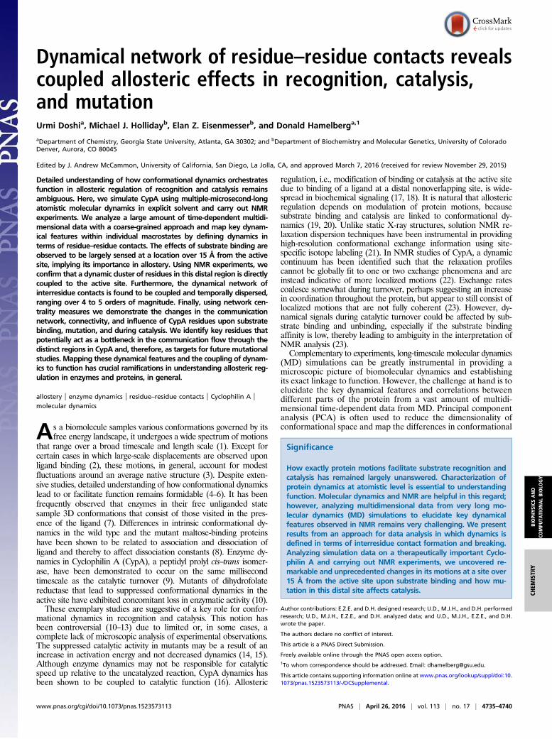
Dynamical network of residue–residue contacts revealscoupled allosteric effects in recognition, catalysis,and mutationUrmi Doshia, Michael J. Hollidayb, Elan Z. Eisenmesserb, and Donald Hamelberga,1
aDepartment of Chemistry, Georgia State University, Atlanta, GA 30302; and bDepartment of Biochemistry and Molecular Genetics, University of ColoradoDenver, Aurora, CO 80045
Edited by J. Andrew McCammon, University of California, San Diego, La Jolla, CA, and approved March 7, 2016 (received for review November 29, 2015)
Detailed understanding of how conformational dynamics orchestratesfunction in allosteric regulation of recognition and catalysis remainsambiguous. Here, we simulate CypA using multiple-microsecond-longatomistic molecular dynamics in explicit solvent and carry out NMRexperiments. We analyze a large amount of time-dependent multidi-mensional data with a coarse-grained approach and map key dynam-ical features within individual macrostates by defining dynamics interms of residue–residue contacts. The effects of substrate binding areobserved to be largely sensed at a location over 15 Å from the activesite, implying its importance in allostery. Using NMR experiments, weconfirm that a dynamic cluster of residues in this distal region is directlycoupled to the active site. Furthermore, the dynamical network ofinterresidue contacts is found to be coupled and temporally dispersed,ranging over 4 to 5 orders of magnitude. Finally, using network cen-trality measures we demonstrate the changes in the communicationnetwork, connectivity, and influence of CypA residues upon substratebinding, mutation, and during catalysis. We identify key residues thatpotentially act as a bottleneck in the communication flow through thedistinct regions in CypA and, therefore, as targets for future mutationalstudies. Mapping these dynamical features and the coupling of dynam-ics to function has crucial ramifications in understanding allosteric reg-ulation in enzymes and proteins, in general.
allostery | enzyme dynamics | residue–residue contacts | Cyclophilin A |molecular dynamics
As a biomolecule samples various conformations governed by itsfree energy landscape, it undergoes a wide spectrum of motions
that range over a broad timescale and length scale (1). Except forcertain cases in which large-scale displacements are observed uponligand binding (2), these motions, in general, account for modestfluctuations around an average native structure (3). Despite exten-sive studies, detailed understanding of how conformational dynamicslead to or facilitate function remains formidable (4–6). It has beenfrequently observed that enzymes in their free unliganded statesample 3D conformations that consist of those visited in the pres-ence of the ligand (7). Differences in intrinsic conformational dy-namics in the wild type and the mutant maltose-binding proteinshave been shown to be related to association and dissociation ofligand and thereby to affect dissociation constants (8). Enzyme dy-namics in Cyclophilin A (CypA), a peptidyl prolyl cis–trans isomer-ase, have been demonstrated to occur on the same millisecondtimescale as the catalytic turnover (9). Mutants of dihydrofolatereductase that lead to suppressed conformational dynamics in theactive site have exhibited concomitant loss in enzymatic activity (10).These exemplary studies are suggestive of a key role for confor-
mational dynamics in recognition and catalysis. This notion hasbeen controversial (10–13) due to limited or, in some cases, acomplete lack of microscopic analysis of experimental observations.The suppressed catalytic activity in mutants may be a result of anincrease in activation energy and not decreased dynamics (14, 15).Although enzyme dynamics may not be responsible for catalyticspeed up relative to the uncatalyzed reaction, CypA dynamics hasbeen shown to be coupled to catalytic function (16). Allosteric
regulation, i.e., modification of binding or catalysis at the active sitedue to binding of a ligand at a distal nonoverlapping site, is wide-spread in biochemical signaling (17, 18). It is natural that allostericregulation depends on modulation of protein motions, becausesubstrate binding and catalysis are linked to conformational dy-namics (19, 20). Unlike static X-ray structures, solution NMR re-laxation dispersion techniques have been instrumental in providinghigh-resolution conformational exchange information using site-specific isotope labeling (21). In NMR studies of CypA, a dynamiccontinuum has been identified such that the relaxation profilescannot be globally fit to one or two exchange phenomena and areinstead indicative of more localized motions (22). Exchange ratescoalesce somewhat during turnover, perhaps suggesting an increasein coordination throughout the protein, but appear to still consist oflocalized motions that are not fully coherent (23). However, dy-namical signals during catalytic turnover could be affected by sub-strate binding and unbinding, especially if the substrate bindingaffinity is low, thereby leading to ambiguity in the interpretation ofNMR analysis (23).Complementary to experiments, long-timescale molecular dynamics
(MD) simulations can be greatly instrumental in providing amicroscopic picture of biomolecular dynamics and establishingits exact linkage to function. However, the challenge at hand is toelucidate the key dynamical features and correlations betweendifferent parts of the protein from a vast amount of multidi-mensional time-dependent data from MD. Principal componentanalysis (PCA) is often used to reduce the dimensionality ofconformational space and map the differences in conformational
Significance
How exactly protein motions facilitate substrate recognition andcatalysis has remained largely unanswered. Characterization ofprotein dynamics at atomistic level is essential to understandingfunction. Molecular dynamics and NMR are helpful in this regard;however, analyzing multidimensional data from very long mo-lecular dynamics (MD) simulations to elucidate key dynamicalfeatures observed in NMR remains very challenging. We presentresults from an approach for data analysis in which dynamics isdefined in terms of interresidue contact formation and breaking.Analyzing simulation data on a therapeutically important Cyclo-philin A and carrying out NMR experiments, we uncovered re-markable and unprecedented changes in its motions at a site over15 Å from the active site upon substrate binding and how mu-tation in this distal site affects catalysis.
Author contributions: E.Z.E. and D.H. designed research; U.D., M.J.H., and D.H. performedresearch; U.D., M.J.H., E.Z.E., and D.H. analyzed data; and U.D., M.J.H., E.Z.E., and D.H.wrote the paper.
The authors declare no conflict of interest.
This article is a PNAS Direct Submission.
Freely available online through the PNAS open access option.1To whom correspondence should be addressed. Email: [email protected].
This article contains supporting information online at www.pnas.org/lookup/suppl/doi:10.1073/pnas.1523573113/-/DCSupplemental.
www.pnas.org/cgi/doi/10.1073/pnas.1523573113 PNAS | April 26, 2016 | vol. 113 | no. 17 | 4735–4740
BIOPH
YSICSAND
COMPU
TATIONALBIOLO
GY
CHEM
ISTR
Y
ensembles. PCA, which is usually performed on Cartesian co-ordinates, may sometimes mask certain important long-rangedynamical relations and complex features of biomolecular con-formational dynamics. Comparing residue–residue contactsin various isoforms from difference contact maps built fromsimulation trajectories has aided in determining certain similarstructural properties and some unique to a particular isoform(24). Another analysis that involves monitoring the time evolu-tion of residue–residue contact formation and breaking has beenproven useful in identifying certain characteristic events duringconformational transitions (25). Correlated motions betweendifferent biomolecular segments have been identified usingcross-correlation analysis and building dynamical networks (25–29). However, interpretation of the results of such analysis incase of CypA, which exhibit subtle changes upon substratebinding and catalysis, has been ambiguous.Here, we characterize the conformational dynamics of CypA
using very long atomistic standard MD simulations in explicit sol-vent, and the results are validated using NMR experiments. CypA isan archetypal and extensively studied enzyme belonging to thefamily of peptidyl prolyl isomerases (PPIases), speeding the cis–transisomerization of peptidyl prolyl ω-bond in its protein substrates bymore than 105 times (30–34). In five independent microsecond-longsimulations, we monitored the dynamics in wild-type CypA, V29Lvariant of CypA, CypA bound to a substrate analogue in the trans,transition state, and cis configurations. When we apply our methodof analyzing the trajectories at a coarse-grained level, i.e., inter-residue contact interactions, and use PCA in contact space, thespecific differences in CypA dynamics upon association to its sub-strate, during the catalytic process and upon alteration of a singleresidue distant from the active site are revealed.
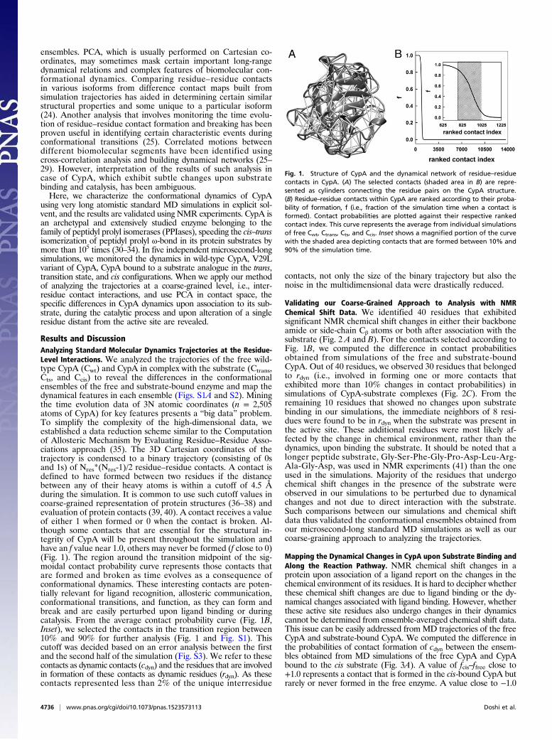
Results and DiscussionAnalyzing Standard Molecular Dynamics Trajectories at the Residue-Level Interactions. We analyzed the trajectories of the free wild-type CypA (Cwt) and CypA in complex with the substrate (Ctrans,Cts, and Ccis) to reveal the differences in the conformationalensembles of the free and substrate-bound enzyme and map thedynamical features in each ensemble (Figs. S1A and S2). Miningthe time evolution data of 3N atomic coordinates (n = 2,505atoms of CypA) for key features presents a “big data” problem.To simplify the complexity of the high-dimensional data, weestablished a data reduction scheme similar to the Computationof Allosteric Mechanism by Evaluating Residue–Residue Asso-ciations approach (35). The 3D Cartesian coordinates of thetrajectory is condensed to a binary trajectory (consisting of 0sand 1s) of Nres*(Nres-1)/2 residue–residue contacts. A contact isdefined to have formed between two residues if the distancebetween any of their heavy atoms is within a cutoff of 4.5 Åduring the simulation. It is common to use such cutoff values incoarse-grained representation of protein structures (36–38) andevaluation of protein contacts (39, 40). A contact receives a valueof either 1 when formed or 0 when the contact is broken. Al-though some contacts that are essential for the structural in-tegrity of CypA will be present throughout the simulation andhave an f value near 1.0, others may never be formed (f close to 0)(Fig. 1). The region around the transition midpoint of the sig-moidal contact probability curve represents those contacts thatare formed and broken as time evolves as a consequence ofconformational dynamics. These interesting contacts are poten-tially relevant for ligand recognition, allosteric communication,conformational transitions, and function, as they can form andbreak and are easily perturbed upon ligand binding or duringcatalysis. From the average contact probability curve (Fig. 1B,Inset), we selected the contacts in the transition region between10% and 90% for further analysis (Fig. 1 and Fig. S1). Thiscutoff was decided based on an error analysis between the firstand the second half of the simulation (Fig. S3). We refer to thesecontacts as dynamic contacts (cdyn) and the residues that are involvedin formation of these contacts as dynamic residues (rdyn). As thesecontacts represented less than 2% of the unique interresidue
contacts, not only the size of the binary trajectory but also thenoise in the multidimensional data were drastically reduced.
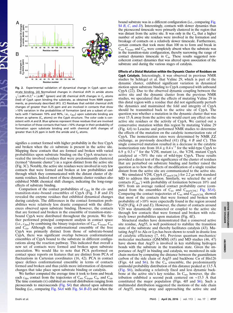
Validating our Coarse-Grained Approach to Analysis with NMRChemical Shift Data. We identified 40 residues that exhibitedsignificant NMR chemical shift changes in either their backboneamide or side-chain Cβ atoms or both after association with thesubstrate (Fig. 2 A and B). For the contacts selected according toFig. 1B, we computed the difference in contact probabilitiesobtained from simulations of the free and substrate-boundCypA. Out of 40 residues, we observed 30 residues that belongedto rdyn (i.e., involved in forming one or more contacts thatexhibited more than 10% changes in contact probabilities) insimulations of CypA-substrate complexes (Fig. 2C). From theremaining 10 residues that showed no changes upon substratebinding in our simulations, the immediate neighbors of 8 resi-dues were found to be in rdyn when the substrate was present inthe active site. These additional residues were most likely af-fected by the change in chemical environment, rather than thedynamics, upon binding the substrate. It should be noted that alonger peptide substrate, Gly-Ser-Phe-Gly-Pro-Asp-Leu-Arg-Ala-Gly-Asp, was used in NMR experiments (41) than the oneused in the simulations. Majority of the residues that undergochemical shift changes in the presence of the substrate wereobserved in our simulations to be perturbed due to dynamicalchanges and not due to direct interaction with the substrate.Such comparisons between our simulations and chemical shiftdata thus validated the conformational ensembles obtained fromour microsecond-long standard MD simulations as well as ourcoarse-graining approach to analyzing the trajectories.
Mapping the Dynamical Changes in CypA upon Substrate Binding andAlong the Reaction Pathway. NMR chemical shift changes in aprotein upon association of a ligand report on the changes in thechemical environment of its residues. It is hard to decipher whetherthese chemical shift changes are due to ligand binding or the dy-namical changes associated with ligand binding. However, whetherthese active site residues also undergo changes in their dynamicscannot be determined from ensemble-averaged chemical shift data.This issue can be easily addressed from MD trajectories of the freeCypA and substrate-bound CypA. We computed the difference inthe probabilities of contact formation of cdyn between the ensem-bles obtained from MD simulations of the free CypA and CypAbound to the cis substrate (Fig. 3A). A value of fcis–ffree close to+1.0 represents a contact that is formed in the cis-bound CypA butrarely or never formed in the free enzyme. A value close to −1.0
Fig. 1. Structure of CypA and the dynamical network of residue–residuecontacts in CypA. (A) The selected contacts (shaded area in B) are repre-sented as cylinders connecting the residue pairs on the CypA structure.(B) Residue–residue contacts within CypA are ranked according to their proba-bility of formation, f (i.e., fraction of the simulation time when a contact isformed). Contact probabilities are plotted against their respective rankedcontact index. This curve represents the average from individual simulationsof free Cwt, Ctrans, Cts, and Ccis. Inset shows a magnified portion of the curvewith the shaded area depicting contacts that are formed between 10% and90% of the simulation time.
4736 | www.pnas.org/cgi/doi/10.1073/pnas.1523573113 Doshi et al.
signifies a contact formed with higher probability in the free CypAand broken when the cis substrate is present in the active site.Mapping these contacts that are formed and broken with variedprobabilities upon substrate binding on the CypA structure re-vealed the involved residues that were predominantly clustered(termed “dynamic cluster”) in a region distinct from the active site(Fig. 3C). Notably, the active site residues were involved in very fewcontacts that were formed and broken at low probabilities andthrough which they communicated with the distant cluster of dy-namic residues. Indeed most of these dynamic cluster residues alsoexhibited NMR chemical shift changes, indicating the long-rangeeffects of substrate binding.Comparison of the contact probabilities of cdyn in the cis- and
transition-state–bound ensembles of CypA (Fig. 3 B and D)revealed the enzyme residues that exhibited dynamical changesduring catalysis. The differences in the contact formation prob-abilities were relatively less drastic compared with the differ-ences observed upon substrate binding. However, the contactsthat are formed and broken in the ensemble of transition-state–bound CypA were distributed throughout the protein. We fur-ther performed principal component analysis in contact space(i.e., cdyn) by combining the binary trajectories of Cwt, Ctrans, Cts,and Ccis. Although the conformational ensemble of the freeCypA was primarily distinct from those of substrate-boundCypA, there was significant overlap between conformationalensembles of CypA bound to the substrate in different configu-rations along the reaction pathway. This indicated that overall anew set of contacts were formed and broken upon substrateassociation. We would like to note that PCA performed oncontact space reports on features that are distinct from PCA offluctuations in Cartesian coordinates (16, 42). PCA in contactspace defines conformational ensemble in terms of contactsformed and broken and thus yields more information of dynamicchanges that take place upon substrate binding or catalysis.We further computed the average time it took to form and break
each cdyn contact from the trajectories of Cwt, Ctrans, Cts, and Ccis.The time constants (τ) exhibited an overall distribution ranging frompicoseconds to microseconds (Fig. S4) that altered upon substratebinding (i.e., comparing Fig. S4A with Fig. S4 B–D) and when the
bound substrate was in a different configuration (i.e., comparing Fig.S4 B, C, and D). Interestingly, contacts with slower dynamics thanthe picosecond timescale were primarily clustered in the region thatwas distant from the active site. It was only in the Cts that a highernumber of active site residues were involved in the formation andbreakage of contacts on a relatively slower timescale. Furthermore,certain contacts that took more than 100 ns to form and break inCwt, Ctrans, and Ccis were completely absent when the substrate wasin the transition-state configuration, thereby narrowing the range ofcontact dynamics timescale in Cts. These results suggested non-coherent contact dynamics that was altered upon association of thesubstrate and during the various stages of catalysis.
Effects of a Distal Mutation within the Dynamic Cluster of Residues onCypA Catalysis. Interestingly, it was observed in previous NMRstudies by Schlegel et al. that Valine 29, which is part of thedynamic cluster, exhibited significant variation in dynamicalmotion upon substrate binding to CypA compared with unboundCypA (22). Due to the observed dynamic coupling between theactive site and the dynamic cluster from the previous NMRstudies, we speculated that the effects of mutating Valine 29 inthis distal region with a residue that did not significantly perturbthe dynamics and maintained the fold and integrity of CypAshould be transmitted back to the active site of CypA. Thequestion was whether a mutation in the dynamic cluster that wasover 15 Å away from the active site would exert any effect on theactive site residues or the activity of CypA. We carried out aconservative mutation within this region by mutating Valine 29(Fig. 4A) to Leucine and performed NMR studies to determinethe effects of the mutation on the catalytic isomerization rate ofCypA. The isomerization rates were determined by NMR ZZexchange, as previously described (41) (Fig. 4 B and C). Thissingle conserved mutation resulted in a decrease in the catalyticisomerization rate from 10.4 ± 0.4 s−1 for the wild-type CypA to6.9 ± 0.3 s−1 for the V29L mutant i.e., the V29L mutant CypAcatalyzed at ∼70% the rate of wild-type CypA. These resultsprovided a direct test of the significance of the cluster of residuesthat are perturbed on substrate binding and further raised thequestion as to how the effects of modification of a single residuedistant from the active site are communicated to the active site.We simulated V29L CypA (CmutV29L) for 2.2 μs with standard
MD to address this question. Similarly, we selected the set ofcontacts (Cdyn′) with probability of formation between 10% and90% from an average ranked contact probability curve (com-puted from the ensembles of Cwt and CmutV29L; Fig. S5A).Comparing the contact trajectories of Cwt and CmutV29L showedthat most of the contacts that were formed and broken withprobability of >10% were expectedly found in the region aroundVal29 (Fig. 4 D and E). However, the cluster of contacts aroundV29 was dynamically connected or coupled to the active sitethrough few contacts that were formed and broken with rela-tively lower probabilities upon mutation (Fig. 4E).Structural studies have demonstrated that a conserved active
site residue, Arg55, is well positioned to stabilize the transitionstate of the substrate and thereby facilitates catalysis (43). Mu-tating Arg55 to Ala or Lys has been shown to result in drastic lossof catalytic efficiency (7, 44). Previous quantum mechanics/molecular mechanics (QM/MM) (45) and MD studies (46, 47)have shown that Arg55 is involved in key stabilizing hydrogenbonds with the substrate in the transition state. Given the im-portance of Arg55 in binding and catalysis, we monitored its sidechain motion by computing the distance between the guanidiniumcarbon of the side chain of Arg55 and backbone Cα of His126(Figs. 4A and S6). In the Cts ensemble, the predominantlyunimodal probability distribution of this distance peaked at 13.5 Å(Fig. S6), indicating a relatively fixed and less dynamic back-bone at the active site’s key residue. In Cwt, however, the dis-tribution exhibited a second peak centered on ∼15.5 Å thatconstituted the major population (Figs. 4H and S6). Such amultimodal distribution suggested the motions of the side chainof Arg55, moving away and approaching the active site and
Fig. 2. Experimental validation of dynamical change in CypA upon sub-
strate binding. (A) Normalized changes in chemical shift in amide atomsffiffiffiffiffiffiffiffiffiffiffiffiffiffiffiffiffiffiffiffiffiffiffiffiffiffiffiffiffiffiffiffiffiffiffiffiffiffiffiffiffiffiffiffiffiffiffiðΔδH ×0.2Þ2 + ðΔδNÞ2
q(green) and (B) chemical shift changes in Cβ atoms
(red) of CypA upon binding the substrate, as obtained from NMR experi-ments, as previously described (41). (C) Residues that exhibit chemical shiftchanges of greater than 0.25 ppm and are involved in contacts that show>10% variation in the probabilities of formation (and are a subset of con-tacts with f between 10% and 90%, i.e., cdyn) upon substrate binding areshown as spheres (Cα atoms) on the CypA structure. The color code is con-sistent with A and B. Blue spheres represent those residues that are involvedin formation of those contacts that have >10% change in their probability offormation upon substrate binding and with chemical shift changes ofgreater than 0.25 ppm in both the amide and Cβ atoms.
Doshi et al. PNAS | April 26, 2016 | vol. 113 | no. 17 | 4737
BIOPH
YSICSAND
COMPU
TATIONALBIOLO
GY
CHEM
ISTR
Y
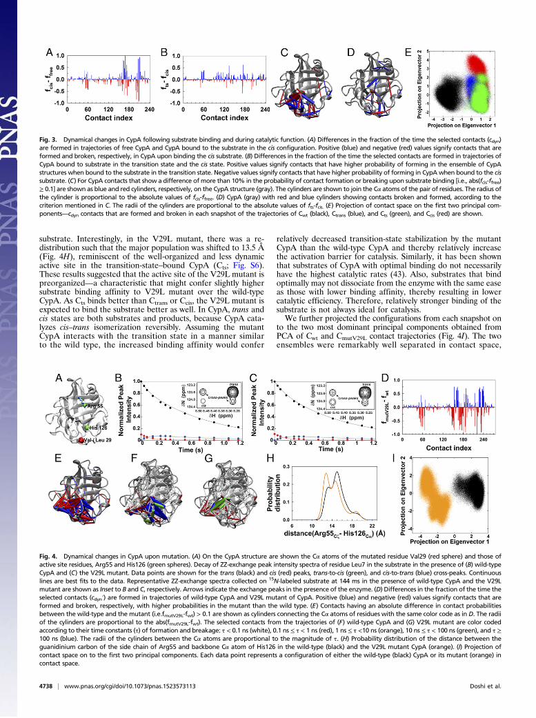
substrate. Interestingly, in the V29L mutant, there was a re-distribution such that the major population was shifted to 13.5 Å(Fig. 4H), reminiscent of the well-organized and less dynamicactive site in the transition-state–bound CypA (Cts; Fig. S6).These results suggested that the active site of the V29L mutant ispreorganized—a characteristic that might confer slightly highersubstrate binding affinity to V29L mutant over the wild-typeCypA. As Cts binds better than Ctrans or Ccis, the V29L mutant isexpected to bind the substrate better as well. In CypA, trans andcis states are both substrates and products, because CypA cata-lyzes cis–trans isomerization reversibly. Assuming the mutantCypA interacts with the transition state in a manner similarto the wild type, the increased binding affinity would confer
relatively decreased transition-state stabilization by the mutantCypA than the wild-type CypA and thereby relatively increasethe activation barrier for catalysis. Similarly, it has been shownthat substrates of CypA with optimal binding do not necessarilyhave the highest catalytic rates (43). Also, substrates that bindoptimally may not dissociate from the enzyme with the same easeas those with lower binding affinity, thereby resulting in lowercatalytic efficiency. Therefore, relatively stronger binding of thesubstrate is not always ideal for catalysis.We further projected the configurations from each snapshot on
to the two most dominant principal components obtained fromPCA of Cwt and CmutV29L contact trajectories (Fig. 4I). The twoensembles were remarkably well separated in contact space,
Fig. 4. Dynamical changes in CypA upon mutation. (A) On the CypA structure are shown the Cα atoms of the mutated residue Val29 (red sphere) and those ofactive site residues, Arg55 and His126 (green spheres). Decay of ZZ-exchange peak intensity spectra of residue Leu7 in the substrate in the presence of (B) wild-typeCypA and (C) the V29L mutant. Data points are shown for the trans (black) and cis (red) peaks, trans-to-cis (green), and cis-to-trans (blue) cross-peaks. Continuouslines are best fits to the data. Representative ZZ-exchange spectra collected on 15N-labeled substrate at 144 ms in the presence of wild-type CypA and the V29Lmutant are shown as Inset to B and C, respectively. Arrows indicate the exchange peaks in the presence of the enzyme. (D) Differences in the fraction of the time theselected contacts (cdyn′) are formed in trajectories of wild-type CypA and V29L mutant of CypA. Positive (blue) and negative (red) values signify contacts that areformed and broken, respectively, with higher probabilities in the mutant than the wild type. (E) Contacts having an absolute difference in contact probabilitiesbetween the wild-type and the mutant (i.e.fmutV29L-fwt) > 0.1 are shown as cylinders connecting the Cα atoms of residues with the same color code as in D. The radiiof the cylinders are proportional to the abs(fmutV29L-fwt). The selected contacts from the trajectories of (F) wild-type CypA and (G) V29L mutant are color codedaccording to their time constants (τ) of formation and breakage: τ < 0.1 ns (white), 0.1 ns ≤ τ < 1 ns (red), 1 ns ≤ τ <10 ns (orange), 10 ns ≤ τ < 100 ns (green), and τ ≥100 ns (blue). The radii of the cylinders between the Cα atoms are proportional to the magnitude of τ. (H) Probability distribution of the distance between theguanidinium carbon of the side chain of Arg55 and backbone Cα atom of His126 in the wild-type (black) and the V29L mutant CypA (orange). (I) Projection ofcontact space on to the first two principal components. Each data point represents a configuration of either the wild-type (black) CypA or its mutant (orange) incontact space.
Fig. 3. Dynamical changes in CypA following substrate binding and during catalytic function. (A) Differences in the fraction of the time the selected contacts (cdyn)are formed in trajectories of free CypA and CypA bound to the substrate in the cis configuration. Positive (blue) and negative (red) values signify contacts that areformed and broken, respectively, in CypA upon binding the cis substrate. (B) Differences in the fraction of the time the selected contacts are formed in trajectories ofCypA bound to substrate in the transition state and the cis state. Positive values signify contacts that have higher probability of forming in the ensemble of CypAstructures when bound to the substrate in the transition state. Negative values signify contacts that have higher probability of forming in CypA when bound to the cissubstrate. (C) For CypA contacts that show a difference of more than 10% in the probability of contact formation or breaking upon substrate binding [i.e., abs(fcis-ffree)≥ 0.1] are shown as blue and red cylinders, respectively, on the CypA structure (gray). The cylinders are shown to join the Cα atoms of the pair of residues. The radius ofthe cylinder is proportional to the absolute values of fcis-ffree. (D) CypA (gray) with red and blue cylinders showing contacts broken and formed, according to thecriterion mentioned in C. The radii of the cylinders are proportional to the absolute values of fts-fcis. (E) Projection of contact space on the first two principal com-ponents—cdyn contacts that are formed and broken in each snapshot of the trajectories of Cwt (black), Ctrans (blue), and Cts (green), and Ccis (red) are shown.
4738 | www.pnas.org/cgi/doi/10.1073/pnas.1523573113 Doshi et al.
despite minimal differences in contact probabilities in the activesite region (Fig. 4 E and I). Marked dissimilarities between thedynamics of wild-type CypA and the V29L mutant were noticed inthe average times of formation and breakage for cdyn’s contacts(Fig. 4 F and G). Majority of the contacts that were formed andbroken in the nanosecond timescale (green and blue cylinders inFig. 4F) in the Cwt ensemble took less than 100 ps in the mutant,indicating the speedup in dynamics around the site of mutation.
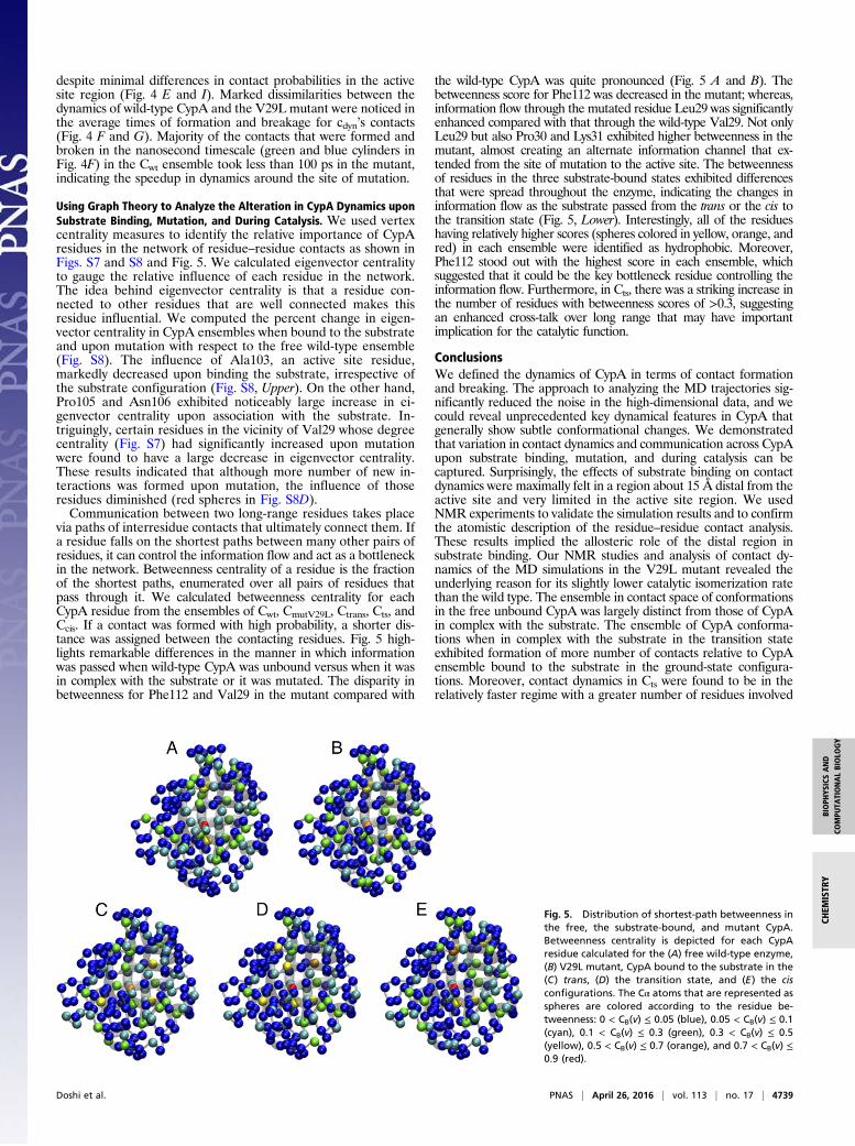
Using Graph Theory to Analyze the Alteration in CypA Dynamics uponSubstrate Binding, Mutation, and During Catalysis. We used vertexcentrality measures to identify the relative importance of CypAresidues in the network of residue–residue contacts as shown inFigs. S7 and S8 and Fig. 5. We calculated eigenvector centralityto gauge the relative influence of each residue in the network.The idea behind eigenvector centrality is that a residue con-nected to other residues that are well connected makes thisresidue influential. We computed the percent change in eigen-vector centrality in CypA ensembles when bound to the substrateand upon mutation with respect to the free wild-type ensemble(Fig. S8). The influence of Ala103, an active site residue,markedly decreased upon binding the substrate, irrespective ofthe substrate configuration (Fig. S8, Upper). On the other hand,Pro105 and Asn106 exhibited noticeably large increase in ei-genvector centrality upon association with the substrate. In-triguingly, certain residues in the vicinity of Val29 whose degreecentrality (Fig. S7) had significantly increased upon mutationwere found to have a large decrease in eigenvector centrality.These results indicated that although more number of new in-teractions was formed upon mutation, the influence of thoseresidues diminished (red spheres in Fig. S8D).Communication between two long-range residues takes place
via paths of interresidue contacts that ultimately connect them. Ifa residue falls on the shortest paths between many other pairs ofresidues, it can control the information flow and act as a bottleneckin the network. Betweenness centrality of a residue is the fractionof the shortest paths, enumerated over all pairs of residues thatpass through it. We calculated betweenness centrality for eachCypA residue from the ensembles of Cwt, CmutV29L, Ctrans, Cts, andCcis. If a contact was formed with high probability, a shorter dis-tance was assigned between the contacting residues. Fig. 5 high-lights remarkable differences in the manner in which informationwas passed when wild-type CypA was unbound versus when it wasin complex with the substrate or it was mutated. The disparity inbetweenness for Phe112 and Val29 in the mutant compared with
the wild-type CypA was quite pronounced (Fig. 5 A and B). Thebetweenness score for Phe112 was decreased in the mutant; whereas,information flow through the mutated residue Leu29 was significantlyenhanced compared with that through the wild-type Val29. Not onlyLeu29 but also Pro30 and Lys31 exhibited higher betweenness in themutant, almost creating an alternate information channel that ex-tended from the site of mutation to the active site. The betweennessof residues in the three substrate-bound states exhibited differencesthat were spread throughout the enzyme, indicating the changes ininformation flow as the substrate passed from the trans or the cis tothe transition state (Fig. 5, Lower). Interestingly, all of the residueshaving relatively higher scores (spheres colored in yellow, orange, andred) in each ensemble were identified as hydrophobic. Moreover,Phe112 stood out with the highest score in each ensemble, whichsuggested that it could be the key bottleneck residue controlling theinformation flow. Furthermore, in Cts, there was a striking increase inthe number of residues with betweenness scores of >0.3, suggestingan enhanced cross-talk over long range that may have importantimplication for the catalytic function.
ConclusionsWe defined the dynamics of CypA in terms of contact formationand breaking. The approach to analyzing the MD trajectories sig-nificantly reduced the noise in the high-dimensional data, and wecould reveal unprecedented key dynamical features in CypA thatgenerally show subtle conformational changes. We demonstratedthat variation in contact dynamics and communication across CypAupon substrate binding, mutation, and during catalysis can becaptured. Surprisingly, the effects of substrate binding on contactdynamics were maximally felt in a region about 15 Å distal from theactive site and very limited in the active site region. We usedNMR experiments to validate the simulation results and to confirmthe atomistic description of the residue–residue contact analysis.These results implied the allosteric role of the distal region insubstrate binding. Our NMR studies and analysis of contact dy-namics of the MD simulations in the V29L mutant revealed theunderlying reason for its slightly lower catalytic isomerization ratethan the wild type. The ensemble in contact space of conformationsin the free unbound CypA was largely distinct from those of CypAin complex with the substrate. The ensemble of CypA conforma-tions when in complex with the substrate in the transition stateexhibited formation of more number of contacts relative to CypAensemble bound to the substrate in the ground-state configura-tions. Moreover, contact dynamics in Cts were found to be in therelatively faster regime with a greater number of residues involved
Fig. 5. Distribution of shortest-path betweenness inthe free, the substrate-bound, and mutant CypA.Betweenness centrality is depicted for each CypAresidue calculated for the (A) free wild-type enzyme,(B) V29L mutant, CypA bound to the substrate in the(C) trans, (D) the transition state, and (E) the cisconfigurations. The Cα atoms that are represented asspheres are colored according to the residue be-tweenness: 0 < CB(v) ≤ 0.05 (blue), 0.05 < CB(v) ≤ 0.1(cyan), 0.1 < CB(v) ≤ 0.3 (green), 0.3 < CB(v) ≤ 0.5(yellow), 0.5 < CB(v) ≤ 0.7 (orange), and 0.7 < CB(v) ≤0.9 (red).
Doshi et al. PNAS | April 26, 2016 | vol. 113 | no. 17 | 4739
BIOPH
YSICSAND
COMPU
TATIONALBIOLO
GY
CHEM
ISTR
Y
in information passage between distant residues. We found thatcontact dynamics is dispersed over a timescale that ranges frompicoseconds to longer than hundreds of nanoseconds. Our resultssuggest that these dynamical features have crucial importance inthe allosteric regulation of biological systems by revealing thecoupling of protein dynamics to function. Mapping these dynamicalfeatures has ramifications in fully understanding the relationshipbetween dynamics and function in biomolecular systems.
Materials and MethodsMolecular Dynamics Simulations. For each system—Cwt, Ctrans, Cts, Ccis, andCmutV29L—the initial structures of CypAwere solvated in individual TIP3P (48) waterboxes. Standard molecular dynamics were carried out for each system at 300 K fora total simulation time of 11 μs. Operational details on molecular dynamics areincluded in the Supporting Information. Contact analysis and calculation of cen-trality measures are described in detail in the Supporting Information.
NMR. The NMR chemical shifts were determined as previously described (41).The catalytic isomerization rates were determined by ZZ exchange as pre-viously described (41). More specifically, ZZ-exchange data were collected on1 mM 15N-labeled peptide with 20 μM unlabeled wild-type CypA or V29LCypA mutant at 10 °C on a Varian 600 MHz spectrometer with a cryogeni-cally cooled probe. Data were fit according to Farrow et al. (49).
ACKNOWLEDGMENTS.We will like to thank Prof. Tongye Shen for insightfulconversations and discussion about his CAMERRA method. This research wassupported by the National Science Foundation (MCB-1517617). We alsoacknowledge support from Georgia State University and the GeorgiaResearch Alliance. A portion of the research was performed using Environ-mental Molecular Sciences Laboratory, a Department of Energy Office ofScience User Facility sponsored by the Office of Biological and EnvironmentalResearch and located at Pacific Northwest National Laboratory. The Infor-mation System and Technology Center of Georgia State University is ac-knowledged for allocating computational time on the IBM System x3850X5 Servers.
1. Frauenfelder H, Sligar SG, Wolynes PG (1991) The energy landscapes and motions ofproteins. Science 254(5038):1598–1603.
2. Henzler-Wildman KA, et al. (2007) A hierarchy of timescales in protein dynamics islinked to enzyme catalysis. Nature 450(7171):913–916.
3. McCammon JA, Gelin BR, Karplus M (1977) Dynamics of folded proteins. Nature267(5612):585–590.
4. Kamerlin SC, Warshel A (2010) At the dawn of the 21st century: Is dynamics themissing link for understanding enzyme catalysis? Proteins 78(6):1339–1375.
5. Doshi U, Hamelberg D (2014) The dilemma of conformational dynamics in enzymecatalysis: Perspectives from theory and experiment. Adv Exp Med Biol 805:221–243.
6. Klinman JP (2013) Importance of protein dynamics during enzymatic C-H bondcleavage catalysis. Biochemistry 52(12):2068–2077.
7. Eisenmesser EZ, et al. (2005) Intrinsic dynamics of an enzyme underlies catalysis.Nature 438(7064):117–121.
8. Seo M-H, Park J, Kim E, Hohng S, Kim H-S (2014) Protein conformational dynamicsdictate the binding affinity for a ligand. Nat Commun 5:3724.
9. Eisenmesser EZ, Bosco DA, Akke M, Kern D (2002) Enzyme dynamics during catalysis.Science 295(5559):1520–1523.
10. Bhabha G, et al. (2011) A dynamic knockout reveals that conformational fluctuationsinfluence the chemical step of enzyme catalysis. Science 332(6026):234–238.
11. Adamczyk AJ, Cao J, Kamerlin SC, Warshel A (2011) Catalysis by dihydrofolate re-ductase and other enzymes arises from electrostatic preorganization, not confor-mational motions. Proc Natl Acad Sci USA 108(34):14115–14120.
12. Loveridge EJ, Behiry EM, Guo J, Allemann RK (2012) Evidence that a ‘dynamicknockout’ in Escherichia coli dihydrofolate reductase does not affect the chemicalstep of catalysis. Nat Chem 4(4):292–297.
13. Kohen A (2012) Enzyme dynamics: Consensus and controversy. J Biocat Biotrans1(1):1–2.
14. Liu H, Warshel A (2007) The catalytic effect of dihydrofolate reductase and its mu-tants is determined by reorganization energies. Biochemistry 46(20):6011–6025.
15. Pisliakov AV, Cao J, Kamerlin SC, Warshel A (2009) Enzyme millisecond conformationaldynamics do not catalyze the chemical step. Proc Natl Acad Sci USA 106(41):17359–17364.
16. Doshi U, McGowan LC, Ladani ST, Hamelberg D (2012) Resolving the complex role ofenzyme conformational dynamics in catalytic function. Proc Natl Acad Sci USA109(15):5699–5704.
17. Gunasekaran K, Ma B, Nussinov R (2004) Is allostery an intrinsic property of all dy-namic proteins? Proteins 57(3):433–443.
18. Tsai CJ, Del Sol A, Nussinov R (2009) Protein allostery, signal transmission and dy-namics: A classification scheme of allosteric mechanisms. Mol Biosyst 5(3):207–216.
19. Nussinov R, Ma B, Tsai CJ (2014) Multiple conformational selection and induced fitevents take place in allosteric propagation. Biophys Chem 186:22–30.
20. Motlagh HN, Wrabl JO, Li J, Hilser VJ (2014) The ensemble nature of allostery. Nature508(7496):331–339.
21. Kleckner IR, Foster MP (2011) An introduction to NMR-based approaches for mea-suring protein dynamics. Biochim Biophys Acta 1814(8):942–968.
22. Schlegel J, Armstrong GS, Redzic JS, Zhang F, Eisenmesser EZ (2009) Characterizingand controlling the inherent dynamics of cyclophilin-A. Protein Sci 18(4):811–824.
23. Holliday MJ, Armstrong GS, Eisenmesser EZ (2015) Determination of the full catalyticcycle among multiple Cyclophilin family members and limitations on the applicationof CPMG-RD in reversible catalytic systems. Biochemistry 54(38):5815–5827.
24. Gorfe AA, Grant BJ, McCammon JA (2008) Mapping the nucleotide and isoform-dependent structural and dynamical features of Ras proteins. Structure 16(6):885–896.
25. Scarabelli G, Grant BJ (2013) Mapping the structural and dynamical features of ki-nesin motor domains. PLOS Comput Biol 9(11):e1003329.
26. Sethi A, Eargle J, Black AA, Luthey-Schulten Z (2009) Dynamical networks in tRNA:protein complexes. Proc Natl Acad Sci USA 106(16):6620–6625.
27. Alexander RW, Eargle J, Luthey-Schulten Z (2010) Experimental and computationaldetermination of tRNA dynamics. FEBS Lett 584(2):376–386.
28. Rivalta I, et al. (2012) Allosteric pathways in imidazole glycerol phosphate synthase.Proc Natl Acad Sci USA 109(22):E1428–E1436.
29. Gasper PM, Fuglestad B, Komives EA, Markwick PR, McCammon JA (2012) Allostericnetworks in thrombin distinguish procoagulant vs. anticoagulant activities. Proc NatlAcad Sci USA 109(52):21216–21222.
30. Fanghänel J, Fischer G (2004) Insights into the catalytic mechanism of peptidyl prolylcis/trans isomerases. Front Biosci 9:3453–3478.
31. Kern D, Kern G, Scherer G, Fischer G, Drakenberg T (1995) Kinetic analysis of cyclo-philin-catalyzed prolyl cis/trans isomerization by dynamic NMR spectroscopy.Biochemistry 34(41):13594–13602.
32. Dugave C, Demange L (2003) Cis-trans isomerization of organic molecules and bio-molecules: Implications and applications. Chem Rev 103(7):2475–2532.
33. Lu KP, Finn G, Lee TH, Nicholson LK (2007) Prolyl cis-trans isomerization as a moleculartimer. Nat Chem Biol 3(10):619–629.
34. Wedemeyer WJ, Welker E, Scheraga HA (2002) Proline cis-trans isomerization andprotein folding. Biochemistry 41(50):14637–14644.
35. Johnson QR, Lindsay RJ, Nellas RB, Fernandez EJ, Shen T (2015) Mapping allosterythrough computational glycine scanning and correlation analysis of residue-residuecontacts. Biochemistry 54(7):1534–1541.
36. Karanicolas J, Brooks CL, 3rd (2003) The structural basis for biphasic kinetics in thefolding of the WW domain from a formin-binding protein: Lessons for protein de-sign? Proc Natl Acad Sci USA 100(7):3954–3959.
37. Shehu A, Kavraki LE, Clementi C (2009) Multiscale characterization of protein con-formational ensembles. Proteins 76(4):837–851.
38. Noel JK, Whitford PC, Onuchic JN (2012) The shadow map: A general contact defi-nition for capturing the dynamics of biomolecular folding and function. J Phys ChemB 116(29):8692–8702.
39. Brinda KV, Vishveshwara S (2005) A network representation of protein structures:Implications for protein stability. Biophys J 89(6):4159–4170.
40. Sethi A, Tian J, Derdeyn CA, Korber B, Gnanakaran S (2013) A mechanistic un-derstanding of allosteric immune escape pathways in the HIV-1 envelope glycopro-tein. PLOS Comput Biol 9(5):e1003046.
41. Holliday MJ, et al. (2015) Structure and dynamics of GeoCyp: A thermophilic Cyclo-philin with a novel substrate binding mechanism that functions efficiently at lowtemperatures. Biochemistry 54(20):3207–3217.
42. Nagaraju M, McGowan LC, Hamelberg D (2013) Cyclophilin A inhibition: Targetingtransition-state-bound enzyme conformations for structure-based drug design.J Chem Inf Model 53(2):403–410.
43. Howard BR, Vajdos FF, Li S, Sundquist WI, Hill CP (2003) Structural insights into thecatalytic mechanism of cyclophilin A. Nat Struct Biol 10(6):475–481.
44. Zheng J, Koblinski JE, Dutson LV, Feeney YB, Clevenger CV (2008) Prolyl isomerasecyclophilin A regulation of Janus-activated kinase 2 and the progression of humanbreast cancer. Cancer Res 68(19):7769–7778.
45. Li G, Cui Q (2003) What is so special about Arg 55 in the catalysis of cyclophilin A?Insights from hybrid QM/MM simulations. J Am Chem Soc 125(49):15028–15038.
46. Hamelberg D, McCammon JA (2009) Mechanistic insight into the role of transition-state stabilization in cyclophilin A. J Am Chem Soc 131(1):147–152.
47. McGowan LC, Hamelberg D (2013) Conformational plasticity of an enzyme duringcatalysis: Intricate coupling between cyclophilin A dynamics and substrate turnover.Biophys J 104(1):216–226.
48. Jorgensen WL, Chandrasekhar J, Madura JD, Impey RW, Klein ML (1983) Comparisonof simple potential functions for simulating liquid water. J Chem Phys 79(2):926–935.
49. Farrow NA, Zhang O, Forman-Kay JD, Kay LE (1994) A heteronuclear correlation ex-periment for simultaneous determination of 15N longitudinal decay and chemicalexchange rates of systems in slow equilibrium. J Biomol NMR 4(5):727–734.
50. Case DA, et al. (2015) AMBER 2015 (University of California, San Francisco).51. Hornak V, et al. (2006) Comparison of multiple Amber force fields and development
of improved protein backbone parameters. Proteins 65(3):712–725.52. Maier JA, et al. (2015) ff14SB: Improving the accuracy of protein side chain and
backbone parameters from ff99SB. J Chem Theory Comput 11(8):3696–3713.53. Doshi U, Hamelberg D (2009) Reoptimization of the AMBER force field parameters for
peptide bond (Omega) torsions using accelerated molecular dynamics. J Phys Chem B113(52):16590–16595.
54. Ryckaert J, Cicotti G, Berendsen H (1977) Numerical integration of the Cartesianequations of motion of a system with constraints: Molecular dynamics of n-alkanes.J Comput Phys 23:327–341.
55. Darden T, York D, Pedersen L (1993) Particle mesh Ewald-an N Log(N) method forEwald sums in large systems. J Chem Phys 98:10089–10092.
4740 | www.pnas.org/cgi/doi/10.1073/pnas.1523573113 Doshi et al.