Embed Size (px)
Citation preview
Diagnostic and Therapeutic Endoscopy, Vol. 1, pp. 75-78Reprints available directly from the publisherPhotocopying permitted by license only
(C) 1994 Harwood Academic Publishers GmbHPrinted in Malaysia
Early Localization of Bronchogenic CarcinomaS. LAMl, C. MACAULAYl, J. C. LERICHE2, N. IKEDA3, and B. PALCIC
Cancer Imaging, British Columbia Cancer Research Centre and The University ofBritish Columbia, Vancouver, B.C. V5Z IL3Canada; Laboratory Medicine, British Columbia Cancer Agency, Vancouver, B.C. V5Z 4E6, Canada; Department ofSurgery,
Tokyo Medical College Hospital, Tokyo, Japan3
(April 4, 1994; infinalform May 20, 1994)
The performance ofa fluorescence imaging device wascomparedwith conventional white-lightbron-choscopy in 100 patients with lung cancer, 46 patients with resected stage non-small cell lung can-cer, l0 patients with head and neck cancer, and 67 volunteers who had smoked at least pack ofcigarettes per day for 25 years or more. Using differences in tissue autofluorescence btween pre-malignant, malignant, and normal tissues, fluorescence bronchoscopy was found to detect signifi-cantly more areas with moderate/severe dysplasia or carcinoma in situ than conventional white-lightbronchoscopy with a similar specificity. Multiple foci of dysplasia or cancer were found in 13-24%of these individuals. Fluorescence bronchoscopy may be an important adjunct to conventional bron-choscopic examination to improve our ability to detect and localize premalignant and early lung can-cer lesions.
KEY WORDS: autofluorescence, bronchoscopy, dysplasia, early lung cancer
INTRODUCTION
The best results of photodynamic therapy (PDT) for lungcancer are seen in patients with carcinoma in situ or mi-croinvasive cancers. Complete eradication of these earlylung cancer lesions without loss of normal lung tissue orlung function capacity can be seen in over 90% of thesepatients (Hayata et al., 1993; Furuse et al., 1993; EdellandCortese, 1992). Whenprecancerous lesions are found,chemoprevention agents, such as 13-cis-retinoic acid orRetinol can be used to regress the lesions (Lippman et al.,1990, Pastorino et al., 1993). Despite the availability ofthese treatment modalities, very few patients benefit fromthem because dysplasia and early lung cancer lesions arevery difficult to detect and localize with conventionalwhite-light bronchoscopic examination. In a study byWoolner and coworkers (Woolner et al., 1984), only 29%of carcinoma in situ (CIS) were visible to an experiencedendoscopist. Even for pathologists who had the opportu-nity to carefully examine the resected specimens, they
Address forcorrespondence: Dr. Stephen Lam,CancerImaging, BritishColumbia Cancer Research Centre, 601 West 10th Avenue, VancouverB.C., V5Z 1L3, Canada.
75
were able to visualize the site of the CIS lesions in only41% of the cases (Woolner et al., 1984). In an attempt toovercome this problem, a lung imaging fluorescence en-doscopic (LIFE) device was developed to detect precan-cerous and CIS lesions using differences in tissueautofluorescence between normal and abnormal tissues(Hung et al., 1991; Lain et al., 1993; Palcic et al., 1991;Lain and Palcic, 1993).The objective of this study was to determine if fluores-
cence bronchoscopy using the LIFE device can improvethe ability of conventional white-light bronchoscopy todetect bronchial dysplasia and CIS.
MATERIALS AND METHODS
Life
LIFE is comprised of a helium-cadmium laser as a lightsource (442 nm), two image-intensified CCD cameraswith green (520 nm) and red (>630 nm) filters, respec-tively, acomputerwithanimagingboard, andacolorvideomonitor (Lam and Palcic, 1993). Two images at different(red and green) wavelengths are simultaneously capturedin precise registration by the imaging board. The images
76 S. LAM et al.
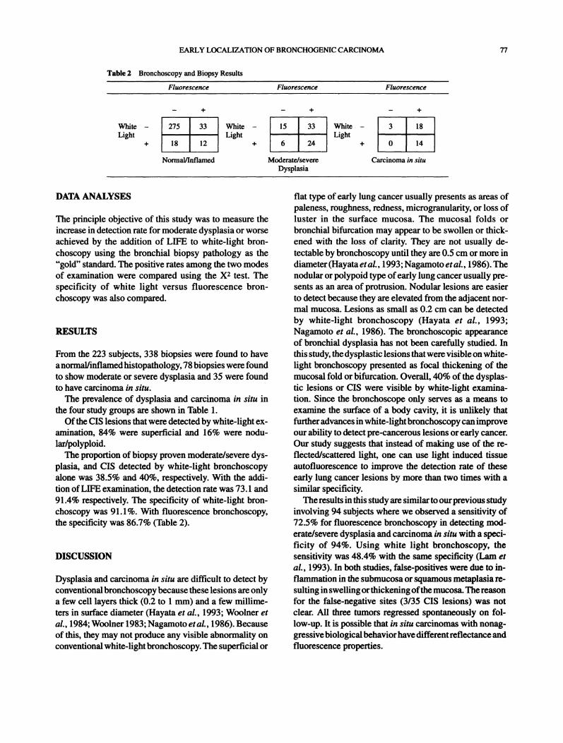
are then combined and processed by an imaging boardusing a specially developed algorithm that allows normaltissue to be clearly distinguished from malignant tissuewhen displayed as apseudocolorimage on the video mon-itor. The computed image is displayed in real time (atvideo rates). The detection oflung tumors is based on theobservation that tumors in vivo have considerably lowerautofluorescence in the green than normal tissue, whileemission in the red is similar (not as reduced) (Hung etal., 1991). The computed image is independent of the dis-tance between the bronchoscope tip and the bronchialwall. The processed image can be displayed as desired,for example, normal tissue as green and tumor as brownor brownish red (Fig. 1A and B). An abnormal area canbe biopsied under direct vision for pathologic confirma-tion.
SUBJECTS
Four groups of subjects were studied. Group I consistedof 100 patients with lung cancer (age 63 _+ 9 years, male/fe-male, 67/33). The pathology of the primary lung cancerwas squamous 50%, adenocarcinoma 25%, large cell car-cinoma 16%, small cell carcinoma 7%, and 2% othertumor types. Group II consisted of 46 patients with stageI completely resected lung cancer (age 66 9 years,male/female, 34/12). Group III consisted of 10 patientswith head and neck cancer (age 62 10 years, all males).Group IV consisted of 67 volunteers who had smokedmore than 1 packofcigarettes per day for 25 years or more(age 56
_8 years), male/female, 51/16). There were 48
current smokers and 19 ex-smokers. They had smoked anaverage of 50 +_ 27 pack years.
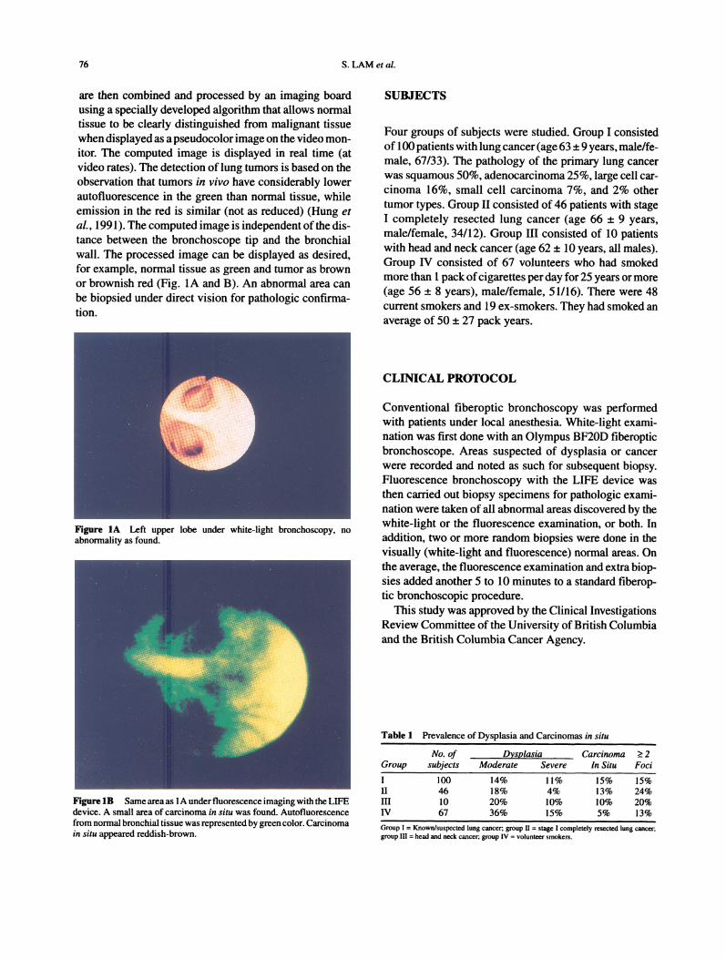
Figure IA Left upper lobe under white-light bronchoscopy, noabnormality as found.
CLINICAL PROTOCOL
Conventional fiberoptic bronchoscopy was performedwith patients under local anesthesia. White-light exami-nation was first done with an Olympus BF20D fiberopticbronchoscope. Areas suspected of dysplasia or cancerwere recorded and noted as such for subsequent biopsy.Fluorescence bronchoscopy with the LIFE device wasthen carried out biopsy specimens for pathologic exami-nation were taken of all abnormal areas discovered by thewhite-light or the fluorescence examination, or both. Inaddition, two or more random biopsies were done in thevisually (white-light and fluorescence) normal areas. Onthe average, the fluorescence examination and extra biop-sies added another 5 to 10 minutes to a standard fiberop-tic bronchoscopic procedure.
This study was approved by the Clinical InvestigationsReview Committee of the University of British Columbiaand the British Columbia Cancer Agency.
Figure IB Same area as 1A under fluorescence imaging with the LIFEdevice. A small area of carcinoma in situ was found. Autofluorescencefrom normal bronchial tissue was represented by green color. Carcinomain situ appeared reddish-brown.
Table I Prevalence of Dysplasia and Carcinomas in situ
No. of Dsplasia CarcinomaGroup subjects Moderate Severe In Situ
>2Foci
100 14% 11% 15% 15%II 46 18% 4% 13% 24%III 10 20% 10% 10% 20%IV 67 36% 15% 5% 13%
Group Known/suspected lung cancer; group II stage completely resected lung cancer,group III head and neck cancer; group IV volunteer smokers.
EARLY LOCALIZATION OF BRONCHOGENIC CARCINOMA 77
Table 2 Bronchoscopy and Biopsy Results
Fluorescence Fluorescence Fluorescence
WhiteLight
+
275 33
18 12
Normal/Inflamed
White 15 33 White 3 18Light Light
+ 6 24 + 0 14
Moderate/severe Carcinoma in situDysplasia
DATA ANALYSES
The principle objective of this study was to measure theincrease in detection rate for moderate dysplasia or worseachieved by the addition of LIFE to white-light bron-choscopy using the bronchial biopsy pathology as the"gold" standard. The positive rates among the two modesof examination were compared using the X2 test. Thespecificity of white light versus fluorescence bron-choscopy was also compared.
RESULTS
From the 223 subjects, 338 biopsies were found to haveanormal/inflamedhistopathology, 78 biopsies were foundto show moderate or severe dysplasia and 35 were foundto have carcinoma in situ.The prevalence of dysplasia and carcinoma in situ in
the four study groups are shown in Table 1.Ofthe CIS lesions that were detected by white-light ex-
amination, 84% were superficial and 16% were nodu-lar/polyploid.The proportion of biopsy proven moderate/severe dys-
plasia, and CIS detected by white-light bronchoscopyalone was 38.5% and 40%, respectively. With the addi-tion ofLIFE examination, the detection rate was 73.1 and91.4% respectively. The specificity of white-light browchoscopy was 91.1%. With fluorescence bronchoscopy,the specificity was 86.7% (Table 2).
DISCUSSION
Dysplasia and carcinoma in situ are difficult to detect byconventionalbronchoscopybecause these lesions are onlya few cell layers thick (0.2 to 1 ram) and a few millime-ters in surface diameter (Hayata et al., 1993; Woolner etal., 1984; Woolner 1983; Nagamoto etal., 1986). Becauseof this, they may not produce any visible abnormality onconventional white-lightbronchoscopy. The superficial or
flat type of early lung cancer usually presents as areas ofpaleness, roughness, redness, microgranularity, or loss ofluster in the surface mucosa. The mucosal folds orbronchial bifurcation may appear to be swollen or thick-ened with the loss of clarity. They are not usually de-tectable by bronchoscopy until they are 0.5 cm or more indiameter(Hayata etal., 1993; Nagamoto etal., 1986). Thenodular or polypoid type ofearly lung cancer usually pre-sents as an area of protrusion. Nodular lesions are easierto detect because they are elevated from the adjacent nor-mal mucosa. Lesions as small as 0.2 cm can be detectedby white-light bronchoscopy (Hayata et al., 1993;Nagamoto et al., 1986). The bronchoscopic appearanceof bronchial dysplasia has not been carefully studied. Inthis study, the dysplastic lesions thatwere visible on white-light bronchoscopy presented as focal thickening of themucosal fold or bifurcation. Overall, 40% of the dysplas-tic lesions or CIS were visible by white-light examina-tion. Since the bronchoscope only serves as a means toexamine the surface of a body cavity, it is unlikely thatfurtheradvances in white-lightbronchoscopycanimproveour ability to detect pre-cancerous lesions or early cancer.Our study suggests that instead of making use of the re-fleeted/scattered light, one can use light induced tissueautofluorescence to improve the detection rate of theseearly lung cancer lesions by more than two times with asimilar specificity.The results in this study are similarto ourprevious study
involving 94 subjects where we observed a sensitivity of72.5% for fluorescence bronchoscopy in detecting mod-erate/severe dysplasia and carcinoma in situ with a speci-ficit of 94%. Using white light bronchoscopy, thesensitivity was 48.4% with the same specificity (Lam eta/., 1993). In both studies, false-positives were due to in-flammatio in the submucosa or squamous metaplasia re-sulting in swellingorthickening ofthe mucosa. Thereasonfor the false-negative sites (3/35 CIS lesions) was notclear. All three tumors regressed spontaneously on fol-low-up. It is possible that in situ carcinomas with nonag-gressivebiological behaviorhave differentreflectance andfluorescence properties.
78 S. LAMet al.
The concept offluorescence detection has intrigued theminds of many since the beginning of the twentieth cen-tury. In 1924, Policard observed that in an experimentalmodel of sarcoma, the tumor tissue fluoresced red uponillumination by Wood’s light (Policard, 1924). In 1933,Sutro and Burman observed that when surgically excisedbreast tissue was exposed to Wood’s light, normal breasttissue fluoresced green, while breast cancer tissues fluo-resced purple (Sutro ar. Burman, 1993). These observa-tions were confirmed by others in cancer of the skin andmouth in addition to breast cancer (Ronchese, 1954). Redfluorescence was found to be associated with advancedcancers only (Ronchese, 1954). The color of the naturaltissue autofluorescence induced by filtered ultravioletlight is variable and in addition, the emitted light is ofverylow intensity. For these reasons, it has been very difficultto observe it visually, and therefore, almost all of the re-search in fluorescence bronchoscopy since the 1960s em-ployed exogenous fluorescent compounds to enhancehumans ability to detect early lung cancer. It was not untilthe advent of image-intensified cameras and computerimage processing technologies that tissue autofiuores-cence alone could be used for detecting small thin earlycancers and premalignant lesions.
Synchronous or second primary cancers occur com-monly in patients with lung cancer. An autopsy study ofAuerbachandco-workers showedthatin patientswhodiedof lung cancer, CIS could be found in 15% of these pa-tients (Auerbach et al., 1961). In the same study, in smok-ers who died ofnon-lung cancer causes, CIS was found in4.3% of those who smoked 1-2 packs per day and 11.4%of those who smoked more than 2 packs a day (Auerbachet al., 1961). In patients resected stage I lung cancer, sec-ond primary tumors occur in 10-20% of cases (Pastorinoet al., 1993; Thomas et al., 1990; Cortese, et al., 1983).The prevalence of dysplasia and CIS observed in our pa-tients with lung cancer and in the smoking volunteers areconsistent with the findings in these earlier studies.Our study suggests that fluorescence bronchoscopy, in
conjunction with standard white-light bronchoscopy, maybe very useful in the detection ofsynchronous and secondprimary tumors in patients with lung cancer. In high riskpopulations such as heavy smokers, fluorescence bron-choscopy may also be useful for localizing precancerouslesions and CIS. This technology, in combination withlocal treatments such as photodynamic therapy which caneradicate these lesions without loss ofnormal lung tissues,offers hope that the traditionally poor prognosis of lungcancer may be altered in a significant way.
ACKNOWLEDGMENTS
This study was supported by the National Cancer Instituteof Canada, the Pacific Pulmonary Research Society, andXillix Technologies Corp., Vancouver, Canada
REFERENCES
Auerbach, O., Stout, A. P., Hammond, E. C. and Garfinkel, L. (1961)Changes in bronchial epithelium in relation to cigarette smokingand in relation to lung cancer. N. Engl. J. Med., 265:253-267.
Cortese, D. A., Pairolero, P. D., Bergstralh, E. J. B. et al. (1983)Roentgenographically occult lung cancer: A ten year experience.J. Thorac. Cardiovasc. Surg., 86:373-80.
Edell, E. S., Cortese, D. A. (1992) Photodynamic therapy in the man-agement ofearly superficial squamous cell carcinoma as an alter-native to surgical resection. Chest 102:1319-1322.
Furuse, K., Fukuoka, M., Kato H. et al. (1993) A prospective study onphotodynamic therapy with Photofrin II for centrally locatedearly-stage lung cancer. J. Clin. Oncol., 11:1852-1857."
Hayata Y., Kalo, H., Konaka, C., Okunaka, T. (1993) Photodynamictherapy in early stage lung cancer. Lung Cancer, 9:287-294.
Hung, J., Lam, S., LeRiche, J. C., Palcic B. (1991) Autofluorescence ofnormal and malignant bronchial tissue. Lasers Surg. Med.,11:99-105.
Lam, S., MacAulay C., Hung, J. et al. (1993) Detection ofdysplasia andcarcinoma in-situ using a lung imaging fluorescence endoscope(LIFE) device. J. Thorac. Cardiovasc. Surg., 105:1035-1040.
Lam, S. and Palcic, B. (1993) Fluorescence detection. In: Roth, J. A.,Cox, J. D., Hong, W. K. (eds.) Lung Cancer, Blackwell ScientificPublications 325-338.
Lippman, S. M., Lee, J. S. and Lotan, R. et al. (1990) Chemopreventionof upper aerodigestive tract cancers: A report of the Third UpperAerodigestive Cancer Task Force Workshop. Head and Neck12:5-20.
Nagamoto, N., Saito, Y., Iami, T. et al. (1986) Roentgenographicallyoccult squamous cell carcinoma: Location in the bronchi, depth ofinvasion and length of axial involvement of the bronchus. TohokuJ. Exp. Med., 148:241-256.
Palcic B., Lain, S., Hung J. and MacAulay C. (1991) Detection and lo-calization of early lung cancer by imaging techniques. Chest,99:742-3.
Pastorino, U., Infante, M., Maioli, M., Chiesa, G. et al. (1993) Adjuvanttreatment of stage lung cancer with high dose vitamin A. J. CliOncol., 11:1216-1222.
Policard, A. (1924) Etude sur les aspects offerts par des tumeurs exper-imentales examinees a la lumiere de Wood. Comptre-rendus Soc.Biol., 91:1423-24.
Ronchese, F. (1954) The fluorescence of cancer under the Wood light.Oral. Surg. Oral. Med. Oral. Pathol., 7:967-971.
Sutro, C. J. and Burman, M. S. (1933) Examination of pathogenic tis-sue by filtered ultraviolet radiation. Arch. Path., 16:346-349.
Thomas, P., Rubinstein, L. and the Lung Cancer Study Group (1990)Cancer recurrence after resection (TINO non small-cell lung can-cer). Ann. Thorac. Surg., 48:242-247.
Woolner, L. B. (1983) Pathology of cancer detected cytologically. In:Atlas ofearly lung cancer, National Cancer Institute CooperativeEarly Lung Cancer Group, Igaku-Shoin, Tokyo, New York., pp.10/-213.
Woolner, L. B., Fontana, R. S., Cortese, D. A. et al. (1984)Roentgenographically occult lung cancer: Pathologic findings andfrequency of multicentricity during a 10-year period. Mayo. Clin.Proc., 59:453-466.

Submit your manuscripts athttp://www.hindawi.com
Stem CellsInternational
Hindawi Publishing Corporationhttp://www.hindawi.com Volume 2014
Hindawi Publishing Corporationhttp://www.hindawi.com Volume 2014
MEDIATORSINFLAMMATION
of
Hindawi Publishing Corporationhttp://www.hindawi.com Volume 2014
Behavioural Neurology
EndocrinologyInternational Journal of
Hindawi Publishing Corporationhttp://www.hindawi.com Volume 2014
Hindawi Publishing Corporationhttp://www.hindawi.com Volume 2014
Disease Markers
Hindawi Publishing Corporationhttp://www.hindawi.com Volume 2014
BioMed Research International
OncologyJournal of
Hindawi Publishing Corporationhttp://www.hindawi.com Volume 2014
Hindawi Publishing Corporationhttp://www.hindawi.com Volume 2014
Oxidative Medicine and Cellular Longevity
Hindawi Publishing Corporationhttp://www.hindawi.com Volume 2014
PPAR Research
The Scientific World JournalHindawi Publishing Corporation http://www.hindawi.com Volume 2014
Immunology ResearchHindawi Publishing Corporationhttp://www.hindawi.com Volume 2014
Journal of
ObesityJournal of
Hindawi Publishing Corporationhttp://www.hindawi.com Volume 2014
Hindawi Publishing Corporationhttp://www.hindawi.com Volume 2014
Computational and Mathematical Methods in Medicine
OphthalmologyJournal of
Hindawi Publishing Corporationhttp://www.hindawi.com Volume 2014
Diabetes ResearchJournal of
Hindawi Publishing Corporationhttp://www.hindawi.com Volume 2014
Hindawi Publishing Corporationhttp://www.hindawi.com Volume 2014
Research and TreatmentAIDS
Hindawi Publishing Corporationhttp://www.hindawi.com Volume 2014
Gastroenterology Research and Practice
Hindawi Publishing Corporationhttp://www.hindawi.com Volume 2014
Parkinson’s Disease
Evidence-Based Complementary and Alternative Medicine
Volume 2014Hindawi Publishing Corporationhttp://www.hindawi.com