Embed Size (px)
Citation preview
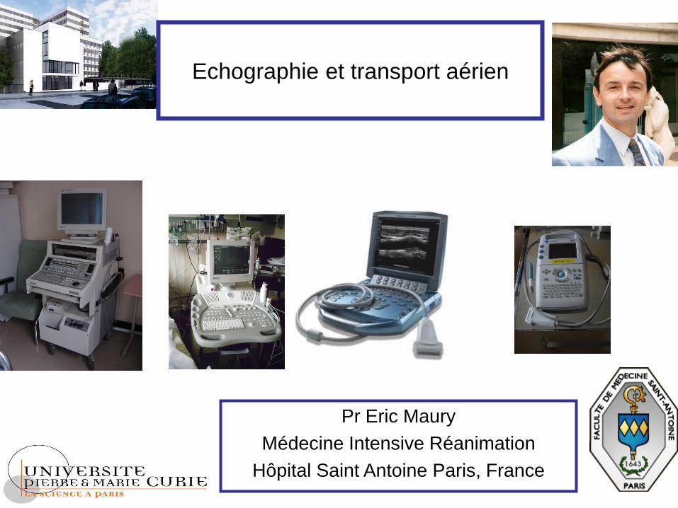
Pr Eric Maury
Médecine Intensive Réanimation
Hôpital Saint Antoine Paris, France
Echographie et transport aérien
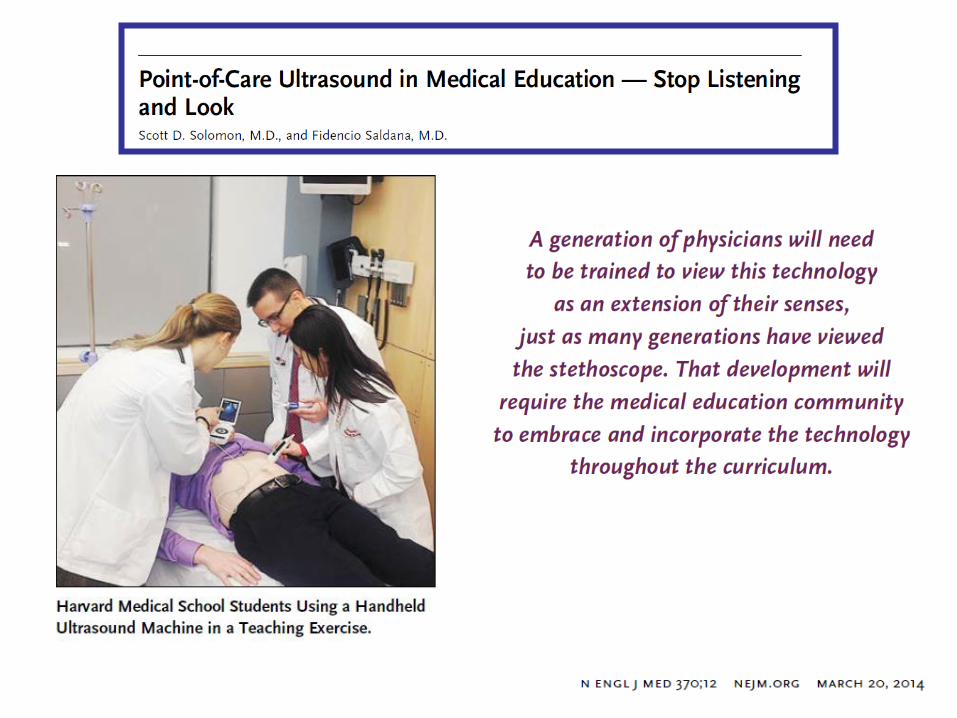
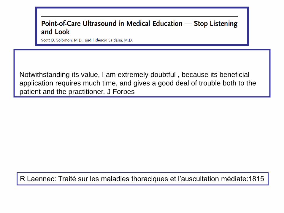
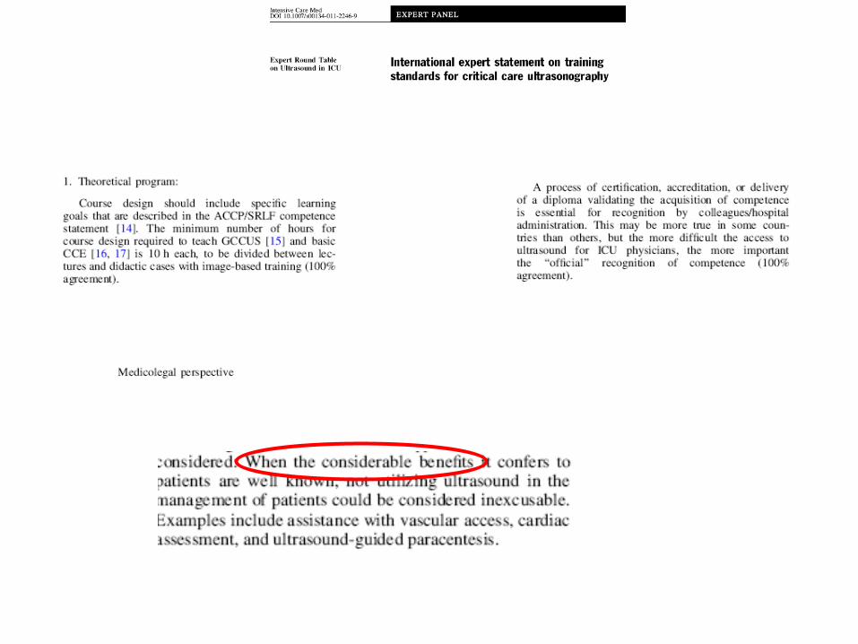
Notwithstanding its value, I am extremely doubtful , because its beneficial
application requires much time, and gives a good deal of trouble both to the
patient and the practitioner. J Forbes
R Laennec: Traité sur les maladies thoraciques et l’auscultation médiate:1815
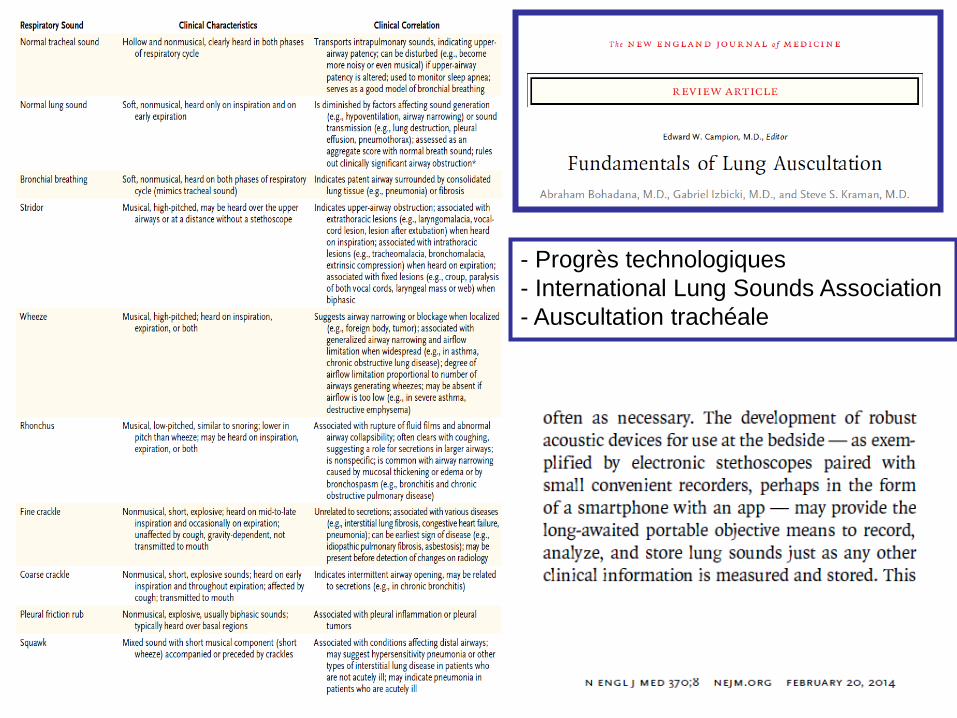
- Progrès technologiques
- International Lung Sounds Association
- Auscultation trachéale
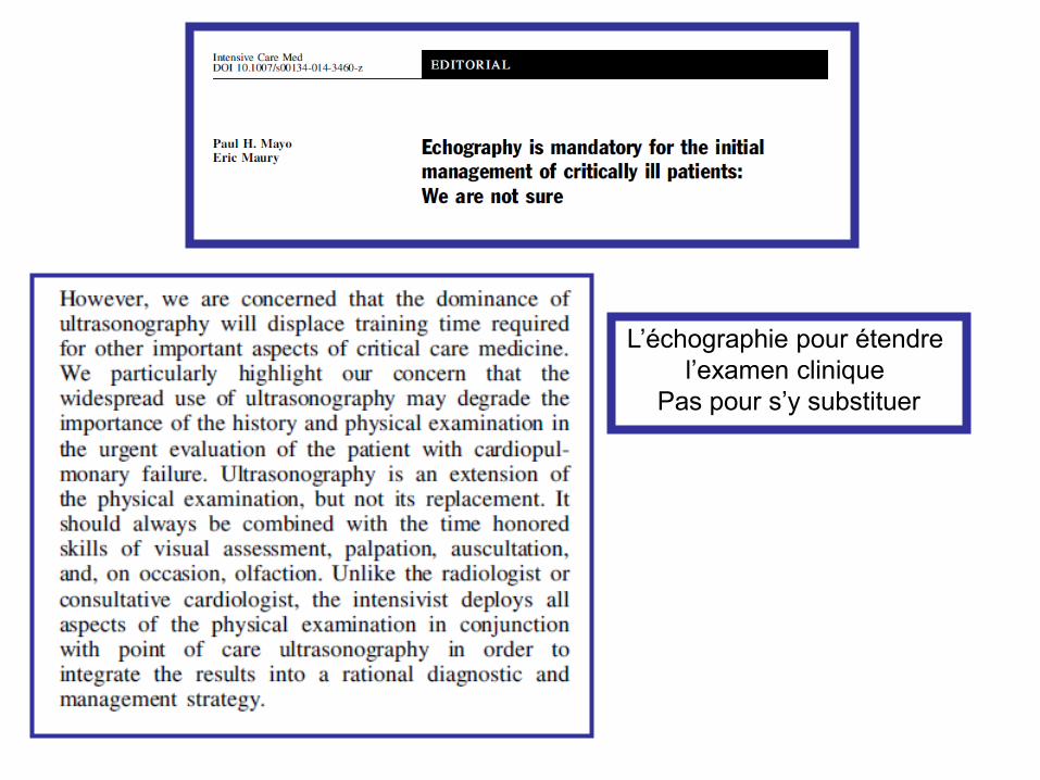
L’échographie pour étendre
l’examen clinique
Pas pour s’y substituer
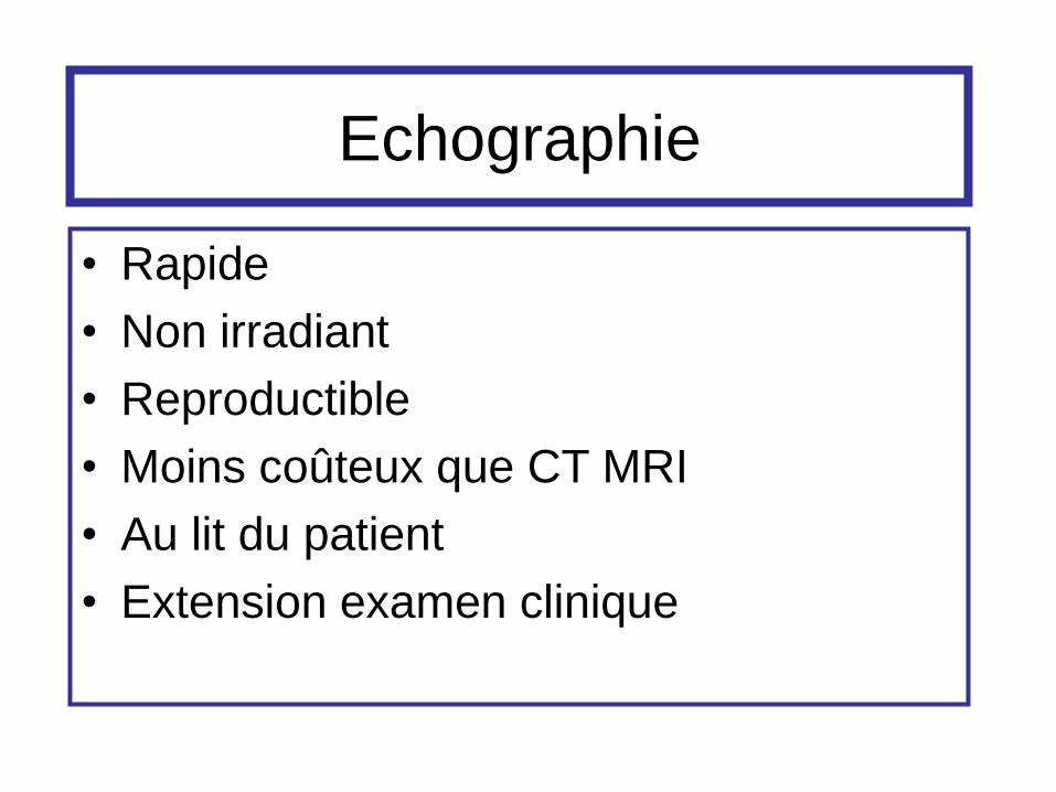
Echographie
• Rapide
• Non irradiant
• Reproductible
• Moins coûteux que CT MRI
• Au lit du patient
• Extension examen clinique
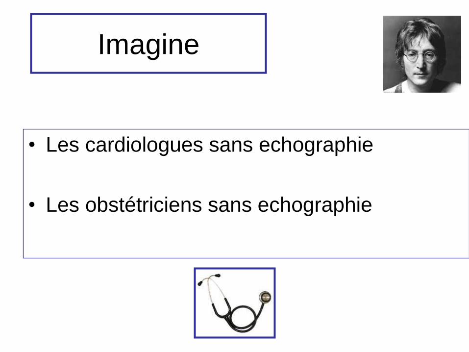
Imagine
• Les cardiologues sans echographie
• Les obstétriciens sans echographie
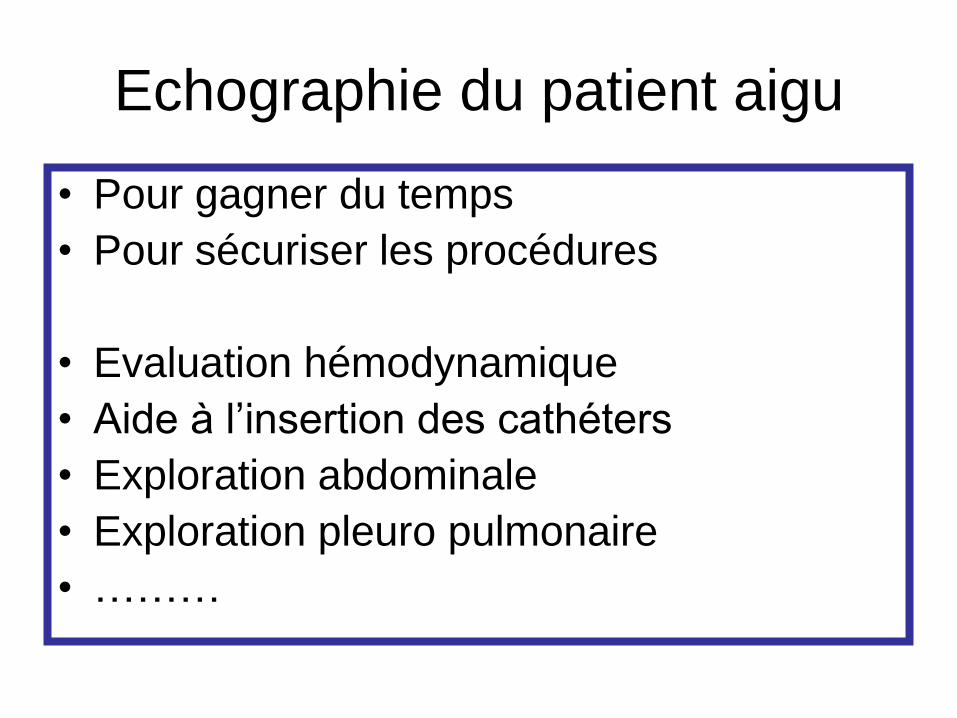
Echographie du patient aigu
• Pour gagner du temps
• Pour sécuriser les procédures
• Evaluation hémodynamique
• Aide à l’insertion des cathéters
• Exploration abdominale
• Exploration pleuro pulmonaire
• ………
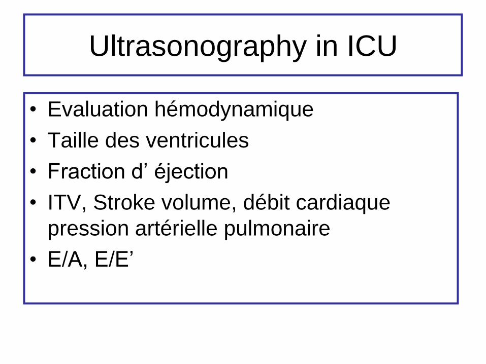
Ultrasonography in ICU
• Evaluation hémodynamique
• Taille des ventricules
• Fraction d’ éjection
• ITV, Stroke volume, débit cardiaque
pression artérielle pulmonaire
• E/A, E/E’
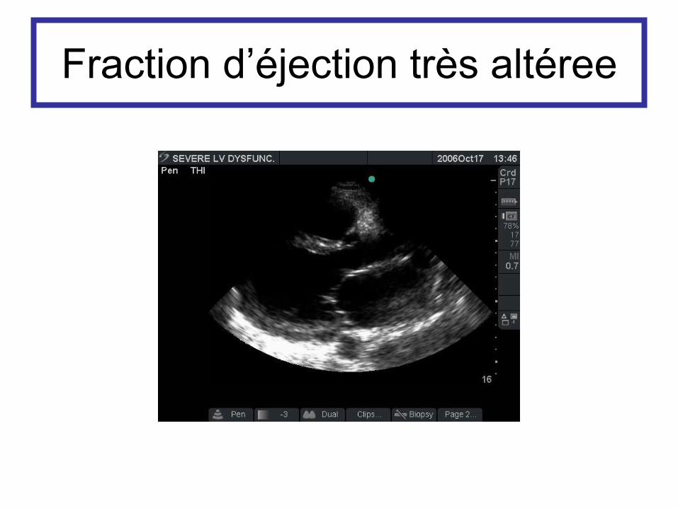
Fraction d’éjection très altéree
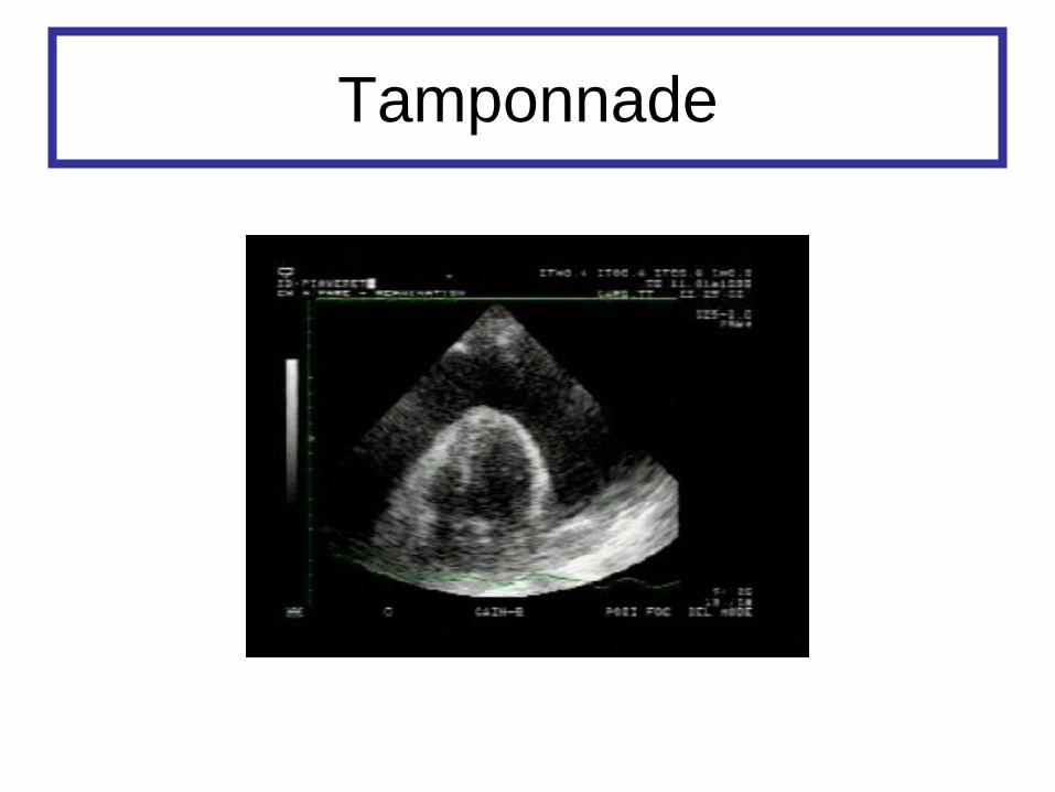
Tamponnade
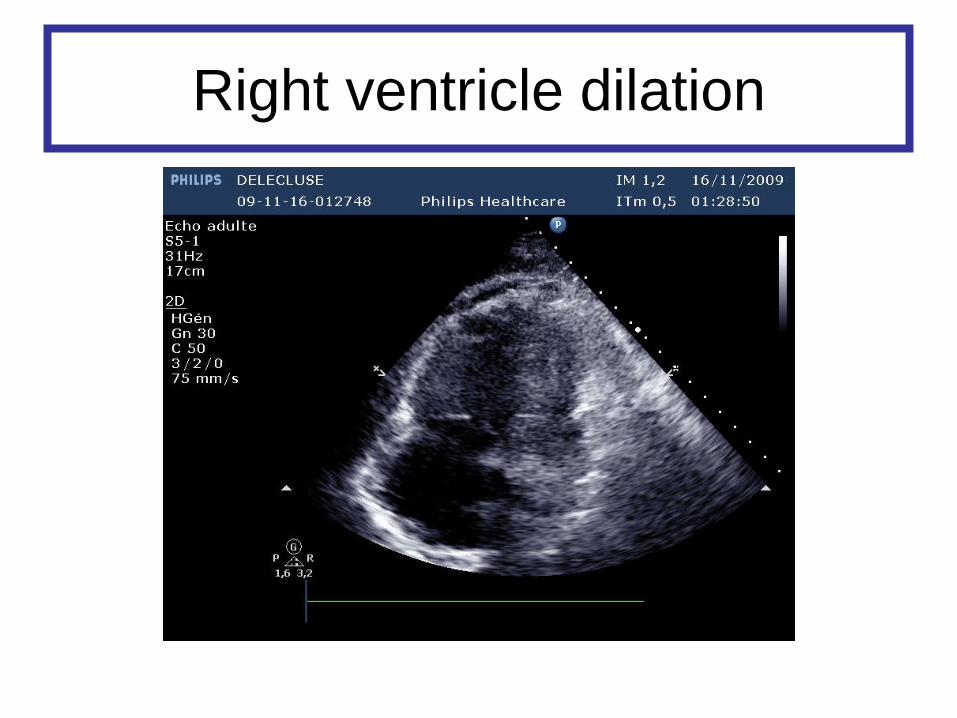
Right ventricle dilation
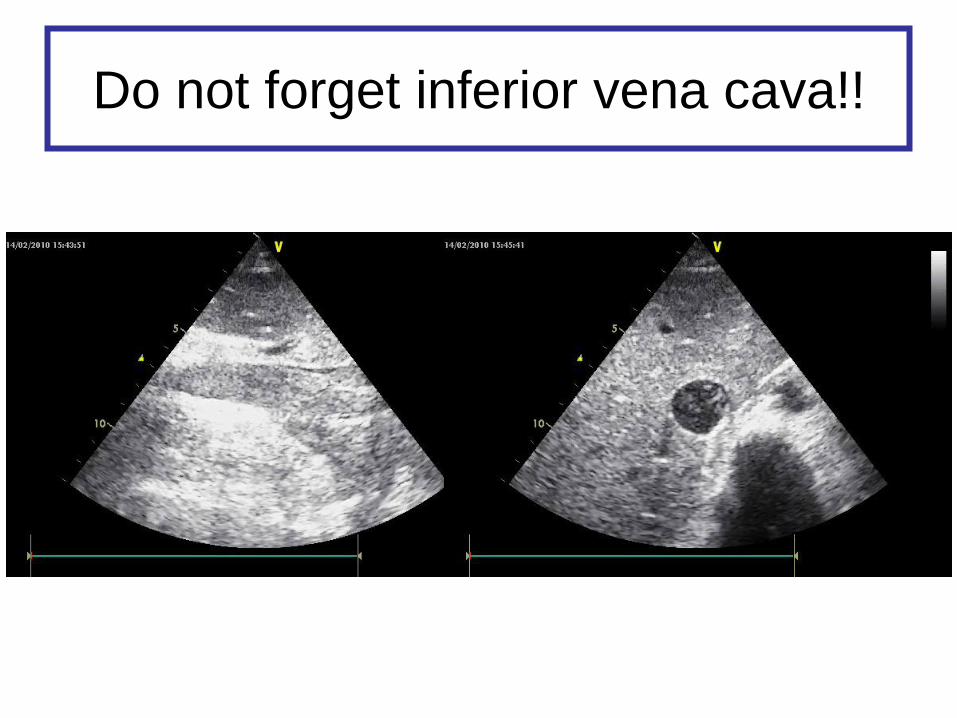
Do not forget inferior vena cava!!
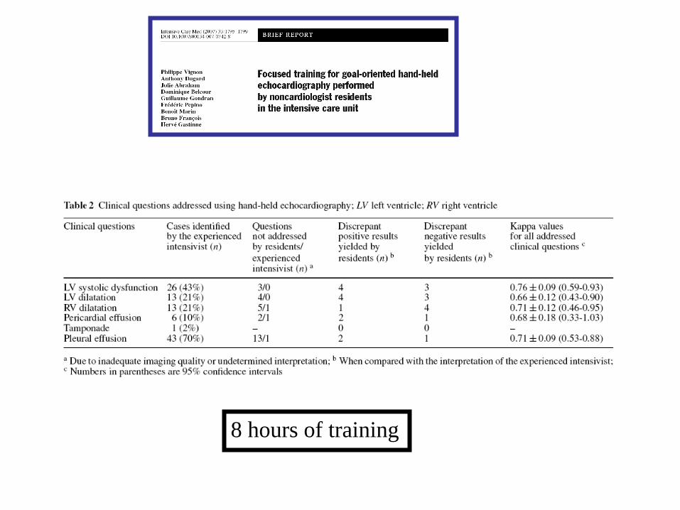
8 hours of training
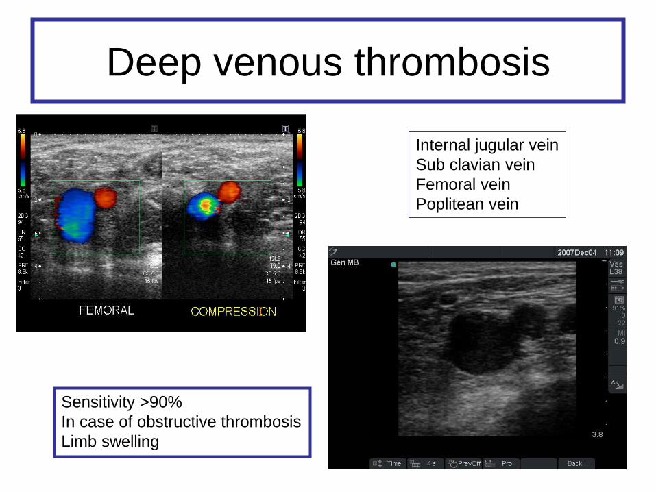
Deep venous thrombosis
Internal jugular vein
Sub clavian vein
Femoral vein
Poplitean vein
Sensitivity >90%
In case of obstructive thrombosis
Limb swelling
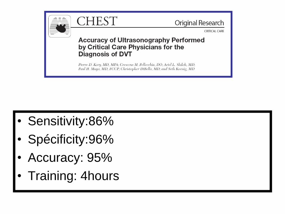
• Sensitivity:86%
• Spécificity:96%
• Accuracy: 95%
• Training: 4hours
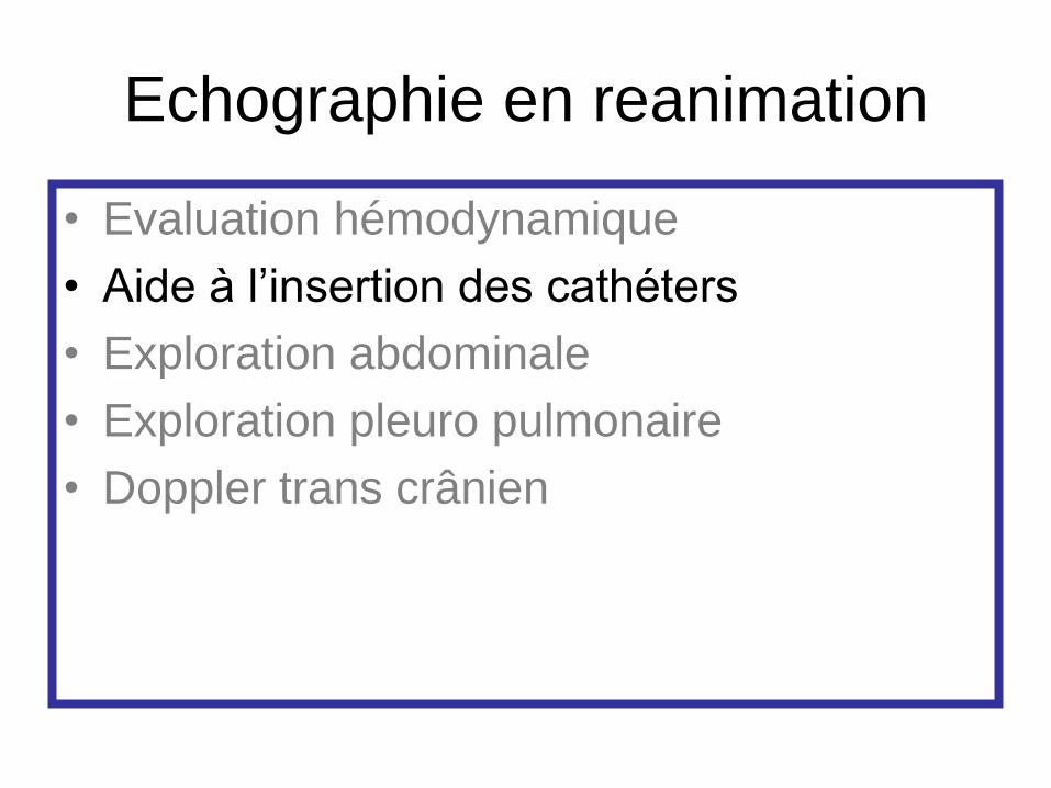
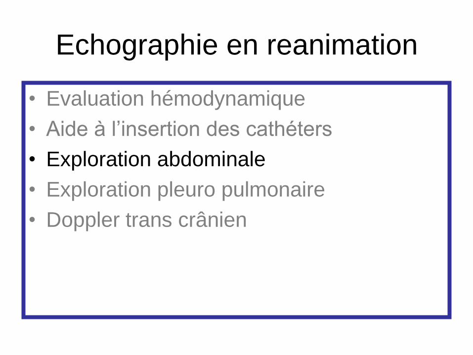
Echographie en reanimation
• Evaluation hémodynamique
• Aide à l’insertion des cathéters
• Exploration abdominale
• Exploration pleuro pulmonaire
• Doppler trans crânien
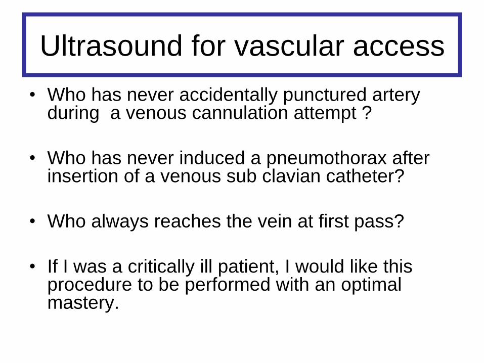
• Who has never accidentally punctured artery during a venous cannulation attempt ?
• Who has never induced a pneumothorax after insertion of a venous sub clavian catheter?
• Who always reaches the vein at first pass?
• If I was a critically ill patient, I would like this procedure to be performed with an optimal mastery.
Ultrasound for vascular access
To see or not…
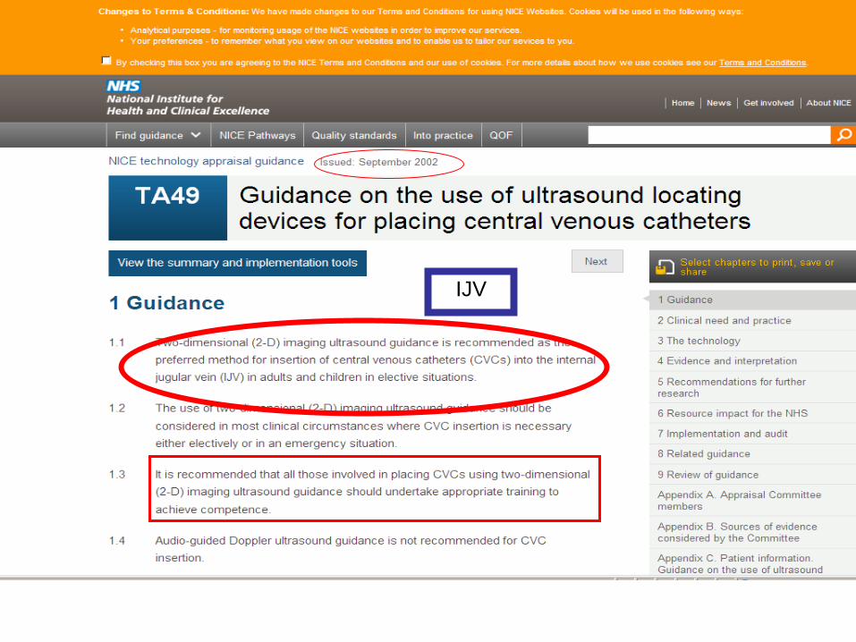
IJV
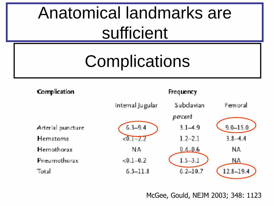
Complications
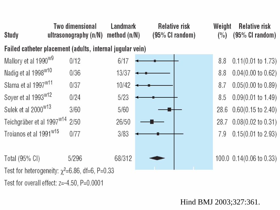
McGee, Gould, NEJM 2003; 348: 1123
Anatomical landmarks are
sufficient
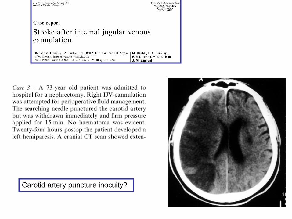
Carotid artery puncture inocuity?
Why such a rate of complications?
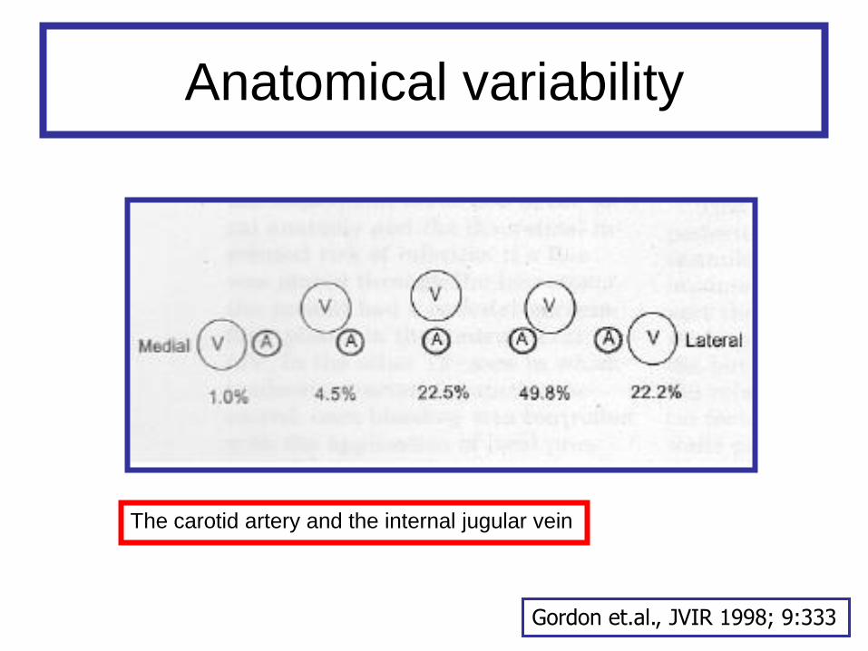
Anatomical variability
Gordon et.al., JVIR 1998; 9:333
The carotid artery and the internal jugular vein
0
10
20
30
40
50
60
70
80
90
100
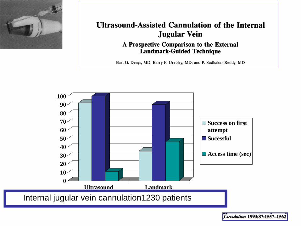
Ultrasound Landmark
Success on first
attempt
Sucessful
Access time (sec)
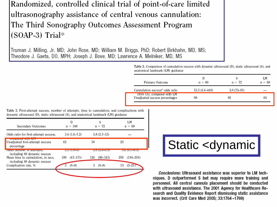
Internal jugular vein cannulation1230 patients
Hind BMJ 2003;327:361.
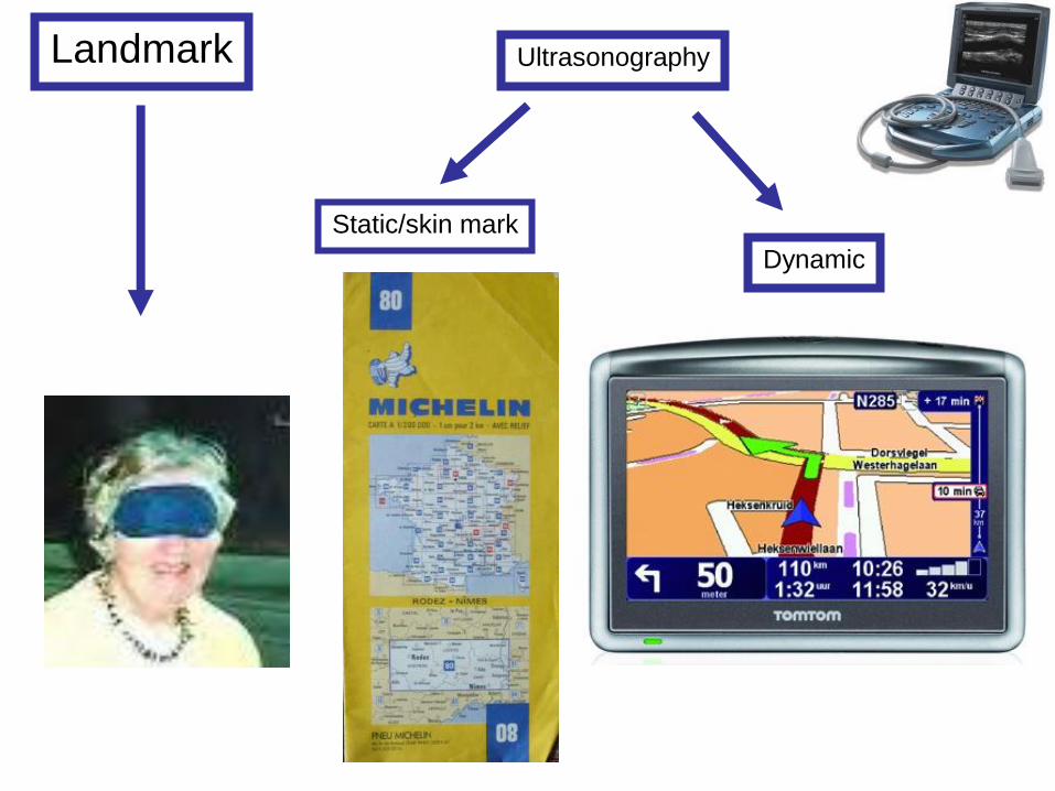
Static/skin mark
Dynamic
Landmark Ultrasonography
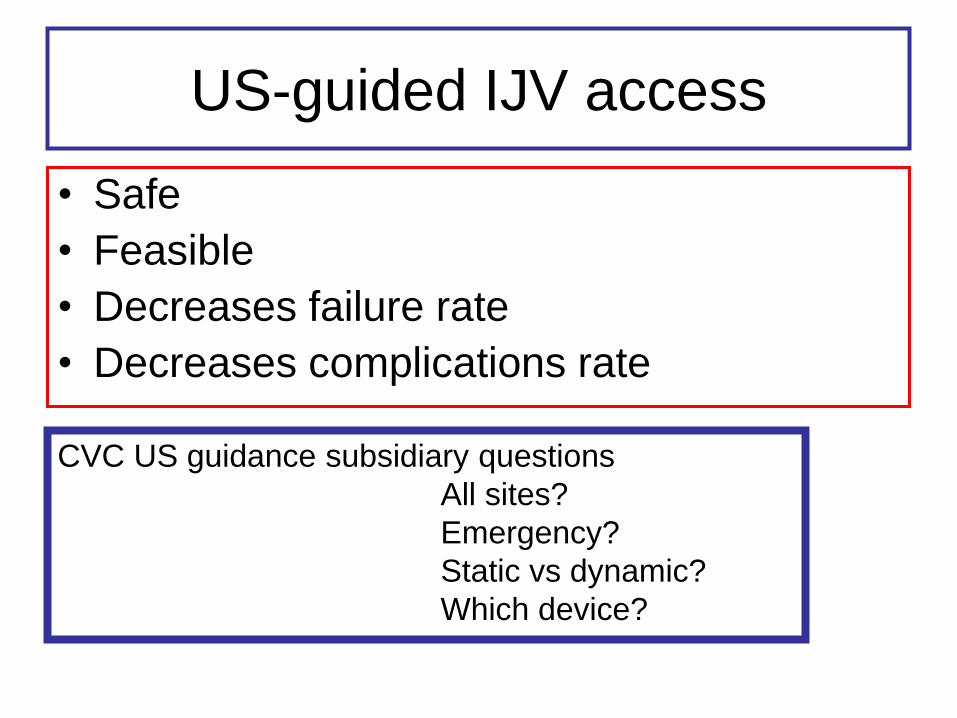
US-guided IJV access
• Safe
• Feasible
• Decreases failure rate
• Decreases complications rate
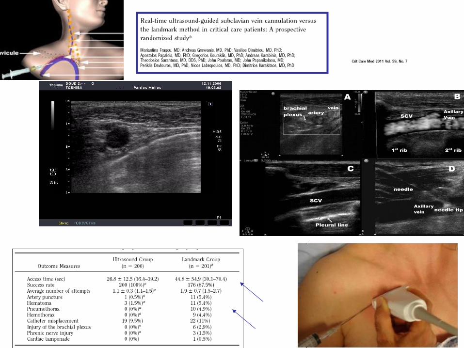
CVC US guidance subsidiary questions
All sites?
Emergency?
Static vs dynamic?
Which device?
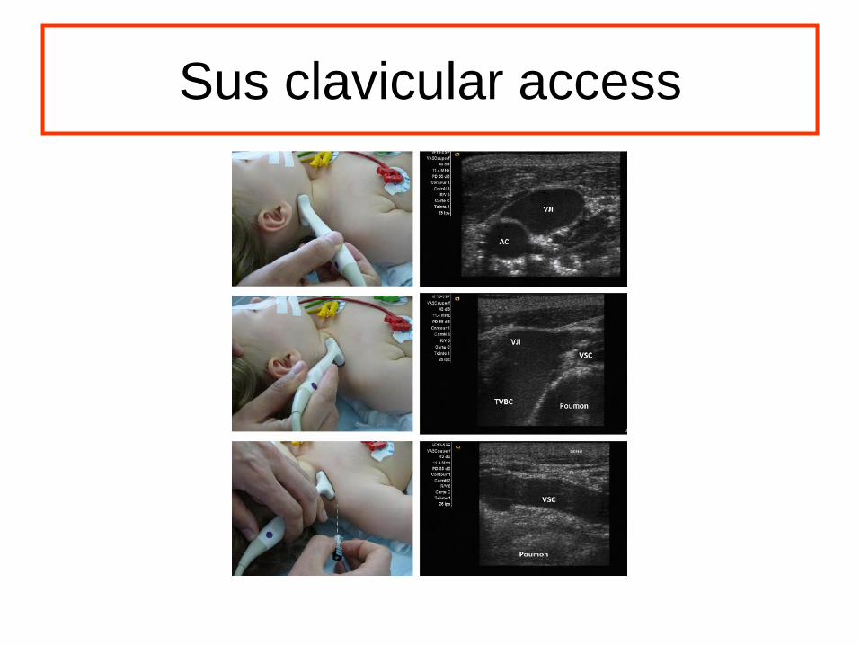
Sus clavicular access
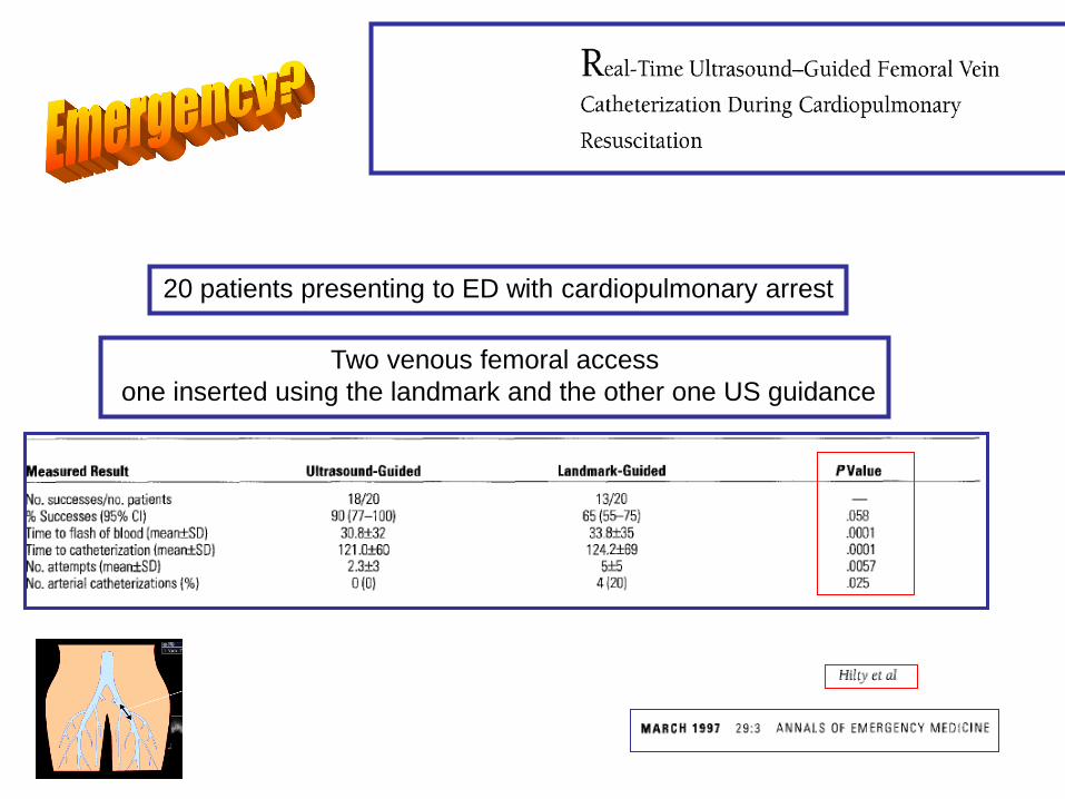
20 patients presenting to ED with cardiopulmonary arrest
Two venous femoral access
one inserted using the landmark and the other one US guidance
Static <dynamic
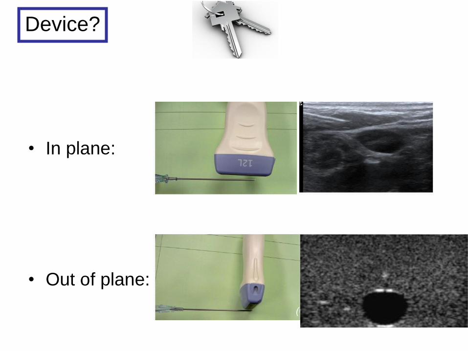
• In plane:
• Out of plane:
Device?
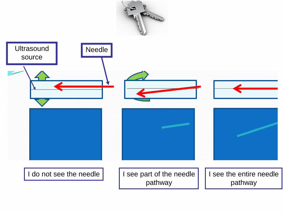
Ultrasound
source Needle
I do not see the needle I see part of the needle
pathway
I see the entire needle
pathway
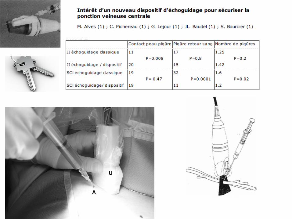
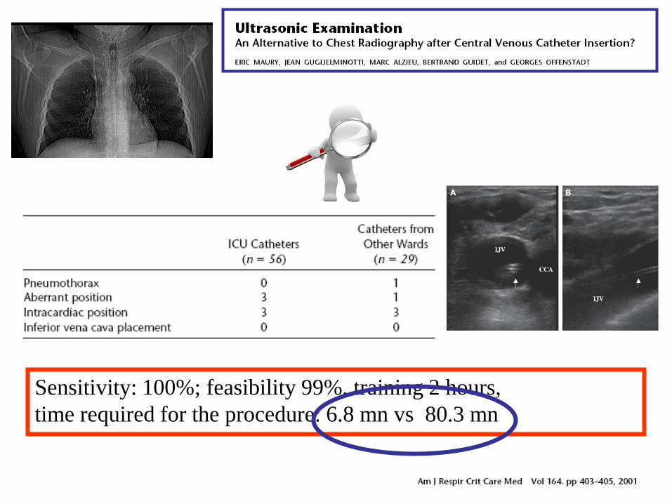
Sensitivity: 100%; feasibility 99%, training 2 hours,
time required for the procedure: 6.8 mn vs 80.3 mn
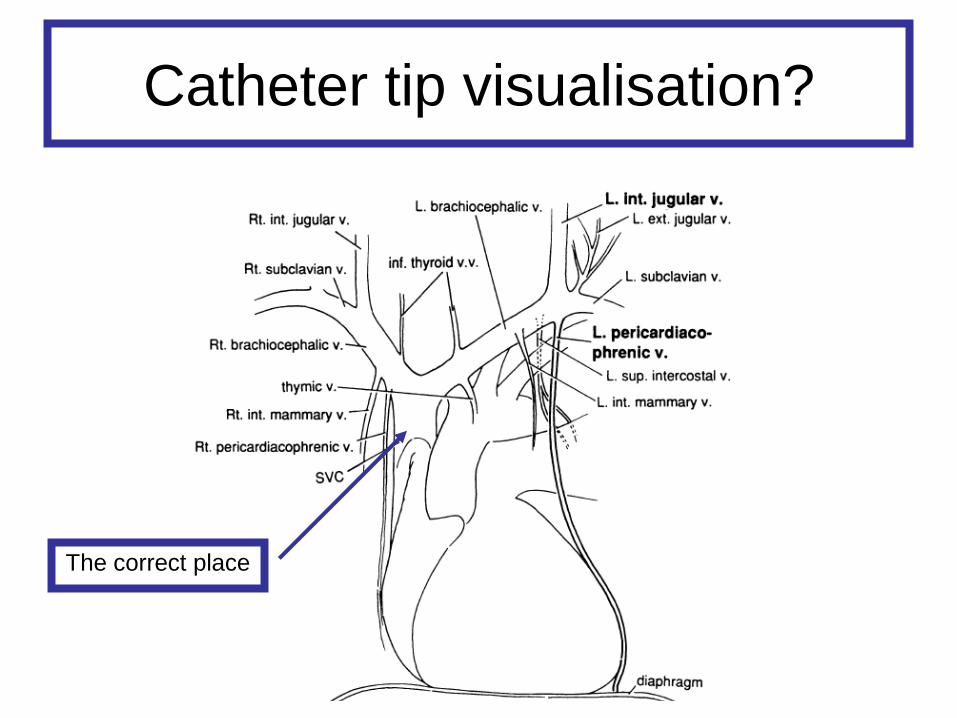
Catheter tip visualisation?
The correct place
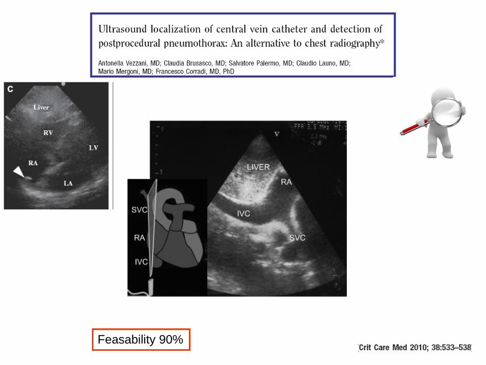
Feasability 90%
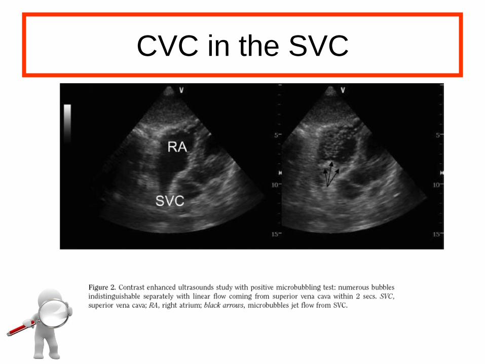
CVC in the SVC
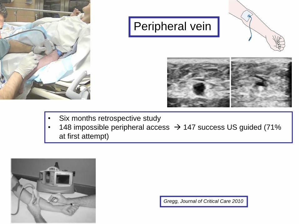
Gregg, Journal of Critical Care 2010
• Six months retrospective study
• 148 impossible peripheral access 147 success US guided (71%
at first attempt)
Peripheral vein
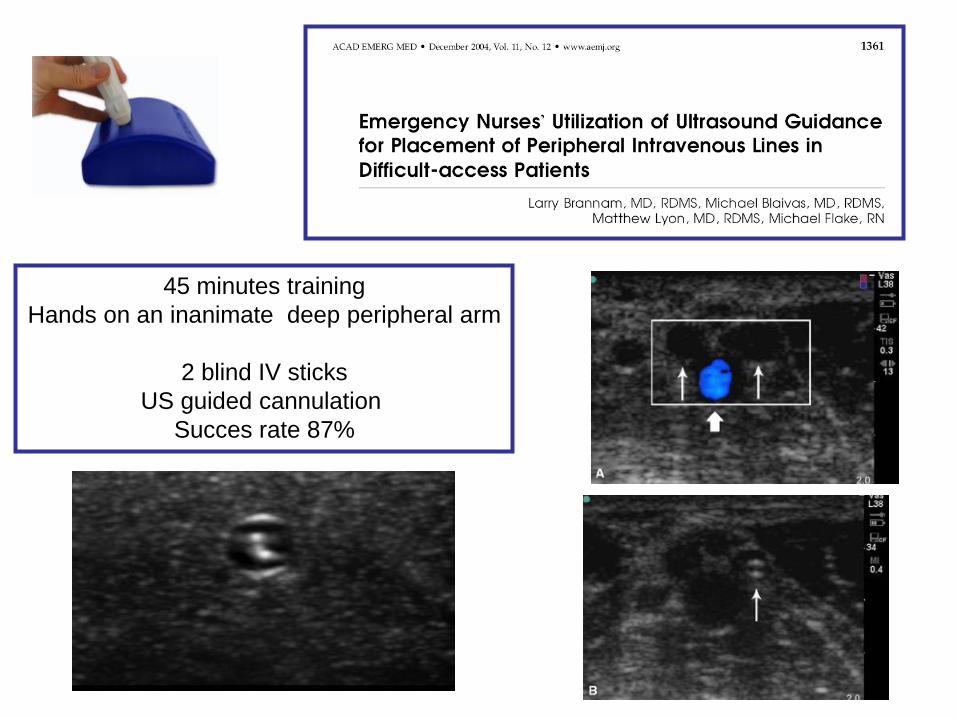
45 minutes training
Hands on an inanimate deep peripheral arm
2 blind IV sticks
US guided cannulation
Succes rate 87%
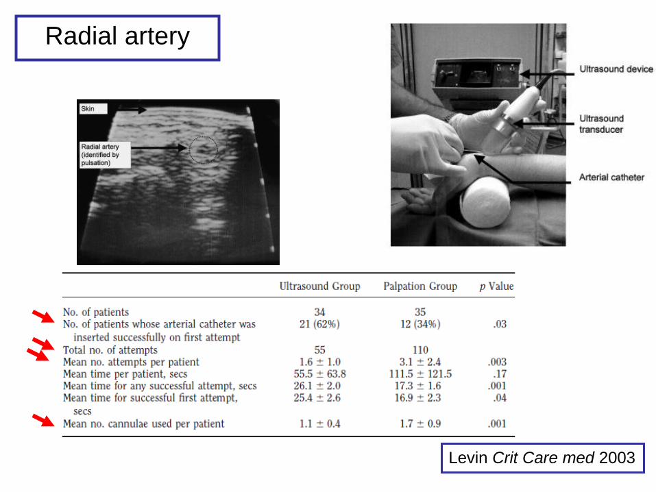
Radial artery
Levin Crit Care med 2003
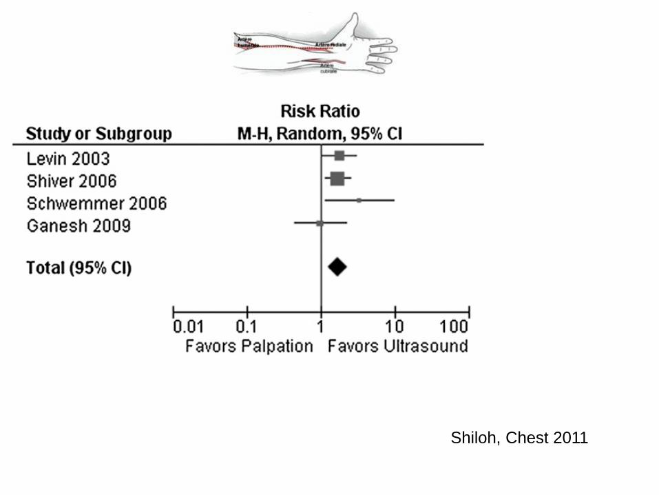
Shiloh, Chest 2011
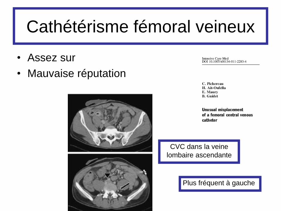
Cathétérisme fémoral veineux
• Assez sur
• Mauvaise réputation
CVC dans la veine
lombaire ascendante
Plus fréquent à gauche
Echographie en reanimation
• Evaluation hémodynamique
• Aide à l’insertion des cathéters
• Exploration abdominale
• Exploration pleuro pulmonaire
• Doppler trans crânien
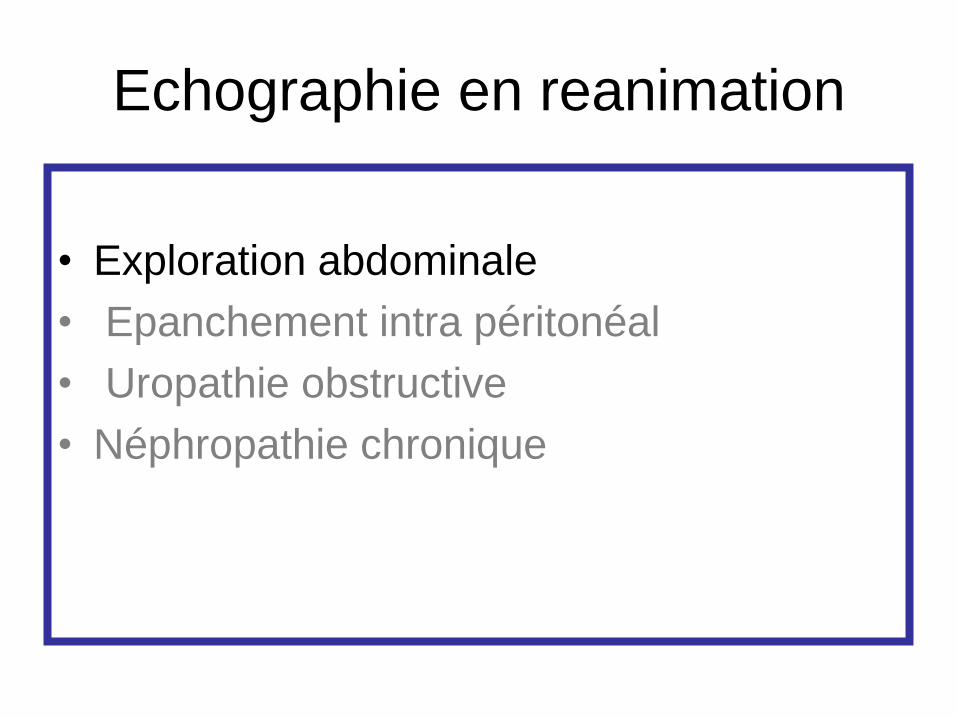
Echographie en reanimation
• Exploration abdominale
• Epanchement intra péritonéal
• Uropathie obstructive
• Néphropathie chronique

Safety
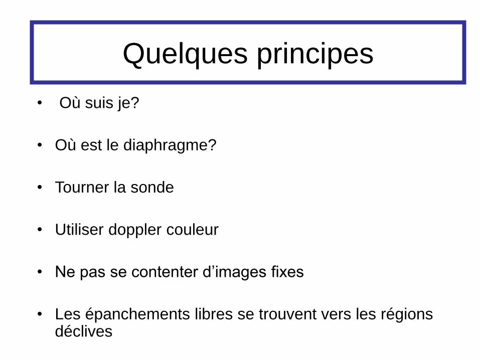
Quelques principes
• Où suis je?
• Où est le diaphragme?
• Tourner la sonde
• Utiliser doppler couleur
• Ne pas se contenter d’images fixes
• Les épanchements libres se trouvent vers les régions déclives
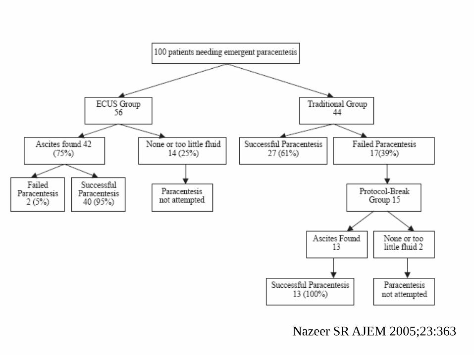
Nazeer SR AJEM 2005;23:363
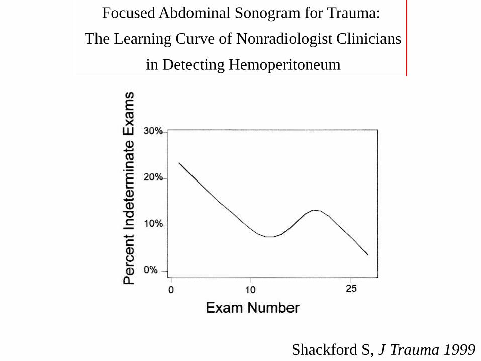
Shackford S, J Trauma 1999
Focused Abdominal Sonogram for Trauma:
The Learning Curve of Nonradiologist Clinicians
in Detecting Hemoperitoneum
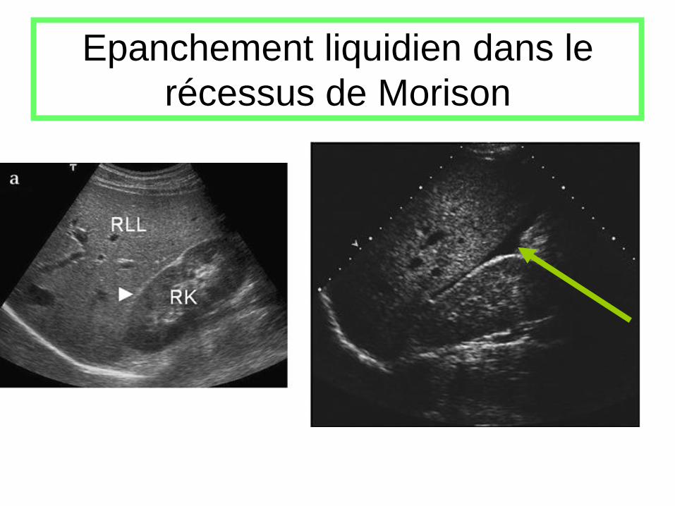
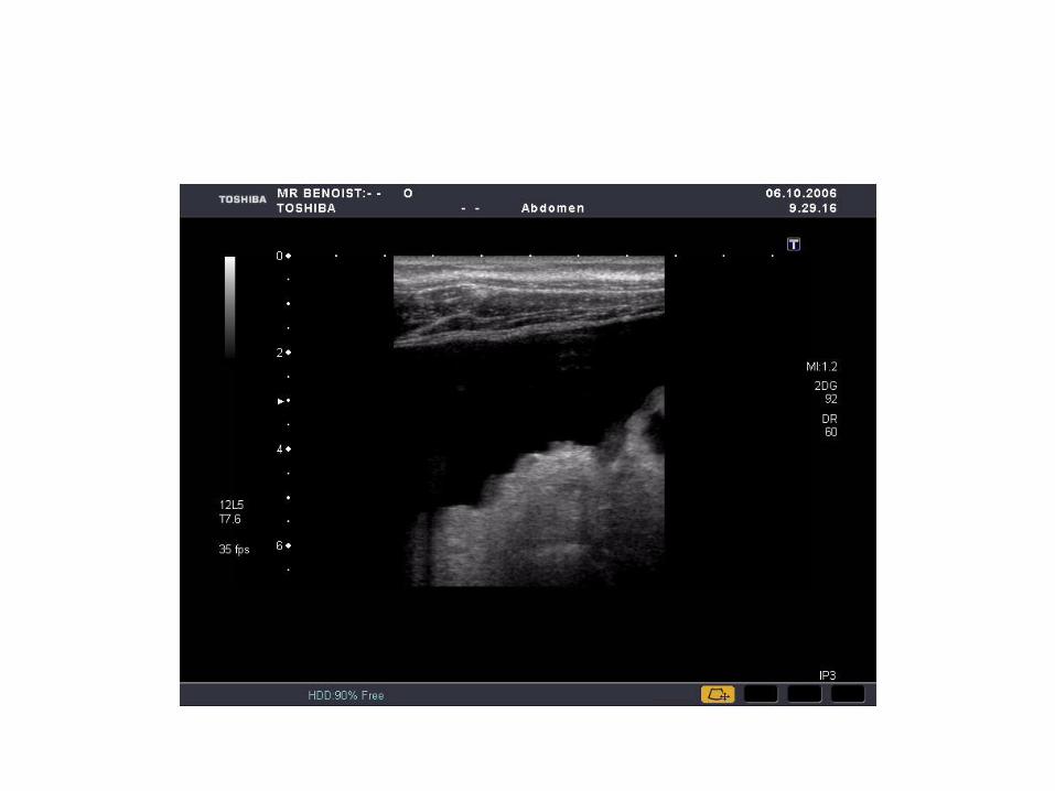
Epanchement liquidien dans le
récessus de Morison
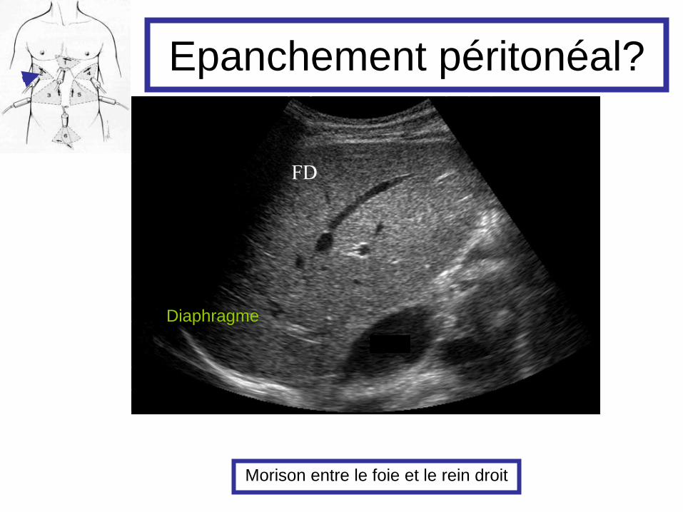
Epanchement péritonéal?
Diaphragme
Morison entre le foie et le rein droit
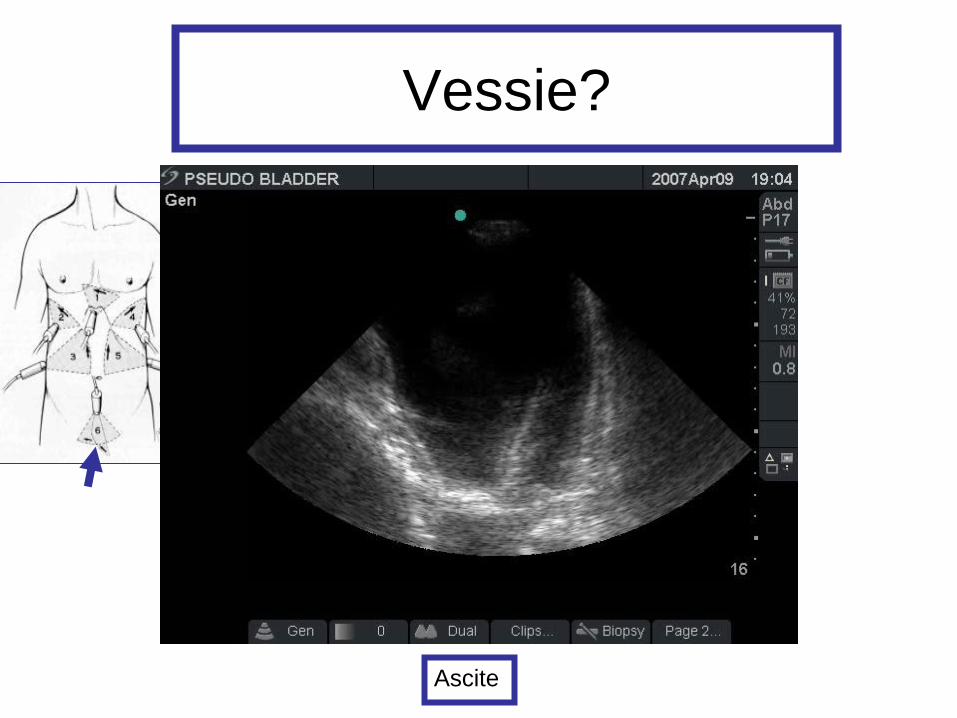
Vessie?
Ascite
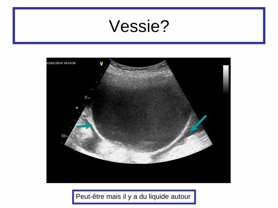
Vessie?
Peut-être mais il y a du liquide autour
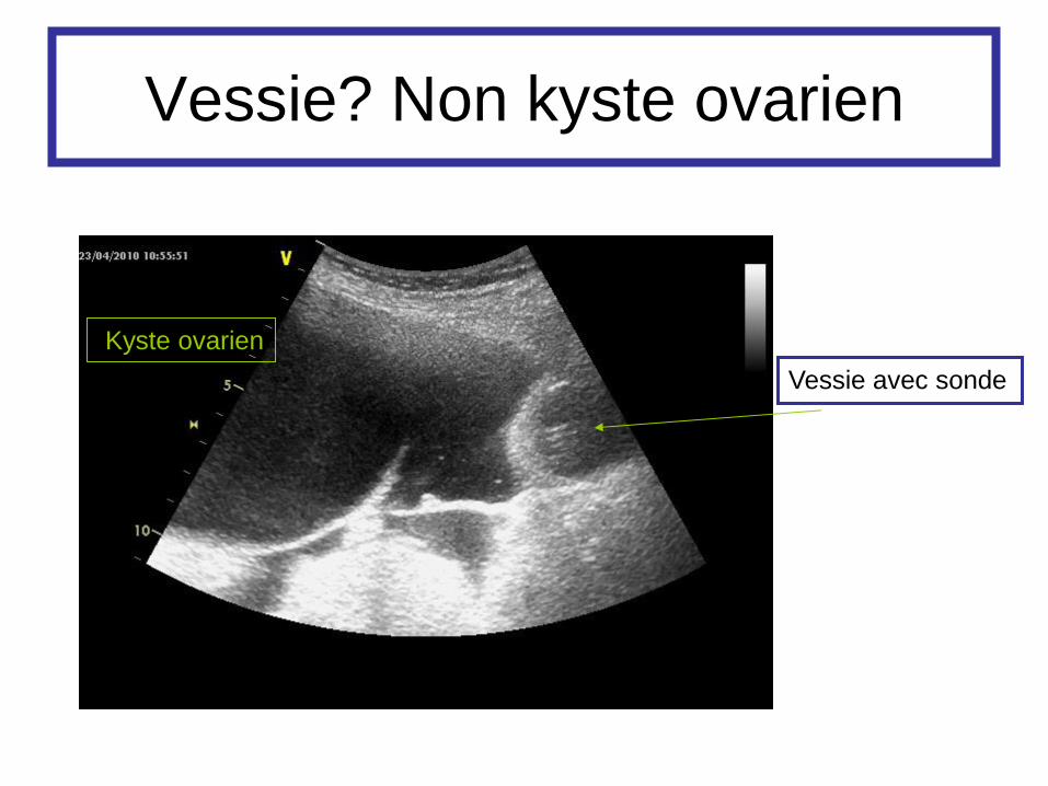
Vessie? Non kyste ovarien
Vessie avec sonde
Kyste ovarien
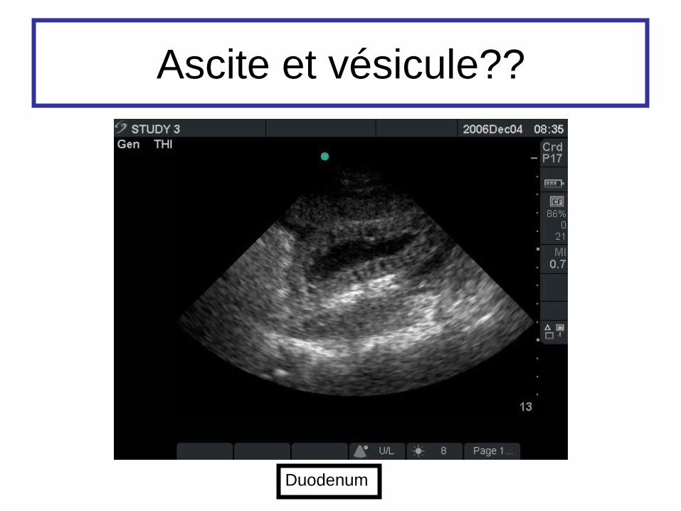
Ascite et vésicule??
Duodenum
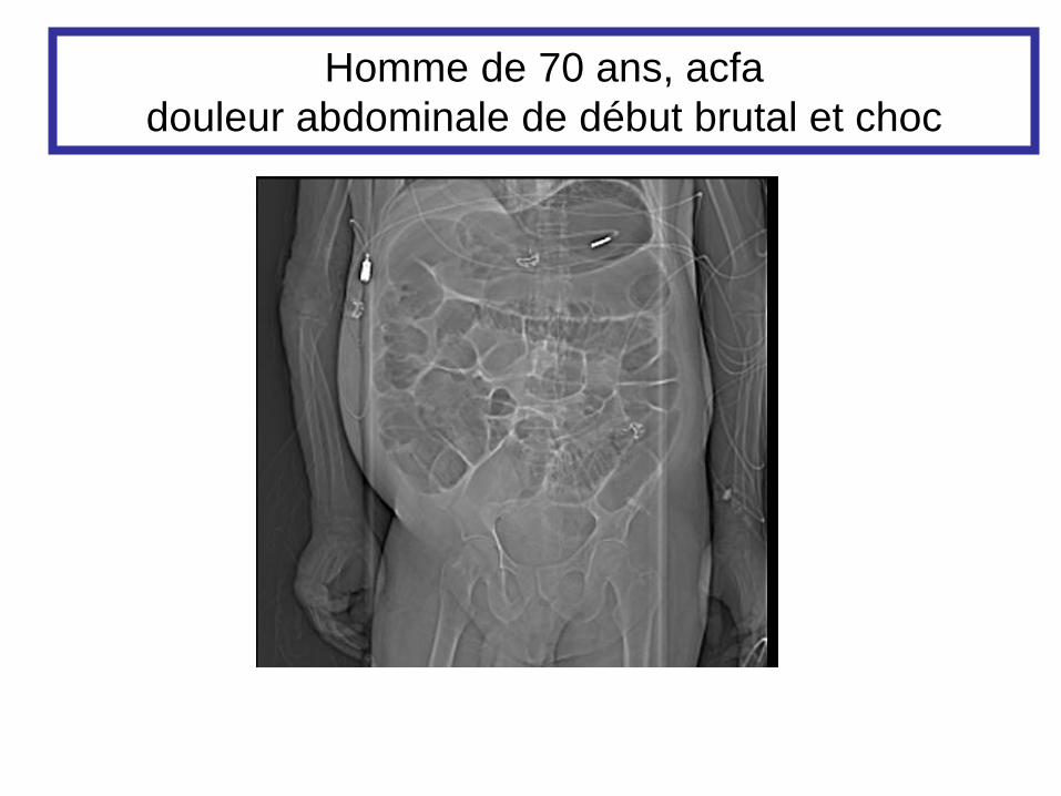
Homme de 70 ans, acfa
douleur abdominale de début brutal et choc
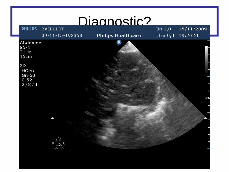
Diagnostic?
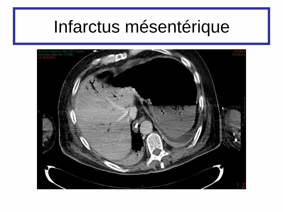
Infarctus mésentérique
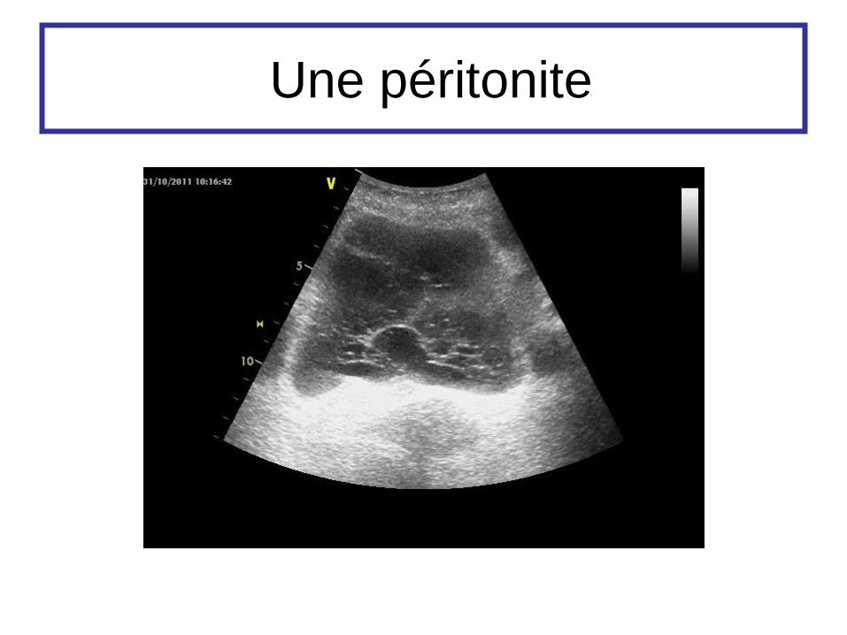
Une péritonite
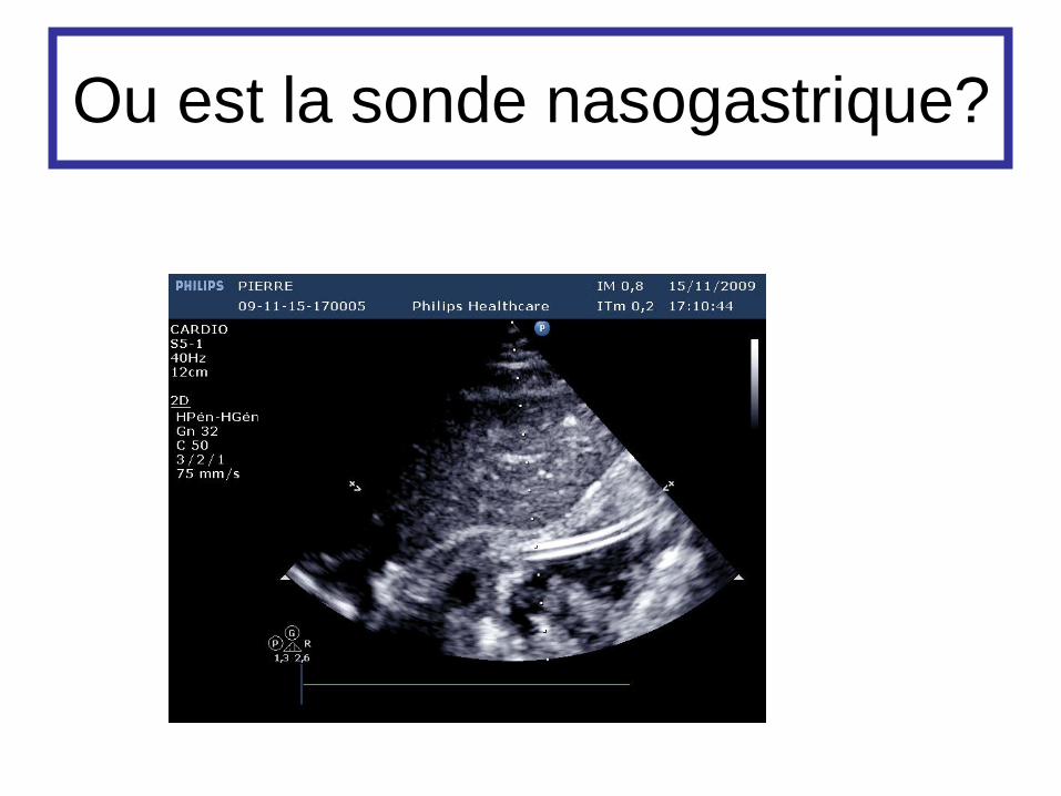
Ou est la sonde nasogastrique?
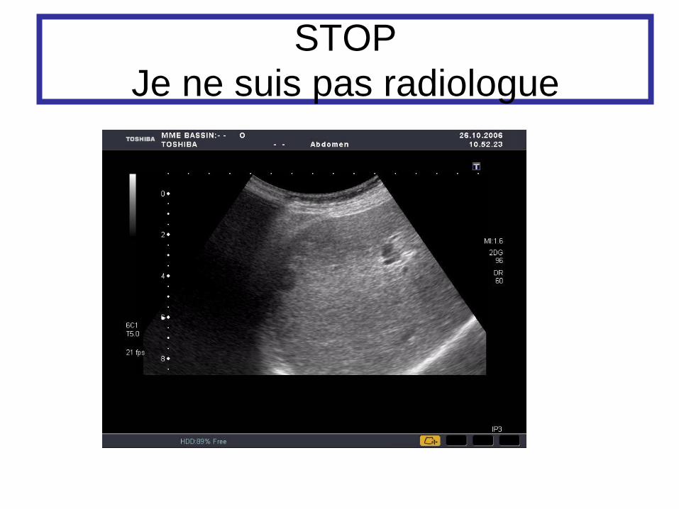
STOP
Je ne suis pas radiologue
Uropathie obstructive
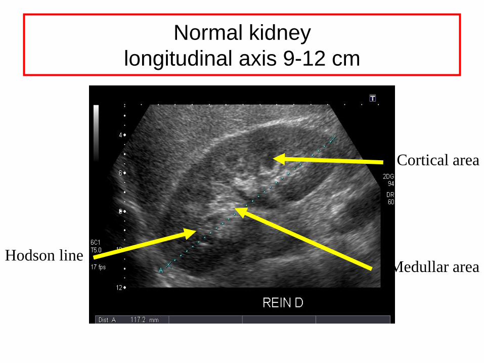
Normal kidney
longitudinal axis 9-12 cm
Cortical area
Medullar area Hodson line
Images anéchogènes rondes
confluentes
en boule de gui ou en oreille
de Mickey non limitées au rein
aboutissant à un uretère dilaté
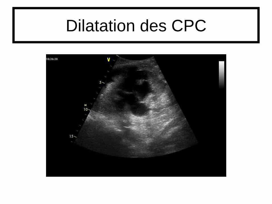
Dilatation des CPC
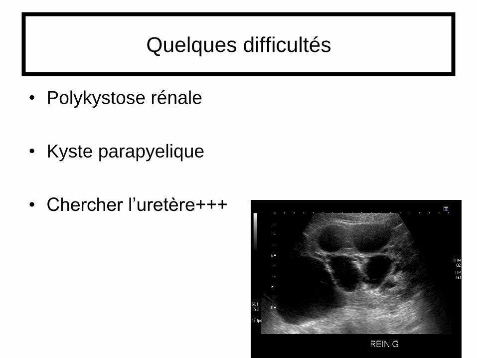
Quelques difficultés
• Polykystose rénale
• Kyste parapyelique
• Chercher l’uretère+++
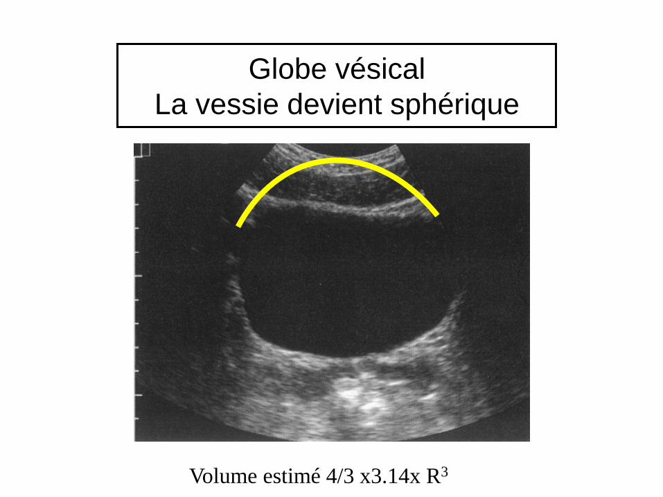
Globe vésical
La vessie devient sphérique
Volume estimé 4/3 x3.14x R3
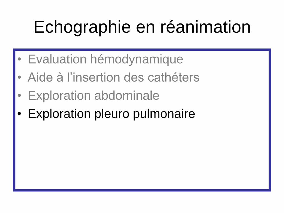
Echographie en réanimation
• Evaluation hémodynamique
• Aide à l’insertion des cathéters
• Exploration abdominale
• Exploration pleuro pulmonaire
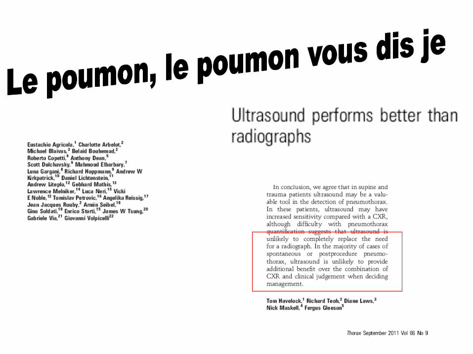
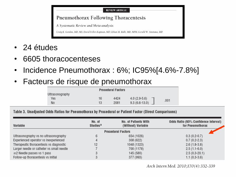
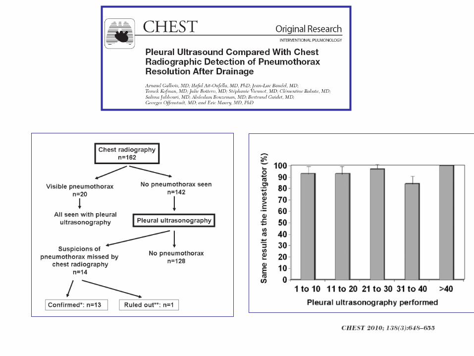
• 24 études
• 6605 thoracocenteses
• Incidence Pneumothorax : 6%; IC95%[4.6%-7.8%]
• Facteurs de risque de pneumothorax
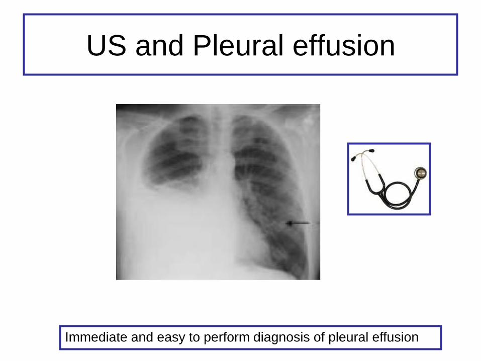
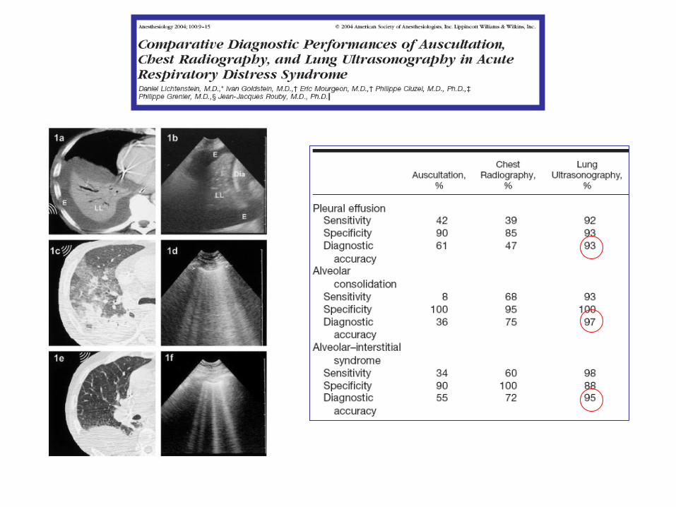
US and Pleural effusion
Immediate and easy to perform diagnosis of pleural effusion
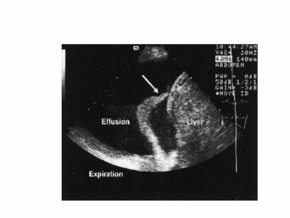
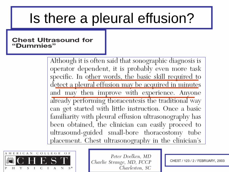
Is there a pleural effusion?
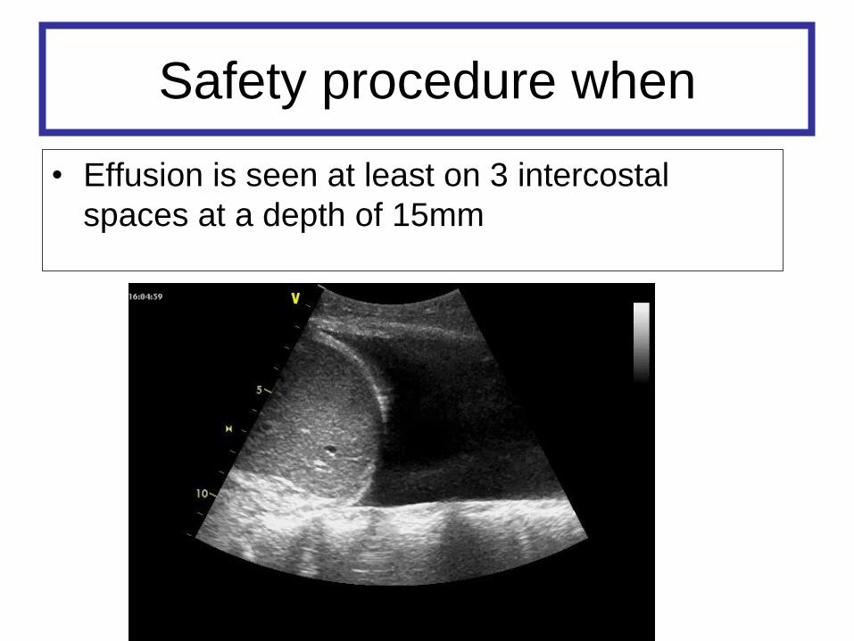
Safety procedure when
• Effusion is seen at least on 3 intercostal
spaces at a depth of 15mm
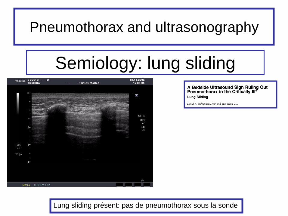
Pneumothorax and ultrasonography
Semiology: lung sliding
Lung sliding présent: pas de pneumothorax sous la sonde
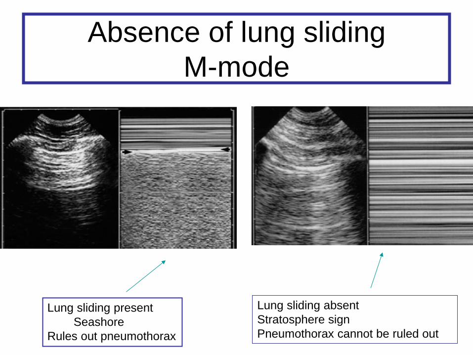
Absence of lung sliding
M-mode
Lung sliding present
Seashore
Rules out pneumothorax
Lung sliding absent
Stratosphere sign
Pneumothorax cannot be ruled out
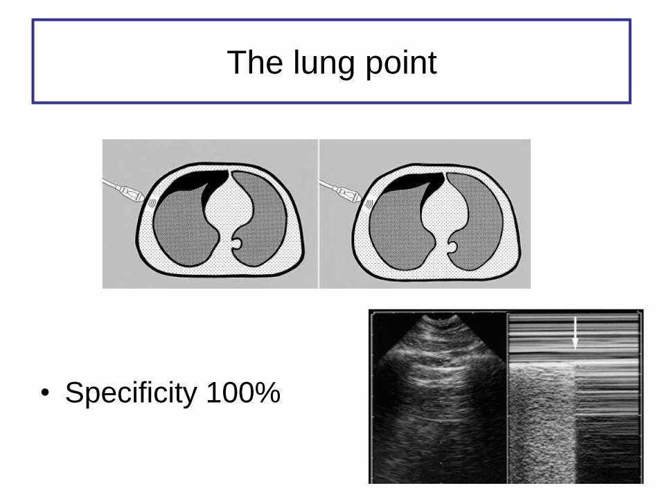
The lung point
• Specificity 100%
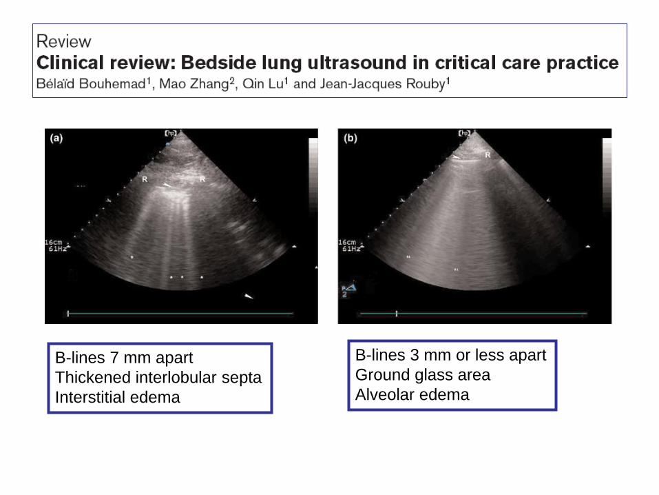
B-lines 7 mm apart
Thickened interlobular septa
Interstitial edema
B-lines 3 mm or less apart
Ground glass area
Alveolar edema
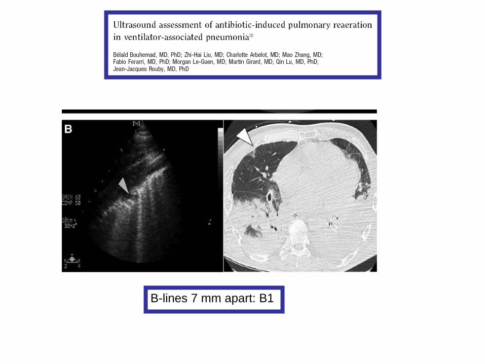
B-lines 7 mm apart: B1
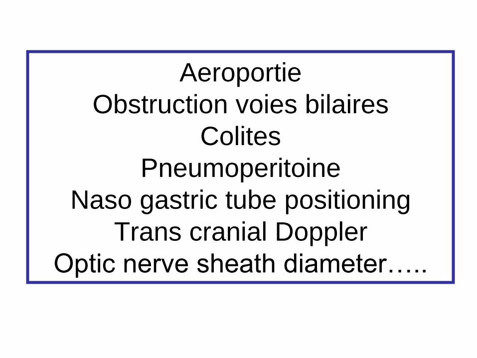
Aeroportie
Obstruction voies bilaires
Colites
Pneumoperitoine
Naso gastric tube positioning
Trans cranial Doppler
Optic nerve sheath diameter…..
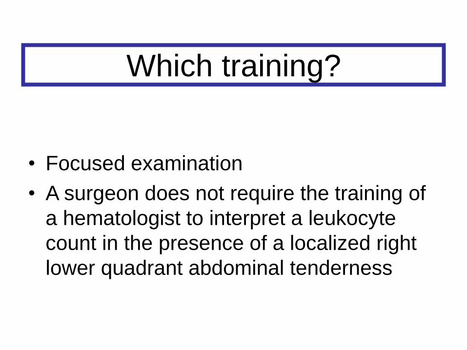
Which training?
• Focused examination
• A surgeon does not require the training of
a hematologist to interpret a leukocyte
count in the presence of a localized right
lower quadrant abdominal tenderness
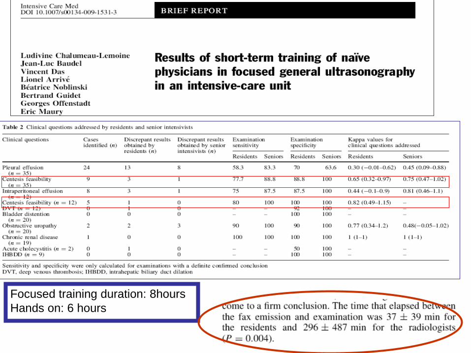
Focused training duration: 8hours
Hands on: 6 hours
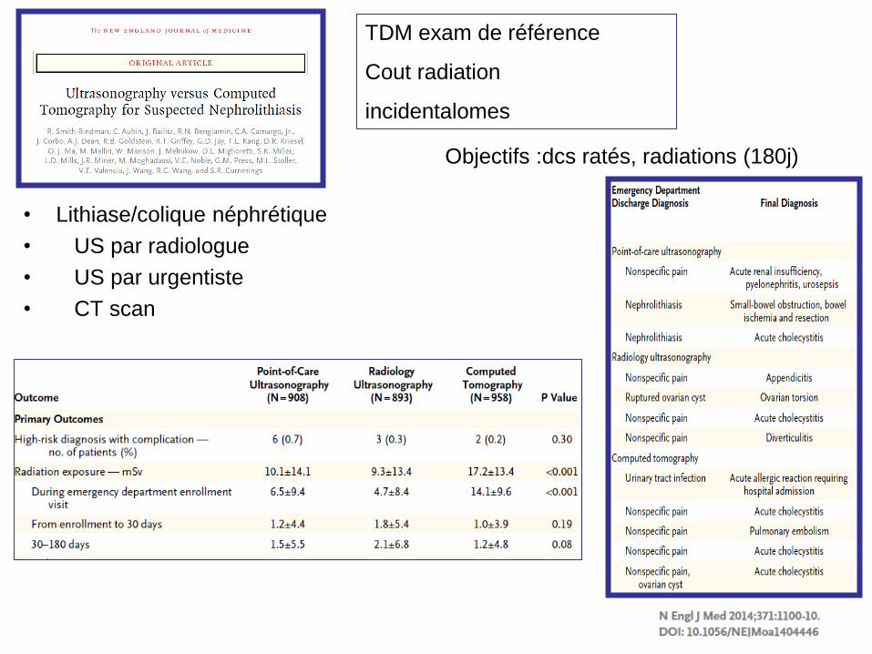
• Lithiase/colique néphrétique
• US par radiologue
• US par urgentiste
• CT scan
TDM exam de référence
Cout radiation
incidentalomes
Objectifs :dcs ratés, radiations (180j)
Ultrasonography: All it can do for you